Note: Descriptions are shown in the official language in which they were submitted.
CA 02766971 2011-12-29
WO 2011/013015 PCT/IB2010/052927
SUBGLOTTIC SUCTIONING SYSTEM
BACKGROUND
Tracheal intubation involves the insertion of a hollow tubular device, known
as a tracheal tube, into the trachea of a patient. The tube may be inserted
through
the mouth or, less desirably, the nose or may be inserted through the neck by
way
of an incision in the front of the throat. If inserted through the mouth or
nose the
tube is referred to as an endotracheal tube, if through the front of the
throat the
tube is referred to as a tracheostomy or trach tube. The two types of tubes
will be
referred to as tracheal tubes herein. The tracheal tube passes into the
trachea
io and terminates at a position above the carina, anterior to a position
between the
second and fourth thoracic vertebrate. Gases may then be introduced through
the
central lumen of the tracheal tube and into the lungs of the patient.
The primary purpose of tracheal intubation is to mechanically ventilate the
patient's lungs when the patient is incapable of normal breathing induced
is ventilation. Intubation may also be used to apply anesthetic gases during
surgical
intervention. It is desirable to seal the passageway around the tracheal tube
in
order to maintain enough air pressure to force the air into the lungs during
mechanical ventilation and to prevent escape of gases past the tube (i.e.
"short
circuiting" or bypassing of the lungs). Such a seal may be produced by the use
of
20 an inflatable cuff or balloon surrounding the tracheal tube near its distal
end.
When the tracheal tube has been introduced into the patient's trachea, the
inflatable cuff will normally be located about 3 to 5 centimeters above the
carina
and within the tube-like trachea.
Once inflated, the cuff will engage the wall of the trachea and thereby seal
25 the trachea and prevent the gases being introduced through the tracheal
tube from
simply reversing course after exiting the distal end of the tube and traveling
back
up and around the tube to exit the mouth. While treatment of this sort has
proved
successful for patients having chronic or acute respiratory diseases, there is
a
constant risk of several complications.
CA 02766971 2011-12-29
WO 2011/013015 PCT/IB2010/052927
One of the most common complications is known as ventilator associated
(or acquired) pneumonia or VAP. Patients receiving tracheal intubation
sometimes
develop this pneumonia from an infection of the lungs, possibly induced by
contaminated pooled secretions entering the trachea and the lungs after
bypassing
the epiglottis while intubated. The epiglottis normally operates as a valve
which
selectively closes the entry into the trachea and lungs to prevent the
introduction of
secretions and particulate matter. However, when an tracheal tube is in place,
the
epiglottis is held in an open position, and secretions which would normally be
directed away from the trachea and into the digestive system instead follow
the
io path of the tracheal tube and pool above the inflatable cuff.
One of the times of greatest risk of such infectious secretions reaching the
lungs is upon the cessation of mechanical ventilation. In particular, when the
need
for tracheal intubation ends, the inflatable cuff of the tracheal tube is
deflated so
that the tracheal tube may be withdrawn from the patient. The infectious
is secretions which have pooled in the space above the inflatable cuff are
then
released and are free to flow into the lungs, where bronchitis or pneumonia
may
develop. There is also a risk of the infectious secretions reaching the lungs
during
the time the tracheal tube is in place by aspiration of the secretions that
may leak
past the tracheal tube cuff.
20 Removing these secretions from above the tracheal tube cuff would likely
reduce the risk of such infections and tracheal tubes having inflatable cuffs
and
suction means are broadly known in the prior art. It is known, for example, to
combine a single lumen suction tube with a tracheal tube. The suction tube
provides means for constant suction or evacuation of any pooled secretions
which
25 accumulate in the trachea above the inflatable cuff. There remain a number
of
concerns with such prior art tubes, however. A single lumen for the suction
tube
under near constant suction often causes direct suction to be exerted on the
tracheal mucosa, which may then result in damage to the mucosa. Another major
problem with a single suction lumen is that it is also subject to clogging or
30 occlusion, and as a result may be rendered completely useless. Secretions
may
be quite viscous and can block the opening of the suction lumen above the cuff
2
CA 02766971 2011-12-29
WO 2011/013015 PCT/IB2010/052927
(the suction port) or can travel into the suction lumen and build up on the
inside
walls to the point where flow in the lumen is stopped.
A number of attempts have been made to solve some of these problems.
US patent 4,305,392 for example, provides a tracheal tube having a suction
lumen
that terminates in a suction chamber in the shape of a bulge having four ports
in
order to avoid damaging the tracheal mucosa. US patent 4,840,173 provides a
suction tube with multiple openings which may be used to evacuate secretions
that
may pool above the inflatable cuff, again in the hope that the suction line
will not
adhere to the trachea. US patent 5,143,062 discloses a double lumen through
io which air may be circulated, creating an indirect gentle suction through a
suction
eye communicating with the distal ends of the lumens. This design, however,
does
not provide adequate suction necessary for aspirating secretions and is easily
occluded. US patent publication 2008/0121236 discloses a suction apparatus and
connectors that allow a solution to be injected into a suction line. There is
no
is mechanism in the `236 publication to allow the valve to return to a fail-
safe or
default position where suction is restored to the suction lumen after the user
is
finished using the apparatus.
The current solution to occlusion of the suction lumen is to remove the
tracheal tube and replace it with another one, thus opening the system, or to
20 dispense with suctioning the space above the cuff altogether. Clearly these
solutions are unsatisfactory as they negate the purpose for having the suction
lumen present. Dispensing with suctioning of secretions from the space above
the
cuff results in a buildup of such fluids and, when the tube is eventually
removed,
can allow the fluids present to flow into the lungs, possibly causing VAP.
25 Removing the tube and replacing it involves opening the system and exposes
the
patient to all of the risks of intubation; low blood oxygen, irritation of the
trachea
and possible damage to the glottis, etc., as well as the movement of
secretions
from the space above the cuff to the lungs. Maintaining the patency of the
tracheal tube can reduce or delay the risks of extubation, contributing to the
30 likelihood of a successful outcome for the patient.
3
CA 02766971 2011-12-29
WO 2011/013015 PCT/IB2010/052927
What is needed is a multilumen tracheal tube or catheter capable of
suctioning secretions which have pooled in the space above the inflatable cuff
in
an effective manner, having a lumen and port that are capable of being cleaned
of
accumulated secretions without removal of the tube from the patient, so that
the
system may remain closed. It is also desirable that the system be simple,
preferably intuitive, to operate, so that it may be used on a regular basis by
nominally trained personnel. The instant disclosure addresses these problems
by
providing a multilumen tracheal tube and suction lumen system with a rinse
function, having a valve that is straight-forward and easy to operate.
SUMMARY
This disclosure relates to a system for a tracheal tube and associated items
used for mechanical ventilation. In particular, the present disclosure relates
to a
tracheal tube having means for irrigating and/or evacuating contaminated
secretions accumulating above the tracheal tube cuff and thereby reducing the
risk
is of such contaminated secretions entering the lungs of the patient. The
present
disclosure improves upon a tracheal tube by incorporating a suction lumen,
easily
operated valve and ultrathin cuff therein. The suction lumen communicates with
the space in the trachea above the cuff where secretions accumulate.
Desirably,
the tube includes a novel shape for the suction lumen and an enhanced design
for
the suction lumen port.
The valve is in fluid communication with the suction lumen and with a
source of vacuum that may be selectively applied to the suction lumen by a
caregiver or user. The valve also has a source of rinsing fluid. The valve may
be
used to change the suction lumen between communication with the source of
vacuum and with the source of rinsing fluid by the caregiver. The alternate
supply
of rinsing fluid or vacuum to the suction lumen at the discretion of the
caregiver
allows the suction lumen and the space proximal to the cuff in the trachea to
be
rinsed and suctioned to loosen and remove secretions that may build up. An
excess of secretions has the potential to pass by the cuff into the lower
respiratory
tract and cause ventilator associated (or acquired) pneumonia (VAP).
Various embodiments of valve designs are provided. All of the valves have
4
CA 02766971 2011-12-29
WO 2011/013015 PCT/IB2010/052927
the common feature of blocking the source of suction and opening a path for
rinsing fluid to the suction lumen when manipulated by the caregiver, and
automatically moving back to the source of suction after release. They are
designed so that the user may easily and repeatedly alternate suction and
rinsing
fluid through the suction lumen, i.e., the user may "pulse" the line to
loosen, break
up and remove secretions and deposits that may partially or completely block
or
clog the suction lumen, while maintaining a closed system.
In one embodiment, the tracheal tube is formed from a flexible cannula
having a length, a distal end, and a proximal end. The cannula consists of a
io plurality of walls extending substantially along the length of the cannula,
dividing
the cannula into a plurality of separate lumens including a respiratory lumen,
a
suction lumen and an inflation lumen. An inflatable cuff surrounds the cannula
proximal to the distal end. The inflatable cuff is adapted to seal the trachea
of a
patient. The inflation lumen is in fluid communication with the inflatable
cuff. A
is port extends through a side wall of the cannula proximal to the inflatable
cuff and
the port is in fluid communication with the suction lumen.
In other embodiments, the tracheal tube may have a plurality of suction
lumens. A rinsing fluid is adapted to be flushed through the suction lumen and
extracted via the suction lumen once vacuum is restored.
20 In still other embodiments, the tracheal tube may be a tracheostomy tube
and may have an inflatable cuff having a shape to block a trachea beneath the
glottis of the patient. The inflatable cuff surrounds the cannula above the
distal
end and is adapted, upon inflation, for expansion of the cuff around the
distal end
portion of the cannula and the proximal end portion of the cannula below a
25 proximal plane of the cannula. The cuff thus seals the trachea below the
tracheal
stoma and avoids sealing the trachea above the tracheal stoma.
The rinsing fluid may be water, saline, as well as other biocompatible liquids
and mucolytic agents. The rinsing fluid may also comprise air or combinations
of
air and liquids. A medicament, for example, an antiseptic or an antibiotic, or
a
30 treatment such as a surfactant may be added to the rinsing fluid to obtain
a desired
effect on the patient, or to ease suctioning or cleaning of the suction lumen.
5
CA 02766971 2011-12-29
WO 2011/013015 PCT/IB2010/052927
BRIEF DESCRIPTION OF THE DRAWINGS
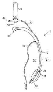
Figure 1 is a depiction of an endotracheal tube embodiment of a multilumen
catheter in accordance with the present disclosure;
Figure 2 is a depiction of a trach tube embodiment of a multilumen catheter in
accordance with the present disclosure.
Figure 3 is a cross-sectional view of the catheter of either Figure 1 or 2
taken
longitudinally through the catheter at 3-3.
Figure 4 is a drawing of a cuff for a tracheostomy tube as described in US
patent
6,612,305.
io Figure 5 is a drawing of a cuff for a tracheostomy tube as described in US
application 60/994,664.
Figure 6 A - H shows various desirable shapes of suction lumens.
Figure 7 depicts an elongated suction port on a cannula.
Figure 8 depicts a rotational rinsing adaptor valve (rotational valve).
is Figure 9 depicts a push-type rinsing adaptor valve (push valve).
Figure 10 depicts a straight rinsing adaptor valve (straight valve).
Figures 11A and B depict a bellows-type rinsing adaptor valve (bellows valve).
Figures 12A and B depict a trigger activated rinsing adaptor valve (trigger
valve).
Figures 13A and B depict an in-line pinching rinsing adaptor valve (pinch
valve).
20 Figure 14 depicts an in-line valve having a trigger tab that blocks flow.
Figure 15 depicts an in-line valve having a trigger bar that blocks flow.
6
CA 02766971 2011-12-29
WO 2011/013015 PCT/IB2010/052927
DETAILED DESCRIPTION
Reference will now be made to the drawings in which the various elements
of the present disclosure will be given numeral designations and in which the
disclosure will be discussed so as to enable one skilled in the art to make
and use
the disclosure. It is to be understood that the following description is only
exemplary of the principles of the present disclosure, and should not be
viewed as
narrowing the pending claims. Those skilled in the art will appreciate that
aspects
of the various embodiments discussed may be interchanged and modified without
departing from the scope and spirit of the disclosure.
Referring to Figures 1, 2 and 3, a tracheal tube 10 in accordance with two
embodiments of the present disclosure are depicted. Figure 1 depicts an
endotracheal tube, Figure 2 depicts a tracheostomy (trach) tube and Figure 3
depicts a cross-section taken at 3-3 in either Figure 1 or 2.
The tracheal tube 10 in the depicted embodiments is a multilumen cannula
is 12 having at least one respiratory lumen 14, at least one suction lumen 16,
and at
least one inflation lumen 18. In these embodiments, each of these lumens is at
least partially internal to the cannula 12 (Figure 3). The respiratory lumen
14 is the
largest lumen in the tube, extends through the entire cannula 12 and is
adapted to
mechanically ventilate a patient (not shown). When the tube 10 is installed in
a
patient, the distal end 20 of the cannula 12 is situated within the upper
respiratory
system of the patient.
A balloon, bladder, or inflatable cuff 22 is provided proximal to the distal
end
20. An inflation lumen 18 terminates within the cuff 22 on the exterior
surface 28
of the cannula 12. The inflation lumen 18 may be within the wall 25 of, or
along
the surface 28 of the cannula 12 until it is near the proximal end 38 of the
tube 10,
at which point it becomes a separate tubing line 40 adapted to be used to
supply
an inflation fluid, generally air, to the cuff 22 . The cuff 22 is shaped so
that when it
is inflated, it blocks the patient's trachea beneath the glottal area. This is
known
and understood by those skilled in the art to eliminate or at least to
minimize the
undesirable flow of fluids from the glottal and subglottal regions of the
patient into
the bronchus and lungs of the patient.
7
CA 02766971 2011-12-29
WO 2011/013015 PCT/IB2010/052927
The suction lumen 16 is, similarly to the inflation lumen 18, within the wall
25 or along the external surface 28 of the cannula 12 and terminates at a port
24
on the exterior surface 28 of the cannula 12. The port 24 in the depicted
embodiment is near an upper surface of the cuff 22. In this manner, the
suction
lumen 16 is adapted to suction fluids that collect in the space above the cuff
22 in
the patient's trachea (the subglottic area) without negatively impacting
ventilation
of the patient through the respiratory lumen 14. The suction lumen 16 extends
proximally from the suction port 24, along or within the wall 25 of the
cannula 12 to
a point where it separates from the cannula 12 and becomes a separate tubing
line
io 30. The tubing line 30 is attached to a valve body 36 that is adapted to
allow a
user to provide suction to the suction lumen 16 from a source of suction (not
shown) that is attached to the valve body 36 at a connector 34. The valve body
36
may also be used to provide a rinsing fluid contained within a bullet 32 or
other
appropriate container, to the suction lumen 16. The functioning of the valve
body
is 36 will be discussed in more detail below.
The rinsing fluid may be introduced into the suction lumen 16 from the bullet
32 while the suction is blocked off by the valve body 36. The rinsing fluid
travels
down (in a distal direction) the suction lumen 16 as far as is allowed by the
condition of the lumen. Desirably the lumen is not completely occluded and
allows
20 rinsing fluid to exit at the suction port 24 above the cuff 22 in the
trachea. Since
the rinsing fluid is usually of a lower viscosity than typical secretions, it
has the
effect of lowering the viscosity of all of the liquid mix found in the space
above the
cuff 22 in the trachea once it is introduced. Once the rinsing fluid has been
introduced to the suction lumen 16 or the space above the cuff 22 through the
25 suction lumen 16 and the port 24, suction may be restored to the suction
lumen 16
and the liquid and any secretions it may have loosened or dissolved may be
removed, i.e. sucked out through the suction port 24 and suction lumen 16.
This
procedure may be repeated as deemed necessary. This procedure is performed
at the discretion of the caregiver or user in order to clean secretions and
other
30 liquids that may collect and potentially clog the suction lumen 16 or
suction port 24.
It is important to keep the suction lumen 16 open so that potentially
deleterious
secretions may be removed from the area above the cuff 22.
8
CA 02766971 2011-12-29
WO 2011/013015 PCT/IB2010/052927
The rinsing fluid may comprise water, saline, as well as other biocompatible
liquids or mucolytic agents. Mucus may narrow or block the airways, making it
difficult to breath. Mucolytic drugs are designed to modify the properties of
the
mucus to help loosen and clear the mucus from the airways by breaking up the
sputum. Common mucolytic agents include erdosteine, acetylcysteine,
bromheksin, carbocysteine and guiafenesin. The rinsing fluid may also comprise
air or combinations of air and liquids. A medicament, for example, an
antiseptic or
an antibiotic, or a treatment such as a surfactant may be added to the rinsing
fluid
to obtain a desired effect on the patient, or to ease suctioning or cleaning
of the
io suction lumen 16.
As can be seen in Figure 3, the cross sectional view of Figures 1 and 2, one
possible configuration of the tracheal tube 10 is depicted, more specifically
a
potential lumen arrangement is depicted within the cannula 12. As can be seen,
the suction lumen 16 and the inflation lumen 18 are formed into the wall of
the
is cannula 12. This configuration is of course only meant to suggest one
possible
arrangement and other arrangements are included in the spirit and scope of the
disclosure. The arrangement of lumens within the cannula 12 is not limited in
scope to any particular configuration. The layout of the lumens within the
cannula
12 may be altered for example or the suction and inflation lumens may be
separate
20 lumens not embedded within any one of the walls of the cannula 12.
In other embodiments, a plurality of suction lumens 16 may be provided.
Each suction lumen would be configured essentially as described above, in that
each would be rinsed by a rinsing fluid provided by a valve 36. A single
central
valve 36 may be provided to service all suction lumens 16 or a separate,
dedicated
25 valve 36 could be provided for each suction lumen 16. Such an arrangement
may
prove beneficial in more thorough rinsing of the suction lumen or lumens.
Alternatively, a plurality of lumens would allow for another lumen to be used
should
the previous lumen become clogged. Any of these embodiments are easily
understood by one of skill in the art as they merely increase the number and
3o arrangement of lumens provided. As such no specific drawings are needed for
an
understanding of these variations.
9
CA 02766971 2011-12-29
WO 2011/013015 PCT/IB2010/052927
As discussed above, the tracheal tube 10 has a cuff 22 around its
circumference on a lower (distal) portion of the tube that serves to block the
normal
air flow in the trachea so that assisted breathing takes place through the
tracheal
tube using a ventilator. The cuff is desirably made from a soft, pliable
polymer
such as polyethylene teraphathalate (PETP), low-density polyethylene (LDPE),
polyvinyl chloride (PVC), polyurethane (PU) or polyolefin. It should be very
thin; on
the order of 25 microns or less, e.g. 20 microns, 15 microns, 10 microns or
even
as low as 5 microns in thickness. The cuff should also desirably be a low
pressure
cuff operating at about 30 mmH2O or less, such as 25 mmH2O, 20 mmH2O, 15
io mmH2O or less. Such a cuff is described in US patents 6,802,317 which
describes
a cuff for obturating a patient's trachea as hermetically as possible,
comprising: a
cuff which blocks the trachea below a patient's glottis, an air tube, the cuff
being
attached to the air tube and being sized to be larger than a tracheal diameter
when
in a fully inflated state and being made of a soft, flexible foil material
that forms at
is least one draped fold in the cuff when inflated in the patient's trachea,
wherein the
foil has a wall thickness below or equal to 0.01 mm and the at least one
draped
fold has a loop found at a dead end of the at least one draped fold, that loop
having a small diameter which inhibits a free flow of secretions through the
loop of
the at least one draped fold. Another description of such a cuff is in US
patent
20 6,526,977 which describes a dilator for obturating a patient's trachea as
hermetically as possible, comprising a cuff which blocks the trachea below a
patient's glottis, an air tube, the cuff being attached to the air tube and
being sized
to be larger than a tracheal diameter when in a fully inflated state and being
made
of a sufficiently soft, flexible foil material that forms at least one draped
fold in the
25 cuff when fully inflated in the patient's trachea, wherein the at least one
draped fold
formed has a capillary size which arrests free flow of secretions across the
cuff by
virtue of capillary forces formed within the fold to prevent aspiration of the
secretions and subsequent infections related to secretion aspiration. It has
been
found that the very thin cuff described above is particularly suitable for
blocking the
30 flow of lower viscosity fluids that are present above the cuff after the
introduction of
the rinsing fluid as described herein.
CA 02766971 2011-12-29
WO 2011/013015 PCT/IB2010/052927
Alternatively, in the particular case of tracheostomy tubes, the cuff may be
of
a shape as described in US patent application 60/994,664, now 12/206,517 or US
patent 6,612,305. In the `305 patent, the cuff expands not only around the
tube, as
do the current models, but also cranially to it and to the stoma, sealing the
stoma
(Figure 4). This is achieved because the proximal point of attachment and the
distal point of attachment of the inflatable cuff on the tube are not
contiguous or, in
other words, are at an angle (a) other than 180 degrees, relative to
conventional
devices. In the `644 application, the cuff has a distal cuff portion
substantially
centered about and attached to the distal end portion of the tube. The cuff
also
io has a proximal cuff portion attached to the bend region of the tube and
positioned
substantially off-center about the bend region below the proximal plane of the
device. Upon inflation, this configuration provides for expansion of the cuff
around
the distal end portion of the tube and the proximal end portion of the tube
below
the proximal plane of the device to seal the trachea below the tracheal stoma
and
is avoid sealing the trachea above the tracheal stoma (Figure 5). Desirably,
this
configuration of the cuff will allow secretions to exit the stoma.
The tracheostomy tube device may have cuff walls that are non-uniform in
thickness. For example, the device may have a first portion of the cuff in
which the
walls have a thickness of about 20 to 30 micrometers and a second portion of
the
20 cuff in which the walls have a thickness of about 5 to about 15
micrometers.
Desirably, the first portion of the cuff is the portion of the cuff contacting
the upper
portion of a cross-sectional region of the tracheal lumen and the second
portion of
the second cuff is the portion of the cuff contacting the lower portion of the
same
cross-sectional region of the tracheal lumen.
25 The inflatable cuff component may include a distal end, a distal attachment
zone, a proximal end, a proximal attachment zone, an upper region and a lower
region, wherein the upper region has a thickness of from about 15 to about 30
micrometers and the lower region has a thickness of from about 5 to about 15
micrometers.
30 The cuff component may desirably be formed from thermoplastic
polyurethane polymers, thermoplastic polyolefin elastomers, thermoplastic
11
CA 02766971 2011-12-29
WO 2011/013015 PCT/IB2010/052927
polyolefin block copolymers, SBS di-block elastomers, SEBS tri-block
elastomers,
polyvinyl chloride, polyethylene terephthalate and blends and mixtures
thereof.
The suction lumen 16 shown in Figure 3 may be round, oval or elliptical in
shape in currently available commercial tracheal tubes. In an attempt to
investigate an improved shape, however, many different configurations were
tested. The tests showed that significant differences existed in the flow rate
through the lumen, based simply on the shape chosen. It was found that the
fluid
that accumulated on the proximal side of the cuff was a complicated
combination
of secretions which were by no means newtonian. The viscosity of the fluid
varied
io substantially, depending on the amount of shear the fluid was subjected to.
As
various shapes of lumens were investigated, it was found that shear varied
within
the lumen, with more shear at the bends or corners and less in the center,
thus
affecting the viscosity and impacting flow. As a result of the investigation,
it was
found that a more elongated, slightly bent shape performed better than a
simple
is circle, ellipse or oval. A number of such "bent oval", "bean", or "banana"
shaped
lumens are shown in Figure 6 A - H, which are cross sectional views of a trach
tube having a ventilating lumen 14 and a suction lumen 16. More desirably, the
bent oval shaped lumens of Figure 6 G, H performed better.
Production of bent oval shaped lumens should prove no more difficult than
20 the production of known round or oval lumens. The tube used to produce
tracheal
catheters is typically extruded. Changing the extrusion shape is not a
difficult
matter for those skilled in the art of polymer extrusion.
The suction port opening 24 of Figures 1 and 2 has been found to be
susceptible to attaching itself to the back of the trachea and causing tissue
25 damage. Continuous suctioning is more of a danger than intermittent
suctioning
but the potential for suction related tissue damage exists in either method.
The
suction port 24 is conventionally placed on the cannula 12 in a position where
it will
be at the lowest point in the trachea above the cuff when the patient is
laying on
his back, and is conventionally a circular port. This area is where the
secretions
30 will naturally accumulate in the greatest amount. This is also an area of
the
cannula 12 that is subjected to high bending stress and so is more likely to
allow
12
CA 02766971 2011-12-29
WO 2011/013015 PCT/IB2010/052927
the suction port 24 to come into contact with the trachea. One solution to
this
problem is to move the suction port 24 to a position where it will not come
into
contact with the trachea should the cannula 12 bend excessively. A position 90
or
180 degrees away from the conventional position depicted in Figures 1 and 2
would make tracheal damage less likely, but would also be much less effective
in
suctioning secretions from the patient lying on his back.
Figure 7 depicts a suction port 24 on a cannula 12 above the cuff 22 where
the suction port 24 is elongated circumferentially around the cannula 12 for
some
distance. This suction port 24 connects to the suction lumen 16 and also has a
1o shallow extension on either side that reduces the likelihood that the
suction port 24
will attach itself to the tracheal wall. The shallow extensions extend a short
depth,
e.g. a millimeter or two, into the outer surface 28 of the cannula 12 but do
not go
all the way through the cannula 12 except in the area where the suction port
24
communicates with the suction lumen 16.. Should the central part of the
elongated
1s suction port 24 come into contact with the trachea, complete suction
against the
tracheal wall would be avoided because the elongated portions of the suction
port
24 would still not be in contact with the tracheal wall. The elongated suction
port
24 disclosed herein thus reduces damage to the trachea and helps maintain the
suction line open by preventing the cannula 12 from being sucked against the
20 tracheal wall. The elongated suction port 24 may be two to five times wider
than
the conventional circular port, desirably about three times as wide.
The valve is an important part of the tracheal tube and system of suctioning
secretions disclosed herein. The valve is used to suction and rinse the tubing
line
30 and by extension the suction lumen 16, suction port 24 and the space in the
25 trachea above the cuff 22. It is desired that the valve have the capability
of easily
and repeatedly alternately suctioning and providing rinsing fluid through the
suction
lumen, i.e., the user may "pulse" the line to loosen, break up and remove
secretions and deposits that may partially or completely block or clog the
suction
lumen. It is also desired that the valve automatically (i.e., by itself,
without
30 intervention by a user) return to a normal, default or "fail-safe" position
in which
suction is applied to the system so that secretions are removed, after the
user has
finished using the valve. If the valve remains in the "rinse" position once
the bullet
13
CA 02766971 2011-12-29
WO 2011/013015 PCT/IB2010/052927
providing rinsing fluid is empty, secretions will build up in the space above
the cuff
22 and the purpose for having a suctioning line will be defeated. It is also
important that the valve close the access to the suction line prior to opening
access to the rinsing fluid bullet, otherwise the fluid will be sucked out of
the bullet
to the source of suction and wasted. It is also desirable that the valve
require a
positive action on the part of the user to move it to the rinse position, so
that the
inadvertent movement of the patient will not activate the valve. Should the
patient
roll over onto the valve, for example, the valve should remain in the position
the
caregiver desires, generally the fail-safe or suction position, or should move
back
1o to the fail-safe position by itself relatively quickly when the force
exerted by the
patient is removed. Lastly, it is desired that rinsing and suctioning be
capable of
being performed and the system kept closed. Removing the bullet, for example,
each time suctioning was applied, would repeatedly open the system and allow
for
the entry of germs. It is true, of course, that the bullet would eventually
need to be
1s replaced but this occurs much more rarely than if it needed to be removed
each
time suctioning were performed. Valves that allow for suctioning and rinsing
while
the source of rinsing fluid remains in place are desired because maintaining a
closed system helps to reduce the chance of infection. The valves described
below meet these criteria.
20 In one embodiment, the valve 36 may be as depicted in Figure 8. This
Figure depicts a rotational rinsing adaptor valve (rotational valve) that
accommodates a sterile rinsing fluid (e.g. saline) bullet 32 or syringe. The
valve 36
may be connected to a vacuum source by a connector 34 and to the suction lumen
(not shown) by a tubing line 30. The normal or fail-safe position of the valve
is to
25 allow constant suction to the tubing line 30. When rinsing of the suction
lumen 16
is desired, a bullet 32 is inserted and is rotated as indicated by the arrow.
This
rotational movement of the bullet 32 turns a three way valve, blocking the
source
of suction or vacuum and opening up fluid access from the bullet 32 to the
tubing
line 30. The tubing line 30 is in fluid communication with the suction lumen
16.
3o The bullet 32 may be squeezed to force the rinsing fluid into the tubing
line 30.
Once the user has finished instilling rinsing fluid into the lumen, releasing
the bullet
32 allows a spring or other automatic means (not shown) to rotate the bullet
32 in
14
CA 02766971 2011-12-29
WO 2011/013015 PCT/IB2010/052927
the opposite direction, back to its original (normal) position, closing the
fluid access
from the bullet 32 and re-opening the flow path to the source of vacuum. Re-
establishing the fluid communication between the source of vacuum and the
tubing
line 30 results in suctioning of the suction lumen and the space above the
cuff 22
through the suction port 24. The user may repeatedly alternate between suction
and rinsing fluid as desired, thus pulsing the system to loosen and remove
secretions.
In another embodiment, the valve 36 may be as depicted in Figure 9. This
Figure depicts a push-type rinsing adaptor valve (push valve) that
accommodates
io a sterile rinsing fluid bullet 32 or syringe. The valve 36 may be connected
to a
vacuum source by a connector 34 and to the suction lumen (not shown) by a
tubing line 30. The normal or fail-safe position of the valve is to allow
constant
suction to the tubing line 30. When rinsing of the suction lumen 16 is desired
a
bullet 32 containing a rinsing fluid is inserted into the valve 36 as shown.
When
is the bullet 32 is pushed downward toward the valve 36 the bullet 32 blocks
the
source of vacuum and opens access from the bullet 32 to the tubing line 30
that is
in fluid communication with the suction lumen. The bullet 32 may be squeezed
to
force the rinsing fluid into the tubing line 30. Once the user has finished
instilling
rinsing fluid into the lumen, releasing the bullet 32 allows a spring or other
20 automatic means(not shown) to move the bullet 32 in the opposite direction,
back
to its original (normal) position, closing the fluid access from the bullet 32
and re-
opening the flow path to the source of vacuum. The user may repeatedly
alternate
between suction and rinsing fluid as desired, thus pulsing the system to
loosen and
remove secretions.
25 In yet another embodiment, the valve 36 may be as depicted in Figure 10.
This Figure depicts a straight rinsing adaptor valve (straight valve) that
accommodates a sterile rinsing fluid bullet 32 or syringe. The valve 36 may be
connected to a vacuum source by a connector 34 and to the suction lumen (not
shown) by a tubing line 30. The normal or fail-safe position of the valve is
to allow
30 constant suction to the tubing line 30. When rinsing of the suction lumen
is desired
a bullet 32 containing a rinsing fluid is inserted. When the bullet 32 is
pushed
downward, it blocks the flow path from the suction source to the tubing line
30 and
CA 02766971 2011-12-29
WO 2011/013015 PCT/IB2010/052927
establishes a fluid connection between the bullet 32 and the tubing line 30.
The
bullet 32 may be squeezed to force the rinsing fluid into the tubing line 30.
Once
the user has finished instilling rinsing fluid into the lumen, releasing the
bullet 32
allows a spring or other automatic means (not shown) to move the bullet 32 in
the
opposite direction, back to its original (normal) position, closing the fluid
access
from the bullet 32 and re-opening the flow path to the source of vacuum. The
user
may repeatedly alternate between suction and rinsing fluid as desired, thus
pulsing
the system to loosen and remove secretions.
Figures 1 1A and 11 B depict a bellows activated suction valve where the
1o cross sectional view is that of Figure 11 B. During suction operation, the
connector
34 is connected to a source of suction (not shown). Suctioned fluid or air can
travel through the tubing line 30 from the patient end 42, through the center
lumen
51 of the piston 46, and then pass through an orifice 47 into the annular
space 45
in which is found the spring 44. The suctioned fluid or air may pass around
the
1s perpendicular section 52 of the pin 48 and out to the source of suction
beyond the
connector 34. The pin 48 does not block fluid or air flow but only serves to
center
and hold in place the other components. Squeezing the bellows 41 flattens the
bellows 41 out and forces the spring 44 into the suction end 43. Immediately
after
the piston 46 begins moving, the piston 46 blocks the flow of fluid or air
through the
20 orifice 47. Further squeezing of the bellows opens the rinse (one way)
check valve
50, allowing the contents of the bellows 41 to travel into the tubing line 30
toward
the patient. Relaxing the squeezing of the bellows 41 allows the spring 44 to
force
the piston 46 away from the suction end 43 and simultaneously closes the rinse
check valve 50. As this occurs and before the orifice 47 is uncovered by the
piston
25 46, the fluid (one-way) check valve 49 opens and rinsing fluid flows from
the bullet
32 into the bellows 41. Continued relaxing of the squeezing of the bellows 41
uncovers the orifice 47 and restores suction to the bellows 41. Such a course
of
action would of course result in the rinsing fluid being sucked out of the
bellows 41
toward the source of suction, and would be unproductive. Rather than relax the
30 squeezing of the bellows 41 completely, however, the squeezing of the
bellows 41
may be only partially relaxed, allowing rinsing fluid to fill the bellows 41
but not
opening the orifice 47. Squeezing of the bellows 41 may be reinitiated,
resulting in
16
CA 02766971 2011-12-29
WO 2011/013015 PCT/IB2010/052927
the closing of the fluid check valve 49 and the rinsing fluid being forced out
of the
bellows 41, through the rinse check valve 50 and into the tubing line 30
toward the
patient. By repeating this squeezing and relaxing of the bellows 41, the
caregiver
may pulse rinsing fluid into the tubing line 30 and on to the suction port 24
for
delivery to the space in the trachea above the cuff 22. This alternating of
rinsing
fluid and suction may provide a more effective method of removing deposits and
secretions than steady state suctioning. The valve 40 may also include a
tethered
cap 53 that may be used to protect the saline check valve 49 and the rest of
the
valve 40 from contamination when the bullet 32 is not in place.
Figures 12A and 12B depict a trigger activated suction valve 60 where the
cross sectional view is that of Figure 12B. Squeezing the trigger 61 moves a
pivot
valve 62 into the closed position, squeezing closed the tubing line 30 within
the
valve 60 and blocking the source of suction. There are two one-way check
valves
in the stationary piston 65, a suction lumen check valve and a rinsing fluid
check
is valve. As the trigger continues to move inwards as it is squeezed, it
compresses
the spring 62, and forces the rinsing fluid in the trigger space 63 through a
suction
lumen check valve 65, through a narrow tube 66 and into the tubing line 30 at
the
point 67 where the narrow tube 66 connects to the main tubing line 30.
Releasing
the trigger 61 allows the spring 62 to push the trigger 61 outward, opening
the
rinsing fluid check valve and allowing rinsing fluid to flow from the bullet
32,
through tubing (not visible) and into the trigger space 63. The pivot valve 62
remains closed until the trigger 61 is entirely released, allowing the user to
send
repeating pulses of rinsing fluid through the tubing line 30 to the suction
port 24.
The user may alternatively pulse rinsing fluid into the tubing line 30 and
restore
suction to the tubing line 30. Upon release of the valve, the spring
automatically
opens access from the source of suction to the suction lumen and closes access
from the bullet to the suction lumen. One skilled in the art may readily see
that the
trigger may be positioned on an upper surface of the valve 60 or on a side, as
desired, and still be within the teachings and inventive spirit of the active
trigger
valve presented herein.
Figures 13A and its exploded view 13B depict an in-line pinching rinse valve
70. This relatively less complicated valve has a body 71 that, in this
embodiment,
17
CA 02766971 2011-12-29
WO 2011/013015 PCT/IB2010/052927
is made from two mirror image halves; a right half 72 and a left half 73 where
the
forward portion of the valve is defined as the patient facing end that
connects to
the tubing line 30. Note that the body may alternatively be made from a top
and
bottom half or may be made as a single piece. The rear portion of the valve 70
is
a connector 34 for the source of suction (not shown). At least one of the body
halves has a peninsular tab 74 (only visible on the left half 73 in the
Figures) that is
separated from its body half by a slight gap 75 for much of its length. The
peninsular tab 74 remains attached to its body half at one end. The gap 75
allows
the peninsular tab 74 to flex and move relative to the body half without
breaking,
io provided the material from which the body half is made is sufficiently thin
and or
flexible, and to spring back to its original position upon release of the
squeezing
force. On the interior of at least one body half should be a ridge 76,
desirably
placed perpendicularly to the crimp tubing 77, that is sized so that when the
peninsular tab 74 is squeezed by a user's fingers, the ridge 76 will contact
an
is internal length of crimp tubing 77 and, if sufficient force is applied,
close the lumen
of the crimp tubing 77 and block the communication of the source of suction
through the crimp tubing 77. The exact size and shape of the peninsular tab 74
and the ridge 76 may be varied according to the desire of the valve designer
and
remain within the teachings of this disclosure, provided the lumen of the
crimp
20 tubing 77 may be closed by squeezing the peninsular tab(s) 74. For example,
the
ridge 76 may, instead of being a rectangular feature as shown in the Figure,
be
another shape like a round or oval bump that is placed in a position to come
into
contact with the crimp tubing 77 when the peninsular tab(s) 74 are squeezed
together. It is also possible to design the peninsular tab(s) 74 in a way such
that
25 the ridge 76 is deleted entirely from the body halves and the peninsular
tab(s) 74
directly impinge upon the crimp tubing 77 and close its lumen.
The patient facing end of the crimp tubing 77 is in fluid communication with
the tubing line 30 that in turn communicates with the suction lumen 16 and
suction
port 24, discussed previously. The other end of the crimp tubing 77 is in
fluid
30 communication with the connector 34 that further communicates with the
source of
suction. The valve 70 has an inlet 78 to receive rinsing fluid from a bullet.
The
valve 70 desirably has an adapter 79 that is designed to accept the bullet and
fit
18
CA 02766971 2011-12-29
WO 2011/013015 PCT/IB2010/052927
snugly against it to reduce fluid leakage, and a check valve 80 through which
the
saline solution will flow from the bullet. The check valve 80 requires more
force to
open it than is exerted by the source of suction alone, thus requiring the
user to
squeeze the bullet to provide sufficient force to open the check valve 80 and
move
rinsing fluid into the tubing line 30. An optional tethered cap (not shown)
adapted
to cover the inlet 78 when a bullet is not in place may be provided.
When fully installed and in use, a user may simply squeeze the peninsular
tab(s) 74 on the body 71 with one hand to close the lumen of the crimp tubing
77
and block the source of suction from the tubing line 30, and, keeping the
crimp
io tubing 77 closed, squeeze the fluid bullet with the other hand to force
liquid into the
tubing line 30 and on toward the space above the cuff 22 in the trachea. Is
has
been found that users generally prefer to perform one function with each hand,
and
that requiring more than one function to be performed with one hand can cause
confusion. It is advantageous, therefore, that this valve has one function for
each
is hand. Once the desired amount of fluid is dispensed, the user may stop
squeezing
the bullet and relax pressure on the peninsular tab(s) 74. This permits the
peninsular tab(s) 74 to spring back to the original position, allowing the
crimp
tubing 77 to resume its normal shape and restoring suction to the tubing line
30.
The user may repeatedly alternate between suction and rinsing fluid as
desired,
20 thus pulsing the system to loosen and remove secretions, without removing
the
bullet.
In embodiments where the body 71 of Figure 13A is made of a single piece,
the tubing line 30 may be slid into the interior of the body 71 and mated with
the
connector 34, thus dispensing with a separate piece acting as the crimp tubing
77.
25 In this case, the tubing line 30 may be punctured for access by the source
of
rinsing fluid.
Should gloves become caught in the gap 75, the body 71 may be wrapped
with a polymeric material like a "shrink wrap" plastic that shrinks in place
in
response to heat, for example, to cover over the gap 75 and prevent glove
3o entrapment in the gap 75. Further, the peninsular tab(s) 74 may have
surface
topography 80 like lines, chevrons, dimples, reverse dimples and the like, in
order
19
CA 02766971 2011-12-29
WO 2011/013015 PCT/IB2010/052927
to improve the tactile sensation felt by a user wearing gloves and to improve
the
quality of the user's grip on the body 71.
In a particular embodiment, the valve 70 of Figure 13 A may have body
halves that are between 3 and 10 cm in length, desirably about 7 cm in length
and
between 0.5 and 2 cm in diameter, desirably about 1.7 cm. Two peninsular tabs
74 located opposite each other may be between 1 and 5 cm in length, desirably
about 2.5 cm and a ridge 76 may be located approximately in the lengthwise
center of each peninsular tab 74. The gap 75 may be between 0.3 and 3 mm in
width, desirably about 1 mm.
Figure 14 shows another embodiment that, like the valve of Figure 13,
allows for one handed operation of the valve. This valve 70 is similar in many
ways to the valve of Figure 13 but differs in the method of activation. In the
valve
70 of Figure 14, depressing a trigger tab 81 on the bottom of the valve 70
results in
crimping the tubing inside the body and closing it. Releasing the trigger tab
81
is allows the crimp tubing (not shown) to re-open and re-establishes
communication
between the source of suction and the suction lumen.
Figure 15 shows another embodiment that, like the valve of Figure 13,
allows for one handed operation of the valve. This valve 70 is similar in many
ways to the valve of Figure 13 but differs in the method of activation. In the
valve
70 of Figure 14, moving a trigger bar 82 on the bottom of the valve 70
rearward
(toward the connector 34 for the source of suction) results in crimping the
tubing
inside the body and closing it. Releasing the trigger bar 82 allows the crimp
tubing
(not shown) to re-open and re-establishes communication between the source of
suction and the suction lumen.
The materials of construction of the valves disclosed herein may be, for the
bodies, polyolefins like polyethylene and polypropylene, nylons,
polycarbonates,
acrylonitrile butadiene styrene (ABS), acrylics, PVC and the like.
Particularly
suitable is high density polyethylene (HDPE). Materials of construction of the
flexible parts like the check valves and tubing include silicones,
polyurethanes,
polyethylene terephthalate (PET), low-density polyethylene (LDPE), polyvinyl
chloride (PVC), or elastomeric-based polyolefins.
CA 02766971 2011-12-29
WO 2011/013015 PCT/IB2010/052927
In use, a medical care provider would insert the tracheal tube 10 into the
patient's trachea in a manner known and understood by those of skill in the
art;
through oral or nasal intubation or through a tracheostomy. The inflatable
cuff 22
would be inflated by air supplied through the inflation lumen 18 so as to
sealingly
engage the walls of the patient's trachea. This would effectively prevent or
at least
minimize flow of undesirable fluids from the subglottic space into the
bronchus and
lungs. Ventilation of the patient through the respiratory lumen 14 may occur
at this
time and continue for as long as necessary.
At the discretion of the caregiver, the subglottic space within the patient's
io trachea may be suctioned through the suction lumen 16 via the port 24
through the
wall 25 of the cannula 12. Such suctioning may be performed continuously or
intermittently as desired. Also at the discretion of the caregiver, the
suction lumen
16 and/or the space above the cuff 22 may be rinsed and suctioned. This is
accomplished by blocking the source of suction from the suction lumen 16
through
is the use of the valve 36 and introducing a rinsing fluid from a bullet 32 to
the
suction lumen 16 as described in more detail above. After the liquid has been
introduced, the valve 36 is re-opened to the source of vacuum and suction
restored, thus evacuating the suction lumen 16 and removing any secretions and
other liquids in the suction lumen 16 and, desirably, any secretions
accumulated
20 above the cuff 22. A treatment may be added to the rinsing fluid such as a
medicament, for example, an antiseptic, antibiotic or mucolytic agent. In that
case,
it may be desirable to allow more time between the introduction of the rinsing
fluid
and the evacuation of the rinsing fluid from the lumen and cuff area so as to
gain
the desired therapeutic effect prior to suctioning.
25 While the invention has been described in detail with respect to specific
embodiments thereof, it will be apparent to those skilled in the art that
various
alterations, modifications and other changes may be made without departing
from
the spirit and scope of the invention. It is therefore intended that the
claims cover
all such modifications, alterations and other changes encompassed by the
3o appended claims.
21