Note: Descriptions are shown in the official language in which they were submitted.
CA 02767113 2011-12-30
WO 2011/120006 PCT/US2011/030077
DETECTION SYSTEM FOR DROPLET-BASED ASSAYS
Cross-Reference to Priority Application
This application is based upon and claims the benefit under 35 U.S.C.
119(e) of U.S. Provisional Patent Application Serial No. 61/317,684, filed
March 25, 2010, which is incorporated herein by reference in its entirety for
all
purposes.
Cross-References to Other Materials
This application incorporates by reference in their entireties for all
purposes the following materials: U.S. Patent No. 7,041,481, issued May 9,
2006; U.S. Patent Application Publication No. 2010/0173394 Al, published
July 8, 2010; and Joseph R. Lakowicz, PRINCIPLES OF FLUORESCENCE
SPECTROSCOPY (2nd Ed. 1999).
Introduction
Many biomedical applications rely on high-throughput assays of
samples. For example, in research and clinical applications, high-throughput
genetic tests using target-specific reagents can provide high-quality
information about samples for drug discovery, biomarker discovery, and
clinical diagnostics, among others. As another example, infectious disease
detection often requires screening a sample for multiple genetic targets to
generate high-confidence results.
Emulsions hold substantial promise for revolutionizing high-throughput
assays. Emulsification techniques can create billions of aqueous droplets that
function as independent reaction chambers for biochemical reactions. For
example, an aqueous sample (e.g., 200 microliters) can be partitioned into
droplets (e.g., four million droplets of 50 picoliters each) to allow
individual
sub-components (e.g., cells, nucleic acids, proteins) to be manipulated,
processed, and studied discretely in a massively high-throughput manner.
Aqueous droplets can be suspended in oil to create a water-in-oil
emulsion (W/O). The emulsion can be stabilized with a surfactant to reduce or
prevent coalescence of droplets during heating, cooling, and transport,
thereby enabling thermal cycling to be performed. Accordingly, emulsions
CA 02767113 2011-12-30
WO 2011/120006 PCT/US2011/030077
2
have been used to perform single-copy amplification of nuclei acid target
molecules in droplets using the polymerase chain reaction (PCR). The fraction
of the droplets that are positive for a target can be analyzed with Poisson
statistics to estimate the concentration of the target in a sample.
Droplet-based assays often use one or more fluorophores as labels in
droplets to report the occurrence of a reaction, such as amplification, and
thus
the presence or absence of at least one copy of a target in individual
droplets.
The droplets may be generated and reacted (e.g., thermally cycled), and then
light emission is measured from each droplet to determine whether or not a
target is present in the droplet. The presence or absence of multiple
different
targets can be measured in each droplet if a different, distinguishable
fluorophore serves as a reporter for each different target. However, there are
many technical hurdles to producing a light detection system for droplets that
is relatively low cost, capable of distinguishably detecting two or more
colors
(fluorescence from two or more distinct fluorophores) at a single point,
collects
droplet data of high resolution, works with popular dyes (such as FAM and
VIC dyes), and/or efficiently identifies droplets within a signal.
Improved light detection systems for droplets are needed.
Summary
The present disclosure provides a system, including methods and
apparatus, for light detection and signal processing for droplet-based assays.
Brief Description of the Drawings
Figure 1 is a flowchart listing exemplary steps that may be performed in
a droplet-based assay, in accordance with aspects of the present disclosure.
Figure 2 is a schematic view of selected aspects of an exemplary
detection unit including an illumination assembly to irradiate droplets with
light
and a collection assembly to gather and detect light from the droplets, in
accordance with aspects of the present disclosure.
Figure 3 is a schematic view of an exemplary detection system
including the detection unit of Figure 2, in accordance with aspects of
present
disclosure.
CA 02767113 2011-12-30
WO 2011/120006 PCT/US2011/030077
3
Figure 4 is a graph of normalized absorption and emission spectra for a
pair of exemplary dyes that may be utilized in the detection systems disclosed
herein.
Figure 5 is a schematic view of selected aspects of another exemplary
detection system for droplet-based assays, in accordance with aspects of the
present disclosure.
Figure 6 is a series of graphs illustrating how exemplary data collected
with the detection system of Figure 5 correspond with pulses of illumination
generated by an illumination assembly of the system, in accordance with
aspects of present disclosure.
Figure 7 is a schematic view of selected aspects of an exemplary
controller for the system of Figure 5, with the controller collecting data
from
only one of the corresponding pairs of light source and detector based on
which light source is providing illumination of the examination region, in
accordance with aspects of the present disclosure.
Figure 8 is a schematic view of selected aspects of yet another
exemplary detection system for droplet-based assays, in accordance with
aspects of the present disclosure.
Figure 9 is a schematic view of selected aspects of an exemplary
illumination assembly and capillary that may be included in the signal
detection systems disclosed herein.
Figure 10A is a sectional view of the illumination assembly Figure 9,
taken generally along line 10A--10A of Figure 9 to illustrate an exemplary
relationship between a slit and a light beam of the illumination assembly.
Figure 10B is a sectional view of the capillary of Figure 9, taken
generally along line 10B-10B of Figure 9 to illustrate a cross-sectional
configuration of a beam shaped by the slit of Figure 10A.
Figure 10C is a schematic view of selected aspects of an exemplary
detection unit and capillary that may be included in the signal detection
systems disclosed herein.
CA 02767113 2011-12-30
WO 2011/120006 PCT/US2011/030077
4
Figure 11 is a series of graphs illustrating an exemplary approach to
processing data collected with the detection systems disclosed herein, in
accordance with aspects of the present disclosure.
Figure 12 is a view of an optical layout of an exemplary detection unit
for the detection systems disclosed herein, such as the system of Figure 5, in
accordance with aspects of the present disclosure.
Figure 13 is the graph of Figure 4 supplemented with exemplary
wavebands of illumination and detection that may be suitable for the dyes of
Figure 4 used in the detection system of Figure 5 equipped with the detection
unit of Figure 12, in accordance with aspects of the present disclosure.
Figure 14 is a schematic view of selected aspects of still yet another
exemplary detection system for droplet-based assays, with the system
including a series of detection units arranged to define a discontinuous
examination region formed of spaced examination sites disposed along a
channel, in accordance with aspects of the present disclosure.
Figure 15 is a schematic view of selected aspects of another
exemplary detection system for droplet-based assays, with the system
including multiple detection units from the system of Figure 5 arranged to
define a discontinuous examination region formed of spaced, multi-color
examination sites disposed along a channel, in accordance with aspects of
the present disclosure.
Figure 16 a view of selected aspects of an embodiment of a detection
system constructed according the system configuration of Figure 14, in
accordance with aspects of the present disclosure.
Detailed Description
The present disclosure provides a system, including methods and
apparatus, for light detection and signal processing for droplet-based assays.
A method of detection for droplets in provided. In the method, an
examination region of a channel may be illuminated with first pulses of light
interleaved with second pulses of light as droplets pass through the
examination region. The first pulses may be spectrally distinct from the
second pulses. Data representing light detected during illumination of the
CA 02767113 2011-12-30
WO 2011/120006 PCT/US2011/030077
examination region with the first pulses and the second pulses may be
collected.
Another method of detection for droplets is provided. In the method, an
examination region of a channel may be illuminated alternately with pulses of
5 light emitted by a first light source and a second light source as droplets
pass
through the examination region. Light may be detected from the examination
region illuminated by the pulses of light. A first signal and a second signal
may
be generated. The first signal may represent light detected at least
predominantly when the first region is illuminated with pulses of light from
the
first light source, and the second signal may represent light detected at
least
predominantly when the second region is illuminated with pulses of light from
the second light source.
A system for detection for droplet-based assays is provided. The
system may comprise a channel and an illumination assembly. The
illumination assembly may be configured to illuminate an examination region
of the channel with first pulses of light interleaved with second pulses of
light
as droplets pass through the examination region. The first pulses may be
spectrally distinct from the second pulses. The system also may comprise one
or more detectors configured to detect light from the examination region and
may further comprise a controller that collects data representing light
detected
during illumination of the examination region with the first pulses and the
second pulses.
Another system for detection for droplet based assays is provided. The
system may comprise a channel and an illumination assembly configured to
produce a beam of light that illuminates an examination region of the channel
as droplets pass through such region. The system also may comprise a
detector configured to detect light received from the examination region and a
controller that collects data representing light detected by the detector. The
beam of light may be elongated in cross section where the beam intersects
the channel.
CA 02767113 2011-12-30
WO 2011/120006 PCT/US2011/030077
6
Yet another system for detection for droplet-based assays is provided.
The system may comprise a channel and a light source that illuminates an
examination region of the channel as droplets pass through such region. The
system also may comprise a detector configured to detect light received from
the examination region and a controller that collects data representing light
detected by the detector. Light emitted by the light source may travel through
at least one slit between the light source and the detector.
Still another method of detection for droplets is provided. In the
method, an examination region of a channel may be illuminated with a beam
of light that is elongated in cross section. Data representing light detected
over time from the region may be collected as a plurality of droplets pass
through the examination region.
Another method of detection for droplet-based assays is provided. In
the method, at least two separate signals may be generated, with each
separate signal representing light detected with a different detection
configuration during a series of time intervals from a stream of fluid
carrying
droplets. The at least two separate signals may be combined to form a
combined signal. The combined signal may be processed to identify time
intervals that correspond to droplets.
Yet another method of detection for droplet-based assays is provided.
In the method, at least two separate signals may be generated, with each
separate signal representing a respective different wavelength or waveband
of light detected during a series of time intervals from a stream of fluid
carrying droplets. Light detected from each wavelength or waveband may
report the presence or absence of a different target in individual droplets.
The
at least two separate signals may be combined to form a combined signal.
The combined signal may be processed to identify time intervals that
correspond to droplets. Droplets containing each different target may be
determined based on values of each separate signal detected during the
identified time intervals.
CA 02767113 2011-12-30
WO 2011/120006 PCT/US2011/030077
7
Still another method of detection for droplet-based assays is provided.
In the method, at least two signals may be generated, with each signal
representing a respective different waveband of light detected during a series
of time intervals from a stream of fluid with droplets. Values of the at least
two
signals may be combined to form a combined signal. Portions of the
combined signal that correspond to droplets may be identified. Values of each
of the at least two signals may be processed, with the values corresponding to
the portions identified, to determine which droplets contain each target.
Another system for detection for droplet-based assays is provided. The
system may comprise one or more detectors configured to detect light from a
stream of fluid carrying droplets containing at least two different dyes. The
system also may comprise a controller configured to generate separate
signals each representing light detected with a different detection
configuration during a series of time intervals from a stream of fluid
carrying
droplets, to combine the at least two separate signals to form a combined
signal, and to process the combined signal to identify time intervals that
correspond to droplets.
Still yet another method of detection for droplets is provided. In the
method, droplets may be obtained, with the droplets including a first dye and
a
second dye. An emission spectrum of the first dye and an absorption
spectrum of the second dye may define a waveband of overlap and overlap
sufficiently to produce at least half-maximal emission from the first dye if
the
first dye is excited at a maximal absorption wavelength of the second dye.
The droplets may be illuminated with excitation light capable of exciting the
first dye and the second dye, with the excitation light being emitted by one
or
more LEDs and including only a shorter-wavelength segment of the
waveband of overlap. Light emitted by the first dye and the second dye may
be detected. Light emitted from the second dye may be detected in a
wavelength range including only a longer-wavelength segment of the
waveband of overlap that is spaced from the shorter-wavelength segment.
CA 02767113 2011-12-30
WO 2011/120006 PCT/US2011/030077
8
Another method of detection for droplets is provided. In the method, a
beam of light may be generated. The beam of light may be split into a main
beam and at least one sampling beam. An intensity of the sampling beam
may be monitored. An intensity of the beam of light may be adjusted based on
one or more measurements from the step of monitoring. An examination
region of a channel may be illuminated with light from the main beam as
droplets pass through the examination region. Data representing light
detected from the examination region may be collected.
Further aspects of the present disclosure are described in the following
sections: (I) overview of detection systems for droplet-based assays, (II)
detection system with pulsed illumination, (III) detection unit with a slit,
(IV)
droplet identification with combined signals, (V) optical layout for a
detection
unit, (VI) detection system with spaced examination sites, and (VII) selected
embodiments.
I. Overview of Detection Systems for Droplet-based Assays
Figure 1 shows an exemplary system 50 for performing a droplet-, or
partition-, based assay. In brief, the system may include sample preparation
52, droplet generation 54, reaction (e.g., amplification) 56, detection 58,
and
data analysis 60. The system may be utilized to perform a digital PCR
(polymerase chain reaction) analysis. More specifically, sample preparation
52 may involve collecting a sample, such as a clinical or environmental
sample, treating the sample to release associated nucleic acids, and forming
a reaction mixture involving the nucleic acids (e.g., for amplification of a
target
nucleic acid). Droplet generation 54 may involve encapsulating the nucleic
acids in droplets, for example, with about one copy of each target nucleic
acid
per droplet, where the droplets are suspended in an immiscible carrier fluid,
such as oil, to form an emulsion. Reaction 56 may involve subjecting the
droplets to a suitable reaction, such as thermal cycling to induce PCR
amplification, so that target nucleic acids, if any, within the droplets are
amplified to form additional copies. Detection 58 may involve detecting some
signal(s), such as radiation, from the droplets indicative of whether or not
there was amplification. Finally, data analysis 60 may involve estimating a
CA 02767113 2011-12-30
WO 2011/120006 PCT/US2011/030077
9
concentration of the target nucleic acid in the sample based on the
percentage (e.g., the fraction) of droplets in which amplification occurred.
The
detection systems disclosed herein may perform any suitable combination of
the steps of Figure 1, in any suitable order, but particularly may perform
detection 58 and/or data analysis 60. Further aspects of droplet-based assay
systems that may be suitable for the detection systems of the present
disclosure are described in the Cross-References listed above, which are
incorporated herein by reference, particularly U.S. Provisional Patent
Application Serial No. 61/317,684, filed March 25, 2010; U.S. Patent No.
7,041,481, issued May 9, 2006; and U.S. Patent Application Publication No.
2010/0173394 Al, published July 8, 2010.
Droplet-based assay systems, and detection step 58 in particular,
generally may involve sensing or detecting droplets themselves and/or
contents of the droplets. The detection of droplets themselves may include
determining the presence or absence of a droplet (or a plurality of droplets)
and/or a characteristic(s) of the droplet, such as its size (e.g., radius or
volume), shape, type, and/or aggregation state, among others. The detection
of the contents of droplets may include determining the nature of the contents
(e.g., whether or not the droplet contains a target(s)) and/or a
characteristic of
the contents (e.g., whether or not the contents have undergone a reaction,
such as PCR, the extent of any such reaction, etc.). The detection of droplets
and their contents, if both are detected, may be performed independently or
coordinately, in any suitable order. For example, the detection may be
performed serially (one droplet at a time), in parallel, in batch, and so
forth.
Detection generally may be performed using any technique(s) or
mechanism(s) capable of yielding, or being processed to yield, the desired
information. These mechanisms may include optical techniques (e.g.,
measuring absorbance, transmission, reflection, scattering, birefringence,
dichroism, fluorescence, phosphorescence, etc.), electrical techniques (e.g.,
measuring bulk resistance, conductance, capacitance, etc.), and/or acoustic
techniques (e.g., ultrasound), among others. The fluorescence techniques, in
turn, may include fluorescence intensity, fluorescence polarization (or
CA 02767113 2011-12-30
WO 2011/120006 PCT/US2011/030077
fluorescence anisotropy) (FP), fluorescence correlation spectroscopy (FCS),
fluorescence recovery after photobleaching (FRAP), total internal reflection
fluorescence (TIRF), fluorescence resonance energy transfer (FRET),
fluorescence lifetime, and/or fluorescence imaging, among others.
5 Figure 2 shows an exemplary detection unit 70 for detecting light 72
from droplets 74 disposed in a channel 76. The detection unit may include at
least one illumination assembly 78 and at least one collection assembly 80.
Light may include ultraviolet radiation, visible light, infrared radiation, or
any
combination thereof.
10 The droplets may have any suitable diameter relative to the channel.
For example, the droplets may have about the same diameter as the channel
(e.g., slightly larger or smaller than the channel). With this relative size
arrangement, each droplet may be substantially centered in the channel,
thereby avoiding variability in measurements that may be produced by off-
center droplets. Alternatively, the diameter of the channel may be
substantially greater than the diameter of the droplets, such as at least
about
50% greater. In this case, some of the droplets may be off-center when they
are detected, which may change the signal intensity.
Illumination assembly 78 illuminates channel 76 with at least one beam
of light (also termed radiation) produced by at least one light source, such
as
light sources 82, 84. Illumination also or alternatively may be described as
irradiation, and a light source as a radiation source. Exemplary light sources
include light-emitting diodes (LEDs), lasers, and so on. Each light source may
be an excitation source configured to emit radiation at a particular
wavelength
or range of wavelengths. Each source in a multiple source excitation system
may (or may not) be configured to emit radiation having a different spectral
signature, to react with fluorophores that are responsive to those various
signatures. For example, the excitation sources may be LEDs configured to
emit radiation with peak amplitudes at different frequencies, i.e., radiation
of
different colors. Light from each light source may be transmitted to channel
76
via illumination optics 85. The illumination optics may modify the spectral
CA 02767113 2011-12-30
WO 2011/120006 PCT/US2011/030077
11
signature of light emitted by each light source, such as by limiting the range
of
wavelengths used for illumination.
Collection assembly 80 gathers and detects light from channel 76, such
as light produced in response to illumination of the channel by illumination
assembly 78. Collection assembly 80 may include at least one detector, such
as detectors 86, 88, and collection optics 90 that transmit light from the
channel to the detector(s). Exemplary detectors include photomultiplier tubes
(PMTS), photodiodes, avalanche photodiodes, charge-coupled devices
(CODs), CMOS devices, or the like. Accordingly, each detector may be a
point detector or an imaging detector. At least one of the detectors may be a
scatter detector configured to detect scattered light, such as light that is
forward-scattered. A scatter detector can provide information about droplet
sizes (e.g., volume and/or diameter), in some cases with the assumption that
droplets are traveling at a constant velocity. Further aspects of scatter
detectors and detection of light scattered from droplets are described in U.S.
Patent Application Publication No. 2010/0173394 Al, published July 8, 2010,
which is incorporated herein by reference.
Illuminations optics 85 and collection optics 90 each may include one
or more optical elements that transmit light from each light source to channel
76 (for optics 85) or from the channel to each detector (for optics 90).
Accordingly, the illumination optics may define an optical path traveled by
light
from each light source to the channel, and the collection optics may define an
optical path traveled by light from the channel to each detector. Each optical
path may be branched or unbranched. If two or more light sources are used in
the detection unit, illumination optics 85 may combine beams from the light
sources, such that radiation incident on the channel is a combined beam from
multiple light sources. In a combined beam, individual beams from the light
sources overlap one another. If two or more detectors are used in the
detection unit, collection optics 90 may split collected light (e.g., emission
light) received from the channel, to send a portion of the collected light to
each detector. In some cases, within a single detection unit, the illumination
optics may combine beams from multiple light sources, and the collection
CA 02767113 2011-12-30
WO 2011/120006 PCT/US2011/030077
12
optics may distribute collected light between or among multiple detectors.
Alternatively, in some embodiments, the illumination optics may combine
beams, or the collection optics may split a collected beam, but not both.
An optical element can be any structure or device that collects, directs,
and/or focuses light and/or selectively blocks undesired light, among others.
An optical element may function by any suitable mechanism, such as light
refraction, reflection, diffraction, blocking, and/or filtering, among others.
Exemplary optical elements include lenses, mirrors, gratings, prisms, filters,
beam splitters, transmissive fibers (fiber optics), apertures, diffusers, or
the
like. The walls of the channel 76 also may act as an optical element. For
example, the channel may be defined by a tube forming a cylindrical lens that
helps to focus light for illumination for collection.
In some cases, illumination optics and/or collection optics are not used
in the detection unit. For example, a light beam from a light source may
travel
directly to the channel without being transmitted by any interposed optical
element(s). Alternatively, or in addition, light from the channel may travel
directly to a detector (e.g., a detector close to the channel) without being
transmitted by any interposed optical element(s).
The illumination assembly and collection assembly collectively define
an examination region 92 of the channel. The examination region includes
any portion or portions of the channel illuminated by the illumination
assembly
(or assemblies), from which light is detected by the collection assembly (or
assemblies). Accordingly, an examination region may be continuous or may
be discontinuous, with two or more spaced examination sites forming the
examination region. In some cases, at least two light sources (and/or pulses
of light that are spectrally distinct from each other) may illuminate
overlapping
portions or volumes of an examination region and/or at least two detectors
may detect light from overlapping portions or volumes of the examination
region. The overlapping portions can be considered the same portion if there
is more than 50% overlap.
CA 02767113 2011-12-30
WO 2011/120006 PCT/US2011/030077
13
Droplets 74 may be a dispersed phase of an emulsion 96 including a
continuous phase 98. The emulsion, and the droplets and continuous phase
thereof, may be driven, indicated by motion arrows at 100, along the channel
through examination region 92. Accordingly, light may be detected from a
stream of fluid carrying droplets, with droplets passing, such as serially as
shown here, through the examination region. The droplets may travel through
the examination region in single file and spaced from each other, to permit
detection of light from individual droplets as each passes through the
examination region. The droplets may be separated from one another by
travel through at least one spacer (also termed a singulator) disposed
upstream of the examination region. The spacer, which may be a flow-
focusing region, may place droplets in single file. The spacer also or
alternatively may dilute the emulsion in which the droplets are disposed, by
adding a carrier fluid (e.g., additional continuous phase) to the emulsion.
Exemplary structures for the spacer are shaped as a cross and a T, among
others. The examination region may be relatively close to the spacer, such as
less than about 100, 50, 25, or 10 droplet or channel diameters from a
separation region or confluence region of the spacer.
Collected light 102 (e.g., emitted light) from the droplets (and/or
emulsion) may be generated in response to incident light 104 (e.g., excitation
light) from the illumination assembly. The respective optical paths 106, 108
of
collected light 102 from the channel and incident light 104 to the channel may
have any suitable directional relationship. Here, to simplify the
presentation,
the optical paths of incidence (106) and collection (108) are shown as being
in
a trans configuration, with illumination and collection performed on opposing
sides of the channel. In other exemplary configurations, collection may be
performed transversely (e.g., orthogonally) to illumination or in an epi
configuration, where the directions of illumination and collection are anti-
parallel to each another.
Detection unit 70 may include any suitable combination of one or more
light sources and one or more detectors. For example, a single light source
may be used with a single detector, a single light source may be used with
CA 02767113 2011-12-30
WO 2011/120006 PCT/US2011/030077
14
multiple detectors, two or more light sources may be used with a single
detector, or two or more light sources may be used with two or more
detectors.
In some embodiments, detection unit 70 may include only one light
source 82 configured to emit radiation at a particular wavelength or range of
wavelengths, and at least two detectors 86, 88. Collection optics 90 may split
radiation collected from examination region 92, to provide a suitable portion
of
the radiation for each detector. The radiation emitted by an illuminated
droplet
may be split by a beam-splitter and/or filtered by one or more filters, so
that
only radiation within a particular wavelength regime will arrive at a
particular
detector. This allows detection of multiple dyes with potentially overlapping
emission spectra. If one or more targets are present in an illuminated
droplet,
reporters for those targets will be excited by incident radiation from the
single
light source and will fluoresce at a particular wavelength or range of
wavelengths. The signature (i.e., color) of the resulting fluorescence will
depend upon which target or combination of targets was present in the
droplet.
The one or more detection units of a detection system may illuminate
the examination region with any suitable wavelengths of light. The light may
be ultraviolet radiation, visible light, infrared radiation, or any
combination
thereof, in overlapping or separate illumination volumes. Also, the detection
units may detect light of any suitable wavelength, such as ultraviolet
radiation,
visible light, and/or infrared radiation, from overlapping or separate
detection
volumes. For example, in some embodiments, the droplets may include an
absorbing or fluorescent dye that absorbs and/or emits in the infrared range
(e.g., near infrared or shortwave infrared, among others,) upon excitation or
illumination with ultraviolet radiation, visible light, or infrared radiation.
The
infrared dye may be a droplet marker used to identify droplet regions (and
thus droplets) in a signal representative of detected infrared radiation
and/or
may serve an internal reference for instrument calibration (e.g., adjusting
the
detector gain). Alternatively, the infrared dye may a reporter (e.g., a probe)
for
a target in the droplets. The use of an infrared dye expands the range of
CA 02767113 2011-12-30
WO 2011/120006 PCT/US2011/030077
wavelengths available for detection and thus may enable a higher level of
multiplexing, more accurate droplet identification, or a combination thereof,
among others.
One or more detection units may provide multiple detection
5 configurations that are different from each other. A detection configuration
generally includes an operative combination of a light source, illumination
optics (if used), collection optics (if used), and a detector. Accordingly,
different detection configurations may be created by changing the light source
used for illumination, the wavelength filter(s) (if any) used to filter
illumination
10 light from the light source, the wavelength filter(s) (if any) used to
filter light
collected from the examination site, the detector, or any combination thereof,
among others. In some cases, separate signals may be generated from
respective different detection configurations and/or multiple signals may be
generated from a corresponding number of different detection configurations.
15 With two or more detection configurations, each detection configuration may
have different sensitivities to dyes present in droplets. For example, in an
assay with two dyes, two separate signals generated from two different
detection configurations may be deconvolved to infer dye-specific signals.
Figure 3 shows an exemplary detection system 120 including detection
unit 70 of Figure 2. The detection system also may include fluidics 122, one
or
more feedback sensors 124 (also termed monitoring sensors) for the
illumination assembly, and at least one controller 126.
Fluidics 122 may include any suitable combination of fluidic elements in
addition to channel 76. These fluidic elements may include least one pump
128 to drive flow of fluid through the channel, one or more valves 130 to
regulate or direct flow into, through, and/or out of the channel, other
channels
that communicate with channel 76, or the like. The other channels may
include one or more dilution channels that add a dilution fluid, to separate
droplets from each other at one or more droplet spacers (e.g., a T-shaped or
cross-shaped spacer/singulator 132), disposed upstream of the examination
region.
CA 02767113 2011-12-30
WO 2011/120006 PCT/US2011/030077
16
A feedback sensor 124 may detect the intensity of a sample of light
from each light source. In some cases, the illumination optics may split the
beam from the light source(s) into an illumination beam and a sampling beam.
The sampling beam may be directed to feedbacks sensor 124, instead of
channel 76, via sampling optics 134. The sensor monitors the intensity of the
sampling beam, which may be proportional to the intensity of the illumination
beam. The sampling beam may be split from a main beam of light from a light
source at any position along the optical path of the illumination optics, such
as
before or after a waveband for illumination has been defined by one or more
filters. Intensity information from sensor 124 may be communicated to
controller 126, which may adjust the voltage or power supplied to the light
source, to maintain a more constant intensity of the light source over time,
such as within an assay or between assays. In other words, the light source,
feedback sensor, and controller may form a feedback loop to maintain a more
constant intensity of illumination with changes in temperature, light source
age, etc. Positioning the feedback sensor after the illumination waveband has
been defined may be particularly advantageous, because some light sources
(such as LEDs) may exhibit a wavelength shift in their emission maximum
with changes in temperature or age, among others. By detecting sampled light
after waveband definition, the feedback loop can maintain a more uniform
intensity of illumination, because any effect of spectral change on
illumination
intensity is measured by the sensor. Exemplary feedback sensors include any
of the detectors disclosed herein, such as a photodiode, among others.
Further aspects of monitoring light source intensities are described below in
Section V.
Controller 126 may control operation of, receive inputs from, and/or
otherwise communicate with any other components of the system, such as
the light sources, illumination optics, fluidics, collection optics,
detectors,
feedback sensors, or any combination thereof. For example, the controller
may control when and how much power is supplied to each light source (e.g.,
to control when each light source is turned on and off), the sensitivity of
each
detector (e.g., by adjusting the gain), creation of signals from detected
light, a
CA 02767113 2011-12-30
WO 2011/120006 PCT/US2011/030077
17
shuttering function of the optics, and/or any combination thereof.
Alternatively,
or in addition, the controller may control generation of detector-specific
and/or
periodic signals, may process signals for droplet identification, may
determine
whether each identified droplet should be excluded from an analysis and/or
contains one or more targets, may estimate one or more target
concentrations, or any combination thereof, among others. The controller may
include one or more processors (e.g., digital processors, also termed
central/computer processing units (CPUs)) for data processing and also may
include additional electronic components to support and/or supplement the
processors, such as amplifiers, frequency filters, analog to digital
converters,
busses, one or more data storage devices, etc. The controller may be
connected to any suitable user interface, such as a display, a keyboard, a
touchscreen, a mouse, etc.
II. Detection System with Pulsed Illumination
This Section describes an exemplary detection system that uses time
multiplexing of excitation light (e.g., pulsed illumination) and/or emission
readings to provide "single point" detection of light from fluorescent dyes
with
overlapping absorption and emission spectra; see Figures 4-8. (Fluorescent
dyes are compounds including a fluorophore.)
The wavelength regimes in which fluorescence emission occurs may,
in some cases, overlap. For example, in some embodiments, there may be
two or more fluorophores in the same (or different) droplets, with the
excitation spectrum of one fluorophore overlapping the absorption spectrum of
another fluorophore (e.g., a first fluorophore might absorb in the blue and
emit
in the green, while a second fluorophore might absorb in the green and emit in
the red). In such cases, it is desirable to separate light detected from the
two
fluorophores, either by spatially separating the fluorophores (e.g., by
spatially
separating different droplets containing different fluorophores) and/or by
temporally separating the emissions from the fluorophores (e.g., by first
exciting and detecting fluorescence from one type of fluorophore, and then
exciting and detecting fluorescence from a different type of fluorophore).
CA 02767113 2011-12-30
WO 2011/120006 PCT/US2011/030077
18
Figure 4 shows a graph illustrating spectral properties of a pair of
exemplary dyes with the type of overlap described above. The graph shows
normalized absorption and emission spectra for the dyes, which may be
utilized in the detection systems disclosed herein. Absorption is used here to
represent the excitation spectrum of each dye. The dyes, carboxyfluorescein
(FAM) and VIC (Applied Biosystems), are popular as labels for fluorescent
oligonucleotide probes, such as TAQMAN probes for real-time PCR assays.
Other popular labels that may pose similar problems to FAM dye and VIC dye
are TAMRA dye and ROX dye, among others.
FAM and VIC dyes could be used as labels for "two-color" assays in
droplets, where light emitted by each label is distinguishable, such as to
report
the presence or absence of two different targets in each droplet. However, the
dyes exhibit a problematic overlap 140 in their spectra: the absorption
spectrum of VIC dye and the emission spectrum of FAM dye overlap
substantially, and their maxima are nearly at the same wavelength. Overlap
140 extends for about 45 nm and is defined as the waveband where the
spectra overlap at 20% or more of their respective maximum values.
Accordingly, any light from within a relatively large waveband suitable for
VIC
dye excitation (i.e., overlap 140) could be detected erroneously as light
emission from FAM, thereby giving false or inaccurate results. Also, it may be
difficult to select excitation and emission wavelengths for FAM dye and VIC
dye that provide sufficient sensitivity to detect VIC emission and sufficient
discrimination from FAM emission. In other words, it may be difficult to
determine whether strong emission detected with a "VIC" detection
configuration results from a droplet producing VIC emission, producing strong
FAM emission that is picked up by the VIC detection configuration, or both.
The problem caused by overlap 140 may be eliminated by detecting
emitted light from FAM and VIC dyes at respective spatially-shifted
examination sites within an examination region of a detection system. In other
words, emission from FAM dye could be detected selectively in response to
FAM-selective excitation at one examination site and from VIC dye after VIC-
selective excitation at the other site. However, separating the examination
CA 02767113 2011-12-30
WO 2011/120006 PCT/US2011/030077
19
sites may cause problems correlating fluorescence data from the two sites. In
particular, it may be difficult to align or match droplet signals from one
site
with those of the other site because the relative spacing of droplets and thus
the time it takes each droplet to travel between the sites can vary. In other
words, droplets can speed up or slow down relative to each other as they
travel through an examination region. Stated differently, droplet signals from
the sites may not match up with each other with application of only one time
offset. Accordingly, two-color droplet assays with spaced examination sites
may not be capable of determining how two targets are distributed relative to
each other among droplets.
The use of lasers as light sources may overcome some or all of the
problems caused by overlap 140. Lasers can provide excitation light of high
intensity at a single wavelength, which can promote relatively strong
fluorescence emission of a corresponding dye. Because laser excitation may
occur at a single wavelength, the laser does not place a substantial limit on
the size of the remaining, nonoverlapping range of wavelengths available for
detection of emitted light. Also, the strong fluorescence emission stimulated
by a laser can enable collection of sufficient emitted light from a relatively
narrow waveband, which also helps to avoid any overlap between excitation
and emission light. Despite these and other advantages, lasers of sufficient
intensity for droplet-based assays can be expensive and in some cases
dangerous.
Light-emitting diodes (LEDs) provide a much cheaper and safer light
source for fluorescence measurements. However, LEDs would appear to be
impractical for many types of fluorescence assays for several reasons. First,
LEDs produce light of low intensity compared to lasers, which results in low,
and sometimes undetectable, levels of emitted light. The problem can be
compounded if much of the emitted light for a dye must be discarded (i.e.,
filtered out) to minimize or avoid contamination with emitted light from
another
dye or with excitation light. Second, LEDs emit light over a relatively broad
range of wavelengths, such as up to 50 nm or more, while a laser is a single
wavelength source. Accordingly, it can be difficult to prevent LED excitation
CA 02767113 2011-12-30
WO 2011/120006 PCT/US2011/030077
light from contaminating a detection waveband. This problem is greatly
exaberated in a two-color assay, such as with FAM and VIC dyes, for the
reasons described above. Third, the spectrum of light produced by an LED is
not constant. For example, the wavelength maximum of the LED and/or the
5 shape of its spectral profile may change with temperature, physical changes
to the LED itself (such as through aging), or based on the voltage used to
energize the LED, among others. In combination, the problems inherent in
LEDs as light sources would appear to be insurmountable for a two-color
droplet-based assay, particularly with a pair of dyes having spectral overlap
10 140.
Pulsed illumination, as disclosed herein, may solve some of the
problems posed by use of LEDs. Radiation from multiple sources may be
configured to intersect with droplets at substantially the same spatial
location,
for example, by employing a dichroic surface that allows one wavelength
15 range to pass through while reflecting others. In this case, it may be
desirable
to pulse the excitation sources sequentially so that radiation from only one
of
the sources arrives at the excitation region at one time. This allows
detection
of an unambiguous emission signal corresponding to one excitation source at
any given instant. Radiation within a particular wavelength regime will arrive
at
20 a particular detector. This allows detection of multiple and potentially
overlapping emission signals from the same droplet, indicating the presence
of multiple different targets in the droplet. The sources may be pulsed
sufficiently rapidly that each droplet in an emulsion will be exposed to
radiation at least once or multiple times from each source before passing
through and out of the excitation region.
The frequency with which different fluorophores are pulsed may be
determined by (or at least informed by) the respective lifetimes of the
fluorophores. In particular, it may make sense to wait at least a few (e.g.,
two,
three, five, ten, or more) fluorescence lifetimes after exciting one type of
fluorophore before exciting (and/or detecting) another type, so that
fluorescence from the first type of fluorophore will have sufficiently decayed
before exciting (and/or detecting) the other type of fluorophore, to avoid
CA 02767113 2011-12-30
WO 2011/120006 PCT/US2011/030077
21
significant signal contamination. In exemplary embodiments, pulse
frequencies in the kilohertz and higher range can be achieved with common
fluorophores (which can have fluorescence lifetimes in the nanosecond range,
among others).
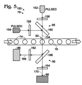
Figure 5 shows an exemplary detection system 150 that utilizes pulsed
illumination, such as from pulsed light sources. In alternative embodiments,
illumination optics 85 transmits pulses of light produced from one or more
continuous beams generated by continuous light sources. For example, optics
85 may include an electro-optical shutter that blocks a continuous beam of
light from a light source between each light pulse. In still other
embodiments,
the illumination may not be pulsed.
System 150 may offer the same advantage as a system with a pair of
light sources at spatially separated examination sites for dye discrimination,
without the potential disadvantage of being unable to correlate data from the
sites with each other. Furthermore, system 150 enables the use of LEDs as
light sources. System 150 may include any suitable combination of the
elements, aspects, and features disclosed elsewhere herein for detection
systems, such as detection system 120 of Figure 3.
Detection system 150 may include a pair (or more) of pulsed light
sources, such as pulsed source 152 and pulsed source 154. Each light source
may emit light of a different (single) wavelength or different wavelength
range
from the other light source(s). Alternatively, the light sources may emit the
same wavelength range of light, but the emitted light may be filtered
differently
for each source. Illumination assembly 78 of system 150, via pulsed light
sources and/or optics 85, may be configured to illuminate examination region
92 in alternation with pulses of light that are spectrally distinct.
Spectrally
distinct pulses have different wavelength maxima, cover different wavelength
ranges, and/or have spectral profiles with distinct shapes, among others.
Detection system 150 can use pulsed sources 152, 154 and detectors
86, 88 as if they form two spatially separated examination sites. The pulses
of
light from each source may be synchronized with periodic data collection from
the detectors. Each source of pulsed illumination may have a corresponding
CA 02767113 2011-12-30
WO 2011/120006 PCT/US2011/030077
22
detector, as indicated by the matching hatch patterns: Source 152 is
operatively paired with detector 86, and source 154 is operatively paired with
detector 88. The examination site defined by source 152 and detector 86 may
overlap the examination site defined by source 154 and detector 88, but
signals from the source/detector pairs can be distinguished temporally by
time-shifted detection and/or data collection from the detectors.
A controller of the system may be operatively connected to the light
sources and the detectors (and/or the optics). The controller may generate a
separate periodic signal for each source/detector pair. The periodic signal
corresponding to a light source/detector pair may result from periodic data
collection from the detector, at least predominantly or exclusively during
illumination by pulses of light from the light source. Alternatively, or in
addition, the periodic signal may result from periodically changing the gain
of
the detector, synchronized with pulses of light from the light source, or
synchronizing pulsed transmission of collected light to the detector with
pulses
of illumination from the light source. In any event, the periodic signal may
represent light detected during illumination of the examination region at
least
predominantly or exclusively with light emitted by only one of the light
sources. Stated differently, the signal may represent light detected by a
detector at least predominantly or exclusively during a plurality of spaced
time
intervals when the examination region is illuminated by a corresponding light
source for the detector.
Illumination optics 85 and/or collection optics 90 may help to limit or
define wavebands of light used for illumination and detection.
Each light source 152, 154 may be operatively connected to at least
one dedicated (or shared) wavelength filter, such as illumination filters 156,
158. The filters may be disposed on dedicated branches of the optical path
from each source to channel 76, namely, before beams from the light sources
are combined at a combining element 160 (e.g., a dichroic element), such that
each filter only affects light from one of the light sources. Illumination
filters
156, 158 may function to remove at least one tail formed by the emission
spectrum of a light source and/or may improve the ability of each light source
CA 02767113 2011-12-30
WO 2011/120006 PCT/US2011/030077
23
to selectively excite a particular dye in the droplets. In other words, the
filters
may improve the ability of excitation light to discriminate between two or
more
dyes.
Each detector 86, 88 may be operatively connected to at least one
dedicated (or shared) wavelength filter, such as collection filters 162, 164.
The
filters may be disposed on dedicated branches of the optical path from
channel 76 to the detectors. In other words, each filter may be disposed
between a beam splitter 166 (e.g., a dichroic filter) and a detector.
Collection
filters 162, 164 may function to transmit different wavebands of detected
light
168, 170 to each detector. Accordingly, the collection filters may be
configured to enable each detector to selectively receive emitted light from a
particular dye in the droplets. Alternatively, or in addition, the collection
filters
may be configured, in combination with the illumination filters, to prevent
wavelength overlap between incident light 104 and detected light 168, 170. In
some embodiments, system 150 also may include a scatter detector to detect
light scattered from droplets, which may enable determination of the size of
individual droplets passing through the examination region.
Figure 6 shows a series of graphs 180-184 all representing the same
time span and illustrating periodic data that is synchronized with
illumination
from each of light sources 152, 154. To simplify the presentation, source 152
and source 154 are arbitrarily designated in graphs 180, 182 as "Source 1"
and "Source 2," respectively. Also, data representing light detected during
pulses of illumination with light from Source 1 is designated as "Signal 1,"
and
data representing light detected during pulses of illumination with light from
Source 2, "Signal 2."
Graphs 180, 182 show alternating light pulses 186, 188 of light from
Source 1 and Source 2. Each pulse 186, 188 is followed by a pause 190 or
192, with the pulses of light from each respective source occurring during a
plurality of spaced time intervals 194. Pulses of illumination of light from
each
source may be separated by a succession of pauses, generally with the light
source emitting (or transmitting) substantially less or no light (e.g., the
light
source is turned on and off repeatedly, or an electro-optical shutter is
opened
CA 02767113 2011-12-30
WO 2011/120006 PCT/US2011/030077
24
and closed repeatedly), with each pulse and pause defining one pulse cycle
196. The pulses and pauses for illumination with a light source may be of
about the same length or may be of different lengths. Also, the pulse lengths
for illumination with each source may be the same or different from each
other. The pulses of illumination with the light sources may be at the same
frequency (e.g., pulses per second) relative to each other and thus with the
same length of pulse cycle, but with a time offset from each other that
interleaves pulses of illumination from the light sources. For example, one
light source may emit a pulse of light each time the other light source
pauses,
and vice versa. The time offset between pulses of illumination from the light
sources may be about one-half of the duration of one pulse cycle. The
interleaved pulses of light from the light sources may exhibit a short time
gap
where no illumination is occurring (as shown here), may occur in immediate
succession with no time gap, or may overlap slightly, among others. Pulsed
illumination from each light source may occur at any suitable frequency, such
as at least about 100 Hz, 1 kHz, 10 kHz, or 100 kHz, among others. In
exemplary embodiments, pulses of illumination with light from each light
source more occur at a frequency of about 100 kHz. The pulse cycle may be
10 microseconds, with each pulse and each pause lasting about 5
microseconds. In other embodiments, each pulse may last for less than about
1 millisecond, or less than about 100, 10, or 1 microseconds, among others.
In some cases, the pulse frequency of illumination may be selected to
illuminate each droplet with at least one pulse, or two or more pulses, of
light
from each light source. Accordingly, a suitable pulse frequency may depend
on the residence time for a droplet in an examination site and the number of
measurements (e.g., signal values) desired for each droplet. The pulse rate
may be faster than the time it takes for a droplet to traverse an examination
site, such that the droplet is illuminated at least once or multiple times
with
light from a light source. The pulse rate may be increased for smaller
droplets
and/or droplets that travel faster. A faster detector may be needed if the
pulse
rate is increased.
CA 02767113 2011-12-30
WO 2011/120006 PCT/US2011/030077
Graph 184 shows how periodic signals 210, 212 generated from light
detected by detectors 86, 88 (see Figure 5) may be synchronized with pulses
186, 188. Periodic Signal 1 (at 210) may be generated from light that is
detected by detector 86 at least predominantly or exclusively during pulses of
5 illumination with light from Source 1 (source 152), and periodic Signal 2
(at
212) may be generated from light that is detected by detector 88 at least
predominantly or exclusively during pulses of light from Source 2 (source
154).
Graph 184 marks portions of Signals 1 or 2 where each signal is
10 stronger due to the presence of a droplet that is positive for a target
(i.e.,
"Droplet A" for Signal 2 and "Droplet B" for Signal 1). Each droplet is
represented by two or three signal values 216 from each of Signal 1 and
Signal 2. In some embodiments, more than one signal value 216 may be
generated from light detected at different times during each pulse.
15 The pulse frequency of illumination may be selected to illuminate each
droplet with at least one pulse, or two or more pulses, of light from each
light
source. Accordingly, a suitable pulse frequency may depend on the time of
occupancy for a droplet in an examination site and the number of signal
values (from different pulses) desired for each droplet. In exemplary
20 embodiments, illumination may be pulsed at 100 kHz for each light source,
1000 droplets per second may pass through the examination site, droplets
may be separated from each other on average by two droplet diameters, and
about thirty signal values of each signal may be generated for each droplet
during thirty pulses of illumination with light from each light source.
25 Figure 7 shows an exemplary controller 230 for system 150. Controller
230 may have any of the properties, structures, or features described
elsewhere herein, such as for controller 126 of Figure 3. Controller may
include any combination of a gate 232, an amplifier 234, a low-pass frequency
filter 236, an analog-to-digital converter 238, and a processor 240, among
others. Any of these components may be dedicated to detector 86 or may be
shared with detector 88. Amplifier 234 may amplify a signal received from the
detector. Filter 236 may remove high frequency components of the signal, to
CA 02767113 2011-12-30
WO 2011/120006 PCT/US2011/030077
26
improve the signal-to-noise ratio. Converter 238 may convert an analog signal
to a digital signal. Processor 240 may manipulate and/or store the digital
signal.
In the configuration shown here, controller 230 is generating a signal
value from light 242 detected by only one of the detectors, namely, detector
86 ("DET 1"). Signal generation is indicated by a series of arrows extending
between controller components to processor 240. The absence of arrows on
the lower line of controller components indicates no signal generation from
detector 88.
Light 242 may be detected predominantly from a first dye 244 during a
pulse of light from source 152. Controller 230 is not generating a signal
value
from unwanted light 246 detected by the other detector (detector 88 ("DET
2")), and source 154 is off. Unwanted light 246 may be produced by various
mechanisms, such as emission 248 from first dye 244, and emission 250 from
a second dye 252 that may absorb light in the pulse from source 152, among
others.
Gate 232 is configured to synchronize signal generation from each
detector with pulses of illumination with light from the light source
corresponding to the detector. The gate may be configured to permit signal
generation, and particularly one or more signal values thereof, during each
pulse of illumination, while blocking signal generation from the other
detector
during the pulse. In the present illustration, the gate is blocking signal
generation from light detected by detector 88, while permitting signal
generation from detector 86. During a subsequent pulse of light from source
154, gate 232, as indicated schematically in phantom at 254, may have the
opposite effect on signal generation by the detectors. Gate 232 may be
described as a time gate because the gate may operate according to a
temporal schedule that corresponds to the schedule of illumination.
The gate may operate on any suitable component(s) to permit and
block signal generation. For example, the gate may control operation of the
detectors themselves, such as by alternately increasing and decreasing the
gain of each detector 86, 88 in substantial synchrony with each pulse of
light.
CA 02767113 2011-12-30
WO 2011/120006 PCT/US2011/030077
27
In some cases, the gate may be an optical gate, such as an electro-optical
shutter, that blocks collected light from reaching the wrong detector (i.e.,
detector 88 in the configuration of Figure 7) during a pulse of illumination.
Alternatively, the gate may periodically block processing of a substantially
continuous signal detected by the detector, in substantial synchrony with
illumination pulses from the non-corresponding light source, to convert the
continuous signal into a periodic signal. The continuous signal may be analog
or digital, and processing may be blocked by gate 232 with the continuous
signal in analog or digital form. The gate may, for example, block input of
the
continuous signal from the detector to amplifier 234, input of the amplified
continuous signal to filter 236, or input of the filtered signal to converter
238,
among others. In some cases, a continuous signal may be processed digitally
by processor 240, such as by selective removal of signal values, to generate
a periodic signal. However, passing a continuous signal through analog filter
236, and then converting the continuous signal into a periodic signal by
digital
processing may be undesirable. The analog filter can degrade the quality of
the resulting periodic signal, because the analog filter can smear together
portions of the continuous signal, making them difficult to separate when the
periodic signal is formed digitally.
Figure 8 shows yet another exemplary detection system 280 for
droplet-based assays. System 280 is similar to detection system 150 of Figure
5, with the capability of producing pulsed illumination, such as with at least
two light sources 152, 154. However, a single detector 282 may be utilized to
detect light during illumination with light from each light source. Collection
optics of the system may include at least one wavelength filter 284 that
selectively excludes one or more wavelength ranges of light. The wavelength
filter(s) may be selected to exclude collected light 102 that was emitted by
one
or both of the light sources, while transmitting collected light emitted by at
least a pair of fluorophores in the droplets. Discrimination between signals
corresponding to different targets may be based on the ability of the light
sources to selectively excite different fluorophores, but generally not based
on
CA 02767113 2011-12-30
WO 2011/120006 PCT/US2011/030077
28
different emission spectra of the fluorophores, since only a single detector
is
used.
Detector 282 may create a substantially continuous signal that is
representative of light detected during pulsed illumination with light from
both
of the light sources. The system may use a controller to convert the
continuous signal into two or more periodic signals each representing light
detected during pulses of illumination with light from a different light
source.
III. Detection Unit with a Slit
This Section describes a slit that may be incorporated into the
illumination optics and/or collection optics of any of the detection systems
disclosed herein; see Figures 9 and 10A-C.
Figures 9, 10A, and 10B shows selected aspects of an exemplary
illumination assembly 310 and a tube 312 (e.g., a capillary) that may be
included in the detection systems disclosed herein. Tube 312 defines channel
76 in which droplets 74 are illuminated. In exemplary embodiments, tube 312
and channel 76 are cylindrical.
Illumination assembly 310 may include at least one light source 314
and illumination optics 316 that transmit light from the light source to tube
312.
The illumination optics may include an aperture element 318 that defines a
slit
320. The slit may be disposed before and/or after one or more optical
elements 322, 324 on an optical path traveled by light from the light source
to
channel 76 (see Fig. 9).
A beam 326 of light from light source 314 may be incident on aperture
element 318, but only a portion of the beam is permitted to travel through
slit
320, to form a shaped beam, namely, a blade 328 of light. In particular,
aperture element 318 may include an optically transmissive substrate 330,
such as glass, and a blocking layer 332 formed on the substrate. The blocking
layer may be substantially opaque, such that it blocks passage of light. In
exemplary embodiments, the blocking layer may be formed by selectively
removing layer 332, such as by etching. A mask may be formed over layer
332, such as by photolithography, to restrict etching of layer 332 to the
position of the slit. The blocking layer may have any suitable composition. In
CA 02767113 2011-12-30
WO 2011/120006 PCT/US2011/030077
29
exemplary embodiments, the blocking layer may be composed of gold and
chromium. An opposing surface of substrate 330 may include a coating of
Mg F2.
Blade 328 is elongated in cross section, namely, in a cross-sectional
plane taken orthogonal to the direction of travel of the blade of light (as in
Figure 10B). Blade 328 may be described as a planar beam with opposing
sides 334 that are at least generally planar. The blade of light may, for
example, be formed by illuminating the slit with a substantially collimated
beam from a light source of the detection unit. Alternatively, the light
source
may be imaged onto the slit. In some cases, imaging the light source onto the
slit may produce higher intensity illumination of the examination site,
everything else being equal, because more light from the light source is
incident on the slit (and thus passes through slit) and less light from the
light
source is incident on and blocked by non-transmissive material flanking the
slit.
Slit 320 may have any suitable properties. The slit may be about the
same length as, longer than, or shorter than the diameter of beam 326. For
example, in shown in Figure 10A, slit 320 is longer than the beam's diameter,
such that opposing ends 336 of blade 328 may not be shaped by the slit (see
Figures 10A and 10B). The slit may have any suitable width, based on the
desired volume of channel 76 to be illuminated, and the amount of
magnification or demagnification that will occur between the slit and the
channel. For example, blade 328 may travel through at least one lens 324
before the blade illuminates a region of channel 76. Lens 324 may focus an
image of the slit onto channel 76 and, optionally, may demagnify the slit's
image relative to the slit itself. In exemplary embodiments, slit 320 is about
10
to 200 microns wide, and the image of the slit at the channel is demagnified,
such that the thickness of blade 328 at the channel is about 5 to 150 microns,
or about 50 to 100 microns, among others.
Figure 10B shows a cross-sectional area 338 of blade 328 taken at
one-half of the distance across channel 76. Blade 328, in cross section, is
elongated transversely to a long axis 340 defined by channel 76. The blade
CA 02767113 2011-12-30
WO 2011/120006 PCT/US2011/030077
may be elongated at least substantially orthogonally to axis 340, namely,
within about 200, 10 , or 5 of orthogonal. A length 342 of the area is
substantially greater than its width 344, such as at least about 2, 5, or 10
times as great. Length 342 may be greater than the diameter of channel 76
5 and/or tube 312. Accordingly, the cross-sectional area of blade 328 at this
position along the blade may only partially overlap the channel and/or tube,
with the area projecting from one or both opposing sides of the channel and/or
tube. A blade in general, and a blade with a cross-sectional length that is
greater than the diameter of the channel and/or tube in particular, may offer
10 one or more advantages over illumination with a cylindrical or conical beam
of
light. These advantages may include a greater tolerance for mis-alignment of
the illumination optics with the channel and/or a decreased tendency for
illumination light from two light sources to illuminate non-overlapping
regions
of the channel.
15 Blade 328 may illuminate a volume 346 of channel 76 (see Fig. 9).
Volume 346 may have a cross sectional shape that corresponds to the cross-
sectional shape of channel 76. Accordingly, volume 346 may be substantially
disk-shaped if the channel has a substantially cylindrical shape. Volume 346
may have opposing planar sides and a dimension (i.e., width 344 in Fig. 10B),
20 measured parallel to channel axis 340, that is greater than, about the same
as, or less than the diameter of a droplet (as shown here). A thinner blade
328
(i.e., a smaller width 344) may permit a higher resolution signal to be
created
from light detected from volume 346. For example, a blade thinner than the
diameter of a droplet may permit collection of more accurate data on droplet
25 size and shape, and better resolution of droplets that are close together
in the
channel. Also, the use of a slit may permit the use of a lens with a higher
numerical aperture to collect the emitted light because illumination is more
precise. Alternatively, the use of a slit may permit detection without
collection
optics.
30 Figure 9 shows additional aspects of tube 312. The tube may include a
coating or sheath 348 that restricts transmission of light across tube 312.
Coating 348 may be selectively removed along a segment 350 of the tube
CA 02767113 2011-12-30
WO 2011/120006 PCT/US2011/030077
31
where illumination is conducted. It may be difficult to accurately remove only
a
short portion of coating 348, so the length of segment 350 may be much
greater than the diameter of a droplet. Accordingly, blade 328 may restrict
illumination to a short region of segment 350. An exemplary coating is formed
of polyimide. In some embodiments, only a short section of coating 348 may
be removed (e.g., on the order of the diameter of a droplet or less).
Figure 10C shows selected aspects of another exemplary detection
unit 360 that may be included in the signal detection systems disclosed
herein. An illumination assembly 362 may illuminate channel 76 with a beam
364 to produce an illuminated volume 366. The illuminated volume may have
a dimension, measured along channel 76, that is greater than the diameter of
the droplets. Alternatively, the illuminated volume may be produced a blade of
light (e.g., see Figs. 9, 10A, and 10B). In any event, aperture element 318
with slit 320 may be included in collection optics 368 of unit 360. The slit
may
be oriented substantially orthogonal to a long axis defined by channel 76. The
slit may be very close to the channel, such as abutted with a tube or other
member that defines the channel. Alternatively, the slit may be disposed in an
image plane formed by the collection optics between the channel and a
detector 370. A slit included in the collection optics may restrict collection
of
light to only a portion of an illuminated volume. In some cases, the use of a
slit
in the collection optics may permit illumination without any illumination
optics.
In some embodiments, a slit may be included in the illumination optics,
and another slit may be included in the collection optics. The slits may be
parallel to each other. The use of a double slit configuration may help to
reduce background by more precisely defining illumination and collection
volumes of the channel. In some cases, the use slits on both the illumination
and collection sides may permit illumination and collection without any other
illumination or collection optics.
IV. Droplet Identification with Combined Signals
This Section describes an exemplary approaching to droplet
identification by using a combined signal; see Figure 11.
CA 02767113 2011-12-30
WO 2011/120006 PCT/US2011/030077
32
Figure 11 shows a flowchart illustrating, with graphs 380-388, an
exemplary approach to processing data collected with the detection systems
disclosed herein.
Graph 380, which has been described already in relation to Figure 6,
shows a pair of separate signals 210, 212 representative of light detected as
droplets in a continuous phase pass through an examination region. Each
separate signal includes droplet data 400 for individual droplets interspersed
with baseline data 402 for droplet-free regions. One goal is to efficiently
identify droplet data for further analysis, free of the baseline data.
However,
with two or more separate signals (e.g., for two or more targets), the
separate
signals may not always be in agreement about the location of droplet data,
particularly when one or both of the separate signals is close to background.
For example, droplet A is identified clearly from Signal 2, but not Signal 1,
while the converse is true for droplet B. A signal processing algorithm could
examine each signal individually to look for the signature of a droplet, but
sometimes a droplet will be identified in one signal but not the other. An
approach is need for identifying droplet data that benefits from the
information
in two or more separate signals representing the same time period of light
detection.
Graph 382 illustrates the results of combining the separate signals of
graph 380 to form a combined signal 404. In particular, individual signal
values 406, 408 representing light detected during the same time interval 410
and from each separate signal 210, 212 may be combined, for each of a
succession of time intervals, to produce combined values 412 forming
combined signal 404. In some embodiments, signal values from more than
two separate signals may be combined. The signal values combined for each
time interval may represent light detected during overlapping or
nonoverlapping portions of the time interval. For example, in the present
illustration, the two separate signals are periodic and temporally offset from
each other, and the individual signal values that are combined represent
successive, instead of overlapping, portions of the time interval. In other
CA 02767113 2011-12-30
WO 2011/120006 PCT/US2011/030077
33
examples, the two (or more) separate signals may be synchronized instead of
temporally offset.
Signal values from separate signals may be combined in any suitable
fashion. For example, two (or more) signal values for each time interval may
be combined to form a linear combination using the following formula:
Y= aXi + bX2
where Y is a combined value of the combined signal, a and b are constants,
and X, and X2 are corresponding individual signal values from the two
separate signals. Additional signal values from other separate signals (e.g.,
cX3, dX4, etc.) also may be included. The constants may be the same or
different. In exemplary embodiments, the constants are at least substantially
the same, such that equal proportions of the separate signals are used to
generate the combined signal. Accordingly, the combined signal may
correspond to an average of separate signals 210, 212.
Individual droplet regions 414 (e.g., peaks or valleys) of the combined
signal representing droplets may be identified, as indicated in graph 384.
Each droplet region may include a temporal sequence of combined values
412 that collectively produce the signature of a droplet. Droplet
identification
may be performed by processing the combined signal with any suitable
algorithm to look for a droplet signature. Exemplary droplet identification
algorithms may be based on one or more predefined conditions
corresponding to an acceptable range for the height (or depth), width,
smoothness, and/or monotonicity of a peak 415 (or valley) formed by the
combined signal.
Signal values corresponding to each droplet region 414, from each
separate signal, indicated at 416, 418, may be processed selectively relative
to other signal values, as indicated in graphs 386, 388. This selective
processing may ignore any signal values, not shown in the graphs, disposed
outside of identified droplet regions. The selective processing may determine
whether a target represented by each separate signal is present or absent in
droplets corresponding to the droplet regions.
CA 02767113 2011-12-30
WO 2011/120006 PCT/US2011/030077
34
V. Optical Layout for a Detection Unit
This Section describes an exemplary detection unit 440, particularly an
exemplary optical layout thereof, for the detection systems disclosed herein;
see Figures 12 and 13. Detection unit 440 may, for example, be incorporated
into detection system 150 (see Fig. 5).
Detection unit 440 may include an illumination assembly 442, a
collection assembly 444, and a monitoring assembly 446, among others.
Illumination assembly 442 may illuminate a capillary 448 defining channel 76.
The collection assembly may collect and detect light received from channel
76, particularly emitted light.
The illumination assembly may be equipped with a blue LED 450 and a
cyan LED 452 that emit light at about 440-520 nm (maximum at 480-485 nm)
and 470-550 nm (maximum at 505-510 nm), respectively. The LEDs may
produce a luminous flux of about 10 to 200 lumens, among others, at a drive
current of about 300 to 1000 mA. Each LED may be configured to be pulsed
at any suitable frequency, such as about 100 kHz each, with the pulses of the
light sources interleaved with each other.
Light from each LED 450, 452 may travel through a lens doublet 454
that collimates light emitted by the LED, to form a collimated beam 456. Each
collimated beam may be filtered through a respective filter, namely,
wavelength filter 458 or 460. Filter 458 may be a short-pass filter that
permits
passage of light of 485 nm or less, and filter 460 may be a band-pass filter
that permits passage of light of 497-518 nm, to produce filtered beams 462,
464. The filtered beams may be combined at a dichroic element 466 oriented
at 45 degrees to optical axes 468, 469 extending to the beams from the LEDs.
Dichroic element 466 may have a nominal reflection cut off of about 495 nm,
such that light from blue LED 450 and cyan LED 452 are combined efficiently.
In some embodiments, dichroic element 466 may be rotated 90 degrees
about axis 469. Also, the optical axis of filtered blue beam 462 may extend to
the rotated dichroic element from back to front in the current view, with blue
LED 450 positioned behind dichroic element 466 in the current view.
CA 02767113 2011-12-30
WO 2011/120006 PCT/US2011/030077
In any event, filtered beams from the LEDs may be combined by
dichroic element 466, to produce a combined beam 470 that is split by a
dichroic beam splitter 471, to form a main beam 472 and a sampling beam
474. A majority of the light (e.g., 95%) may form the main beam and a
5 minority of the light (e.g., 5%) may form the sampling beam. The sampling
beam will be described further below in relation to monitoring assembly 446.
Main beam 472 may travel through a slit 476 defined by an aperture element
478 (e.g., see aperture element 318 of Figs. 9 and 10A-C), to form a blade of
light 480, which may be focused onto capillary 448 by a pair of spaced lenses
10 482. The blade of light may define a plane that that is substantially
orthogonal
to the long axis of the capillary 448 where the blade of light intersects the
capillary.
Collection assembly 444 gathers and detects light received from
capillary 448. The optical axis of the collected light may be substantially
15 orthogonal to both the long axis of the capillary and to the axis of
illumination
defined by the illumination light of blade 480. The collection assembly 444
may, for example, be equipped with a pair of photomultiplier tubes (PMTS)
484, 486 that serve as detectors of light collected from the capillary by
collection optics 488. Optics 488 receives light for the two detectors from
the
20 capillary along a shared optical axis that branches into a pair of optical
axes
extending to the respective PMTs.
The shared optical axis extending from the capillary may include an
aspheric lens 490 disposed close to the capillary. Lens 490 may provide a
high numerical aperture (10 mm diameter, 0.625 NA) for efficient collection of
25 emitted light. Also, the examination site of the capillary that is
illuminated may
be substantially enclosed by a chamber 492, which permits entry of incident
illumination light and exit of emitted light via the aspheric lens, but
otherwise
blocks light. The chamber may be lined with a light-absorbing material (i.e.,
a
blackened chamber) and may minimize or eliminate the occurrence of objects
30 or edges, to minimize scattered and reflected light. The chamber also may
contain at least one mirror 493 that reflects emitted light toward aspheric
lens.
In other words, the mirror may help collect light that is emitted away from
the
CA 02767113 2011-12-30
WO 2011/120006 PCT/US2011/030077
36
aspheric lens, to improve the efficiency which emitted light is detected. The
mirror may, for example, be an elliptical mirror. In some cases, the
examination site may be disposed at least generally between the mirror (or at
least a region thereof) and a collection optical element (e.g., aspheric lens
490) and/or a detector (e.g., if no collection optics are used).
The collected light from aspheric lens 490 may travel through a clean-
up dichroic element 494, which rejects light of less than 500 nm, to remove
residual excitation light, if any, from the LEDs, particularly light emitted
by blue
LED 450. Light transmitted through dichroic element 494 next encounters
dichroic beam splitter 496, which splits the light to form a reflected split
beam
portion 498 of less than about 550 nm and a transmitted split beam portion
500 of greater than about 550 nm. Split beam portion 498 is transmitted to
PMT 484, and split beam portion 500 to PMT 486. Each beam portion may
travel through one or more wavelength filters, such as respective filters 502
or
504, at least one lens 506, and an optional aperture element 508 before
reaching the respective PMT. Filter 502 may be a long-pass filter that rejects
light of less than about 540 nm. Filter 504 may be a band-pass filter that
rejects light off less than about 520 nm and of greater than about 555 nm.
The filters used in the illumination optics and the collection optics may
effectively prevent all excitation light from reaching the detectors. However,
contamination of detected light with excitation light also or alternatively
may
be reduced or eliminated by the use of polarization filters that are cross-
polarized with respect to each other. Illumination light from each light
source
may be polarized on the optical path to the capillary, after the beams are
combined, with a first polarization filter 510 (an illumination filter), to
form a
polarized light beam from the light sources. Collected light may be
transmitted
through a second polarization filter 512 (a collection filter) before the
collected
light beam has been split. Accordingly, second filter 512 will block light
polarized by first filter 510 because such light is polarized in the cross
plane
that is blocked by the second filter. In this way, illumination/excitation
light that
is collected can be blocked by the second polarization filter from reaching
either detector. This arrangement of filters may be particularly suitable with
a
CA 02767113 2011-12-30
WO 2011/120006 PCT/US2011/030077
37
pair of dyes, such as VIC and FAM dyes, where one of the dyes (e.g., VIC
dye) has an excitation spectrum that overlaps the emission spectrum of the
other dye. Also, or alternatively, this filter arrangement may reduce channel
cross-talk. Filters 510 and 512 may be absorptive linear polarizers that
polarize light in respective orthogonal planes. Light emitted from capillary
448
generally is unpolarized, so a substantial part of the emitted light (e.g.,
about
half) may be capable of traveling through polarization filter 512.
Monitoring assembly 446 may monitor the illumination intensity of
illumination light from each LED 450, 452. The monitoring assembly may be
arranged as part of a feedback loop with a controller and LEDs 450, 452 to
maintain the illumination intensity at the capillary substantially constant.
Sampling beam 474 is received by assembly 446 from beam splitter 470 after
the waveband of capillary illumination by each LED has been determined by
respective filters 458, 460. Accordingly, any intensity change for each
waveband that is produced by an overall increase or decrease in light output
by an LED, in addition to any intensity change produced by a shift or other
alteration in the spectral profile of the corresponding LED, can be measured
by assembly 446.
Monitoring assembly 446 may include a corresponding sensor 514 or
516 for each light source. The sensor may, for example, be a photodiode.
Sampling beam 474 may be split by a dichroic beam splitter 518, which may
have at least substantially the same reflection properties as dichroic element
466 (e.g., a nominal reflection cut-off of 495 nm). In other words, beam
splitter
518 acts to reverse the effect of dichroic combining element 466. Beam
splitter 518 thus may produce respective blue and green beam portions 520,
522 corresponding respectively to LEDs 450, 452. Green beam portion 522
may be reflected by a mirror 524 toward sensor 516. Each beam portion may
be passed through a diffuser 526 before reaching the respective sensor 514
or 516.
Figure 13 shows the graph of Figure 4 supplemented with exemplary
wavebands of illumination and detection that may be suitable for the dyes of
Figure 4, namely FAM dye and VIC dye, in the detection system of Figure 5
CA 02767113 2011-12-30
WO 2011/120006 PCT/US2011/030077
38
equipped with detection unit 440 of Figure 12. One of the light sources,
namely, blue LED 450, may illuminate capillary 448 with a waveband 530 of
less about 485 nm. Waveband 530 may or may not have a defined shorter
wavelength boundary. The other light source, namely, cyan LED 452 may
illuminate capillary 448 with a waveband 532 of about 495-515 nm.
Accordingly, there may (or may not) be overlap between the wavebands. In
some cases, a gap 534 may be formed between illumination wavebands 530,
532. Gap 534 may, for example, be at least about 2, 5, or 10 nm, among
others. In some cases, one or both of the light sources (e.g., a laser) may
emit
light of a single wavelength instead of a range of wavelengths. In some cases,
a single light source may produce excitation light for both dyes. In any
event,
light from waveband 530 is selectively absorbed by one of the dyes (FAM
dye) and light from waveband 532 is selectively absorbed by the other dye
(VIC dye). Accordingly, LED 450 selectively excites FAM dye, and LED 452
selectively excites VIC dye.
Light may be collected in detection wavebands 536, 538. The detection
wavebands may or may not overlap. If there is overlap, the amount of overlap
may be about 0-20 or 0-10 nm, among others. Detection waveband 536 is
selective for emission from one of the dyes (FAM dye), and detection
waveband 538 for emission from the other dye (VIC dye). Accordingly,
detection waveband 536 corresponds to illumination waveband 530, blue LED
450, and FAM dye, and detection waveband 538 corresponds to illumination
waveband 532, cyan LED 452, and VIC dye.
Illumination waveband 532 and detection waveband 536 represent
different dyes. These wavebands potentially could overlap if light is detected
from detection waveband 536 only when cyan LED 452 is off, e.g., by using
interleaved pulses of light as described above in Section II. However, the
detector for detection waveband 536 may become saturated by the light from
illumination waveband 532, during a pulse from LED 452. The pulse may
render the detector incapable of accurately measuring light during the next
pulse with the other light source, after LED 452 is turned off, because the
recovery time for the detector may be much longer than the pulse duration.
CA 02767113 2011-12-30
WO 2011/120006 PCT/US2011/030077
39
Therefore, it may be desirable to separate illumination waveband 532 from
detection waveband 536. Alternately, or in addition, it may be desirable to
gate off a detector (e.g., detector 486) for waveband 536 during the period of
illumination with waveband 532. This can, for example, be accomplished by
decreasing the dynode voltage of a PMT detector or the bias voltage on an
APD (avalanche photodiode) detector, or using an electro-optical shutter to
block light from reaching the detector, among others.
An exemplary strategy for dividing up overlap 140, in a balanced
manner, between illumination waveband 532 and detection waveband 536 is
shown in the graph. A shorter wavelength segment (i.e., waveband 532) may
be dedicated to illumination, and a nonoverlapping, longer wavelength
segment (i.e., waveband 536) may be dedicated to detection. Segments of
about the same size (within about 50% of each other in length) from overlap
140 may be assigned to illumination and detection. Wavebands 532 and 536
may be separated by a gap 540 of at least about 2, 5, or 10 nm, to prevent
any excitation light from reaching the detector. Gap 540 may be positioned
near (e.g., within about 10 nm or 20 nm) a maximum value of the absorption
spectrum of one of the dyes and/or a maximum value of the emission
spectrum of the other dye.
VI. Detection System with Spaced Examination Sites
This Section describes exemplary detection systems that define
spaced examination sites with spatially separated excitation/emission
volumes; see Figures 14-16.
In some system embodiments, radiation from multiple excitation
sources may be spatially shifted, i.e., may intersect with droplets at
substantially different spatial locations. In this case, the excitation
sources
need not be pulsed or otherwise alternating, since radiation from only one
source reaches any particular droplet at a given time. As in the case of
systems with a single common excitation region or point, systems having
multiple spatially shifted excitation regions may use a beam splitter and/or
filters in conjunction with multiple detectors to distinguish between droplet
emission signals, or may use a single detector capable of distinguishing
CA 02767113 2011-12-30
WO 2011/120006 PCT/US2011/030077
between emission spectra resulting from excitation of different target
molecules.
Figure 14 shows an exemplary detection system 550 with spatially
shifted examination sites for droplet-based assays. System is equipped with
5 multiple detection units 552 arranged to define a discontinuous examination
region 554 formed of spaced examination sites 556-560 disposed along
channel 76.
The examination sites may have any suitable spacing from one another
(e.g., less than about 5 cm, 1 cm, or 1 mm, or less than about 100, 50, 25, or
10 10 droplet or channel diameters, among others). It may be desirable to
place
the examination sites as close together as possible because closer
examination sites make correlating data detected from different examination
sites less problematic. There is less time for droplets to change their
relative
separations from one another in the flow stream as such droplets travel
15 between examination sites. In some configurations, a series of examination
sites may be close enough to one another that each droplet travels through all
of the sites before the next droplet enters the examination region. With sites
this close, there is no problem syncing droplet data collected from the
examination sites.
20 Each detection unit 552 may include at least one light source 562,
illumination optics 564, collection optics 566, and a detector 568. The
detection units, relative to one another, may provide different wavelengths or
wavebands of illumination light and/or may detect different wavelengths or
wavebands of collected light.
25 Figure 15 shows yet another exemplary detection system 580 for
droplet-based assays. System 580 includes multiple detection units 582
arranged to define a discontinuous examination region 584 formed of spaced
examination sites 586-590 disposed along channel 76. Each detection unit
may include at least one or at least a pair of light sources 592, 594 and at
30 least one or at least a pair of detectors 596, 598. In some embodiments,
one
or more of the detection units may incorporate any combination of features
disclosed above for detection system 150 or 280 (Figs. 4-8), illumination
CA 02767113 2011-12-30
WO 2011/120006 PCT/US2011/030077
41
assembly 310 (Figs. 9 and 10A-C), and/or detection unit 440 (Fig. 12), such
as pulsed illumination and generation of a periodic signal corresponding to
pulses of illumination, a slit to shape and/or selectively block a light beam,
use
of a combined signal to identify droplets, balanced division of a region of
excitation/emission overlap for dyes to an illumination waveband for one dye
and a detection waveband for the other dye, and/or closed-loop monitoring of
illumination intensities, among others.
Figure 16 shows an embodiment 610 of detection system 550. A
capillary 612 defining channel 76 extends through each detection unit 552.
The capillary is clamped in place by brackets 614. Each light source 562
includes an LED. An alternative light source or light collector, an optical
fiber
616, extends through the detection units. Collection optics 566 of each unit
552 may include a filter 618, which may be interchangeable readily by a user.
Light may be detected by photomultiplier tubes 620.
VII. Selected Embodiments
This section describes additional aspects and features of detection
systems for droplet-based assays, presented without limitation as a series of
numbered paragraphs. Each of these paragraphs can be combined with one
or more other paragraphs, and/or with disclosure from elsewhere in this
application, in any suitable manner. Some of the paragraphs below expressly
refer to and further limit other paragraphs, providing without limitation
examples of some of the suitable combinations.
1. A method of detection for droplets, comprising: (A) illuminating
an examination region of a channel with first pulses of light interleaved with
second pulses of light as droplets pass through the examination region, the
first pulses being spectrally distinct from the second pulses; and (B)
collecting
data representing light detected during illumination of the examination region
with the first pulses and the second pulses.
2. The method of paragraph 1, wherein the first pulses define a
first range of wavelengths of light and the second pulses define a second
range of wavelengths of light, and wherein the first range is different from
the
second range.
CA 02767113 2011-12-30
WO 2011/120006 PCT/US2011/030077
42
3. The method of paragraph 1, wherein only the first pulses are
produced by a single wavelength of light, or both the first pulses and the
second pulses are produced by respective single wavelengths of light.
4. The method of any of paragraphs 1 to 3, wherein the first pulses
are produced by a pulsed light source.
5. The method of any of paragraphs 1 to 4, wherein the first pulses
and the second pulses are produced by respective pulsed light sources.
6. The method of paragraph 1, wherein the first pulses, the second
pulses, or both the first and second pulses are produced by at least one
continuous light beam that is transmitted intermittently to the examination
region.
7. The method of paragraph 1, wherein the first pulses and the
second pulses include light emitted by at least one LED.
8. The method of paragraph 7, wherein the first pulses and the
second pulses are emitted by respective pulsed LEDs.
9. The method of paragraph 1, wherein overlapping volumes of the
examination region are illuminated by the first pulses and the second pulses.
10. The method of paragraph 1, further comprising a step of
detecting light from overlapping volumes of the examination region during the
first pulses and the second pulses.
11. The method of paragraph 1, wherein each droplet is illuminated
with at least one first pulse and at least one second pulse.
12. The method of paragraph 11, wherein each droplet is illuminated
with multiple first pulses and multiple second pulses.
13. The method of paragraph 1, wherein each droplet is illuminated
with a beam of light that is narrower than a diameter of the droplets.
14. The method of paragraph 1, wherein the first pulses and second
pulses contain light emitted by respective first and second light sources,
further comprising a step of passing the light emitted by the first and second
sources through a slit before such light illuminates the examination region.
15. The method of paragraph 1, wherein the step of collecting data
includes a step of generating a first signal and a second signal representing
CA 02767113 2011-12-30
WO 2011/120006 PCT/US2011/030077
43
light detected during illumination of the examination region with the first
pulses
and the second pulses, respectively.
16. The method of paragraph 15, wherein the droplets include a first
dye and a second dye, wherein the first signal is generated from a first
detection configuration and the second signal is generated from a second
detection configuration, and wherein the first detection configuration has a
different relative sensitivity to the first and second dyes than the second
detection configuration.
17. The method of paragraph 15, further comprising a step of
detecting light from the examination region with a first detector and a second
detector, and wherein the first signal represents light detected at least
predominantly by the first detector and the second signal represents light
detected at least predominantly by the second detector.
18. The method of paragraph 17, wherein each detector detects
light during the first pulses and the second pulses.
19. The method of paragraph 17, wherein a gain of each detector is
adjusted over time according to whether a first pulse or a second pulse is
illuminating the examination region of the channel.
20. The method of paragraph 15, wherein the first signal and the
second signal are periodic signals.
21. The method of paragraph 1, wherein the data represents light
detected with a same detector during illumination of the examination region
with the first pulses and the second pulses.
22. The method of paragraph 1, further comprising a step of
detecting light from the examination region during the first pulses and the
second pulses, wherein the step of detecting light creates a first signal and
a
second signal, and wherein the step of collecting data includes a step of
periodically gating the first signal and the second signal in correspondence
with the first pulses and the second pulses, respectively.
23. The method of paragraph 1, wherein the step of illuminating
includes a step of intersecting the channel with a beam of light having a
cross
section that is elongated transversely to a long axis defined by the channel.
CA 02767113 2011-12-30
WO 2011/120006 PCT/US2011/030077
44
24. The method of paragraph 1, wherein the step of illuminating
includes a step of illuminating a disk-shaped volume of the channel.
25. The method of paragraph 1, wherein the step of illuminating
includes a step of illuminating the examination region of the channel with
third
pulses of light that are spectrally distinct from the first pulses and the
second
pulses.
26. A method of detection for droplets, comprising: (A) illuminating
an examination region of a channel alternately with pulses of light emitted by
a first light source and a second light source as droplets pass through the
examination region; (B) detecting light from the examination region
illuminated
by the pulses of light; and (C) generating a first signal and a second signal,
the first signal representing light detected at least predominantly when the
first
region is illuminated with pulses of light from the first light source and the
second signal representing light detected at least predominantly when the
second region is illuminated with pulses of light from the second light
source.
27. The method of paragraph 26, further comprising a step of
estimating a concentration of a first target and a second target in the
droplets
based on the first signal and the second signal.
28. The method of paragraph 26, further comprising a step of
determining whether an amplification reaction occurred in individual droplets.
29. A system for detection for droplet-based assays, comprising: (A)
a channel; (B) an illumination assembly configured to illuminate an
examination region of the channel with first pulses of light interleaved with
second pulses of light as droplets pass through the examination region, the
first pulses being spectrally distinct from the second pulses; (C) one or more
detectors configured to detect light from the examination region; and (D) a
controller that collects data representing light detected during illumination
of
the examination region with the first pulses and the second pulses.
30. The system of paragraph 29, wherein the first pulses define a
first range of wavelengths of light and the second pulses define a second
range of wavelengths of light, and wherein the first range is different from
the
second range.
CA 02767113 2011-12-30
WO 2011/120006 PCT/US2011/030077
31. The system of paragraph 29, wherein only the first pulses are
produced by a single wavelength of light, or both the first pulses and the
second pulses are produced by respective single wavelengths of light.
32. The system of paragraph 29, wherein the illumination assembly
5 includes at least one pulsed light source.
33. The system of paragraph 29, wherein illumination assembly
include a pair of pulsed LEDs.
34. The system of paragraph 29, wherein the illumination assembly
includes at least one continuous light source configured to emit a beam of
10 light that is transmitted intermittently to the examination region.
35. The system of paragraph 29, wherein the first pulses and the
second pulses are configured to illuminate overlapping volumes of the
examination region.
36. The system of paragraph 29, wherein the one or more detectors
15 are configured to detect light from overlapping volumes of the examination
region during the first pulses and the second pulses.
37. The system of paragraph 29, wherein the illumination assembly
is configured to illuminate the examination region with a beam of light that
is
elongated in cross section and in a direction transverse to a long axis
defined
20 by the channel.
38. The system of paragraph 29, wherein the illumination assembly
includes a least one slit through which light travels before illuminating the
examination region.
39. The system of paragraph 29, wherein the controller is configured
25 to generate a first signal and a second signal representing light detected
during illumination of the examination region with the first pulses and the
second pulses, respectively.
40. The system of paragraph 39, wherein the one or more detectors
include a first detector and a second detector, and wherein the first signal
30 represents light detected at least predominantly by the first detector and
the
second signal represents light detected at least predominantly by the second
detector.
CA 02767113 2011-12-30
WO 2011/120006 PCT/US2011/030077
46
41. The system of paragraph 40, wherein each detector is
configured to detect light during the first pulses and the second pulses.
42. The system of paragraph 39, wherein the first signal and the
second signal are periodic signals.
43. The system of paragraph 29, wherein the controller is configured
to adjust a gain of each detector over time according to whether a first pulse
or a second pulse is illuminating the examination region of the channel.
44. The system of paragraph 29, wherein the one or more detectors
create a first signal and a second signal that are each at least substantially
continuous, and wherein the controller is configured to periodically gate the
first signal and the second signal in correspondence with the first pulses and
the second pulses, respectively, to make the signals periodic.
45. The system of paragraph 29, wherein the illumination assembly
forms a beam of light having a cross section that is elongated transversely to
a long axis defined by the channel.
46. The system of paragraph 29, wherein the illumination assembly
is configured to illuminate a disk-shaped volume of the channel.
47. The system of paragraph 29, wherein the illumination assembly
includes a first light source and a second light source, further comprising at
least one pump configured to drive the droplets through the examination
region as the light sources illuminate overlapping volumes of the channel.
48. A system for detection in droplet-based assays, comprising: (A)
a channel; (B) an illumination assembly configured to produce a beam of light
that illuminates an examination region of the channel as droplets pass through
such region; (C) a detector configured to detect light received from the
examination region; and (D) a controller that collects data representing light
detected by the detector, wherein the beam of light is elongated in cross
section where the beam intersects the channel.
49. The system of paragraph 48, wherein the illumination assembly
includes a light source and a slit, and wherein light emitted by the light
source
travels through the slit before reaching the examination region.
CA 02767113 2011-12-30
WO 2011/120006 PCT/US2011/030077
47
50. The system of paragraph 48, wherein the beam of light is
elongated in cross section in a direction transverse to a long axis defined by
the channel.
51. The system of paragraph 48, wherein a cross section of the
beam, at a position halfway across the channel, extends outside opposing
surfaces of the channel.
52. The system of paragraph 51, where the channel is defined by a
tube, and wherein a cross section of the beam, at a position halfway across
the channel, is longer than a diameter of the tube.
53. The system of paragraph 52, wherein the cross section at a
position halfway across the channel has opposing ends that do not intersect
the tube.
54. The system of paragraph 48, wherein the beam of light
illuminates a disk-shaped volume of the channel.
55. The system of paragraph 48, wherein the beam of light has
opposing planar sides.
56. The system of paragraph 48, wherein the beam of light has a
dimension measured parallel to a long axis of the channel where the channel
and the beam intersect, and wherein the dimension is less than a diameter of
the channel.
57. A system for detection in droplet-based assays, comprising: (A)
a channel; (B) a light source that illuminates an examination region of the
channel as droplets pass through such region; (C) a detector configured to
detect light received from the examination region; and (D) a controller that
collects data representing light detected by the detector, wherein light
emitted
by the light source travels through at least one slit between the light source
and the detector.
58. The system of paragraph 57, wherein the at least one slit
includes a slit disposed on an optical path from the light source to the
examination region.
59. The system of paragraph 57, wherein the at least one slit
includes a slit disposed between collection optics and the channel.
CA 02767113 2011-12-30
WO 2011/120006 PCT/US2011/030077
48
60. A method of detection for droplets, comprising: (A) illuminating
an examination region of a channel with a beam of light that is elongated in
cross section; and (B) collecting data representing light detected over time
from the region as a plurality of droplets pass through the examination
region.
61. The method of paragraph 60, wherein the step of illuminating
includes a step of transmitting light through a slit disposed on an optical
path
between a light source and the examination region.
62. The method of paragraph 60, wherein a disk-shaped volume of
the examination region is illuminated.
63. The method of paragraph 60, wherein the beam of light is
elongated in cross section in a direction that is transverse to a long axis
defined by the channel.
64. The method of paragraph 60, wherein the step of illuminating
includes a step of illuminating droplets with a beam of light that is thinner
than
a diameter of the droplets.
65. A method of detection for droplet-based assays, comprising: (A)
generating at least two separate signals each representing light detected with
a different detection configuration during a series of time intervals from a
stream of fluid carrying droplets; (B) combining the at least two separate
signals to form a combined signal; and (C) processing the combined signal to
identify time intervals that correspond to droplets.
66. The method of paragraph 65, wherein the step of combining
includes a step of forming a linear combination of values from the separate
signals for individual time intervals.
67. The method of paragraph 66, wherein the step of forming a
linear combination includes a step of forming a linear combination of the
values in equal proportions.
68. The method of paragraph 65, wherein the step of combining is
performed with the at least two signals in digital form.
69. The method of paragraph 65, wherein the step of combining is
performed at least in part as the at least two signals are being generated.
CA 02767113 2011-12-30
WO 2011/120006 PCT/US2011/030077
49
70. The method of paragraph 65, wherein the step of combining
includes a step of combining values from the separate signals for individual
time intervals, and wherein each value that is combined for a given time
interval represents light detected during a different part of the given time
interval.
71. The method of paragraph 70, wherein each value that is
combined for a given time interval represents light detected during
nonoverlapping portions of the given time interval.
72. The method of paragraph 65, wherein the step of combining
includes a step of combining values from the separate signals for individual
time intervals, and wherein each value that is combined for a given time
interval represents light detected during a same part or all of the given time
interval.
73. The method of paragraph 65, wherein the separate signals
include a first signal and a second signal representing light detected from a
region of a channel holding the stream of fluid during illumination of the
region
with alternating pulses of light from a first light source and a second light
source.
74. The method of paragraph 73, wherein the first signal at least
predominantly represents light detected by a first detector during pulses from
the first light source, and wherein the second signal at least predominantly
represents light detected by a second detector during pulses from the second
light source.
75. The method of paragraph 65, wherein each different dye
includes a fluorophore.
76. A method of detection for droplet-based assays, comprising: (A)
generating at least two separate signals each representing a respective
different wavelength or waveband of light detected during a series of time
intervals from a stream of fluid carrying droplets, wherein light detected
from
each wavelength or waveband reports the presence or absence of a different
target in individual droplets; (B) combining the at least two separate signals
to
form a combined signal; (C) processing the combined signal to identify time
CA 02767113 2011-12-30
WO 2011/120006 PCT/US2011/030077
intervals that correspond to droplets; and (D) determining which droplets
contain each different target based on values of each separate signal
detected during the identified time intervals.
77. A method of detection for droplet-based assays, comprising: (A)
5 generating at least two signals each representing a respective different
waveband of light detected during a series of time intervals from a stream of
fluid with droplets; (B) combining values of the at least two signals to form
a
combined signal; (C) identifying portions of the combined signal that
correspond to droplets; and (D) processing values of each of the at least two
10 signals that correspond to the portions identified, to determine which
droplets
contain each target.
78. A system for detection for droplet-based assays, comprising: (A)
one or more detectors configured to detect light from a stream of fluid
carrying
droplets containing at least two different dyes; and (B) a controller
configured
15 to generate separate signals each representing light detected with a
different
detection configuration during a series of time intervals from a stream of
fluid
carrying droplets, to combine the at least two separate signals to form a
combined signal, and to process the combined signal to identify time intervals
that correspond to droplets.
20 79. A method of detection for droplets, comprising: (A) obtaining
droplets including a first dye and a second dye, wherein an emission
spectrum of the first dye and an absorption spectrum of the second dye define
a waveband of overlap and overlap sufficiently to produce at least half-
maximal emission from the first dye if the first dye is excited at a maximal
25 absorption wavelength of the second dye; (B) illuminating the droplets with
excitation light capable of exciting the first dye and the second dye, the
excitation light being emitted by one or more LEDs and including only a
shorter-wavelength segment of the waveband of overlap; and (C) detecting
light emitted by the first dye and the second dye, wherein light emitted from
30 the second dye is detected in a wavelength range including only a longer-
wavelength segment of the waveband of overlap that is spaced from the
shorter-wavelength segment.
CA 02767113 2011-12-30
WO 2011/120006 PCT/US2011/030077
51
80. The method of paragraph 79, wherein the absorption spectrum
and the emission spectrum have respective maxima at wavelengths that are
within about 20 nm of each other.
81. The method of paragraph 79, wherein the one or more LEDs
include a first LED that selectively excites the first dye and a second LED
that
selectively excites the second dye.
82. The method of paragraph 79, further comprising a step of
collecting a first set of data and a second set of data representing light
detected selectively from the first dye and the second dye, respectively.
83. The method of paragraph 79, wherein the first dye is FAM dye
and the second dye is VIC dye.
84. The method of paragraph 79, wherein the waveband of overlap
is defined where the spectra overlap at 20% or more of maximal absorption or
emission, and wherein the waveband of overlap extends for least 25 nm.
85. A system for detection in droplet-based assays, comprising: (A)
a channel configured to receive droplets including a first dye and a second
dye, wherein an emission spectrum of the first dye and an absorption
spectrum of the second dye define a waveband of overlap and overlap
sufficiently to produce at least half-maximal emission from the first dye if
the
first dye is excited at a maximal absorption wavelength of the second dye; (B)
an illumination assembly including one or more LEDs and configured to
illuminate the droplets with excitation light capable of exciting the first
and
second dyes, the excitation light being emitted by the LEDs and including only
a shorter-wavelength segment of the waveband of overlap; and (C) one or
more detectors configured to detect light emitted by the first dye and the
second dye, wherein the light from the second dye is detected in a
wavelength range including only a longer-wavelength segment of the
waveband of overlap that is spaced from the shorter-wavelength segment.
86. The system of paragraph 85, wherein the illumination assembly
includes one or more filters that define the shorter-wavelength segment.
CA 02767113 2011-12-30
WO 2011/120006 PCT/US2011/030077
52
87. The system of paragraph 85, further comprising a collection
assembly including the one or more detectors, were in the collection assembly
includes one or more filters that define the longer-wavelength segment.
88. A method of detection for droplets, comprising: (A) generating a
beam of light; (B) splitting the beam of light into a main beam and at least
one
sampling beam; (C) monitoring an intensity of the sampling beam; (D)
adjusting an intensity of the beam of light based on one or more
measurements from the step of monitoring; (E) illuminating an examination
region of a channel with light from the main beam as droplets pass through
the examination region; and (F) collecting data representing light detected
from the examination region.
89. The method of paragraph 88, wherein the step of generating a
beam of light includes a step of filtering light emitted from a light source
to
change a spectrum of the emitted light, and wherein the step of splitting is
performed after the step of filtering.
90. The method of paragraph 88, wherein the step of filtering is
performed with a band-pass wavelength filter, a long-pass wavelength filter, a
short-pass wavelength filter, or a combination thereof.
91. The method of paragraph 88, wherein the step of generating a
beam of light includes a step of combining beams of light emitted from at
least
two light sources, and wherein the step of combining is performed after the
step of filtering.
92. The method of paragraph 88, wherein the step of generating a
beam of light includes a step of emitting light with an LED.
93. The method of paragraph 88, wherein the step of generating a
beam of light includes a step of combining light from a first light source and
a
second light source, wherein the step of splitting includes a step of
splitting
the beam of light into a first sampling beam and a second sampling beam,
and wherein the first sampling beam corresponds to the first light source and
the second sampling beam corresponds to the second light source.
CA 02767113 2011-12-30
WO 2011/120006 PCT/US2011/030077
53
94. The method of paragraph 93, wherein the step of adjusting
keeps substantially constant an intensity of a portion of the main beam
corresponding to each light source.
The disclosure set forth above may encompass multiple distinct
inventions with independent utility. Although each of these inventions has
been disclosed in its preferred form(s), the specific embodiments thereof as
disclosed and illustrated herein are not to be considered in a limiting sense,
because numerous variations are possible. The subject matter of the
inventions includes all novel and nonobvious combinations and
subcombinations of the various elements, features, functions, and/or
properties disclosed herein. The following claims particularly point out
certain
combinations and subcombinations regarded as novel and nonobvious.
Inventions embodied in other combinations and subcombinations of features,
functions, elements, and/or properties may be claimed in applications claiming
priority from this or a related application. Such claims, whether directed to
a
different invention or to the same invention, and whether broader, narrower,
equal, or different in scope to the original claims, also are regarded as
included within the subject matter of the inventions of the present
disclosure.