Note: Descriptions are shown in the official language in which they were submitted.
CA 02767825 2012-01-06
WO 2011/005930 PCT/US2010/041327
CARDIAC TISSUE-DERIVED CELLS
CROSS REFERENCE TO RELATED APPLICATION
[0001] The present invention claims priority to application serial number
61/224,446, filed July
9, 2009.
FIELD OF THE INVENTION
[0002] The present invention is directed to methods and compositions for
repairing damaged
myocardium using human cardiac tissue-derived cells. In particular, the
present
invention provides methods and compositions for repairing damaged myocardium
using
expanded human cardiac tissue-derived cells that do not express telomerase.
BACKGROUND
[0003] Acute myocardial infarction (AMI) is the leading cause of death in the
US. AMI is
caused by a sudden and sustained lack of blood flow to an area of the heart,
commonly
caused by narrowing of a coronary artery. Without adequate blood supply, the
tissue
becomes ischemic, leading to the death of myocytes and vascular structures.
This area of
necrotic tissue is referred to as the infarct site, and will eventually become
scar tissue.
The remaining cardiomyocytes are unable to reconstitute the necrotic tissue,
and the heart
deteriorates with time. The deterioration may be in the form of a loss of
function of the
heart muscle associated with remodeling of the damaged myocardium.
[0004] Some current therapies for acute myocardial infarction focus on
thrombolysis or,
alternatively, angioplasty, to open up the clotted vessel and restore blood
supply to the
infarct site. These treatments may effectively reduce infarct site size and
improve cardiac
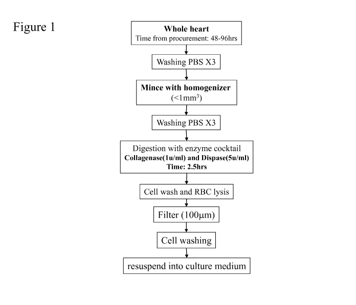
systolic function, but do not reverse the loss of function of the heart muscle
associated
with remodeling of the damaged myocardium. Other therapies, such as, for
example,
angiotensin converting enzyme inhibitors (ACEI) and beta-blockers also improve
global
function and survival. However, the therapeutic effects from these medications
may only
improve survival by less than 5% in post-AMI patients.
1
CA 02767825 2012-01-06
WO 2011/005930 PCT/US2010/041327
[0005] Cell transplantation may be another potential therapy for acute
myocardial infarction.
For example, Orlic et at (Nature 410: 701 - 705 (2001)) report the injection
of Lin c-kit+
bone marrow cells into damaged myocardium. Orlic et at state: "Our studies
indicate
that locally delivered bone marrow cells can generate de novo myocardium,
ameliorating
the outcome of coronary artery disease."
[0006] In another example, Nygren et at (Nature Medicine 10: 494 - 501 (2004))
state: "We
show that unfractionated bone marrow cells and a purified population of
hematopoietic
stem and progenitor cells efficiently engraft within the infarcted myocardium.
Engraftment was transient, however, and hematopoietic in nature. In contrast,
bone
marrow-derived cardiomyocytes were observed outside the infarcted myocardium
at a
low frequency and were derived exclusively through cell fusion."
[0007] However, the mechanism by which bone marrow-derived cells treat AMI is
unclear. For
example, Murry et at (Nature 428: 664 - 668 (2004)) state: "[W]e used both
cardiomyocyte-restricted and ubiquitously expressed reporter transgenes to
track the fate
of haematopoietic stem cells after 145 transplants into normal and injured
adult mouse
hearts. No transdifferentiation into cardiomyocytes was detectable when using
these
genetic techniques to follow cell fate, and stem-cell-engrafted hearts showed
no overt
increase in cardiomyocytes compared to sham-engrafted hearts. These results
indicate
that haematopoietic stem cells do not readily acquire a cardiac phenotype, and
raise a
cautionary note for clinical studies of infarct repair."
[0008] In another example, Werner et at (Nature Clinical Practice
Cardiovascular Medicine 5:
78-79 (2008)) state: "There are many questions, however, still to be answered
with
regard to the most effective progenitor cell subpopulation, the best technique
for
progenitor cell augmentation, the underlying mechanisms of action, and the
long-term
safety and effectiveness of the method. Moreover, several trials of [bone
marrow cell]
therapy in patients with AMI have produced negative results, possibly because
of
variation in the timing of [bone marrow cell] administration after AMI,
differences in the
methods of progenitor cell preparation used, or both."
2
CA 02767825 2012-01-06
WO 2011/005930 PCT/US2010/041327
[0009] In another example, Balsam et at (Nature 428: 668 - 673 (2004)) state:
"Our data
suggest that even in the microenvironment of the injured heart, c-kit-enriched
BM cells,
Lin c-kit+ BM cells and c-kit+ Thy 1.110 Lin- Sca-1+ long-term reconstituting
haematopoietic stem cells adopt only traditional haematopoietic fates."
[0010] Another possible source of cells is embryonic stem cells. For example,
Gold et at
(W02005090558) discloses methods for generating cardiomyocyte lineage cells
from
embryonic stem cells for use in regenerative medicine.
[0011] In another example, Gold and Hassanipour (W02007002136) disclose
methods for the
differentiation of primate pluripotent stem cells into cardiomyocyte-lineage
cells.
[0012] Another possible source of cells is cardiac progenitor cells. Cardiac
progenitor cells have
been identified in the human and rat heart. Cardiac progenitor cells are self-
renewing and
multipotent giving rise to all cardiac lineages.
[0013] For example, U.S. Patent Application US20040126879A1 disclose the use
of cardiac
stem cells that are CD31+, CD38+ and c-kif to treat damaged myocardium.
[0014] In another example, Oh et at (PNAS 100: 12313 - 12318 (2003)) disclose
the existence
of adult heart-derived cardiac progenitor cells, expressing Sca-1, CD31 and
CD38, and
lacking the expression of CD4, CD8, B220, Gr-1, Mac-1, TER119, c-kit, Flk-1, e-
Cadherin, von Willebrand factor, CD45 and CD34.
[0015] In another example, U.S. Patent Application US 20080241111A1 disclose a
method for
preparing mammalian cardiac tissue-derived cells prepared through the steps
of. (i)
enzymatically treating a cardiac tissue fragment from a mammal to prepare a
cell
suspension; (ii) separating a group of cardiac tissue-derived cells from said
cell
suspension by a density gradient method; and (iii) suspension culturing the
obtained
group of cardiac tissue-derived cells in a culture medium containing
fibroblast growth
factor and epidermal growth factor, and then selecting and separating cells
forming a
floating sphere.
3
CA 02767825 2012-01-06
WO 2011/005930 PCT/US2010/041327
[0016] In another example, U.S. Patent Application US 20080213231A1 disclose a
pluripotent
stem cell group composed of pluripotent stem cells derived from a human or
mouse
skeletal muscle tissue, the pluripotent stem cells being c-met-negative, Pax-7-
negative,
Myf-5-negative, MyoD-negative, Myogenin-negative, M-cadherin-negative, CD105-
positive, CD90-positive, c-kit-negative and CD45-negative, the pluripotent
stem cells
being CD34-negative in the case of the human-derived stem cells and being CD34-
positive in the case of the mouse-derived stem cells, and the pluripotent stem
cell group
being obtained by proliferation of a single cell.
[0017] In another example, Laugwitz et at (Nature 433: 647 - 653 (2005)
discloses isll-1+
cardiac progenitor cells in postnatal rat, mouse and human myocardium.
[0018] In another example, Messina at at (Circulation Research 95: 911 - 921,
(2004)) disclose
the "isolation of undifferentiated cells that grow as self-adherent clusters
(that we have
termed "cardiospheres") from subcultures of postnatal atrial or ventricular
human biopsy
specimens and from murine hearts. These cells are clonogenic, express stem and
endothelial progenitor cell antigens/markers, and appear to have the
properties of adult
cardiac stem cells." Messina at at state: "[N]ewly developing human and mouse
cardiospheres revealed expression of endothelial (KDR (human)/flk-1 [mouse],
CD-3 1)
and stem cell (CD-34, c-kit, sca-1) markers."
[0019] In another example, Smith et at (Circulation 115(7): 896 - 908 (2007)
state:
"Percutaneous endomyocardial biopsy specimens grown in primary culture
developed
multicellular clusters known as cardiospheres, which were plated to yield
cardiosphere-
derived cells (CDCs)."
[0020] In another example, U.S. Patent Application US20070020758 discloses a
method for the
isolation, expansion and preservation of cardiac stem cells from human or
animal tissue
biopsy samples to be employed in cell transplantation and functional repair of
the
myocardium or other organs.
[0021] In another example, Beltrami et at (Cell 114(6): 763 - 776 (2003))
disclose "the
existence of Lin- c-kitPOS cells with the properties of cardiac stem cells.
They are self-
4
CA 02767825 2012-01-06
WO 2011/005930 PCT/US2010/041327
renewing, clonogenic, and multipotent, giving rise to myocytes, smooth muscle,
and
endothelial cells."
[0022] In another example, WO 2008054819 discloses cardiovascular stem cells
positive for
markers isll+/ Nkx 2.5+/ ik1+ and cardiovascular stem cells which can
differentiate along
endothelial, cardiac, and smooth muscle cell lineages.
[0023] In another example, WO 2008109839A1 discloses an enriched population of
stem cells
comprising a CXCR4 polypeptide and an FIk-I polypeptide, wherein said stem
cells are
capable of differentiating into cells that express Mef2C, GATA-4, Myocardin,
and
Nkx2.5 polypeptides.
[0024] In another example, WO 2008081457A2 discloses a method of isolating
cardiac stem
cells, the method comprising contacting a tissue which comprises the cardiac
stem cells
with a composition which comprises dispase 11 under conditions sufficient to
induce cell
dissociation, thereby isolating the cardiac stem cells.
[0025] In another example, WO 2008058273A2 discloses a method for obtaining
mammalian
stem-cell-like myocyte-derived cells (MDCs) from atrial or ventricular heart
tissue,
comprising the steps of. isolating cells from atrial or ventricular heart
tissue to form a
cell suspension; and culturing the cells in a medium comprising a mitogen
thereby
forming a composition comprising MDCs.
[0026] In another example, WO 2008054819A2 discloses a method for isolating
cardiovascular
stem cells, the method comprising contacting a population of cells with agents
reactive to
Isletl, Nkx2.5 and ikl, and separating reactive positive cells from non-
reactive cells.
[0027] In another example, U.S. Patent Application US 20070212423A1 discloses
a method of
isolating a c-kit/c-mef cardiomyocyte precursor cell of muscular origin,
comprising
separating cells of less than 40 m in diameter from a suspension of muscle
cells;
culturing the cells in a tissue culture medium on a solid substrate; and
isolating the cells
in suspension in the medium; thereby isolating the c-kit/c-mef cardiomyocyte
precursor
cell of muscular origin.
CA 02767825 2012-01-06
WO 2011/005930 PCT/US2010/041327
[0028] In another example, U.S. Patent Application US 20050058633 an isolated
mammalian c-
kit-/c-met-cardiomyocyte precursor cell of muscular origin.
[0029] In another example, WO 2004019767 discloses an isolated mammalian
cardiomyocyte
stem cell having c-kitneg/CD31+ /CD38+ and expressing telomerase reverse
transcriptase.
[0030] In another example, WO 2008083962A1 discloses [c]ardiomyocyte
progenitor cells
(CMPCs) which are characterized by Sca-1 or a Sca-1 like epitope and CD31 on
their cell
surface.
[0031] In another example, U.S. Patent Application 20080213230A1 discloses
method of
preparing an isolated cell population enriched in stem cells or progenitor
cells,
comprising: (a) culturing a tissue sample;(b) obtaining cells that migrate
above adherent
fibroblasts during said culturing;(c) cloning one or more cells obtained in
(b) to produce
one or more clonogenic populations;(d) identifying one or more clonogenic
populations
having a desired phenotype;(e) isolating stem cells or progenitor cells from
the one or
more clonogenic populations identified in step (d) by cell sorting using one
or more cell
surface or internal markers of stem cells or progenitor cells; and(f)
culturing the isolated
stem cells or progenitor cells in conditioned media in the absence of feeder
cells; thereby
obtaining an isolated cell population enriched in stem cells or progenitor
cells.
[0032] However, one obstacle for the use of cardiac progenitor cells is the
lack of an efficient
method to isolate or expand the cells. Therefore, there still remains a need
for the
efficient isolation and expansion of cardiac progenitor cells in order for
their
effectiveness as a therapy for damages myocardium to be assessed.
SUMMARY
[0033] The present invention provides methods to isolate and expand cells
derived from human
cardiac tissue. Cells isolated and expanded according the methods of the
present
invention do not express telomerase, and are useful to treat or repair damaged
myocardium.
6
CA 02767825 2012-01-06
WO 2011/005930 PCT/US2010/041327
[0034] The present invention provides a purified population of human cardiac
tissue-derived
cells that do not express telomerase.
[0035] The present invention provides a method to produce human cardiac tissue-
derived cells
that do not express telomerase, comprising the steps of:
a. Obtaining heart tissue,
b. Dissociating the heart tissue,
c. Digesting the heart tissue to release cells,
d. Removing the cardiomyocytes from the released cells, and
e. Culturing the remaining cells.
[0036] In one embodiment, the present invention provides a method to treat or
repair damaged
myocardium in a patient comprising the steps of:
a. Obtaining a population of human cardiac tissue-derived cells that do not
express
telomerase, and
b. Administering the population of human cardiac tissue-derived cells to the
patient
in an amount sufficient to treat or repair the damaged myocardium.
[0037] In one embodiment, the human cardiac tissue-derived cells used to treat
the patient have
been cryopreserved.
BRIEF DESCRIPTION OF THE FIGURES
[0038] Figure 1 outlines the procedure by which the cells of the present
invention are isolated.
The details of the process to obtain the cell populations are described in
Example 1.
[0039] Figure 2 outlines the isolation of the human cardiac tissue-derived
cells of the present
invention. The details of the process to obtain the cell initial populations
are described in
Example 1.
7
CA 02767825 2012-01-06
WO 2011/005930 PCT/US2010/041327
[0040] Figure 3 outlines an alternate method to isolate the human cardiac
tissue-derived cells of
the present invention.
[0041] Figure 4 shows the morphology of the human cardiac tissue-derived cells
(hCTC's) of
the present invention. All images shown are at 100X magnification, unless
otherwise
indicated. Panel a: is an image showing a cell suspension obtained from pre-
plating the
cells obtained from the initial digestion. The black arrow shows phase-bright
non-
adherent S cells; Panel b: The black arrow shows a phase-bright S cell
cluster. The
white arrow shows adherent cells obtained from the initial plating (the image
shown is at
200X magnification); Panel c shows A2 cells, derived from replating S cell
cultures;
Panel d shows replated phase-bright S cells in an A2 cell culture.
[0042] Figure 5 shows the effect of seeding density on hCTC growth potential.
The x-axis
shows the days in culture after plating a mixture of hCTC (Al) and hCTC (S)
cells from
frozen vials. The y-axis shows the accumulative total population doublings of
the hCTC
(A3) cells.
[0043] Figure 6 shows the effect of reduced oxygen levels on hCTC (A3) cell
growth potential.
[0044] Figure 7 shows the growth potential of rat cardiac tissue-derived
rCTCA2 cells (rCTC
(A2)). The x-axis shows the days in culture after replating rCTC (S) cells
from frozen
vials. The y-axis shows the accumulative total population doublings of rCTC
(A2) cells.
[0045] Figure 8 shows the recovery and viability of hCTC (A3) cells, following
cryopreservation and simulated delivery with a potential administration device
(consisting of a 30-gauge needle). The viable cell number recovered is
indicated on the
left y- axis. The cell viability is indicated on the right y-axis. Filled
diamonds depict cell
viability; open squares depict cell recovery. Details are described in Example
6.
[0046] Figure 9 shows the recovery and viability of rCTC (A2) cells following
cryopreservation
and simulated delivery with a potential administration device (consisting of a
30-gauge
needle). The viable cell number recovered is indicated on the left y-axis. The
cell
viability is indicated on the right y-axis. Filled squares depict cell
recovery prior to
needle passage; filled triangles depict cell viability prior to needle
passage; open squares
8
CA 02767825 2012-01-06
WO 2011/005930 PCT/US2010/041327
depict cell viability post needle passage; open triangle depict cell recovery
post needle
passage. Details are described in Example 6.
[0047] Figure 10 shows hCTC (A3) cell surface marker expression as determined
by flow
cytometry. In each histogram, the dotted line is the isotype antibody control.
The solid
line is the antigen staining. Antigens were shown in the panels. The x-axis is
the
phycoerythrin (PE) intensity in logarithmic scale. The y-axis is the cell
count.
[0048] Figure 11 shows the comparison of cell surface marker expression
between hCTC (A3)
cells (upper panels) and adult human dermal fibroblasts-NHDF (catalogue
number: CC-
2511, Lonza, lower panels). In each histogram, the x-axis is the PE intensity
in
logarithmic scale. The y-axis is the cell count. The dotted line depicts
antibody isotype
control. The solid line is the antigen staining. The positive population was
gated based
on I% positive population in isotype controls. The individual markers are
indicated in
each histogram.
[0049] Figure 12 shows rCTC (A2) cell surface marker expression as determined
by flow
cytometry. The dotted line depicts the isotype control. The solid line is the
antigen
staining for CD31 (left panel), and CD90 (right panel).
[0050] Figure 13 shows the gene expression of cardiac specific genes in hCTC
(A3) cells as
determined by quantitative real-time polymerase chain reaction (qRT-PCR).
Details are
described in Example 8. The y-axis shows the percentage of GAPDH, and is split
into
lower and upper scales. The lower scale ranges from 0 to 0.01%, and the upper
scale
ranges from 0.05% to 0.15%. The acronyms have the following meanings: MHC
means
myosin heavy chain; cardiac TF means transcription factor; NHDF means Neonatal
human dermal fibroblast; h-heart means human heart. Data are expressed as Mean
S.D
(n=3).
[0051] Figure 14 shows the elevated expression of mouse specific myosin heavy
chain (MHC)
in a co-culture of mouse cardiac tissue-derived (A2) cells (mCTC (A2)) with
rat neonatal
cardiomyocytes (Catalogue # 8357-6W, Cell Application, Austin, TX). The mouse
MHC gene expression level was presented as the percentage of mouse GAPDH in
each
9
CA 02767825 2012-01-06
WO 2011/005930 PCT/US2010/041327
sample. The y-axis indicates the percentage of mouse GAPDH. The acronyms have
the
following meanings: mCTC means mCTC (A2) cells cultured in differentiation
medium,
as described in Example 9; CM means cardiomyocytes; mCTC+CM means mCTC co-
cultured with rat cardiomyocytes CM in differentiation medium. Data are
expressed as
Mean S.D (n=3).
[0052] Figure 15 shows the growth curve observed for porcine cardiac tissue-
derived (A3) cells
(pCTC (A3)), cultured according to the methods described in Example 10. The x-
axis
shows time in culture following plating. The y-axis shows the accumulative
total
population doublings.
[0053] Figure 16 shows pCTC (A3) cell surface marker expression as detected by
flow
cytometry. The dotted line depicts the isotype control. The solid line is the
antigen
staining for CD90 (upper left panel), CD 105 (upper right panel), pig
endothelial cell
marker (lower left panel), CD 16 (lower middle panel), CD45 (lower right
panel).
[0054] Figure 17 shows a cartoon of the cardiac remodeling which follows acute
myocardial
infarction. The cartoon was reproduced from Pfeffer M. in Atlas of heart
failure (Colucci
W, editor, 1999).
[0055] Figure 18 shows the effect of the administration of the cardiac tissue-
derived cells of the
present invention on fractional shortening (FS) in animals wherein acute
myocardial
infarctions have been induced, as measured by echocardiography. Fractional
shortening
is the percent change (FS%) in systole from diastole in each cardiac cycle,
and reflects
the global function of the heart. Data shown is the fractional shortening
recorded in an
individual animal at 5 (D5) or 28 days (D28) after induction of AMI. Animals
were
dosed with rCTC (A2) cells, or hCTC (A3) cells at the doses indicated on the x-
axis.
[0056] Figure 19 shows the effect of the cardiac tissue-derived cells of the
present invention on
regional wall motion score (RWMS) in animals wherein acute myocardial
infarctions
have been induced, as measured by echocardiography. Each panel separated by a
vertical
solid line is an experimental arm in the study. 5D and 28D reflect 5 days and
28 days
after induction of AMI respectively. The RWMS was measured at 5D as baseline
and
CA 02767825 2012-01-06
WO 2011/005930 PCT/US2010/041327
28D as a follow-up. Animals were dosed with rCTC (A2) or hCTC (A3) cells at
the
doses indicated on the x-axis.
[0057] Figure 20 shows the effect of the cardiac tissue-derived cells of the
present invention on
left ventricular end diastolic dimension (LVEDD) in animals wherein acute
myocardial
infarctions have been induced. LVEDD is a measurement of left chamber
dimension at
the end of diastole. LEVDD was measured at 5 days (5D) and 28 days (28D) after
induction of myocardial infarction. Data shown is the relative change [(28D-
5D)/5D] of
individual animals. Animals were dosed with rCTC (A2), or hCTC (A3) cells at
the
doses indicated on the x-axis.
[0058] Figure 21 shows the statistical analysis, comparing the relative change
of LVEDD in
each experimental group. An F-test was applied to the data using one-way
analysis of
variance (ANOVA). Group 1: vehicle; group 2: rCTC (A2) cells 1 x 10⁶ cells
(target
dose); group 3: hCTC (A3) cells 1 x 10⁴ cells (target dose); group 4: hCTC
(A3) cells 1 x
105 cells (target dose); groups: hCTC (A3) cells 1 x 10⁶ cells (target dose).
[0059] Figure 22 shows the effect of human cardiac tissue-derived cell
administration on left
ventricular end systolic dimension (LVESD). LVESD is a measurement of chamber
dimension at the end of systole in each cardiac cycle. Each panel separated by
vertical
solid line was an experimental arm in the study. LVESD was measured at 5 days
(5D)
and 28 days (28D) after induction of myocardial infarction. Data shown is the
recordings
from a single animal at each time point. Animals were dosed with rCTC (A2) or
hCTC
(A3) cells at the doses indicated on the x-axis.
[0060] Figure 23 shows the cardiac function at day 5 and day 28 post
infarction and human
cardiac tissue-derived cell administration, in individual animals in four
parameters (FS,
RWMS, LVESD, LVEDD) measured by echocardiography. Each black dot depicts
individual animal's cardiac function at the time point indicated on the X
axis. The black
solid line shows the trend of change from 5D to 28D in each animal. Each panel
depicts
one parameter measured by echocardiography.
11
CA 02767825 2012-01-06
WO 2011/005930 PCT/US2010/041327
[0061] Figure 24 shows the correlation between fractional shortening and the
dose of cardiac
tissue-derived cells. Y axis is the absolute change at day 28 from day 5 post
infarction
and cell administration (28D-5D). X axis is the dose of hCTC (A3) on linear
scale. Data
are expressed as Mean S.D (n=35).
[0062] Figure 25 shows the correlation between LVEDD change and the dosage of
the human
cardiac-derived tissue of the present invention. The y-axis depicts the
absolute change at
day 28 from day 5 post infarction and cell administration (28D-5D). The x-axis
depicts
the dose of hCTC (A3) cells on a linear scale. Data are expressed as Mean
S.D (n=35).
[0063] Figure 26 shows the retention of hCTC (A3) cells administered to the
heart of animals
that have myocardial infactions. Retention was estimated from the level of
beta-
microglobulin expression detected in rat hearts. Panel "a" shows hCTC (A3)
cell
retention at 4 weeks after administration at the doses indicated on the x-
axis. Panel "b"
shows the time course of hCTC (A3) cell retention where the x-axis depicts the
number
of days after cell administration in rat MI heart, and the y-axis shows the
percentage of
the target dose. Panel "c" shows hCTC (A3) cell retention over time, using an
average
percentage of the cells detected, setting the amount of human cells detected
immediately
after cell administration at 100%. The x-axis depicts the number of days after
cell
administration in rat MI heart.
[0064] Figure 27 shows the correlation between hCTC (A3) retention and
prevention of
remodeling in rat MI. Left panel shows the correlation graph. The x-axis
depicts cell
number on logarithmic scale; the y-axis depicts remodeling changes (delta
LVEDD, 28D-
5D). Each animal's cell number at 4 weeks after administration and the
corresponding
delta LVEDD were plotted in the graph. The right panel shows the statistical
analysis of
the linear regression.
[0065] Figure 28 shows human NuMA (Nuclear Matrix Antigen) localization in rat
myocardium
treated with hCTC (A3) cells (a targeted dose of 1 x 10⁶ cells). Left panel
shows the
positive human NuMA staining observed the target dose of 1 x 10⁶ hCTC-treated
rat
myocardium at 400-fold magnification. The right panel shows the staining in a
vehicle
control animal.
12
CA 02767825 2012-01-06
WO 2011/005930 PCT/US2010/041327
[0066] Figure 29 shows human NuMA localization in another animal receiving a
targeted dose
of 1 x 10⁶ hCTC (A3) cells. The top left panel shows a low power image (100-
fold
magnification) showing two clusters of NuMA positive cells. The top right
panel is a
high magnification image (400-fold magnification), of clusters of NuMA
positive cells.
The bottom left panel is a high magnification image of NuMA positive cell
cluster. The
bottom right panel is a high magnification image showing NuMA positive cell
nuclei
with myocyte-like morphology.
[0067] Figure 30 shows the staining of antibody controls for NuMA observed in
human and rat
myocardium. The top two images show NuMA positive staining (left panel) with
high
nuclear specificity (non-staining with isotype control, right panel) in human
heart. The
bottom two images show rat heart controls demonstrating the NuMA antibody's
specificity to human cells.
[0068] Figure 31 shows the scoring evaluation of myocardial hypertrophy in
hCTC (A3)-treated
or vehicle-treated groups. The target dose is indicated on the y-axis. Animals
receiving
hCTC (A3) cells received a target dose of either 1 x 10⁴, 1 x 10⁵, or 1 x 10⁶
cells. The
light grey area shows the proportion of non-hypertrophy sections in whole
heart. The
dark grey area shows the proportion of hypertrophic sections in whole heart.
[0069] Figure 32 shows infarct size assessment. Left panel shows the relative
infarct size
(percentage of infarct area in total left ventricular area); Right panel shows
the absolute
infarct area. The black dots depict each individual animal. The average size
of infarct of
the group was shown as solid black line.
[0070] Figure 33 shows the staining of capillary density in hCTC (A3) cell-
treated, or vehicle-
treated groups. Animals receiving hCTC (A3) cells received a target dose of
either 1 x
104, 1 x 10⁵, or 1 x 10⁶ cells. Panel "a" shows capillary density at the
border zone of the
infarct in myocardium, as detected with isolectin-B4 staining. Panel "b" shows
capillary
density at the border zone, as detected by CD31 staining.
[0071] Figure 34 shows the myocyte density at the non-infarcted area. Panel
"a" shows
representative images of H&E staining of the myocardium from vehicle treated
animals
13
CA 02767825 2012-01-06
WO 2011/005930 PCT/US2010/041327
(left panel) and animals treated with 1 x10⁵ hCTC (A3) cells (right panel).
Panel "b"
shows the myocyte density observed in non-infarcted areas of the heart,
expressed as
myocyte numbers per mm2. Data are shown as Mean SD (n=6); Panel c shows
proliferating myocytes that were observed by double-staining of Ki-67 and
myosin heavy
chain. Data are expressed as Mean SD (n=6).
[0072] Figure 35 shows differentially expressed genes in rat myocardium in
response to hCTC
(A3) cell treatment at all target doses.
[0073] Figure 36 shows the quantification of the effect of hCTC and hMSC cell
administration
on cardiac tissue in rats suffering from an acute MI. Panel "a" shows the
ratio of infarct
area vs. healthy tissue in the left ventricular free wall. Panel "b" shows the
ranking of
dilatation observed in hearts from animals in all groups. Panel "c" shows
viable
myocardium.
[0074] Figure 37 shows the effects of hCTC (A3) cell and human mesenchymal
stem cell (Cat #
PT-2501, Lonza, Walkersville, MD) administration on the cardiac tissue in rats
suffering
from an acute MI. Two sections are shown side-by-side from each animal: one
taken
from the mid line between the papillary muscle and atrial level and one taken
from the
papillary muscle. The left two columns are from the vehicle treated group; the
middle
two columns were from hMSC treated group (1 x 10⁶ targeted dose); the right
two
columns were from the hCTC (A3) cell treated group (1 x 10⁵ targeted dose).
[0075] Figure 38 shows the effect of hCTC (A3) cell administration on cardiac
function in rats
suffering from an acute MI, at day 28 after infarction and cell
administration. Three
parameters (FS, LVESD, LVEDD) were measured by echocardiography. Relative
change from baseline (day 7 post infarction and cell administration) at day 28
post cell
administration and infarction is presented. Three hCTC (A3) cell lots from
different
donors, human dermal fibroblasts and pCTC (A3) cells are shown.
[0076] Figure 39 shows the effect of hCTC (A3) cell administration on cardiac
function in rats
suffering from an acute MI, at day 84 post infarction and cell administration.
Three
parameters (FS, LVESD, LVEDD) were measured by echocardiography. Relative
14
CA 02767825 2012-01-06
WO 2011/005930 PCT/US2010/041327
change from baseline (day 7 post infarction and cell administration) at day 84
post cell
administration and infarction is presented. Human dermal fibroblasts and three
different
hCTC (A3) cell lots that were prepared from different donors were examined.
DETAILED DESCRIPTION
[0077] For clarity of disclosure, and not by way of limitation, the detailed
description of the
invention is divided into the following subsections that describe or
illustrate certain
features, embodiments, or applications of the present invention.
Definitions
[0078] As used herein, the term "damaged myocardium" refers to myocardial
cells which have
been exposed to ischemic conditions. These ischemic conditions may be caused
by a
myocardial infarction, or other cardiovascular disease or related complaint.
[0079] "Acute myocardial infarction" as used herein refers to the condition
commonly known as
a "heart attack," wherein when the blood supply to part of the heart is
interrupted causing
some heart cells to die. This is most commonly due to occlusion (blockage) of
a
coronary artery following the rupture of a vulnerable atherosclerotic plaque,
which is an
unstable collection of lipids (like cholesterol) and white blood cells
(especially
macrophages) in the wall of an artery. The resulting ischemia (restriction in
blood
supply) and oxygen shortage, if left untreated for a sufficient period, can
cause damage
and/or death of heart muscle tissue (myocardium).
[0080] The term "hCTC (S) population" or "hCTC (S)" as used herein refers to a
non-adherent
population of human cardiac tissue-derived cells that is obtained following
the initial
culture of cells after the human cardiac tissue has been dissociated,
enzymatically
digested, and filtered according to the methods of the present invention.
[0081] The term "hCTC (Al) population" or "hCTC (Al) cells" as used herein as
used herein
refers to an adherent population of human cardiac tissue-derived cells that is
obtained
following the initial culture of cells after the human cardiac tissue has been
dissociated,
enzymatically digested, and filtered according to the methods of the present
invention.
CA 02767825 2012-01-06
WO 2011/005930 PCT/US2010/041327
[0082] The term "hCTC (A2) population" or "hCTC (A2) cells" as used herein
refers to a
population of adherent cells that result from the in vitro culture of hCTC (S)
cells.
[0083] The term "hCTC (A3) population" or "hCTC (A3) cells" as used herein
refers to a
population of adherent cells that result from the in vitro culture of a
mixture of hCTC (S)
and hCTC (Al) cells.
Methods to Derive the Cells of the Present Invention
[0084] The present invention provides a method to produce human cardiac tissue-
derived cells
that do not express telomerase, comprising the steps of:
a. Obtaining heart tissue,
b. Dissociating the heart tissue,
c. Digesting the heart tissue to release cells,
d. Removing the cardiomyocytes from the released cells and
e. Culturing the remaining cells.
[0085] The heart tissue may be dissociated manually. Alternatively, the heart
tissue may be
dissociated mechanically.
[0086] The cardiomyocytes may be removed from the released cells by any
suitable method.
For example, the cardiomyocytes may be removed by filtration, centrifugation,
or by
FACS.
[0087] In one embodiment, the cells released from the digestion of the cardiac
tissue are filtered
to remove the cardiomyocytes. The purpose of the filtration step is to exclude
cells that
are larger in size than the human cardiac tissue-derived cells of the present
invention. In
one embodiment, the human cardiac tissue derived cells of the present
invention are from
about 5 microns to about 50 microns in diameter, and a filter of a pore size
of 50 microns
is chosen to allow the human cardiac tissue-derived cells of the present
invention to pass
through the filter.
16
CA 02767825 2012-01-06
WO 2011/005930 PCT/US2010/041327
[0088] In one embodiment, the human cardiac tissue-derived cells that pass
through the filter are
cultured in vitro. In one embodiment, the human cardiac tissue-derived cells
that are
cultured in vitro after the filtration step are a mixture of non-adherent
cells and adherent
cells.
[0089] The human cardiac tissue-derived cells of the present invention may
adhere to any solid
substrate. In one embodiment, the solid substrate is polycarbonate.
Alternatively, the
solid substrate may be polystyrene. Alternatively, the solid substrate may be
glass. The
solid substrate may also be coated with an adlayer comprising an extracellular
matrix
protein, such as, for example, collagen or laminin, and the like.
[0090] The adherent cells of the present invention that are obtained after the
initial culture step
are referred to herein as the human cardiac tissue-derived (Al) population of
cells, or
hCTC (Al) cells. The non-adherent cells of the present invention that are
obtained after
the initial culture step are referred to herein as the human cardiac tissue-
derived (S)
population of cells, or hCTC (S) cells.
[0091] In one embodiment, hCTC (Al) cells are expanded in culture. The hCTC
(Al) cells of
the present invention may be cultured in any suitable tissue culture medium.
For
example, in one embodiment, the cardiac tissue-derived cells may be cultured
in DMEM,
supplemented with 1,000 g/1 D-glucose, 584 mg/l L-glutamine, and 110 mg/l
sodium
pyruvate, and 10% FBS. Antibiotics such as, for example, penicillin 50 U/ml
and
streptomycin 50 g/ml may be added to the culture medium. Alternatively,
antibiotics
may be added to the suspension of cells obtained following dissociation and
enzymatic
digestion of the heart tissue. The hCTC (Al) cells of the present invention
may be plated
at a seeding density of about 1,000 to about 10,000 viable cells / cm2 on
tissue culture
substrates. The hCTC (Al) cells of the present invention may be incubated
under 5-20
%v/v atmospheric oxygen.
[0092] In one embodiment, the hCTC (Al) cells of the present invention are
passaged once the
cells reach approximately 80% confluence. Alternatively, the hCTC (Al) cells
of the
present invention are passaged once the cells reach approximately 90%
confluence.
17
CA 02767825 2012-01-06
WO 2011/005930 PCT/US2010/041327
Alternatively, the hCTC (Al) cells of the present invention are be passaged
every one to
seven days.
[0093] In one embodiment, hCTC (S) cells are expanded in culture. In one
embodiment, the
hCTC (S) cells of the present invention may be cultured in any suitable tissue
culture
medium. For example, in one embodiment, the cardiac tissue-derived cells may
be
cultured in DMEM, supplemented with 1,000 g/1 D-glucose, 584 mg/l L-glutamine,
and
110 mg/l sodium pyruvate, and 10% FBS. Antibiotics such as, for example,
penicillin 50
U/ml and streptomycin 50 g/ml may be added to the culture medium.
Alternatively,
antibiotics may be added to the suspension of cells obtained following
dissociation and
enzymatic digestion of the heart tissue. The hCTC (S) cells of the present
invention may
be incubated under 5-20 % v/v atmospheric oxygen. In one embodiment, the
tissue
culture medium is replaced every three days.
[0094] In one embodiment, the hCTC (S) cells become adherent with time in
culture. The time
in culture in which the hCTC (S) cells become adherent is from about 1 days to
about 7
days. The population of adherent cells that result from the hCTC (S) cells
becoming
adherent is referred to herein as the human cardiac tissue-derived (A2)
population of
cells, or hCTC (A2) cells.
[0095] In one embodiment, hCTC (A2) cells are expanded in culture. The hCTC
(A2) cells of
the present invention may be cultured in any suitable tissue culture medium.
For
example, in one embodiment, the cardiac tissue-derived cells may be cultured
in DMEM,
supplemented with 1,000 g/1 D-glucose, 584 mg/l L-glutamine, and 110 mg/l
sodium
pyruvate, and 10% FBS. Antibiotics such as, for example, penicillin 50 U/ml
and
streptomycin 50 g/ml may be added to the culture medium. Alternatively,
antibiotics
may be added to the suspension of cells obtained following dissociation and
enzymatic
digestion of the heart tissue. The hCTC (A2) cells of the present invention
may be plated
at a seeding density of about 1,000 to about 10,000 viable cells / cm2 on
tissue culture
substrates. The hCTC (A2) cells of the present invention may be incubated
under 5-20 %
v/v atmospheric oxygen.
18
CA 02767825 2012-01-06
WO 2011/005930 PCT/US2010/041327
[0096] In one embodiment, the hCTC (A2) cells of the present invention are
passaged once the
cells reach approximately 80% confluence. Alternatively, the hCTC (A2) cells
of the
present invention are passaged once the cells reach approximately 90%
confluence.
Alternatively, the hCTC (A2) cells of the present invention are passaged every
one to
seven days.
[0097] In one embodiment, a mixture of hCTC (Al) cells and hCTC (S) cells are
expanded in
culture. In one embodiment, the mixture of hCTC (Al) cells and hCTC (S) cells
form a
population of adherent cells with time in culture. The time in culture in
which the hCTC
(S) cells become adherent is from about 2 days to about 14 days. The
population of
adherent cells that result from the mixture of hCTC (Al) cells and hCTC (S)
cells
becoming adherent is referred to herein as the human cardiac tissue-derived
(A3)
population of cells, or hCTC (A3) cells.
[0098] In one embodiment, hCTC (A3) cells are expanded in culture. The hCTC
(A3) cells of
the present invention may be cultured in any suitable tissue culture medium.
For
example, in one embodiment, the cardiac tissue-derived cells may be cultured
in DMEM,
supplemented with 1,000 g/1 D-glucose, 584 mg/l L-glutamine, and 110 mg/l
sodium
pyruvate, and 10% FBS. Antibiotics such as, for example, penicillin 50 U/ml
and
streptomycin 50 g/ml may be added to the culture medium. Alternatively,
antibiotics
may be added to the suspension of cells obtained following dissociation and
enzymatic
digestion of the heart tissue. The hCTC (A3) cells of the present invention
may be plated
at a seeding density of about 1,000 to about 10,000 viable cells / cm2 on
tissue culture
substrates. The hCTC (A3) cells of the present invention may be incubated
under 5-20 %
v/v atmospheric oxygen.
[0099] In one embodiment, the hCTC (A3) cells of the present invention are
passaged once the
cells reach approximately 80% confluence. Alternatively, the hCTC (A3) cells
of the
present invention are passaged once the cells reach approximately 90%
confluence.
Alternatively, the hCTC (A3) cells of the present invention are be passaged
every one to
seven days.
19
CA 02767825 2012-01-06
WO 2011/005930 PCT/US2010/041327
[0100] One method by which to obtain the human cardiac tissue-derived cells of
the present
invention is outlined in Figure 1. An alternate method by which to obtain the
human
cardiac tissue-derived cells of the present invention is outlined in Figure 2.
An alternate
method by which to obtain the human cardiac tissue-derived cells of the
present invention
is outlined in Figure 3.
[0101] The cells of the present invention may be derived from dissociating the
whole heart, and
subsequently digesting the dissociated tissue. Alternatively, cells of the
present invention
may be derived from dissociating portions of heart tissue, and subsequently
digesting the
dissociated tissue. The portions may be obtained from any part of the heart,
such as, for
example, the atria, or the ventricles, the apex of the heart, or either side
of the heart.
[0102] The dissociated heart tissue can be digested using enzymes such as, for
example
collagenase and dispase. The enzymes may be used separately or alternatively
in
combination. In one embodiment, the dissociated heart tissue is digested using
a mixture
of collagenase and dispase.
[0103] The collagenase may be used at a concentration from about 0.1 U/ml to
about 10 U/ml.
Alternatively the collagenase may be used at a concentration from about 0.1
U/ml to
about 5 U/ml. Alternatively the collagenase may be used at a concentration of
about 5
U/ml. Alternatively the collagenase may be used at a concentration of about 1
U/ml.
[0104] The dispase may be used at a concentration from about 0.5 U/ml to about
50 U/ml.
Alternatively the collagenase may be used at a concentration from about 0.5
U/ml to
about 10 U/ml. Alternatively the collagenase may be used at a concentration of
about 10
U/ml. Alternatively the collagenase may be used at a concentration of about 5
U/ml.
[0105] The dissociated tissue may be treated with the enzymes for about 5
minutes to about 5
hours. Alternatively the dissociated tissue may be treated with the enzymes
for about 30
minutes to about 5 hours. Alternatively the dissociated tissue may be treated
with the
enzymes for about 30 minutes to about 4 hours. Alternatively the dissociated
tissue may
be treated with the enzymes for about 30 minutes to about 3 hours.
Alternatively the
dissociated tissue may be treated with the enzymes for about 30 minutes to
about 2 hours.
CA 02767825 2012-01-06
WO 2011/005930 PCT/US2010/041327
Alternatively the dissociated tissue may be treated with the enzymes for about
30 minutes
to about 1 hour. In one embodiment, the dissociated tissue is treated with the
enzymes
for about 2.5 hours.
[0106] If desirable, the cardiac tissue-derived cells of the present invention
may be exposed, for
example, to an agent (such as an antibody) that specifically recognizes a
protein marker
expressed by the cardiac tissue-derived cells, to identify and select cardiac
tissue-derived
cells, thereby obtaining a substantially pure population of cardiac tissue-
derived cells.
The Cells of the Present Invention
[0107] The present invention provides a human cardiac tissue-derived cell
population that does
not express telomerase that can be maintained and expanded in culture, and is
useful in
the treatment and repair of damaged myocardium. The properties of the cardiac
tissue-
derived-cells of the present invention are summarized in Table 1.
[0108] In one embodiment, the human cardiac tissue-derived cells of the
present invention that
do not express telomerase, express at least one of the following markers:
CD49e,
CD 105, CD59, CD54, CD90, CD34, and CD 117.
[0109] In one embodiment, the human cardiac tissue-derived cells of the
present invention that
do not express telomerase do not express at least one of the following
markers: MDR,
CD19, CD16, CD31, CD45 and Isl-1.
[0110] In one embodiment, the human cardiac tissue-derived cells of the
present invention that
do not express telomerase, express the following markers: CD49e, CD105, CD59,
CD54,
CD90, CD34, and CD 117.
[0111] In one embodiment, the human cardiac tissue-derived cells of the
present invention that
do not express telomerase do not express the following markers: MDR, CD 19, CD
16,
CD31, CD45 and Isl-1.
[0112] In one embodiment, the human cardiac tissue-derived-cells of the
present invention are
further differentiated into cardiomyocytes. This differentiation may be prior
to, or,
alternatively after, administration into the patient. Differentiation refers
to the act of
21
CA 02767825 2012-01-06
WO 2011/005930 PCT/US2010/041327
increasing the extent of the acquisition or possession of one or more
characteristics or
functions, which differ from that of the original cell (i.e., cell
specialization). This can be
detected, for example, by screening for a change in the phenotype of the cell
(i.e.,
identifying morphological changes in the cell and/or surface markers on the
cell). Any
method capable of differentiating the cardiac tissue-derived cells of the
present invention
into cardiomyocytes may be used.
[0113] For example, the cardiac tissue-derived-cells of the present invention
may be further
differentiated into cardiomyocytes according to the methods disclosed in U.S.
Patent
Application US20040126879.
[0114] In another example, the cardiac tissue-derived-cells of the present
invention may be
further differentiated into cardiomyocytes according to the methods disclosed
in
W02004019767.
Methods to Treat or Repair Damaged Myocardium
[0115] Damaged myocardium results from a variety of cardiac diseases, such as,
for example
acute myocardial infraction, chronic myocardial infraction, congestive heart
failure, and
the like. The cardiac tissue-derived cells of the present invention may be
used a therapy
to repair damaged myocardium. In one embodiment, the cardiac tissue-derived
cells of
the present invention are used as a therapy to repair myocardium that is
damaged as a
result of acute myocardial infarction.
[0116] In one embodiment, the present invention provides a method to treat
damaged
myocardium in a patient comprising the steps of:
a. Obtaining a population of human cardiac tissue-derived cells that do not
express
telomerase, and
b. Administering the population of human cardiac tissue-derived cells to the
patient
in an amount sufficient to treat the damaged myocardium.
[0117] In one embodiment, the present invention provides a method to repair
damaged
myocardium in a patient comprising the steps of:
22
CA 02767825 2012-01-06
WO 2011/005930 PCT/US2010/041327
a. Obtaining a population of human cardiac tissue-derived cells that do not
express
telomerase, and
b. Administering the population of human cardiac tissue-derived cells to the
patient
in an amount sufficient to repair the damaged myocardium.
[0118] In one embodiment, the population of human cardiac tissue-derived cells
are prepared
and administered to the patient without culturing the cells. In an alternate
embodiment,
the population of cardiac tissue-derived cells are prepared and cultured in
vitro, prior to
administering to the patient.
[0119] In the case where the population of human cardiac tissue-derived cells
are cultured and
expanded in vitro, the population of cells that is cultured and expanded may
be a
population of hCTC (Al) cells. Alternatively, the population of cells that is
cultured and
expanded may be a population of hCTC (A2) cells. Alternatively, the population
of cells
that is cultured and expanded may be a population of hCTC (A3) cells.
[0120] The human cardiac tissue-derived cells of the present invention may be
administered to a
patient suffering from damaged myocardium by any suitable method in the art.
Such
methods are readily selected by one of ordinary skill in the art.
[0121] For example, administration of the human cardiac tissue-derived cells
of the present
invention to the damaged myocardium may be via direct injection of the damaged
myocardium. Alternatively, the human cardiac tissue-derived cells of the
present
invention may be administered systemically. Where the human cardiac tissue-
derived
cells of the present invention are delivered systemically, the efficiency of
delivering the
cells to the damaged myocardium may be enhanced, for example, by treating the
patient
with at least one other agent, or by another method capable of enhancing the
delivery of
cells to the damaged myocardium.
[0122] For example, the human cardiac tissue-derived cells of the present
invention may be
administered along with another agent selected from the group consisting of.
stem cell
factor (SCF), granulocyte-colony stimulating factor (G-CSF), granulocyte-
macrophage
colony stimulating factor (GM-CSF), stromal cell-derived factor-1, steel
factor, vascular
23
CA 02767825 2012-01-06
WO 2011/005930 PCT/US2010/041327
endothelial growth factor, macrophage colony stimulating factor, granulocyte-
macrophage stimulating factor, and Interleukin-3.
[0123] In one embodiment, the human cardiac tissue-derived cells of the
present invention are
administered along with another agent according to the methods disclosed in
U.S. Patent
Application US20020061587.
[0124] In one embodiment, the human cardiac tissue-derived cells of the
present invention are
administered along with another agent according to the methods disclosed in
U.S. Patent
Application US2002162796.
[0125] For example, the delivery of cells to the damaged myocardium may be
enhanced by
isolating the patient's cardiac circulation from the patient's systemic
circulation and
perfusing a solution comprising cells into the cardiac circuit. An example of
this method
is disclosed in W02007067502.
[0126] In one embodiment, the human cardiac tissue-derived cells of the
present invention are
administered to the patient according o the methods disclosed by Iwasaki,
Kawamoto et
at. (Circulation 113: 1311-1325; 2006).
[0127] In an alternate embodiment, the human cardiac tissue-derived cells of
the present
invention are administered to the patient using a catheter that can be
inserted into
coronary artery according to the methods disclosed by Sherman, Martens et at.
(Nature
Clinical Practice Cardiovascular Medicine 3, suppl 1: S57-64; 2006).
[0128] In an alternate embodiment, the human cardiac tissue-derived cells of
the present
invention are administered to the patient using a catheter that is capable of
mapping the
electrical activity of the myocardium. In one embodiment, the cardiac tissue-
derived
cells of the present invention are administered to the patient using a
catheter that is
capable of mapping the electrical activity of the myocardium according to the
methods
disclosed by Perin, Dohmann et at. (Circulation 107: 2294-2302; 2003); and
Perin, Silva
et at. (Journal of Molecular and Cellular Cardiology 44: 486-495; 2008). In an
alternate
embodiment, the cardiac tissue-derived cells of the present invention are
administered to
24
CA 02767825 2012-01-06
WO 2011/005930 PCT/US2010/041327
the patient according to methods disclosed by Sherman, Martens et at. (Nature
Clinical
Practice Cardiovascular Medicine 3, suppl 1: S57-64; 2006).
[0129] In an alternate embodiment, the human cardiac tissue-derived cells of
the present
invention are administered to the patient using a catheter that is capable of
intra-
myocardium injection according to the methods disclosed by Hashemi, Ghods et
at.
(European Heart Journal 29: 251-259; 2008).
[0130] The present invention is further illustrated, but not limited by, the
following examples.
Example 1
Isolation of Human Cardiac Tissue-Derived Cells
[0131] Materials and methods -digestion enzyme cocktail preparation: The
digestion enzyme
cocktail used in the present invention to isolate cardiac tissue-derived cells
from human
heart was prepared as 2X cocktail stock solutions in Phosphate Buffered Saline
(PBS,
Gibco, Invitrogen, Carlsbad, CA). The concentrations are 0.2U/ml or 2U/ml
Collagenase
(Serva Electrophoresis GmbH, Heidelberg, Germany) and l0U/ml Dispase II (Roche
Applied Science, Indianapolis, Indiana). These enzyme cocktail stocks were
stored at -
40 C. Prior to digestion, the enzyme cocktail was filtered through 0.22 m
vacuum filter
system (Corning Incorporated, Acton, MA). For human heart digestion, the 2U/ml
Collagenase stock was used in the digestion procedure. The final
concentrations of
digestion enzymes are 1 U/ml collagenase and 5U/ml dispase. The process of
digestion
of the whole human heart and the isolation of human cardiac tissue-derived
cells (hCTC)
is summarized in Figure 1.
[0132] Material and methods - human heart: Transplant-discard whole hearts
were obtained
from the National Development and Research Institutes (NDRI, New York, NY).
The
procurement time of the transplant-discard hearts was between 40-98 hours. The
whole
organ was immerged into growth medium (DMEM+10% fetal bovine serum) and stored
at 4 C until being processed for cell isolation.
CA 02767825 2012-01-06
WO 2011/005930 PCT/US2010/041327
[0133] Materials and methods - human heart tissue processing: The whole heart
tissue was
transferred to a biosafety cabinet and placed into a square bioassay dish
(Coming Inc.,
Acton, MA). The excess fat tissue was removed using sterile scalpels (Bard
Parker,
Becton Dickinson, Hancock, NY). The first whole heart was cut into small
pieces (2-3
cm3). Three quarters of the small tissue pieces were then minced manually to
fine pieces
(1-2 mm3 in size). This procedure took two hours to complete. One quarter of
the pieces
were transferred to the PR0250 homogenizer chamber (Pro Scientific, Oxford,
CT). The
lid was placed on the chamber and the PR0250 generator attached with the speed
of the
generator set to 3. The tissue pieces were homogenized for 10 seconds at room
temperature with no addition of any buffer and the tissue was visually
inspected. The
homogenizing was complete when the tissue was finely minced (resulting in
fragments
less than lmm3 in size).
[0134] Materials and methods - human tissue digestion: The tissue pieces, both
manually
processed and homogenized-were transferred to separate 250 ml conical tubes
(Coming
Inc., Coming, NY). The tissue in each tube was washed three times by adding
100 ml
room temperature PBS and inverting the tube five times. The tube was then
placed
upright and the tissue allowed to settle. The supernatant was aspirated using
a 2 ml
aspirating pipette (BD falcon, BD Biosciences, San Jose, CA). The digestion
enzyme
cocktail stock (2X) was added to the 250 ml tube at an enzyme to tissue ratio
of 1:1. The
final concentration of the enzymes was lU/ml Collagenase and 5U/ml Dispase II.
The
tubes containing the tissue and enzymes were transferred to a 37 C orbital
shaker set for
225 rpm (Bamstead Lab, Melrose Park, IL) and incubated for 2.5 hours. After
incubation, the tube was transferred back to the biosafety cabinet. The cell
suspension
was diluted by filling the tubes with room temperature PBS. In order to remove
any
remaining undigested tissue, the cell suspension was filtered through an 8-
inch diameter
250 m standard testing sieve (Sigma-Aldrich, St. Louis, MO) and into a 500 ml
glass
beaker. Following this step, the cell suspension in the glass beaker was
further filtered
through 100 m cell strainers (BD Falcon) and into multiple 50 ml conical
tubes (BD
Falcon). The cell suspension was then washed by centrifuging at 338 x g for 5
minutes at
room temperature using a Sorvall Legend T centrifuge (Thermo Fisher
Scientific, Inc,
26
CA 02767825 2012-01-06
WO 2011/005930 PCT/US2010/041327
Waltham, MA) to pellet the cells. The supernatant was aspirated off and the
cell pellets
resuspended in PBS and pooled into separate 50 ml tubes, one each for the
manually
minced process and the homogenizing process. The cell suspension was washed
three
more times with 40 ml room temperature PBS. After washing, the pellet was
resuspended in 20 ml ACK lysing buffer (Lonza, Walkersville, MD) and incubated
for 10
minutes at room temperature to lyse any remaining red blood cells. After
incubation the
cell suspension was washed two more times with 40 ml room temperature PBS.
Following the final centrifugation, the pellet was resuspended in 20 ml room
temperature
growth medium (DMEM, 1,000 mg/L D-glucose, L-glutamine, and 110 mg/L sodium
pyruvate, 10% fetal bovine serum, Penicillin 50U/ml, Streptomycin 50ug/ml) and
counted.
[0135] Materials and methods - Cell counting: The total viable cell density
and viability
analysis was performed using the Vi-CellTM XR (Beckman Coulter, Fullerton,
CA). The
Vi-CellTM cell viability analyzer automates the trypan blue dye exclusion
method for cell
viability assessment using video captures technology and image analysis of up
to 100
images of cells in a flow cell. The Vi-Cell has a counting accuracy of 6%.
Samples
were prepared and analyzed according to the manufacturer's instructions
(Reference
Manual PN 383674 Rev.A). Briefly, a 500 L aliquot of the final cell suspension
obtained after RBC lysis was transferred to a Vi-CellTM 4 ml sample vial and
analyzed
using a Vi-Ce11TM XR Cell Viability Analyzer. The default cell type profile
was used:
Cell brightness 85%
Cell sharpness 100
Viable cell spot brightness 75%
Viable cell spot area 5%
Minimum circularity 0
Decluster degree Medium
27
CA 02767825 2012-01-06
WO 2011/005930 PCT/US2010/041327
Minimum diameter 5 microns
Maximum diameter 50 microns
Images 50
Aspirate cycles 1
Trypan blue mixing cycles 3
[0136] Results - Cell yield obtained from a whole heart digestion: From the
first whole heart,
after digestion, the yield from the manually minced process produced 43
million viable
cells after dissociation and enzymatic digestion. The viability was 65%. The
mechanical
homogenization procedure produced 12 million viable cells and viability was
63%. Since
3-times more tissue went through the manual procedure, there were no
differences in
yield and viability between the 2 procedures. Based on the results, subsequent
human
hearts were processed using mechanical homogenization. After digestion, total
yield was
typically 34-64 million viable cells per heart. The viability was 55-81%, as
shown in
Table 2.
Example 2
Selection and In Vitro Culture of the Human Cardiac Tissue-Derived Cells of
the
Present Invention
[0137] The cell suspension obtained following the dissociation and enzymatic
digestion of a
human heart was expanded for further experimental analysis.
[0138] The initial plating of the cells obtained from dissociation and
enzymatic digestion of a
human heart: The cell suspension obtained from the dissociation and enzymatic
digestion of a human heart, according to the methods described in Example 1
was added
to T225 tissue culture flasks (Coming Inc., Coming, NY) flasks. 10 ml of the
cell
suspension was added to each flask, which contained 50 ml growth medium (DMEM,
1,000 mg/L D-glucose, 584 mg/L L-glutamine, and 110 mg/L sodium pyruvate, 10%
fetal bovine serum, Penicillin 50U/ml, Streptomycin 50 g/ml, Invitrogen,
Calsbald,
28
CA 02767825 2012-01-06
WO 2011/005930 PCT/US2010/041327
CA). The final volume of the initial culture was 60 ml. The cells were
incubated at 37 C
in an atmosphere comprising 20% 02 and 5% C02, for 2 days. After this time, a
heterogeneous cell culture was observed. Non-adherent, phase bright cells were
observed
(referred to herein as hCTC (S) cells), and adherent cells were observed
(referred to
herein as hCTC (Al) cells). See Figure 4.
[0139] The hCTC (S) cells obtained after the initial culture step had a
similar morphology to the
cells described as cardiac stem cells by Anversa (Beltrami, Barlucchi et at.
2003; Cell
114(6): 763-76). See Figure 4a. In addition, cell clusters, similar to those
described by
Messina et at (Messina, De Angelis et at. 2004; Circulation Research 95(9):
911-21)
were observed, shown in Figure 4b.
[0140] Expansion of the hCTC (S) population: In one study, the hCTC (S) cells
were removed
from the culture flasks after the initial two day culture period. The hCTC (S)
cells were
transferred to 50 ml conical tubes. The hCTC (S) cells were centrifuged at 338
x g for 5
minutes at room temperature. The supernatant was discarded and the cell pellet
re-
suspended in 20 ml fresh growth medium. hCTC (S) cells were counted after
resuspension. The total number of hCTC (S) cells obtained from the initial
culture step
was about 10-14 million total cells. A fraction of the hCTC (S) cells were
cryo-preserved
in preservation medium at 1-1.5million/ml and stored at -140 C. The remainder
were
expanded in culture. The hCTC (S) cells were replated in flasks at seeding
density of
5,000 cells/cm2. After 2 days in culture, hCTC (S) cells became adherent, and
formed a
homogeneous adherent cell population, referred to herein as the hCTC (A2)
population,
or hCTC (A2) cells. Once the hCTC (A2) cells of the present invention reached
80%
confluency at about 10-14 days after hCTC (S) plating, the cells were
trypsinized by
aspirating off the growth medium, washed with 60 ml room temperature PBS, and
then 4
ml Trypsin-EDTA (Invitrogen, Carlsbad, CA) was added to each flask. The hCTC
(A2)
cells were incubated for approximately 5 minutes at room temperature until the
cells had
detached. To each flask, 6 ml growth medium was added and the cell suspension
was
transferred to a fresh 50 ml conical tube (BD Falcon, BD Biosciences, San
Jose, CA). A
500 L aliquot was removed and transferred to a Vi-cell sample cup for
counting using
29
CA 02767825 2012-01-06
WO 2011/005930 PCT/US2010/041327
the Vi-Ce11TM Cell Viability Analyzer as described in Example 1. These cells
were
replated at 5,000 cells/cm2 or 3,000 cells/cm2.
[0141] hCTC (S) cell expansion was also performed using cryopreserved cells. A
frozen vial of
S cells was thawed at 37 C and was washed with PBS once. Then cells were
counted
and plated at 5,000 cells/cm2 in growth medium in culture flasks. Cell
expansion and
confluency was observed each day.
[0142] By visual observation, non-adherent hCTC (S) cells were significantly
reduced after 2
days in culture and the number of non-adherent hCTC (S) did not increase in
culture over
a period of 10-14 days. hCTC (S) cells in culture started attaching to flask
after 2 days in
culture and grew as adherent cells. They reached 80% confluency at 10-14 days
after
seeding of hCTC (S) cells. Although in culture some hCTC (S) cells were still
observed,
the number of this non-adherent population did not increase in culture. This
was possibly
due to the morphology change of non-adherent hCTC (S) to adherent hCTC (A2) in
culture. Since the hCTC (S) cells attached to the flask after 2 days, the
number of non-
adherent cells became very low and was not counted.
[0143] In contrast, the adherent hCTC (A2) cells that were derived from hCTC
(S) cells
demonstrated some growth potential, up to 10 PDL shown in Figure 5a. The
growth rate
of this population was between 1-3 PDL/passage until it reached plateau at 9-
10 PDL,
when the hCTC (A2) cells were seeded at either 5,000 cells/cm2 or at 3,000
cells/cm2.
However, the PDL calculation is based on the initial cell number plated into
the culture
(i.e. the hCTC (S) cell number), the estimation of PDL may not be entirely
accurate.
[0144] Expansion of the hCTC (Al) population: After the medium containing the
hCTC (S)
cells was removed from the flasks containing the cells from the initial two
day plating, 60
ml fresh growth medium was added to the remaining adherent cells present in
the flasks.
The hCTC (Al) cells were cultured until the cells reached 80% confluency.
After this
time, the cells were trypsinized by aspirating off the growth medium, washed
with 60 ml
room temperature PBS and adding 4 ml Trypsin-EDTA (Invitrogen, Carlsbad, CA).
Cells were incubated for approximately 5 minutes at room temperature until the
cells had
detached. To each flask, 6 ml growth medium was added and the cell suspension
was
CA 02767825 2012-01-06
WO 2011/005930 PCT/US2010/041327
transferred to a fresh 50 ml conical tube (BD Falcon, BD Biosciences, San
Jose, CA). A
500 L aliquot was removed and transferred to a Vi-cell sample cup for
counting using a
Vi-Ce11TM Cell Viability Analyzer. A portion of the cells were then re-
suspended with
cryo-preservation medium (90% FBS and 10% DMSO) and saved at 1-1.5 million
cells/ml and stored at -140 C. The remaining cells were expanded by replating
the cells
frozen vials at 3,000 cells/cm2. The spent medium was replaced three days
after replating
and cells were passaged at day 7. These cells were passaged every 7 days with
medium
replacement at day 3, using trypsinization.
[0145] Expansion of the hCTC (A3) population: A vial of hCTC (Al) and a vial
hCTC (S) cells
was washed and then combined into a 50 ml conical tube (BD Falcon, BD
Biosciences,
San Jose, CA). Either a mixture of 5,000 cells/cm2 or 3,000 cells/cm2 of the
combined
cell suspension was added into separate T225 flasks. Each flask was filled
with fresh
growth medium to 60 ml per flask, and the cells incubated at 37 C, 20%
atmospheric 02,
for 2 days. After this time, the majority of the cells formed an adherent cell
population,
referred to herein as the hCTC (A3) population, or hCTC (A3) cells. Once the
cells had
reached 80% confluency, the cells were passagaged by trypsinization and
replating at
seeding density of either 5,000 cells/cm2 or 3,000 cells/cm2 to identify the
appropriate
seeding density and incubated at 37 C, 20% atmosphere 02. hCTC (A3) cells
typically
reached 80% confluency in 7 days after seeding. Non-adherent hCTC (S) cells
were
visually observed daily, and were significantly reduced after 2 days in
culture, such that
the hCTC (A3) cells became a homogeneous population of cells. On average, this
took
about 2 days. The hCTC (A3) population was capable of expanding of a rate of 1-
3 PDL
per passage. When hCTC (A3) cells were seeded at a density of 5,000 cells/cm2,
the
hCTC (A3) were capable of reaching 9-10 PDL before reaching senescence. In
contrast,
when hCTC (A3) cells were seeded at a density of 3,000 cells/cm2, the hCTC
(A3) were
capable of reaching 24 PDL before reaching senescence. See Figure 5b.
[0146] Characterization of the human cardiac tissue-derived cells of the
present invention:
hCTC (A2) and hCTC (A3) cells demonstrated similar growth rate of 1-3 PDL at
each
passage. They both required about 7 days for cells to reach 80-90% confluency
for
passaging. The differences in the total PDL observed between the two cell
populations
31
CA 02767825 2012-01-06
WO 2011/005930 PCT/US2010/041327
may possibly be due to the initial underestimated PDL in hCTC (A2) cells when
they
were derived from hCTC (S) cell population.
[0147] There were no differences observed in the expression of cell surface
makers or genes in
all the populations of human cardiac tissue-derived cells isolated by the
methods of the
present invention. hCTC (Al), hCTC (S), hCTC (A2), and hCTC (A3) cells did not
express telomerase. See Table 1 for a list of genes expressed in the human
cardiac
tissue-derived cells of the present invention, and cardiac progenitor cells in
the art. All
the cell populations isolated according to the methods of the present
invention
demonstrated positive gene expression of GATA4 and Nkx2.5. No expression or
myosin
heavy chain was observed. The stem cell marker c-kit was detected by gene
expression
in all the cell populations of the present invention. See Table 8. By flow
cytometry, the
human cardiac tissue-derived cells of the present invention were positive for
CD 105 and
CD90. The cells of the present invention did not express CD31, CD45 and CD16.
See
Table 8.
[0148] There were no significant differences observed in the characteristics
of the populations of
human cardiac tissue-derived cells of the present invention. The hCTC (A3)
population
was selected for further characterization.
Example 3
In Vitro Cell Culture of Human Cardiac Tissue-Derived Cells
[0149] Cell density and hypoxia have impact on cell growth (Tavaluc R et at,
Cell Cycle 6:20,
2554-2562, 15 October 2007). Cell-cell contact can reduce cell growth
potential and low
seeding density reduced the opportunity for cell contact and enhances growth
potential.
Hypoxia, or low 02 tension has been shown to reduce contact inhibition of cell
growth
(Nonomura Y. et al; The,lournal aiZ is inaiolct ~ April 1, 20Ã 9 vol. 36 no. 4
698-17105).
In current invention, seeding density at 3,000 cells/cm2 demonstrated more
growth
potential compared to 5,000 cells/cm2.
[0150] To compare the effect of 02 levels on cell growth, hCTC (A3) cells were
seeded at 3,000
cells/ cm2 after each passage in T225 flasks. The cells were incubated in an
atmosphere
32
CA 02767825 2012-01-06
WO 2011/005930 PCT/US2010/041327
of either 20% 02 or 5% 02. On day 3, the spent medium was replaced with 60 ml
fresh
growth medium. On day 7, hCTC (A3) cells were harvested according to the
methods
described in Example 2. The growth kinetics was determined by examining the
accumulative total PDL until senescence was observed. The total duration of
the
experiment was greater than 100 days, during which, the cells were passaged 16-
17
times. The hCTC (A3) cells cultured in normal oxygen conditions (20% 02), the
growth
curve reached a plateau at PDL 24. However, when hCTC (A3) cells at PDL 12
were
cultured in low oxygen conditions (5% 02), the growth curve reached a plateau
at PDL
28, as shown in Figure 6.
Example 4
Isolation of Rat Cardiac Tissue-Derived Cells
[0151] A Sprague-Dawley rat at 8-12 weeks old was anesthetized by isofluorane,
and the
abdominal cavity was opened. The intestines were displaced and the aorta was
severed.
A 27-guage needle was inserted into the thoracic vena cava and the heart was
perfused
with 10 ml PBS, containing 5U/ml heparin. Retrograde perfusion of the heart
was then
performed by injecting 10 ml PBS, containing 5U/ml heparin through the
thoracic aorta.
Care was taken to ensure the heart remained beating throughout this procedure.
The
whole heart was then removed from the chest cavity, and placed in ice-cold
Hank's
buffer. Five isolated rat hearts were combined together for dissociation and
enzymatic
digestion.
[0152] The isolated rat hearts were then washed twice with 20m1 room
temperature PBS, and the
supernatant discarded. The hearts were then manually minced with surgical
scalpels at
room temperature and the chopped tissue was transferred to three 50-ml tubes.
The
chopped tissue was then washed three times with 25 ml PBS and the tube was
inverted
five times.
[0153] The tissue pieces were transferred to separate 50 ml conical tubes
(Coming Inc., Coming,
NY). The tissue in each tube was washed three times by adding 30 ml room
temperature
PBS and inverting the tube five times. The tube was then placed upright and
the tissue
33
CA 02767825 2012-01-06
WO 2011/005930 PCT/US2010/041327
allowed to settle. The supernatant was aspirated using a 2 ml aspirating
pipette (BD
falcon, BD Biosciences, San Jose, CA). The digestion enzyme cocktail stock
(2X) was
added to the 50 ml tube at an enzyme to tissue ratio of 1:1. The final
concentration of the
mixed enzymes was lU/ml Collagenase and 5U/ml Dispase II. The tubes containing
the
tissue and enzymes were transferred to a 37 C orbital shaker set for 225 rpm
(Bamstead
Lab, Melrose Park, IL) and incubated for 2.5 hours. After incubation, the tube
was
transferred back to the biosafety cabinet. The cell suspension was diluted by
filling the
tubes with room temperature PBS. In order to remove any remaining undigested
tissue,
the cell suspension was filtered through an 8-inch diameter 100 m cell
strainer (BD
Falcon), and then a 40 m cell strainer (BD Falcon) and into six 50 ml conical
tubes (BD
Falcon). The filter size for rat CTC was smaller than the ones used for human
cells
because of the myocyte size difference between rat and human. The cell
suspension was
then washed by centrifuging at 338 x g for 5 minutes at room temperature using
a Sorvall
Legend T centrifuge (Thermo Fisher Scientific, Inc, Waltham, MA) to pellet the
cells.
The supernatant was aspirated off and the cell pellets resuspended in growth
medium and
pooled into one 50 ml tube in 20 ml growth medium, and a sample removed to
determine
cell yield. Typical yields obtained were 10 million cells per heart, with a
viability of
70%.
[0154] In the preparation of rat cardiac tissue-derived cells, either 0.1U/ml
or lU/ml collagenase
stocks were used to digest the cardiac tissue. After the 3-hour incubation, 20
ml growth
medium was added to each of the tubes, as described in Example 1. However, rat
cardiac tissue digested with 0.1 U/ml collagenase did not yield any cells.
[0155] The cell suspension obtained from the dissociation and enzymatic
digestion of the cardiac
tissue was seeded into T225 tissue culture flasks (Coming Inc., Coming, NY),
by
transferring 10 ml into each flask. To each flask, 35 ml growth medium (DMEM,
1,000
mg/L D-glucose, 584 mg/L L-glutamine, and 110 mg/L sodium pyruvate, 10% fetal
bovine serum, Penicillin 50U/ml, Streptomycin 50 g/ml, Invitrogen, Calsbald,
CA) was
added, bringing the final volume inside each flask to 45m1. The initial cell
culture was
for two days at 37 C in an atmosphere of 20% 02 and 5% CO2. After the initial
two day
culture, the non-adherent rCTC (S) cells were removed and transferred into 50
ml conical
34
CA 02767825 2012-01-06
WO 2011/005930 PCT/US2010/041327
tubes, and centrifuged at 338 x g for 5 minutes at room temperature. The
supernatant is
discarded and the cell pellet re-suspended in 20 ml growth medium. The cells
were
counted and reseeded into T225 flasks at a seeding density of 5,000 cells/cm2.
The rCTC
(S) cells were cultured in growth medium. After an additional two days in
culture, it was
noted that the rCTC (s) cells became adherent. The adherent cell population
that formed
from the rCTC (S) cells that became adherent was refereed to as the rCTC (A2)
population of cells, or rCTC (A2) cells.
[0156] rCTC (A2) cells were harvested and passaged at day 7 by trypsinization,
according to the
methods described in Example 2. rCTC (A2) cells were plated at a density of
5,000
cells/cm2, in T225 flasks, with 45 ml growth medium in each flask. Cells were
passaged
when the cells reached approximately 80%. The growth curve of rCTC (A2) cells
observed is shown in Figure 7.
Example 5
Isolation of GFP Expressing Mouse Cardiac Tissue-Derived Cells
[0157] Five FVB.Cg-Tg(ACTB-EGFP)B5Nagy/J mice (GFP mice, Jackson Lab, Bar
Harbor,
Maine) at 8-12 weeks old were anesthetized by isofluorane, and the abdominal
cavity was
opened. The intestines were displaced and the aorta was severed. A 27-guage
needle
was inserted into the thoracic vena cava and the heart was perfused with 10 ml
PBS,
containing 5U/ml heparin. Retrograde perfusion of the heart was then performed
by
injecting 10 ml PBS, containing 5U/ml heparin through the thoracic aorta. Care
was
taken to ensure the heart remained beating throughout this procedure. The
whole heart
was then removed from the chest cavity, and placed in ice-cold Hank's buffer.
[0158] Five isolated GFP mouse hearts were combined for dissociation and
enzymatic digestion.
The isolated mouse hearts were then washed twice with 20 ml room temperature
PBS,
and the supernatant discarded. The hearts were then manually minced with
surgical
scalpels at room temperature and the chopped tissue was transferred to three
50-ml tubes.
The chopped tissue was then washed three times with 25 ml PBS and inverting
the tube
five times. The tissue pieces were transferred to separate 50-ml conical tubes
(Coming
CA 02767825 2012-01-06
WO 2011/005930 PCT/US2010/041327
Inc., Coming, NY). The tissue in each tube was washed three times by adding 30
ml
room temperature PBS and inverting the tube five times. The tube was then
placed
upright and the tissue allowed to settle. The supernatant was aspirated using
a 2 ml
aspirating pipette (BD falcon, BD Biosciences, San Jose, CA). The digestion
enzyme
cocktail stock (2X) was added to the 50 ml tube at an enzyme to tissue ratio
of 1:1. The
final concentration of the mixed enzymes was lU/ml Collagenase and 5U/ml
Dispase II.
The tubes containing the tissue and enzymes were transferred to a 37 C orbital
shaker set
for 225 rpm (Bamstead Lab, Melrose Park, IL) and incubated for 2.5 hours.
After
incubation, the tube was transferred back to the biosafety cabinet. The cell
suspension
was diluted by filling the tubes with room temperature PBS. In order to remove
any
remaining undigested tissue, the cell suspension was filtered through an 8-
inch diameter
100 m cell strainer (BD Falcon), and then a 40 m cell strainer (BD Falcon)
and into 6
50-ml conical tubes (BD Falcon). The filter size for rat CTC was smaller than
the ones
used for human cells because of the myocyte size difference between rat and
human. The
cell suspension was then washed by centrifuging at 338 x g for 5 minutes at
room
temperature using a Sorvall Legend T centrifuge (Thermo Fisher Scientific,
Inc,
Waltham, MA) to pellet the cells. The supernatant was aspirated off and the
cell pellets
resuspended in growth medium and pooled into one 50 ml tube in 20 ml growth
medium,
and a sample removed to determine cell yield. Typical yields obtained were 10
million
cells per heart, with a viability of 70%.
[0159] After the 3-hour incubation, 20 ml growth medium as described in
Example was added
to each of the tubes. In order to remove any remaining undigested tissue, the
cell
suspension was filtered through an 8-inch diameter 100 m cell strainer (BD
Falcon), and
then a 40 m cell strainer (BD Falcon) and into six 50 ml conical tubes (BD
Falcon).
The cell suspension was washed by centrifuging at 338 x g for 5 minutes at
room
temperature using a Sorvall Legend T centrifuge (Thermo Fisher Scientific,
Inc,
Waltham, MA) to pellet the cells. The supernatant was aspirated off and the
cell pellets
resuspended in growth medium and pooled into one 50 ml tube in 20 ml growth
medium,
and a sample removed to determine cell yield. Typical yields obtained were 10
million
36
CA 02767825 2012-01-06
WO 2011/005930 PCT/US2010/041327
cells per heart with a viability of 70%, based on 2 isolations The mCTC (A2)
population
was used in subsequent studies. The cells were expanded for two passages prior
to study.
Example 6
Cell Cryopreservation, Viability and Recovery
[0160] Rat and human cardiac tissue-derived cells of the present invention
were prepared for
cryopreservation. Briefly, cells from either the hCTC (A3), or the rCTC (A2)
populations were obtained by expanding cryopreserved hCTC (A3) and rCTC (A2)
cells
at earlier passages. Cells were seeded at 3,000 cells/ cm2, incubated at 37 C,
under 20%
atmospheric 02, and passaged 7 days after in culture with medium replacement
at day 3
in culture. They were collected at 12-14 PDLs.
[0161] Cells were trypsinized and resuspended for cryopreservation in CRYOSTOR
D-LITETM
(Biolife Solutions, Inc, Bothell, WA), containing 2% DMSO was cryopreserved in
Nalgene 2 mL Polypropylene, Sterile, Internal Thread with Screw Cap Cryovials
(Nalgne
Nunc, Rochester, NY), using a Integra 750 Plus programmable freezer (Planer,
Middlesex, U.K,) with DeltaT software. Cell and solutions were at room
temperature
prior to loading into the programmable freezer, which was held at 15 C. A
sample
temperature probe was placed in a vial of freezing buffer. The following
program was
used for cryopreservation:
Step No. Rate ( C/min) End Temp ( C) Trigger
1 -1 -6 Sample
2 -25 -65 Chamber
3 +10 -19 Chamber
4 +2.16 -14 Chamber
-1 -100 Chamber
6 -10 -140 Chamber
37
CA 02767825 2012-01-06
WO 2011/005930 PCT/US2010/041327
[0162] When the temperature reached -140 C, samples were transferred to
liquid nitrogen tank
for storage.
[0163] Viability and recovery of the human cardiac tissue-derived cells of the
present invention
following cryopreservation: After a one-month storage in liquid nitrogen tank
(-140 C),
one vial of hCTC (A3) cells in CRYOSTOR D-LITETM at 1 million cells/vial was
thawed
at room temperature. The vial was then transferred to the biosafety cabinet. A
50 L
(containing 0.5 million cells) sample was transferred to a 1.8nL microfuge
tube
containing 50 L of trypan blue solution. Duplicate counts were taken from this
cell
preparation by transferring 10 L to a hemacyometer and counted. These counts
determined the to pre-needle recovery and viability. To determine cell
viability and
recovery post-needle passage without room temperature incubation (to post-
needle),
100 L of cell suspension was drawn into a 1 mL tuberculin syringe (BD cat#
309602)
through a 30 gauge needle (BD cat# 305106). The sample was then passed through
the
needle again and into a 1.8 mL microfuge tube. To this tube, 100 L of trypan
blue was
added and duplicate counts were performed as described above. This procedure
was
performed after incubation times of 10 min, 20 min, 30 min at room
temperature, and
also at 30 minutes with no passage through the needle.
[0164] After one-month storage, hCTC (A3) cell viability was determined to be
94% following
thawing. The recovery of cells was 0.54 million, similar to the original cell
number
before cryopreservation as shown in Table 3 and Figure 8.
[0165] The viability of the human cardiac tissue-derived cells tested after
passing through a 30
gauge needle administration needle was above 90% after 30 minutes incubation
at room
temperature, which is the required time for cell administration during rat
infarction
procedure. The recovery was similar to the cell number prior to needle
passage, as
shown in Table 3 and Figure 8.
38
CA 02767825 2012-01-06
WO 2011/005930 PCT/US2010/041327
[0166] hCTC (A3) cells were expanded to PDL 12 and were banked for future in
vivo studies.
Samples of the banked cells were examined for any karyotype abnormality. The
results
are summarized in Table 4.
[0167] Rat CTC Biocompatibility: One vial of rCTC (A2) cells in CRYOSTOR D-
LITETM at 2
million cells/vial was thawed as described above. The vial was then
transferred to the
biosafety cabinet. A 50 L sample was transferred to a 1.8mL microfuge tube
containing
50 L of trypan blue solution. Triplicate counts were taken from this cell
preparation by
transferring 10 L to a hemacyometer and counted. To determine the baseline for
post-
needle cell yield and viability, the cell counts were done both prior to
needle passage and
post needle passage, with incubation time of 0, 10, 20, 30 mins. At each time
point,
100 L of cell suspension was drawn into a 1 mL tuberculin syringe (BD cat#
309602)
through a 30 gauge needle (BD cat# 305106). The sample was then passed through
the
needle again and into a 1.8 mL microfuge tube. To this tube, 100 L of trypan
blue was
added and triplicate counts were performed as described above. This procedure
was
performed after incubation times of 10 min, 20 min, and 30 min at room
temperature to
simulate the potential procedure of cell administration in the rat acute
myocardial
infarction model.
[0168] After one-month storage in liquid nitrogen, rCTC (A2) cell viability
following thawing
was 94%. The recovery of cells was 1.4 million/ml, about 70% of the original
cell
concentration (2 million/ml). The viability of rCTC (A2) cells after passing
through
injection needle was above 90% after 30 minutes incubation at room
temperature, which
is the required time frame for injection during rat infarction procedure. The
recovery was
similar to prior to needle passage, as shown in Table 5 and Figure 9.
Example 7
Characterization of the Cardiac Tissue-Derived Cells of the Present Invention
[0169] The expression of cell surface proteins was determined on populations
of cardiac tissue-
derived cells, obtained by the methods of the present invention from rat and
human
39
CA 02767825 2012-01-06
WO 2011/005930 PCT/US2010/041327
cardiac tissue. The cell surface markers tested are shown in Table 6.
Populations of
human dermal fibroblasts were included as a control.
[0170] Greater than 90% of the population of hCTC (A3) cells expressed CD59,
CD 105, CD54
and CD90 (analysed separately). Approximately 30% of the population of hCTC
(A3)
cells expressed CD34, a stem cell marker for endothelial progenitor cells.
Also, about
30% hCTC (A3) showed positivity for c-Kit. In contrast, less than 5% of the
population
of hCTC (A3) cells expressed either CD31, CD45 or CD16. See Figures 10, 11 and
Table 7. Populations of hCTC (Al), hCTC (Al) and hCTC (S) cells also showed
similar
cell surface marker expression. See Table 8. Furthermore, similar results were
observed
a population of rCTC (A3) cells. See Figure 12. CD54, Intercellular Adhesion
Molecule-1 (ICAM), binds to integrins on leukocytes and mediate the
transmigration of
leukocytes through vascular barrier into tissues (Yang L et al, Blood 106 (2):
584-92,
July, 2005). Thus, cell surface expression of this molecule may facilitate the
translocation of hCTC (A3) from the vasculature into myocardium, when
administered
into coronary artery.
Example 8
Gene Expression Analysis of Cardiac Tissue-Derived Cells
[0171] The RNA samples from the following cardiac tissue -derived cell
populations were
collected: hCTC (Al), hCTC (A2), hCTC (A3), rCTC (A2), and mCTC (A2) (RNA was
collected from one million cells of each cell population).
[0172] The expression of a panel of genes was determined via real-time PCR in
the samples
collected. The real-time PCR reaction was initiated according to the reaction
mix defined
in Table 9, and the primers for the genes tested are shown in Table 10. Two
categories
of genes were examined: cardiac -specific genes and stem cell genes. Cardiac
specific
genes were further separated into differentiated markers such as myosin heavy
chain
(MyHC) and undifferentiated cardiac markers such as GATA-4 and Nkx2.5. The
stem
cell genes were further categorized as the stem cell marker, c-kit; embryonic
cardiac
marker islet- 1, and cell division marker, telomerase. A housekeeping gene-
CA 02767825 2012-01-06
WO 2011/005930 PCT/US2010/041327
Glyceraldehyde 3-phosphate dehydrogenase (GAPDH) was used as benchmark to
normalize the expression levels in each sample.
[0173] The expression of the genes tested was found to be similar in the hCTC
(Al), hCTC
(A2), and hCTC (A3) populations. See Table 10. The stem cell marker c-kit was
expressed (Ct: 27-29), while the expression of telomerase and islet-1 was
undetectable in
the hCTC (Al), hCTC (A2), and hCTC (A3) populations: No message for the genes
was
detected at a Ct value of 40. The cardiac markers GATA 4 and Nkx2.5 were
expressed at
a CT value of 25 and 32-34 respectively, in the hCTC (Al), hCTC (A2), and hCTC
(A3)
populations, while neither myosin heavy chain, or cardiac actin expression was
observed
in any of the hCTC (Al), hCTC (A2), and hCTC (A3) populations. See Table 10.
[0174] These data suggest that the cardiac tissue-derived cells of the present
invention are
"progenitor-like", namely that the ratio of progenitor cell marker v.
differentiated cell
marker expression was greater than 50,000 in the hCTC (Al), hCTC (A2), and
hCTC
(A3) populations, compared to cardio myocytes (1%) and human fibroblast cells
(12%).
See Table 10, Table 11, and Figure 13.
[0175] rCTC (A2) cells also expressed cardiac lineage genes such as GATA-4
(Ct: 28) and
Nkx2.5 (Ct: 27). However, unlike human cardiac tissue-derived cells, the
expression of
Nkx2.5 was at a higher level in rat cardiac tissue-derived cells than that
observed in
human cardiac tissue-derived cells. The markers c-kit, Islet-1 and telomerase
were also
expressed in rCTC (A2) cells. See Table 12. Similar cardiac tissue-derived
cells
obtained from rat heart, mouse cardiac tissue derived cells also expressed
Nkx2.5, c-kit,
Islet-1 and telomerase. See Table 13.
Example 9
Cardiac Tissue-Derived Cells can Differentiate into Cardiomyocytes
[0176] mCTC (A2) cells (200K) obtained according to the methods described in
Example 5
were first cultured in growth medium for 2 days and then were collected by
trypsinization
and counted before being mixed with rat cardiac myocytes (1 million, Cat #
R357, Cell
Application, Inc. Austin TX), at a ratio of 1:5. Rat cardiac myocytes were in
culture for 5
41
CA 02767825 2012-01-06
WO 2011/005930 PCT/US2010/041327
days before being trypsinized and counted, then mixed with mCTC (A2) cells.
The
mixture of cells was plated on to a laminin-treated 6-well plate (Cat # 354595
BD
Biosciences, NJ) for 5 days. The ability of the mCTC (A2) cells to
differentiate was
tested by incubating the mixture of cells in tissue culture medium comprising
DMEM-
F12(1:1) +10% horse serum (Sigma), hereinafter referred to as differentiation
medium.
The cells were incubated in differentiation medium for 5 days in an atmosphere
of 20%
02, at 37 C. After this time, cells were harvested, and RNA extracted. Total
RNA from
co-cultures of mCTC (A2) cells and rat cardiomyocytes and from parallel
cultures of
mCTC (A2) cells was tested for the gene expression of murine myosin heavy
chain. The
following murine myosin heavy chain primers were used:
Type Name Sequence
Forward Primer MHC-mouse-F GAAACACCTGAAGAATTCTCAAGCT
Reverse Primer MHC-mouse-R TTGGCATGGACAGCATCATC
Probe MHC-mouse-P ACTTGAAGGACACCCAGC
[0177] The co-culture of mCTC (A2) cells with rat cardiomyocytes resulted in
the 9-fold
increase in expression of murine myosin heavy chain, compared to parallel
cultures of
mCTC (A2) cells alone. See Figure 14. These data suggest that the cardiac-
derived cells
of the present invention are capable of differentiating into cardiomyocytes,
and co-
culturing the cells of the present invention with cardiomyocytes may enhance
the
differentiation.
42
CA 02767825 2012-01-06
WO 2011/005930 PCT/US2010/041327
Example 10
Isolation, Expansion and Characterization of Porcine Cardiac Tissue Derived
Cells
[0178] A single heart from a Gottingen mini swine at 8-12 weeks of age was
obtained at each
isolation from Marshall Bioresources (North Rose, NY). The heart was perfused
to
deplete blood prior to collection and the whole organ was emerged in DMEM +
10%
FBS on ice during shipment. The time from procurement to tissue digestion was
between
48-96 hours. Four separate isolations were performed according to the
procedures
described below.
[0179] The hearts were cut into small pieces (approximately 2 to 3 cm3 in
size). These tissue
pieces were homogenized via mechanical homogenization, as described in Example
1 to
yield heart tissue fragments of less than lmm3 in size, and then were
transferred to one
250 ml conical tube (Coming Inc., Coming, NY) and washed three times. The
digestion
enzyme cocktail stock (2X) was added to the 250 ml tube at an enzyme to tissue
ratio of
1:1. The final concentration of the enzymes was lU/ml Collagenase and 5U/ml
Dispase
II. The tubes containing the tissue and enzymes were transferred to a 37 C
orbital shaker
set for 225 rpm (Bamstead Lab, Melrose Park, IL) and incubated for 2.5 hours.
After
incubation, in order to remove any remaining undigested tissue, the cell
suspension was
filtered through an 8-inch diameter 250 m standard testing sieve to eliminate
the
undigested connective tissue and adipose tissue (Sigma-Aldrich, St. Louis, MO)
and then
further filtered through 100 m cell strainers to eliminate cardiomyocytes (BD
Falcon).
The medium, containing the cells that passed through the filter was
transferred into
multiple 50-ml conical tubes (BD Falcon). The cell suspension was then washed.
After
washing, the pellet was resuspended in 20 ml ACK lysing buffer (Lonza,
Walkersville,
MD) and incubated for 10 minutes at room temperature to lyse any remaining red
blood
cells. After incubation the cell suspension was washed two more times with 40
ml room
temperature PBS. Following the final centrifugation, the pellet was
resuspended in 20 ml
room temperature growth medium and counted. After dissociation and enzymatic
digestion, the yield of cells was typically 27 million cells, in a volume of
20 ml. The
viability was typically 80%.
43
CA 02767825 2012-01-06
WO 2011/005930 PCT/US2010/041327
[0180] The cell suspension obtained from the dissociation and enzymatic
digestion was added to
T225 tissue culture flasks (Coming Inc., Coming, NY) flasks. 10 ml of the cell
suspension was added to each flask, which contained 50 ml growth medium (DMEM,
1,000 mg/L D-glucose, 584 mg/L L-glutamine, and 110 mg/L sodium pyruvate, 10%
fetal bovine serum, Penicillin 50U/ml, Streptomycin 50 g/ml, Invitrogen,
Calsbald,
CA). The final volume of the initial culture was 60 ml. The cells were
incubated at 37 C
in an atmosphere comprising 20% 02 and 5% CO2, for 2 days. After this time, a
heterogeneous cell culture was observed. Non-adherent, phase bright cells were
observed
(referred to herein as pCTC (S) cells), and adherent cells were observed
(referred to
herein as pCTC (Al) cells).
[0181] Dissociation and enzymatic digestion of porcine heart according to the
methods of the
present invention, and subsequent expansion of the cells resulted in the
following cell
populations: pCTC (S), pCTC (Al), pCTC (A2), and pCTC (A3) cells. The
morphology
of the porcine cardiac tissue-derived cells of the present invention was
similar to the
human cardiac tissue-derived cells of the present invention. The pCTC (A3)
population
was selected for further characterization and subsequent in vivo studies.
[0182] The pCTC (S) cells and pCTC (Al) cells were initially expanded in
culture as a mixture
in T225 flasks. Each flask was filled with fresh growth medium to 60 ml per
flask, and
the cells incubated at 37 C, 20% 02, for 2 days. After this time, the majority
of the cells
formed an adherent cell population, referred to herein as the pCTC (A3)
population, or
pCTC (A3) cells. After 2 days in culture, by visual observation, the number of
non-
adherent cells declined, such that the pCTC (A3) cells became a homogeneous
population
of cells. On average, this took 2 days. pCTC (A3) cells were passaged once the
cells
reached 90-100% confluency, and were re-seeded at 3000 cells/cm2. The
expansion of
pCTC (A3) cells in culture exceeded the growth of the human cardiac tissue-
derived
cells. pCTC (A3) cells grew to above 90% confluence in 3-4 days. See Figure 15
for the
growth curve observed for the pCTC (A3) cells of the present invention.
[0183] pCTC (A3) cells did not express telomerase or myosin heavy chain, as
determined by
real-time PCR. However, pCTC (A3) cells expressed GATA-4. The expression of
44
CA 02767825 2012-01-06
WO 2011/005930 PCT/US2010/041327
Nkx2.5 was not examined in the porcine cardiac tissue-derived cells of the
present
invention. Single staining of cell surface markers demonstrated that greater
than 90% of
the population of pCTC (A3) cells was positive for the expression of CD 105
and CD90.
Less than 5% of the pCTC (A3) cells expressed either CD45, CD16, or porcine
endothelial cell marker (Cat # MCA1752, Serotec). This marker is a
histocompatibility
complex class II molecule, which has been identified on capillary endothelium
in a wide
range of tissues, shown by Wilson et at (Immunology. 1996 May; 88(1):98-103).
See
Figure 16 and Table 14. Other cell populations such as pCTC (Al), or pCTC (A2)
cells
were not examined.
Example 11
Treatment of Acute Myocardial Infarction with the Cardiac Tissue-Derived Cells
of
the Present Invention
[0184] Rat Acute Myocardial Infarction Model: The rat myocardial infarction
model has been
used successfully to test the efficacy of agents such as ACEI and beta-
blockers as
therapies for human AMI. At all stages of the experiment, the animals were
treated in
accordance with local institutional guidelines.
[0185] The rat myocardial infarction model is well established to simulate
human
pathophysiology post myocardial infarction and further deterioration of the
cardiac
function post infarction (Pfeffer M. A. et at, Circ Res 1979; 44: 503-12;
Litwin S. E. et
at, Circulation 1994; 89: 345-54; Hodsman G. P. et at Circulation 1988; 78:
376-81).
[0186] Female nude rats (weight, 250 to 300 g; Shizuoka Agricultural
Cooperation Association,
Shizuoka, Japan) were anesthetized with ketamine and xylazine (60 and 10 mg/kg
IP,
respectively), and positive-pressure respiration was applied through an
endotracheal tube.
The thorax was opened at the fourth left intercostal space, the heart was
exteriorized, and
the pericardium was incised. Thereafter, the heart was held with forceps, and
a 6-0
Proline suture was looped under the left anterior descending coronary artery,
approximately 2 mm from its origin. AMI was induced in the heart by pulling
the
ligature, occluding the artery permanently. Discoloration of the infracted
myocardium
CA 02767825 2012-01-06
WO 2011/005930 PCT/US2010/041327
was visually observed. A suspension of cardiac tissue-derived cells, or the
vehicle was
injected at the border zone of the discolored area about 20 mins after
infarction was
induced, as described below in cell administration. After the injection, the
thorax was
closed, and the rats were returned to their cages. At each specified time
after surgery, the
rats were sacrificed by excision of the heart under anesthesia.
[0187] Crypreserved populations of hCTC (A3) and rCTC (A2) cells that were
stored at -80 C
were thawed on ice, and their viability determined prior to administration to
the test
animals. Cell viability was above 95% in all cell populations employed in this
investigation.
[0188] Cells were administered to test animals 20 minutes after the ligation
of the left anterior
descending coronary artery. Crypreserved populations of hCTC (A3) cells were
injected
at the border zone of the discolored area. Test animals received one of the
following
target doses in 120 l Cryostor D-lite (15 animals per target dose): 1x10⁴
cells (low
dose), 1x10⁵ cells (mid dose), or 1x10⁶ cells (high dose). In parallel,
crypreserved
populations of rCTC (A2) cells were injected at the border zone of the
discolored area.
Test animals received one of the following target doses in 120 l Cryostor D-
lite (15
animals per target dose): 1x10⁶ cells.
[0189] In all test animals, the cryopreserved cells were in a total volume of
l20 1 of
cryopreservation medium. The cells of one target dose were injected into five
separate
sites around the discolored area of the heart. A control group, receiving an
injection of
cryopreservation medium (l20 1) was also included in the study.
[0190] Transthoracic echocardiography (SONOS 5500, Philips Medical Systems)
was performed
to evaluate left ventricle (LV) function at 5 and 28 days after the induction
of AMI. Rats
were anesthetized with ketamine and xylazine while echocardiography was
performed.
LV end-diastolic and end-systolic dimensions (LVEDD and LVESD, respectively)
and
fractional shortening (FS) were measured at the mid-papillary muscle level. FS
reflects
the pumping effect of the heart by measuring the percent difference between
systolic
diameter (end of contraction) and diastolic diameter (end of filling).
Regional wall
46
CA 02767825 2012-01-06
WO 2011/005930 PCT/US2010/041327
motion score (RWMS) was evaluated per published criteria: Score 1: normal wall
motion
and thickening; Score 2: reduced wall motion and thickening; Score 3: absence
of wall
motion and thickening; Score 4: outward motion or bulging. (See for example,
Schiller,
Shah et at. (Journal of American Society of Echocardiography vol 2: 358-367;
1989)).
[0191] Briefly, seventeen serial sectional images were obtained from
echocardiogram and each
section was given a wall motion score based on the definitions in Table 15. A
sum of the
score of all 17 segments was used as the indication of wall contractility.
RWMS is a
direct measurement of contraction. A reduction in RWMS indicates an
improvement in
contraction and reflects improved function of the cardiac muscle. Table 15
describes the
criteria for each score.
[0192] The observed mortality rate was 16% in the study. There was no
significant difference
for mortality between groups as shown in Table 16.
Results
[0193] Figure 17 is reproduced from the Atlas of Heart Failure by Pffeffer et
at (1999),
demonstrates the pathological changes observed in the heart, following
infarction. The
rat acute myocardial infarction model has been established to simulate AMI and
chronic
heart failure in human patients. After infarction, as in human, the ventricle
undergoes
series of pathophysiological alterations, starting with the replacement of
myocardium
with fibrotic tissue at the infarct area. The contraction and the pressure in
the ventricle
causes the extension of the infarct, gradually the expansion of the
ventricular chamber,
and ultimately results in remodeling of the left ventricle, demonstrated by
the geometry
change of the chamber from elliptical to globular, and cellular changes as
myocyte
hypertrophy.
[0194] Cryopreserved populations of hCTC (A3) cells improved global cardiac
function and
cardiac contractility, as measured by fractional shortening (FS) and regional
wall motion
score (RWMS) respectively. Improvements in global cardiac function and cardiac
contractility were observed at all target doses of hCTC (A3) cells. See
Figures 18 and
19.
47
CA 02767825 2012-01-06
WO 2011/005930 PCT/US2010/041327
[0195] At five days post administration, animals dosed with rCTC (A2) cells,
or the target dose
of lx 106 hCTC (A3) cells demonstrated a FS of 3.3% (hCTC (A3)) and 3.8% (rCTC
(A2)) less than vehicle treated animals. At four weeks post cell
administration, the
absolute value of fractional shortening (calculated by subtracting the FS
value observed
at day five from the FS value observed at day 28) was improved. See Figure 18
and
Table 17 and 18. The absolute value of FS in animals treated with 1 x 10⁴ hCTC
(A3)
cells was 9.687 1.329% (n= 12, P less than 0.001). The absolute value of FS
in animals
treated with 1 x 10⁵ hCTC (A3) cells was 10.9 1.6% (n=l 1, P<0.001). The
absolute
value of FS in animals treated with 1 x 10⁶ hCTC (A3) cells was 12.9 1.8%
(n=10, P
less than 0.001). Several possible reasons may explain the inefficacy of rCTC
cells in the
experimental model used in the present invention. Although nude rats are
immune-
compromised, their rejection to foreign cells was not completely eliminated.
In the
current study, rCTC may be more susceptible to immune rejection by nude rats
than
human cells. Their retention in the myocardium compromised because of immune
rejection, and thus their effect on myocardium can be affected.
[0196] A reduction of RWMS was also observed in animals treated with hCTC (A3)
cells, four
weeks post cell administration. The RWMS score in animals treated with 1 x 10⁴
hCTC
(A3) cells was 24.42 1.4 at 5 days post infarction and cell administration,
but was
reduced to 21.08 1.7 (n= 12, P less than 0.001) at 4 weeks after infarction
and cell
administration. The RWMS score in animals treated with 1 x 10⁵ hCTC (A3) cells
was
25.58 1.4 at 5 days post infarction and cell administration, and was reduced
to 21.08
1.9 (n=l 1, P less than 0.001). The RWMS score in animals treated with 1 x 10⁶
hCTC
(A3) cells was 25.91 1.6 at 5 days post infarction and cell administration,
and was
reduced to 20 1.7 (n=10, P less than 0.001) at 4 weeks after infarction and
cell
administration. While rCTC (A2) treatment did not appear to reduce fractional
shortening, a slight reduction of RWMS was observed. The RWMS score in animals
treated with 1 x 10⁶ rCTC (A2) cells was 25.29 1.9 at 5 days post infarction
and cell
administration, and was reduced to 23.86 2.3 (n=12, P=0.09) at four weeks
post cell
administration. See Figure 19 and Table 17 and 19. The data observed in rCTC
(A2)
treated animals suggest that while rCTC (A2) cells did not improve global
function, they
improved cardiac contractility, as shown by RWMS.
48
CA 02767825 2012-01-06
WO 2011/005930 PCT/US2010/041327
[0197] On the other hand, the data observed in animals treated with hCTC (A3)
cells suggest that
human cardiac tissue-derived cells improved global cardiac function and
cardiac
contractility.
[0198] Cardiac remodeling was also prevented in animals receiving hCTC (A3)
cells. Cardiac
remodeling refers to the changes in size, shape, and function of the heart
that are
observed after ischemic injuries, such as, for example, myocardial infarction.
The
changes observed include myocardial cell death and a disproportionate thinning
of the
chamber wall at the infarct zone. The thin chamber wall is unable to withstand
the
pressure and volume load on the heart. As a result there is dilatation of the
chamber
arising from the infarct region, spreading to the compensating non-infarcted
cardiac
muscle. Over time, as the heart undergoes ongoing dilatation, the ventricle
enlarges in
size, and becomes less elliptical and more spherical in shape as demonstrated
by
increased dimension in echocardiography. The increases in ventricular mass and
volume
adversely affect cardiac function even further. The increased volume at the
end of
diastole eventually impairs with the heart's ability to relax between
contractions, resulting
in a further decline in function. The severity of the enlargement of the
ventricle
determines the prognosis of patients. The enlargement of the chamber
correlated with
shortened life expectancy in heart failure patients.
[0199] The degree of cardiac remodeling in the left ventricle of animals
following induction of
an acute myocardial infarction was determined by measuring the dimension of
left
ventricle at the end of diastole (left ventricle end diastolic dimension,
LVEDD) and
systole (left ventricle end systolic dimension, LVESD) via echocardiography.
An
increase of LVEDD and LVESD denotes an increase in the severity of cardiac
remodeling. Conversely, a reduction in observed values of LVEDD and LVESD
denoted
a reversal of cardiac remodeling, or an improvement in cardiac function.
[0200] In vehicle treated animals, LVEDD increased from 0.74 0.020 mm at five
days post cell
administration to 0.83 0.019 mm at 4 weeks post cell administration. This
corresponded
to a 12% relative increase [100%(D28-D5)/D5] in the left ventricle. In animals
treated
with rCTC (A2) cells, LVEDD increased from 0.69 0.022 mm at five days post
cell
49
CA 02767825 2012-01-06
WO 2011/005930 PCT/US2010/041327
administration to 0.80 0.018 mm at 4 weeks post cell administration. In
animals treated
with 1 x 10⁴ hCTC (A3) cells, LVEDD increased from 0.70 0.012 mm at five days
post
cell administration to 0.77 0.022 mm at 4 weeks post cell administration.
[0201] In animals treated with 1 x 10⁵ hCTC (A3) cells, LVEDD did not appear
to change
significantly, wherein LVEDD was 0.73 0.012 mm at five days post cell
administration,
and 0.74 0.023 mm at 4 weeks post cell administration, a relative change of
1.4% (p less
than 0.01, compared to vehicle group). Similarly, animals treated with 1 x 10⁶
hCTC
(A3) cells, LVEDD also did not appear to change significantly, wherein LVEDD
was
0.76 0.011 mm at five days post cell administration, and 0.71 0.028 mm at 4
weeks post
cell administration a relative change of 6.6% reduced from five days after
cell
administration (p less than 0.001, compared to vehicle group). These data
suggest that
the 1 x 10⁵ hCTC (A3) dose and the 1 x 10⁶ hCTC (A3) dose prevented cardiac
remodeling. See Figure 23, Table 17 and Table 20. The relative change in LVEDD
(100%(28D-5D)/5D) is shown in Table 21 and Figures 20-21.
[0202] In animals treated with 1 x 10⁴ hCTC (A3) cells, LVEDD was 0.71 0.045
mm at day 5
and 0.78 0.079 mm at day 28. In animals treated with 1 x 10⁶ rCTC (A2) cells,
LVEDD
was 0.70 0.083 mm at day 5 and 0.80 0.071 mm at day 28. There was no
significant
difference from vehicle-treated animals in LVEDD, suggesting no improvement in
remodeling compared to vehicle group. See Table 17 and Figure 23.
[0203] Animals treated with human cardiac tissue-derived cells also
demonstrated a reduction in
left ventricle end systolic dimension (LVESD). LVESD measures the size of the
ventricle at the end of contraction. This parameter not only represents
remodeling but
also indicates contractility of the cardiac muscle. A reduction in LVESD
corresponds to
an increase in the strength of contraction.
[0204] LVESD was increased from day 5 to day 28 in vehicle treated animals.
LVESD was
maintained at the same level in animals treated with 1 x 10⁴ hCTC (A3) cells
(0.56 0.05cm at day 5 and 0.54 0.08cm at day 28). LVESD was reduced in animals
treated with 1 x 10⁵ (0.58 0.04cm at day 5 and 0.51 0.08cm at day 28) and 1 x
106
CA 02767825 2012-01-06
WO 2011/005930 PCT/US2010/041327
hCTC (A3) cells 0.62 0.05cm at day 5 and 0.48 0.09cm at day 28). See Figure 22
and
Table 22. Functional data by all four parameters measured by echocardiography
from
each animal at each time points were shown in Figure 23. The trend changes
between
day 5 and day 28 are consistent within each group.
[0205] Analysis of the targeted dose of hCTC (A3) cell administration and
cardiac function, as
determined by global function measured by fractional shortening (FS)
demonstrated a
correlation (p=0.001, n=35) between cell dose and functional improvement. See
Figure
24.
[0206] Similarly, analysis of the targeted dose of hCTC (A3) cell
administration and cardiac
remodeling, as determined by the absolute change of LVEDD from day 5 to day 28
(28D-
5D) was observed (p=0.0002, n=35). See Figure 25. A correlation was
established and
it appears to be exponential, instead of linear.
Example 12
Retention of Human Cardiac Tissue-Derived Cells in Rat Model of Acute
Myocardial Infarction
[0207] To further understand the mechanisms and the biological benefits of
human cardiac
tissue-derived cells as a therapy for damaged myocardium, tissue samples were
taken
from the hearts of the animals treated with human cardiac cells in the
previous example,
to determine the retention of human cardiac tissue-derived cells in animals
four weeks
post administration.
[0208] Hearts were removed from the animals treated with human cardiac tissue-
derived cells in
the previous example were collected a four weeks post cell administration.
Cell retention
was determined by histology (n=6 per cell dose), and quantitative real-time
PCR (n=4 per
cell dose).
[0209] To establish base-line cell retention values, hCTC (A3) cell were
administered to test
animals 20 minutes after the ligation of the left anterior descending coronary
artery.
Crypreserved populations of hCTC (A3) cells were injected at the border zone
of the
51
CA 02767825 2012-01-06
WO 2011/005930 PCT/US2010/041327
discolored area in five separate injections sites per animal. Animals were
administered
target doses of either 1 x 10⁴, 1 x 10⁵, or 1 x 10⁶ cells. Animals were
sacrificed at 0, 1, 3
and 7 days, and the hearts removed for cell retention analysis by quantitative
real-time
PCR (n=3 per treatment group).
[0210] In the cases where samples were taken for quantitative real-time PCR,
heart tissues were
processed to obtain total RNA. Retention of human cells was estimated, based
on the
amount of human RNA detected in the heart samples.
[0211] RNA from human cardiac tissue-derived cells was detected at 4 weeks
post cell
administration in animals treated with hCTC (A3) cells. The cell retention
appeared to be
dose-dependent, with animals receiving 1 x 10⁶ hCTC (A3) cells demonstrating
more cell
retention than animals receiving 1 x 10⁵ hCTC (A3) cells. In animals receiving
1 x 10⁴
hCTC (A3) cells, cell retention was estimated to be at background levels,
based on the
amount of human RNA detected in the samples.
[0212] Human RNA was detected in hearts from animals sacrificed at 0, 1 day, 3
days, and 7
days post cell administration. Cell retention dropped rapidly immediately
after
administration, and declined still further at 24 hours post cell
administration. As shown
in Figure 26, panels c and d, immediately after cell administration, only
about 8% of the
target dose remained, as estimated by the amount of human RNA detected in the
heart
samples. Using the 0 time point as the baseline, the level of cell retention
declined still
further, when determined at 24 hours post cell administration, to
approximately 10% of
the 0 time point. The level of cell retention remained at the same level at
day 7 post cell
administration.
[0213] A correlation was observed between human cardiac tissue-derived cell
retention and the
prevention of cardiac remodeling. In animals receiving human cardiac tissue-
derived
cells, the change in LVEDD (D28-D5) correlated with the retention of human
cardiac
tissue-derived cells. As can be seen in Figure 27, the trend of the
correlation is
significant with a p value of 0.023, and an r2 value of 4l %. Ina clinical
pharmacology
study (Lilian Murray et at in Br J Clin Pharmacol. 1998; 45(6): 559-566) of
Enalapril, a
well-prescribed medicine for hypertension, a significant correlation of
enalapril and
52
CA 02767825 2012-01-06
WO 2011/005930 PCT/US2010/041327
reduction in blood pressure was observed in the study (p less than 0.01).
However, "the
predictive power of the model increased (r2 = 23.6%, p less than 0.01) but
left the
majority of the variability in response unexplained".
[0214] In the cases where samples were taken for immunohistochemistry, the
hearts were
embedded in OCT media and flash-frozen in liquid nitrogen (n=6 per group).
Sections
were cut at the basal, middle and apex level of the heart. The embedded frozen
tissues
were sent to QualTek Technology (Santa Barbara, CA) for further histology
evaluation.
The tissues were thawed at room temperature and re-fixed in formalin and
embedded in
paraffin and sectioned into 5 m sections. Sections were stained with an
antibody against
human Nuclear Matrix Antigen (hu NuMA) in order to discern human cardiac
tissue-
derived cells within rat myocardium.
[0215] The immunohistochemistry results were consistent with the results from
qPCR. Positive
human NuMA staining was identified in myocardium from animals receiving the
target
dose of 1 x 10⁶ hCTC (A3) cells. See Figure 28, panel a, and Figure 29. NuMA
positive cells that stain dark brown and similar in staining characteristics
seen with
human tissue controls are shown here in the two oval circles and with no
background
staining present. The estimate of cell number was approximately 100 human
cells
/section. Under high magnification, myocyte-like human cells were also
identified as
shown in Figure 29, panel d. No staining for human NuMA was observed in
vehicle
treated animals. See Figure 28, panels a and b, Figure 29 and Figure 30.
Example 13
Human Cardiac Tissue-Derived Cells Reduced Hypertrophy in an Animal Model of
Acute Myocardial Infarction
[0216] To further understand the mechanisms and the biological benefits of
human cardiac
tissue-derived cells as a therapy for damaged myocardium, tissue samples were
taken
from the hearts of the animals treated with human cardiac cells in Example 11,
to
determine the effect of administration of human cardiac tissue-derived cells
on the infarct
size general pathology of the heart.
53
CA 02767825 2012-01-06
WO 2011/005930 PCT/US2010/041327
[0217] Histopathology was evaluated by a pathologist at QualTek (Santa
Barbara, CA). The
pathologist was blinded to the study treatment. Heart tissues were embedded in
paraffin
blocks. Sections were obtained at every 5 m through the whole organ and
evaluated for
general pathology. Hypertrophy evaluation was performed by a scoring system.
In
hypertrophic myocardium (score 1), i.e. myocardial cells with enlarged
cytoplasm and
odd nuclei were commonly found. Otherwise, the myocardium is scored 0. The
number
of sections with hypertrophy and without hypertrophy was counted and presented
in
Figure 31 as proportion of total sections to represent the proportion of
hypertrophy in the
whole heart.
[0218] Myocardial hypertrophy was observed in the hearts of vehicle treated
animals, wherein
approximately 70% of the myocardium showed hypertrophy (score 1). See Figure
31.
Treatment with human cardiac tissue-derived cells significantly reduced the
hypertrophy
observed in hearts, when compared to vehicle treated animals. In hearts
receiving hCTC
(A3) cells at either 1 x 10⁴, 1 x 10⁵, or 1 x 10⁶ target doses, hypertrophic
myocardium
was reduced to 30%-50%. See Figure 31.
[0219] To elucidate the severity of myocardial infarction, Masson trichrome
staining was
performed on sections at the papillary muscle level from each heart. Infarct
size was
determined by direct measurement of the infarct area and the non-infarcted
area. The
relative infarct size was estimated by 100% [infarct area/(infarct area +non-
infarct area)].
All morphometric studies were performed according to the methods described in
Iwasaki
et at in Circulation. 2006; 113:1311-1325.
[0220] A trend towards reduction in the relative infarct size was observed in
animals receiving
either 1 x 10⁵ (16.5 7.3%, p=0.02), or 1 x 10⁶ hCTC (A3) cells (14.8 8.6%,
p=0.01),
compared with vehicle group (24.1 2.9%). See Figure 32, panel a. Similarly, a
trend
towards reducing infarct size by the actual infarct area was also observed in
animals
receiving either 1 x 10⁵ (557 221, p=0.09), or 1 x 10⁶ (537 261, p=0.08) hCTC
(A3)
cells, compared with the vehicle treated group (748 191). See Figure 32, panel
b.
54
CA 02767825 2012-01-06
WO 2011/005930 PCT/US2010/041327
[0221] Myocardial hypertrophy was observed in the hearts of vehicle treated
animals, wherein
approximately 70% of the myocardium showed hypertrophy (score 1). See Figure
31.
Treatment with human cardiac tissue-derived cells significantly reduced the
hypertrophy
observed in hearts, when compared to vehicle treated animals. In hearts
receiving hCTC
(A3) cells at either 1 x 10⁴, 1 x 10⁵, or 1 x 10⁶ target doses, hypertrophic
myocardium
was reduced to 30%-50%. See Figure 31. The reduction of hypertrophy achieved
by
hCTCs may be attributed directly via trophic or paracrine effects, i.e.
cytokines secreted
by hCTC, as shown in Table 24 and/or due to a secondary effect of increased de
novo
myocyte generation as discussed in Example 14 below.
Example 14
Human Cardiac Tissue-Derived Cells Increased capillary Density in an Animal
Model of Acute Myocardial Infarction
[0222] To further understand the mechanisms and the biological benefits of
human cardiac
tissue-derived cells as a therapy for damaged myocardium, tissue samples were
taken
from the hearts of the animals treated with human cardiac cells in Example 11,
to
determine the effect of administration of human cardiac tissue-derived cells
on the
capillary density at the border zone of the infarcted area.
[0223] Five tissue sections of the left ventricles from each heart, taken at
the border zone of the
infracted area were selected at random, and capillary density was
morphometrically
evaluated by histological examination, wherein the capillaries were visualized
using an
antibody to isolectin B4 (Vector Laboratories, Burlingame, CA), or CD3 1.
Isolectin B4
is specific for endothelial cell surface sugar residues and has been
documented to
recognize endothelial cells in many settings as described by Vasudevan et al
in Nature
Neuroscience 11: 429-439 (2008) and by Schmidt et at in Development 134, 2913-
2923
(2007). CD3 1, also known as platelet endothelial cell adhesion molecule
(PECAM) has
been applied extensively to identify endothelial cells and thus, vasculature
in various
tissues including the heart (Tabibiazar and Rockson Eur Heart J 2001vol 22;
903-918).
CA 02767825 2012-01-06
WO 2011/005930 PCT/US2010/041327
[0224] Visualization of capillaries either by isolectin B4, or CD3 1,
demonstrated that
administration of human cardiac tissue-derived cells increased capillary
density at the
border zone of the infracted area. Administration of hCTC (A3) cells at all
doses resulted
in the increase in capillary density, compared to vehicle treated groups, four
weeks post
cell administration. See Figure 33, panels a and b. (p=0.0068 for Isolectin-B4
staining
and p=0.0005 for CD31 staining).
[0225] The increase in capillary density may have been due, in part, to the
secretion of factors
from the human cardiac tissue-derived cells of the present invention. These
trophic
factors may act, for example, in a paracrine manner on the heart cells. The
trophic
factors may affect, either directly, or indirectly, blood vessel formation,
blood vessel
function and hemodynamics, cardiac muscle remodeling and function, myocyte
proliferation (such as myogenesis), myocyte hypertrophy, fibrosis or
increasing cardiac
cell survival. The trophic factors may also regulate the recipient's immune
response. To
determine whether human cardiac tissue-derived cells secrete trophic factors,
culture
media was collected from populations of hCTC (A3) cells that had been cultured
in vitro
for seven days. Samples of the media were stored at -80 C, prior to assaying
for the
presence of secreted cytokines.
[0226] Cytokines secreted by hCTC (A3) cells included vascular endothelial
growth factor
(VEGF) and angiopoietin 2 (ANG2). See Table 24. , These cytokines play a
significant
role in angiogenesis. More importantly, the combination of VEGF and ANG2 can
synergistically initiate and enhance capillary sprouting process, as has been
documented
by Maisonpi_erre et at in Science 277:55-60 (1997) and reviewed by Ramsauer et
at in
Journal of Clinical Investigation 110: 1615-1617 (2002).
Example 15
Human Cardiac Tissue-Derived Cells Increased Myocyte Density in The Non-
Infarcted Area in Animals Receiving the Human Cardiac Tissue-Derived Cells of
the Present Invention
56
CA 02767825 2012-01-06
WO 2011/005930 PCT/US2010/041327
[0227] To further understand the mechanisms and the biological benefits of
human cardiac
tissue-derived cells as a therapy for damaged myocardium, tissue samples were
taken
from the hearts of the animals treated with human cardiac cells in Example 11,
to
determine the effect of administration of human cardiac tissue-derived cells
on the
proliferation of rat myocytes at the border zone of the infarcted area, and
the density of
myocytes in non-infarcted regions of the heart.
[0228] Formalin-fixed, paraffin-embedded tissue samples were sectioned at 4 m.
One slide at
approximately 17th section was selected from each animal. The sections were
incubated
with an antibody to Ki-67 (MIB-5) for 60 minutes at room temperature, washed
in PBS,
and incubated with a micropolymer labeled affinity mouse IgG secondary
antibody. The
slides were washed in PBS and then developed with a Vector SG Substrate that
produces
a navy blue/gray reaction product. The slides rinsed in PBS counterstained
using DAPI
(KPL Gaithersburg, Maryland). Positive and negative controls were included in
each
staining protocol.
[0229] Parallel formalin-fixed sections were incubated with an antibody to
cardiac myosin for 45
minutes at room temperature, washed in PBS, and incubated with a biotinylated
mouse
IgG secondary antibody. After the secondary incubation was complete,
Vectastain ABC-
AP reagent (Vectastain Universal ABC-AP Kit, Vector Laboratories, Inc.,
Burlingame,
CA) was applied for 30 minutes. The slides were washed in PBS and then
developed
using Liquid Permanent Red Chromogen (Dako, Carpinteria, CA) that produces a
dark
pink to red reaction product. The slides rinsed in PBS counterstained using
DAPI (KPL
Gaithersburg, Maryland). Positive and negative controls were included in each
staining
protocol.
[0230] Proliferating myocytes were measured by double staining of Ki-67 and
myosin heavy
chain (MHC). Total myocytes, recognized by MHC staining were counted. The
number
of total myocytes in one high-power field was similar between vehicle and cell
treated
groups at all doses. The ratio of proliferating myocytes among total myocytes
was higher
in animals receiving either 1 x 10⁴ (3.8 0.02%) or 1 x 10⁵ (3.7 0.02%) hCTC
(A3) cells,
57
CA 02767825 2012-01-06
WO 2011/005930 PCT/US2010/041327
compared to vehicle treated (2.3 0.01 %) animals, or animals recieving 1 x 10⁶
(1.2 0.01%) cells. See Table 25 and Figure 34.
[0231] One possible explanation for the lower ratio of proliferating myocytes
among total
myocytes in animals receiving 1 x 10⁶ cells may be due to the myocytes
entering into GO
in response to hCTC (A3) treatment. Ki-67 is a cell proliferating marker,
present in all
phases during cell cycle. However, when cycling cells exit into GO phase, Ki-
67 is no
longer present. See, for example, (Thomas Scholzen 2000; Journal of Cellular
Physiology; 182 (3), 311-322).
[0232] H&E staining: Slides were deparaffinized with 2 changes of xylene, 10
minutes per
slide, then re-hydrated in 2 changes of absolute alcohol, 5 minutes each, then
95%
alcohol for 2 minutes and 70% alcohol for 2 minutes. Slides were washed
briefly in
distilled water, then stained in hematoxylin solution for 8 minutes, washed in
running tap
water for 5 minutes, differentiated in I% acid alcohol for 30 seconds, washed
running tap
water for 1 minute, stained in 0.2% ammonia water for 30 seconds to 1 minute.
Then the
slides were washed in running tap water for 5 minutes, rinsed in 95% alcohol
(10 dips),
then counterstained in eosin-phloxine B solution for 30 seconds to 1 minute,
dehydrated
with 95% alcohol, 2 changes of absolute alcohol, 5 minutes each, washed in 2
changes of
xylene, 5 minutes each, and mounted with xylene based mounting medium.
[0233] For H&E stained slides, one level was sampled in each animal. In each
level, five 400X
fields (67,500 m2 per field) containing transversely cut myofibrils with
mostly cross-
sectioned capillaries in the left ventricular wall remote from the infarct
were chosen.
Myocyte density was reported for each level as the average of five fields and
expressed in
mm2. The mean, standard deviation, and standard error of the mean were
calculated for
each treatment group.
[0234] Individual myocytes were generally visible in the hematoxylin and eosin
(H&E) stained
tissue at 400X magnification. Figure 35 shows representative images obtained
from
samples of hearts receiving 1 x 10⁵ hCTC (A3) cells, together with samples
obtained
from vehicle treated animals. In Table 26, the mean for the vehicle group
(1813
84/mm) was lower than the mean for any of the treatment groups (1 x 10⁴ hCTC
(A3)
58
CA 02767825 2012-01-06
WO 2011/005930 PCT/US2010/041327
cells: 2210 227, 1 x 10⁵ hCTC (A3) cells: 2220 186, 1 x 10⁶ hCTC (A3)
cells: 2113
186). The increased myocyte density may be attributed by the increased
proliferating
myocytes (myogenesis) and/or by the reduced myocyte hypertrophy, such as that
described in Example 13.
Example 16
Human Cardiac Tissue-Derived Cells Treatment Induced Differential Gene
Expression in Rat Myocardium
[0235] In order to understand the molecular alterations induced by human
cardiac tissue-derived
cell administration, a gene profiling study was conducted to compare gene
expression
levels in vehicle and human cardiac tissue-derived cell treated groups. Rat
hearts were
collected from animals that had received 1 x 10⁴, 1 x 10⁵, 1 x 10⁶ hCTC (A3)
cells, or
vehicle, four weeks after cell administration, from animals used in the study
described in
Example 11. Total RNA was collected from the samples.
[0236] The HG-U133_Plus_2 gene chip from Affymetrix was used to perform the
analysis of
gene expression in the samples. Using Spotfire DecisionSite the microarray
data set was
normalized across the microarray chips by the "Normalize by mean" function.
The
individual chips were organized into groups for comparison (1 X10⁴, 1 X10⁵,
and 1 X10⁶
hCTC (A3) target dose, and vehicle). Using Spotfire DecisionSite, the p-Values
and Fold
Change between groups where established. Genes were filtered out that did not
have a P
in the Present Call column of the data in at least 3 columns, have a group
comparison p-
Value less than or equal to 0.05 in at least 2 columns, and any genes that did
not have a
fold change greater than or equal to 2.0 or less than or equal to 0.5 in at
least 2 columns.
The filtered set comprised 45 genes of interest. The 45 genes were then
entered into the
Principle Component Analysis (PCA) program from Spotfire DecisionSite to
visually
show group separation using this subset of genes. See Table 27, wherein the
differentially expressed genes were identified and listed.
[0237] Among the genes identified, transforming growth factor-beta receptor
(TGF(3R) was
down regulated in animals that received hCTC (A3) cells, at any dose. See
Figure 35.
59
CA 02767825 2012-01-06
WO 2011/005930 PCT/US2010/041327
The TGF(3R pathway has been previously implicated to an enhanced hypertrophy
(see,
for example, Watkins, Jonker et at. Cardiovasc Res. 2006 Feb 1; 69 (2):432-9)
and
remodeling after infarction in myocardium (Ellmers, Scott et at.
Endocrinology. 2008
Nov;149(1 1):5828-34.). Blockade of TGF(3 and TGF(3R has been reported to
reduce
remodeling and fibrosis following hypertrophy (see, for example, Ellmers,
Scott et at.
Endocrinology. 2008 Nov; 149(11): 5828-34). In addition, activation of the
TGF(3 and
TGF(3R pathway post infarction has been reported to increase myocyte and
ventricular
hypertrophy and remodeling (see, for example, Matsumoto-Ida, Takimoto et at.
Am J
Physiol Heart Circ Physiol. 2006 Feb; 290(2): H709-15).
[0238] Another gene that was identified by differential gene expression
analysis was neuronal
nitric oxide synthase (NOS1). Post infarction, the expression of NOSl in the
heart
increased. The over-expression of NOS l has been reported to reduce the
contractility of
myocardium (see, for example, (Burkard, Rokita et at. Circ Res. 2007 Feb 16;
100(3):
e32-44). In a human failing heart, NOS1 expression at mRNA and protein level
has been
reported to increase significantly, indicating a role of NOSl in the
pathogenesis of
cardiac dysfunction (see, for example, Damy, Ratajczak et at. Lancet. 2004 Apr
24; 363
(9418):1365-7). In hCTC (A3) cell-treated myocardium, at all doses, NOS1
expression
was reduced compared with vehicle-treated myocardium, by more than 10 fold.
Example 17
Human Cardiac Tissue-Derived Cell Treatment Reduced Infarct Size and
Prevented Hypertrophy in a Rat Model of Acute Myocardial Infarction
[0239] The efficacy of the human cardiac tissue-derived cells to treat damaged
myocardium was
compared to bone marrow-derived mesenchymal stem cells, in a rodent model of
acute
myocardial infarction. 96 female nude rats (Charles River Laboratories) at 8-
10 weeks
old were used for this study. Surgical procedures were performed as described
in
Example 11. 1 x 10⁵ hCTC (A3) cells (Lot 1) were administered in a volume of
100 l
CryoStor D-lite. In parallel, 1 x 10⁶ human mesenchymal stem cells (Cat# PT-
2501,
Lonza) were administered in a volume of 100 l Cryostor D-lite. A similar
procedure to
CA 02767825 2012-01-06
WO 2011/005930 PCT/US2010/041327
that described in Example 11 was used with the following modifications based
on
surgeon's preference: cells were injected into two sites with 50 l each at
border zone of
infarct, using a 0.3 ml insulin syringe fitted with a 29-gauge needle, roughly
10 minutes
post-LAD ligation when discoloration of infarct area was clearly observed.
Animals
were sacrificed at 28 days post cell administration and the hearts removed for
subsequent
analysis.
[0240] The atria were trimmed and ventricles were flushed with saline. The
hearts were
immersed in 10% neutral buffered formalin (NBF) for 24 h before being cut into
four 2
mm slices, Each slice was processed for microscopic examination, embedded in
paraffin,
sectioned at 5 m, and stained with hematoxylin and eosin (H&E) and/or
Masson's
Trichrome. Two sections are shown side-by-side from each animal: one taken
from the
mid line between the papillary muscle and basal level and one taken from the
papillary
muscle.
[0241] Tissue sections from all groups and time points were blinded and put
into rank order
according to severity of disease (decompensation/dilatation and hypertrophy)
from worst
to least. Each ordinal was assigned a number, with the highest number
corresponding to
greatest severity of disease. The blind was then broken and all rank values
from each
group compiled.
[0242] All images were collected throughout the left ventricular free wall,
which included the
infarct. Low magnification (2X) images were collected from 2 tissue slices
stained with
trichrome from each animal and analyzed for infarct size. Image-Pro Plus v 5.1
software
(Media Cybernetics, Inc., Bethesda, MD) was used to perform the automatic
morphometric analysis of infarct size on the collected images. The perimeter
of left
ventricular free wall was traced and designated as the area of interest (AOI).
The percent
occupied by blue staining, representing infarct, and the percent by red
staining,
representing functional myocardium, were the two measurements collected from
each
image.
[0243] Tissue sections of heart stained with H&E and/or Masson's Trichrome
were examined 28
days post-infarction. Animals treated with 1 x 10⁵ hCTC (A3) cells, as well as
animals
61
CA 02767825 2012-01-06
WO 2011/005930 PCT/US2010/041327
treated with 1 x 10⁶ human mesenchymal stem cells showed a reduced infarct
area,
compared to vehicle treated animals. A reduction in the dilatation of both the
left and
right ventricles was also observed. See Figure 36, panels a and b.
Additionally, there
was preservation of functional myocardium in the left ventricular free wall in
animals
that had received 1 x 10⁵ hCTC (A3) cells, as well as animals treated with 1 x
106 human
mesenchymal stem cells. See Figure 36, panel c.
[0244] Interestingly, hCTC (A3) cells and human mesenchymal stem cells
demonstrated
differential effects on the interventricular septum (IVS), with hMSC showing
hypertrophic enlargement of IVS, while no such change was observed in animals
that
received hCTC (A3) cells. See Figure 37. Evidence of hypertrophic changes of
the
cardiomyocytes was present in the septa of both groups, but was more
pronounced in the
animals that received hMSC. The hypertrophic myocytes and IVS contribute to
the
eventual remodeling in the heart, suggesting the human cardiac tissue-derived
cells of the
present invention may have more beneficial effects on cardiac function than
human
mesenchymal stem cells.
Example 18
Multiple-Lots and Long-Term Efficacy of the Human Cardiac Tissue-Derived Cells
of the Present Invention in an Animal Model of Acute Myocardial Infarction
[0245] Multiple lots of human cardiac tissue-derived cells were prepared from
three donors. The
donor information is described in Table 28. Briefly, lot 1 is from a
transplant-grade heart
organ; lot 2 from a healthy heart but donor failed age criteria for
transplantation; lot 3
from a donor diagnosed with dilated cardiomyopathy, a failing heart condition.
[0246] Female nude rats (weight, 250 to 300 g) were anesthetized with ketamine
and xylazine
(60 and 10 mg/kg IP, respectively). The thorax was opened at the fourth left
intercostal
space, the heart was exteriorized, and the pericardium was incised.
Thereafter, the heart
was held with forceps, and a 6-0 Proline suture was looped under the left
anterior
descending coronary artery, approximately 2 mm from its origin. AMI was
induced in
62
CA 02767825 2012-01-06
WO 2011/005930 PCT/US2010/041327
the heart by pulling the ligature, occluding the artery. Discoloration of the
infracted
myocardium was visually observed.
[0247] Twenty minutes after induction of MI, rats received an intramyocardial
transplantation of
1x10⁶ of hCTC (A3) cells from either lot 1, 2, or 3 hCTC (A3). In parallel,
animals
received 1x10⁶ of pCTC (A3) cells or human neonatal dermal fibroblast cells
(Cat # CC-
2509, Lonza) or vehicle. All cell populations had been cryopreserved prior to
administration and were injected into the heart in a final volume of 120 l in
CryoStor
Dlite. Cells were administered at 2 sites, 50 l cell suspension or CryoStor
Dlite was
injected at each site. After the injection was completed, the thorax was
closed. 10
animals were enrolled in each group. The observed mortality rate was 32% in
the study.
There was no significant difference for mortality between groups as shown in
Table 29.
Absolute values of cardiac function are shown in Table 30.
[0248] Transthoracic echocardiography (SONOS 5500, Philips Medical Systems)
was performed
to evaluate LV function at 1, 4, and 12 weeks after cell administration. The
following
parameters were measured: left ventricular (LV) end-diastolic dimension
(LVEDD), LV
end-systolic dimension (LVESD), fractional shortening (FS), regional wall
motion score
(RWMS).
[0249] Administration of hCTC (A3) cells from Lot 1 improved cardiac function
in all the
parameters tested, at 28 days and at 84 days post cell administration. See
Figures 38 and
39. Administration of hCTC (A3) cells from Lot 2 was able to improve cardiac
contractility and global cardiac function at 84 days after infarction, as
measured by
regional wall motion score RWMS and FS.
[0250] FS at 7 days post cell administration was similar between vehicle and
cell treated groups.
However, 84 days post cell administration, cardiac function measured by FS was
improved by 9.5 6.0% (n= 8, p<O.01), 8.9 4.1% (n=8, p<O.001) in hCTC (A3)
cells
Lot 2 and hCTC (A3) cells from 1 treated groups, respectively. See Figure 39
and Table
30. Minimal to no change in FS was observed in vehicle treated groups (-1.0
3.3%),
hCTC lot 3 (-0.4 3.0%) and human fibroblast (4.1 2.1%) groups. See Figure 39
and
Table 30. Consistent with the global function, RWMS was also reduced in
animals that
63
CA 02767825 2012-01-06
WO 2011/005930 PCT/US2010/041327
received either hCTC (A3) lot 1 from 24.83 1.64 at day 7 to 22.13 1.5 (N=8, p
less than
0.05) or lot 2 cells from 25.44 1.0 to 23.13 2.2 (N=8, p less than 0.05),
compared to
vehicle treated animals. See Table 30.
[0251] In the current study, hCTC (A3) cells from either lot 1 or lot 2
prevented remodeling at
28 and 84 days after infarction, demonstrated by LVESD. At 28 days after cell
administration, LVESD was reduced in animals that had received hCTC (A3) cells
from
lot 1 (-8.9 4.1 %). LVESD in animals that had received hCTC (A3) cells from
lot 2 was
maintained at baseline (-0.5 4.3%). Conversely, in vehicle treated animals, at
28 days
post cell administration, LVESD increased by 16.4 5.2%. In animals that had
received
hCTC (A3) cells from lot 3, or human fibroblasts, LVESD was increased by 9.1
2.3%
and 5.4 3.6%, respectively. See Figure 38 and Table 30.
[0252] At 84 days after cell administration, in vehicle group, LVESD was
increased by 16.3
2.8%. Similarly, the animals that had received hCTC (A3) cells from lot 3, or
human
fibroblast treated animals also showed enlargement of left ventricle at 84
days. LVESD
was increased by 12.5 3.7% and 7.6 3.7%, respectively. In contrast, cardiac
remodeling
did not occur in animals that had received hCTC (A3) cells from lot 1 (-3.9
5.2%), or in
animals that had received hCTC (A3) cells from lot 2 (-1.8 4.2%). See Figure
39 and
Table 30.
[0253] The increase in the dilatation of left ventricle, as measured by LVEDD,
at the end of
diastole was prevented in animals that had received hCTC (A3) cells from lot 1
(7 days:
0.80 0.10 cm 84 days: 0.84 0.07 cm, 5% increment) and in animals that had
received
hCTC (A3) cells from lot 2 (7 days: 0.74 0.07 cm 84 days: 0.82 0.06 cm; 6.7%
increment). Conversely, LVEDD was increased in vehicle treated animals (7
days:
0.75 0.03 cm; 84 days: 0.86 0.06 cm; 14.6% increment), and animals that had
received
human fibroblasts (7 days: 0.73 0.034 cm; 84 days: 0.83 0.06 cm; 13.7%
increment).
Animals that had received hCTC (A3) cells from lot 3 also showed an increase
in
LVEDD (7 days: 0.73 0.04 cm; 84 days: 0.82 0.06 cm; 12.3% increment). See
Figure
39 and Table 30.
64
CA 02767825 2012-01-06
WO 2011/005930 PCT/US2010/041327
Example 19
Cardiac Tissue-Derived Cell Size
[0254] Methods and Materials: Cell size of the cardiac tissue-derived cells,
obtained from
human, mouse, pig and rat hearts, according to the methods of the present
invention was
analyzed during cell counting. The total viable cell counting was performed
after
digestion and before replating of the cell populations using the Vi-Ce11TM XR
(Beckman
Coulter, Fullerton, CA). The Vi-CellTM cell viability analyzer automates the
trypan blue
dye exclusion method for cell viability assessment using video captures
technology and
image analysis of up to 100 images of cells in a flow cell.
[0255] Samples were prepared and analyzed according to the manufacturer's
instructions
(Reference Manual PN 383674 Rev.A). Briefly, a 500 L aliquot of the final cell
suspension obtained after RBC lysis was transferred to a Vi-CellTM 4 ml sample
vial and
analyzed using a Vi-Ce11TM XR Cell Viability Analyzer. Cell size was
determined by the
diameter of the average of the counted cells.
[0256] The average of the diameter of hCTC (A3) cells was 16.7 2.13 m. The
diameter of
rCTC (A2) cells was 18.4 1.02 m and the diameter of pCTC(A3) cells was
17.2 0.42 m. Based on these data, the filter size of greater than or equal to
20 m would
allow the cardiac tissue-derived cells of the present invention to pass
through the filter to
be collected, and exclude other cell types.
Example 20
Cryopreservation of the Cardiac Tissue-Derived Cells of the Present Invention
[0257] It is advantageous to generate a product that can be administered
directly without further
processing at clinics. To generate such a product, cryopreservation of human
cardiac
tissue-derived cells was tested using a clinically approved cryopreservation
solution. In
addition, the toxicity of the cryopreservation solution in myocardium was also
tested.
CA 02767825 2012-01-06
WO 2011/005930 PCT/US2010/041327
[0258] For cryopreservation, hCTC (A3) cells were collected from flasks by
trypsinization. Cell
banks were cryopreserved in CryoStor D1iteTM (BioLife Solutions, Inc. Bothell,
WA)
containing 2% v/v DMSO. CryoStor Dlite is an animal-origin-free
cryopreservation
designed to prepare and preserve cells in ultra low temperature environments (-
80 C to -
196 C) according to the principles described in Advances in Biopreservation
edited by
J.G. Baust and J.M. Baust. Other solutions that provide, for example,
necessary
electrolyte, osmotic and buffering conditions for hypothermic storage may also
be used.
[0259] The cell suspensions were cryopreserved in Nalgene 2 mL polypropylene,
sterile, internal
thread with Screw Cap cryovials (Nalgne Nunc, Rochester, NY) using an Integra
750
Plus programmable freezer (Planer, Middlesex, U.K.) with DeltaT software. Cell
and
solutions were at room temperature prior to loading into the programmable
freezer, which
was held at 15 C. A sample temperature probe was placed in a vial of freezing
buffer.
The following program was used to cryopreserve cells:
Step No. Rate ( C/min) End Temp ( C) Trigger
1 -1 -6 Sample
2 -25 -65 Chamber
3 +10 -19 Chamber
4 +2.16 -14 Chamber
-1 -100 Chamber
6 -10 -140 Chamber
[0260] When the temperature reached -140 C, samples were transferred to
liquid nitrogen tank
for storage.
[0261] Publications cited throughout this document are hereby incorporated by
reference in their
entirety. Although the various aspects of the invention have been illustrated
above by
reference to examples and preferred embodiments, it will be appreciated that
the scope of
66
CA 02767825 2012-01-06
WO 2011/005930 PCT/US2010/041327
the invention is defined not by the foregoing description but by the following
claims
properly construed under principles of patent law.
67
CA 02767825 2012-01-06
WO 2011/005930 PCT/US2010/041327
hCTC rCTC Anversa Marban Schneider Chien
Tissue source Transplant- 8-12 weeks Human: Atrial Biopsies (1- Mouse: whole
Moue whole
discard whole whole heart biopsies(20-100mg) 2mm3) heart heart
heart Rat: 6-12 weeks old Mouse: whole
whole heart heart
Digestion enzyme Collagenase Collagenase Collagenase II Trypsin Collagenase II
Collagenase II
(GMP grade) (GMP grade) Collagenase IV
Dispase (GMP and
grade) Dispase(GMP
grade)
Enzyme lu/ml 1u1ml No information 0.2% 0.1% 240u/ml
concentration 5u/ml 5u/ml 0.1% (800u/ml)
Digestion time 2.5 hr 2.5 hr No information 5 mins 30 mins 10 mins 4
continuous continuous continuous round
Filter 70u 70u none N/a 70u None
Sorting N/a N/a c-kit N/a Sca-l Islet-1
Pre-plating Cell suspension tissue specimen on Tissue specimen N/a 2 round 1-
hr on
on uncoated uncoated petri dish on fibrinectin plastic
culture flask
Pre-plating DMEM+10%F DMEM+10% DMEM+F12+5- IMDM+10%FB N/a DMEM/M199(
medium BS FBS 10% FBS+insulin- S+2 mmol/L L- 4:1)+10%
transferrin glutamine-t 0.1 horse serum+
mmol/L 2- 5% FBS
mercaptoethanol
Time to harvest 2 days 2 days No information 1-3 weeks N/a 2 hrs
cells
Cells harvest Phase-bright Phase-bright Sorting c-kit + cells Sphere-forming
N/a Sorting Islet-1+
non-adherent non-adherent cells cells
Cardiac gene +: GATA4, +: GATA 4, +: GATA4; Nkx2.5 +: GATA4; +: GATA4; +:
GATA4
expression Nkx2.5 NKx2.5, Islet-1 -: MyHC Nkx2.5 -: MyHC Nkx2.5; Islet-1
MyHC; Islet- -: MyHC -: MyHC -: MyHC
Stem cell gene +: c-kit, CD34 +: c-kit, +: c-kit, telomerase, +: telomerase, c-
+: Sca-1( +: islet-1, Sca-
expression -: telomerase, telomerase, Mdr kit mouse), 1(mouse),
Mdr Nestin -: CD34 -: CD34 telomerase telomerase
-: Mdr -: c-kit, CD34
Surface marker +:CD49e, +: CD90, +: c-kit, Mdr +: CD105, CD90 +: Sca-1 +: Sca-
1
CD59, CD105 -: CD31, CD45 -: Cd45, CD34, -: CD45, CD31 -: c-kit, CD31 -: c-
kit, CD45,
CD34, c-kit CD31, CD90 CD31
-: CD16,
CD31, CD45
Reference N/a N/a (Beltrami, Barlucchi (Messina, De (Oh, Bradfute (Laugwitz,
et al. 2003) Angelis et al. et al. 2003) Moretti et al.
2004; Smith, 2005)
Barile et al.
2007
68
CA 02767825 2012-01-06
WO 2011/005930 PCT/US2010/041327
Table 1: Comparison of hCTC with other cardiac derived cells.
69
CA 02767825 2012-01-06
WO 2011/005930 PCT/US2010/041327
Table 2: Yield and cell viability after digestion of heart tissue
sltliit
...............................................................................
............
20061130 61.9 65%
20071116 49.2 78%
20071127 64 55%
20080116* 34 81%
*: half of the heart was processed
CA 02767825 2012-01-06
WO 2011/005930 PCT/US2010/041327
Table 3: Viability after cryopreservation and needle passage
.....................................................................
................................................................
l:iÃti 1Iif 0:::: ibllit
................................................. .......................
.................................................................
...............................................................................
.......................................................
...............................................................................
.........................................................
...............................................................................
.........................................................
...............................................................................
.........................................................
Pre-Needle 0.54 93.9
0 0.44 93.2
0.46 92.4
0.43 93.5
0.46 93.3
30-no needle 0.46 94.4
71
CA 02767825 2012-01-06
WO 2011/005930 PCT/US2010/041327
Table 4: Karyotype of hCTC (A3)
Cell count 20
Cell analyzed 20
Kar of pe: 5
Normal Kar of pe 5
72
CA 02767825 2012-01-06
WO 2011/005930 PCT/US2010/041327
Table 5: Rat CTC recovery and viability after cryopreservation and needle
passage.
...............................................................................
...............................................................................
...........................................................
{ ......................... ells :;:;::;:;:;:. iab ::[ts : .;1:.t tab:il:t
............................................... .....................
........... .................... ..................................... .......
........................................
0 1.43 94.9% 1.54 94.2%
1.44 94.1% 1.54 94.7%
1.56 93.0% 1.30 94.6%
1.23 94.4% 1.27 93.5%
* n = 3, data represents the average. Time indicates the incubation time at
room
temperature, which reflects the preparation time during cell injection
procedure in
myocardial infarction model.
73
CA 02767825 2012-01-06
WO 2011/005930 PCT/US2010/041327
Table 6: Antibodies used in the 96-well plate flow cytometry assay
Mi G1 BD harmin en 550083
MigG2a BD harmin en 349053
MigG2b R & D Systems IC0041P
CD9 BD harmin en 555372
CD11a BD phan-ningen 555380
CD16 Caltag Laboratories MHCD1604
CD29 BD harmin en 556049
CD31 BD harmin en 555446
CD34 BD harmin en 550761
CD44 BD phan-ningen 550989
CD45 BD phan-ningen 555483
CD49b BD phan-ningen 555669
CD49e BD harmin en 555617
CD54 BD phan-ningen 347977
CD59 BD harmin en 555764
CD62E BD harmin en 551145
CD62L BD harmin en 555544
CD62P BD phan-ningen 555524
CD63 BD harmin en 556020
CD73 BD harmin en 550257
CD81 BD harmin en 555676
CD90 BD harmin en 555596
CD104 BD phan-ningen 555720
CD105 Caltag Laboratories MHCD10504
CD106 BD harmin en 555647
CD117 BD harmin en 340529
CD140b BD phan-ningen 558821
CD141 BD harmin en 559781
CD142 BD harmin en 550312
CD146 BD harmin en 550315
CD147 Serotec MCA1876PE
CD184 BD harmin en 555974
MDR BD phan-ningen 557003
74
CA 02767825 2012-01-06
WO 2011/005930 PCT/US2010/041327
Table 7: Mean fluorescence intensity (MFI) and delta MFI
percent of
Antibody isotype Delta MFI total
populaiton
CD 16 127.05 108.21 18.84 2.27%
CD31 105.16 99.36 5.8 1.45%
CD45 109.37 99.36 10.01 5.26%
CD34 178.41 99.36 79.05 16.29%
CD59 1039.5 166.59 872.91 95.55%
CD105 394.58 99.36 295.22 94.61%
c-Kit 67.63 24.75 42.88 30.60%
CA 02767825 2012-01-06
WO 2011/005930 PCT/US2010/041327
Table 8: Cell surface markers in three hCTC cell populations.
Surface marker Al A2 A3
isotype 1 % 1 % 1 %
CD16 2.5% 0.8% 2.27%
CD31 2.49% 0.64% 1.45%
CD45 7.5% 2.94% 5.26%
CD34 42.8% 6.27% 16.29%
CD49e 97.7% 85.9% N/A
CD59 96.5% 93.3% 95.55%
CD 105 97.8% 95.6% 94.61%
c-Kit 22.1% 2.54% 30.6%
76
CA 02767825 2012-01-06
WO 2011/005930 PCT/US2010/041327
Table 9: Primer sets used in PCR
Primer Catalog number
Cardiac Actin Hs00606316 ml
C-kit Hs00174029 ml
GAPDH Hs99999905m1
GATA4 Hs00171403_ml
Isl-1 Hs01099687 ml
Myh7 Hs00165276-m1
Nestin Hs00156568 ml
Nkx2.5 Hs00231763-ml
Telomerase Hs0162669 ml
77
CA 02767825 2012-01-06
WO 2011/005930 PCT/US2010/041327
Table 10: comparison of different cells obtained from the procedure
...............................................................................
...............................................................................
...........................................................................
........................................................
...............................................................................
...............................................................................
................................................................
House- GAPDH 17.48 18.61
keeping 18.62
lineage GATA4 24.87 24.8 25.64
Cardiac commitment Nkx2.5 32.45 31.89 34.77
specific Differentiation actin 1 n/a n/a n/a
MyHC 39.05 38.06 38.27
Stem cell Multi potent c-kit 27.38 29 27.37
marker Cell division Telomerase Undetectable Undetectable undetectable
embryonic Islet-1 Undetectable Undetectable undetectable
GATA4/Actin n/a n/a n/a
Lineage/differ GATA4/MyHC 18561.2 9809.7 7098.8
entiation Nkx2.5/Actin n/a n/a n/a
Nkx2.5/MyHC 93.70 72.00 11.31
78
CA 02767825 2012-01-06
WO 2011/005930 PCT/US2010/041327
Table 11: Gene expression in expanded hCTC
...............................................................................
...............................................................................
..............................................
.
....:...............................
.................GAPDH.................16.51.....................18.85
.......................17.79...........
lineage GATA4 24.64 23.54 37.71
Cardiac specific cam7tment Nk <2.5 34.78 23.81 Undetectable
Differentiation actin 1 n/a n/a n/a
Myl-c 33.67 16.89 34.69
Multi tent c -kit 25.86 29.77 27.95
Stem cell marker Cell division Telomerase Undetectable Undetectable 36.34
aTbryonic Islet-1 Undetectable Undetectable Undetectable
GATA4/Actin n/a n/a n/a
Lineage/differentia GAT 522.76 0.01 0.12
tion Nkx2SActin n/a n/a n/a
Nl<x2.5tMyFC 0.46 0.01 0.00
Fibroblast: NHDF
79
CA 02767825 2012-01-06
WO 2011/005930 PCT/US2010/041327
Table 12: rCTC gene expression
data' t7i e eat .d :> Gane T rHeart
........... ............... .:.....................................
...............................................................................
.............................
House-keeping GAPDH 18.89 16.2
lineage GATA4 28.32
Cardiac specific commitment Nkx2.5 27.93 27.61
Differentiation actin 1 33.78 26.65
MyHC 37.4 20.36
Multi potent c-kit 34.85 27.95
Stem cell marker Cell division Telomerase 30.06 30.73
embryonic Islet-1 26.93 30.87
GATA4/Actin 44.02
Lineage/differentia GATA4/M HC 541.19
tion Nkx2.5/Actin 57.68 0.51
Nkx2.5/MyHC 709.18 0.01
* : rHeart: Rat heart
CA 02767825 2012-01-06
WO 2011/005930 PCT/US2010/041327
Table 13: Mouse GFP-CTC gene expression
...............................................................................
...............................................................................
.....................
...............................................................................
...............................................................................
.........
House- GAPDH 19.07 19.41
keeping
lieage GATA4 24.64 25.38
Cardiac commitment Nkx2.5 30.48 26.71
specific actin 1 33.78 18.01
Differentiation MyHC 38.05 19.59
Stem cell Multi potent c-kit 34.74 32.31
marker Cell division Telomerase 26.58 30.04
embryonic Islet-1 33.52 38.78
GATA4/Actin 564 0.006
Lineage/differ GATA4/MyHC 10884 0.02
entiation Nkx2.5/Actin 9.9 0.002
Nkx2.5/MyHC 190 0.007
81
CA 02767825 2012-01-06
WO 2011/005930 PCT/US2010/041327
Table 14: Gene expression in pCTC
...............................................................................
........................................
A l"'4 28.54 23.86
tl: undetectable 22.21
``eterrre> undetectable undetectable
irterrtterl rat::::: 15.25 15.07
82
CA 02767825 2012-01-06
WO 2011/005930 PCT/US2010/041327
Table 15: Regional Wall Motion Score Definition
Score Wall motion Definition
1 Normal Normal inward motion and
thickening
2 Hypokinesis Reduced wall motion and
thickening
3 Akinesis Absence of motion or thickening
4 Dyskinesis Outward motion "bulging"
83
CA 02767825 2012-01-06
WO 2011/005930 PCT/US2010/041327
Table 16: Summary of the observed mortality rate
Group Mortality
Vehicle 6.7%(1/15)
rCPC 1e6 13.3% (2/15)
hCPC 1e4 13.3% (2/15)
hCPC 1e5 26.7%(4/15)
5)
hCPC 1e6 20%(3/1
84
CA 02767825 2012-01-06
WO 2011/005930 PCT/US2010/041327
Table 17: Cardiac function Summary
...............................................................................
...............................................................................
...............................................
...............................................................................
...............................................................................
...............................................
...............................................................................
...............................................................................
...............................................
...............................................................................
...............................................................................
...............................................
...............................................................................
...............................................................................
...............................................
":#eri#uEarrieE:: SEE:veritrirala:r:rteE::::>::R ria:9ta:lE:::>
..................................................
......... ................
::::::::::::::::::: : ::::::::::::>:::>:::.>:
>:::;::.>:.>:::>::>::::'.:'.>:'.:'.: sl:k:'.li:-$tktEca-
3::'.:'.::'.C3i&ESE...A~>:'.:'.>:'.:
aarE ,vr~.:pa.:am t$r
......................................................:::::::.>:........:::::::
::::I:::::::::::: ::::::: ;:::>:::>:
...............................................................................
...............................................................................
...............................................
...............................................................................
...............................................................................
...............................................
...............................................................................
...............................................................................
................................................
...............................................................................
...............................................................................
...............................................
...............................................................................
...............................................................................
...............................................
...............................................................................
...............................................................................
...............................................
...............................................................................
...............................................................................
...............................................
.. . ~eaEE::>: >:::>::Si :......
A4:e ri>>:::>:SE k3::....ari .. .. ...
:..... .........
...
22.19 3 8:::::. 0.58 0.077 0.750.07 524.932.1
tkE33E
20.82 3.1 0.65 0.063 0.83 0.072 26.14 1.4
'd ys'??? 18.99 4.2 0.56 0.081 0.70 0.083 25.29 1.9
"`>> :::>::8:t~ 23.06 6.1 0.62 0.082 0.80 0.071 23.86 2.3
#E7~G1$}:::::.7Iyg 21.28 3.7 0.56 0.049 0.71 0.045 24.42 1.4
..........................................
a .s 30.51 3.7 0.54 0.085 0.78 0.079 21.08 1.7
# E7~G 13) ' yg 21.33 2.3 0.58 0.037 0.73 0.045 25.58 1.4
< 05 > 2 da .s'.'. 31.88 5.0 0.51 0.080 0.74 0.082 21.08 1.9
#E7~G13}::::::7y 18.38 3.2 0.62 0.047 0.76 0.038 25.91 1.6
:>>>28:da '.s'.'. 33.99 6.2 0.48 0.091 0.72 0.094 20 1.7
CA 02767825 2012-01-06
WO 2011/005930 PCT/US2010/041327
Table 18: Statistic analysis of absolute change of fractional shortening
95%
Percent Confidence Standar
Stud DifferencStandar Interval Point d Point Standard
y Comparison e d (Lower Estimate Error of Estimate Error of
Da (A vs. B) Estimate Error Upper) P-value of A A of B B
rCPC vs
Vehicle -15 6 -26 -3 .16 18.6 0.9 21.9 1.0
1e4 vs.
5 Vehicle -4 7 -17 10 0.538 21.0 1.1 21.9 1.0
1e5 vs.
5 Vehicle -3 7 -16 11 0.650 21.2 1.1 21.9 1.0
1e6 vs.
5 Vehicle -17 6 -28 -4 a. 18.1 1.0 21.9 1.0
rCPC vs
28 Vehicle 8 7 -5 24 0.242 22.3 1.1 20.6 1.0
1e4 vs.
28 Vehicle 47 10 28 69 . 30.3 1.6 20.6 1.0
1e5 vs.
28 Vehicle 53 11 33 76 31.5 1.6 20.6 1.0
1e6 vs.
28 Vehicle 62 12 41 87 .00. 33.5 1.8 20.6 1.0
86
CA 02767825 2012-01-06
WO 2011/005930 PCT/US2010/041327
Table 19: Statistic analysis of absolute change of regional wall motion score
95%
Percent Confidence
Stud Differenc Standar Interval Point Standard Point
y Comparison e d (Lower Estimate Error of Estimate Standard
Day (A vs. B) Estimate Error Upper) P-value of A A of B Error of B
rCPC vs
Vehicle 1 3 -4 7 0.616 25 1 25 1
1e4 vs.
5 Vehicle -2 3 -8 4 0.530 24 1 25 1
1e5 vs.
5 Vehicle 3 3 -3 9 0.363 26 1 25 1
1e6 vs.
5 Vehicle 4 3 -2 11 0.199 26 1 25 1
rCPC vs
28 Vehicle -9 3 -14 -4 001 24 0 26 1
1e4 vs.
28 Vehicle -19 2 -24 -15 21 0 26 1
1e5 vs.
28 Vehicle -20 2 -24 -15 0.00i 21 0 26 1
1e6 vs.
28 Vehicle -24 2 -28 -19 001 20 0 26 1
87
CA 02767825 2012-01-06
WO 2011/005930 PCT/US2010/041327
Table 20: Statistic analysis of absolute change of LVEDD
95%
Percent Confidence
Stud Differenc Standar Interval Point Standard Point
y Comparison e d (Lower Estimate Error of Estimate Standard
Da (A vs. B) Estimate Error Upper) P-value of A A of B Error of B
rCPC vs
Vehicle -7 3 -14 0 0.690 0.018 0.743 0.019
1e4 vs.
5 Vehicle -5 4 -12 3 0.199 0.707 0.020 0.743 0.019
1e5 vs.
5 Vehicle -1 4 -9 6 0.716 0.733 0.020 0.743 0.019
1e6 vs.
5 Vehicle 2 4 -5 10 0.565 0.760 0.022 0.743 0.019
rCPC vs
28 Vehicle -4 4 -11 3 0.294 0.796 0.021 0.828 0.021
1e4 vs.
28 Vehicle -7 4 -14 1 0.069 0.772 0.022 0.828 0.021
1e5 vs.
28 Vehicle -11 3 -17 -4 0.04 0.740 0.021 0.828 0.021
1e6 vs.
28 Vehicle -14 3 -20 -7 < 0.001 0.712 0.021 0.828 0.021
88
CA 02767825 2012-01-06
WO 2011/005930 PCT/US2010/041327
Table 21: Statistic analysis of relative change of LVEDD
Simple 95% Individual 95%
Difference Standard Confidence Point Standard Confidence
Comparison Estimate Error Interval P-value Group Estimate Error Intervals
rCPC vs. -0.0018 0.0466 -0.0952 0.0916 0.969 rCPC 0.1083 0.0330 0.0423 0.1743
Vehicle
Vehicle 0.1101 0.0330 0.0441 0.1761
1e4 vs. -0.0260 0.0485 -0.1232 0.0712 0.594 1e4 0.0841 0.0356 0.0128 0.1554
Vehicle
Vehicle 0.1101 0.0330 0.0441 0.1761
1e5 vs. -0.1170 0.0485 -0.2142 -0.0199 01e5 -0.0069 0.0356 -0.0782 0.0644
Vehicle
Vehicle 0.1101 0.0330 0.0441 0.1761
1e6 vs. -0.1561 0.0497 -0.2556 -0.0565 ' 1e6 -0.0459 0.0372 -0.1204 0.0285
Vehicle
Vehicle 0.1101 0.0330 0.0441 0.1761
89
CA 02767825 2012-01-06
WO 2011/005930 PCT/US2010/041327
Table 22: Statistic analysis of absolute change of LVESD
95%
Percent Confidence Point Point
Study Comparison Difference Standard Interval Estimate Standard Estimate
Standard
Day (A vs. B) Estimate Error (Lower Upper) P-value of A Error of A of B Error
of B
rCPC vs
Vehicle -3 5 -12 7 0.517 0.559 0.020 0.577 0.021
5 le4 vs. Vehicle -4 5 -13 7 0.483 0.556 0.021 0.577 0.021
5 le5 vs. Vehicle 0 5 -10 11 0.982 0.576 0.022 0.577 0.021
5 le6 vs. Vehicle 7 6 -4 19 0.222 0.616 0.025 0.577 0.021
rCPC vs
28 Vehicle -6 5 -15 4 0.201 0.611 0.022 0.652 0.023
28 1e4 vs. Vehicle -17 4 -26 -8
0.538 0.021 0.652 0.023
28 le5 vs. Vehicle -23 4 -30 -14 ; 0.503 0.019 0.652 0.023
28 le6 vs. Vehicle -28 4 -35 -20 . 001 0.468 0.019 0.652 0.023
CA 02767825 2012-01-06
WO 2011/005930 PCT/US2010/041327
Table 23: Rat CTC isolation from different part of heart
Date of
isolation Atria A ex Remainnig Ventricle
Yield Viability Yield Viability Yield Viability
71706 2.70E+06 81.10% 4.90E+05 72.70% 5.20E+06 77.90%
72406 6.00E+06 68.70% 1.20E+06 55.90% 5.60E+06 55.70%
80206 4.00E+06 89% 1.02E+06 73.50% 4.80E+06 78.60%
Average 4.23E+06 79.6% 9.03E+05 67.4% 5.20E+06 70.7%
S.D. 1.66E+06 10.2% 3.69E+05 9.9% 4.00E+05 13.0%
91
CA 02767825 2012-01-06
WO 2011/005930 PCT/US2010/041327
Table 24: Cytokine secretion from hCTC
Cytokine Unit hCTC conditioned medium Blank medium
Pierce Searchlight
hTIMP1 pg/ml 120000.0 225.6
hKGF pg/ml 118.8 21.2
hL-Selectin pg/ml 1086.8 329.0
hHGF pg/ml 787.4 259.6
hVCAM pg/ml 1458.6 548.0
hHB-EGF pg/ml 169.6 63.8
hICAM1 pg/ml 116.6 44.8
hVEGF-Rl pg/ml 95.8 41.2
hANG2 pg/ml 549.0 240.6
hE-Selectin pg/ml 238.4 108.2
hPDGF-BB pg/ml 41.2 23.2
hVEGF pg/ml 1170.0 700.8
hE Cadherin pg/ml 175.4 116.8
hP-selectin pg/ml 4433.8 3040.0
hFGFb pg/ml 8.6 6.2
hTPO pg/ml 1119.8 892.6
hVEGF-Rll pg/ml 11.6 15.6
Rules-Based Medicine
PAI-1 ng/mL 68 <LOW>
TIMP-1 ng/mL 58 <LOW>
IL-6 pg/mL 736 <LOW>
VEGF pg/mL 346 <LOW>
MCP-1 pg/mL 623 <LOW>
IL-8 pg/mL 54 1.5
MIP-lalpha pg/mL 8.1 0.73
Cancer Antigen 1 U/mL 0.35 <LOW>
FGF basic pg/mL 107 <LOW>
Endothelin-1 pg/mL 7.1 <LOW>
Eotaxin pg/mL 26 <LOW>
ICAM-1 ng/mL 0.13 0.044
Alpha-Fetoprotei ng/mL 0.12 0.065
IL-1 beta pg/mL 0.16 0.098
IL-10 pg/mL 0.36 0.32
IL-12p70 pg/mL 7.4 7.1
Cancer Antigen 1 U/mL 0.86 <LOW>
IL-7 pg/mL 13 13
Glutathione S-Tr ng/mL 0.14 0.14
IL-13 pg/mL 7.0 7.6
SGOT ug/mL 1.5 1.7
MMP-2 n /mL 20 <LOW>
IFN-gamma pg/mL 0.59 <LOW>
EGF pg/mL 0.86 <LOW>
92
CA 02767825 2012-01-06
WO 2011/005930 PCT/US2010/041327
Table 25: Ratio of proliferating myocytes in total myocytes at border zone
...............................................................................
...............................................................................
...........
r; tEtlrr"`:c' t>>>tclcl>>>>>E>>>
2.34% 1.36% 0.55%
17 T'C'101t 3.85% 1.79% 0.73%
OT 1: 0 000 3.69% 2.49% 1.02%
[iih. C..*iii--.Iio.*..O...*.o..oooiiiI 1.16% 1.33% 0.54%
93
CA 02767825 2012-01-06
WO 2011/005930 PCT/US2010/041327
Table 26: Myocyte density (mm2)
hCTC hCTC hCTC
group vehicle 10000 100000 1000000
mean 1813 2210 2220 2113
std 205 555 455 456
SEM 84 227 186 186
94
CA 02767825 2012-01-06
WO 2011/005930 PCT/US2010/041327
Table 27: Differentially Expressed Genes In Treatment Groups Over Vehicle
Group
ccession ID Gene Symbol Regulation
1368674_at Pygl
1368858_at Ugt8 T
1369015_at Nosl
1369653_at gfbr2
1370225 at Cited4 T
1370297_at PIk1
1370597at Stx1 T
1371457_at
1373799_at
1373896_at Syt1
1374356_at T
1376856_at RGD1310414 T
1377059_at Mapk10
1377073_at
1378206_a_at T
1378647_at T
1378834_at rcc5 T
1378867_at T
1379930_at T
1380586_at Ggpsl
1380727_at T
1381588_at RGD1310623 T
1382433_at ores1_predicted 1383656_at T
1383736_at ElavI2 T
1387415_a_at Stxbp5
T
1387445_at Phkgl T
1387492at Slco2al
T
1389240at T
1389948at T
1391144_at T
1391331_at
1392688_at Rassf1 T
1392727_at RGD1307365 T
1393261at T
1393526at T
1393812_at Plac9 predicted
1394401_at EIovI6 T
1394603at
1394641at
1395227at
1395415_at T
1397577_at
1397911 at
1398686 at T 95
CA 02767825 2012-01-06
WO 2011/005930 PCT/US2010/041327
Table 28: hCTC Donor information
Donor Age Gender Cause Cardiac related Medical Others
(lot) of diagnosis history
Death
1 49 Female ICH None Hypertension Transplant-
(lot 1) grade
2 65 Female ICH None Hypertension
lot 2
3 65 Male ICH Dilated Hypertension
(lot 3) CardioMyopathy;
Coronary Artery
Disease
96
CA 02767825 2012-01-06
WO 2011/005930 PCT/US2010/041327
Table 29: Mortality in multiple-lot and long-term efficacy study
Group Mortality
Vehicle 40% (4/10)
hCTC lot 1 20% (2/10)
hCTC lot 2 20% (2/10)
hCTC lot 3 40% (4/10)
hFibroblast 40% (4/10)
pCTC 10%(1/10)
97
CA 02767825 2012-01-06
WO 2011/005930 PCT/US2010/041327
Table 30: Cardiac function measured by echocardiography (absolute value)
...............................................................................
...............................................................................
...............................................
...............................................................................
...............................................................................
...............................................
...............................................................................
...............................................................................
...............................................
...............................................................................
...............................................................................
...............................................
...............................................................................
...............................................................................
...............................................
...............................................................................
...............................................................................
...............................................
e r: cam ar.: n:: a ::'a r c ar:: h n ..
.................................................
.s Ql: ::Oim ext ra:::: :Eas ofi 6-D innentton n
sh rtee~~i :'.: F$....:1 ~ ....:.....:....::.::.::.::.::.::.
LCD :aat ::::>::::>::::::::>::::>:::: trlt n::::::::::::::::
:::..~. ''::: # ::::::::::::::::::::::::.::.:.::::::::.:.::.::.::.::.:.......
yy..............s.....................................r........yy.......
......................................
...............................................................................
...............................................................................
...............................................
...............................................................................
...............................................................................
...............................................
...............................................................................
...............................................................................
...............................................
...............................................................................
...............................................................................
...............................................
...............................................................................
...............................................................................
...............................................
...............................................................................
...............................................................................
...............................................
...............................................................................
...............................................................................
...............................................
...............................................................................
...............................................................................
...............................................
...............................................................................
...............................................................................
...............................................
...............................................................................
...............................................................................
...............................................
...............................................................................
...............................................................................
...............................................
...............................................................................
...............................................................................
...............................................
> iB:aEt::>::>::>::>: >::>::>::::: i::>: .>::>::
7`" 21.25 3.478 0.5872 0.02992 0.7458 0.02612 25.5 1.643
Vehi 1 28 days 18.58 3.265 0.6812 0.05998 0.8357 0.04769 26.17 1.602
::...........:..:...............:......
.............................................
84 da: s'.'. 18.58 3.265 0.6833 0.06113 0.8572 0.05673 26.5 1.517
7Ey 21.23 4.155 0.573 0.04188 0.7268 0.0365 21.38 0.9574
28:ds 26.85 1.848 0.6025 0.0298 0.824 0.02617 25.75 1.155
rat las
$4::rIa: s'.'. 25.38 3.453 0.6153 0.02916 0.8238 0.02848 23 1.708
7tays': 20.1 4.155 0.6341 0.1115 0.7896 0.09621 24.83 1.642
1G18 dIays 28.54 4.371 0.5699 0.06522 0.7985 0.07923 25.88 1.246
.......:......:......................
.............................................
$4tla: s'.'. 29.03 5.39 0.6001 0.07568 0.8435 0.0709 22.13 1.506
7tays 21.88 4.086 0.5804 0.05531 0.7425 0.06553 25.44 1.069
1Ã 1 . E 8 days
......... 26.19 1.485 0.575 0.0684 0.7784 0.06983 25.5 1.642
............................
.............................................
8 4 da:.. 31.36 4.205 0.5699 0.08822 0.8269 0.06365 23.13 2.204
7tays': 22.15 6.682 0.5712 0.03337 0.7332 0.03658 21 1.751
1Ã ; E B Mays 22.18 2.234 0.6235 0.05712 0.8005 0.05026 25.67 1.549
$4tla: s'.'. 21.75 3.291 0.6433 0.07528 0.8203 0.05551 25 1.835
98