Note: Descriptions are shown in the official language in which they were submitted.
CA 02768367 2012-01-13
WO 2011/011471 PCT/US2010/042680
1
Multiple Axes Scanning System and Method for Measuring Radiation
from a Radiation Source
Field of Invention
The invention generally relates to radiation therapy equipment and radiation
treatment, and in particular to systems and methods for measuring and
localizing,
spatially and/or temporally, the dose in a phantom for commissioning treatment
planning
systems in radiation therapy beam delivery.
0 Background
Best standard practice for commissioning a linear accelerator for clinical use
typically requires a three dimensional (3D) water tank dosimetry scanner
(3DS). A 2008
AAPM report' "Accelerator Beam Data Commissioning Equipment and Procedures:
Report of the TG-106 of the Therapy Physics Committee of the AAPM" (Indra Das -
Chair) highlights the importance of the 3DS as well as a lack of easy to use
systems
currently available. The following excerpts provide guidance for the
Performance
Objectives of the 3DS.
From the above referenced TG-1 06 report (see Abstract): "For commissioning a
linear accelerator for clinical use, medical physicists are faced with many
challenges
?0 including the need for precision, a variety of testing methods, data
validation, the lack of
standards, and the time constraints. Since commissioning beam data is treated
as a
reference and ultimately used by treatment planning systems, it is vitally
important that
the collected data should be of the highest quality to avoid dosimetric and
patient
treatment errors that may subsequently lead to a poor radiation outcome. Beam
?5 commissioning data should be independent of the user and should be
performed with
appropriate knowledge and proper tools. To achieve this goal, Task Group 106
(TG-
106) of the Therapy Physics Committee of the American Association of
Physicists in
Medicine (AAPM) was formed to review the practical aspects as well as the
physics of
linear accelerator commissioning."
30 Again, from the TG-106 report (see Introduction): Beam data commissioning
should be independent of users and scanning systems if it is performed with
appropriate
CA 02768367 2012-01-13
WO 2011/011471 PCT/US2010/042680
2
knowledge and proper tools. Data variation among collectors should be as
minimal as
possible (<1 %). To achieve this goal, the TG-1 06 report was prepared to aid
users in all
aspects of accelerator beam data commissioning by describing specific set-up
and
measurement techniques, reviewing different types of radiation phantoms and
detectors, discussing possible sources of error, and recommending procedures
for
acquiring specific photon and electron beam parameters."
Also, the NEED, the PROBLEMS (issues), and the EFFORT of these
measurements are defined with the following points that head a discussion on
each. In
particular, the time burden is emphasized in the third point:
0 0 "Need for commissioning data"
= "Issues with beam commissioning measurements"
"Commissioning effort
"...The amount of commissioning data requirements depends on the of the user's
clinical need, including the treatment planning system (TPS), monitor unit
5 programs, in-house data tables, and the like. To account for equipment
setup,
change in machine parameters, machine faults, etc, the typical time for photon
beam scanning is 1.5 weeks. An additional week is needed for point data
collections, 1-2 week for electrons and a week for verification. Typically, 1-
2
weeks are needed in analysis and report writing. The typical time allotted for
the
?0 commissioning process is 4-6 weeks..."
Therefore, there is a need for an accurate scan measurement of relative dose
in
a water phantom. Furthermore, there is a need for the 3DS water tank size to
permit at
least a 40x40 cm2 field and a scanning depth of 40 cm'- ILA "Phantom material"
Furthermore,
there is a need for the 3DS system to allow scanning in both cross- and in-
plane (X and
?5 Y directions) and diagonal or star profiles'-Tabiel and ii.A. Quoting from
the TG-106 report,
(Section II. A Phantom Material) "Scanning in both dimensions provides
convenience
and avoids alignment problems associated with tank rotation."
Further consider guidance from the TG-106 report (Section II.B Dimension of
phantom ):
30 "The size of water tank should be large enough to allow scanning of beam
profiles up to the largest field size required (e.g., for photon beams, 40x40
cm2
CA 02768367 2012-01-13
WO 2011/011471 PCT/US2010/042680
3
with sufficient lateral buildup (5 cm) and over-scan distance. Some planning
systems require larger lateral scans and diagonal profiles for the largest
field size
and at a depth of 40 cm for modeling. When considering the size of the
scanning
tank, the over scan and the beam divergence at 40 cm depth should be
considered. A factor of 1.6 times the maximum field size should provide a safe
limit. Simple calculation shows that at a maximum depth with consideration of
over-scan and diagonal distance, a tank size of 75 x 75 cm2 is recommended. If
the scanning software does not have the ability to perform diagonal scans, the
table pedestal should be rotated to acquire the desired data. ... The size of
the
0 tank still needs to be much larger than 75 x 75 cm2 to achieve- the data
with the
same over-scan distance for diagonal profiles. In practical terms, however,
very
few commercial scanning systems are capable of scanning the full diagonal plus
5 cm over-scan at depths of >30 cm for 40x40 cm2 field at 100 cm SSD. Some
compromise could be made by taking only half scans. Consequently, half scans
5 will have to be collected for these maximum field sizes, which require an
offset of
the tank relative to the central axis. Before setting up for half scans, it is
important to verify that the open beam show minimal asymmetry (<0.5%)...
These guidelines are written by users of 3DS systems, keeping in mind the
general concepts of 3DS systems that are commercially available. The
guidelines,
0 although published in 2008, are not new concepts since the 40 cm field sizes
and TPS
requirements have been around for decades. Thus, there has been a disconnect
between a desirable scanning system to meet the performance needs of the
application
and what has actually been commercially available.
Water tank scanning dosimetry systems have been commercially available since
the 1970s and probably earlier. Their designs incorporate orthogonal linear
axes, the
earlier units being a two axes system, one for depth and the other for
horizontal "beam
intensity profile" scans. To change a scan axis from beam in-plane to beam
cross-plane,
the operator would typically rotate the tank. Later as design sophistication
came about,
another horizontal axis was added (orthogonal to the other horizontal axis)
making a
;0 three dimensional system, with the ability to scan to any location within
the axes'
scanning range. By the nature of the scan axes, these 2D and 3D systems used a
CA 02768367 2012-01-13
WO 2011/011471 PCT/US2010/042680
4
"rectangular or orthogonal axis" geometry and were mounted in rectangular
tanks that
hold water. By way of example, Artronix Incorporated provided System 3302
three axis
system in rectangular shape. It is of interest to note that a journal
advertisement
appeared in Medical Physics, 1976. This was a natural evolution to the
radiation
machines such as Co-60 units and linear accelerators (LINAC). The collimators
ride on
two axes, the in-plane and cross-plane, which produce square or rectangular
radiation
fields. Computer controls on linear axis drive systems were commonly
available, making
linear axes a natural selection. Scanning the beam to measure the radiation
intensity
distribution requires means to periodically measure the radiation "field"
sensor, radiation
0 detector, output at temporal or spatial increment positions as the sensor is
moved
through the water, and means to record these measurements for later analysis.
The
sensor will move perpendicular to the beam axis to measure the profile of beam
intensity as a function of distance from the central axis of the beam. Such a
movement
will normally be parallel to the water surface when the beam is directed into
the open
5 top of the water tank, but could also be perpendicular to the water surface
if the beam is
directed through the sidewall of the water tank.
A measurement with the detector movement parallel to the beam axis would be a
depth dose curve, i.e., the change in beam intensity as it transmits through
the water
and suffers beam divergence, otherwise known as "percent depth dose" (PDD).
The
!0 measurement of the sensor is normally done in conjunction with a reference
sensor that
is stationary in the beam and positioned such that it does not interfere with
the
detector/sensor. Both sensors, radiation and reference, are measured
simultaneously
so that any change in beam intensity from the LINAC itself is normalized out
by taking a
ratio of the measurements.
?5 Nearly all LINACs have a maximum field size of 40x40 cm; Varian 2 LINACs
have
a primary collimator beam limiting geometry with rounded corners that result
in a 50 cm
maximum diagonal in a 40x40 cm field. Other manufacturers may have similar
geometries. As discussed in the TG-1 06 report, this defines the tank geometry
requirements if the scanner is to measure the beam and 5 cm outside of beam at
both
30 sides at a maximum depth of 40 cm. There is a need for scanning systems to
perform
these measurements. To overcome this in typical systems, the scanning system
(and
CA 02768367 2012-01-13
WO 2011/011471 PCT/US2010/042680
the tank) is shifted off center in order to measure the diagonal and 5 cm out
of beam.
For example, with the source to surface distance (SSD - water surface to LINAC
target)
at 90 cm, the 40x40 cm field at 40 cm depth extends to 47x47 cm. A 5 cm out of
beam
measurement extension requires an additional 10 cm, or a scan dimension of
57x57
5 cm. This exceeds the capabilities of most if not all commercially available
scanners. The
PTW3 MP3-M has approximate inner tank dimensions of 59.6 cm x 59.4 cm and 50.6
cm depth. However, the scan dimensions are typically limited by the mechanical
overhead of pillow blocks and stops that restrict the scan dimensions to
approximately
54 cm x 50 cm and 40.8 cm depth. The IBA Scanditronix Wellhofer4 RFA-300 has
49.5
0 cm x 49.5 cm x 49.5 cm scanning dimensions on the 3 linear orthogonal axes,
again
smaller than the desired 57 x 57 cm scan range when scanning all the
geometries of a
40x40 cm field.
When the profile measurement nears the beam edge, there is a steep drop off in
beam intensity as the sensor moves out of the beam. This beam edge, or
"penumbra"
5 region includes important. information for the planning system and is used
in
commissioning the dose model of the treatment planning system (TPS) for the
LINAC
being commissioned. The shape of the penumbra region can be affected by the
sensor
geometry and if the sensor does not have scan direction symmetry, the relative
penumbra shape may also be dependent upon the scan direction if the sensor is
not re-
0 oriented before scanning, i.e. does not have the same orientation for both
scan
directions. (See TG-1 06 IV.A.4 Beam Profiles). Using a conventional three
axis
scanner, in order to keep the same detector orientation in profile scans that
are
orthogonal (ex: X and Y, cross-plane and in-plane, transverse and radial), the
detector
mount would be rotated 90 degrees. Some of the scanners have this provision
with a
5 detector mount that can be rotated, but this requires a trip into the LINAC
room and runs
the risk of disturbing the setup. A two dimensional scanner (one vertical, one
horizontal)
would require rotation of the scanner itself to make the orthogonal scan. It
would keep
the detector properly oriented but with the burden of a trip into the room and
disturbance
of the scanner setup.
0 The sensors are typically chosen by the medical physicists from an array of
available sensors that may or may not be best suited for the measurement
conditions,
CA 02768367 2012-01-13
WO 2011/011471 PCT/US2010/042680
6
such as electrometer noise and signal (gain), field size, beam intensity from
the LINAC,
beam edge penumbra width, and beam type (electrons or X-rays). These issues
are
discussed in the TG-1 06 report and generally contribute to the problem of the
beam
scan measurement results not being unique to the beam but dependent on the
operator
and equipment.
Sensor size plays an important role on penumbra measurement, with larger
dimensions in the scan direction contributing a larger error in the penumbra
measurement. There are methods to correct for these "convolution" errors
resulting from
volume averaging of the sensor, as reported by JF. Dempsey5. However, this "de-
0 convolution" correction method is complex and typically not available in the
scanning
systems. If corrected, as demonstrated by G Yan6, it would be done so after
scanning,
outside of the scanner system analysis software.
Therefore, there is a need for scan analysis, concurrent with the scanning
system
profile measurement, which provides a de-convolution of the chamber scan data
that
5 results in an accurate determination of the true beam profile shape and
which provides
the user the confidence to continue with the other beams before closing the
LINAC
measurements. A consistent data set is important for commissioning the TPS
system,
as stated in both TG-1 061 and TG-537 reports. Consistency is best achieved in
a
contiguous measurements work flow that results when there is no need to repeat
!0 measurement in repeated setups.
The measurement session of the LINAC beam scanning can take many days as
discussed in the TG-106 report. During these long scanning times, there are no
assurances from the scanner system to indicate that the scanner system or the
LINAC
has not changed during scans in a way that would affect the measurement data.
It is
!5 incumbent upon the operator to perform periodic quality assurance (QA)
tests that
would reveal such changes in the scanner system. This was the basic scope for
TG-106
report, to provide insight to the operator who only occasionally performs the
scanner
measurements. There is a need in the scanner system to provide system QA tests
which would reveal changes in the scanner operation that could cause or
influence a
30 change in scan measurements over the duration of the scanning sessions,
both
CA 02768367 2012-01-13
WO 2011/011471 PCT/US2010/042680
7
intersession i.e., between sessions separated by setup change, beam condition
change
(6 MV vs. 15 MV), day change, etc, and intrasession, i.e., within a session
itself.
The measurement session of the LINAC beam scanning will consist of many
setups and data structuring as discussed in the TG-106 report. During these
many
setup changes and tedious measurements, the operator may incorrectly identify
data
with particular setups. For example, unintentionally interchanging the labels
on scan
axes; or not changing the LINAC energy when the scan queue changed; or the
collimator of the LINAC is rotated 90 degrees on a symmetric field without the
user
being aware. These types of setup errors are difficult to see after the
sessions have
0 ended and the data saved. The operator can open the data and examine the
profiles,
but there is generally not enough characteristic uniqueness to the data to
easily identity
an error, particularly if the operator is not very experienced, or even with
experienced
operators, when the error is a collimator rotation of 90 degrees. There is a
need in the
scanner system to provide setup QA tests that would reveal unique
characteristics
5 associated with the setup identifiers in the data that is to be saved.
Summary
Based on the forgoing described needs, embodiments of the invention may
comprise a multiple axes scanning system for measuring radiation from a
radiation
>.0 source. Such a system may comprise a processor having means for analysis
and data
storage and a controller operable with the processor. A ring drive may be
operable with
the controller for providing a rotational movement about a first axis
responsive to a
command therefrom. A horizontal drive may be operable with the controller for
providing horizontal movement along a second axis, wherein the horizontal
drive may
?5 be operable with the ring drive for receiving a rotational movement
therefrom about the
first axis. A vertical drive may be operable with the controller for providing
a vertical
movement of the horizontal drive along a third axis responsive to a command
therefrom.
A radiation detector may then be carried by the horizontal drive for receiving
the
horizontal movement therefrom. (For fixed radius circular scanning, a
radiation detector
30 may also be carried by the Ring drive.) The radiation detector provides
sensing signals
CA 02768367 2012-01-13
WO 2011/011471 PCT/US2010/042680
8
to the processor for locations of the radiation detector orientated through
circular,
horizontal, and/or vertical movement along the axes as commanded by the
controller.
Yet further, the present invention may provide a system and method, wherein
one embodiment may be provided as herein referred to as 3DSTM and 3D ScannerTM
that may comprise a scanning system having three axes, one forming a circular
ring
drive, one forming a linear drive essentially along the diameter of the ring
drive, and one
forming another linear drive essentially vertical to the diameter of the ring
drive, with
electronics capability to independently control all three drives such that a
radiation
sensor mounted to the horizontal drive can be located, using any or all of the
axes, at
0 any desired location for beam measurement and connected to electronics
capability to
measure the sensor's response to radiation (such as an electrometer, by way of
example) and record the sensors response and location on the three axes. This
described cylindrical three axis scanning system will scan the LINAC beam
profile axis
(X, Y, diagonals, and star through beam center) using the same drive and same
5 detector orientation without the need to disturb the scanner system. This is
accomplished with remote electronics control capability. The use of the same
drive for
all profiles provides the benefits including, by way of example, each profile
measurable
on the same axis drive locator which cancels differences in linear transfer
functions that
may exist between different axis drives, for example in a 3 axis linear system
where the
0 X and Y are two distinct drives even if they are identical in design. Each
profile
measurement may have a minimum and identical mass movement in a water scan,
providing minimum water disturbance that may influence the beam transmission
through
water. (See TG-106) In a 3 axis linear system, one axis (e.g. X) carries the
sensor
which provides minimum mass movement, but the other axis (eg. Y) must move the
5 entire axis (e.g. X) that carries the sensor when the other axis (eg. Y) is
required for the
orthogonal profile, i.e., Y axis profile is orthogonal to X axis profile. Each
profile may be
measured with the same sensor orientation with respect to the scan direction.
Normally,
the sensor will be oriented with its smallest dimension moving along the scan
axis,
providing the least volume averaging in the penumbra. In a conventional 3 axis
linear
=0 system, one axis carries the sensor so that the dimension 'X' moves along
the axis, but
when the orthogonal axis profile is selected for measurement, dimension 'Y'
moves
CA 02768367 2012-01-13
WO 2011/011471 PCT/US2010/042680
9
along the scanning axis, carrying the X scanning axis and the sensor oriented
for X axis
scanning.
The cylindrical three axis scanning system may be mounted in a circular
cylindrical tank. A circular cylindrical tank is not a requirement to benefit
from the
cylindrical three axis scanning system. The tank circular cylinder may be a
more rigid
structure to hold water than a rectangular (or square) tank, which may have
deformation
on the sidewalls due to water pressure if the tank walls are not sufficiently
thick. A
circular cylinder also is a more efficient use of area footprint when
considering scan
dimensions. Other tank geometries may also be used with the cylindrical three
axis
0 scanning system with suitable mechanical mounting.
The present invention also provides an improvement in a method of mounting the
radiation detector with an offset from the center of the scan axis which
enables full
access to the tank edge, thereby extending the scanning range and eliminating
the
need to shift the system (scanner and/or tank) when scanning large field
sizes. In
5 particular to the embodiment herein described by way of example with a 66 cm
inner
diameter tank, it becomes possible to scan the largest field size (40 cm x 40
cm at
isocenter, to the axes and diagonals, as addressed above) in two segments per
profile
with their scan centers offset on either side from the ring drive center. The
segments
can be scanned with both directions without user intervention by a ring
rotation, or the
!0 user can reposition the offset mount and rescan in the same direction. The
present
invention provides further improvement with reference detector and one or more
radiation detectors wherein the multiple detectors, connected to electronic
means, are
mounted at various points on the scan axis, offset on both sides of the axis
center as
well as the possibility of one detector on the axis center, enabling the full
field scan in
one scan of the detectors. The overlapping scan regions provide means by which
analysis means may normalize and concatenate partial scans of all detectors
into one
field scan.
A water surface sensor may be provided including an ultrasonic surface sensor
or capacitive surface sensor having a sharp conductive point connected to
electronic
30 means of contact detection, that when mounted on the linear horizontal
drive, measures
the water surface at three or more non-collinear position (a surface) by
adjusting the
CA 02768367 2012-01-13
WO 2011/011471 PCT/US2010/042680
vertical drive until the surface is located. The software may then analyze the
level error
of the three dimensional cylindrical scanning system with respect to the water
surface
and either instruct the operator to make level adjustments with leveling means
that may
include scaled adjustment controls, or adjust motorized leveling screws, or
compensate
5 for the level error with instructions to the three dimensional cylindrical
scanning system
control in such a way that keeps the radiation field sensor level (parallel)
to the water
surface.
The present invention provides an improvement in a method of profile analysis
that determines the beam penumbra from the scanned penumbra by de-convolution
of
0 the scanned detector response, where the de-convolution method uses the
chamber
spread function that is determined in these analysis means from data measured
with the
scanning detector that exhibits penumbra spreading and another detector that
does not
exhibit significant penumbra spreading, both scanned with these scanning
means. The
present invention provides an improvement in the method of confidence in beam
5 scanning by providing analysis means that may calculate ratios of normalized
orthogonal scanned beam profiles, resulting in a values (plot) of the radial
homogeneity
of the radiation field, and provides a comparison of the penumbra shape that
is
characteristic to beam shapers such as jaw positions (upper and lower), MLC
leaf ends
and leaf sides, as two examples. Ratios of percent depth dose curves (PDD) may
be
10 calculated, normalized at same depth, measured at different beam energies,
resulting in
values that trend up or down, depending upon the actual beam energy, thereby
providing comparison means with expected trend. Given the same field size,
higher
beam energy PDD divided by lower beam energy will produce values than trend
upward, i.e., increase in value with increase in depth. Ratios of normalized
profiles
!5 measured with the same beam geometry but measured at different times in the
queuing
process may be calculated, resulting in a values (plot) that that should not
change if no
physical setup changes occurred, no physical operating conditions of the
scanning
system occurred, no changes in the LINAC radiation delivery occurred. Analysis
of
these ratio values will reveal slight shifts in the scanner reference position
by causing
i0 differences in the penumbra regions, changes in the beam limiters, changes
in the
LINAC beam shape, to name a few. Ratios of normalized scans (profiles and PDD
CA 02768367 2012-01-13
WO 2011/011471 PCT/US2010/042680
11
curves) may be calculated that have corresponding LINAC setup conditions that
were
measured in a reference set of LINAC measurements that are known to be good,
i.e.,
golden or benchmark data. Such a data set was determined by Sun Nuclear Corp
under
an SBIR contract "Establishment of Benchmark Data Sets for Radiotherapy
Quality
Assurance" with the National Institute of Health's National Cancer Institute
Contract No.
HHSN261200522014C, ADB Contract No. N43-CM-52214.
Brief Description of Drawings
Embodiments of the invention are described by way of example with reference to
0 the accompanying drawings and illustrations in which:
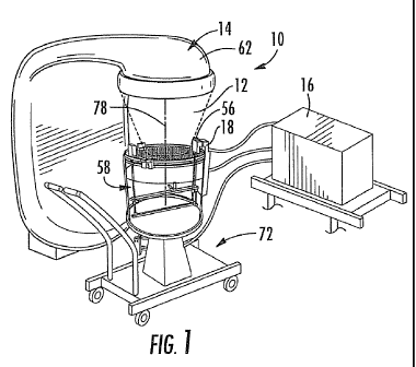
FIG. 1 is a partial perspective view of one embodiment of the invention
operable
with a LINAC for radiation dose measurements;
FIG. 2 is a diagrammatical illustration of a cylindrical scanning system in
keeping
with the teachings of the present invention;
5 FIG. 3 is a perspective view of one embodiment of the invention illustrated
in
FIG. 1;
FIGS. 4 and 5 are partial diagrammatical illustrations of detector offset
mounts
used with single and multiple radiation detectors;
FIG. 6 is a perspective view of one embodiment of a table for carrying a water
,0 tank illustrated in FIG. 1;
FIG. 6A is an underside open view of one adjustment means providing a
motorized level and shift platform;
FIG. 7 illustrates a 2 cm radiation field profile, produced by in-plane jaws
of a
Varian linear accelerator that is measured by various detectors, one of which
exhibits
the need for de-convolution;
FIG. 8 and 9 are diagrammatical illustrations of de-convolution analyses; and
FIG. 10 illustrate radiation profiles for a Varian radiation field uniformity
at two
depths (1.5 cm and 10 cm) within a water tank (Varian Radial Homogeniety of
Profile
scan field, 90 SSD, Raw Symmetrized Scan Data);
CA 02768367 2012-01-13
WO 2011/011471 PCT/US2010/042680
12
FIG. 11 illustrates in-line profile comparisons for 10cm X 10cm field, 1.5 cm
depth, zoom in-line (axial, IEC Y axis) on Varian, Siemens and Electa machines
(90
SSD, Raw Symmetrized Scan Data); and
FIG. 12 illustrates a point normalization of 4cm X 4cm PDD curves between
Varian, Elekta and Siemens machines.
Description of Embodiments
The present invention will now be described more fully hereinafter with
reference
to the accompanying drawings, in which embodiments of the invention are shown.
This
0 invention may, however, be embodied in many different forms and should not
be
construed as limited to the embodiments set forth herein. Rather, the
embodiments
herein presented are provided so that this disclosure will be thorough and
complete,
and will fully convey the scope of the invention to those skilled in the art.
By way of example, and with reference initially to FIG. 1, one embodiment of
the
invention, herein described by way of example, is a multiple axes scanning
system 10
for measuring radiation 12 emitted from a radiation source 14. The system 10
comprises a processor 16 having analysis and data storage capabilities and a
controller
18 operable with the processor.
With reference to FIG. 2, a ring drive 20 is operable with the controller 18
for
?0 providing a rotational movement 22 about a first axis 24 responsive to
commands from
the controller. A horizontal drive 26 is operable with the controller 18 for
providing
horizontal movement 28 along a second axis 30. For the embodiment herein
described
by way of example, the horizontal drive 26 is operable with the ring drive 20
for
receiving the rotational movement 22. A vertical drive 32 is operable with the
controller
?5 18 for providing vertical movement 34 of the horizontal drive 26 along a
third axis 36
responsive to the commands from the controller 18. A radiation detector 38
carried by a
mount 40 affixed to the horizontal drive 26 for locating it along the drive 26
by the
horizontal movement 28. The radiation detector 38 provides sensing signals to
the
processor 16 for selected locations of the radiation detector when orientated
through the
30 circular (rotational), horizontal, and vertical movements 22, 28, 34 along
the first,
CA 02768367 2012-01-13
WO 2011/011471 PCT/US2010/042680
13
second and third axes 24, 30, 36, respectively, as a result of the commands
from the
controller 18.
As illustrated with reference to FIG. 3 for the embodiment herein described by
way of example, the ring drive 20 comprises a circular gear 42, wherein the
controller
18 operates with a first motor 44 operable with the circular gear for
providing the
rotational movement 22. The horizontal drive 26 comprises a belt and pulley
assembly
46 driven by a second motor 48 and shaft 49operable for moving the radiation
detector
38 horizontally. The vertical drive 32 comprises a screw gear 50 driven by a
third motor
52 mounted to the ring drive 20. While individual motors are herein described
by way of
0 example, alternate gearing and linkages will come to the mind of those
skilled in the art
now having the benefit of the teachings of the present invention.
With reference again to FIG. 2 the system 10, herein described by way of
example, comprises an electrometer 54 operable between the processor 16 and
the
radiation detector 38. In addition, a reference detector 56 is located at a
fixed location
5 for comparing the sensing signals from the radiation detector 38 to the
reference
detector. As illustrated with reference again to FIGS. 1 and 3, a cylindrical
water tank
58 carrying water 60 is dimensioned for movement of the radiation detector 38
is
provided and supports the drives 20, 26, 32 described above. The controller 18
is
capable of communicating movement commands and receiving encoder information
from the motors and bi-directional communication of movement command and
encoder
position data to the programmable processor 16, synchronized to the bi-
directional
communication of the detectors 38 and 56 from the electrometer 54.
The embodiment herein described is by way of example only for one application
for the cylindrical scanning system 10. However, other geometrically shaped
vessels
!5 may be employed without compromising the benefits of the cylindrical
scanning system.
During scans, the vessel may contain water, or air scans may also be performed
with an
empty vessel, depending upon the requirements of the operator and the TPS.
Furthermore, the system 10 may be implemented without a vessel and assembled
in a
self supporting frame that rests on the treatment couch or mounted to the head
of the
30 radiation source such as a LINACTM for testing radiation beam
characteristics as the
gantry 62 is moved, as referenced in FIG.1.
CA 02768367 2012-01-13
WO 2011/011471 PCT/US2010/042680
14
With reference now to FIG. 4, the mount 40 may comprise an offset mount 64
operable with the horizontal drive 26, wherein the offset carries the
radiation detector
38. The offset mount 64 is dimensioned for extending the radiation detector 38
up to or
closely proximate an inside wall surface 66 of the vessel 58 during a scanning
movement of the radiation detector. The offset mount 64 may comprise multiple
detectors 38a, 38b, 38c as desired and illustrated with reference to FIG. 5,
by way of
example.
With reference again to FIG. 3, a surface sensor 68 is included for
determining a
location of a surface 70 of the water. The surface sensor 68 in one embodiment
is
0 carried by the horizontal drive for allowing measurements to be taken at
multiple non-
collinear positions by movement of the vertical drive 32 for locating the
surface of the
water 70. The surface sensor 68 may include an ultrasonic sensor, a capacitive
sensor,
or alternatives as desired.
With reference again to FIG. 1 and now to FIG. 6, a table 72 is employed for
5 supporting the vessel 58. Adjustment means 74 is operable with the table 72
for
providing a leveling of the table and an axis adjustment 76 for operation with
the
radiation detector 38 thus providing an alignment of the first axis 24 of the
ring drive 20
to a beam axis 78 from the radiation source 14.
With continued reference to FIGS 1 and 6, and with reference now to FIG. 6A,
!0 one embodiment of the adjustment means 74 includes two leveling motors 74A
(operable with a leveling foot) and 74B (operable with a leveling foot)
provide
independent vertical movement of a platform 73 carried by the table 72. In
addition, two
linear shift motors 74C (providing a linear shifting for +/- Y axis movement)
and 74D
(providing a linear shifting for +/- X axis movement) are each operable with
rails 75 that
?5 provide independent horizontal movement of the platform 73 in an orthogonal
X and Y
direction. The water tank, vessel 58 is positioned on a top side of the
platform 73A.
During operation of the system 10, the processorl 6, using the water surface
sensor 68
locates the first axis 24 and any tilt error with the water surface and then
commands the
74A and 74B motors to adjust the platform 73 to be level with the water
surface 70. The
30 processor 16, using the horizontal drive 26 for scanning the radiation
detector 38,
locates the beam axis 78 (a LINAC central axis (CAX) by way of example) on the
X
CA 02768367 2012-01-13
WO 2011/011471 PCT/US2010/042680
axis, then rotates the circular gear 42 by 90 degrees. The same operation is
performed
on the Y axis (CAY). The processor 16 commands the X and Y motors 74C, 74D to
adjust the platform 73 to the X CAX and Y CAX.
As will be appreciated by those skilled in the art, now having the benefit of
the
5 teachings of the present invention, an additional gear 76 may be used to
rotate the
extended detector 38,as illustrated with reference again to FIG. 4, by way of
example.
Alternatively, multiple offsets may be employed, as illustrated in FIG. 5 with
the mount
40 showing a center mount for a center field detector 38b, a right offset
mount for a right
field detector 38c, and a left offset mount for a left field detector 38a.
Other mount
0 offsets with various numbers of detectors will become obvious for those
skilled in the
art. Note that the center mounting may be omitted in some configurations.
With regard to functionality of the system 10 and to further aid the reader,
the
following discussion is provided as reported by W Simon8 for "Scan De-
Convolution and
Chamber Spread Function."
5 Experimental dosimeters always measure weighted integral dose over a limited
geometric region. This effect can be characterized by a spread function, rc,
that
represents physical phenomena of the spatial extension and spatial sensitivity
of the
dosimeter. The influence of a detector spread function can be eliminated by
deconvolving the spread function from the measured dose distribution Dm
employing
!0 the Fourier deconvolution theorem:
where and
are the Fourier transforms of the actual dose distribution D, the measured
dose
distribution Dm, and the spread function of the dosimeter ic. A numerical
method for
?5 performing the deconvolution can be developed using the following model for
the shape
of MV photon beam penumbra: (JF Dempsey, "A Fourier analysis of IMRT dose grid
resolution", Med Phys, 32, 380-388, 2005)
N
P(X)=- 1 Y a' elf. x + b; - erf x -
2 i=1
CA 02768367 2012-01-13
WO 2011/011471 PCT/US2010/042680
16
where P is the dose profile, a1 is an amplitude, bi is an effective field
size, Gi the
standard deviation of the erf function, given by:
erf (x) _ T . bx e-`Z = dt
Radiation dose profiles 80 are illustrated with reference to FIG. 7, wherein a
2 cm field
produced by in-plane jaws of a Varian linear accelerator at 6 MV and modeled
(fit) with
P(x) for N=2 is illustrated by way of example.
The Fourier transform of P is known to be:
N nZ-6Z=w=
P(w) _ Y 2 = a; =v; = e = sinc(2cov; )
0 '=1
Thus, we can find the Fourier transform of the spread function, x, by a
deconvolution of
the measured and true profile. The raw scan data in FIG. 7 illustrate the
significance of
the measurement uncertainty with a chamber 82, and also the measurement
redundancy between a diode 84 and film 86. Notice that the Edge 84 and EBT 86
trace
5 out an identical profile, thus validating each other. The IC15 (Wellhofer
model M3 ion
chamber 82 dimensions 6 mm ID x 5.8mm length, 0.13 cm3 volume) has a
significant
amount of volume averaging in the penumbra illustrated by the shallower
slopes. The
penumbrae, measured from 20 to 80 percent by Edge and EBT, are 2.2 to 2.3 mm
on
both sides that clearly exceeds a Nyquist spatial frequency limit of the IC15.
Each
D measured profile has an associated modeled profile that utilizes the error
function erf(x)
as discussed above. The deconvolution will eliminate the effect of the
detector-spread
function on the measured data set. In order to apply the deconvolution the
field edge
must be scanned until the dose reaches the collimator transmission level plus
the
distance where the spread function has effectively fallen to zero. The film
dose
5 calibration curve was measured several weeks apart from this profile
measurement.
This resulted in the deviation from the diode data outside the penumbra in the
low dose
region. The EBT film may require frequent calibration for application in low
dose
regions.
By way of further example, and with reference to FIG. 8, one overview of the
de-
0 convolution is illustrated with a scanned profile 88 being processed with
the de-
CA 02768367 2012-01-13
WO 2011/011471 PCT/US2010/042680
17
convolution analysis 90 that results in a modified profile shape 92 that
represents an
improvement of the true beam intensity of the LINAC. An illustration of the
this process
is represented with reference to FIG. 9 by which one scanned profile 94 from
one field
detector, an IBA4 ion chamber sensor named "CC13", is spread out compared to a
scanned profile B96 96 from the radiation detector 38, earlier described with
reference
to FIG. 2, a Sun Nuclear9 small diode sensor named "EDGE". The EDGE detector
profile was analyzed for the true beam shape from which was derived the de-
convolution parameter (2.4083) which, by de-convolution analysis, produced the
de-
convolved profile 98. The de-convolution parameter is used in the analysis of
all the
0 M3 scans as they are produced, resulting in nearly "real-time" visualization
of the true
profile immediately after scan. This allows immediate comparison to other
known
profiles before saving the scan data for clinical use.
By way of example, and with reference to a data plot of FIG. 10, one overview
of
results from a QA method of the present invention is illustrated as ratios X/Y
of the
5 profiles 100 scanned on the X and Y axes of the Varian 2 LINAC at two
different scan
depths of the radiation detector 38 below the surface 70; 10 cm 102 and 1.5 cm
104
below the water surface. In the flat portion 106, the profile ratio varies
less than 1 %
between the two axes, in 86% of the field size. Then as the scan enters the
penumbra
region as earlier described, the ratio increases to almost 12% and then drops,
which
indicate the X axis (in-plane) has a sharper penumbra, wherein the scan value
stays
higher as the scanned radiation detector 38 enters the beam edge penumbra, and
lower
jaw transmission. The location of the Varian X axis collimator jaw is lower
(closer to the
scanning detector 38 and therefore will have a sharper penumbra, thus
verifying the
setup jaw and scanner orientation geometry. Similar analysis may be performed
on
!5 scans where there is no change in setup but only a repeat, indicating that
a ratio of unity
(flat line) is expected, again a QA function that may be automated.
By way of another example, and with reference to the data plot of FIG. 11,
another overview of the QA method is illustrated as the profiles scanned on
different
LINAC's Variant, Elekta10, and Siemens", with the scans being made in the same
30 beam geometry (setup) and depth. The profile from the Varian 108, the
profile from the
Elekta 110 and the profile from Siemens 112 look similar until they are
overlaid and
CA 02768367 2012-01-13
WO 2011/011471 PCT/US2010/042680
18
zoomed in to examine the finer detail, where the differences in the flattening
filters, a
device in the LINAC to shape the raw X-ray beam, are clearly seen, producing
an
identity print of the of the three machines. These profile shapes should be
characteristic
to the manufacturer, i.e., this is one of the principle differences between
machine
makes, the other being the beam edge shape in the penumbra region as described
with
FIG. 10. Such measurements and analysis provide another QA check of the
scanning
and the LINAC when the profile is compared to a library of benchmark scans
from
known machine makes, or when comparing historical scan data that verifies
clinical use.
In this example, the ratios were not illustrated because a demonstration of
the profile
0 shapes of the three manufacturers would have been hidden, however for the
same
manufacturer but different machines, such a ratio would reveal the matching or
non-
matching characteristics of the LINACs.
By way of yet another example, and with reference to the data plot of FIG. 12,
another overview of the QA method is illustrated as the ratios of the percent
depth dose
5 (PDD, fractional depth dose FDD) curves scanned on different LINAC's
Varian2,
Elekta10, and Siemens", with the scans being made in the same beam geometry
(setup). A comparison of machine PDD provides a convenient and quantitative
method
to compare the "beam matching" between machines. From each machine data, the
PDD's were compared by taking point-to-point ratios. These ratio plots are for
field sizes
4x4 cm. A linear regression fit between 3 cm and 30 cm on the 4x4 cm field
size
resulted in the following:
a. V/E: Y=-0.0018x+1.0176 R2 = 0.9383 114
b. V/S: Y=-0.0011x+1.0098 R2 = 0.8559 116
c. E/S: y = 0.0006x + 0.9921 R2 = 0.6903 118
>.5 The quality of the linear fit is expressed_ as R2, with perfect being
1.00. The plots are
reasonably linear in this small 4x4 cm field. The slope term is a statement of
the beam
matching, where two beams that are identical will have a PDD ratio that is
unity
throughout the depth, and a slope of zero without trend. From the values
above, the
machine ratios have slopes less than 0.2% from zero. The best matching is
between
30 Elekta and Siemens, with a 0.06% change in attenuation per cm; i.e., over a
30 cm
depth range, the attenuation difference between beams is 1.8.
CA 02768367 2012-01-13
WO 2011/011471 PCT/US2010/042680
19
As earlier described with reference to FIG. 3, the water surface sensor 68 may
be provided including an ultrasonic surface sensor or capacitive surface
sensor having
a sharp conductive point connected to electronic means of contact detection,
that when
mounted on the linear horizontal drive, measures the water surface at three or
more
non-collinear positions (a surface) by adjusting the vertical drive 32 until
the surface 70
is located. The software may then analyze the level error of the three
dimensional
cylindrical scanning system with respect to the water surface and either
instruct the
operator to make level adjustments with leveling means that may include scaled
adjustment controls, or adjust motorized leveling screws, or compensate for
the level
0 error with instructions to the three dimensional cylindrical scanning system
control in
such a way that keeps the radiation field sensor level (parallel) to the water
surface.
To further aid the reader, the risks and possible mitigations of a cylindrical
design
are herein presented by way of example and may include:
Misalignment of the scan ring axis 24 to beam axis 78, as earlier described
with
5 reference to FIGS. 1 and 2: The scan ring axis may desirably be autoset to
the beam
axis with an additional hardware (ex: a small drive or axis adjustment device
76 on the
detector mount 40 as earlier described with reference to FIG. 2).
Alternatively, there
may be provision in a ring axis mount that would allow for such adjustments.
Beam axis
alignment is critical in SRS fields below 2 cm diameter or square. In the
present
embodiment, there are two fine X-Y adjustments 120 in the lift table 72 that
provide the
alignment capability. The software analysis of two sets of two orthogonal
scans, each
set measured at two different depths along the beam axis, results in the
determination
of beam centers that define the collinear beam axis that result in the
required
adjustments which the user then performs before collecting clinical beam data.
With
?5 scaled adjustment controls, these scale adjustments can be the output of
the analysis
whereby the operator makes a quantitative adjustment to align the ring axis to
the beam
axis or the software controller performs quantitative adjustment with
motorized X and Y
controls.
Inability to scan on an off axis chord: This may be a risk if the ring motor
44 is not
30 part of the scan control, i.e., if the ring movement 22 does not have the
precision to
locate a detector to a precision of -0.2mm, then it cannot be used to drive
the detector
CA 02768367 2012-01-13
WO 2011/011471 PCT/US2010/042680
38, in conjunction with the horizontal drive 26, on a chord for scanning. In-
the
embodiment above described by way of example, the ring drive 20 has a
precision of
better than 0.1 mm and a hysteresis of 0.03mm, which provides, along with the
precision of the vertical and horizontal drives 32, 26, an accurate scanning
mechanism
5 through any X, Y, Z point in the water.
For example, asymmetric fields that are offset from the beam axis are chord
scans in this cylindrical system when scanned profiles run through the offset
"field"
center and parallel to the linac axes (inplane or crossplane). This scan
geometry is a
chord offset to the center of the circle. TPS beam data do not call for chord
tracing in
0 asymmetric fields. However, chord tracing would be required if the penumbra
profile at
the MLC leaf end were required for leaves that are off central axis, beyond
the reach of
the detector motor. The present embodiment with the precision ring drive,
along with the
other two axes, enables any chord scanning as well as scanning between any two
spatial locations that are defined within the scanning range of the three
dimensional
5 cylindrical scanning system. A shift in the X or Y direction can also enable
scanning on
a chord.
In this cylindrical geometry, any PDD can be ray traced (using the vertical
and
linear horizontal axis drives) after the ring is rotated to align the
divergent ray parallel to
the scanning arm. A PDD in an asymmetric off axis field is the most likely
requirement
!0 of this geometry.
Many modifications and other embodiments of the invention will come to the
mind of one skilled in the art having the benefit of the teachings presented
in the
foregoing descriptions and the associated drawings. Therefore, it is
understood that the
invention is not to be limited to the specific embodiments disclosed, and that
?5 modifications and embodiments are intended to be included within the scope
of the
claims supported by this disclosure.
CA 02768367 2012-01-13
WO 2011/011471 PCT/US2010/042680
21
References cited in the specification
1. Indra J. Das, Chee-Wai Cheng, Ronald J. Watts, Anders Ahnesjo, John
Gibbons,
X. Allen Li, Jessica Lowenstein, Raj K. Mitra, William E. Simon, and Timothy
C.
Zhu, "Accelerator beam data commissioning equipment and procedures: Report
of the TG-106 of the Therapy Physics Committee of the AAPM", Med. Phys. 35
4186 (2008)
2. Varian Medical Systems, Palo Alto CA
3. PTW, Freiburg Germany
0 4. IBA Scanditronix Wellhofer, Schwarzenbruck, Germany
5. JF Dempsey, et at., "A Fourier analysis of IMRT dose grid resolution", Med
Phys,
32, 380-388, 2005
6. Guanghua Yan, Christopher Fox, Chihray Liu, and Jonathan G. Li, "The
extraction of true profiles for TPS commissioning and its impact on IMRT
patient-
5 specific QA", Med. Phys. 35 3661 (2008)
7. B. Fraass, K. Doppke, M. Hunt, G. Kutcher, George Starkschall, R. Stern, J.
Van
Dyke, "American Association of Physicists in Medicine Radiation Therapy
Committee Task Group 53: Quality assurance for clinical radiotherapy treatment
planning", Med. Phys. 25. 1773-1829, Oct 1998
:0 8. W. Simon et al "LINAC Dosimetry: Benchmark Data Set Uncertainty", Med.
Phys.
33 2118 SU-FF-T-311: (2006
9. Sun Nuclear Corporation, Melbourne Florida
10. Elekta, Crawley, England
11. Siemens, Erlangen, Germany