Note: Descriptions are shown in the official language in which they were submitted.
CA 02768475 2012-01-17
WO 2011/009114 PCT/US2010/042400
Page 1 of 37
TITLE
METHODS AND KITS USED IN ASSESSING CANCER RISK
INVENTORS
PAMELA POLLOCK
PAUL GOODFELLOW
BACKGROUND OF THE INVENTION
Endometrial cancer is the most common gynecological cancer. Endometrial
carcinoma is
subdivided into Type I and Type II disease. Type I endometrioid endometrial
accounts for
approximately 80-85% of endometrial cancers and is classified as being
estrogen-dependent and
well differentiated. Type II endometrial cancers comprise poorly
differentiated endometrioid,
clear cell, and papillary serous histological subtypes that display high
biological aggressiveness
and are associated with poor prognosis. Approximately 75% of type I
endometrioid tumors are
diagnosed as Stage I/II. These patients have a 5 year overall survival of 80-
90%, a 5 year cancer
specific survival of 90-95% and a recurrence rate of 4-8% (Creutzberg et al.
2000). However, for
those women that recur, or present with advanced stage or progressive disease,
survival is poor
as there are no adjuvant therapies proven to be effective. The median survival
after recurrence is
10 months and the 5-year survival for patients who have recurred is only 13%.
There is a clear
need to develop additional prognostic markers to identify those patients at
risk for recurrence.
BRIEF SUMMARY OF THE INVENTION
The present invention provides among other things a method of assessing the
risk of
disease recurrence in patients diagnosed with endometrial cancer.
It is an object of the invention to classify a subject into a cohort of
increased risk of
recurrence of endometrial cancer based upon FGFR2 mutation status.
It is an object of the invention to provide a kit used to classify a subject
into a cohort of
increased risk of recurrence of endometrial cancer based upon FGFR2 mutation
status.
It is an object of the invention to identify endometrial cancer patients with
a higher risk of
recurrence of disease that would be otherwise predicted based on existing
clinico-pathological
risk factors such as stage, grade, age, or race among others.
1
CA 02768475 2012-01-17
WO 2011/009114 PCT/US2010/042400
Page 2 of 37
The above and other objects may be achieved through the use of methods
involving
obtaining a sample from the subject and subjecting the sample to conditions
that allow detection
of a mutant of either SEQ ID NO. 1 or SEQ ID NO. 2. The subject is known to
have had
endometrial cancer and the sample comprises a tumor cell. The cohort comprises
two or more
individuals with an increased risk of recurrence of endometrial cancer. The
mutant may comprise
any mutation in SEQ ID NO. 1 or SEQ ID NO. 2, including those that lead to one
or more the
following amino acid changes: S252W, P253R, S373C, Y376C, C383R, G385R, 1548V,
N550K,
N550H, K660E, M392R, V396D, L398M, and IVS 10+2A>C. The endometrial cancer may
be
of the endometrioid subtype. The stage may be any stage, including Stage IA,
Stage IB, Stage
IC, Stage IIA, and Stage IIB. The grade may be any grade, including Grade 1,
Grade 2, and
Grade 3. The conditions may allow detection of a mutant of SEQ ID NO. 1. In
this example, the
conditions may comprise the use of a technology selected from the group
consisting of nucleic
acid sequencing, microarray analysis, PCR amplification, allele specific PCR
amplification,
restriction fragment length polymorphism, allele specific hybridization,
allele specific primer
extension, and/or Southern Blot. The conditions may comprise detection of a
mutant of SEQ ID
NO. 2. In this example, then the conditions may comprise use of a technology
selected from the
group consisting of HPLC, mass spectrometry, ELISA, flow cytometry,
immunohistochemistry
or radioimmunoassay. The conditions may alternatively comprise measuring the
activity of
FGFR2 protein.
The above and other objects may be achieved through the use of kits comprising
a first
reagent capable of detecting a mutant of a sequence selected from the group
consisting of SEQ
ID NO. 1 and SEQ ID NO. 2 and an indication of a result that signifies
classification of a subject
into a cohort where the cohort comprises two or more individuals with an
increased risk of
recurrence of endometrial cancer. The mutant may comprise any mutation in SEQ
ID NO. 1 or
SEQ ID NO. 2, including those that lead to one or more the following amino
acid changes:
S252W, P253R, S373C, Y376C, C383R, G385R, 1548V, N550K, N550H, K660E, M392R,
V396D, L398M, and IVS 10+2A>C. The first reagent may be capable of binding to
a mutant of
SEQ ID NO. 1. In that example, the kit may further comprise a component that
facilitates the use
of a technology selected from the group consisting of nucleic acid sequencing,
microarray
analysis, PCR amplification, allele specific PCR amplification, restriction
fragment length
polymorphism, allele specific hybridization, allele specific primer extension,
and Southern blot.
2
CA 02768475 2012-01-17
WO 2011/009114 PCT/US2010/042400
Page 3 of 37
The kit may comprise a first reagent that is capable of binding a mutant of
SEQ ID NO. 2. The
first reagent may comprise a first antibody. In this example, the kit may
further comprise a
component that facilitates the use of a technology selected from the group
consisting of ELISA,
flow cytometry, and radioimmunoassay. The result may be any result that
signifies the detection
of a mutant including a particular nucleic acid sequence or optical density
value. The indication
may be any indication including a positive control or a writing. A writing may
be any writing
including a writing on paper or a writing made available via a website. A
writing may comprise a
photograph. The indication may also comprise software configured to detect the
result as input
and the classification of the subject as output. Such software may be
incorporated into a machine
configured to detect the mutant.
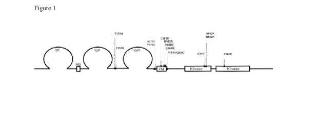
BRIEF DESCRIPTION OF THE FIGURES
Figure 1 depicts the location of mutations identified in FGFR2 in endometrioid
endometrial cancers. The majority of the mutations occur at seven hotspots.
Figure 2 depicts progression free survival curves in intermediate risk
patients with (Yes)
and without (No) an FGFR2 mutation.
Figure 3 depicts overall survival in intermediate patients with (Yes) and
without (No) an
FGFR2 mutation.
Elements and acts in the figures are illustrated for simplicity and have not
necessarily
been rendered according to any particular sequence or embodiment.
DETAILED DESCRIPTION OF THE INVENTION
Endometrial cancer includes all forms and subtypes of the disease, including
for example,
serous, mucinous, and endometrioid histological subtypes or any other cancer
that starts in the
endometrium, which includes the lining of the uterus. Endometrial cancer is
currently surgically
staged using the International Federation of Gynecology and Obstetrics (FIGO)
system, which
emphasizes complete surgico-pathologic assessment of data. In response to the
dismal prognosis
associated with progressive or recurrent endometrial cancer, multiple efforts
have been made to
identify patients at risk for disease progression and recurrence. Patients who
present with
advanced extrauterine disease (stage III/IV) at diagnosis have a high risk of
recurrence. In those
patients that present with cancer confined to the uterus (Stage I/II), an
increased risk of
3
CA 02768475 2012-01-17
WO 2011/009114 PCT/US2010/042400
Page 4 of 37
recurrence is associated with histologic cell type, tumor grade, depth of
myometrial invasion,
occult extension into the cervix and tumor cell invasion of lymphatic vessels
(lymphovascular
space invasion: LVSI). Table 4 demonstrates the stage and grade
classifications of patients
considered to have a low, intermediate or high risk of recurrence, where the
intermediate risk is
further broken down to patients with a low-intermediate risk and high-
intermediate risk.
The concept of the FGFR2 gene encompasses a gene of human origin with a coding
nucleotide sequence set forth in SEQ ID NO 1, or homologs including allelic
variants and
orthologs. The FGFR2 protein encompasses a protein, also preferably of human
origin, having
the amino acid sequence set forth in SEQ ID NO 2 or homologs, including
orthologs thereof.
Figure 1 displays the various domains of the FGFR2 protein and the FGFR2
mutations
mapped in relation with the domains. FGFR2 belongs to a family of structurally
related tyrosine
kinase receptors (FGFRs 1-4) encoded by four different genes. FGFR2 is a
glycoprotein
composed of three extra-cellular immunoglobulin-like (1g) domains, a
transmembrane domain,
and a split tyrosine kinase domain. Alternative splicing in the IgIII domain
is primary
determinant of both the patterns of redundancy and specificity in FGF/FGFR
binding and
signaling. This splicing event is tissue specific and gives rise to the IIIb
and IIIc receptor
isoforms for FGFRI-FGFR3 which possess distinct ligand specificities
(Mohammadi M, Olsen
SK and Ibrahimi OA. (2005), Cytokine Growth Factor Rev 16: 107-137, Ornitz DM
and Itoh N.
(2001). Genome Bioi 2: REVIEWS3005). For FGFR2, cells of an epithelial linage
only express
the "IIIb" isoform encoded by exon 8 (FGFR2b; SEQ ill NO:2; NP_07529.2), while
mesenchymally derived cells exclusively express the "IIIc" isoform utilizing
exon 9 (FGFR2c;
SEQ ill NO:3; NP000132.1) (Scotet E and Houssaint E. (1995). Biochim Biophys
Acta 1264:
238-242). The FGFR2b iosform predominantly binds FGF1, FGF3, FGF7 and FGF10,
while
FGFR2c does not bind FGF7 and FGF10 but does bind FGF1, FGF2, FGF4, FGF6,and
FGF8
with high affinity (Ibrahimi OA, Zhang F, Eliseenkova AV, ltoh N, Linhardt RJ
and Mohammadi
M. (2004), Hum Mol Genet 13: 2313-2324).
An FGFR2 mutation with increased activity in a test subject or a biological
sample may
also be called an activation mutation. Activation mutations display higher
total FGFR2 activity
in the test subject or biological sample in comparison with a control, e.g., a
healthy subject or a
standard sample. Preferably, although not necessarily, the activity is at
least 10%, at least 50%, at
least 100%, or at least 150% higher in the test subject or sample than in the
control. The
4
CA 02768475 2012-01-17
WO 2011/009114 PCT/US2010/042400
Page 5 of 37
increased activity, for example, may result from increased basal FGFR2
activity in the absence
of ligand, increased level of activation in the presence of ligand, prolonged
stimulation, delayed
degradation or over-expression, e.g., due to enhanced ligand binding,
promiscuous or
inappropriate ligand binding, constitutive receptor dimerization, impaired
recycling resulting in
augmentation of signaling, delayed degradation, or kinase activation.
A higher expression level of FGFR2 may result from, for example, a mutation in
a non-
coding region of a FGFR2 gene or a mutation in a coding or non-coding gene
involved in
FGFR2 transcription or translation. The expression level of FGFR2 can be
determined, for
example, by comparing FGFR2 mRNA or the level of FGFR2 protein in a test
subject as
compared to a control, for example by comparing the tumor to normal
endometrium (e.g., a
normal adjacent endometrium sample).
Conserved variants encompass any mutation or other variant in which a given
amino acid
residue in a protein or enzyme has been changed without altering the overall
conformation and
function of the polypeptide, including, but not limited to, replacement of an
amino acid with one
having similar properties (such as, for example, polarity, hydrogen bonding
potential, acidic,
basic, hydrophobic, aromatic, and the like). Amino acids with similar
properties are well known
in the art. For example, arginine, histidine and lysine are hydrophilic-basic
amino acids and may
be interchangeable. Similarly, isoleucine, a hydrophobic amino acid, may be
replaced with
leucine, methionine or valine. Depending on the location of the mutation in
the overall context of
the protein, the substitution may have little or no effect on the apparent
molecular weight or
isoelectric point of the protein or polypeptide. A conserved variant can still
result in receptor
activation by a wide variety of mechanisms.
Amino acids other than those indicated as conserved may differ in a protein or
enzyme so
that the percent protein or amino acid sequence similarity between any two
proteins of similar
function may vary and may be, for example, from 70% to 99% as determined
according to an
alignment scheme such as by the Cluster Method, wherein similarity is based on
the
MEGALIGN algorithm. The concept of a variant further encompasses a polypeptide
or enzyme
which has at least 60%, 75%, 85%, 90%, or 95%, amino acid identity as
determined by
algorithms such as BLAST or FASTA and which has the same or substantially
similar properties
and/or activities as the native or parent protein or enzyme to which it is
compared.
One example of such a variant is a gain-of-function variant. Gain of function
variants of
5
CA 02768475 2012-01-17
WO 2011/009114 PCT/US2010/042400
Page 6 of 37
polypeptides encompass any variant in which a change in one or more amino acid
residues in a
protein or enzyme improves the activity of the polypeptide. Examples of
activities of a
polypeptide that may be improved by a change resulting in a gain of function
variant include but
are not limited to enzymatic activity, binding affinity, phosphorylation or
dephosphorylation
efficiency, activation, deactivation, or any other activity or property of a
protein that may be
quantitatively measured by some method now known or yet to be disclosed.
Proteins that possess a common evolutionary origin may be homologous or
similar to one
another. Examples of homologous or similar proteins include proteins from
superfamilies (e.g.,
the immunoglobulin superfamily) and homologous proteins from different
species. Such proteins
and their encoding genes have sequence homology with one another. The homology
may be
expressed in terms of percent similarity or the presence of specific residues
or motifs at
conserved positions.
A mutation may be any detectable change in genetic material such as DNA, or a
corresponding change in the RNA or protein product of that genetic material. A
mutant may be
any biological material in which one or more mutations are detected when
compared to a control
material. Examples of mutations include gene mutations, in which the DNA
sequence of a gene
or any controlling elements surrounding the gene is altered. Controlling
elements include
promoter, enhancer, suppressor or silencing elements capable of controlling a
given gene. Other
examples of mutations include alterations in the products of DNA expression
such as RNA or
protein that result from corresponding mutations in the DNA. Mutants may also
be
interchangeably called variants. The concept of a mutant includes any change
in DNA sequence
specific to the tumor cell (not present in DNA prepared from normal, non-
neoplastic tissues).
Assessing the risk of a particular disease outcome includes the performing of
any type of
test, assay, examination, result, readout, or interpretation that correlates
with an increased or
decreased probability that an individual has had, currently has, or will
develop a particular
disease, disorder, symptom, syndrome, or any condition related to health or
bodily state.
Examples of disease outcomes include, but need not be limited to survival,
death, progression of
existing disease, remission of existing disease, initiation of onset of a
disease in an otherwise
disease-free subject, or the continued lack of disease in a subject in which
there has been a
remission of disease. Assessing the risk of a disease outcome also encompasses
the concept of
prognosis. A prognosis may be any assessment of the risk of disease outcome in
an individual in
6
CA 02768475 2012-01-17
WO 2011/009114 PCT/US2010/042400
Page 7 of 37
which a particular disease has been diagnosed.
A sample may be any cell source from which DNA, including genomic, somatic,
and
germline DNA may be obtained. In endometrial cancer, a biological sample is
often obtained
from the uterus and generally includes one or more endometrial tumor cells.
Circulating tumor
cells may be found and obtained from serum. Tumor cells may be obtained by any
method now
known in the art or yet to be disclosed, including for example, surgical
resection, laser capture
microdissection, isolation from blood or other fluids including lavage fluid,
or any other method
capable of obtaining and, if necessary, concentrating endometrial tumor cells.
Alternatively a
sample may comprise free DNA from a tumor extracted directly from serum (See
Reference 32).
A subject includes any human or non-human mammal, including for example: a
primate,
cow, horse, pig, sheep, goat, dog, cat, or rodent, capable of developing
endometrial cancer
including human patients that are suspected of having endometrial cancer, that
have been
diagnosed with endometrial cancer, or that have a family history of
endometrial cancer. Methods
of identifying subjects suspected of having endometrial cancer include but are
not limited to:
physical examination, family medical history, subject medical history,
endometrial biopsy, or a
number of imaging technologies such as ultrasonography, computed tomography,
magnetic
resonance imaging, magnetic resonance spectroscopy, or positron emission
tomography.
Methods of diagnosing endometrial cancer as well as the staging, grading, or
other clinical
delineation of endometrial cancer are well known to those of skill in the
medical arts.
Sequence-specific oligonucleotides include sets of oligonucleotides that can
be used to
detect specific variations or mutations in the FGFR2 gene. Probes include
oliognucleotides
capable of forming a hybrid structure with a sequence in a target region due
to complementarity
of at least one nucleic acid base in the probe with a sequence in the target
protein of the subject.
Prognostic methods encompass detecting a mutation in the FGFR2 protein
including
mutations that result in increased activity of the FGFR2 protein. Examples of
such mutations
include mutations occurring in the junction between the immunoglobulin-like
(Ig) domains II
and III; mutations occurring in the IgIll domain; mutations occurring in the
junction between the
IgIll domain and the transmembrane (TM) domain; mutations occurring in the TM
domain;
mutations occurring in the junction between the TM domain and the tyrosine
kinase domain I;
mutations occurring in the tyrosine kinase domain I, or mutations occurring in
the tyrosine
kinase domain II. Such mutations likely induce an amino acid substitution.
Examples of such
7
CA 02768475 2012-01-17
WO 2011/009114 PCT/US2010/042400
Page 8 of 37
amino acid substitutions induced by mutations include but are not limited to:
an S to W mutation
at position 252, a P to R mutation at position 253, an S to C mutation at
position 373, a Y to C
mutation at position 376, a C to R mutation at position 383, an M to R
mutation at position 392,
a V to D mutation at position 396, an L to M mutation at position 398, an Ito
V mutation at
position 548, an N to K mutation at position 550, an N to H mutation at
position 550, and a K to
E mutation at position 660 with position numbers as indicated in SEQ ID NO. 2.
In one
nonlimiting embodiment, the mutation is consist of a deletion of nucleotide C
and T at position
2290-91 of the nucleotide sequence (NM-02297.2) or an IVS10+2A>C splicing
mutation with
position numbers as indicated in SEQ ID. NO. 1 or any other somatic mutation
found in an
endometrial tumor cell.
A detected FGFR2 receptor activation mutation may increase activation of the
receptor
by, for example, enhancing ligand binding, promoting altered or promiscuous
ligand affinity
with reduced selectivity, constitutive receptor dimerization, delayed
degradation, impaired
recycling from the cell membrane, signaling inappropriately from intracellular
membranes,
overexpression, or kinase activation.
In one embodiment, the prognosis of endometrial cancer in a subject may be
assessed by
determining an activity level of the FGFR2 protein in an endometrial cancer
cell of a test subject
and comparing it to the activity in endometrial cells of a control subject,
wherein an increased
activity of FGFR2 protein in the test subject compared to the control subject
is indicative of an
increased risk of recurrence of endometrial cancer. The level of FGFR2
activity may be assessed
by determining the level of activity in a FGFR2 signaling pathway through any
method now
known or yet to be developed. Examples include but need not be limited to,
assessing the
expression of targets up- or down-regulated upon FGFR2 signaling, assessing
the
phosphorylation status of proteins phosphorylated or dephosphorylated on FGFR2
signaling, or
any other method capable of detecting an increase in FGFR2 activity or ligand
promiscuity.
Mutated forms of FGFR2 nucleic acids, such as in FGFR2 DNA or any transcripts
(including any splice variants now known or yet to be disclosed) as well as a
deregulated
expression (including overexpression or underexpression) of FGFR2 or other
elements of a
FGFR2 pathway may be detected by any of a variety of suitable methods.
Any method capable of detecting a mutated nucleic acid in a biological sample
now
known or yet to be disclosed may be employed and many strategies of genotypic
analysis are
8
CA 02768475 2012-01-17
WO 2011/009114 PCT/US2010/042400
Page 9 of 37
now known to those skilled in the art. Some of these methods use nucleic acid
sequences such as
specific oligonucleotides to detect mutations in an FGFR2 nucleic acid in a
biological sample.
Such oligonucleotides may specifically hybridize to a nucleic acid sequence
containing the
specific mutation, or to a region adjacent to the site of mutation. Other
methods use primers that
permit amplification of all or part of an FGFR2 nucleic acid. Alternatively,
or in combination
with such techniques, oligonucleotide sequencing described herein or known to
the skilled
artisan may be applied to detect the FGFR2 mutations. One skilled in the art
may use
hybridization probes in solution and in embodiments employing solid-phase
procedures. In such
procedures, the test nucleic acid is adsorbed or otherwise affixed to a
selected matrix or surface.
The fixed, single-stranded nucleic acid is then subjected to specific
hybridization with selected
probes. Alternatively, one skilled in the art may use oligonucleotide primers
in an amplification
technique, such as PCR or reverse-PCR ("reverse polymerase chain reaction"),
to specifically
amplify a target DNA or mRNA, respectively. Such primers include primers that
permit
amplification of FGFR2 exons.
One example of such a method includes but is not limited to the following:
contacting a
biological sample containing DNA with specific oligonucleotides permitting the
amplification of
all or part of the FGFR2 gene, the DNA contained in the sample having being
rendered
accessible, where appropriate, to hybridization, and under conditions
permitting a hybridization
of the primers with the DNA contained in the biological sample; amplifying
said DNA; detecting
the amplification products; and comparing the amplified products as obtained
to the amplified
products obtained with a normal control biological sample, and thereby
detecting an abnormality
in the FGFR2 gene if such abnormality is present and not detecting an
abnormality if such
abnormality is not present.
Alternatively, a sample may be sequenced directly with no amplification. In
such
methods, the sequenced DNA is compared to a normal genomic control sequence.
The control
sequence may be obtained from another subject or from a noncancerous sample
from the same
subject. One such method of sequencing is allele specific primer extension in
which sample
DNA hybridized to a chip is used as a synthesis template with the affixed
oligonucleotide as a
primer. Only the added dNTP's are labeled. Incorporation of the labeled dNTP
then serves as a
signal indicating the presence of the mutation. The fluorescent label may be
detected by any of a
number of instruments configured to read at least four different fluorescent
labels on a DNA
9
CA 02768475 2012-01-17
WO 2011/009114 PCT/US2010/042400
Page 10 of 37
chip. In an alternative method, the identity of the final dNTP added to the
oligonucleotide may
be assessed by mass spectrometry. In this method, the dNTP's may, but need not
be labeled with
a label of known molecular weight.
Other methods of detecting abnormalities in FGFR2 include those that detect
abnormalities in the transcript of the FGFR2 gene. Such methods include
amplifying mRNA
transcripts in a biological sample by techniques such as RT-PCR. One example
of such a method
includes but is not limited to the following: producing cDNA from mRNA
contained in a
biological sample; contacting said cDNA with specific oligonucleotides capable
of amplifying of
all or part of the transcript of the FGFR2 gene, under conditions capable of
hybridizing the
primers with said cDNA; amplifying said cDNA; detecting the amplification
products;
comparing the amplified products as obtained to the amplified products
obtained with a normal
control biological sample, and thereby detecting an abnormality in the
transcript of the FGFR2
gene if such an abnormality is present and not detecting an abnormality if
such an abnormality is
not present. A control may be any noncancerous endometrial tissue control
sample known as
noncancerous to those skilled in the art, for example, a normal adjacent
endometrium sample or a
normal FGFR2 mRNA or DNA, obtained from blood, buccal swab or other source.
Samples to be used in mRNA analysis may be obtained from any cell source, as
described above, including a biopsy tissue. RNA may be then isolated from the
sample using
standard methods well known to those of ordinary skill in the art. Examples
include but are not
limited to: guanidium thiocyanate-phenolchloroform extraction (Chomocyznski et
al., Anal.
Biochem., 1987, 162:156), isolation through the use of resin, Trizol or other
reagents, or any
other appropriate method. The isolated RNA is then subjected to coupled
reverse transcription
and amplification by polymerase chain reaction (RT-PCR), using specific
oligonucleotide
primers that are specific for a selected region of the cDNA sequence. Primer
annealing
conditions are chosen to ensure specific reverse transcription and
amplification; thus, the
appearance of an amplification product is diagnostic of the presence of a
particular genetic
variation. In another embodiment, RNA is reverse-transcribed and amplified.
Mutations in the
amplified sequences (if present) may then be detected by any of a number of
methods including
direct sequencing, restriction fragment length polymorphism, hybridization of
a specific probe to
the amplified sequence, or be cloning into a plasmid followed by sequencing.
If mutations are
not present, then they will not be detected.
CA 02768475 2012-01-17
WO 2011/009114 PCT/US2010/042400
Page 11 of 37
Nucleic acids that hybridize to mutant forms of FGFR2 may be used as probes in
prognostic assays such a probe may comprise a substantially purified
oligonucleotide that further
includes a region having a nucleotide sequence that is capable of hybridizing
specifically to a
region of a FGFR2 gene that may be mutant or polymorphic. Such probes can then
be used to
detect specifically which, if any, mutation of the FGFR2 gene is present in a
sample taken from a
subject. The mutant or polymorphic region can be located in the promoter,
exon, or intron
sequences of the FGFR2 gene. In general, such probes have a sufficient number
of nucleotides to
allow specific hybridization to the target nucleotide sequence. Probes
complementary to mutant
sequences with the appropriate specificity may be constructed by those skilled
in the art. For
example, a portion of the FGFR2 gene may first be amplified and isolated from
chromosomal
DNA and hybridized to a probe. In such a case a probe of 10, 15, 20, 30, 50,
or 100 nucleotides
may be used.
The probe or primer may include a label. A label may be any substance capable
of aiding
a machine, detector, sensor, device, or enhanced or unenhanced human eye from
differentiating a
sequence that contains a particular allele from a cell that does not contain
the allele. Examples of
labels include but are not limited to: a radioactive isotope or chelate
thereof, a dye (fluorescent or
nonfluorescent,) stain, enzyme, or nonradioactive metal. Specific examples
include but are not
limited to: fluorescein, biotin, digoxigenin, alkaline phosphatase, biotin,
streptavidin, 3H, 14C,
32P, 35S5 or any other compound capable of emitting radiation, rhodamine, 4-
(4'-dimethylamino-
phenylazo)benzoic acid ("Dabcyl"); 4-(4'-dimethylamino-phenylazo)sulfonic acid
(sulfonyl
chloride) ("Dabsyl"); 5-((2-aminoethyl)-amino)-naphtalene-1-sulfonic acid
("EDANS");
Psoralene derivatives, haptens, cyanines, acridines, fluorescent rhodol
derivatives, cholesterol
derivatives; ethylenediaminetetraaceticacid ("EDTA") and derivatives thereof
or any other
compound that signals the presence of bound ligand to an allele. In one
embodiment of the
invention, the label includes one or more dyes optimized for use in
genotyping. Examples of
such dyes include but are not limited to: dRl 10, 5-FAM, 6FAM, dR6G, JOE, HEX,
VIC, TET,
dTAMRA, TAMRA, NED, dROX, PET, and LIZ.
Alternatively, the probe may be modified to be more stable. Exemplary nucleic
acid
molecules that may be used to modify the probe to increase stability include
phosphoramidate,
phosphothioate and methylphosphonate analogs of DNA (see also U.S. Pat. Nos.
5,176,996;
5,264,564; and 5,256,775).
11
CA 02768475 2012-01-17
WO 2011/009114 PCT/US2010/042400
Page 12 of 37
One may use HPLC or denaturing HPLC (DHPLC) techniques to analyze the FGFR2
nucleic acids. DHPLC was developed when observing that, when HPLC analyses are
carried out
at a partially denaturing temperature, homoduplexes can be separated from
heteroduplexes
having the same base pair length (Hayward-Lester, et al., Genome Research,
1995,5:494;
Underhill, et al., Proc. Natl. Acad. Sci. USA, 1996, 93:193; Doris, et al.,
DHPLC Workshop,
1997, Stanford University). Thus, the use of DHPLC was applied to mutation
detection
(Underhill, et al., Genome Research,1997, 7:996; Liu, et al., Nucleic Acid
Res., 1998, 26; 1396).
DHPLC can separate heteroduplexes that differ by as little as one base pair.
"Matched Ion
Polynucleotide Chromatography" (MIPC), or Denaturing "Matched Ion
Polynucleotide
Chromatography" (DMIPC) as described in U.S. Pat. Nos. 6,287,822 or 6,024,878,
are additional
separation methods.
Alternatively, one can use the DGGE method (Denaturing Gradient Gel
Electrophoresis),
or the SSCP method (Single Strand Conformation Polymorphism) for detecting an
abnormality
in the FGFR2 gene. DGGE is a method for resolving multiple DNA fragments of
identical length
on the basis of sequence differences as small as a single base pair change,
using electrophoresis
through a gel containing varying concentrations of denaturant (Guldberg et
al., Nuc. Acids Res.
1994,22:880). SSCP is a method for detecting sequence differences between two
DNAs,
comprising hybridization of the two species with subsequent mismatch detection
by gel
electrophoresis (Ravnik-Glavac et al., Hum. Mol. Genet. 1994, 3:801). "HOT
cleavage", a
method for detecting sequence differences between two DNAs, comprising
hybridization of the
two species with subsequent mismatch detection by chemical cleavage (Cotton,
et al, Proc. Natl.
Acad. Sci. USA 1988, 85:4397), can also be used.
Additionally, RT-PCR allows visualization of the consequences of a splicing
mutation
such as exon skipping or aberrant splicing due to the activation of a cryptic
site.
Techniques using microarrays including microarrays that utilize high-
throughput
screening, may also be advantageously implemented to detect genetic
abnormalities or assess
gene expression. Gene expression may be that of the FGFR2 gene or the
expression of another
gene upstream or downstream in a pathway of which FGFR2 is a component or any
other gene
the expression of which correlates with FGFR2 expression. Microarrays may be
designed so that
the same set of identical oligonucleotides is attached to at least two
selected discrete regions of
the array, so that one can easily compare a normal sample, contacted with one
of said selected
12
CA 02768475 2012-01-17
WO 2011/009114 PCT/US2010/042400
Page 13 of 37
regions of the array, against a test sample, contacted with another of said
selected regions. These
arrays use microfluidic conduits to avoid the mixture of normal sample and
test sample.
Examples of microarray techniques include those developed by Nanogen, Inc (San
Diego, Calif.)
and those developed by Affymetrix. However, all types of microarrays, also
called "gene chips"
or "DNA chips", may be adapted for the identification of mutations. Such
microarrays are well
known in the art.
The solid support on which oligonucleotides are attached may be made from
glass,
silicon, plastic (e.g., polypropylene, nylon), polyacrylamide, nitrocellulose,
or other materials
now known or yet to be disclosed. One method for attaching the nucleic acids
to a surface is by
printing on glass plates, as is described generally by Schena et al., Science
1995, 270:467-470.
This method is especially useful for preparing microarrays of cDNA. See also
DeRisi et al.,
Nature Genetics 1996, 14:457-460; Shalon et al., Genome Res. 1996, 6:639645;
and Schena et
al., Proc. Natl. Acad. Sci. USA 1995,93:10539-11286.
Other methods for making microarrays, e.g., by masking (Maskos and Southern,
Nuc.
Acids Res. 1992,20:1679-1684), may also be used. In principal, any type of
array, for example,
dot blots on a nylon hybridization membrane (see Sambrook et al., Molecular
Cloning A
Laboratory Manual (2nd Ed.), Vol. 1-3, Cold Spring Harbor Laboratory, Cold
Spring Harbor,
N.Y., 1989) could be used, although, as will be recognized by those of skill
in the art. For these
assays nucleic acid hybridization and wash conditions are chosen so that the
attached
oligonucleotides specifically hybridize to at least a portion of the FGFR2
gene present in the
tested sample sequence but does not hybridize to a site with a non-
complementary nucleic acid
sequence. The terms "hybridize" and "bind" are used interchangeably.
Alternatively, one may use allele specific hybridization to detect the mutant.
In allele-
specific hybridization, oligonucleotide sequences representing all possible
variations at a
polymorphic site are included on a DNA chip. The chip and sample are subject
to conditions
under which the labeled sample DNA will only bind to an oligonucleotide with
an exact
sequence match. In allele-specific primer extension, sample DNA hybridized to
the chip may be
used as a synthesis template with the affixed oligonucleotide as a primer.
Under this method,
only the added dNTP's are labeled. Incorporation of the labeled dNTP then
serves as the signal
indicating the presence of the allele. The fluorescent label may be detected
by any of a number of
instruments configured to read at least four different fluorescent labels on a
DNA chip. In
13
CA 02768475 2012-01-17
WO 2011/009114 PCT/US2010/042400
Page 14 of 37
another alternative, the identity of the final dNTP added to the
oligonucleotide may be assessed
by mass spectrometry. In this alternative, the dNTP's may, but need not be
labeled with a label
of known molecular weight.
One polynucleotide sequence is considered complementary to another when, if
the
shorter of the polynucleotides is less than or equal to 25 bases, there are no
mismatches using
standard base-pairing rules or, if the shorter of the polynucleotides is
longer than 25 bases, there
is no more than a 5% mismatch. Preferably, the polynucleotides are perfectly
complementary (no
mismatches). It can easily be demonstrated that specific hybridization
conditions result in
specific hybridization by carrying out a hybridization assay including
negative controls (see, e.g.,
Shalon et al, supra, and Chee et al., Science 1996,274:610-614).
A variety of methods are available for detection and analysis of the
hybridization events.
Depending on the label used, detection and analysis may be carried out, for
example
fluorimetrically, colorimetrically or by autoradiography. By observing and
measuring
emitted radiation, such as fluorescent radiation or a particle emission,
information may be
obtained about the hybridization events. When fluorescently labeled probes are
used, the
fluorescence emissions at each site of transcript array can be detected by,
for example, scanning
confocal laser microscopy. In scanning confocal laser microscopy, a separate
scan using the
appropriate excitation line, is carried out for each of at least two
fluorophores used to label
probes. Alternatively, a laser that allows simultaneous specimen illumination
at wavelengths
specific to the two fluorophores and emissions from the two fluorophores may
be used (see
Shalon et al. Genome Res. 1996, 6:639-695).
One may also detect mutations in the FGFR2 protein, or assess dysregulated
expression
of the FGFR2 protein. FGFR2 may be detected by immunoassay. For example,
Western blotting
permits detection of a specific variant, or the presence or absence of FGFR2
expression. In
particular, an immunoassay is capable of detecting a specific amino acid
sequence in a FGFR2
protein. Other examples of immunoassays include ELISA. In ELISA assays, an
antibody raised
against whole FGFR2, or a fragment of FGFR2, or any mutant form of FGFR2 is
immobilized
onto a solid surface capable of binding proteins nonspecifically. One example
of such a surface
is polystyrene. Alternatively, purified FGFR2 or FGFR2 mutant, or any fragment
thereof is
immobilized onto the solid surface directly. After washing to remove
incompletely adsorbed
polypeptides, a blocking protein such as a solution of bovine serum albumin
(BSA) or whole
14
CA 02768475 2012-01-17
WO 2011/009114 PCT/US2010/042400
Page 15 of 37
serum may be added to the selected surface. This allows for blocking of
nonspecific adsorption
sites on the immobilizing surface and thus reduces the background caused by
nonspecific
bindings of antibodies onto the surface. The surface with the immobilized
antibodies is then
contacted with a sample and incubated under conditions that facilitate immune
complex
(antigen/antibody) formation. Examples of such conditions include dilution of
the sample with
one or more diluents solutions of BSA, bovine gamma globulin (BGG) and/or
phosphate
buffered saline - detergent such as PBS/Tween and incubating the sample from
30 minutes to 72
hours at temperatures from 4 to 37 degrees C.
Following incubation, the surface is washed to remove nonimmunocomplexed
material.
The washing procedure may include washing with a solution, such as PBS/Tween
or borate
buffer. Following formation of specific immunocomplexes between the test
sample and the
bound antibody, and subsequent washing, the occurrence, and an even amount of
immunocomplex formation may be determined by subjecting the immunocomplex to a
second
antibody against FGFR2 mutants, that recognizes a mutated epitope on the
protein. In general,
the second antibody may have an associated activity such as an enzymatic
activity that will
generate, for example, a color development upon incubating with an appropriate
chromogenic
substrate. Alternatively, the second antibody may be labeled with a small
molecule such as biotin
and the enzymatic activity linked to a ligand for the small molecule, such as
streptavidin.
Quantification of FGFR2 in the sample may then be achieved by measuring the
degree of
color generation using, for example, a visible spectra spectrophotometer.
Examples of the
enzyme to which the second antibody is conjugated include but are not limited
to peroxidase and
alkaline phosphatase. Examples of the substrate include a peroxidase substrate
such as
tetramethylbenzidine or any other substrate that changes the color or another
property of a
solution in response to the presence of a particular enzyme. The test protein
concentration may
be determined by comparison with a standard curve.These protocols are detailed
in Current
Protocols in Molecular Biology, V. 2 Ch. 11 and Antibodies, a Laboratory
Manual, Ed Harlow,
David Lane, Cold Spring Harbor Laboratory (1988) pp 579-593.
Other examples of immunoassays that may be used to detect mutant forms of
FGFR2
protein include radioimmunoassay, sandwich immunoassays, immunoradiometric
assays, gel
diffusion precipitin reactions, immunodiffusion asays, in situ immuoassays or
immunohistochemistry assays (IHC), precipitation reactions, agglutination
assays, complement
CA 02768475 2012-01-17
WO 2011/009114 PCT/US2010/042400
Page 16 of 37
fixation assays, immunofluorescence assays, protein A assays,
immunoelectrophoresis assays,
flow cytometry based assays or any other technique now known or yet to be
developed that
utilizes a specific antibody to detect mutant FGFR2.
Antibodies to be used in immunoassays that detect the presence of mutant forms
of
FGFR2 may be produced by any of a number of techniques that include but are
not limited to the
techniques below. Such antibodies include but are not limited to polyclonal,
monoclonal,
chimeric, single chain, Fab fragments, Fab expression library, and for
example, humanized
antibodies.
Various procedures known in the art may be used for the production of
polyclonal or
monoclonal antibodies to FGFR2 polypeptides or derivative or analog thereof.
For the
production of antibody, various host animals can be immunized by injection
with the antigenic
polypeptide, including but not limited to rabbits, mice, rats, sheep, goats,
chickens, etc. For
preparation of monoclonal antibodies directed toward the FGFR2 polypeptides,
any technique
that provides for the production of antibody molecules by continuous cell
lines in culture may be
used.
These include but are not limited to the hybridoma technique originally
developed by
Kohler and Milstein (Nature 256:495497, 1975), as well as the trioma
technique, the human B-
cell hybridoma technique (Kozbor et al., Immunology Today 4:72, 1983; Cote et
at, Proc. Natl.
Acad. Sci. U.S.A. 80:2026-2030, 1983), and the EBV-hybridoma technique to
produce human
monoclonal antibodies (Cole et al., in Monoclonal Antibodies and Cancer
Therapy, Alan R. Liss,
Inc., pp. 77-96, 1985). In an additional embodiment of the invention,
monoclonal antibodies can
be produced in germ-free animals (International Patent Publication No. WO
89/12690, published
Dec. 28, 1989).
Techniques described for the production of single chain antibodies (U.S. Pat.
Nos.
5,476,786 and 5,132,405 to Huston; U.S. Pat. No. 4,946,778) may be adapted to
produce FGFR2
polypeptide-specific single chain antibodies. Alternatively the techniques
described for the
construction of Fab expression libraries (Huse et al., Science 246:1275-1281,
1989) may be used
to allow rapid and easy identification of monoclonal Fab fragments with
specificity for a FGFR2
polypeptide, or its derivatives, or analogs.
Antibody fragments which contain the idiotype of the antibody molecule may be
generated by known techniques. For example, such fragments include but are not
limited to: the
16
CA 02768475 2012-01-17
WO 2011/009114 PCT/US2010/042400
Page 17 of 37
F(ab')2 fragment which can be produced by pepsin digestion of the antibody
molecule; the Fab'
fragments which can be generated by reducing the disulfide bridges of the
F(ab')2 fragment, and
the Fab fragments which can be generated by treating the antibody molecule
with papain and a
reducing agent.
In the production of antibodies, screening for the desired antibody can be
accomplished
by techniques known in the art, e.g., radioimmunoassay, ELISA (enzyme-linked
immunosorbant
assay), "sandwich" immunoassays, immunoradiometric assays, gel diffusion
precipitin reactions,
immunodiffusion assays, in situ immunoassays (using colloidal gold, enzyme or
radioisotope
labels, for example), western blots, precipitation reactions, agglutination
assays (e.g., gel
agglutination assays, hemagglutination assays), complement fixation assays,
immunofluorescence assays, protein A assays, and immunoelectrophoresis assays,
etc.
Any biochemical assay can be used to detect expression, or accumulation of
FGFR2
protein, e.g., by detecting the presence or absence of a band in samples
analyzed by
polyacrylamide gel electrophoresis; by the presence or absence of a
chromatographic peak in
samples analyzed by any of the various methods of high performance liquid
chromatography,
including reverse phase, ion exchange, and gel permeation; by the presence or
absence of FGFR2
in analytical capillary electrophoresis chromatography, or any other
quantitative or qualitative
biochemical technique known in the art.
The presence or absence of mutant FGFR2 may be used to predict the presence or
absence of a particular physiological characteristic. Prediction of a cellular
or physiological
characteristic includes the prediction of any cellular or physiological state
that may be predicted
by assessing the expression of a marker. Examples include the identity of a
cell as a particular
cell including a particular normal or cancer cell type, the likelihood that
one or more diseases is
present or absent, the likelihood that a present disease will progress, remain
unchanged, or
regress, the likelihood that a disease will respond or not respond to a
particular therapy, or any
other disease outcome. Further examples include the likelihood that a cell
will move, senesce,
apoptose, differentiate, metastasize, or change from any state to any other
state or maintain its
current state.
One type of cellular or physiological characteristic is the risk that a
particular disease
outcome will occur. Assessing this risk includes the performing of any type of
test, assay,
examination, result, readout, or interpretation that correlates with an
increased or decreased
17
CA 02768475 2012-01-17
WO 2011/009114 PCT/US2010/042400
Page 18 of 37
probability that an individual has had, currently has, or will develop a
particular disease,
disorder, symptom, syndrome, or any condition related to health or bodily
state. Examples of
disease outcomes include, but need not be limited to survival, death,
progression of existing
disease, remission of existing disease, initiation of onset of a disease in an
otherwise disease-free
subject, or the continued lack of disease in a subject in which there has been
a remission of
disease. Assessing the risk of a particular disease encompasses diagnosis in
which the type of
disease afflicting a subject is determined. Assessing the risk of a disease
outcome also
encompasses the concept of prognosis. A prognosis may be any assessment of the
risk of disease
outcome in an individual in which a particular disease has been diagnosed.
Assessing the risk
further encompasses prediction of therapeutic response in which a treatment
regimen is chosen
based on the assessment. Assessing the risk also encompasses a prediction of
overall survival
after diagnosis.
Determining whether or not the presence or absence of an FGFR2 mutation
signifies a
physiological or cellular characteristic may be assessed by any of a number of
methods. In
assessing disease outcome or the effect of treatment, a population of
patients, all of which have,
a disease such as cancer, may be followed for a period of time. After the
period of time expires,
the population may be divided into two or more groups. For example, the
population may be
divided into a first group of patients whose disease progresses to a
particular endpoint and a
second group of patients whose disease does not progress to the particular
endpoint. Examples of
endpoints include disease recurrence, death, metastasis or other states to
which disease may
progress. If presence or absence of an FGFR2 mutation in a sample is more
similar to the
predetermined expression of the marker in one group relative to the other
group, the sample may
be assigned a risk of having the same outcome as the patient group to which it
is more similar.
For example, Receiver Operating Characteristic curves, or "ROC" curves, may be
calculated by plotting the value of a variable versus its relative frequency
in two populations. For
any particular marker, a distribution of marker expression levels for subjects
with and without a
disease may overlap. This indicates that the test does not absolutely
distinguish between the two
populations with complete accuracy. The area of overlap indicates where the
test cannot
distinguish the two groups. A threshold is selected. Expression of the marker
in the sample above
the threshold indicates the sample is similar to one group and expression of
the marker below the
threshold indicates the sample is similar to the other group. The area under
the ROC curve is a
18
CA 02768475 2012-01-17
WO 2011/009114 PCT/US2010/042400
Page 19 of 37
measure of the probability that the expression correctly indicated the
similarity of the sample to
the proper group. See, e.g., Hanley et at., Radiology 143: 29-36 (1982) hereby
incorporated by
reference.
Other methods may be used to assess how accurately the presence or absence of
an
FGFR2 mutation signifies a particular physiological or cellular
characteristic. Such methods
include a positive likelihood ratio, negative likelihood ratio, odds ratio,
and/or hazard ratio. In
the case of a likelihood ratio, the likelihood that the expression of the
marker would be found in
a sample with a particular cellular or physiological characteristic is
compared with the likelihood
that the expression of the marker would be found in a sample lacking the
particular cellular or
physiological characteristic.
An odds ratio measures effect size and describes the amount of association or
non-
independence between two groups. An odds ratio is the ratio of the odds of a
marker being
expressed in one set of samples versus the odds of the marker being expressed
in the other set of
samples. An odds ratio of 1 indicates that the event or condition is equally
likely to occur in both
groups. An odds ratio grater or less than 1 indicates that expression of the
marker is more likely
to occur in one group or the other depending on how the odds ratio calculation
was set up.
A hazard ratio may be calculated by estimate of relative risk. Relative risk
is the chance that a
particular event will take place. It is a ratio of the probability that an
event such as development
or progression of a disease will occur in samples that exceed a threshold
level of expression of a
marker over the probability that the event will occur in samples that do not
exceed a threshold
level of expression of a marker. Alternatively, a hazard ratio may be
calculated by the limit of
the number of events per unit time divided by the number at risk as the time
interval decreases.
In the case of a hazard ratio, a value of 1 indicates that the relative risk
is equal in both the first
and second groups; a value greater or less than 1 indicates that the risk is
greater in one group or
another, depending on the inputs into the calculation.
Additionally, multiple threshold levels of expression may be determined. This
can be the
case in so-called "tertile," "quartile," or "quintile" analyses. In these
methods, multiple groups
can be considered together as a single population, and are divided into 3 or
more bins having
equal numbers of individuals. The boundary between two of these "bins" may be
considered
threshold levels of expression indicating a particular level of risk of a
disease developing or
signifying a physiological or cellular state. A risk may be assigned based on
which "bin" a test
19
CA 02768475 2012-01-17
WO 2011/009114 PCT/US2010/042400
Page 20 of 37
subject falls into.
The present invention further provides kits for the determination of the
sequence within
the FGFR2 gene in a subject to diagnose or classify endometrial cancer. Kits
include any
combination of components that facilitates the performance of an assay. A kit
that facilitates
detection of mutant FGFR2 may include suitable nucleic acid-based and
immunological reagents
as well as suitable buffers, control reagents, and printed protocols.
Kits that facilitate nucleic acid based methods may further include one or
more of the
following: specific nucleic acid probes or primers such as sequencing primers,
labeling reagents,
and reagents that facilitate hybridization.
Kits that facilitate antibody based methods of detecting mutant FGFR2 proteins
may
further include one or more of the following: a labeled or unlabeled antibody
with specificity to
an FGFR2 mutant, a labeled secondary antibody, and an enzyme substrate.
A kit may also contain an indication of a result that signifies a particular
physiological or
cellular characteristic. An indication includes any result that, using the kit
in which the indication
is provided, would signal the presence or absence of any physiological or
cellular state that the
kit is configured to detect. The indication may be expressed numerically, as a
nucleic acid or
protein sequence, expressed as a color, expressed as an intensity of a band,
derived from a
standard curve, or compared to a control. The indication may be printed on a
writing that may be
included in the kit or it may be posted on the internet or embedded in a
software package.
EXAMPLE
476 frozen endometrioid endometrial tumors collected at the Washington
University
University School of Medicine were examined for mutation in FGFR2 by direct
sequencing. The
relationship between FGFR2 mutations status and clinicopathological variables
including overall
and progression free survival were evaluated using Kaplan-Meier survival
analysis and Cox
proportional hazard models.
FGFR2 mutations were detected in 49/476 (10%) of cases. FGFR2 mutations were
more
common in FIGO grade 1 and 2 tumors than grade 3 tumors (p<0.03) and were
associated with
microsatellite instability (P=0.01). Mutation of FGFR2 was not significantly
associated with age
at diagnosis, tumor stage, or overall or progression free survival. However,
in women with early
stage, intermediate risk disease (314 cases) univariate analysis found that
FGFR2 mutation was
CA 02768475 2012-01-17
WO 2011/009114 PCT/US2010/042400
Page 21 of 37
associated with decreased progression free survival (hazard ratio [HR] = 2.51;
95% CI, 1.10 to
5.77; p=0.03) and decreased overall survival (HR 2.00; 95% CI 1.08 to 3.68;
p=0.03;).
Furthermore, multivariate analysis revealed that FGFR2 mutation had
independent prognostic
value (HR 3.04; 95% CI, 1.26 to 7.35; p=0.03) in the cohort of women with an
intermediate risk
of recurrence.
FGFR2 mutation is associated with worse prognosis in patients with early
stage,
intermediate risk endometrial tumors.
Since 1991, the Division of Gynecologic Oncology at Washington University
School of
Medicine (St Louis, MO) has prospectively collected tumor samples from
patients undergoing a
hysterectomy for suspected uterine cancer. For all cases, surgery was
performed by a
gynecologic oncologist at Washington University School of Medicine/Barnes-
Jewish Hospital.
Surgical staging and tumor grade was assigned on the basis of International
Federation of
Gynecology and Obstetrics (FIGO) 1988 criteria by experienced gynecologic
pathologists. None
of these patients underwent preoperative radiation or chemotherapy. All
participants consented to
molecular analyses and follow-up monitoring. All prospectively collected
clinical and pathologic
information was stored in a computerized database. Following their initial
treatment, these
patients were typically followed at 3-month intervals for the first 2 years,
then at 6-month
intervals for at least 2 years, and then annually thereafter. Disease
surveillance included physical
examination and periodic vaginal cuff cytology. Diagnostic imaging and
directed biopsies were
performed as clinically indicated. Histological confirmation of all
recurrences was performed
when appropriate. Follow-up data were extracted from clinic charts, hospital
records, and the
Barnes-Jewish Hospital/Siteman Cancer Center's tumor registry surveillance
database.
Within this cohort there were 476 patients with endometrioid endometrial
cancer that were
informative for survival analyses. Of this group, there were 314 cases
classified as early stage,
intermediate risk. For the purposes of this study, intermediate risk was set
at Stage 1 (G3), Stage
IB, IC, IIA, JIB (G1-G3).
Tissue specimens and blood were obtained at the time of surgery, snap frozen,
and stored
at -70 C. Tumors were evaluated to select tissues with >66% neoplastic
cellularity for DNA
preparations. DNA was isolated using proteinase K and phenol extraction or
through the use of a
commercially available kit. DNA was extracted from peripheral blood leukocytes
as previously
described. When blood was not available, normal DNA was extracted from
uninvolved
21
CA 02768475 2012-01-17
WO 2011/009114 PCT/US2010/042400
Page 22 of 37
myometrium (See References 10 and 11)
Exons 7, 8, 10, 13 and 15 of FGFR2 (See Figure 1) were tested for mutations by
direct
DNA sequencing. The M13 tailed PCR primers and conditions used were
essentially as
previously described (See Reference 8). Sequences were analyzed using
Sequencher (Gene
Codes). All potential mutations were confirmed with repeat amplification and
sequencing of the
exon of interest. Matched normal DNA was analyzed to confirm the mutation
arose somatically.
The relationship between FGFR2 mutation status and covariates was performed
using
Fisher's exact test or Student's t-test as appropriate. Overall survival (OS)
was defined as the
time from date of surgery to death due to any cause. Survivors were censored
at the date of last
contact. Disease free survival (DFS) was defined as the time from surgery to
recurrence or
progression. The Kaplan-Meier product limit method was used to estimate OS and
DFS.
Univariate and multivariate Cox proportional hazard models were fitted to
assess the effects of
the covariates on OS and DFS, and the proportional hazard assumptions were
checked using
scaled Schoenfeld residuals (See Reference 12). In the analysis of DFS, Gray's
competing risk
methods were also used to account for the potential competing effect of death
(See Reference
13). All analyses were two-sided and significance was set at a P-value of
0.05. Statistical
analyses were performed using SAS (SAS Institutes, Cary, NC), as well as the
cmprsk R
(http://biowww.dfci.harvard.edu/-gray) statistical packages for competing risk
analysis.
A total of 476 surgically staged endometrioid endometrial cancers that were
informative
for survival analyses were assessed for FGFR2 mutations (Table 1). The mean
age at diagnosis
was 63.6 years with a mean follow-up time of 68 months (0.7-176). The majority
of patients
presented with early-stage disease (394 or 83% stage I or II). Among those,
314 were considered
to have an intermediate risk of recurrence based on stage and grade (IA G3;
IB, IC, IIA, IIB, Gl-
G3). The mean age at diagnosis in this group was 64.7 year, all patients were
>2 years post
surgery and the mean time of follow-up was 72 months (0.7-176).
Overall, we have identified mutations in 49/476 (10.3%) endometrial tumors
with
endometrioid histology (Table 2), including those originally reported in 116
of these cases (See
Reference 8). One FGFR2 sequence alteration (frameshift) originally reported
as a mutation was
excluded because of uncertainty as to whether the sequence change is
pathogenic. The most
common mutations were S252W (n=18; 37%) and N550K (n=12, 25%). All together, 7
mutations affecting 6 codons (S252W, P253R, Y376C, C383R, N550K, N550H and
K660E)
22
CA 02768475 2012-01-17
WO 2011/009114 PCT/US2010/042400
Page 23 of 37
account for 90% of the mutations identified. We identified two additional
novel mutations in the
transmembrane domain not previously described (V396D and L398M).
There was no association between FGFR2 mutation and stage (stage I, 9%, stage
II, 18%,
stage III/IV, 10%) or age at diagnosis. FGFR2 mutations were more common in
Caucasian/Asian
cases (45/420, 11%) than African American patients (2/56, 3%) but the
difference was not
statistically significant (p= 0.10). FGFR2 mutation was, however,
significantly associated with
grade. Mutations were more common in well (FIGO grade 1) and moderately
differentiated
(grade 2) tumors (29/250 and 18/156; 11.5%) compared to poorly differentiated
(grade 3) tumors
(2/69; 3%) (p< 0.03). FGFR2 mutation was strongly associated with defective
DNA mismatch
repair (tumor MSI). Twenty-five of 159 MSI-positive cases had FGFR2 mutations
(15.7%)
whereas 24 of 316 MSI-stable cases (7.6%) had mutations (P=0.01).
In the entire cohort, univariate analyses revealed shorter progression free
survival (PFS)
and overall survival (OS) is associated with advanced stage (III/IV)
(p<0.0001) and a poorly
differentiated tumors- FIGO grade 3 (p<0.0001). FGFR2 mutations are not
significantly
associated with overall or progression free survival (p<0.29). Multivariate
analysis revealed age,
stage and grade were significantly associated with poor PFS and OS (Table 3).
FGFR2 mutation is associated with outcome in patients with so-called
intermediate risk
tumors, the 314 stage IA (G3), IB, IC or II cases that comprise 66% of our
cohort. FGFR2
mutations were detected in 33/314 (10.5%) of these intermediate risk cases.
FGFR2 mutations
were more common in those patients that recurred (7/35; 20%) versus those that
did not (26/279;
9.3%). Univariate analysis revealed FGFR2 was significantly associated with
decreased
progression free survival (HR=2.5 1; 95% CI 1.10-5.77; P=0.03) and decreased
overall survival
(HR=2.00; 95% CI 1.08-3.68; P=0.03). Kaplan Meier survival plots for PFS and
OS according to
FGFR2 mutation status are presented in Figure 2. Consistent with the
literature, a poorly
differentiated histology was associated with reduced PFS (p<0.0042) and OS
(P<0.0002) (See
References 1, 18). Several other clinicopathological variables showed a weak
association with
PFS and OS respectively including: age (p<0.06; p<0.06), race (p<0.26; p<O.11)
and stage II
(p<O.16; p<O.06).
Multivariate analysis revealed that FGFR2 demonstrated independent prognostic
value to
that provided by the existing clinicopathologic features of age, stage, grade
and race (HR=3.04,
C.I. 1.26-7.35) in the cohort of 314 patients with an intermediate risk of
recurrence.
23
CA 02768475 2012-01-17
WO 2011/009114 PCT/US2010/042400
Page 24 of 37
TABLES
Table 1 - Sample description
Entire cohort of 484 Cohort of 314 intermediate risk
Endometrioid Endometrial Endometrioid Endometrial
Cancers WashU Cancers (WashU)
Mean Age at Dx (SD) 63.4 (11.7) 64.7 (11.2)
Follow-up time (mean) 68 months (0.7-176) 72 months (0.7-176)
Race
Caucasian/Asian 420 (88%) 277 (88%)
African American 56 (12%) 37(12%)
FIGO stage
IA 82(17%) 2(1%)
IB 202 (42%) 202 (64%)
1 C 71 (15%) 71 (23%)
IIA 16(3%) 16(5%)
IIB 23 (5%) 23 (7%)
III 66 (13%) -
IV 16(2%)
-
Grade
1 250 (53%) 163 (52%)
2 157 (33%) 108 (34%)
3 69(14%) 43 (14%)
Recurrence
No 406 (85%) 279(89%)
Yes 70(15%) 35(11%)
Vital Status
Alive 327 (69%) 226 (72%)
Dead 149(31%) 88 (28%)
MSI
No 317 (67%) 203 (65%)
Yes 159 (33%) 111 (35%)
24
CA 02768475 2012-01-17
WO 2011/009114 PCT/US2010/042400
Page 25 of 37
Table 2. Clinicopathological features of endometrial tumors with FGFR2
mutations
Case ID Stage Grad Recur FGFR2b Nucleotidea FGFR2
1133 IA 1 N c.755C>G p.S252W
1141 IB 2 N c.755C>G p.S252W
1195 IA 1 N c.755C>G p.S252W
1410 IIIC 2 N c.755C>G p.S252W
1431 IIA 1 N c.755C>G p.S252W
1536 IA 1 N c.755C>G p.S252W
1604 IA 1 N c.755C>G p.S252W
1806 IB 1 N c.755C>G p.S252W
1829 IC 1 N c.755C>G p.S252W
1958 IC 1 Y c.755C>G p.S252W
1987 IB 1 N c.755C>G p.S252W
1359c IB 2 Y c.755C>G p.S252W
1574c IC 2 Y c.755C>G p.S252W
1484c IIIC 3 Y c.755C>G p.S252W
1316c IIIC 1 Y c.755C>G p.S252W
1792` IIIC 1 N c.755C>G p.S252W
1482c IVA 2 N c.755C>G p.S252W
1130 IC 1 N c.758C>G p.P253R
1590 1B 2 N c.758C>G p.P253R
1684c IB 1 N c.1118C>G p.S373C
1363 1B 2 N c.1127A>G p.Y376C
1655c IIIC 2 Y c.1127A>G p.Y376C
1361c IB 1 Y c.1175T>G p.M392R
2033 1B 1 N c.1187 1188delinsAT p.V396D
1524 IC 2 N c.1192C>A p.L398M
1744c IIIC 2 N c.1642A>G p.1548V
1220 IB 1 N c.1650T>A p.N550K
1231 IB 1 N c.1650T>A p.N550K
1249 IB 1 N c.1650T>A p.N550K
1347 IB 1 N c.1650T>A p.N550K
1464 IIA 3 N c.1650T>A p.N550K
1631 IIB 1 N c.1650T>A p.N550K
1714 IA 1 N c.1650T>A p.N550K
1877 IIA 1 N c.1650T>A p.N550K
1946 IIB 1 N c.1650T>G p.N550K
1267c IIA 2 Y c.1650T>A p.N550K
1391c IIIC 2 N c.1650T>A p.N550K
1528c IVA 2 N c.1650T>A p.N550K
2056 IB 2 N c.1648A>C p.N550H
2066 IB 2 N c.1648A>C p.N550H
1550 IB 1 N c.1978A>G p.K660E
1587 IC 1 N c.1978A>G p.K660E
2024 IB 2 N c.1978A>G p.K660E
CA 02768475 2012-01-17
WO 2011/009114 PCT/US2010/042400
Page 26 of 37
1717c IC 2 N c.1978A>G p.K660E
1164 IC 2 N c.1147T>C p.C383R
1729 IA 1 N c.1147T>C p.C383R
1094c IB 1 Y c.1147T>C p.C383R
1492c IC 1 Y c.[755C>GC755G(+)l 127A>Gl p.[S252W(+) Y376C1
1272c IA 1 N Intron10 A>C+2
a Numbering relative to NM_022970.2 b Numbering relative to NP_075259.2 `These
mutations have been reported previously (See Reference 8).
Table 3 Multivariate Analysis
. . . . . . . . . . . . . . . . . . . . . . . . . . . . . . . . . . . . . . .
. . . . . . . . . . . . . . . . . . . . . . . . . . . . . . . . . . . . . . .
. . . . . . . . . . . . . . . . . . . . . . . . . . . . . . . . . . . . . . .
. . . . . . . . . . . . . . . . . . . . . . . . . . . . . . . . . . . . . . .
. . . . . . . . . . . . . . . . . . . . . . . . . . . . . . . . . . . . . . .
. . . . . . . . . . . . . . . . . . . . . . . . . . . . . . . . . . . . .
1
nf:Ãre f ter i d'iaf Risk Cohn:-t (1
HR 95% CI P :HR P
iG\.. \td ~M~\lM ,tM
f~\ \c r+` Z...\~' J 'M V.53 iR ~a = , \ 8i, i\ \. t..:. Z , Ji..t,~.C Z6, ~.b
s = 83õ~ ~=. 'Z;; ,j N
, ~ ~:: ~~V .]d~ Z\ ~" . ~ Z.~ \.C TZ.,i, \. \.. \. .W .2 NIvZ.Z'~' >a`~: 2
\.a,~,.E
Table 4 FIGO Staining Classifications and Risk of Recurrence
Stage Grade 1 Grade 2 Grade 3
IA - Tumor limited to endometrium Low risk Low risk High-int risk
IB - Invasion to less than 1/2 myometrium Low-int risk Low-int risk High-int
risk
IC - Invasion to more than 1/2 myometrium Low-int risk Low-int risk High-int
risk
IIA - Endocervical glandular involvement only Low-int risk Low-int risk High-
int risk
IIB - Cervical stromal invasion High-int risk High-int risk High-int risk
IIIA - Tumor invades serosa and/or adnexa and/or positive High risk High risk
High risk
peritoneal cytology
IIIB - Metastases to pelvic and/or paraaortic lymph nodes High risk High risk
High risk
IVA - Tumor invasion of bladder and/or bowel mucosa High risk High risk High
risk
IVB - Distant metastases including intraabdominal and/or High risk High risk
High risk
inguinal lymph nodes
SEQUENCES
SEQ ID NO 1:
ggcggcggct ggaggagagc gcggtggaga gccgagcggg cgggcggcgg gtgcggagcg 60
ggcgagggag cgcgcgcggc cgccacaaag ctcgggcgcc gcggggctgc atgcggcgta 120
cctggcccgg cgcggcgact gctctccggg ctggcggggg ccggccgcga gccccggggg 180
26
CA 02768475 2012-01-17
WO 2011/009114 PCT/US2010/042400
Page 27 of 37
ccccgaggcc gcagcttgcc tgcgcgctct gagccttcgc aactcgcgag caaagtttgg 240
tggaggcaac gccaagcctg agtcctttct tcctctcgtt ccccaaatcc gagggcagcc 300
cgcgggcgtc atgcccgcgc tcctccgcag cctggggtac gcgtgaagcc cgggaggctt 360
ggcgccggcg aagacccaag gaccactctt ctgcgtttgg agttgctccc cgcaaccccg 420
ggctcgtcgc tttctccatc ccgacccacg cggggcgcgg ggacaacaca ggtcgcggag 480
gagcgttgcc attcaagtga ctgcagcagc agcggcagcg cctcggttcc tgagcccacc 540
gcaggctgaa ggcattgcgc gtagtccatg cccgtagagg aagtgtgcag atgggattaa 600
cgtccacatg gagatatgga agaggaccgg ggattggtac cgtaaccatg gtcagctggg 660
gtcgtttcat ctgcctggtc gtggtcacca tggcaacctt gtccctggcc cggccctcct 720
tcagtttagt tgaggatacc acattagagc cagaagagcc accaaccaaa taccaaatct 780
ctcaaccaga agtgtacgtg gctgcgccag gggagtcgct agaggtgcgc tgcctgttga 840
aagatgccgc cgtgatcagt tggactaagg atggggtgca cttggggccc aacaatagga 900
cagtgcttat tggggagtac ttgcagataa agggcgccac gcctagagac tccggcctct 960
atgcttgtac tgccagtagg actgtagaca gtgaaacttg gtacttcatg gtgaatgtca 1020
cagatgccat ctcatccgga gatgatgagg atgacaccga tggtgcggaa gattttgtca 1080
gtgagaacag taacaacaag agagcaccat actggaccaa cacagaaaag atggaaaagc 1140
ggctccatgc tgtgcctgcg gccaacactg tcaagtttcg ctgcccagcc ggggggaacc 1200
caatgccaac catgcggtgg ctgaaaaacg ggaaggagtt taagcaggag catcgcattg 1260
gaggctacaa ggtacgaaac cagcactgga gcctcattat ggaaagtgtg gtcccatctg 1320
acaagggaaa ttatacctgt gtagtggaga atgaatacgg gtccatcaat cacacgtacc 1380
acctggatgt tgtggagcga tcgcctcacc ggcccatcct ccaagccgga ctgccggcaa 1440
atgcctccac agtggtcgga ggagacgtag agtttgtctg caaggtttac agtgatgccc 1500
agccccacat ccagtggatc aagcacgtgg aaaagaacgg cagtaaatac gggcccgacg 1560
ggctgcccta cctcaaggtt ctcaaggccg ccggtgttaa caccacggac aaagagattg 1620
aggttctcta tattcggaat gtaacttttg aggacgctgg ggaatatacg tgcttggcgg 1680
gtaattctat tgggatatcc tttcactctg catggttgac agttctgcca gcgcctggaa 1740
gagaaaagga gattacagct tccccagact acctggagat agccatttac tgcatagggg 1800
tcttcttaat cgcctgtatg gtggtaacag tcatcctgtg ccgaatgaag aacacgacca 1860
agaagccaga cttcagcagc cagccggctg tgcacaagct gaccaaacgt atccccctgc 1920
ggagacaggt aacagtttcg gctgagtcca gctcctccat gaactccaac accccgctgg 1980
tgaggataac aacacgcctc tcttcaacgg cagacacccc catgctggca ggggtctccg 2040
agtatgaact tccagaggac ccaaaatggg agtttccaag agataagctg acactgggca 2100
agcccctggg agaaggttgc tttgggcaag tggtcatggc ggaagcagtg ggaattgaca 2160
aagacaagcc caaggaggcg gtcaccgtgg ccgtgaagat gttgaaagat gatgccacag 2220
agaaagacct ttctgatctg gtgtcagaga tggagatgat gaagatgatt gggaaacaca 2280
agaatatcat aaatcttctt ggagcctgca cacaggatgg gcctctctat gtcatagttg 2340
agtatgcctc taaaggcaac ctccgagaat acctccgagc ccggaggcca cccgggatgg 2400
agtactccta tgacattaac cgtgttcctg aggagcagat gaccttcaag gacttggtgt 2460
catgcaccta ccagctggcc agaggcatgg agtacttggc ttcccaaaaa tgtattcatc 2520
27
CA 02768475 2012-01-17
WO 2011/009114 PCT/US2010/042400
Page 28 of 37
gagatttagc agccagaaat gttttggtaa cagaaaacaa tgtgatgaaa atagcagact 2580
ttggactcgc cagagatatc aacaatatag actattacaa aaagaccacc aatgggcggc 2640
ttccagtcaa gtggatggct ccagaagccc tgtttgatag agtatacact catcagagtg 2700
atgtctggtc cttcggggtg ttaatgtggg agatcttcac tttagggggc tcgccctacc 2760
cagggattcc cgtggaggaa ctttttaagc tgctgaagga aggacacaga atggataagc 2820
cagccaactg caccaacgaa ctgtacatga tgatgaggga ctgttggcat gcagtgccct 2880
cccagagacc aacgttcaag cagttggtag aagacttgga tcgaattctc actctcacaa 2940
ccaatgagga atacttggac ctcagccaac ctctcgaaca gtattcacct agttaccctg 3000
acacaagaag ttcttgttct tcaggagatg attctgtttt ttctccagac cccatgcctt 3060
acgaaccatg ccttcctcag tatccacaca taaacggcag tgttaaaaca tgaatgactg 3120
tgtctgcctg tccccaaaca ggacagcact gggaacctag ctacactgag cagggagacc 3180
atgcctccca gagcttgttg tctccacttg tatatatgga tcagaggagt aaataattgg 3240
aaaagtaatc agcatatgtg taaagattta tacagttgaa aacttgtaat cttccccagg 3300
aggagaagaa ggtttctgga gcagtggact gccacaagcc accatgtaac ccctctcacc 3360
tgccgtgcgt actggctgtg gaccagtagg actcaaggtg gacgtgcgtt ctgccttcct 3420
tgttaatttt gtaataattg gagaagattt atgtcagcac acacttacag agcacaaatg 3480
cagtatatag gtgctggatg tatgtaaata tattcaaatt atgtataaat atatattata 3540
tatttacaag gagttatttt ttgtattgat tttaaatgga tgtcccaatg cacctagaaa 3600
attggtctct ctttttttaa tagctatttg ctaaatgctg ttcttacaca taatttctta 3660
attttcaccg agcagaggtg gaaaaatact tttgctttca gggaaaatgg tataacgtta 3720
atttattaat aaattggtaa tatacaaaac aattaatcat ttatagtttt ttttgtaatt 3780
taagtggcat ttctatgcag gcagcacagc agactagtta atctattgct tggacttaac 3840
tagttatcag atcctttgaa aagagaatat ttacaatata tgactaattt ggggaaaatg 3900
aagttttgat ttatttgtgt ttaaatgctg ctgtcagacg attgttctta gacctcctaa 3960
atgccccata ttaaaagaac tcattcatag gaaggtgttt cattttggtg tgcaaccctg 4020
tcattacgtc aacgcaacgt ctaactggac ttcccaagat aaatggtacc agcgtcctct 4080
taaaagatgc cttaatccat tccttgagga cagaccttag ttgaaatgat agcagaatgt 4140
gcttctctct ggcagctggc cttctgcttc tgagttgcac attaatcaga ttagcctgta 4200
ttctcttcag tgaattttga taatggcttc cagactcttt ggcgttggag acgcctgtta 4260
ggatcttcaa gtcccatcat agaaaattga aacacagagt tgttctgctg atagttttgg 4320
ggatacgtcc atctttttaa gggattgctt tcatctaatt ctggcaggac ctcaccaaaa 4380
gatccagcct catacctaca tcagacaaaa tatcgccgtt gttccttctg tactaaagta 4440
ttgtgttttg ctttggaaac acccactcac tttgcaatag ccgtgcaaga tgaatgcaga 4500
ttacactgat cttatgtgtt acaaaattgg agaaagtatt taataaaacc tgttaatttt 4560
tatactgaca ataaaaatgt ttctacagat attaatgtta acaagacaaa ataaatgtca 4620
cgcaacttat ttttttaata aaaaaaaaaa aaaa 4654
SEQ ID NO. 2
MVSWGRFICL VVVTMATLSL ARPSFSLVED TTLEPEEPPT KYQISQPEVY VAAPGESLEV 60
28
CA 02768475 2012-01-17
WO 2011/009114 PCT/US2010/042400
Page 29 of 37
RCLLKDAAVI SWTKDGVHLG PNNRTVLIGE YLQIKGATPR DSGLYACTAS RTVDSETWYF 120
MVNVTDAISS GDDEDDTDGA EDFVSENSNN KRAPYWTNTE KMEKRLHAVP AANTVKFRCP 180
AGGNPMPTMR WLKNGKEFKQ EHRIGGYKVR NQHWSLIMES VVPSDKGNYT CVVENEYGSI 240
NHTYHLDVVE RSPHRPILQA GLPANASTVV GGDVEFVCKV YSDAQPHIQW IKHVEKNGSK 300
YGPDGLPYLK VLKAAGVNTT DKEIEVLYIR NVTFEDAGEY TCLAGNSIGI SFHSAWLTVL 360
PAPGREKEIT ASPDYLEIAI YCIGVFLIAC MVVTVILCRM KNTTKKPDFS SQPAVHKLTK 420
RIPLRRQVTV SAESSSSMNS NTPLVRITTR LSSTADTPML AGVSEYELPE DPKWEFPRDK 480
LTLGKPLGEG CFGQVVMAEA VGIDKDKPKE AVTVAVKMLK DDATEKDLSD LVSEMEMMKM 540
IGKHKNIINL LGACTQDGPL YVIVEYASKG NLREYLRARR PPGMEYSYDI NRVPEEQMTF 600
KDLVSCTYQL ARGMEYLASQ KCIHRDLAAR NVLVTENNVM KIADFGLARD INNIDYYKKT 660
TNGRLPVKWM APEALFDRVY THQSDVWSFG VLMWEIFTLG GSPYPGIPVE ELFKLLKEGH 720
RMDKPANCTN ELYMMMRDCW HAVPSQRPTF KQLVEDLDRI LTLTTNEEYL DLSQPLEQYS 780
PSYPDTRSSC SSGDDSVFSP DPMPYEPCLP QYPHINGSVK T
REFERENCES
The inventors herein expressly incorporate by reference into the
specification, all of the
following materials to the greatest extent allowed.
1. Creutzberg CL et al, Lancet 355, 1404-1411 (2000).
2. Tangjitgamol S et al, Lancet Oncol 10, 1119-1127 (2009).
3. Lu KH, Semin Oncol 36, 137-144 (2009).
4. Cragun JM et al, J Clin Oncol 23, 3668-3675 (2005).
5. Barton DP et al, Int J Gynecol Cancer 19, 1465 (2009).
6. Benedetti Panici P et al, JNatl Cancer Inst 100, 1707-1716 (2008).
7. Kwon JS et al, Obstet Gynecol 114, 736-743 (2009).
8. Pollock PM et al, Oncogene 26, 7158-7162 (2007).
9. Dutt A et al, Proc Natl Acad Sci USA, 105, 8713-8717 (2008).
10. Miller SA et al, Nucleic Acids Res 16, 1215 (1988).
11. Lahiri DK and Numberger, Jr. JI, Nucleic Acids Res, 19, 5444 (1991).
12. Grambsch P and Therneau T, Biometrika 81, 515-526 (1994).
13. Fine JP and Gray RJ, JAm Stat Assoc 94, 496-509 (1999).
14. Meyers GA et al, Nat Genet 11, 462-464 (1995).
15. van Rhijn BW et al, EurJHum Genet 10, 819-824 (2002).
16. He L and Hristova K, JMol Biol 384,1130-1142 (2008).
29
CA 02768475 2012-01-17
WO 2011/009114 PCT/US2010/042400
Page 30 of 37
17. Monsonego-Ornan, E et al, Mol Cell Biol 20, 516-522 (2000).
18. Keys HM et al, Gynecol Oncol 92, 744-51 (2004).
19. Tsuboi R, Jlnvest Dermatol 101, 49-53 (1993).
20. Putnins EE et al, Cell Adhes Commun 7, 211-221 (1999).
21. Zheng Jet al, Eur J Cell Biol 69, 128-134 (1996).
22. Putnins EE et al, Jlnvest Dermatol 104, 989-94 (1995).
23. Putnins, EE et al, Matrix Biol 15, 21-29 (1996).
24. Madlener M et al, Biochem J 320 (Pt 2), 659-664 (1996).
25. Uitto VJ et al, Am JPathol 152, 1489-1499 (1998).
26. Nomura S et al, BrJCancer 99, 305-313 (2008).
27. Toyokawa T et al, Oncol Rep 21, 875-880 (2009).
28. Creasman WT et al, Cancer 60, 2035-2041 (1987).
29. Morrow CP et al, Gynecol Oncol 40, 55-65 (1991).
30. Engelsen IB et al, APMIS, 117, 693-707 (2009).
31. Susini T et al, Cancer 109, 882-890 (2007).
32. Leary RJ et al, Sci Transl Med 2, 20ra14 (Feb, 2010).
33. Pollock and Goodfellow, WO/2008/118877, international filing date 24 March
2008.