Note: Descriptions are shown in the official language in which they were submitted.
CA 02768496 2017-02-03
- 1
INTRODUCER FOR PROSTHETIC HEART VALVE
FIELD
[001] The present invention pertains to an introducer that can be used to
facilitate implantation of a prosthetic heart valve.
BACKGROUND
[002] Prosthetic heart valves have been used for many years to treat cardiac
valvular disorders. The native heart valves (such as the aortic, pulmonary,
and
mitral valves) serve critical functions in assuring the forward flow of an
adequate supply of blood through the cardiovascular system. These heart valves
can be rendered less effective by congenital, inflammatory, or infectious
conditions. Such conditions can eventually lead to serious cardiovascular
compromise or death.
[003] When a native valve is replaced, surgical implantation of a prosthetic
valve typically requires an open-chest surgery during which the heart is
stopped
and patient placed on cardiopulmonary bypass (a so-called "heart-lung
machine"). In one common surgical procedure, an incision is made in the aorta
and the diseased native valve leaflets are excised. An array of implant
sutures
are secured around the periphery of the native valve, and the opposite ends of
the sutures are pulled through the incision and then threaded through the
sewing
ring of the prosthetic valve. The prosthetic valve is then "parachuted" down
the
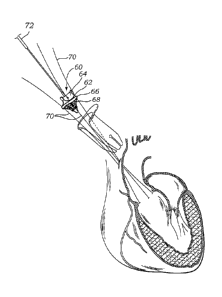
array of sutures until it rests against the native annulus. Thereafter, the
sutures
can be tied off and severed to secure the prosthetic valve to the annulus.
[004] One specific technique that is used to introduce the prosthetic valve
into
the aorta is referred to as the "shoehorn" technique. In the shoehorn
technique,
a transverse incision is made in the aortic root, which typically is smaller
than
the cross-sectional profile of the prosthetic valve. In order to pass the
valve
through the incision, the valve is inserted into the aortic root at an angle
relative
to a plane defined by the incision, much like passing a button through a
button
#11568603
CA 02768496 2017-02-03
- 2 -
hole. As can be appreciated, this technique adds complexity to the procedure,
can cause laceration of the tissue, and can cause damage to the prosthetic
valve.
[005] Another technique used to implant a prosthetic valve involves making an
oblique or "hockey stick" shaped incision in the aorta. This type of incision
creates a larger opening for the passage of the valve, but is more difficult
to
close and therefore is more prone to leakage than a straight transverse
incision.
[006] Accordingly, there exists a need for new and improved apparatus and
methods for introducing a prosthetic valve into the vasculature of a patient.
SUMMARY
[007] The present disclosure concerns embodiments of an introducer that is
adapted to facilitate insertion of a prosthetic device, such as a prosthetic
heart
valve, into a patient's vasculature. In particular embodiments, the introducer
comprises an elongated body defining a central lumen extending between distal
and proximal ends of the body. The body is adapted to be inserted through an
incision, such as a transverse aortotomy, and into the vasculature of the
patient.
A prosthetic valve can be introduced into the patient's vasculature by
advancing
the valve through the introducer.
[008] The introducer body can have a longitudinally extending gap extending
along the length of the body between the distal and proximal ends, which
allows
the introducer to radially expand and dilate the incision as the valve is
advanced
through the incision. The gap in the introducer also allows a user to place
the
introducer around implant sutures by passing the sutures through the gap and
into the central lumen. Similarly, the gap allows the introducer to be easily
removed from the sutures and/or a delivery device by passing them outwardly
through the gap. The introducer body can have a tapered distal portion to
facilitate insertion of the introducer through an incision.
[009] In one representative embodiment, a method of implanting a prosthetic
heart valve comprises making an incision in the vasculature of a patient's
body,
*11568603
CA 02768496 2017-02-03
- 3 -
threading one or more sutures through a native annulus of the heart and
extending the one or more sutures outwardly through the incision and through a
portion of the prosthetic valve, placing an introducer around the one or more
sutures and inserting the introducer into and through the incision such that a
distal end and a proximal end of the introducer are on opposite sides of the
incision, sliding the prosthetic valve along the one or more sutures and
through
the introducer until the prosthetic valve engages the annulus, removing the
introducer from the incision, and securing the valve to the annulus with the
one
or more sutures.
[010] In another representative embodiment, a method of implanting a
prosthetic heart valve comprises making an incision in the vasculature of a
patient's body, inserting the introducer through the incision and into the
vasculature of the patient, and providing a prosthetic valve mounted on a
delivery device comprising an elongated handle. The method further comprises
advancing the prosthetic valve and a portion of the handle through the
introducer until the prosthetic valve engages a native annulus of the heart,
and
removing the introducer from the incision and away from a position
surrounding the handle by passing the handle through a gap formed in a side of
the introducer.
[011] In another representative embodiment, an introducer for introducing a
prosthetic device into the vasculature of a patient comprises an elongated
body
having distal and proximal ends and a central passageway extending through the
body from the distal end to the proximal end. The body comprises two
opposing longitudinal edges defining a gap therebetween, the gap extending
from the proximal end to the distal end.
[012] The foregoing and other features and advantages of the invention will
become more apparent from the following detailed description, which proceeds
with reference to the accompanying figures.
BRIEF DESCRIPTION OF THE DRAWINGS
#11568603
CA 02768496 2017-02-03
- 4 -
[013] FIG. 1 is a perspective view of an introducer, according to one
embodiment, that can be used to introduce a prosthetic heart valve into the
vasculature of a patient.
[014] FIG. 2 is a bottom plan view of the introducer shown in FIG. 1.
[015} FIG. 3 is a perspective view of another embodiment of an introducer.
[016] FIGS. 4-10 illustrate one specific procedure for implanting a prosthetic
aortic valve into the heart using the introducer shown in FIG. 1.
[017] FIG. l 1 illustrates another procedure for implanting a prosthetic
aortic
valve into the heart using the introducer shown in FIG. 1.
[018] FIG. 12 shows an introducer assembly, according to another
embodiment, that can be used to introduce a prosthetic heart valve into the
vasculature of a patient.
1019] FIGS. 13-17 illustrate one specific procedure for implanting a
prosthetic
aortic valve into the heart using the introducer assembly shown in FIG. 12.
[020] FIG. 18 is a perspective view of another embodiment of an introducer
that can be used to introduce a prosthetic heart valve into the vasculature of
a
patient.
DETAILED DESCRIPTION
[021] The present disclosure concerns embodiments of an introducer that is
adapted to facilitate insertion of a prosthetic device, such as a prosthetic
heart
valve, into a patient's vasculature. The examples described below involve the
implantation of a prosthetic valve in the aortic annulus of the heart.
However,
the introducer can also be used to facilitate implantation of prosthetic
valves
into the other native annuluses of the heart. Further, the introducer can also
be
used to introduce various other prosthetic devices into other body lumens.
#11568603
CA 02768496 2017-02-03
- 5 -
[022] Referring first to FIGS. 1 and 2, an introducer 10, according to one
embodiment, comprises an elongated body 12 having a proximal end 14 and a
distal end 16. The body 12 has a length L extending from the proximal end 14
to the distal end 16. The body 12 has a proximal opening 18, a distal opening
20, a central lumen, or passageway, 22 extending longitudinally through the
body from the proximal opening to the distal opening. The body 12 desirably
has a proximal flange 24 that extends circumferentially around the proximal
opening and radially outwardly therefrom. The proximal flange 24 can have an
enlarged portion 26 that functions as a handle for grasping and manipulating
the
introducer during use.
[023] The body 12 in the illustrated embodiment has a generally cylindrical
shape along the majority of its length and a tapered distal end portion 28
that
extends about one third the length of the body. Thus, as can be seen in FIGS.
1
and 2, the proximal opening 18 has a larger diameter than that of the distal
opening 20. The tapered distal end portion 28 facilitates insertion of the
introducer into a surgical incision, as described below. The body 12 desirably
has a plurality of angularly spaced, longitudinally extending gaps 30 defining
a
plurality of longitudinally extending, circumferentially arrayed fingers 32.
The
fingers 32 can flex radially outwardly from each other from a non-expanded, or
contracted, state (FIG. 1) to an expanded state (FIG. 8) to expand the distal
opening to accommodate a prosthetic valve as it is pushed through the distal
end
portion of the introducer. The proximal end of each gap 30 can terminate at an
enlarged circular aperture 34 that facilitates bending or flexing of the
fingers at
their proximal ends where each finger joins the remaining portion of the body
12.
[024] The body 12 desirably has a longitudinal gap 36 extending the entire
length of the body from the proximal end 14 to the distal end 16 and defining
longitudinal edges 38. In the illustrated embodiment, the gap 36 has a
constant
width from the distal end 16 to a location near the proximal end and then
flares
or widens from this location toward the proximal end 14 to form an enlarged
#11568603
CA 02768496 2017-02-03
- 6 -
mouth 40 of the gap at the proximal end. The gap 36 allows the introducer 10
to be easily placed around and removed from sutures and/or a delivery device,
as further described below. The gap 36 also allows the introducer body 12 to
radially expand to accommodate a prosthetic valve having a larger diameter
than that of the introducer as the valve is advanced through the introducer.
In
the illustrated embodiment, the gap 36 can be referred to as a permanent gap
because there is a gap or opening between the longitudinal edges 36 when the
introducer is in its nonexpanded, or contracted, state shown in FIG. 1.
[025] In an alternative embodiment, the entire length of the gap 36 or a
portion
thereof can be replaced with a longitudinally extending slit between the
longitudinal edges 38. When the body is in a non-expanded state, the edges 38
can contact each other to close the slit. Because the introducer body is made
of
a flexible material, the edges 38 can be separated to create an opening or gap
therebetween, such as for placing the introducer around sutures or a delivery
device, as described below. Similarly, the distal end portion 28 can be formed
with a plurality of slits defining the fingers 32, rather than permanent gaps
30
between adjacent fingers as shown in the illustrated embodiment.
[026] The introducer 10 can be formed from any of various suitable materials,
including metals (e.g., stainless steel or Nitinol), alloys, polymers (e.g.,
nylon or
PTFE), composites, or combinations thereof. In certain embodiments, the
introducer can be sized for use with a variety of valve sizes (e.g., 17-35 mm
valves). Alternatively, the introducer can be provided in a range of different
sizes with each size adapted for use with one valve size or a range of valve
sizes.
[027] FIG. 3 shows an introducer 50, according to another embodiment. The
introducer 50 is similar to the introducer 10 shown in FIGS. 1 and 2 in most
respects. Unlike the introducer 10, the introducer 50 has a flange portion 52
formed with an opening 54. The opening 54 can be adapted to receive the distal
#11568603
CA 02768496 2017-02-03
- 7 -
end of an elongated handle 56 that can be used to assist in positioning and
manipulating the introducer during use.
[028] FIGS. 4-11 illustrate the use of the introducer 10 in one specific
procedure for implanting a prosthetic heart valve 60 in the aortic annulus.
The
prosthetic valve 60 in the illustrated embodiment includes a substantially
rigid,
non-collapsible annular frame 62, a plurality of leaflets 64 supported by the
frame, a sewing ring 66, and a plastically expandable stent, or support frame,
68
extending downwardly from the sewing ring. The prosthetic valve 60 can be
referred to as a hybrid valve in that it combines a non-collapsible surgical
valve
and an expandable stent that is typically incorporated in expandable
prosthetic
valves that are delivered in minimally invasive procedures. The prosthetic
valve 60 can be mounted to the distal end of an elongated shaft or handle of a
delivery device 72.
[029] As shown in FIG. 4, an incision can be made in the aorta to access the
aortic annulus. Typically, although not required, the native leaflets are
excised
before implanting the prosthetic valve, as shown in the figures. A plurality
of
implant sutures 70 can be threaded through the periphery of the aortic
annulus,
extended outside of the body through the incision and then threaded through
the
sewing ring 66 of the prosthetic valve 60 in a conventional manner. It has
been
found that three implant sutures are sufficient to adequately secure the
prosthetic valve in place at the implantation site when the securement of the
valve to the annulus is supplemented by the stent 68. A greater or fewer
number of implant sutures can be used in other applications.
[0301 To assist in passing the prosthetic valve 60 through the incision in the
aorta using the introducer, the introducer 10 is first placed around the
sutures 70
and inserted through the incision. Because the introducer has a gap 36
extending the length of the introducer, it can be easily placed around the
sutures
at a location between the incision and the valve by passing the sutures
through
the gap 36, as depicted in FIGS. 4 and 5. The enlarged mouth portion 40 of the
#11568603
CA 02768496 2017-02-03
- 8 -
gap facilitates this process in that it allows the operator to more easily
direct the
implant sutures 70 into and through the gap 36.
[031] As shown in FIG. 6, the introducer 10 can then be inserted through the
incision into the aorta. The tapered distal end portion 28 facilitates the
insertion
of the introducer through the incision. The introducer desirably is positioned
such that the distal end 16 is positioned in the native annulus or in the
Valsalva
sinuses immediately adjacent the native annulus. In this manner, the
prosthetic
valve can be guided directly to the desired implantation position as it is
advanced from the distal end of the introducer. After the introducer is
inserted
into the aorta, the surgeon can push the prosthetic valve through the
introducer
and into the aorta. If the outer diameter of the prosthetic valve 60 is
greater
than the diameter of the introducer, the introducer can radially expand as the
prosthetic valve passes through the introducer due to the presence of the gap
36,
as depicted in FIGS. 6-8. In addition the fingers 32 can expand radially
outwardly from each other to accommodate the passage of the valve through the
tapered distal end portion 28 of the introducer.
[032] In the illustrated example, the incision can be made smaller than the
cross-sectional profile of the prosthetic valve taken at sewing ring 66 (the
cross-
sectional profile is taken at a plane that extends through the sewing ring and
is
perpendicular to the central axis of the valve). Thus, as the valve passes
through the incision in this example, the incision is caused to dilate by the
radial
force of the valve against the introducer, allowing the valve to slide through
the
introducer at the location of the incision while minimizing or preventing
laceration of the tissue. Moreover, the prosthetic valve can be pushed through
the incision while the valve is maintained at a position in which the cross-
sectional profile of the valve is generally perpendicular to the line of
movement
through the introducer and the incision; in other words, the valve need not be
tilted or canted in order to pass the valve through the incision, as required
to
perform a conventional "shoehorn" technique.
#11568603
CA 02768496 2017-02-03
- 9 -
[033] As shown in FIG. 9, the valve can be advanced out of the distal end of
the introducer 10 and seated against the aortic annulus. At this time, the
introducer 10 can be removed from the incision and from its position extending
around the sutures 70 and the delivery device 72. The introducer can be
removed from its position extending around the sutures 70 and the delivery
device 72 by simply withdrawing the introducer laterally away from the sutures
and the delivery device while directing them to pass through the gap 36 in the
introducer. Due to the presence of the gap, the introducer can be removed
sideways with respect to the delivery device and does not need to be withdrawn
off of the proximal end of the delivery device, which is being held in one
hand
of the surgeon. Advantageously, this allows the surgeon to easily and quickly
remove the introducer without having to remove the hand from the proximal
end of the delivery device.
[034] As further shown in FIG. 9, after removing the introducer 10, the
expandable stent 68 can be deployed by advancing a balloon catheter 74 of the
delivery device proximally relative to the valve to displace a nose cone 76
out
of engagement with the stent 68 and to position a balloon 78 of the balloon
catheter to extend through the stent 68. The balloon 78 can then be inflated
to
cause the stent 68 to radially expand and engage surrounding tissue.
Thereafter,
the balloon is deflated, the sutures 70 are tied off to secure the sewing ring
66 to
the aortic annulus, and the delivery device is detached from the valve 60 and
removed from the body. As can be appreciated, in the illustrated example, the
prosthetic valve is secured in place against the native annulus by a
combination
of the sutures 70 and the radial outward force of the expandable stent 68.
[035] It can be appreciated that the introducer can be used to assist in the
implantation of other types of prosthetic valves. For example, the introducer
can be used to introduce a conventional surgical valve (i.e., one that does
not
have an expandable stent, like stent 68 of valve 60) into the patient's
vasculature. When implanting a conventional surgical valve, a greater number
of implant sutures 70, typically about 15-21 sutures, are used to secure the
valve
#1156B603
CA 02768496 2017-02-03
- 10 -
to the native annulus. The introducer can also be used to introduce into a
patient's vasculature a transcatheter heart valve that can be radially
compressed
to a reduced diameter for insertion into the patient's vasculature and
radially
expandable to its functional size at the deployment site inside the body.
[036] In addition, the introducer 10 can be used introduce a valve into the
patient's vasculature via any type of aortotomy (incision in the aorta). For
example, FIG. 11 illustrates the implantation of a prosthetic valve 60 via a
transverse aortotomy where a transverse incision is made through the aortic
root. A plurality of implant sutures can be threaded through the native
annulus
and the sewing ring 66 of the valve, as described above. As shown, the
introducer 10 can be placed around the sutures by passing the sutures through
the gap 36, and then inserting the distal end portion 28 of the introducer
into the
aortic root until the distal end is just above the native annulus. The
prosthetic
valve 60 can then be pushed through the introducer, into the aorta and into
the
native annulus. The valve can be secured in place by deploying the stent 68
and
tying off the implant sutures 70, as described above. If the prosthetic valve
has
a diameter greater than that of the portion of the aorta through which the
valve
passes, the introducer 10 causes the aorta to dilate around the valve as it is
passed through the introducer toward the annulus, thereby allowing for
atraumatic passage of the valve and protecting against damage to the valve
itself. Moreover, the valve can be introduced while held perpendicular to the
line of movement into and through the aorta; that is, the valve need not be
tilted
or canted relative to the line of movement in order pass the relatively larger
valve into the aorta.
[037] FIG. 12 illustrates an introducer assembly 100, according to another
embodiment, that can be used to introduce a prosthetic device, such as a
prosthetic valve 60, into the vasculature of a patient. The assembly 100 in
the
illustrated embodiment comprises an introducer 101 (also referred to as a
sleeve) and an applicator frame, or mounting frame, 102 configured to mount
the introducer 101 around the prosthetic valve. The introducer 101 can
#11588603
CA 02768496 2017-02-03
- 11 -
comprise a tubular sleeve made of a stretchable, flexible and/or resilient
material, such as a fabric. The applicator 102 is configured to retain the
introducer 101 in an expanded state having a generally cylindrical
configuration
to allow the prosthetic valve to be placed within the introducer during use.
The
applicator 102 can comprise proximal and distal rings 104, 106, respectively,
interconnected by a plurality of longitudinal members 108 extending between
and interconnecting the rings. The applicator 102 desirably is made of a
relatively rigid material as compared to the introducer, such as metal or
plastic.
[038] FIGS. 13-17 illustrate the implantation of the valve into the aortic
annulus via a transverse aortotomy made at the aortic root. However, it should
be understood that various other aortotmies can be performed to gain access to
the native annulus for implanting the prosthetic valve using the introducer
assembly 100. In any case, as shown in FIG. 13, implant sutures 70 can be
threaded through the native annulus and the sewing ring 66 of the valve. The
introducer 101 is first placed around the applicator 102, and the assembly 100
is
placed around the sutures 70, as depicted in FIG. 13, by inserting the
proximal
ends of the sutures 70 and the delivery device (the proximal ends are not
shown
in FIG. 13) into the distal opening of the applicator 102 and sliding the
assembly downwardly to the position shown in FIG. 13. The inner diameter of
the applicator 102 (defined by proximal and distal rings 104, 106) is
dimensioned large enough to permit the valve to pass easily through the
applicator.
[039] Referring to FIG. 14, the valve 60 can then be positioned within the
expanded introducer 101. With the valve inside of the introducer 101, the
introducer is removed from the applicator, such as by holding the introducer
and
the valve stationary and retracting the applicator 102 upwardly in the
direction
of arrow 110. The applicator 102 can be removed from its position surrounding
the sutures and the delivery device 72 by withdrawing the applicator upwardly
beyond the proximal ends of the sutures and the delivery device. When the
applicator is removed, the introducer 101 collapses around and desirably
#11568603
CA 02768496 2017-02-03
- 12 -
extends over and covers the entire valve such that a distal end portion 112 of
the
introducer extends beyond the lower end of the valve, as depicted in FIG. 15.
[040] Referring to FIG. 16, the distal end portion of the introducer 101 is
then
inserted into the aortic root. The valve 60 can then be inserted into the
aortic
root by maintaining slight upward tension on the introducer 101 (in the
direction
of arrow 114) while pushing downwardly on the valve 60 via the delivery
device (in the direction of arrow 116). As the valve is pushed through the
introducer and into the aortic root, the vessel dilates and allows the valve
to be
advanced through the vessel to the implantation site at the aortic annulus.
When
the valve is at the desired position, the valve is held firmly in place via
the
delivery device and the introducer 101 is pulled out of the aortic root and
then
removed from the delivery device (FIG. 17) by moving the introducer upwardly
beyond the proximal ends of the suture and the delivery device and the
sutures.
Thereafter, the valve can be secured in place by deploying the stent 68 and
tying
off the implant sutures 70, as described above.
[041] FIG. 18 shows an introducer 150, according to another embodiment, that
can be used to introduce a prosthetic device, such as a prosthetic valve 60,
into
the vasculature of a patient. The introducer 150 comprises a thin-walled,
generally tubular body 152 defining a central lumen, or passageway, 154. The
introducer body 152 is constructed from a thin membrane made of a flexible
material, such as a flexible polymer, that forms a tubular outer membrane 156
and a tubular inner membrane 158. The outer and inner membranes are joined
to each other at the opposite ends of the body so as to form a sealed inner
cavity
that can contain a fluid, such as a sterile liquid. The introducer body
exhibits
sufficient flexibility to allow the positions of the inner and outer members
to be
reversed; that is, the outer membrane 156 can be turned and pushed inwardly
into the lumen 154 at one end of the body to become part of the inner membrane
(as indicated by arrows 160) while the inner membrane 158 at the opposite end
of the body is caused to turn and move outwardly (as indicated by arrows 162)
so as to become part of the outer membrane.
#11568603
CA 02768496 2017-02-03
- 13 -
[042] In use, a prosthetic valve 60 (or other prosthetic device), which can be
mounted to the distal end of a delivery device as described herein, can be
inserted into the lumen 154 of the introducer 150. This can be accomplished by
pushing or urging the valve into the opening at one end of the introducer. The
rolling or reversing action of the membranes causes the introducer to "roll"
onto
and over the valve. With the introducer 150 covering the valve, one end of the
introducer is inserted into an incision made in the aorta (or another part of
the
patient's vasculature). The valve can then be pushed through the introducer
and
into the aorta. As the valve is advanced through the introducer, the membranes
156, 158 can roll relative to the valve to facilitate its passage through the
lumen
154.
[043] In view of the many possible embodiments to which the principles of the
disclosed invention may be applied, it should be recognized that the
illustrated
embodiments are only preferred examples of the invention and should not be
taken as limiting the scope of the invention. Rather, the scope of the
invention
is defined by the following claims. We therefore claim as our invention all
that
comes within the scope and spirit of these claims.
#11568603