Note: Descriptions are shown in the official language in which they were submitted.
CA 02768681 2012-09-28
SYSTEM FOR DIGITIZING POSITION AND ORIENTATION
INFORMATION OF HIP JOINT IMPLANT COMPONENTS
CROSS-REFERENCE TO RELATED APPLICATION
This patent application is a divisional of
Canadian Patent Application No. 2,537,594, filed on
September 7, 2004, by the present applicant.
FIELD OF THE INVENTION
The present invention generally relates to
computer-assisted hip replacement surgery and, more
particularly, to a device for positioning hip joint
implant components during surgery, and to a system and
method associated with the device.
BACKGROUND OF THE INVENTION
Computer-assisted surgery (CAS) systems
provide position and orientation information in
different forms throughout the operative steps, to
guide the surgeon in his/her decision making. CAS
systems are used for instance to assist surgeons in hip
replacement surgery. In hip replacement surgery, the
hip joint implants being implanted must assure a
desired posture to the patient. Accordingly, the
position and orientation information provided to the
surgeon must be precise and accurate to obtain the
desired posture.
The femoral implant and the acetabular
implant generally form a spherical joint, in which the
center of a ball head of the femoral implant coincides
with the center of an hemispherical socket of the
acetabular implant, at a center of rotation of the hip
joint implant. During surgery, the femur is separated
from its associated pelvis for the implants to be
implanted. Through the separation of the femur from
the pelvis, position and orientation information is
still provided from the tracking of the femur, the
pelvis and the various tools being used. For instance,
a rasping tool altering the intramedullary
1
CA 02768681 2012-02-15
canal of the femur may be tracked such that the
center of rotation of the femoral implant (i.e., the
center of the ball head) may be calculated as a
function of the geometry of the femoral implant and
of the altered intramedullary canal.
Some types of femoral implants come
separate with the ball head being fixable to the
femoral implant body. The femoral implant body has a
frusto-conical connector end (e.g., a Morse 12/14
lo taper) upon which the ball head is slid in a friction
fit. In
calculating the position of the center of
rotation of the femoral implant, some precision is
lost considering that the fit between the ball head
and the frusto-conical connector end is unpredictable
to some extent.
Alternatively, it may be desired to confirm
the position and orientation of the femoral implant.
Referring to the above-described example in which the
center of rotation of the femoral implant is
calculated as a function of the geometry of the
femoral implant and of the altered intramedullary
canal, it is possible that the femoral implant is not
completely fitted as expected in the altered
intramedullary canal. In such a case, a confirmation
of the position and orientation of the femoral
implant would be appropriate.
SUMMARY OF THE INVENTION
Accordingly, an object of the present
invention is to provide a device for obtaining
position information for hip joint implant components
in computer-assisted surgery.
It is a further object of the present
invention to provide a method and system for
obtaining position information for hip joint implant
components in computer-assisted surgery.
2
_
CA 02768681 2012-02-15
It is a further object of the present
invention to provide a device for obtaining the
center of rotation of an implant.
It is a further object of the present
invention to provide a method for obtaining the
center of rotation of an implant.1
Therefore, in accordance with the present
application, there is provided a device for
digitizing a center of rotation of a hip joint
implant component with respect to a bone element in
computer-assisted surgery, comprising a detectable
member trackable for position and orientation by a
computer-assisted surgery system; and a body
connected to the detectable member in a known
is geometry, the body having a coupling portion adapted
to be coupled to the hip joint implant component in a
predetermined configuration, the center of rotation
of the hip joint implant component being calculable
in the predetermined configuration as a function of
the known geometry and of the position and
orientation of the detectable member.
Further in accordance with the present
invention, there is provided a method for digitizing
a center of rotation of a pelvic implant component
with a computer-assisted surgery system, comprising
the steps of providing a device being trackable for
position and orientation by the computer-assisted
surgery system, the device being releasably coupled
in a known configuration to the pelvic implant
component; tracking a position and orientation of a
pelvis implanted with the pelvic implant component
and a position and orientation of the device; and
calculating a center of rotation of the pelvic
implant component with respect to the position and
orientation of the pelvis by relating the known
3
CA 02768681 2012-02-15
configuration of the device with the position and
orientation tracking of the pelvis and of the device.
Still further in accordance with the
present invention, there is provided a method of
doing surgical treatment with a position tracking
system in computer-assisted surgery for guiding an
operator in inserting a femoral implant of a hip
joint implant in a resected femur tracked for
position and orientation, comprising the steps of
positioning a trackable device on the femoral implant
in a predetermined configuration, the trackable
device being trackable in space for position and
orientation; registering implant geometry information
for the femoral implant with respect to the trackable
device as a function of said predetermined
configuration between the femoral implant and the
trackable device; and inserting the femoral implant
in the femur by obtaining implant position and
orientation information, the implant position and
orientation information being calculated from said
implant geometry information as a function of the
tracking for position and orientation of the
trackable device with respect to a frame of reference
of the femur.
Still further in accordance with the
present invention, there is provided a computer-
assisted surgery system for guiding an operator in
inserting a femoral implant of a hip joint implant in
a resected femur tracked for position and
orientation, comprising a trackable reference device
positionable onto the femoral implant in a
predetermined configuration and trackable in space
for position and orientation; a registration device
trackable in space for position and orientation and
handled by the operator to register surface
information; a sensing apparatus, for tracking any
4
CA 02768681 2012-02-15
one of the devices for position and orientation; a
controller connected to the sensing apparatus, the
controller being provided to: i)
calculate a
position and orientation of the devices as a function
of the tracking by the sensing apparatus;
ii) digitize surface information of the femoral
implant as a function of the tracking of the
registration device by the sensing apparatus; and an
implant geometry information calculator connected to
lo the controller, for calculating geometry information
of the femoral implant from said predetermined
configuration with respect to the trackable reference
device, as a function of said surface information of
the femoral implant; whereby the geometry information
is used to provide implant position and orientation
information related to a frame of reference of the
femur, so as to guide the operator in subsequently
inserting the femoral implant in the resected femur.
BRIEF DESCRIPTION OF DRAWINGS
These and other features, aspects and
advantages of the present invention will become
better understood with regard to the following
description and accompanying drawings wherein:
FIG. 1 is a perspective view of a device
for digitizing position and orientation information
of a femoral implant, in accordance with a preferred
embodiment of the present invention;
FIG. 2 is a perspective view of a device
for digitizing position information of an acetabular
implant, in accordance with a preferred embodiment of
the present invention;
FIG. 3 is a block diagram of a computer-
assisted surgery system to be used with the devices
of FIGS. 1 and 2; and
5
õ- -
CA 02768681 2012-02-15
FIG. 4 is a flow chart illustrating a
method of doing surgical treatment for guiding an
operator in inserting a femoral implant in a resected
femur in hip replacement surgery in accordance with
an embodiment of the present invention.
DESCRIPTION OF THE PREFERRED EMBODIMENT
Referring to the drawings and, more
particularly, to Fig. 1, a device to be used for
obtaining position and orientation information for a
femoral implant is generally shown at 10. A femoral
implant is shown at F, and has a body F10 and a ball
head F20. The body F10 has a stem portion F11, which
is adapted to be received in an intramedullary canal
of a resected femur (not shown). A connector end F12
projects from an end of the stem portion F11. The
connector end F12 is illustrated having a frusto-
conical shape, for instance having a Morse 12/14
taper.
The ball head F20 has a spherical outer
surface, and a connector bore F21.
The connector
bore F21 is illustrated having a frusto-conical
shape, so as to correspond to the shape of the
connector end F12 of the body F10. When the body F10
is suitably received in the intramedullary canal of
the femur, the ball head F20 is secured to the
connector end F12, by the complementary shapes of the
connector end F12 and the connector bore F21.
As mentioned previously, the position of
the center of rotation of the femoral implant F is
useful information, even prior to the ball head F20
being secured thereto. From the calculated center of
rotation, it may be required to further alter the
intramedullary canal in view of an anticipated leg
length discrepancy.
Alternatively, the calculated
center of rotation may be used to calculate the size
6
CA 02768681 2012-02-15
of ball head F20 to be used in the femoral implant F.
Femoral implant orientation information is useful in
calculating information such as the varus/valgus
angle and the offset.
Accordingly, the device 10 is to be used in
digitizing the center of rotation of the femoral
implant F and/or the orientation of the femoral
implant F. The device 10 has a tubular body 11. A
tracker base 12 projects from the tubular body 11.
lo The illustrated tracker base 12 is of the type that
receives the passive type of tracker, i.e., in the
form of three detectable devices 13 in a known
geometrical pattern. Alternatively, the tracker base
12 could be used to secure an active tracker to the
tubular body 11. The tubular
body 11 defines a
cylindrical bore 14 (i.e., cylindrical receptacle),
having a circular edge 15 at its opening in the
tubular body 11. The circular edge 15 has a known
diameter, and a known position and orientation with
respect to the tracker on the tracker base 12.
The device 10 is to be positioned onto the
connector end F12 of the femoral implant F. More
specifically, the connector end F12 is received in
the cylindrical bore 14, such that the circular edge
15 abuts against an outer surface of the connector
end F12. In such a position, the cylindrical bore 14
and the connector end F12 will axially align
themselves, considering that the connector end F12 is
frusto-conically shaped.
Furthermore, the geometric interrelation
(i.e., predetermined known configuration) between the
connector end F12 and the cylindrical bore 14 allows
the calculation of the position and orientation of
the taper of the connector end F12 with respect to
the tracker of the device 10. This
position and
orientation information of the connector end F12 may
7
CA 02768681 2012-02-15
then be used to calculate the anticipated center of
the ball head F20 as a function of the size and
geometry of the ball head F20. This
position and
orientation information of the connector end F12 may
alternatively be related to a reference tracker on
the femur to allow the calculation of navigation
information (e.g., offset, varus/valgus angles, limb
length discrepancy, etc.)
An alternative method of calculating the
lo center of the ball head F20 is contemplated. A
device 10', having the tracker base 12 with the three
detectable devices 13 with a hemispherical hole 14'
can be positioned directly on the ball head F20 once
the ball head F20 is secured to the connector end F12
of the femoral implant F. Ball heads typically come
in 3 defined sizes of 22, 28 and 32 mm, whereby the
device 10' is typically provided with corresponding
diameters for the hemispherical receptacle 14'.
Therefore, when the device 10' is mounted onto the
ball head F20, the relation between the center of the
hemispherical receptacle 14' and the center of the
ball head F20 is known (e.g., the centers are
coincident), such that the center of the ball head
F20 may be established with respect to a frame of
reference on the femur. The
determination of the
position of the center of rotation of the femoral
implant F (through the above described procedure) can
be accomplished on trial ball heads for the
calculation of other parameters (e.g., limb length),
as well as on the definitive ball head F20 installed
on the femoral implant F.
It is also contemplated to provide an
alignment mechanism between the implants F and/or A
and the devices 10 (10') and 20 (20'), respectively,
for the interconnection between the implant and its
8
CA 02768681 2012-09-28
,
,
associated device to be reproducible in position and
orientation.
Referring to Fig. 2, an
alternative
embodiment of the device, to be used to obtain position
and orientation information for an acetabular implant
is generally shown at 20.
An acetabular implant is
shown at A and has a shell A10 and a liner A20. The
shell A10 has a cup-shaped body having an outer surface
All and a receiving cavity Al2. The acetabular implant
A is to be fitted into an acetabulum (not shown), with
the outer surface All being in contact with a surface
of the acetabulum.
The receiving cavity Al2 is
equipped with connector holes such that an impactor
(not shown) can be used to insert the shell A10 into
the acetabulum and adjust its position and orientation.
The liner A20 also has a cup-shaped body.
The liner A20 is sized so as to fit into the receiving
cavity Al2 of the shell A10. More specifically, the
liner A20 has an outer surface A21 and a socket A22.
The outer surface A21 contacts the surface of the
receiving cavity Al2 when the liner A20 is fitted into
the shell A10. The socket A22 will house the ball head
F20 (Fig. 1) of the femoral implant F to form the hip
joint implant.
As mentioned previously, the position of the
center of rotation of the acetabular implant A (i.e.,
the center of rotation of the socket A22) is useful
information prior to the liner A20 being received in
the shell A10.
The center of rotation of the
acetabular implant A is dependent on the socket size of
the liner A20, and on the geometry of the liner A20.
The calculated center of rotation of the acetabular
implant A can be used for calculating navigation
information such as the offset and the limb length
discrepancy.
9
CA 02768681 2012-02-15
The device 20 is to be used in digitizing
the center of rotation of the acetabular implant A.
The device 20 has a generally hemispherical body 21.
A tracker base 22 projects from an underside of the
hemispherical body 21. The illustrated tracker base
22 is of the type that receives the passive type of
tracker, i.e., for instance three detectable spheres
in a known geometrical pattern. The tracker base 22
could be used to secure an active tracker to the body
lo 21. The
hemispherical body 21 defines an outer
surface 24.
The device 20 is to be positioned into the
receiving cavity Al2 of the shell A10 of the
acetabular implant A. More
specifically, the
hemispherical body 21 is sized to fit the receiving
cavity Al2 of the shell A10, such that the center of
rotation of the receiving cavity Al2 of the shell Al0
may be determined. From the
center of rotation of
the receiving cavity Al2, the center of rotation of
the liner A20 may be calculated, knowing the geometry
of the liner A20 (e.g., the CAS system being provided
with geometry data of various sizes of liners). It
is also possible that the liner A20 is of the type
having its center coincident with the center of the
shell A10. Therefore, the anticipated center of the
socket A22 is calculable as a function of the center
of the receiving cavity Al2 and of the geometry of
the liner A20 (stored in the CAS system).
Thereafter, the anticipated center of the
rotation of the socket A22 can be related to a
reference tracker on the acetabulum to allow the
calculation of navigation information, such as the
offset and the limb length discrepancy.
It is pointed out that the device 20 may be
used to determine the center of rotation of the liner
A20 directly. More
specifically, the hemispherical
CA 02768681 2012-02-15
body 21 may be sized so as to be received directly in
the socket A22 of the liner A20, with the liner A20
having beforehand been secured in the receiving
cavity Al2. Moreover,
an alternative configuration
of the device 20, herein illustrated as device 20',
is provided with a flange 25 at a periphery of the
outer surface 24, so as to enable the calculation of
a plane associated to the center of rotation of the
acetabular implant A.
The setting of the femoral implant F in the
intramedullary canal of the femur is an operation
that involves a plurality of factors that will have a
direct impact on the success of the hip replacement
surgery.
Therefore, the setting of the femoral
implant F advantageously involves the creation of
reference systems that will be used to provide
numeric data throughout the surgery to the surgeon
for such anatomical references as varus/valgus angle,
limb length discrepancy and femoral anteversion.
These values are calculable using position and
orientation data of the femoral implant, which will
be available during the setting of the femoral
implant F in the femur.
Therefore, referring to Fig. 4, a method
for doing surgical treatment with a tracking system
in computer-assisted surgery, for guiding an operator
in inserting a femoral implant in a femur as a
function of the limb length and the orientation of
the femoral implant is generally shown at 50.
The insertion of the femoral implant in the
femur takes place after the femoral head has been
resected, and the intramedullary canal has been
altered in view of the insertion of the implant
therein. Such
steps are described in International
Publication No. WO 2004/030556, published on
April 15, 2004, by Jansen et al. At this
point, a
11
CA 02768681 2012-02-15
generic digital model of the implant F is available
through the CAS assisting the operator.
In Step 52 of the method 50, the device 10
(Figs. 1 and 3) is positioned on the connector end
F12 of the implant F. If the ball
head F20 is
already secured to the implant body F10, the device
10' is used (Figs. 1 and 3).
In Step 54, the orientation of the neck
axis of the connector end F12, and the center of
lo rotation of the ball head F20, are calculable as a
function of the position and orientation of the
tracker base 12.
In Step 56, a plane is digitized for the
implant F. More specifically, three non-linear
points are digitized using a registration pointer,
whereby a plane may be digitized with respect to the
device 10 in which all three points lie. For
instance, points are taken at P1, P2 and P3 in
Fig. 1. With
these points and with the neck axis
calculated in Step 54, the position and orientation
digitized and calculated in Steps 54 and 56 may be
associated to the digital model of the implant.
In Step 58, a tip of the implant is
digitized with respect to the device 10, using the
registration pointer. The tip is illustrated at P4
in Fig. 1.
In Step 60, a longitudinal axis of the
implant F is digitized with respect to the device 10.
More specifically, the CCD angle of the implant F is
generic information provided with the digital model
of the implant F. Accordingly, using the neck axis
calculated in Step 54 and the CCD angle, a line
parallel to the longitudinal axis is defined. The
longitudinal axis is then calculated with respect to
the device 10 or 10 as being parallel to this line,
while lying in the plane digitized in Step 56 and
12
CA 02768681 2012-02-15
passing through the tip of the implant digitized in
Step 58.
In Step 62, now that the required geometry
information pertaining to the implant F is known
(i.e., longitudinal axis, neck axis, center of
rotation, with respect to the device 10), the implant
F is inserted in the altered intramedullary canal of
the femur F.
Real-time information may be provided to
io the operator, whereby the device 10 (10') must be
kept onto the implant F during the insertion of the
implant F in the intramedullary canal. Accordingly,
a locking mechanism should be used to secure the
device 10 to the implant F in position and
orientation.
In Step 64, the geometry information
gathered for the implant F is associated to the frame
of reference of the femur. By positioning the device
10 (or 10') on the implant F, the position of the
center of rotation of the implant F is known, as well
as the position of the neck axis.
The orientation of the implant F may be
calculated by knowing the interconnection between the
implant F and the device 10 (or 10') (through an
alignment mechanism, as mentioned previously).
Alternatively, the orientation of the
implant F may be calculated using the digital model
of the altered intramedullary canal with respect to
the frame of reference of the femur, in association
with the position and orientation of the device 10
(or 10'). The digital model of the altered
intramedullary canal is information available as
calculated during the alteration of the
intramedullary canal, as described in International
Publication No. WO 2004/030556, published on
April 15, 2004, by Jansen et al.
13
CA 02768681 2012-02-15
Therefore, when the geometry information of
the implant F is associated to the frame of reference
of the femur, the geometry information can be used to
calculate position and orientation information of the
s implant F with respect to the femur.
For instance, the longitudinal axis of the
implant F, as obtained through the method 50, can be
used in the calculation of the varus/valgus angle of
the femoral implant F. More specifically, the
lo longitudinal axis of the femoral implant is projected
onto a frontal plane of the patient along with an
axis of the intramedullary canal (as described in
International Publication No. WO 2004/030556), with
the angle between these two projections representing
15 the varus/valgus angle.
Also, the neck axis of the implant is
projected onto the transverse plane (as described in
International Publication No. WO 2004/030556), where-
by the femoral anteversion is calculable as the angle
20 between this projection and the intersection of the
transverse and frontal planes.
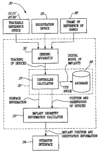
Referring to Fig. 3, a CAS system in
accordance with the present invention is generally
shown at 30. The CAS system 30 has a controller 31
25 that is connected to the sensing apparatus 32.
The sensing apparatus 32 tracks the devices
10, 10', 20 and 20', as well as a registration device
35 (e.g., registration tool), and frames of reference
36 associated to bones (e.g., femoral and pelvic
30 frames of reference as described in International
Publication No. WO 2004/030556). For
instance, the
sensing apparatus 32 is an optical sensing apparatus
that visually detects the position of the passive
detectable devices, such as those illustrated at 13
35 in Fig. 1). The
tracking output of the sensing
apparatus 32 is calculated as position and
14
,
CA 02768681 2012-02-15
orientation of the devices by the controller 31,
whereas registered points, as described in Steps 56
and 58 (Fig. 4), are digitized as surface information
of the implants.
The CAS system 30 has an implant geometry
information calculator 33, that will receive the
position and orientation of the devices 10, 10', 20,
20', as well as the surface information, so as to
calculate geometry information, as mentioned in Steps
54 and 60, and transfer this data in the form of
implant position and orientation information, as
described in Step 62, to an operator through operator
interface 34.
The controller 31 typically has a
controller calculator 37 consisting of a processor
that will calculate the above described information,
and a database 38 that will hold some information
that may be required in the calculation, such as
digital model of implants, to which the geometry
information and the implant position and orientation
information may be associated, as mentioned in the
method 50.