Note: Descriptions are shown in the official language in which they were submitted.
CA 02769106 2015-07-03
OPTICAL SYSTEM FOR OPHTHALMIC SURGICAL LASER
Field of Invention
100021 This invention relates to a system for surgery of the anterior
segment of the
eye with a femtosecond laser, more particularly to embodiments minimizing
optical
distortions of the laser beam while scanning and focusing the laser beam into
the eye.
Background
100031 This application describes examples and embodiments of techniques
and
systems for laser surgery within the anterior segment of the eye the
crystalline lens via
photodisruption caused by laser pulses. Various lens surgical procedures for
removal of the
crystalline lens utilize various techniques to break up the lens into small
fragments that can
be removed from the eye through small incisions. These procedures use manual
instruments,
ultrasound, heated fluids or lasers and tend to have significant drawbacks,
including the need
to enter the eye with probes in order to accomplish the fragmentation, and the
limited
precision associated with such lens fragmentation techniques.
100041 Photodisruptive laser technology can deliver laser pulses into
the lens to
optically fragment the lens without insertion of a probe and thus can offer
the potential for
improved lens removal. Laser-induced photodisruption has been widely used in
laser
ophthalmic surgery and Nd:YAG lasers have been frequently used as the laser
sources,
including lens fragmentation via laser induced photodisruption. Some existing
systems utilize
nanosecond lasers with pulse energies of several mJ (E. H. Ryan etal. America!
Journal of
Ophthalmology 104: 382-386, October 1987; R. R. Kruger et al. Ophthalmology
108: 2122-
2129, 2001), and picosecond lasers with several tens of IAJ (A. Gwon et al. J.
Cataract Refract
Surg. 21, 282-286, 1995). These relatively long pulses deposit relatively
large amounts of
energy into the surgical spots, resulting in considerable limitations on the
precision and
control of the procedure, while creating a relatively high level of risk of
unwanted outcomes.
CA 02769106 2015-07-03
[0005] In parallel, in the related field of cornea surgery it was
recognized that shorter
pulse durations and better focusing can be achieved by using pulses of
duration of hundreds
of femtoseconds instead of the nanosecond and picosecond pulses. Femtosecond
pulses
deposit much less energy per pulse, significantly increasing the precision and
the safety of
the procedure.
[0006] Presently several companies commercialize femtosecond laser
technology for
ophthalmic procedures on the cornea, such as LASIK flaps and corneal
transplants. These
companies include Intralase Corp. / Advanced Medical Optics, USA, 20/10
Perfect Vision
Optische Gerate GmbH, Germany, Carl Zeiss Meditec, Inc. Germany, and Ziemer
Ophthalmic Systems AG, Switzerland.
[0007] However, these systems are designed according to the requirements of
the
cornea surgery. Crucially, the depth range of the laser focus is typically
less than about 1
mm, the thickness of the cornea. As such, these designs do not offer solutions
for the
considerable challenges of performing surgery on the lens of the eye.
Summary
[0008] Briefly and generally, a laser system for ophthalmic surgery
includes a laser
source, to generate a pulsed laser beam, an XY scanner, to receive the pulsed
laser beam,
and to output an XY-scanning beam, scanned in two directions transverse to a Z
axis, a Z
scanner, to receive the XY-scanning beam, and to output an XYZ-scanning beam,
scanned
in addition along the Z axis, the Z scanner including a first lens group to
output a beam
having an intermediate focal plane, and a movable lens group to receive the
beam through
the intermediate focal plane and to collimate the beam in a variable manner,
and an
objective to receive the collimated beam from the Z scanner and to focus the
beam into a
focal spot in a target region.
2
CA 02769106 2015-07-03
[0008a] Certain exemplary embodiments can provide a laser system for
ophthalmic
surgery, comprising: a laser source, to generate a pulsed laser beam; an XY
scanner, to
receive the pulsed laser beam, and to output an XY-scanning beam, scanned in
two
directions transverse to a Z axis; a Z scanner, to receive the XY-scanning
beam; and to
output an XYZ-scanning beam, scanned in addition along the Z axis; the Z
scanner
comprising a first lens group to output a beam having an intermediate focal
plane; and
a movable lens group to receive the beam through the intermediate focal plane
and to
collimate the beam in a variable manner; and an objective to receive the
collimated beam
from the Z scanner and to focus the beam into a focal spot in a target region.
[0008b] Certain exemplary embodiments can provide a method of ophthalmic
surgery, the method comprising the steps of: generating a pulsed laser beam;
XY scanning
the pulsed laser beam in two directions transverse to a Z direction with an XY
scanner; Z
scanning the XY scanned beam along a Z direction with a Z scanner by focusing
the XY
scanned beam onto an intermediate focal plane with a first lens group;
receiving the beam
from the intermediate focal plane by a movable beam scanner; collimating the
beam into an
adjustable entrance pivot point of an objective by the movable beam scanner;
and focusing
the beam onto an XYZ scanned focal spot in a target region by the objective.
[0009] In some implementations the Z scanner is configured to scan a Z
focal depth
of the focal spot in the target region within a Z scanning range of 5
millimeter to 10
millimeter. In some implementations the Z scanner is configured to scan a Z
focal depth of
the focal spot in the target region within a Z scanning range of 0 millimeter
to 15 millimeter.
[0010] In some implementations the movable lens group can be moved along
the Z
axis by a distance between 5 and 50 millimeters.
2a
CA 02769106 2012-01-24
WO 2011/017003
PCT/US2010/042796
[0011] In some implementations the first lens group includes 2-10
lenses, and the
movable lens group includes 2-10 lenses.
[0012] In some implementations the first lens group includes,
sequentially from an
input side a first lens group with a positive refractive power, a meniscus
lens, having a
convex surface facing the input side, and a second lens, having a concave
surface facing the
input side.
[0013] In some implementations the movable lens group includes,
sequentially from
an input side, a meniscus lens, having a concave surface facing the input
side, a negative lens
with a negative refractive power, and a positive lens group with a positive
refractive power.
[0014] In some implementations the movable lens group is adjustable to
change at
least one characteristic of the XYZ scanning beam exiting the Z scanner, the
characteristics
including a convergence, a beam diameter, a Z focal depth, and a numerical
aperture NA.
[0015] In some implementations the movable lens group is configured to
adjust a
numerical aperture NA and a Z focal depth of the XYZ scanning beam essentially
independently from each other.
[0016] In some implementations the Z scanner is configured to focus a
beam,
emanating from an exit pivot point of the XY scanner into an entrance pivot
point of the
objective.
[0017] In some implementations the entrance pivot point of the
objective is inside the
objective. In some implementations a position of the entrance pivot point of
the objective is
adjustable by moving the movable lens group.
[0018] In some implementations the Z scanner is configured to modify
an exit pivot
point of the XY scanner into an entrance pivot point of the objective. In some
implementations the entrance pivot point of the objective is inside the
objective.
[0019] In some implementations the first lens group is one of a movable
lens group
and a fixed lens group.
[0020] In some implementations a method of ophthalmic surgery includes
generating
a pulsed laser beam, XY scanning the pulsed laser beam in two directions
transverse to a Z
direction with an XY scanner, Z scanning the XY scanned beam along a Z
direction with a Z
scanner by focusing the XY scanned beam onto an intermediate focal plane with
a first lens
group, receiving the beam from the intermediate focal plane by a movable beam
scanner,
3
CA 02769106 2012-01-24
WO 2011/017003
PCT/US2010/042796
collimating the beam into an adjustable entrance pivot point of an objective
by the movable
beam scanner, and focusing the beam onto an XYZ scanned focal spot in a target
region by
the objective.
[0021] In some implementations the Z scanning step includes adjusting the
movable
beam scanner to change at least one characteristic of the XYZ scanning beam
exiting the Z
scanner, the characteristics including a convergence, a beam diameter, a Z
focal depth, and a
numerical aperture NA.
[0022] In some implementations the Z scanning step includes adjusting a
numerical
aperture NA and a Z focal depth of the XYZ scanning beam essentially
independently from
each other.
Brief description of Figures
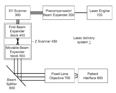
[0023] FIG. 1 illustrates a surgical laser delivery system 1.
[0024] FIG. 2 illustrates a Gaussian wavefront G and an aberrated wavefront
W.
[0025] FIGS. 3A-B illustrate rays at an optimal and a scanned focal plane.
[0026] FIG. 3C illustrates a definition of the focal spot radius.
[0027] FIG. 4 illustrates a relation between a Strehl ratio S and an RMS
wavefront
error co.
[0028] FIG. 5 illustrates reference points for ophthalmic surgery.
[0029] FIGS. 6A-B illustrate conceptually the operation of precompensator
200.
[0030] FIGS. 7A-B illustrate various uses of an efficient Z scanning
functionality.
[0031] FIGS. 8A-D illustrate implementations of the precompensator 200.
[0032] FIG. 9 illustrates an implementation of the laser delivery system 1
with two Z
Scanners.
[0033] FIG. 10 illustrates a table of configurations containing 0, 1, or 2
Z depth
Scanner and 0, 1, or 2 NA modifiers.
[0034] FIGS. 11A-C illustrate an XY Scanner with 2, 3, and 4 scanning
mirrors.
[0035] FIGS. 12A-D illustrate an aberration as a function of a numerical
aperture and
the corresponding optical numerical aperture NAopt(z) as a function of the Z
focal depth.
4
CA 02769106 2012-01-24
WO 2011/017003
PCT/US2010/042796
[0036] FIGS. 13A-B illustrate two settings of the First Beam Expander
block 400 and
the Movable Beam Expander block 500.
[0037] FIG. 14 illustrates the intermediate focal plane of the Z
Scanner 450.
[0038] FIG. 15 illustrates an implementation of the Objective 700.
[0039] FIG. 16 illustrates a curved focal plane in the target region.
[0040] FIG. 17 illustrates a nomogram of the XY Scanner inclination
angle.
[0041] FIG. 18 illustrates a nomogram of the Movable Beam Expander
position.
[0042] FIG. 19 illustrates steps of a computational control method.
5
CA 02769106 2012-01-24
WO 2011/017003
PCT/US2010/042796
Detailed description
[0043] Some embodiments of the present invention include systems for
surgery in the
lens of the eye, utilizing femtosecond laser pulses. Some integrated
embodiments are also
capable of performing both corneal and lens surgical procedures. Performing
ophthalmic
surgery in the lens of the eye is associated with qualitatively different
requirements than
corneal procedures.
[0044] The key differences between the presently described lens
surgical laser system
and corneal systems include:
[0045] 1. Femtosecond laser pulses are to be generated reliably. High
repetition rate
femtosecond pulses allow the use of a much smaller energy per pulse, providing
much higher
control and precision for the operator of the system. However, generating
femtosecond
pulses reliably is a considerably greater challenge than nanosecond or
picosend pulses, used
by some existing systems.
[0046] 2. The surgical laser beam is refracted considerably when
propagating through
up to 5 millimeters of refractive medium, including the cornea and the
anterior aqueous
chamber just to reach the surgical target, the lens. In contrast, the laser
beam used for corneal
surgery is focused at a depth of a fraction of a millimeter, and is thus
essentially not refracted
as it enters the cornea from the surgical system.
[0047] 3. The surgical laser delivery system is configured to scan the
entire surgical
region, for example from the front/anterior of the lens at a typical depth of
5 mm to the
back/posterior of the lens at a typical depth of 10 mm. This 5 mm or more
depth-scanning
range, or "Z scanning range", is considerably more extensive than the 1 mm
depth-scanning
range used for surgery on the cornea. Typically, the surgical optics,
especially the here-used
high numerical aperture optics, is optimized to focus a laser beam to a
specific operating
depth. During corneal procedures the 1 mm depth-scanning causes only moderate
departure
from the optimized operating depth. In contrast, during the scan from 5 to 10
mm during lens
surgery, the system is driven far from a fixed optimized operating depth.
Therefore, the lens-
surgical laser delivery system employs a much¨refined adaptive optics to be
able to scan the
extensive depth-scanning range required by lens surgery.
[0048] 4. Some embodiments are integrated in the sense that they are
configured to
perform surgery on both the cornea and the lens. In these integrated
embodiments the depth-
scanning range can be up to 10 mm instead of 5 mm, posing even harder
challenges.
6
CA 02769106 2012-01-24
WO 2011/017003
PCT/US2010/042796
[0049] 5. During corneal surgical procedures, such as the many
variants of LASIK,
the laser beam is scanned perpendicular to the optical axis ("in the XY
plane"). In typical
procedures the XY scanning range covers only the central portion of the cornea
with a
diameter of 10 mm. However, in integrated surgical systems additional cuts may
be formed
as well. One type of cuts is the entry cuts, providing access to the inside of
the eye for
aspiration needles and conventional surgical tools. Another type of cuts is
the limbal relaxing
incisions (LRIs), which involve making a pair of incisions at the corneal
limbus just anterior
to the vascular arcade. By adjusting the length, depth, and location of these
arcuate incisions,
one can induce changes in the corneal astigmatism. Entry cuts and LRIs can be
placed at the
periphery of the cornea, typically with a diameter of 12mm. While increasing
the XY
scanning diameter from 10 mm to 12 mm diameter is only a 20% increase compared
to the
regular diameter of LASIK flaps, it is a significant challenge to keep off-
axis aberrations of
the laser delivery system under control at such diameters, since off-axis
aberrations grow
proportional to higher powers of the field diameter at the focal plane.
[0050] 6. Lens laser surgical procedures may require guidance from
sophisticated
imaging systems. In some imaging systems limbal blood vessels are identified
to serve as
reference marks on the eye, to calibrate the cyclo-rotational alignment of the
eye during the
time of surgery, in some cases relative to the reference coordinates
identified during
preoperative diagnosis of the eye. Blood vessels chosen on the periphery of
the surgical area
can be the most undisturbed by the surgery and thus the most reliable. Imaging
systems
directed to such peripheral blood vessels, however, require the imaging optics
to image an
area with a radius larger than 10 mm, such as 12 mm.
[0051] 7. The laser beam develops various aberrations while
propagating along the
optical path within the eye. Laser delivery systems can improve precision by
compensating
for these aberrations. An additional aspect of these aberrations is that they
depend on the
frequency of the light, a fact referenced as "chromatic aberration".
Compensating these
frequency dependent aberrations increases the challenge on the system. The
difficulty of
compensating these chromatic aberrations increases with the bandwidth of the
laser beam. a
laser system. It is recalled that the spectral bandwidth of a beam is
inversely proportional to
the pulse length. Accordingly, the bandwidth for femtosecond pulses is often
greater than
that of picosecond pulses by an order of magnitude or more, necessitating a
much better
chromatic compensation in femtosecond laser systems.
7
CA 02769106 2012-01-24
WO 2011/017003
PCT/US2010/042796
[0052] 8. Surgical procedures using high repetition rate femtosecond
laser surgical
systems require high precision in positioning each pulse both in an absolute
sense with
respect to target locations in the target tissue and in a relative sense with
respect to preceding
pulses. For example, the laser system may be required to redirect the beam by
only a few
microns within the time between pulses, which can be of the order of
microseconds. Because
the time between two subsequent pulses is short and the precision requirement
for the pulse
placement is high, manual targeting as used in existing low repetition rate
lens surgical
systems is no longer adequate or feasible.
[0053] 9. The laser delivery system is configured to deliver the
femtosecond laser
pulses into the entire surgical volume of lens of the eye, through a
refractive medium, with
their temporal, spectral and spatial integrity preserved.
[0054] 10. To ensure that only tissue in the surgical region receives
a laser beam with
high enough energy densities to cause surgical effects, such as tissue
ablation, the laser
delivery system has an unusually high numerical aperture (NA). This high NA
results in
small spot sizes and provides necessary control and precision for the surgical
procedure.
Typical ranges for the numerical aperture can include NA values larger than
0.3, resulting in
spot sizes of 3 microns or less.
[0055] 11. Given the complexity of the optical path of the laser for
lens surgery, the
laser delivery system achieves high precision and control by including a high
performance
computer-managed imaging system, whereas corneal surgical systems can achieve
satisfactory control without such imaging systems, or with a low level of
imaging. Notably,
surgical and imaging functions of the system, as well as the customary
observational beams
generally all operate in different spectral bands. As an example, surgical
lasers may operate
at wavelengths in the band of 1.0-1.1 micron, observational beams in the
visible band of 0.4-
0.7 micron, and imaging beams in the band of 0.8-0.9 micron. Combining beam
paths in
common, or shared, optical components places demanding chromatic requirements
on the
optics of the laser surgical system.
[0056] The differences 1-11 illustrate through several examples that
ophthalmic laser
surgery (i) on the lens (ii) with femtosecond pulses introduces requirements
which are
qualitatively different from those of corneal surgery and even from lens
surgery, using only
nanosecond or picosecond laser pulses.
[0057] FIG. 1 illustrates a laser delivery system 1. Before describing
it in detail, we
mention that some embodiments combine the laser delivery system of FIG. 1 with
an
8
CA 02769106 2016-12-21
imaging or an observational system. In some corneal procedures, such as in
LASIK
treatments, eye trackers establish positional references of the eye by visual
clues such an
identification of the center of the iris by imaging and image processing
algorithms, typically
on the surface of the eye. However, existing eye trackers recognize and
analyze features in a
two-dimensional space, lacking depth information, since the surgical
procedures are
performed on the cornea, the outermost layer of the eye. Often, the cornea is
even flattened
to make the surface truly two dimensional.
[0058] The situation is quite different when focusing a laser beam in
the lens, deep
inside the eye. The crystalline lens can change its position, shape, thickness
and diameter
during accommodation, not only between prior measurement and surgery but also
during
surgery. Attaching the eye to the surgical instrument by mechanical means can
also change
the shape of the eye in an ill-defined manner. Such attaching devices can
include fixating
the eye with a suction ring, or aplanating the eye with a flat or curved lens.
Further, the
movement of the patient during surgery can introduce additional changes. These
changes
can add up to as much as a few millimeters of displacement of visual clues
within the eye.
Therefore, mechanically referencing and fixating the surface of the eye such
as the anterior
surface of the cornea or limbus are unsatisfactory when performing precision
laser surgery
on the lens or other internal portions of the eye.
[0059] To address this problem, laser delivery system 1 can be
combined with an
imaging system, as described in co-pending application serial number US Patent
Application
12/205,844 to R.M. Kurtz, F. Raksi and M. Karavitis. The imaging system is
configured to
image portions of a surgical region to establish three dimensional positional
references
based on the internal features of the eye. These images can be created before
the surgery
and updated in parallel with the surgical procedure to account for individual
variations and
changes. The images can be used to direct the laser beam safely to the desired
location with
high precision and control.
[0060] In some implementations, the imaging system can be an Optical
Coherence
Tomography (OCT) system. The imaging beam of the imaging system can have a
separate
imaging optical path, or an optical path partially or fully shared with the
surgical beam.
Imaging systems with a partially or fully shared optical path reduce the cost
and simplify the
calibration of the imaging and surgical systems. The imaging system can also
use the same
or a different light source as the laser of the laser delivery system 1. The
imaging system
9
CA 02769106 2016-12-21
can also have its own beam scanning subsystems, or can make use of the
scanning
subsystems of the laser delivery system 1. Several different architectures of
such OCT
systems are described in the referred co-pending application.
[0061] The laser delivery system 1 can be also implemented in
combination with a
visual observation optics. The observation optics can help the operator of the
surgical laser
to observe the effects of the surgical laser beam and control the beam in
response to the
observations.
[0062] Finally, in some implementations, which use an infrared and
thus invisible
surgical laser beam, an additional tracking laser may be employed operating at
visible
frequencies. The visible tracking laser maybe implemented to track the path of
the infrared
surgical laser. The tracking laser may be operated at a low enough energy not
to cause any
disruption of the target tissue. The observation optics may be configured to
direct the
tracking laser, reflected from the target tissue, to the operator of the laser
delivery system 1.
[0063] In FIG. 1, the beams associated with the imaging system and
the visual
observation optics can be coupled into the laser delivery system 1 e.g.
through a beam
splitter/dichroic mirror 600. The present application will not discuss
extensively the various
combinations of the laser delivery system 1 with the imaging, observational
and tracking
systems.
[0064] FIG. 1 illustrates a laser delivery system 1, which includes a
Laser Engine
100, a Precompensator 200, an XY Scanner 300, a First Beam Expander block 400,
a
Movable Beam Expander block 500, a Beam Splitter/dichroic mirror 600, an
Objective 700
and a Patient Interface 800, wherein the First Beam Expander block 400 and the
Movable
Beam Expander block 500 will be jointly referred to as Z Scanner 450.
[0065] In many implementations below the convention is used that the
Z direction is
the direction essentially along the optical path of the laser beam, or along
the optical axis of
the optical element. The directions transverse to the Z direction are referred
to as XY
directions. The term transverse is used in a broader sense to include that in
some
implementations the transverse and Z directions may not be strictly
perpendicular to each
other. In some implementations the transverse directions can be better
described in terms of
radial coordinates. Thus the terms transverse, XY, or radial directions denote
analogous
directions in the described implementations, all approximately (but
necessarily precisely)
perpendicular to the Z direction.
CA 02769106 2012-01-24
WO 2011/017003
PCT/US2010/042796
1. The Laser Engine 100
[0066] The laser engine 100 can include a laser to emit laser pulses
with
predetermined laser parameters. These laser parameters may include pulse
duration in the 1
femtosecond to 100 picosecond range, or within the 10 femtosecond to 10
picosecond range,
or in some embodiments the 100 femtosecond to 1 picosecond range. The laser
pulses can
have an energy per pulse in the 0.1 microJoule to 1000 microJoule range, in
other
embodiments in the 1 microJoule to 100 microJoule range. The pulses can have a
repeat
frequency in the 10 kHz to 100 MHz range, in other embodiments in the 100 kHz
to 1 MHz
range. Other embodiments may have laser parameters which fall within a
combination of
these range limits, such as a range of pulse duration of 1-1000 femtosecond.
The laser
parameters for a particular procedure can be selected within these wide ranges
e.g. during a
pre-operational procedure, or based on a calculation which is based on certain
data of the
patient, such as his/her age.
[0067] Examples of the laser engine 100 can include Nd:glass and Nd:Yag
lasers, and
other lasers of a wide variety. The operating wavelength of the laser engine
can be in the
infrared or in the visible range. In some embodiments the operating wavelength
can be in the
700 nm ¨ 2 micron range. In some cases the operating wavelength can be in the
1.0-1.1
micron range, e.g. in infrared lasers based on Yb or Nd.
[0068] In some implementations the laser parameters of the laser pulses may
be
adjustable and variable. The laser parameters may be adjustable with a short
switch time,
thus enabling the operator of the surgical laser delivery system 1 to change
laser parameters
during a complex surgery. Such a change of parameters can be initiated in
response to a
reading by a sensing or imaging subsystem of the laser delivery system 1.
[0069] Other parameter changes can be performed as part of a multi-step
procedure
during which the laser delivery system may be first used for a first surgical
procedure,
followed by a second, different surgical procedure. Examples include first
performing one or
more surgical steps in a region of a lens of an eye, such as a capsulotomy
step, followed by a
second surgical procedure in a corneal region of the eye. These procedures can
be performed
in various sequences.
[0070] High repetition rate pulsed lasers operating at a pulse
repetition rate of tens to
hundreds of thousands of shots per second or higher with relatively low energy
per pulse can
11
CA 02769106 2012-01-24
WO 2011/017003
PCT/US2010/042796
be used for surgical applications to achieve certain advantages. Such lasers
use relatively low
energy per pulse to localize the tissue effect caused by the laser-induced
photodisruption. In
some implementations, for example, the extent of the disrupted tissue can be
limited to a few
microns or a few tens of microns. This localized tissue effect can improve the
precision of
the laser surgery and can be desirable in certain surgical procedures. In
various
implementations of such surgeries, many hundreds, thousands or millions of
pulses can be
delivered to a sequence of spots which are contiguous, nearly contiguous, or
are separated by
controlled distances. These implementations can achieve certain desired
surgical effects,
such as tissue incisions, separations or fragmentation.
[0071] The parameters of the pulses and the scan pattern can be selected by
various
methods. For example, they can be based on a preoperative measurement of the
optical or
structural properties of the lens. The laser energy and the spot separation
can also be selected
based on a preoperative measurement of optical or structural properties of the
lens or on an
age-dependent algorithm.
12
CA 02769106 2012-01-24
WO 2011/017003
PCT/US2010/042796
2. Precompensator 200
[0072] FIG. 2 illustrates that the wavefront of the laser beam can
deviate from an
ideal behavior in several different ways and for several different reasons. A
large group of
these deviations are called aberrations. Aberrations (and the other wavefront
distortions)
displace real image points from the ideal paraxial Gaussian image points. FIG.
2 illustrates
wavefronts of light exiting through an exit pupil ExP. The undistorted
spherical wavefront G
emanates from the pupil and converges to a point P1 at the center of curvature
of the
wavefront G. G is also called the Gaussian reference sphere. An aberrated
wavefront W
deviates from G and converges to a different point P2. The aberration AW of
the aberrated
wavefront W at point Q1 can be characterized by the optical length of the
pathway relative to
the undistorted reference sphere G: A W=n1Q1Q2 , where ni is the refractive
index of the
medium in the image space and Q1Q2 is the distance of points Q1 and Q2.
[0073] In general, the aberration AW depends on the coordinates both
at the exit pupil
as well as at the focal plane. Therefore, this aberration A W can be also
thought of as a
correlation function: it represents that the set of points whose image
converges to P2,
removed from P1 on the optical axis by r', are located on a surface W, which
deviates from
the reference sphere G by an amount of A W at the radial distance r at the
Exit pupil ExP. For
a rotationally symmetrical system, A W can be written in terms of a double
power series
expansion in r and r' as:
(r' ;r ,O) =EEE manmr'21" rn cosm O. (1)
1=0 n=1 m=0
[0074] Here r' is the radial coordinate of the image point P2 in the
focal plane and r is
the radial coordinate of point Q1 at the pupil. The angular dependence is
represented by 0,
the spherical angle. n = 2p + m is a positive integer and 21 ma mil are the
expansion
coefficients of the aberrated wavefront W. For reference, see e.g.: Optical
Imaging and
Aberrations, Part I. Ray Geometrical Optics by Virendra N Mahajan, SPIE
Optical
Engineering Press. The order i of an aberration term is given by i = 21 + m
+n.
[0075] The terms up to i = 4 are related to the primary aberrations:
spherical, coma,
astigmatism, field curvature and distortion. The actual relations between
these primary
aberrations and the 21+ ma nm aberration coefficients are documented in the
literature. For a
system imaging a point object, the explicit dependence of the aberration terms
on the image
13
CA 02769106 2012-01-24
WO 2011/017003
PCT/US2010/042796
radius r' can be suppressed by introducing the dimensionless variable p = r/a,
where a is a
transverse linear extent of the exit pupil, such as its radius:
AW(p,0)= EEarimpn cosm 0, (2)
n=1 m=0
where
anm = anE 21+ma nin' = (3)
1=0
[0076] A benefit of this notation is that the aberration coefficients anm
all have the
dimension of length and represent the maximum value of the corresponding
aberration at the
exit pupil. In this notation, for example, the spherical aberration is
characterized by the
aberration coefficient a40.
[0077] While the description of aberration in terms of the aberration
coefficients amn
is mathematically well defined, it is not always the experimentally most
accessible approach.
Therefore, three alternative aberration measures are described next.
[0078] In the same vein of experimental accessibility and testability, it
is noted that
the behavior of a beam in a biological tissue, such as the eye, may not be the
easiest to
measure. Helpfully, studies indicate that rays in the eye may behave very
analogously to rays
in salty water with physiologically appropriate salt concentration, where they
can be
quantitatively measured and described. Therefore, throughout the application
when the laser
delivery system's behavior in the eye is described, it is understood that this
description refers
to behavior either in the described eye tissue, or in corresponding salty
water.
[0079] FIGS. 3A-C illustrate a second measure of aberrations. The laser
delivery
system 1, which was configured to focus a beam at a focal plane 210 at depth
A, can cause a
spherical aberration if it is operated to focus the beam at an operating focal
plane 211 at depth
B instead. Such a situation can occur, for example, during a three dimensional
scanning
procedure, when the focal point of the laser beam is moved from focal plane
210 to focal
plane 211.
[0080] FIG. 3A illustrates the case when the laser delivery system 1
focuses the rays
to their optimal focal plane 210. The rays pass through a spot at the optimal
focal plane 210
(a "focal spot") of very narrow radial extent, or radius, rf(A). This radial
extent rf(A) can be
greater than zero for a variety of reasons, such as the diffraction of the
light beam. The radius
14
CA 02769106 2012-01-24
WO 2011/017003
PCT/US2010/042796
of the focal spot can be defined in more than one ways. A common definition of
rf(A) is the
minimal radius of the light spot on a screen as the screen's position is
varied along the axial,
or Z, direction. This Z depth is often called the "point of least confusion".
This definition is
further refined in relation to FIG. 3C.
[0081] FIG. 3B illustrates the case when the laser delivery system 1 scans
the focus
by some distance, such as a few millimeters, off the optimal focal plane 210,
to an operating
focal plane 211. Visibly, the rays pass through a focal spot of a radius rf(B)
larger than ri(A),
causing a spherical aberration. Mathematical formulae of various accuracy have
been
developed connecting the aberration coefficients anti, and the focal spot
radius rf. In some
cases, the focal spot radius rf is an experimentally more accessible measure
to quantify the
aberrations than the anin aberration coefficients.
[0082] FIG. 3C illustrates a more quantitative definition of the focal
spot radius rf.
FIG. 3C illustrates the energy contained in a spot of radius r, measured from
a centroid of the
beam. A widely accepted definition of the focal spot radius rf is the radius,
within which
50% of the beam's energy is contained. The curve labeled "A" shows that in a
diffraction
limited beam, when the beam is focused to its optimal focal plane 210, as in
FIG. 3A, 50%
percent of the beam's energy can be contained, or enclosed, in a spot of
radius r=0.8 micron,
providing a useful definition of ri(A).
[0083] Surgical procedures based on laser induced optical breakdown
(LIOB) can
have higher precision and efficiency and smaller undesirable effects if the
laser beam's
energy is deposited in a well or sharply defined focal spot. LIOB is a highly
nonlinear
process with an intensity (plasma-) threshold: typically, tissue exposed to a
beam with
intensity higher than the plasma threshold turns into plasma, whereas tissue
exposed to a
beam with intensity below the plasma threshold does not undergo the plasma
transition.
Therefore, a broadening of the focal spot by aberration reduces the fraction
of the beam
which achieves intensity at the focal plane higher than the plasma threshold
and increases the
fraction of the beam whose intensity remains below the threshold. This latter
fraction of the
beam is not absorbed effectively by the target tissue and continues to
propagate through the
eye tissue, in most cases to the retina, potentially causing undesirable
retinal exposure.
[0084] For surgical procedures aimed at correcting the cornea, the focal
plane is
typically scanned, or shifted, in the Z direction (along the optical axis)
only by about 0.6 mm
from its optimal or nominal depth, since the thickness of the cornea is
essentially 0.6 mm, in
rare case thicker but still does not exceed 1 mm. The curve labeled "B"
illustrates that when
CA 02769106 2012-01-24
WO 2011/017003
PCT/US2010/042796
the focal plane of a beam is shifted from its optimal focal plane 210 by lmm
(an upper
estimate for corneal procedures) to the operating focal plane 211, 50% of the
beam's energy
is contained within the focal spot radius of rf(B)=1.8 micron. While this
shift introduces an
aberration, but its measure is limited. Correspondingly, some of the existing
corneal laser
systems do not compensate this aberration at all, while others introduce only
some limited
level of compensation.
[0085] Besides the aberration coefficients anin and the focal spot
radius rf, a third
measure of aberrations is the so-called Strehl ratio S. The Strehl ratio S of
a system can be
defined referring to a beam which emanates from a point source, as a peak
intensity of the
beam at the focal plane of the system divided by the theoretical maximum peak
intensity of
an equivalent perfect imaging system, which works at the diffraction limit.
Equivalent
definitions are also known in the literature and are within the scope of the
definition of the
Strehl ratio S.
[0086] Corresponding to this definition, the smaller the value of S,
the bigger the
aberration. An unaberrated beam has S = 1 and conventionally, when 5> 0.8, the
imaging
system is said to be diffraction limited.
[0087] A fourth definition of the aberrations is a), a root-mean-
square, or RMS,
wavefront error which expresses the deviation A W of the aberrated wavefront W
from the
undistorted wavefront G of FIG. 2, averaged over the entire wavefront at the
Exit pupil ExP.
Nis expressed in units of the wavelength of the beam, making it a
dimensionless quantity.
[0088] FIG. 4 illustrates that for relatively small aberrations wand S
are related by
the following empirical formula:
S e-(2"42 (4),
regardless of the type of aberration, where e is the base of natural
logarithm.
[0089] All four of the above measures of aberration are useful for
diagnosing
problems and optimizing the design of the laser delivery system 1.
Accordingly, below the
general terminology "aberration measure" can refer to any one of these
measures, or their
equivalents. Notably, increasing aberration is captured by an increase of the
aberration
coefficients anin, focal spot radius rf and RMS wavefront error a), but by a
decrease of the
Strehl ratio S.
16
CA 02769106 2012-01-24
WO 2011/017003
PCT/US2010/042796
[0090] The relationship between these aberration measures is
demonstrated by
showing the spherical aberration coefficient a40 and the corresponding Strehl
ratio S in a
specific example. In the example, the surgical laser system focuses the laser
beam in an
ocular tissue at different depths below its surface. The laser beam is
diffraction limited, with
a 1 micrometer wavelength and NA = 0.3 numerical aperture, and is focused at
the surface of
the tissue at normal angle of incidence. The numbers of this example can be
analogous to the
effects of adding a plan parallel plate of thickness equal to the scanned
depth near the focal
plane of the system, and carrying out the calculation for salty water.
[0091] The surface of the tissue introduces aberrations in the beam,
characterized by
Equations (2) and (3). The spherical aberration, characterized by the
aberration coefficient
a40, is zero at the surface, the Strehl ratio, by its very construction, is S
= 1.
[0092] LASIK surgeries typically form flaps in a depth of 0.1 mm. At
these depths,
the Strehl ratio S is reduced to about 0.996, only a small decrease. Even at
0.6 mm depth,
approximately at the posterior surface of the cornea, S is about 0.85. While
this is a non-
negligible decrease of peak intensity, but still can be compensated by
adjusting the laser
beam intensity.
[0093] On the other hand, at 5 mm depth, characterizing the anterior
surface of the
crystalline lens in the eye, the Strehl ratio can decrease to S = 0.054. At
this depth and Strehl
ratio, the beam intensity is reduced considerably below the plasma-threshold,
and thus the
beam is unable to generate LIOB. This drastic loss of peak intensity cannot be
compensated
by increasing the laser power without undesirable effects such as a serious
over-exposure of
the retina or excessively increased bubble size.
17
CA 02769106 2012-01-24
WO 2011/017003
PCT/US2010/042796
[0094] Table 1 illustrates the spherical aberration a40, corresponding
to the just-
described Strehl ratios. Visibly, the spherical aberration increases
approximately linearly
with the tissue-depth, whereas the Strehl ratio S behaves in a non-linear
manner:
Depth in tissue [mm] Spherical aberration a40 [micron] Strehl
ratio S
0 0.00 1.000
0.1 -0.04 0.996
0.6 -0.24 0.856
5 -2.00 0.054
10 -3.99 0.041
Table 1
[0095] In surgical procedures aimed at performing lens lysis,
capsulotomy, or other
surgical procedures on the crystalline lens, the focal plane is often scanned
across the entire
depth of the lens, which can be as much as 5mm. Moreover, in integrated cornea-
lens
systems, the total scanning depth can extend from the cornea to the posterior
surface of the
lens, about 10 mm. The curve labeled "C" in FIG. 3C indicates that in such
cases the focal
spot radius grow up to ri(C)=18 microns, which value is too large to even
appear on the same
plot as ri(A) and ri(B). In some embodiments, the optimal focal plane can be
chosen to lie
halfway in the depth-scanning range and the laser beam maybe scanned in a
plus/minus 5mm
depth range. In this case ri(C) can be reduced to 10 microns.
[0096] These large rf(C) values translate to a great amount of aberration
in the other
three aberration measures a40, S and co. Clearly, in contrast to the corneal
procedures which
scan only a few tenth of a millimeter, these large aberrations of lens surgery
pose numerous
challenges for the design of the laser delivery system 1 to compensate or
manage their
undesirable consequences.
[0097] To address the problem of large aberration measures, associated with
lens
surgery, some embodiments include the Precompensator 200 to precompensate the
spherical
aberration and improve the aberration measures. These aberrations can be
developed in the
target tissue, or along a portion of the optical pathway within the laser
delivery system 1, or
along the entire optical pathway.
18
CA 02769106 2012-01-24
WO 2011/017003
PCT/US2010/042796
[0098] FIG. 5 illustrates (not to scale) that, since the aberration
measures ri(C), a40, S
and co depend on the focal spot's depth z and its radial distance r from the
optical axis, in
what follows when it is described that an aberration measure assumes a value,
this will refer
to the aberration measure assuming the described value at some selected
reference points. A
set of relevant reference points can be described by their cylindrical
coordinates (z, r):
P1=(0,0), P2=(2,6), P3=(5,0), P4=(8,0), P5=(8,3), all in millimeters. Since
the main
structures of the eye exhibit an approximate cylindrical symmetry, these P
reference points
can be located at any azimuth angle 0. Therefore, these P points will be
referred to only by
two of their three cylindrical coordinates, the azimuth angle 0 being
suppressed. P1 is a
typical point for a centrally located corneal procedure, P2 is typical for
peripheral corneal
procedures, P3 is related to the anterior region of the lens, P4 is related to
the posterior of the
lens, and P5 is a peripheral lens reference point. Other reference points can
be adopted to
characterize the aberrations of a laser delivery system as well. In some
cases, an aberration
measure can refer to the aberration measure averaged over the operational
wavefront, or
illuminated area.
[0099] The aberration measures can be determined in several different
ways. A
wavefront of the laser beam can be tracked in a computer-aided design (CAD)
process
through a selected section of the optical pathway, such as a model of the
target tissue, or a
section of the laser delivery system 1. Or, the aberration of the laser beam
can be measured
in an actual laser delivery system, or a combination of these two procedures.
[00100] Accordingly, in some implementations the precompensation, introduced
by the
Precompensator 200 may be selected by determining, calculating or measuring an
aberration
measure along a selected portion of the optical pathway, which may include the
target tissue
itself and then determining an amount of precompensation which is needed to
compensate a
preselected portion of the determined/calculated/measured aberration.
[00101] The Precompensator 200 can correct, or precompensate, the spherical
aberration efficiently, because the spherical aberrations dominantly affect
axial rays. Other
types of aberrations, such as transverse aberrations, astigmatism and coma,
affect non-zero
angle rays as well as field rays, including rays being offset from the optical
axis. While the
laser beam, generated by the laser engine 100 is an essentially axial beam,
the various blocks
in the optical pathway, most notably the XY Scanner 300, transform this axial
beam into a
non-zero angle beam, having field rays.
19
CA 02769106 2012-01-24
WO 2011/017003
PCT/US2010/042796
[00102] Therefore, in designs where a precompensator is placed after the XY
Scanner
300, the field rays of the beam can develop several different aberrations.
This emergence of
different aberrations poses great design challenges because (i) the
optimization of the beam
may require compensating several of the aberrations, and (ii) the different
types of
aberrations are not independent from each other. Thus, compensating one type
of aberration
typically induces unwanted other types of aberration.
[00103] Therefore, in architectures where a compensator is placed after the XY
scanner, the spherical aberrations are typically compensated only to a limited
degree and at
the expense of introducing other types of unwanted aberrations.
[00104] In contrast, embodiments of the present laser delivery system 1 can
have the
Precompensator 200 before the XY Scanner 300. This design allows the
Precompensator 200
to precompensate a spherical aberration without introducing other types of
unwanted
aberrations.
[00105] Some implementations can even exploit the above mentioned inter-
dependence of the on-axis and the off-axis aberrations by introducing an on-
axis
precompensation by the Precompensator 200 to precompensate an off-axis
aberration, caused
by a subsequent segment of the laser delivery system or the target tissue.
[00106] FIGS. 6A-B illustrate schematically an idealized operation of the
Precompensator 200.
[00107] FIG. 6A illustrates a laser delivery system 1 without a
precompensator. In
general, an optical pathway segment 301 can introduce some level of spherical
aberration.
This is shown by an undistorted wavefront entering the optical pathway segment
301 and a
wavefront with aberration leaving the optical pathway segment 301. This
segment can be
any segment of the optical pathway, such as a portion of the target tissue, or
the entire target
tissue, or a portion of the pathway within the laser delivery system 1.
[00108] FIG. 6B illustrates that the Precompensator 200 can introduce a
compensating
(or complementary) distortion of the wavefront. This precompensated wavefront
then enters
the optical pathway segment 301, causing it to output a wavefront with reduced
distortion, or
even without distortion.
[00109] Some existing systems do not have a dedicated compensator at all.
Other
systems may compensate the spherical aberration only in a distributed manner
by the lenses
of lens groups which have other functions as well and are positioned after the
XY scanner. In
CA 02769106 2012-01-24
WO 2011/017003
PCT/US2010/042796
these existing systems, the parameters of the lenses are chosen as a result of
making
compromises between different functionalities, leading to limitations on their
performance.
[00110] In contrast, embodiments of the laser delivery system 1 can have the
dedicated
Precompensator 200 disposed before the XY Scanner 300. In some embodiments,
the
Precompensator 200 is the first optical unit, or lens group, which receives
the laser beam
from the laser engine 100. Since because of its location the laser beam
reaches the
Precompensator 200 without developing non-zero angle rays or field rays (which
could be
caused by the XY Scanner 300), these embodiments can achieve a high level of
precompensation. The precompensation is also efficient because it is a primary
function of
the Precompensator 200 and thus design compromises can be kept very limited,
as opposed to
existing systems, which compensate with lenses serving additional functions.
[00111] For these reasons, in such implementations it is possible to correct
the
spherical aberration to a high degree without affecting or introducing other
types of
aberrations.
[00112] It is known in the theory of aberrations, that the spherical
aberration of a
compound lens system is approximately the sum of spherical aberrations of
individual
components. Therefore, in some implementations of the laser delivery system 1,
an
unwanted amount of spherical aberration can be precompensated by designing the
Precompensator 200 to introduce an equal amount of aberration, but with the
opposite sign.
[00113] As an example, when the depth of the focal spot inside the eye tissue
is moved
by 5mm off its optimal focal plane, the spherical aberration a40 (according to
Table 1) is -2.0
micrometers. Accordingly, in some implementations the Precompensator 200 can
introduce
an aberration measure of a40 = +2.0 micrometers. In a first approximation this
precompensation may essentially eliminate the spherical aberration caused by
the 5 mm shift
of the focal spot and correspondingly increase the Strehl ratio from S = 0.054
back to S = 1.
(This simple example disregarded other sources of aberrations.)
[00114] Some implementations below will be characterized by comparing the
aberration measures of "non-precompensated" laser delivery systems 1, i.e.
laser delivery
systems where the Precompensator 200 has been removed, to "precompensated"
laser
delivery systems, i.e. systems where the Precompensator 200 has not been
removed.
[00115] In some implementations, installing the Precompensator 200 can
increase the
Strehl ratio from a value S<S(precomp) of the non-precompensated laser
delivery system 1 to
21
CA 02769106 2012-01-24
WO 2011/017003
PCT/US2010/042796
a value S>S(precomp) for the precompensated laser delivery system 1. In some
implementations S(precomp) can be 0.6, 0.7, 0.8 or 0.9, for example.
[00116] As stated above, this Strehl ratio S here and below can refer to any
one of the
Strehl ratios S(P1), ... S(P5) at the five reference points P1-P5 above, or to
the Strehl ratio at
some other predetermined reference points, or to an average of the Strehl
ratios over the five
reference points, or to an average over the operational wavefront.
[00117] Also, the Strehl ratio can refer to the entire laser delivery system
1, receiving
the laser beam from Laser Engine 100, ending with the Objective 700 and
forming the focal
spot in an ophthalmic target tissue. In some other cases the term can refer to
other targets,
including air. In some implementations the term can refer to a subsystem of
the laser
delivery system 1.
[00118] In some implementations, the addition of the Precompensator 200 to the
non-
precompensated laser delivery system 1 can increase a Strehl ratio from a non-
precompensated value below S=S(precomp) to a precompensated value above
S=S(precomp)
for pulses having an associated bandwidth at least an order of magnitude
larger than the
transform-limited bandwidth of laser pulses with a duration of a picosecond or
longer. As
above, S(precomp) can be 0.6, 0.7, 0.8, or 0.9, for example.
[00119] In some implementations the addition of the Precompensator 200 to the
laser
delivery system 1 can increase a Strehl ratio from a non-precompensated value
below
S=S(precomp) to a precompensated value above S=S(precomp) over a range of
wavelengths
of 0.4 microns to 1.1 microns. As above, S(precomp) can be 0.6, 0.7, 0.8, or
0.9, for
example.
[00120] In some implementations the addition of the Precompensator 200 can
increase
a system numerical aperture from a non-precompensated value below
NA=NA(precomp),
corresponding to the laser delivery system 1 without the Precompensator 200,
to a
precompensated value above NA=NA(precomp) with the Precompensator 200. In some
implementations, the value of NA(precomp) can be 0.2, 0.25, 0.3 or 0.35, for
example.
[00121] In some implementations adding the Precompensator 200 to a laser
delivery
system 1 without one can decrease the focal spot radius rf in a target tissue
from a non-
precompensated value above rf(precomp) to a precompensated value below
rf(precomp),
corresponding to the laser delivery system 1 with the Precompensator 200. In
some
implementations rf(precomp) can be 2, 3 or 4 microns.
22
CA 02769106 2012-01-24
WO 2011/017003
PCT/US2010/042796
[00122] In some implementations, installing the Precompensator 200 can
increase the
RMS wavefront error from a value co>co(precomp) of the non-precompensated
laser delivery
system 1 to a value co<co(precomp) for the precompensated laser delivery
system 1. In some
implementations w(precomp) can be 0.06, 0.07, 0.08 or 0.09, all in units of
the wavelength of
the laser beam, for example.
[00123] In some implementations, installing the Precompensator 200 can
increase the
spherical aberration coefficient from a value coo> a40(precomp) of the non-
precompensated
laser delivery system 1 to a value c40<a40(precomp) for the precompensated
laser delivery
system 1. In some implementations coo(precomp) can be 2, 3, or 4 micrometers,
for example.
[00124] In some implementations, installing the Precompensator 200 into a non-
precompensated laser delivery system 1 can reduce at least one of the
following aberration
measures: the RMS wavefront error a), the spherical aberration measure a40 and
the focal spot
radius rf from a non-precompensated value by at least a precompensation
percentage
P(precomp), or increase a Strehl ratio S by at least the precompensation
percentage
P(precomp). In some implementations P(precomp) can be 10%, or 20%, or 30%, or
40%, for
example.
[00125] As described above, any one of these aberration measures can belong to
any
one of the reference points Pl, P5,
or to some other predetermined reference points, or to
an average of values at reference points, or can be an average over the
wavefront.
[00126] In some embodiments, the Precompensator 200 can compensate non-
spherical
aberrations, such as first, or higher order aberrations as well. In some cases
it can perform
precompensation of off-axis rays too.
[00127] In some implementations, the Precompensator 200 precompensates other
types
of aberrations, while not increasing the RMS wavefront error by more than
0.075, or by
keeping the Strehl ratio above S(precomp), having a value of e.g. 0.8.
[00128] In some implementations the Precompensator 200 can increase the radius
of
the beam rb exiting the Precompensator 200 to a value above rb=rb(precomp),
where
rb(precomp) can be e.g. 5 mm or 8 mm.
[00129] Some of these functionalities can be reached by including one or more
movable lenses into the Precompensator 200. Position actuators can move the
movable lens
or lenses, changing the distance between some of the lenses of the
Precompensator 200.
23
CA 02769106 2012-01-24
WO 2011/017003
PCT/US2010/042796
[00130] In implementations with one movable lens, the movable lens of the
Precompensator 200 can move the focal plane or spot of the laser delivery
system 1 along the
optical axis by 0.3-4.0 mm. In some other implementations, by 0.5-2.0 mm.
[00131] In some implementations, when at least one of the Strehl ratios S(low)
at the
above described five reference points Pl, ... P5 is below S=S(movable) when
the movable
lens is in a median position, the movable lens can be moved to increase the
Strehl ratio S(low)
to a value above S=S(movable). S(movable) can be 0.6, 0.7, 0.8 or 0.9.
[00132] In some implementations the movable lens can be moved to vary the
Strehl
ratio S in the range 0.6-0.9. In other implementation in the range 0.70-0.85.
[00133] Since the Precompensator 200 is located before the XY Scanner 300 or
other
beam expanders, the beam radius is still small. Therefore, the movable lens
can be small.
And since the movable lens is small, the position actuators can move it very
fast, allowing for
a very quick changing of the focal depth. This feature speeds up the depth
scanning, or Z
scanning in these embodiments and can make the Z scanning speed comparable to
the
typically faster XY scanning speed.
[00134] In some typical existing systems, the aberrations are compensated
dominantly
by optical means, such as lenses. The presently described movable lens
Precompensator 200
can utilize the fast movable lens or lenses to carry out this function well.
In particular, when
the laser beam is scanned with the XY Scanner 300, the movable lens or lenses
can be moved
with a sufficiently high speed so that the aberrations associated with the XY
scanning get
compensated to a desired level.
[00135] FIG. 7A illustrates that this aspect can be useful when a transverse
surgical
cut 206 is performed essentially tracking the contact surface of a planar or
curved patient
interface 208. The speed of the small movable lens makes it possible that the
Z scanning is
performed at the speed required by the XY scanning, forming the desired curved
cut.
[00136] In some implementations a curvature, or radius, of the curved cut, or
curved
target line can be smaller than 1 mm, 10 mm, and 100mm.
[00137] FIG. 7B illustrates another useful aspect of a high Z scanning speed.
The
focal plane of most optical systems is somewhat curved. If it is desired to
create an
essentially straight transversal cut, which therefore does not track the
curvature of the focal
plane, the focal depth needs to be continuously re-adjusted, synchronously
with the fast
transverse XY scanning to compensate for the curvature of the focal plane. For
example, for
24
CA 02769106 2012-01-24
WO 2011/017003
PCT/US2010/042796
radial cuts or planar cuts with a raster scan pattern the change of the
radial, or XY coordinate,
can be very fast. In these procedures a fast Z scanning speed can help forming
the desired
straight cut.
[00138] Finally, the high Z scanning speed can be also useful to perform some
surgical
procedures fast, such as corneal procedures.
[00139] In some implementations, the movable lens Precompensator 200 can
change
the depth of the focal spot of the laser delivery system with an axial speed
at least 5% of the
maximum transversal scanning speed of the focal spot. In some implementations
with an
axial speed at least 10% of the maximum transversal scanning speed of the
focal spot. In
other embodiments with an axial speed at least 20% of the maximum transversal
scanning
speed of the focal spot.
[00140] In some implementations, the movable lens Precompensator 200 can
change
the Z coordinate of the focal spot by 0.5 - 1 millimeter in a Z scanning time.
[00141] In some implementations this Z scanning time can be in the range of 10-
100
nanoseconds, 100 nanoseconds - 1 millisecond, 1 millisecond - 10 milliseconds
and 10
milliseconds - 100 milliseconds.
[00142] In some implementations the movable lens of the lens group is movable
in a Z
moving range to reduce a first aberration measure by at least a movable
percentage
P(movable). Here the first aberration measure can be a spherical aberration
coefficient coo,
an RMS wavefront error a), and a focal spot radius rf; and the movable
percentage
P(movable) can be 10%, 20%, 30% and 40%.
[00143] In some implementations the movable lens of the lens group is movable
in a Z
moving range to increase a Strehl ratio S by at least a movable percentage
P(movable), which
can be 10%, 20%, 30% and 40%.
[00144] In some implementations, the movable lens Precompensator 200 is
capable of
changing a numerical aperture NA of the laser delivery system 1, a Z depth of
the focal spot,
any one of the aberration measures and a beam diameter essentially
independently by moving
the movable lens. In other words, moving the movable lens is capable of
varying any one of
these four characteristics of the laser delivery system 1 without changing the
other two
characteristics. These embodiments offer considerable control for the operator
of the
embodiment.
CA 02769106 2012-01-24
WO 2011/017003
PCT/US2010/042796
[00145] Some of the functions of the Precompensator 200 are sometimes referred
to as
beam conditioning or beam expanding. Correspondingly, in some existing systems
blocks
with analogous functions are referred to as beam conditioner or beam
expanders.
[00146] In some embodiments the Precompensator 200 includes just one lens to
achieve the above functionalities.
[00147] In some embodiments the Precompensator 200 includes two to five lenses
to
achieve the above functionalities.
[00148] FIG. 8A illustrates a three lens embodiment of Precompensator 200,
including
lens 221, lens 222 and lens 223.
[00149] FIG. 8B illustrates a three lens embodiment of movable lens
Precompensator
200', including lens 221', movable lens 222' and lens 223'.
[00150] FIG. 8C illustrates a four lens embodiment of Precompensator 200",
including lenses 231-234.
[00151] FIG. 8D illustrates a four lens embodiment of movable lens
Precompensator
200", including lens 231', movable lens 232', lens 233' and lens 234'.
[00152] Tables 2-4 illustrate various three lens implementations of the
Precompensators 200 and 200' of FIGS. 8A-B. Embodiments of the Precompensator
200
can be implemented using thin lenses. Therefore, they can be described in
terms of refractive
powers of the individual lenses and their distances from the next lens.
[00153] Table 2 illustrates a three fixed lens embodiment of Precompensator
200, also
shown in FIG. 8A. In Table 2 column 1 shows the lens number, column 2 the
refractive
power measured in diopters Di (i=1, 2, 3), and column 3 the distance di (i=1,
2) between
lenses i and i+1.
Lens number Refractive power [1/m] Distance to next lens [mm]
221 D1=(-3, -5) d1=(60, 100)
222 D2=(3, 5) d2=(3, 9)
223 D3=(-3.5, -6)
Table 2 for FIG. 8A
26
CA 02769106 2012-01-24
WO 2011/017003
PCT/US2010/042796
[00154] Table 3 illustrates a possible implementation of Precompensator 200'
with
two movable lenses 222' and 223', as in FIG. 8B, showing lens spacings diA and
diB in two
configurations A and B in columns 3 and 4. The lens spacings di can vary
continuously
between diA and diB.
Distance to next lens [mm],
Distance to next lens [mm],
Lens number Refractive power [1/m] Configuration A
Configuration B
221' D1=(-3, -5)
d1=(60, 100) d1B=(1.0, 9.0)
222' D2=(3, 5)
d2=(3, 9) d2B=(20, 40)
223' D3=(-3.5, -6)
Table 3 for FIG. 8B
[00155] Table 4 illustrates that in various implementations the above
parameters Di
and di can assume values in broad intervals, depending on a large number of
design
considerations, such as different beam sizes and available space. Some of the
parameters of
these implementations can be connected to the embodiments of Tables 2-3 by
scaling: the
refractive powers with a scaling factor a, and the distances with a
corresponding scaling
factor 1/a. Furthermore, the refractive powers can be additionally modified by
tolerance
factors ti trough t3 to allow for differences in tolerances and design
implementations. These
relations are summarized in Table 4:
Lens number Refractive power [1/m] Distance to next lens [mm]
221 D1 *a*t1 dl/a
222 D2*a*t2 d2/a
223 D3 *a*t3
Table 4 for FIGS. 8A-B
[00156] In some implementations the scaling factor a can be in a range of 0.3
to 3, and
the tolerance factors tl, t2, and t3 can be in a range of 0.8 to 1.2.
27
CA 02769106 2012-01-24
WO 2011/017003
PCT/US2010/042796
[00157] Analogously, Table 5 illustrates various four lens implementations of
the
Precompensator 200", wherein the lenses 231, 232, 233 and 234 are fixed, as
shown in FIG.
8C.
Lens number Refractive power [1/m] Distance to next lens [mm]
231 D1=(-15, -20) d1=(100, 130)
232 D2=(-5, -8) d2=(32, 41)
233 D3=(-25, -35) d3=(33, 45)
234 D4=(7, 10)
Table 5 for FIG. 8C
[00158] Table 6 illustrates a four lens implementation of the Precompensator
200" of
FIG. 8D, with one movable lens 232'.
Distance to next lens [mm], Distance to next lens
Lens number Refractive power [1/m] Configuration A [mm],
Configuration B
231 D1=(-15, -20) D1A=(100, 130) d1B=(120, 140)
232 D2=(-5, -8) d2A=(32, 41) d2B=(20, 30)
233 D3=(-25, -35) d3A=(33, 45) d3B=(31, 42)
234 D4=(7, 10)
Table 6 for FIG. 8D
[00159] As in the three lens implementations, the parameters of the four lens
Precompensators 200" and 200" ' can assume values in broad ranges. Parameters
of some of
these implementations again can be related to each other by scaling factors a,
1/a, ti, t2, t3,
and t4, respectively, in analogy to Table 4. The scaling factor a can be in
the range of 0.2 to
5 and the tolerance factors ti, ... t4 can be in a range of 0.7 to 1.3.
[00160] In other embodiments, other combinations and ranges are employed.
Within
these ranges, many embodiments of the laser delivery system 1 are possible, as
the system
can be optimized for many different functionalities resulting in different
choices. Design
compromises and optimization constraints can lead to a large number of
implementations,
each with its own advantages. The large number of possibilities is illustrated
by the ranges of
parameters in the above Tables 2-6.
28
CA 02769106 2012-01-24
WO 2011/017003
PCT/US2010/042796
[00161] In a one movable lens implementation of the Precompensator 200' the
moving
lens can change one of the laser system's characteristics essentially
independently. These
parameters include the Z focal depth, the numerical aperture NA, any one of
the aberration
measures, and a diameter of the exit beam. For example, these implementations
allow the
operator to change e.g. the numerical aperture of the laser delivery system 1,
without
changing e.g. the Z focal depth.
[00162] In some implementations the Precompensator 200 has two independently
moving elements. Such implementations allow the operator to independently
control two
characteristics of the laser beam, such as e.g. the beam diameter and the
numerical aperture
NA, while keeping the aberrations fixed.
[00163] FIG. 9 illustrates an embodiment of the laser delivery system l',
where a Z
scanning functionality of various optical blocks is highlighted. In
particular, the laser engine
100 generates a laser beam, which is received by a first Z Scanner 250. The
first Z Scanner
250 receives the laser beam from the laser engine 100 and scans a focal point
of the laser
delivery system l' over a first Z interval along an optical axis of the laser
delivery system 1'.
The beam, outputted by the first Z Scanner 250 is received by the XY Scanner
300, which
scans the laser beam in a direction essentially transverse to the optical axis
of the laser
system. The outputted XY scanned laser beam is then received by a second Z
Scanner 450,
which scans the focal point of the laser system over a second Z interval along
the optical axis
of the laser system.
[00164] In some embodiments, the first Z Scanner 250 is configured so that the
first Z
interval is suitable for a corneal surgical procedure, and the second Z
Scanner 450 is
configured so that the second Z interval is suitable for an anterior segment
surgical
procedure.
[00165] In some embodiments, the first Z interval is within the range of 0.05-
1 mm
and the second Z interval is within the range of 1-5 mm.
[00166] In some embodiments the first Z interval is within the range of 1-5 mm
and
the second Z interval is within the range of 5-10 mm.
[00167] In some embodiments the first Z Scanner 250 is configured to scan the
focal
point over the first Z interval of 0.05 mm-1 mm in a first Z scanning time.
The first Z
scanning time can be in one of the ranges of 10-100 nanoseconds, 100
nanoseconds ¨ 1
millisecond, 1 millisecond - 10 milliseconds, and 10 milliseconds - 100
milliseconds.
29
CA 02769106 2012-01-24
WO 2011/017003
PCT/US2010/042796
[00168] In some embodiments the second Z Scanner 450 is configured to scan the
focal point over the second Z interval of 1 mm ¨ 5 mm in a second Z scanning
time. The
second Z scanning time can be in one of the ranges of 10-100 milliseconds, and
100
milliseconds ¨ lsecond.
[00169] In some embodiments the first Z Scanner 250 is configured to change
the
numerical aperture of the laser beam by more than 10%.
[00170] In some embodiments the second Z Scanner 450 is configured to change
the
numerical aperture of the laser beam by more than 10 %.
[00171] In some embodiments the first Z Scanner 250 is configured to change
the
numerical aperture of the laser beam by more than 25%.
[00172] In some embodiments the second Z Scanner 450 is configured to change
the
numerical aperture of the laser beam by more than 25%.
[00173] FIG. 10 shows a summary table of the many variations of the above
described
elements. As shown, some implementations can have 0 Z depth scanners, 1 Z
depth scanner
before the XY Scanner 300, 1 Z depth scanner after the XY Scanner 300 and 2 Z
depth
scanners, one before and one after the XY Scanner 300.
[00174] Further, some implementations can have 0 NA controller, 1 NA
controller
before the XY Scanner 300, 1 NA controller after the XY Scanner 300 and 2 NA
controllers,
one before and one after the XY Scanner 300.
[00175] Here, the Z Scanners and NA controllers quite generally refer to a
single lens
or a lens group, which can modify the Z depth and the numerical aperture NA,
respectively.
In some cases these modifiers can be activated, or controlled by a single
electric actuator,
which makes the lenses of the modifier move synchronously to modify the NA or
the Z depth
of the beam.
[00176] Both the Z Scanners and the NA controllers can be housed in the first
Z
Scanner 250 and the second Z Scanner 450 of FIG. 9. In some cases the
corresponding
optical elements are distinct, in other implementations the Z Scanner and the
NA controller
which are housed in the same Z Scanner block 250 or 450, can share one or more
lenses,
movable lenses, or electric actuators.
[00177] As shown in FIG. 10, 0 Z scanners and one or two NA controllers
operate at
fixed Z depth, but can control NA during the XY scanning.
CA 02769106 2012-01-24
WO 2011/017003
PCT/US2010/042796
[00178] 1 Z Scanner and 0 NA controller can perform the Z scanning.
[00179] 1 Z Scanner and 1 or 2 NA controllers can perform, in addition
to the Z
scanning, a control of the NA.
[00180] 2 Z Scanners can perform Z scanning at two speeds and also control the
NA,
when combined with 1 or 2 NA controllers.
[00181] Non-lens optical elements are also used in some implementations, such
as
variable apertures and pupils.
[00182] In addition, most of the illustrated 16 combinations can be further
configured
to precompensate a selected aberration, such as the spherical aberration.
[00183] FIG. 10 illustrates that the various system characteristics, such as
the Z depth
of the beam, its numerical aperture NA and its aberration, represented by its
aberration
measure such as the Strehl ratio S, can be controlled or adjusted
independently of each other.
Such embodiments offer a great control and precision to the operator of laser
delivery system
1.
[00184] In analogous embodiments, such double beam conditioning can be
performed
for other pairings of beam characteristics. For example, similar tables with
4X4=16 pairings
can be created regarding an aberration controller and a beam diameter
controller. Here 0, 1,
or 2 aberration controllers can be paired in all possible combinations with 0,
1 or 2 beam
diameter controllers.
[00185] The list of beam characteristics includes: Z depth of the focal spot,
the
numerical aperture NA, the beam radius, and any aberration measure, such as
the Strehl ratio
S, the focal spot radius rf, the RMS wavefront error co and the spherical
aberration measure
a40.
31
CA 02769106 2012-01-24
WO 2011/017003
PCT/US2010/042796
3. XY Scanner 300
[00186] The XY Scanner 300 may receive the precompensated beam from the
Precompensator 200, either directly of indirectly, having passed through some
intermediate
optical elements. A function of the XY Scanner 300 may be to scan the beam
received from
the Precompensator 200 in a direction essentially transverse to an optical
axis of the laser
delivery system 1. In various embodiments, the "transverse" direction is not
necessarily
perpendicular to the optical axis, and can include any direction which makes a
substantial
angle with the optical axis.
[00187] In some embodiments the XY Scanner 300 outputs a scanning laser beam,
which, having propagated through the laser delivery system 1 and having
reached the surgical
region, scans in a transverse direction from zero to a maximum of an XY
scanning range of
5-14 mm. In some implementations maximum of the XY scanning range is between 8
and 12
mm.
[00188] FIG. 11A illustrates that the XY Scanner 300 can include an X scanner
and a
Y scanner. In some existing designs the X and the Y scanner each include one
mirror: a
single X scanning mirror 310 and a single Y scanning mirror 320. In such
designs the beam
deflected by the X scanning mirror 310 hits the Y scanning mirror 320 at
different points
depending on the orientation of the X scanning mirror 310. In particular, when
the X
scanning mirror 310 is in position 310a, the incident beam 331 is reflected as
beam 332a,
whereas when the X scanning mirror is rotated into position 310b, the incident
beam is
reflected as beam 332b.
[00189] These two beams 332a and 332b hit the Y scanning mirror 320 in
different
positions and therefore even for a fixed Y scanning mirror 320 in position
320a they will give
rise to two different reflected beams 333aa and 333ba, respectively. Worse
yet, when the Y
scanning mirror 320 itself is rotated from position 320a to 320b, the two
incident beams 332a
and 332b give rise to two additional reflected beams 333ab and 333bb, all four
beams 333aa,
333ab, 333ba, and 333bb propagating in different directions.
[00190] The problem can be characterized in terms of the notion of a pivot
point.
One definition of a pivot point of a scanning optical element can be as the
point through
which essentially all rays go through, having exited from the optical scanning
element. This
notion is the analogue of the focal point of non-moving refractive elements,
as applied for
moving optical elements, such as scanners.
32
CA 02769106 2012-01-24
WO 2011/017003
PCT/US2010/042796
[00191] Using this terminology, the above problem can be traced back in FIG.
11A to
the X scanner pivot point 315X being fixed on the X scanning mirror 310 itself
The
outputted scanned beam will appear for the subsequent optical elements as
having emanated
from a single pivot point 315X on the X scanning mirror 310, and thus
propagating into a
wide range of angles. This divergence of the two mirror designs can lead to
several different
types of undesirable aberrations.
[00192] FIG. 11B illustrates an existing three mirror XY Scanner 300', where
the X
scanner 310 includes two mirrors 311 and 312 to address this problem. For
clarity, the
mirrors are shown from the side. In this design, X scanning mirrors 311 and
312 perform the
X scanning function in a coordinated manner. As shown in FIG. 11B, as the
first X scanning
mirror 311 changes its orientation from 311a to 311b, the second X scanning
mirror 312 can
be rotated in a coordinated manner from 312a to 312b. These coordinated
scanning rotations
make it possible that the deflected beams 332a and 332b in the two rotational
states go
through a pivot point 315X, which is lifted off the X scanning mirrors.
[00193] Since the X scanner pivot point 315X has been lifted from the X
scanning
mirror itself, its location can be adjusted. In the design of FIG. 11B, the X
scanning mirrors
are designed to place the pivot point 315X essentially onto the Y scanning
mirror 320. In
such designs the problem of the X scanner 310 in FIG. 11A is essentially
resolved and the
corresponding aberrations are much reduced.
[00194] However, even this design has a problem analogous to that of FIG. 11A,
only
in the context of the Y scanning mirror 320. In the design of FIG. 11B, the Y
scanner pivot
point 315Y is still fixed to the Y scanning mirror.
[00195] The entrance pupil of an optical system is the image of the aperture
stop when
viewed from the front of the system. The exit pupil is the image of the
aperture stop in the
image space. In an optical system with multiple groups of lenses the locations
of the entrance
and exit pupils are often carefully adjusted. In many designs, the exit pupil
of one lens group
matches the entrance pupil of the following lens group.
[00196] For the XY scanner 310 the pivot point can be regarded as the exit
pupil. In
some embodiments this exit pupil matches the entrance pupil of the following
lens group,
such as the Z Scanner 450. However, the entrance pupil of that lens group may
be inside the
physical boundaries of the lens group, where a scanner block cannot be placed.
In that case a
scanner block is desirable for which the pivot point is outside the physical
boundaries of the
scanner block, at a location which can be arbitrarily chosen.
33
CA 02769106 2012-01-24
WO 2011/017003
PCT/US2010/042796
[00197] FIG. 11C illustrates a four mirror design to address this problem. In
the XY
Scanner 300" the X scanner 310 again includes two X scanning mirrors 311 and
312.
However, the Y scanner also includes two Y scanning mirrors, 321 and 322.
[00198] XY Scanner 300" removes the Y scanner pivot point 315Y from the Y
scanning mirror. Accordingly, XY Scanner 300" can control the Y scanner, or
output, pivot
point 315Y to a predetermined location. An example is to move the Y scanning-
output pivot
point 315Y onto the entry pupil 340 of a subsequent lens group. In some
implementations
the X pivot point 315X can be also moved to the same location as well.
[00199] Other aspects of this design include that XY Scanner 300" can control
essentially independently (i) an angle a between the outputted scanned beam
and an optical
axis of the laser delivery system 1, and (ii) a location where the scanning
beam impacts the
entrance pupil of the subsequent optical element, characterized by a distance
d from the
optical axis. Because of the approximate independence of these controls, the
XY Scanner
300" can provide a scanning beam with minimized aberrations, as well as can
control
astigmatism and coma in the peripheral regions, including the peripheral
regions of the
surgical region.
[00200] Some implementations of XY Scanner 300" include only one X scanning
mirror 310 and one Y scanning mirror 320, each of them of the "fast steering"
type. An
individual fast steering mirror is capable of angular motion around two axes
of rotation. A
pair of these fast steering mirrors can also control the beam angle and the
beam position in
the plane transversal to the optical axis.
[00201] In some implementations the XY Scanner 300" is configured to scan the
laser beam over an XY scanning range whose maximum is longer than 5 millimeter
and
shorter than 15 millimeter at the focal plane of the laser system.
[00202] In some implementations the X pivot point generated by the first and
second
XY fast steering mirrors and the Y pivot point generated by the first and
second XY fast
steering mirrors coincide.
34
CA 02769106 2012-01-24
WO 2011/017003
PCT/US2010/042796
4. Z Scanner 450
[00203] As described above, ophthalmic surgical systems are configured to
perform
anterior segment surgery, or lens surgery by having a design which allows
scanning a focal
point over an interval much larger than the scanned interval in corneal
procedures. In some
implementations the Z scanning is performed over a Z scanning path within the
Z scanning
range of 5 mm to 10 mm, or 0 mm to 15 mm. (Throughout this application, the
term
"scanning within a range of x mm to y mm" refers to a scanning path whose
initial value is x
mm or more and ending value is y mm or less, encompassing all scanning paths
which do not
extend across the entire scanning range.)
[00204] Here, it is recalled that the "X, Y, Z" assignments are meant
throughout the
implementations in a broad sense. Z typically denotes an optical axis, which
can be close to a
geometrical axis. But the Z direction inside a target tissue, such as the eye,
may not be fully
parallel to the optical axis of the laser delivery system 1. Any compromise
axis between
these two can be also referred to as the Z direction. Also, the X, Y
directions are not
necessarily perpendicular to the Z axis. They can refer to any direction
making a substantial
angle with the Z direction. Also, in some implementations, a radial coordinate
system may
be more suitable to describe the scanning of the laser delivery system 1. In
those
implementations, the XY scanning refers to any scanning not parallel to the Z
axis,
parametrized by suitable radial coordinates.
[00205] FIG. 1 illustrates that some implementations of the laser delivery
system 1
achieve these challenging large Z scanning ranges by including the First Beam
Expander
block 400 and the Movable Beam Expander block 500 in the Z Scanner 450. In
various
implementations, the First Beam Expander block 400 can be a movable block or a
fixed
block. The distance between the First Beam Expander block 400 and the Movable
Beam
Expander block 500 can be adjusted e.g. by a position actuator.
[00206] As was illustrated already in FIGS. 2A-B, as the focal point is moved
away
from its optimal position in the target tissue, the aberrations increase.
These aberrations are
typically called "geometric aberrations", as they can be understood from
tracing geometric
rays, and originate from the finite extent of the lenses. These geometric
aberrations can be
limited by making a numerical aperture of the Z Scanner 450 smaller. As such,
the geometric
aberrations depend both on the Z focal depth and the numerical aperture NA.
[00207] In addition, with decreasing numerical aperture NA, a second source of
aberrations arises from the wave nature of light. These aberrations give rise
to the so-called
CA 02769106 2012-01-24
WO 2011/017003
PCT/US2010/042796
"diffraction aberration". This second type of aberration increases the focal
spot radius with
decreasing numerical aperture.
[00208] FIGS. 12A-B illustrate the geometric and diffraction aberrations in an
anterior
segment of an eye as a function of the aperture size of the Z Scanner 450,
characterized by
one of the above aberration measures: the focal spot radius rff. Since the
geometric aberration
increases with the aperture size while the diffraction aberration decreases, a
total aberration,
defined as a sum of these two aberrations, exhibits an optimal minimum value
at an optimal
aberration and corresponding optimal numerical aberration NAopt.
[00209] Here the usual definition connects the numerical aperture NA and the
aperture
size: NA = n * Sin ArTan (aperture size/(2* focal length)), where n is the
refractive index of
the material in which the image is formed.
[00210] These curves are for specific Z focal depths, 1 mm Z focal depth in
FIG. 12A
and 8 mm Z focal depth in FIG 12B. As the geometric aberration is different at
different Z
focal depths, the minimum of the total aberration curve and thus the optimal
aperture size and
the optimal numerical aperture NAopt of the whole system depend on the Z focal
depth:
NAopt=NAopt(z). In particular, the optimal aperture size and NAopt decreases
for increasing Z
focal depth, from 32 mm to 25 mm in this specific instance as the Z focal
depth increases
from 1 mm to 8 mm. Therefore, laser delivery systems which are intended to be
used for
both corneal and lens surgeries, need to cover a broader range of apertures
and corresponding
NA ranges. This requirement poses considerable design challenges.
[00211] As discussed further below, FIGS. 12 A-B also illustrate that the
aberration
exhibits a broad flat optimum for the typical corneal Z focal depths of 1 mm,
while it exhibits
a narrower, sharper minimum for Z focal depths typical for lens-surgery.
[00212] The aberration can be also characterized by the other three aberration
measures S, a or coo as well, all yielding curves exhibiting an optimum. Any
of the above
four aberration measures can correspond to any of the five reference points
P(1), ... P(5)
described above, or can be an average taken over some or all of these
reference points, or
may correspond to other reference points.
[00213] In some implementations, in a wide range of Z focal depths, the
aperture size
and the corresponding NA can be adjusted to essentially the optimal numerical
aperture
NAopt(z), minimizing the total aberration, measured by an aberration measure.
This
functionality allows a strong reduction of the total aberration. Here, as
before, the aberrations
36
CA 02769106 2012-01-24
WO 2011/017003
PCT/US2010/042796
can be measured by one of the four aberration measures rf, S, a), or a40, at
any one of the
above five reference points Pl, P5.
The optimal aberration corresponds to a minimum of
aberration measures rf, a), or a40, or a maximum of the Strehl ratio S.
[00214] In some other implementations, where the optimal aberration may not be
reached, or design considerations dictate that an aberration away from the
optimal value
should be used, the Movable Beam Expander Block 500 can still decrease the
values of the
aberration measures rf, a), or coo by at least a P(MovableExpander)
percentage, or
correspondingly increase the value of the Strehl ratio S by at least a
P(MovableExpander)
percentage, compared to the aberration measures of an essentially identical
laser system
where the second block of the Z Scanner 450 is not movable and thus the
numerical aperture
is not adjustable. In some implementations P(MovableExpander) can be 20%, 30%,
40%, or
50%. Here, as before, the aberration measures rf., S, a), or a40, can be
measured at any one of
the five reference points Pl, P5.
[00215] In some implementations, laser systems having the Z Scanner 450 with
the
adjustable numerical aperture NA can increase the Strehl ratio S above 0.8,
relative to
essentially identical laser systems where the Z scanner does not have an
adjustable numerical
aperture, having a Strehl ratio S below 0.8.
[00216] An additional design challenge is not only to minimize the total
aberration at a
fixed Z focal depth by adjusting the laser delivery system to its optimal
aperture size and
corresponding numerical aperture NAopt(z), but also to keep the system at
least close to the Z
dependent optimal numerical aperture NAopt(z) as the Z focal depth is scanned.
In a typical
implementation, the optimal numerical aperture decreases as the focal depth
increases.
[00217] To address this variation of the optimal aperture as the Z focal depth
is
scanned within the Z scanning range, implementations of the laser delivery
system 1 have the
capability of changing the numerical aperture NA(z) as a separate parameter of
the Z Scanner
450, essentially independently from varying the Z focal depth itself
[00218] Implementations, where two quantities are controlled essentially
independently, as presently the Z focal depth and the numerical aperture NA,
typically have a
pair of control parameters to achieve this modality. Examples include the
pairing of a
controllable distance between the First Beam Expander block 400 and the
Movable Beam
Expander block 500 and a position of a movable lens in either of these blocks,
which can be
adjusted by a secondary optical controller. Another example includes two
movable lenses in
37
CA 02769106 2012-01-24
WO 2011/017003
PCT/US2010/042796
any combination in the two blocks of the Z Scanner 450. It is recalled that
the First Beam
Expander block 400 can be implemented as a fixed block or a movable block.
[00219] In some implementations the numerical aperture NA can be adjusted to a
sequence of optimal numerical aperture values NAopt(z), yielding a sequence of
optimal total
aberration values at a sequence of Z focal depth as the Z focal depth is
scanned.
[00220] As before, the optimal total aberration can be captured by the minimum
of any
of the above aberration measures rf, a) or coo, or the maximum of the Strehl
ratio S. The Z
scanning ranges can be e.g. 5-10 mm or 0-15 mm. The Z focal depth can be
scanned at a
radius r1=0 mm, or r2=3mm, or at some other radius r, or at a variable radius
r(z), bounded
e.g. by r<3 mm.
[00221] Table 7 illustrates an example, where the second column describes the
scanning of the Z focal depth within a Z scanning range of (-0.14 mm, 11.65
mm) in an
ocular target tissue and the third column shows the corresponding values of
NAopt(z).
Implementations of the Z Scanner 450 are capable of adjusting the Z focal
depth in this range
and adjusting the numerical aperture NA to its optimal value NAopt(z) at these
focal depths.
Z Position of Movable
Expander 500 [mm] Z focal depth [mm] NA ,(z)
0.00 11.65 0.17
5.00 9.68 0.18
10.00 7.94 0.19
15.00 6.43 0.20
20.00 5.12 0.22
25.00 3.98 0.23
30.00 3.00 0.25
35.00 2.16 0.27
40.00 1.44 0.28
45.00 0.83 0.30
50.00 0.30 0.32
55.00 -0.14 0.34
Table 7
38
CA 02769106 2012-01-24
WO 2011/017003
PCT/US2010/042796
[00222] In some other embodiments, the Z focal depth maybe scanned within a Z
scanning range of 0 mm to 10 mm. In the course of scanning the numerical
aperture may
vary within a range of 0.4 to 0.1, in some other embodiments from 0.35 to
0.15.
[00223] FIG. 12C illustrates an analogous sequence of aberration curves,
corresponding to a sequence of Z focal depths of 8 mm, 4 mm, 2 mm, and 0 mm,
exhibiting
a sequence of corresponding optimal numerical apertures Nopt(z).
[00224] FIG. 12D illustrates explicitly the optimal numerical apertures N01(z)
as a
function of the corresponding Z focal depths.
[00225] As described above, the separate adjustability of the Z focal depth
and the
numerical aperture NA typically requires two independently adjustable control
parameters.
Some implementations, however, may not offer the separate and independent
adjustability of
Z and NA. Instead, for every Z focal depth, these implementations adjust
automatically the
numerical aperture to either its optimal value NAopt(z), or at least to a
vicinity of NAopt(z),
without a separate NA adjusting step by an operator. For example, NA can track
NAopt(z)
within a P (track) percent, where P(track) can be 10%, 20%, or 30%.
[00226] These implementations can have only a single, integrated adjustable
controller. In the just described example, this integrated controller may only
display to a user
of the system that it controls the Z focal depth in the target region.
However, the controller
may contain a coupled aperture adjuster, which simultaneously adjusts the
numerical aperture
NA to track NAopt(z) without a separate tuning step performed by the user of
the laser delivery
system 1.
[00227] In some implementations adjusting the distance between the First Beam
Expander 400 and the Movable Beam Expander 500 may perform this functionality
adequately. In other implementations, a single movable lens can offer this
modality. In yet
other implementations, a combination of two adjusters may be employed.
[00228] These implementations offer a simplified control function for the
operator of
the laser delivery system 1. Since achieving such a single, integrated control
function is a
design challenge, some implementations perform these integrated control
functions in
combination with the other blocks, such as the Precompensator 200, the XY
Scanner 300 and
the Objective 700.
[00229] In some implementations, where the optimal total aberration values
cannot, or
are not, achieved for various design considerations, the numerical aperture NA
can be
39
CA 02769106 2012-01-24
WO 2011/017003
PCT/US2010/042796
adjusted to a sequence of numerical aperture values at a sequence of Z focal
depths along the
Z scanning path within the Z scanning range to reduce the total aberration by
at least a
P(scan) percentage relative to laser systems whose Z Scanner 450 does not have
an
adjustable numerical aperture NA. In some implementations P(scan) can be 20,
30, 40, or 50
percent.
[00230] As before, the total aberration can be characterized by any on of the
previously
introduced aberration measures rf, a) or coo. Equivalently, the reduction of
the aberration can
be characterized by a corresponding increase of the Strehl ratio S. The Z
scanning path can
be a path parallel to the Z axis at a radius R from the optical, or Z axis of
the laser system. In
some implementations the Z scanning path can be located between radii r1=0 mm
and T2=3
mm from the optical Z axis.
[00231] The total aberration can be measured in several different ways. The
total
aberration can refer to a total aberration averaged over the Z scanning path,
or to the
maximum or minimal value of the total aberration along the scanning path. The
reduction of
the total aberration can refer to any one of these possibilities.
[00232] In some implementations, the numerical aperture NA can be adjusted
from a
first value when a corneal procedure is performed to a second value when an
anterior
segment procedure is performed. In some implementations the first value is in
the range of
0.2-0.5 and the second value is in the range of 0.1-0.3. In some other
implementations the
first value can be in the range of 0.25-0.35 and the second value can be in
the range of 0.15-
0.25.
[00233] The present implementation of the Z Scanner 450 is different from
existing
corneal laser delivery systems in several other ways, including the following.
[00234] 1. In corneal laser delivery systems it is typically required
that the numerical
aperture does not change during the Z scan of the focal depth to ensure the
simplicity of the
design. This design is satisfactory for corneal surgery as the total
aberration induced by the
typical 1 mm Z scan is not a serious limiting factor of the precision of the
corneal laser
delivery systems. In contrast, implementations of the laser delivery system 1
have a variable
numerical aperture NA to keep adjusting the aperture to its optimal aperture
over the
extensive surgical Z interval of e.g. 5-10 mm. This, of course, is achieved by
the modality of
the numerical aperture NA being adjustable essentially independently from the
Z focal depth.
CA 02769106 2012-01-24
WO 2011/017003
PCT/US2010/042796
[00235] 2. Also, typical existing corneal systems have their Z scanner in the
Objective
700, or as a part of a complex implementation of the Objective 700, whereas
the present Z
Scanner 450 is disposed before the Objective 700. Here the Objective 700
denotes the final
lens group of the laser delivery system 1 which is disposed in a functional
mechanical
housing separate from the functional mechanical housing of the XY Scanner and
the Z
Scanner. The term functional mechanical housing refers not to the overall
housing of the
delivery system, whose design can be dictated by ergonomic or appearance
considerations,
but to the housing which is holding together the lenses to perform their
actual optical
function. The Objective 700 of the present implementations is typically
positioned in the
optical pathway after the XYZ scanning beam, outputted by the Z Scanner 450,
is deflected
by the mirror 600.
[00236] 3. FIGS. 12A-B illustrate a further challenge in the design of lens-
surgical
optical systems. Visibly, the total aberration exhibits a wide, flat optimal
region for typical
corneal Z focal depths of 1 mm, thus (i) the system parameters can be
optimized for other
considerations, (ii) a broad Z scanning range can be used, and (iii) less
precise tuning of the
system parameters is needed, all without much deterioration of the focal spot
size. In
contrast, for lens-surgical systems the focal spot size deteriorates quickly
when (i) the system
parameters are optimized for other considerations, (ii) a broader Z scanning
range is
implemented, and (iii) the system parameters are tuned less precisely.
[00237] In a further aspect of the embodiments of the Z Scanner 450, it is
recalled that
laser delivery systems which include an imaging sub-system or a visual
observational optics
sub-system, have the beams associated with either of these sub-systems coupled
into the laser
delivery system 1 through the mirror 600. The mirror 600 can be a dichroic
mirror, for
example. In typical surgical systems the Objective 700 refers to the lens
group which is
positioned after the mirror 600 in the optical pathway.
[00238] Implementing the Z Scanner 450 before the mirror 600 and separate from
the
Objective 700 is an important design consideration also because the weight of
the Objective
700 is a critical factor, since the Objective 700 makes essentially direct
contact with the target
tissue, such as the eye of the patient. Therefore, minimizing the weight or
mass of the
Objective 700 makes implementations of the laser delivery system 1 impose a
reduced
pressure on the eye. And since this pressure deforms the eye itself and thus
decreases the
precision of the surgical procedure, designs which reduce the pressure on the
eye increase the
precision of the ophthalmic surgery considerably.
41
CA 02769106 2012-01-24
WO 2011/017003
PCT/US2010/042796
[00239] Tables 8-9 illustrate ranges of some relevant parameters for various
embodiments of the First Beam Expander block 400 and the Movable Beam Expander
block
500. The Beam Expander blocks each can have 2-10 lenses, in some embodiments 3-
5
lenses, which are configured to carry out the above functionalities.
[00240] Table 8 illustrates a five lens embodiment of the First Beam Expander
block
400 using an industry standard convention, describing groups of thick lenses
in terms of the
individual surfaces. First Beam Expander block 400 can include lenses 411,
412, 413, 414
and 415 with parameters in the following ranges (indicated by brackets):
Surface Curvature [1/m] Distance [mm] Refractive Index n
1 (0, 1.5) (5,25) (1.6, 1.93)
2 (22,28) (12,22) (1.6,1.7)
3 (-17, -14) (0.5, 12) 1
4 (7.0, 8.5) (15, 29) (1.65, 1.8)
5 (-19, -13) (3, 14) 1
6 (14, 18) (8, 12) (1.6, 1.7)
7 (0, 9.3) (6, 12) 1
8 (-28,-21) (1,5) (1.65,1.75)
9 (-15,-6)
Table 8
[00241] In some embodiments, the First Beam Expander block 400 includes,
sequentially from an input side facing the XY Scanner 300: a first lens group
with a positive
refractive power, a meniscus lens, haying a convex surface facing the input
side, and a second
lens, haying a concave surface facing the input side.
[00242] Other implementations are related to the implementations of Table 8 by
a
scale factor a, haying five scaled lenses, the curvatures of the second column
being multiplied
by a, the distances of the third column multiplied by 1/a, and haying
unchanged indices of
refraction n. The scale factor a can assume values between 0.3 and 3.
42
CA 02769106 2012-01-24
WO 2011/017003
PCT/US2010/042796
[00243] Table 9 illustrates a four lens embodiment of the Moving Beam Expander
block 500, including lenses 511, 512, 513, and 514, with parameters in the
following ranges:
Surface Curvature [1/m] Distance [mm] Refractive Index n
1 (-25, -10) (3,7) (1.7, 1.8)
2 (-25, -28) (0, 2) 1
3 (-43, -24) (1.5, 5) (1.5, 1.62)
4 (8.5, 19.4) (26, 31) 1
(-6.2, -4.6) (10, 16) (1.53, 1.6)
6 (-18.4, -14.7) (34, 49) 1
7 (1.9,4.2) (8, 14) (1.58, 1.61)
8 (-11,-9.0)
Table 9
5 [00244] Some implementations of the Movable Beam Expander block 500
include,
sequentially from an input side facing the First Beam Expander block 400: a
meniscus lens,
having a concave surface facing the input side, a negative lens with a
negative refractive
power, and a positive lens group with a positive refractive power.
[00245] Other implementations are related to the implementations of Table 9 by
a
scale factor a, having four scaled lenses, having the curvatures of the second
column being
multiplied by a, the distances of the third column multiplied by 1/a, and
having unchanged
indices of refraction n. The scale factor a can assume values between 0.3 and
3.
[00246] FIGS. 13A-B illustrate embodiments of Tables 8-9 in two configurations
with
different distances between the First Beam Expander block 400 and the Moving
Beam
Expander block 500. In some implementations, the Moving Beam Expander block
500 can be
moved relative to the First Beam Expander block 400 by a distance in the range
of d=5-50
mm.
[00247] These figures illustrate the design considerations of the Z Scanner
450 at
work.
[00248] FIG. 13A illustrates the case when the Movable Beam Expander block 500
is
in a position relatively far from the First Beam Expander block 400. In this
case the beam
43
CA 02769106 2012-01-24
WO 2011/017003
PCT/US2010/042796
exiting the combined assembly has (i) convergent rays, (ii) a relatively large
diameter at an
exit pupil ExP, (iii) a shallower Z-depth of the focal spot when a fixed focal
length objective
is placed near the exit pupil of the Z Scanner 450, and thus (iv) the focal
spot is formed by a
beam with a higher numerical aperture NA.
[00249] FIG. 13B illustrates the case when Movable Beam Expander block 500 is
closer to the First Beam Expander 400 than in the case of FIG. 13A. Here the
beam has (i)
divergent rays, (ii) a smaller diameter at the exit pupil ExP, (iii) a deeper
Z-depth of the focal
spot when a fixed focal length objective is placed at the exit pupil of the Z
Scanner 450, and
thus (iv) the focal spot is formed by a beam with a smaller numerical aperture
NA.
[00250] In summary, at shallower Z focal depths the focal spot is created by a
large NA
beam, whereas for increasing Z focal depths the numerical aperture NA
decreases. The
relative change in the numerical aperture NA can be optimized by optimizing
the location of
the exit pupil ExP of the Beam Expander blocks 400 and 500 and the location of
the entrance
pupil of the focusing Objective 700. These implementations are alternative
ways for
optimizing the numerical aperture at different focal depths even without use
of the
functionalities of the pre-compensator 200.
[00251] As discussed above, the numerical aperture NA can be extensively
adjusted
with or without the Precompensator 200. In the overall laser delivery system 1
the numerical
aperture NA can be adjusted by controlling the Precompensator 200, the First
Beam Expander
block 400 or the Movable Beam Expander block 500, or by controlling these
blocks in
combination. The actual choice of implementation in practice depends on other
higher level
system level requirements, such as scanning range, scanning speed, and
complexity.
Implementations with other numerical ranges can also be configured to perform
some or all
of the above described functionalities.
[00252] FIG. 14 illustrates a further aspect of the Z Scanner 450. Three
different
characteristic beams are shown, emanating from an exit pivot point PP(XY) of
the XY
Scanner 300. Remarkably, all three characteristic beams are focused into an
entrance pivot
point PP(0) of the Objective 700 by the Z Scanner 450. The position of PP(0)
can be
adjusted e.g. by moving the Movable Beam Expander 500.
[00253] As discussed below, laser delivery systems which generate a pivot
point PP(0)
located off the mirrors of the XY Scanner 300 have useful features e.g. in
embodiments
where the PP(0) pivot point falls inside the Objective 700.
44
CA 02769106 2012-01-24
WO 2011/017003
PCT/US2010/042796
[00254] In other embodiments, the XY Scanner 300 has an exit pivot point
PP(XY)
farther than the distance to the Z Scanner 450. In these embodiments, the Z
Scanner 450 only
modifies the exit pivot point PP(XY) of the XY Scanner 300 into the entrance
pivot point
PP(0) of the Objective 700.
[00255] In either case, these implementations make use of the existence of an
intermediate focal plane 451, located between the First Beam Expander block
400 and the
Movable Beam Expander block 500. The existence of this intermediate focal
plane 451 is
indicated by the focal points of the three characteristic beams lining up
laterally with
essentially the same z coordinate. Conversely, implementations which do not
possess such
an intermediate focal plane are not well suited to have an adjustable pivot
point PP(0).
5. Objective 700
[00256] In some implementations the laser beam outputted by the Z Scanner 450
is
deflected by the Beam Splitter/Dichroic Mirror 600 onto the Objective 700.
Through this
mirror 600 various auxiliary lights can also be coupled into the laser
delivery system 1. The
auxiliary light sources can include light associated with an optical coherence
tomography
imaging (OCT) system, an illumination system and a visual observational block.
[00257] The Objective 700 can provide a shared optical pathway for an XYZ
scanned
laser beam, propagating from the laser engine 100 through the XY Scanner 300
and the Z
Scanner 450, and the auxiliary light into the surgical target region. In
various
implementations, the Objective 700 may include objective lens groups. In
several
implementations the lenses of the objective lens groups do not move relative
to each other.
As such, while the Objective 700 is an integral part of the Z scanning
functionality, it does
not contribute to the Z scanning in a variable or dynamic manner. In these
implementations
no lens position is adjusted in the Objective 700 to move the Z focal depth of
the focal spot.
[00258] Implementations of the Objective 700 can control at least one of a
spherical
aberration, coma, and higher order aberrations of the surgical pulsed laser
beam.
[00259] Since the Objective 700 is guiding lights of different wavelength,
implementations of the Objective 700 use achromatized lens groups. The
wavelength of the
auxiliary light can be e.g. in the range of 0.4 micron to 0.9 micron, and the
wavelength of the
surgical light can be in the 1.0-1.1 micron range. Implementations of the
Objective 700 keep
the chromatic aberrations below a predetermined value throughout the range of
wavelengths
of the used lights, such as 0.4 micron to 1.1 micron in the above example.
CA 02769106 2012-01-24
WO 2011/017003
PCT/US2010/042796
[00260] The weight or mass of the Objective 700 is an important consideration.
In
some implementations the objective is in mechanical contact with the eye of
the patient. As
such, it exerts pressure on the eye. This pressure can distort the eye from
its relaxed
configuration, making it more difficult to select targets and direct the
surgical laser beam
accurately.
[00261] Furthermore, if the patient moves during the surgical procedure, it
may be
preferable that the objective can move with the smallest resistance in
response to the patient's
movement. Although the weight of the objective can be statically balanced with
a spring
system or counterbalance, these measures may not reduce the dynamic or
inertial forces. In
fact, these forces may be increased by such measures. All of these
considerations point
toward the usefulness of reducing the weight or mass of the Objective 700.
[00262] There are numerous ways to identify critical forces and corresponding
objective masses in relation to eye surgical procedures. A review of various
impacts on the
eye was published e.g. in Determination of Significant Parameters for Eye
Injury Risk from
Projectiles; Duma SM, Ng TP, Kennedy EA, Stitzel JD, Herring IP, Kuhn F. J
Trauma.
2005 Oct; 59(4):960-4. This paper reviewed objects impacting an eye and
provided critical
energy values of the impacting objects, corresponding to (i) different types
of damage to the
eye, including minor injuries like corneal abrasions, moderate ones like lens
dislocations, and
grave injuries like retinal damage. The paper also assigned a probability of
injury, from (ii)
low, representing a few percent chance, to medium, representing an about 50%
chance, to
high, referring to a near certainty of injury. The paper further classified
(iii) the impact
scenarios according to the shape of the impacting object, categorizing
according to total
impacting energy and impacting energy normalized by the impact area.
[00263] These results can be applied to the specific case of eye surgery by
investigating the possibly highest impact injury, caused by a total breakdown
of the
mechanical support system of the Objective 700. Such a breakdown may result in
a freefall
of the entire Objective 700 over a typical vertical path of 20-25mm,
transferring all of the
objective's energy to the eye itself Critical masses can be computed from the
published
critical energy values modeling the freefall of the objective according to
known physical
principles.
[00264] A vertical path of this length can emerge from the following design
principles.
The Objective 700 can be mounted on a vertical sliding stage to provide a safe
and reliable
docking of the laser delivery system 1 by a gantry to the eye. Such designs
ease precision
46
CA 02769106 2012-01-24
WO 2011/017003
PCT/US2010/042796
and force requirements on the gantry because the vertical gantry accommodates
the Objective
700 to be positioned within the vertical travel range. Further, once the eye
is docked, these
designs allow the eye to move vertically relative to laser source 100 without
breaking the
attachment of the eye to the laser delivery system 1. These movements may
occur due to
movement of the patient or movement of the surgical bed. A vertical travel
range of 20 to 25
mm of the Objective 700 mitigates effectively and safely against gantry forces
and patient
motion within this range.
[00265] Finally, (iv) a design consideration also influences the
critical masses in the
sense that the ("optical") mass of the optical elements of the Objective 700,
such as the glass
lenses alone in the objective lens groups define a lower bound on the mass of
the entire
objective, as there are numerous ways to reduce the mass of the housing and
the control
systems of the objective, while it is much harder to reduce the mass of the
lenses. In present
systems the total mass of the objective can be two-three times the "optical"
mass of the lenses
alone.
[00266] Some of these criteria yield sharper definitions of critical
masses, others only a
smooth crossover dependence, not lending themselves to a sharp definition.
[00267] From all the possible combinations of the above (i)-(iv)
classifications, four
relatively sharp and meaningful definitions of critical masses MC can be
identified as
follows:
(1) MC1 ¨ 400 grams: objectives with masses M < MC1 pose essentially no risk
of injury
for a patient even in a worst case breakdown scenario;
(2) MC2 ¨ 750 grams: masses in the MC1 <M < MC2 regime can have a larger than
10%
chance of causing some corneal abrasions via the total impacting energy;
(3) MC3 ¨ 1,300-1,400 grams: masses in the MC2 <M < MC3 regime may have a 50%
chance of causing corneal abrasions in any impacting scenario; and finally
(4) MC4 ¨ 3,300 grams: masses in the MC3 <M < MC4 range in some impacting
scenarios can cause a near certain corneal abrasion, and can develop a non-
zero chance
of injuries of medium severity or worse.
[00268] All of these probabilities, of course, are to be multiplied with the
small
probability of the total breakdown of the mechanical support system of the
objective actually
occurring. However, in ophthalmic applications extreme measures need to be
taken to guard
47
CA 02769106 2012-01-24
WO 2011/017003
PCT/US2010/042796
against all conceivable injury scenarios, however unlikely, making the above
critical masses
relevant.
[00269] Therefore, the above considerations identify four critical masses
according to
clear criteria, regarding total and optical masses of the Objective 700.
Accordingly,
embodiments of the Objective 700 where the design process manages to reduce
the objective
mass below any one of the above critical masses MC4, , MC1, offer
qualitatively better
chances for safe surgical procedures.
[00270] Existing objectives for femtosecond ophthalmic lasers have a mass
above
5000 grams, considerably above even the largest of these four critical masses.
An exception
is US patent application 20030053219 by Manzi, which describes a lens system
where the
optical mass of the lenses alone is about 1000 grams, possibly leading to a
total mass of
2,000-3,000 grams. While Manzi's design is lighter than other existing
objectives, it is still
quite massive. This is primarily due to a Z scanner being an integral part of
the objective
since lens elements inside the objective are used for Z focus control.
Additional mass is
required by Manzi for the precision machined housing, for a precision linear
guide for the
lenses, and for a servo motor, all increasing the total mass to values back
above 5000 grams.
[00271] In contrast, a mass of various embodiments of the Objective 700 can
fall in
any of the above four mass ranges: 0-400 grams, 400-750 grams, 750-1,350
grams, and
1,350-3,300 grams. The mass can be either the optical or the total mass. E.g.
the lenses in an
implementation of the Objective 700 can have a mass of less than 130 grams. It
is feasible to
mount these lenses in a precision metal housing for a total assembly mass of
400 grams.
[00272] Embodiments of the Objective 700 achieve such a remarkable mass
reduction
to below 400 grams, 750 grams, 1,350 grams and 3,300 grams by removing the Z
scanning
functionality to the separate Z Scanner 450, housing it in a separate
functional or mechanical
housing. Here the term "functional or mechanical housing" refers to the fact
that overall,
non-functional design considerations may result in disposing the separate Z
Scanner 450 into
the same general container as the Objective 700, but such a general container
does not serve
an optical function or mechanical purpose.
[00273] In some embodiments, a mass of the Objective 700 can be reduced by a
P(mass) percentage in comparison to analogous objectives, which perform at
least some of
the dynamic Z scanning functionality by adjusting an optical characteristic of
the Objective
700. Such characteristic can be the entire Z Scanner 450 being integrated into
the Objective
700, or the Movable Beam Expander block 500 being integrated into the
Objective 700, or
48
CA 02769106 2012-01-24
WO 2011/017003
PCT/US2010/042796
one or more movable scanning lens being integrated into the Objective 700.
P(mass) can be
10%, 50%, or 100%.
[00274] Another related aspect of the Objective 700 and the corresponding
design of
the surgical laser system 1 was described in relation to FIG. 14, where it was
shown that
embodiments of the Z Scanner 450 can focus the XYZ scanned laser beam onto the
objective's entrance pivot point PP(0). Embodiments, which have the entrance
pivot point
PP(0) inside the Objective 700 have a much-reduced beam radius rb over a large
fraction of
the optical pathway, as the beam converges towards this internal pivot point
PP(0). In turn, a
beam with a reduced beam radius rb can be controlled by smaller lenses,
resulting in
significant reduction of the overall mass of the Objective 700.
[00275] An implementation of the Objective 700 according to the above design
insights is summarized in Table 10 and illustrated in FIG. 15. Implementations
of the
Objective 700 include a first lens group, to receive the surgical pulsed laser
beam from the Z
Scanner 450, and a second lens group, to receive the surgical pulsed laser
beam from the first
lens group and to focus the surgical laser beam onto a target region.
[00276] Table 10 illustrates the Objective 700 of FIG. 15 in more detail via
surfaces 1
trough 16. The Objective 700 has nine lenses L1-L9 and interfaces with the
Patient Interface
800 via surface 17. As before, the brackets indicate the ranges the
corresponding parameters
can assume. (Surfaces 1 and 2 define a doublet of lenses L 1/L2 and surfaces 8
and 9 define a
doublet of lenses L5/L6, hence the 16 surface instead of 18.)
49
CA 02769106 2012-01-24
WO 2011/017003
PCT/US2010/042796
Surface Curvature [1/m] Distance [mm] Index of
refraction n
1 (-1.5, 4.5) (1,6) (1.7, 1.9)
2 (7.8, 45) (6.4, 13) 1.56, 1.8)
3 (-4.2, 3.2) (0, 3.2) 1
4 (22, 36) (10.5, 14) (1.47, 1.62)
(-10, 5) (0, 6.8) 1
6 (-27.2, -12.6) (8.0, 11.6) (1.58, 1.63)
7 (-30.3, 2.5) (0, 6.7) 1
8 (-3.1, 18.9) (4.0, 8.3) (1.65, 1.76)
9 (40.7, 72) (8.2, 17.9) (1.57, 1.69)
(-28.3, -22.1) (0, 3) 1
11 (-37.8, -17.6) (3.0, 26) (1.70, 1.86)
12 (-6.3 14.0) (0, 3.0) 1
13 (37.9, 65) (12.0, 22.3) (1.54, 1.72)
14 (-15.4, 5.2) (0,6.5) 1
(-55.1, -21.6) (2.0, 4.7) (1.56, 1.85)
16 (11.4,26.8) (0,2.0) 1
17 (-60.0,0) (1.0, 1.5) (1.47, 1.54)
Table 10
[00277] In other implementations, different number of lenses can be used with
different parameter ranges, which satisfy the above design considerations
comparably well.
5 [00278]
In some implementations the Objective 700 can be described in terms of lens
groups. For example, the Objective 700 can include a first lens group, to
receive the XYZ
scanned laser beam from the Z Scanner 450, and a second lens group, to receive
a laser beam
from the first lens group. The second lens group can include a first lens with
an index of
refraction in the range of 1.54 to 1.72, an entry surface with a curvature in
the range of 37.9
10 to 65 1/m and an exit surface with a curvature in the range of -15.4 to
5.2 1/m. Further, the
second lens group can also include a second lens, separated from the first
lens by a distance
in the range of 0 to 6.5 mm, with an index of refraction in the range of 1.56
to 1.85, an entry
CA 02769106 2012-01-24
WO 2011/017003
PCT/US2010/042796
surface with a curvature in the range of -55.1 to -21.8 1/m and an exit
surface with a
curvature in the range of 11.4 to 26.8 1/m. The Objective 700 can output the
laser beam onto
the patient interface 800 through the second lens.
[00279] In some implementations an effective focal length of the Objective 700
is less
than 70 mm.
[00280] In some embodiments a distance from the Objective 700 to the patient
interface 800 is less than 20 mm.
[00281] In some designs a curvature of a focal plane of the laser delivery
system 1 is
larger than 20 1/m.
[00282] Numerous other implementations of the Objective 700 and the entire
surgical
laser system 1 can be also created to adhere to the design principles
expressed throughout this
application by using commercially available optical design software packages
such as Zemax
from Zemax Development Corporation or Code V from Optical Research Associates.
6. Overall System Optical Performance
[00283] In the various implementations, the parameters of the subsystems
Precompensator 200, XY Scanner 300, Z Scanner 450 and Objective 700 can be
optimized in
an interdependent manner so that the optical performance of the overall laser
delivery system
1 may exhibit properties which are uniquely useful for e.g. ophthalmic
surgical applications.
[00284] Tables 11A-B summarize the optical performance of the overall laser
delivery
system 1 in a first and a second implementation in terms of the numerical
aperture NA and the
Strehl ratio S. The optical performance is again characterized at reference
points, in analogy
to the above reference points Pl, P5. Tables 11A-B show the optical
performance of the
laser delivery system 1 with its components in configurations A, B, C, and D,
delivering the
laser beam to a center of the cornea (A), a periphery of the cornea (B), a
center of the lens (C)
and a periphery of the lens (D), respectively. These reference points
represent a large
surgical volume, associated with the challenge of performing ophthalmic
surgery on the
crystalline lens.
[00285] Tables 11A-B show the radial coordinates of the reference points
having
specific values. However, in other embodiments NA and S assume values in the
same
respective ranges "around" these specific radial coordinates. In some cases
the term
"around" refers to a range of radial coordinates within the P (radial) percent
of the shown
radial coordinate values, where P (radial) can be one of 10%, 20% and 30%.
E.g. points
51
CA 02769106 2012-01-24
WO 2011/017003
PCT/US2010/042796
having a z radial coordinate in the range of 7.2 mm and 8.8 mm are within the
P(radial)=10% vicinity of the z=8.0 mm radial coordinate of the "lens, center"
reference
point.
[00286] Furthermore, in some embodiments, NA and S fall in only one of their
three
respective ranges listed for the B, C, and D configurations. In some other
embodiments, NA
and S fall into two of their three respective ranges, listed for the B, C, and
D configurations in
Tables 11A-B.
[00287] Visibly, the described laser delivery system is well corrected to
essentially a
diffraction limited optical performance throughout the entire lens-surgical
volume.
Depth z Radius r Numerical Strehl ratio
Configuration Tissue, location [mm] [mm]
aperture NA
A Cornea, center 0.3 0
(0.25, 0.40) (0.90, 1.0)
Cornea, periphery 0.3 6.2 (0.25, 0.40) (0.90, 1.0)
Lens, center 8 0 (0.15, 0.35) (0.90, 1.0)
Lens, periphery 7.3 4 (0.15, 0.35) (0.80, 1.0)
Table 11A
Depth z Radius r Numerical
Strehl ratio
Configuration Tissue, location [mm] [mm]
aperture NA
A Cornea, center 0.3 0 (0.30,
0.35) (0.95, 1.0)
Cornea, periphery 0.3 6.2 (0.30, 0.35) (0.90, 0.95)
Lens, center 8 0 (0.20, 0.25) (0.95, 1.0)
Lens, periphery 7.3 4 (0.20, 0.25) (0.85, 0.90)
Table 11B
[00288] Analogous designs, which have a Strehl ratio S higher than 0.8 can be
considered equivalent to the above listed designs, as all of these designs are
considered
diffraction limited systems.
[00289] Other aberration measures, such as the focal spot radius rf can be
also used
besides the Strehl ratio S to characterize the overall optical performance of
the laser delivery
system 1. Since large Strehl ratios combined with large numerical apertures
NAs translate to
small focal spot radii rf, throughout the configurations A-D the focal spot
radius rf can stay
52
CA 02769106 2012-01-24
WO 2011/017003
PCT/US2010/042796
below 2 microns in some implementations, in others below 4 microns, in yet
others below 10
microns in the ocular target region.
[00290] To characterize the laser delivery system's performance more
accurately, and
to represent the substantial impact of the cornea and lens on the beam
propagation, the NA
and S values of Tables 11A-B have been derived by designing the system
including the eye
as an integral part of the optical design. In some designs, the eye is modeled
in its natural
form. In others, a degree of applanation of the eye is included, to represent
authentic surgical
condition.
[00291] Table 12 summarizes a simple model of the relevant ocular tissues, as
shown
by Model human eye 850 in FIG. 15. (The numbering of the surfaces was chosen
to
continue the numbering of Table 10, starting with surface 18, the surface
connecting the
Patient Interface 800 to the corneal tissue.) The ocular tissue can be modeled
by a 0.6 mm
thick cornea (entered from the Patient Interface via shared surface 18),
aqueous humor
(entered from the cornea via surface 19) and the crystalline lens (entered
from the aqueous
humor via surface 20). The separations of the ocular surfaces are treated
similarly to the
separations of the lens surfaces 1-16.
Surface Curvature [1/m] Distance [mm] Index of
refraction n
18 (-100, -80) 0.6 1.38
19 (-100, -80) (2.0, 4.0) 1.34
(-100, -80) (3.0, 5.0) 1.42
Table 12
[00292] The NA and S values of Tables 11A-B were calculated using this model
of the
ocular tissue. Related models of the eye result in comparable aberration
measures.
20 [00293]
In a separate further aspect, in some implementations of the optical design of
the entire laser delivery system 1 can be simplified by leaving some of the
distortions and
field curvatures uncorrected by optical means.
[00294] FIG. 16 illustrates that in some systems this design principle would
render the
positional accuracy of the surgical system less advantageous. The square dots
indicate the
position of the focal spot as a mirror of the XY Scanner 300 scans in 1 degree
steps and the Z
Scanner 450 scans the Z focal depth by moving the Movable Beam Expander 500 in
5 mm
steps. Visibly, the "focal plane", defined as the XY scanned locations of the
focal spot while
53
CA 02769106 2012-01-24
WO 2011/017003
PCT/US2010/042796
the keeping the Z focal depth constant, is curved. At the lateral periphery
the cutting depth is
shallower, consistent with the known behavior of lenses with uncorrected field
curvature.
[00295] Likewise, if the mirrors of the XY Scanner 300 are kept fixed and the
Z
Scanner 450 scans the Z focal depth, the lateral position of the focal spot
changes. Further
complicating the design, neither the radial lateral XY position nor the Z
focal depth exhibits a
linear dependence on the respective scanner positions. In the XY plane these
distortions are
called barrel or pincushion distortions. (In many implementations, the third
coordinate, the
azimuth angle of the XY Scanner 300 transfers unchanged to the azimuth angle
of the focal
positions, and hence will be suppressed.)
[00296] FIG. 17 illustrates how some implementations of the laser delivery
system 1
offer new, computational solutions to the described challenges. The scanner
coordinates are
given in spherical coordinates (4-, x, 0), where is the position of the Z
Scanner 450, x is an
inclination angle of the XY Scanner 300 from the optical axis, and 0 is the
azimuth angle.
The focal spot positions are given by the cylindrical focal coordinates (z, r,
0), z being the Z
focal depth, r the radial distance from the optical axis, and 0 the azimuth
angle.
[00297] The azimuth angle of the focal position can be essentially the same as
the
azimuth angle of the scanners and thus is not shown. The remaining XY and the
Z scanner
coordinates (4, x) can be discretized within their respective scanning
intervals, defining a
scanning grid and a corresponding scanner matrix Cu, defined as Cu = x).
If the actual
scanner coordinates assume a value (4-th, xj0), then the scanning matrix Cu is
1 at this particular
(i0, j0) pair and zero for all other (i, j) pairs.
[00298] Similarly, the focal spot positions can be characterized by a two
dimensional
focal matrix Ski, where Ski is related to the discretized radial and Z depth
focal coordinates (zk,
rd. In terms of the scanner matrix Cu and focal matrix Ski, the optical
performance of the
laser delivery system 1 can be characterized with a four dimensional transfer
matrix Tyki,
which expresses how the scanner coordinates (4-;, x) transform onto the focal
coordinates (zk,
rd in general: S=TC, or in detail:
S klET
kh, C (5)
[00299] While the transfer matrix Tuki represents a linear connection between
the
scanner matrix Cu and focal matrix Ski, in some other implementations a non-
linear
54
CA 02769106 2012-01-24
WO 2011/017003
PCT/US2010/042796
relationship may exist between the scanner matrix Cu and focal matrix Ski. In
those
implementations Eq. (5) is replaced by a non-linear connection.
[00300] The laser delivery system 1 can be designed to optimize the elements
of the
transfer matrix T by computational ray tracing, physical calibration, or a
combination of both.
An implementation of a physical calibration method is described in US Patent
Application
US20090131921, which could be used for such a purpose.
[00301] Typically, the transfer matrix T is invertible and can be used to
create the
inverse transfer matrix, 74, which connects elements of the focal matrix Ski
to the scanner
matrix Cu.
[00302] Alternatively, in some embodiments the inverse transfer matrix T-1 can
be
determined directly by starting a computational design process with the
desired focal matrix
Ski in the target region and use e.g. ray tracing to reconstruct the
corresponding scanner matrix
[00303] FIGS. 17-18 illustrate such relations. These FIGS. are nomograms,
illustrating which (4:, xj) scanner coordinates the XY Scanner 300 or the Z
Scanner 450 can
be tuned to in order to focus the beam to the (zk, r) focal coordinates, shown
on the z and r
axes.
[00304] FIG. 17 shows the x inclination angle of the XY Scanner 300,
corresponding
to the (z, r) focal coordinates. As an example, to achieve a Z depth of z=6 mm
and a radial
position of r=4 mm, the dashed lines indicate that an XY scanner inclination
angle of x5.4
degrees can be used.
[00305] FIG. 18 shows that, to achieve the same (z, r)= (4, 6) focal
coordinates, a Z
scanner position 4-= 15.5 mm can be used. Computationally, the nomograms can
be stored in
a computer memory as look-up tables. Values in between stored look-up
coordinates can be
quickly determined by two dimensional linear or quadratic interpolation.
[00306] Knowledge of the transfer matrix T and its inverse T-/ allow
embodiments of
the laser delivery system 1 to correct the aberrations of FIG. 16 by
computational methods
instead of optical methods. These embodiments may include a computational
controller,
which can control at least one of the XY Scanner 300 and the Z Scanner 450 to
control an
optical distortion of the laser delivery system 1.
CA 02769106 2012-01-24
WO 2011/017003
PCT/US2010/042796
[00307] FIG. 19 illustrates that, for example, if scanning along a scanning
pattern with
reduced optical distortion is desired in a target region, e.g. along a flat
focal plane at a
predetermined Z focal depth z, the computational controller can perform the
steps of the
following computational control method 900:
(910): receiving at least one of input (zk, rd focal coordinates and elements
of a focal
matrix Ski corresponding to a scanning pattern with reduced optical distortion
in the target
region;
(920): computing, or recalling from a stored memory at least one of the (4:,
xj)
scanner coordinates and the elements of the scanner matrix Cu, corresponding
to the input (zk,
rd focal coordinates or elements of the focal matrix Ski, using a
predetermined inverse
transfer matrix (Ti)ijki; and
(930): controlling at least one of the Z Scanner 450 and the XY Scanner 300
according to the computed (4:, xj) scanner coordinates to scan the focal spot
according to the
input (zk, rd focal coordinates or elements of the focal matrix Ski.
[00308] Laser delivery systems having such a computational controller can
reduce an
optical distortion relative to the same or similar laser systems without such
controllers. The
degree of reduction may be as much as 10% in some embodiments, and as much as
30% in
other embodiments.
[00309] The reduced optical distortion can be any one of an aberration, a
field
curvature, a barrel distortion, a pincushion distortion, a curved focal plane,
and a bent
scanning line, intended to be parallel to the Z axis.
[00310] In some implementations, the computational controller performs these
functions in cooperation with the other blocks of the laser delivery system,
including the
Precompensator 200, the XY Scanner 300, the Z Scanner 450 and the Objective
700, possibly
utilizing any of their above described features.
[00311] The number of possible analogous implementations is very large,
relying on
the principle of computational control to reduce optical aberrations. E.g. the
computational
controller in some embodiments can be capable to scan the focal spot over a
focal plane with
a curvature below a critical curvature value. In some other implementations
surfaces with
predetermined shapes can be scanned with an appropriate operation of the
computational
controller.
56
CA 02769106 2012-01-24
WO 2011/017003
PCT/US2010/042796
[00312] While this document contains many specifics, these should not be
construed as
limitations on the scope of an invention or of what may be claimed, but rather
as descriptions
of features specific to particular embodiments of the invention. Certain
features that are
described in this document in the context of separate embodiments can also be
implemented
in combination in a single embodiment. Conversely, various features that are
described in the
context of a single embodiment can also be implemented in multiple embodiments
separately
or in any suitable subcombination. Moreover, although features may be
described above as
acting in certain combinations and even initially claimed as such, one or more
features from a
claimed combination can in some cases be excised from the combination, and the
claimed
combination may be directed to a subcombination or a variation of a
subcombination.
[00313] A number of implementations of imaging-guided laser surgical
techniques,
apparatus and systems are disclosed. However, variations and enhancements of
the described
implementations, and other implementations can be made based on what is
described.
57