Note: Descriptions are shown in the official language in which they were submitted.
CA 2769320 2017-03-06
CELL CONCENTRATION, CAPTURE AND LYSIS DEVICES AND METHODS OF
USE THEREOF
FIELD OF THE INVENTION
This invention relates to in-vitro diagnostic methods and devices for the
concentration and/or detection of cellular analytes. More particularly, the
invention
relates to microfluidic diagnostic devices involving the concentration and
capture of
cells and the controlled permeabilization or lysis of cells.
BACKGROUND OF THE INVENTION
Increasing the sensitivity and reducing assay run time is often important for
detecting and identifying microorganisms in clinical and environmental
samples. For
example, in the case of sepsis diagnosis, even a moderate increase in
sensitivity or a
decrease in assay time can have life or death consequences for a patient. In
cell
affinity assays in which increased sensitivity is required, it is common to
augment the
concentration of cell numbers at the proximity of the capture ligands, and to
attempt
to increase the frequency at which the cells collide with the capture ligands.
Sample
concentration, in the case of cellular samples, is routinely performed by
centrifugation
or filtration followed by cell re-suspension in an appropriate liquid media.
Unfortunately, the processes require several time consuming manual steps and
are
not easily amenable to automation in a cost effective manner.
While some solutions have proposed the use of electric fields for the
1
CA 02769320 2012-01-27
WO 2011/014946 PCT/CA2010/001176
concentration and capture of species, such methods typically still require
complex
sample preparation steps in order to obtain a precisely controlled ionic
environment. For
example, in prior art devices adapted to produce concentration using
electrophoretic
concentration, it is usually necessary to re-suspend the sample in a buffer
with a low
ionic strength and/or to include oxidation and reduction reagents to avoid or
mitigate
electrolytic effects. A failure to address these effects results in problems
associated with
the difficulty of establishing an electric field inside a raw or minimally
treated aqueous
sample due to screening effects of the dissolved ions, and the onset of
electrochemical
reactions, such as water electrolysis, at the electrode-electrolyte
interfaces. Such
limitations impair the utility of electrical sample concentration approaches
due to the
onerous and costly pre-processing steps.
What is therefore needed is an integrated device that allows for the rapid
concentration of analyte and the subsequent detection of a sample, without
requiring
significant pre-treatment of the sample.
SUMMARY OF THE INVENTION
In a first aspect, there is provided an apparatus for detecting an
intracellular
analyte, the apparatus comprising: a solid support comprising an
immobilization region,
the immobilization region having provided thereon an adherent material for
immobilizing
one or more cells provided in a cell-containing liquid sample; the
immobilization region
further comprising secondary receptors for binding intracellular analyte
released from the
cells.
The adherent material preferably comprises primary receptors having an
affinity
for a surface of the cells, where the primary receptors are preferably
antibodies. The
.. secondary receptors may be immobilized to the adherent material. The
adherent
material may be capable of immobilizing more than one cell type or genus. The
secondary receptors are preferably selected from the group consisting of
antibodies,
aptamers, nucleic acids, and nucleic acid analogs. The cells may be
prokaryotic cells
wherein the intracellular analyte comprises a nucleic acid. The intracellular
analyte is
preferably specific to a type of the cell or a cell genus.
The apparatus may comprise one or more additional immobilization regions,
wherein the immobilization region and the additional immobilization regions
form an
array, and where each immobilization region preferably selective to a
different
intracellular analyte. The adherent material within each immobilization region
preferably
2
CA 02769320 2012-01-27
WO 2011/014946 PCT/CA2010/001176
is selective to a unique cell type or genus. Each immobilization region is
preferably
provided for detecting a unique type, species, strain, and/or genus of a
microorganism.
In one aspect, the solid support may be a surface of a microwell.
In another aspect, the solid support may be an internal surface of a
microfluidic
channel, and may further comprise electrodes for electrically releasing
contents of
immobilized cells, wherein the solid support comprises: a first electrode; a
second
electrode defining in internal surface of the microfluidic channel facing the
solid support;
and a dielectric layer provided on the first electrode for preventing the flow
of a Faradic
current within the microfluidic channel under the application of a voltage
between the
first and second electrodes, wherein the adherent material and the secondary
receptors
are provided on the dielectric layer. The thickness of the dielectric layer
and a dielectric
constant of the dielectric layer are preferably selected to provide an
amplified transient
electric field proximal to the dielectric layer within the microfluidic
channel under the
application of a voltage pulse between the first and second electrodes.
The thickness of the dielectric layer is preferably in the range of
approximately 10
nm to 100 nm, and the dielectric constant of the dielectric layer is
preferably within a
range of approximately 3 to 10. The dielectric layer is preferably aluminum
oxide, and
the first electrode is preferably aluminum. The second electrode is preferably
a
transparent electrode.
The microfluidic channel may further comprise: an electrical concentration
zone
upstream of the immobilization region for concentrating cells within the
liquid sample
when the liquid sample is contacted with the microfluidic channel, wherein the
cells may
be concentrated toward an upstream portion of the solid support prior to
flowing the cells
downstream to the immobilization region under the application of an electric
field.
The concentration zone may comprise a portion of the microfluidic channel in
which the first and second electrodes extend upstream of the immobilization
zone,
wherein the cells may be concentrated to the upstream portion of the solid
support under
the application of a series of unipolar voltage pulses between the first and
second
electrodes. Alternatively, the concentration zone may comprise additional
electrodes
provided on opposing sides of the microfluidic channel upstream of the first
and second
electrodes, wherein the cells may be concentrated to the upstream portion of
the solid
support under the application of a series of unipolar voltage pulses between
the
additional electrodes.
The secondary receptors may be provided adjacent to the adherent material
3
CA 02769320 2012-01-27
WO 2011/014946 PCT/CA2010/001176
within the immobilization region, or may be co-mixed with the adherent
material within
the immobilization region.
In another aspect, there is provided a system for detecting an intracellular
analyte, the system comprising the apparatus as described above, the system
further
comprising a liquid handling means for contacting the sample with the solid
support.
In yet another aspect, there is provided a system for detecting an
intracellular
analyte, the system comprising the apparatus described above, the system
further
comprising a pulsed voltage source for applying one or more voltage pulses
between the
first and second electrodes.
In still another aspect, there is provided a method of providing an
immobilization
region on a solid support for immobilizing one or more cells and binding
intracellular
analyte from the one or more cells, the method comprising: providing the solid
support,
wherein the solid support comprises a surface functionalized to bind an
adherent
material and secondary receptors, wherein the adherent material has an
affinity for a
surface of the one or more cells and the secondary receptors are selected to
bind the
intracellular analyte; dispensing one or more liquid reagents comprising the
adherent
material and the secondary receptors onto a localized region of the solid
support; and
drying the solid support.
The step of dispensing the one or more liquid reagents may comprise dispensing
a pre-mixed reagent comprising the adherent material and the secondary
receptors. The
adherent material and the secondary receptors preferably comprise functional
groups for
covalently binding to the functionalized surface. The functionalized surface
preferably
comprises a heterobifunctional silane layer.
In another aspect, there is provided a microfluidic device for disrupting a
cellular
membrane of a cell, the device comprising: a microfluidic channel for flowing
a cell-
containing liquid sample; a first electrode provided on one surface of the
microfluidic
channel; a second electrode provided on an opposing surface of the
microfluidic
channel; and a dielectric layer provided on the first electrode for preventing
the flow of a
Faradic current within the microfluidic channel under the application of a
voltage
between the first and second electrodes; wherein a thickness of the dielectric
layer and a
dielectric constant of the dielectric layer are selected to provide an
amplified transient
electric field proximal to the dielectric layer within the microfluidic
channel under the
application of a voltage pulse between the first and second electrodes.
The dielectric layer preferably comprises an immobilization region, the
4
CA 02769320 2012-01-27
WO 2011/014946 PCT/CA2010/001176
immobilization region having provided thereon an adherent material for
immobilizing one
or more cells provided by the cell-containing liquid sample. A thickness of
the dielectric
layer is preferably in the range of approximately 10 nm to 100 nm, and a
dielectric
constant of the dielectric layer is preferably within a range of approximately
3 to 10. The
dielectric layer is preferably aluminum oxide.
The microfluidic channel may further comprise: an electrical concentration
zone
upstream of the immobilization region for concentrating cells within the
liquid sample
when the liquid sample is contacted with the microfluidic channel, wherein the
cells may
be concentrated toward an upstream portion of a surface of the microfluidic
channel, the
surface provided on a common side of the microfluidic channel relative to the
immobilization region, prior to flowing the cells downstream to the
immobilization region
under the application of an electric field. The concentration zone preferably
comprises a
portion of the microfluidic channel in which the first and second electrodes
extend
upstream of the immobilization zone, wherein the cells may be concentrated to
the
surface under the application of a series of unipolar voltage pulses between
the first and
second electrodes. The concentration zone may alternatively comprise third and
fourth
electrodes provided on opposing sides of the microfluidic channel upstream of
the first
and second electrodes, wherein the cells may be concentrated to the surface
under the
application of a series of unipolar voltage pulses between the third and
fourth electrodes.
In another aspect, there is provided a system for disrupting a cellular
membrane
of a cell, the system comprising the apparatus according to the above
apparatus, the
system further comprising a liquid handling means for contacting the liquid
sample with
microfluidic channel. The system further may further comprise a pulsed voltage
source
for applying one or more voltage pulses between the first and second
electrodes.
In yet another aspect, there is provided a method of disrupting a cellular
membrane of one or more cells provided in a cell-containing liquid sample, the
method
comprising the steps of: providing a microfluidic device comprising: a
microfluidic
channel; a first electrode provided on one surface of the microfluidic
channel; a second
electrode provided on an opposing surface of the microfluidic channel; and a
dielectric
layer provided on the first electrode for preventing the flow of a Faradic
current within the
microfluidic channel under the application of a voltage between the first and
second
electrodes, the dielectric layer comprising an immobilization region, the
immobilization
region having provided thereon an adherent material for immobilizing cells;
flowing the
liquid sample through the microfluidic channel, wherein one or more cells of
the cell-
5
CA 02769320 2012-01-27
WO 2011/014946 PCT/CA2010/001176
containing liquid sample are immobilized by the immobilization region;
applying one or
more voltage pulses to the electrodes, the voltage pulses having a time
duration and an
amplitude selected to disrupting a cellular membrane of the immobilized cells;
wherein a
thickness of the dielectric layer and a dielectric constant of the dielectric
layer are
selected to provide an amplified transient electric field proximal to the
dielectric layer
within the microfluidic channel under the application of the voltage pulses
between the
first and second electrodes. The amplified transient electric field preferably
exceeds an
electric field that would be obtained in the absence of the dielectric layer.
The method may further comprise the step of flowing a wash reagent through the
microfluidic channel prior to the step of applying one or more voltage pulses
to the
electrodes.
An ionic strength of the cell-containing liquid sample is preferably selected
to be
less than 100 mM. Each pulse of the voltage pulses preferably comprises a time
duration on a millisecond to sub-millisecond timescale.
The disruption of the cellular membrane may comprises the electropo ration or
electro-lysis of the cellular membrane.
The immobilization region may further comprise secondary receptors for binding
intracellular analyte released from the immobilized cells, the method further
comprising
the steps of: performing additional assay steps to detect intracellular
analyte bound to
the secondary receptors. The additional assay steps may comprise flowing a
detector
reagent into the microfluidic channel, the detector reagent comprising a
labeled receptor
specific to the intracellular analyte; and flowing a wash reagent through the
microfluidic
channel; and detecting a signal from detector reagent bound to the bound
intracellular
analyte.
The intracellular analyte preferably comprises a nucleic acid and the
secondary
receptors preferably comprise probes for binding to the nucleic acid. The
nucleic acid
may comprise rRNA and wherein the probes comprise one of a DNA probe and a
synthetic DNA analog probe.
The method may further comprise the step of filling the microfluidic channel
with
a buffer comprising an ionic strength of less than approximately 10 mM prior
to the step
of applying one or more voltage pulses to the electrodes. The method may
further
comprise, where the immobilization region further comprises secondary
receptors for
binding the intracellular analyte, the steps of: performing additional assay
steps to
detect intracellular analyte bound to the secondary receptors.
6
CA 02769320 2012-01-27
WO 2011/014946 PCT/CA2010/001176
The intracellular analyte is charged, in which case prior to the step of
performing
the additional assay steps to detect the intracellular analyte bound to the
secondary
receptors, the following step may be performed: applying a series of unipolar
voltage
pulses between the first and second electrodes after having released the
intracellular
analyte; wherein a polarity of the unipolar voltage pulses is selected to
concentrate the
intracellular analyte proximal to the secondary receptors. The liquid sample
may
comprises a raw biological sample, and the method may comprise screening the
raw
sample for the presence or absence of microorganisms.
Prior to the step of performing the additional assay steps to detect the
intracellular analyte bound to the secondary receptors, the method may further
comprise
the step of filling the microfluidic channel with an additional reagent while
applying the
unipolar voltage pulses. the additional reagent selected to support binding
between the
intracellular analyte and the secondary receptors.
The additional assay steps may comprise: flowing a detector reagent into the
microfluidic channel, the detector reagent comprising a labeled receptor
specific to the
intracellular analyte; and flowing a wash reagent through the microfluidic
channel; and
detecting a signal from detector reagent bound to the bound intracellular
analyte.
The intracellular analyte may comprise a nucleic acid, wherein the secondary
receptors comprise probes for binding to the nucleic acid, and the additional
reagent
comprises a hybridization buffer. The nucleic acid preferably comprises rRNA
and the
probes preferably comprise a DNA probe or a synthetic DNA analog probe.
The method may further comprise the following steps: prior to the step of
applying one or more voltage pulses to the electrodes, flowing a detection
reagent
through the microfluidic channel, the detection reagent selected to produce a
signal
when the detection reagent contacts intracellular analyte released from the
immobilized
cells; and after applying the one or more voltage pulses, detecting the
signal. The signal
is preferably an optical signal, in which case the second electrode is
transparent. The
intracellular analyte is preferably adenosine triphosphate, and wherein the
detection
reagent comprises luciferin and lucif erase.
The device may further comprises one or more additional immobilization
regions,
wherein the immobilization region and the additional immobilization regions
form an
array. Each the immobilization region is preferably selective to a different
intracellular
analyte. The adherent material within each the immobilization region is
preferably
7
CA 02769320 2012-01-27
WO 2011/014946 PCT/CA2010/001176
selective to a unique cell type or genus. Each immobilization region is
preferably
provided for detecting a unique type, species, strain, and/or genus of a
microorganism.
In yet another aspect, there is provided a method of concentrating
electrically
charged cells within a cell-containing liquid sample, the method comprising
the steps of:
providing a microfluidic device comprising: a microfluidic channel; a first
electrode
provided on one surface of the microfluidic channel; a second electrode
provided on an
opposing surface of the microfluidic channel; and a dielectric layer provided
on one of
the first and second electrodes for preventing the flow of a Faradic current
within the
microfluidic channel under the application of a voltage between the first and
second
electrodes; flowing the liquid sample through the microfluidic channel;
applying a series
of unipolar voltage pulses between the first and second electrodes, wherein
the unipolar
voltage pulses have a polarity selected to apply an electrophoretic force
directed toward
a selected side of the microfluidic channel. The liquid sample may comprise a
concentration of ions, and wherein the ratio of a mobility to a diffusivity of
the charged
species significantly exceeds the ratio of a mobility to a diffusivity of the
ions.
The method preferably further comprises the step of flowing a wash liquid
through the fluidic device while applying the unipolar voltage pulses.
A time duration of each voltage pulse is preferably less than approximately a
timescale over which an electrical field within the fluidic channel is
screened by ions
within the sample. An interval between voltage pulses is preferably greater
than
approximately a diffusive relaxation time of ions within the sample. A
duration of each
voltage pulse is preferably greater than about 1 microsecond and less than
about 10
milliseconds. An interval between voltage pulses is preferably greater than
about ten
times the pulse duration, and/or is approximately within the range of 10
microseconds to
100 millisecond.
The method may further comprising performing the following steps prior to
applying the series of voltage pulses: applying one or more voltage pulses
between the
first pair of electrodes, wherein the voltage has a polarity selected to apply
an
electrophoretic force to the charged species in a direction towards the side
of the fluidic
channel common to one of the first pair of electrodes, measuring a current
applied to the
pair of electrodes while applying the one or more voltage pulses; and
selecting a
preferred pulse duration for use when applying the series of voltage pulses by
determining a time interval between the time at which a voltage pulse is
applied and the
time at which the measured current drops below a selected minimum current
threshold.
8
CA 02769320 2012-01-27
WO 2011/014946 PCT/CA2010/001176
The minimum current threshold may be selected to be a fraction of the current
measured immediately after a given voltage pulse is applied. Alternatively,
the current
may be fitted to a exponential function, and wherein the minimum current
threshold is
selected to be approximately equal to the current measured at a time
approximately
equal to a fitted time constant.
The sample may be flowed through the fluidic device using an external pump
means, and the sample may be recirculated through the fluidic device one or
more
times. A motion of the sample may be oscillated within the fluidic device one
or more
times.
The pump means may be an external pump, wherein the external pump is
coupled to the device through tubing and a fluidic interfacing means connected
to an
inlet port of the device, or a pipettor, wherein the pipettor comprises a
pipette tip adapted
to be inserted into an inlet port of the device. An absorbent material may be
provided
downstream of a channel outlet of the device is adapted to induce flow of
liquid in the
channel.
The method may further comprise filtering the sample, wherein the fluidic
device
comprises at least one filter apparatus. The filter apparatus may comprise
packed ion
exchange resins.
In a case where the cells are microorganisms and wherein the selected surface
of the microfluidic channel further comprises an adherent material for
immobilizing the
microorganisms on the selected side of the microfluidic channel, the method
preferably
further comprising the steps of: monitoring an optical signal indicative of an
accumulation
of the microorganisms on the selected side of the microfluidic channel through
a
transparent surface of the microfluidic channel while flowing the sample;
after a pre-
selected accumulation level has been obtained, flowing a wash reagent through
the
microfluidic channel; providing a growth medium into the microfluidic channel;
incubating
the channel for a first time interval while monitoring growth of
microorganisms bound by
the adherent material by measuring the optical signal; flowing a wash reagent
through
the microfluidic channel; providing a growth medium inoculated with an
antibiotic into the
microfluidic channel; measuring the optical signal to determine a baseline
signal;
incubating the microfluidic channel for a second time interval while
monitoring growth of
the microorganisms bound by the adherent material in the presence of the
antibiotic by
measuring the optical signal; and determining growth rate by from a difference
between
9
CA 02769320 2012-01-27
WO 2011/014946 PCT/CA2010/001176
the signal obtained in the presence of the antibiotic and the baseline signal.
The optical signal may comprise an auto-fluorescence signal from the cells.
The
method may alternatively comprise contacting the cell-containing liquid sample
with a
labeled detector reagent prior to the step of flowing the sample through the
microfluidic
channel, the labeled detector reagent comprising receptors having an affinity
for a
surface of the cells, the label comprising a fluorometric label, and wherein
the optical
signal comprises a fluorescence signal from the labeled detector reagent bound
to the
cells. The method may alternatively comprise contacting the cell-containing
liquid
sample with a fluorometric stain prior to the step of flowing the sample
through the
microfluidic channel, wherein the optical signal comprises a fluorescence
signal from the
fluorometric stain bound to the cells.
The method preferably further comprises the step of inferring a susceptibility
of
the microorganism to the antibiotic from the growth rate.
In another aspect, wherein the selected side is a side of the microfluidic
channel
where the dielectric layer is located, the method further may further comprise
the steps
of: applying one or more voltage pulses to the electrodes, the voltage pulses
having a
time duration and an amplitude selected to disrupting a cellular membrane of
the cells
concentrated proximal to the dielectric layer; wherein a thickness of the
dielectric layer
and a dielectric constant of the dielectric layer are selected to provide an
amplified
transient electric field proximal to the dielectric layer within the
microfluidic channel
under the application of the voltage pulses between the first and second
electrodes.
In yet another aspect, there is provided a device for detecting intracellular
analyte, the device comprising: a lateral flow apparatus comprising, in fluid-
flow contact
with one another, a sample receiving zone for receiving a fluid sample and a
capture
zone comprising an immobilized capture reagent that binds directly or
indirectly to one or
more cellular analytes; and an upper electrode in fluid-flow contact with a
top surface of
the capture zone and a lower electrode in fluid-flow contact with a bottom
surface of the
capture zone when the capture zone is moistened by a fluid sample. The device
further
comprises a voltage source for applying a voltage between the upper and lower
electrodes, and may further comprise one or more reagents for detecting the
intracellular
analyte.
The intracellular analyte preferably comprises adenosine-5'-triphosphate and
wherein the one or more reagents comprise luciferin and luciferase.
The one or more reagents are preferably dried within one of the capture zone
CA 02769320 2012-01-27
WO 2011/014946 PCT/CA2010/001176
and the sample receiving zone, or are immobilized in one of the capture zone
and an
additional zone downstream of the capture zone.
The one or more reagents preferably comprise receptors for binding the
intracellular reagent, and are more preferably antibodies, aptamers, or
nucleic acid
probes (or synthetic analogs thereof).
The device may further comprise a labeled detection reagent for producing a
measurable signal from intracellular analyte bound to the one or more
reagents.
The upper electrode is preferably a transparent electrode, and a spacing
between the upper and lower electrodes is preferably less than approximately
100
microns. A voltage of the voltage source and a spacing of between the upper
and lower
electrodes is preferably selected to be capable of providing an internal
electric field
between the upper and lower electrodes that is greater than about 1 kV/cm.
The device preferably further comprises a means for applying a compressive
force to the upper electrode.
A further understanding of the functional and advantageous aspects of the
invention can be realized by reference to the following detailed description
and drawings.
BRIEF DESCRIPTION OF THE DRAWINGS
The embodiments of the present invention are described with reference to the
attached figures, wherein:
Figure 1 shows a schematic of a microfluidic device having concentration and
reaction zones.
Figure 2 shows a schematic cross-sectional view parallel to the flow of the
concentration zone.
Figure 3 shows the equivalent circuit model for the concentration module.
Figure 4 shows the sample pre-treatment module.
Figure 5 shows a schematic cross-sectional view parallel to the flow of the
reaction zone.
Figure 6 shows a schematic of the sample before and after filtering.
Figure 7 shows concentrated layer formation and cell retention at
immobilization
regions.
Figure 8 shows a schematic of a wash process.
Figure 9 shows a cell lysis and ATP-based signal detection step.
Figure 10 shows cell lysis and nucleic acid hybridization.
11
CA 02769320 2012-01-27
WO 2011/014946 PCT/CA2010/001176
Figure 11 shows nucleic acid hybridization-based signal detection.
Figure 12 illustrates a method of determining the antibiotic susceptibility of
a
bacterial sample according to an embodiment of the invention.
Figure 13 shows a lateral flow device comprising electrodes for detecting
cellular
analyte.
Figure 14 illustrates the steps taken to prepare an array of co-immobilized
antibody and capture oligonucleotide probes.
Figure 15 illustrates a comparison of analyte capture by single and co-
immobilized capture probes.
DETAILED DESCRIPTION OF THE INVENTION
Generally speaking, the systems described herein are directed to devices for
the
concentration, capture and detection of cellular analyte. As required.
embodiments of the
present invention are disclosed herein. However, the disclosed embodiments are
merely
exemplary, and it should be understood that the invention may be embodied in
many
various and alternative forms. The Figures are not to scale and some features
may be
exaggerated or minimized to show details of particular elements while related
elements
may have been eliminated to prevent obscuring novel aspects. Therefore,
specific
structural and functional details disclosed herein are not to be interpreted
as limiting but
merely as a basis for the claims and as a representative basis for teaching
one skilled in
the art to variously employ the present invention. For purposes of teaching
and not
limitation, the illustrated embodiments are directed to devices and methods
adapted to
concentrate and detect cellular or membrane bound analyte.
As used herein, the terms, "comprises" and "comprising" are to be construed as
being inclusive and open ended, and not exclusive. Specifically, when used in
this
specification including claims, the terms, "comprises" and "comprising" and
variations
thereof mean the specified features, steps or components are included. These
terms are
not to be interpreted to exclude the presence of other features, steps or
components.
As used herein, the terms "about" and "approximately", when used in
conjunction
with ranges of dimensions of particles, compositions of mixtures or other
physical
properties or characteristics, are meant to cover slight variations that may
exist in the
upper and lower limits of the ranges of dimensions so as to not exclude
embodiments
where on average most of the dimensions are satisfied but where statistically
dimensions may exist outside this region. It is not the intention to exclude
embodiments
12
CA 02769320 2012-01-27
WO 2011/014946 PCT/CA2010/001176
such as these from the present invention.
As used herein, the coordinating conjunction "and/or" is meant to be a
selection
between a logical disjunction and a logical conjunction of the adjacent words,
phrases,
or clauses. Specifically, the phrase "X and/or Y" is meant to be interpreted
as "one or
both of X and Y" wherein X and Y are any word, phrase, or clause.
"Array" and "array surface" as used herein are to be interpreted broadly and
generally relate to a linear or two-dimensional array of discrete
immobilization regions
(here at least two), each having a finite area, formed on a solid support,
usually on a
continuous surface thereof, and supporting one or more binding agents. Ordered
arrays
of nucleic acids, proteins, small molecules, cells or other substances on a
solid support
enable parallel analysis of complex biochemical samples.
"Immobilization region" as used herein relates to a localized area on the
solid
support surface for binding one or more cells or intracellular analyte
released from one
or more cell. The immobilization region may have any desired shape, such as
circular,
rectangular, elliptical, etc, and is often referred to as a "spot".
"Solid support" as used herein is meant to comprise any solid (flexible or
rigid)
substrate onto which it is desired to apply an array of one or more binding
agents. The
substrate may be biological, non-biological, organic, inorganic or a
combination thereof,
and may be in the form of particles, strands, precipitates, gels, sheets,
tubing, spheres,
containers, capillaries, pads, slices, films, plates, slides. etc, having any
convenient
shape, including disc, sphere, circle. etc. The substrate surface supporting
the array may
have any two-dimensional configuration and may include, for example steps,
ridges,
kinks, terraces and the like and may be the surface of a layer of material
different from
that of the rest of the substrate.
"Specific binding pair" (abbreviated "sbp") as used herein describes a pair of
molecules (each being a member of a specific binding pair) which are naturally
derived
or synthetically produced. One of the pair of molecules has a structure (such
as an area
or cavity) on its surface that specifically binds to (and is therefore defined
as
complementary with) a particular structure (such as a spatial and polar
organization) of
the other molecule, so that the molecules of the pair have the property of
binding
specifically to each other. Examples of types of specific binding pairs
(without any
limitation thereto) are antigen-antibody, antibody-hapten, biotin-avidin,
ligand-receptor
(e.g., hormone receptor, peptide-receptor, enzyme-receptor), carbohydrate-
protein,
carbohydrate-lipid, lectin-carbohydrate, nucleic acid-nucleic acid (such as
13
CA 02769320 2012-01-27
WO 2011/014946 PCT/CA2010/001176
oligonucleotide-oligonucleotide).
"Nucleic acid" refers to a deoxyribonucleotide polymer (DNA) or ribonucleotide
polymer (RNA) in either single- or double-stranded form, and also encompasses
synthetically produced analogs that can function in a similar manner as
naturally
occurring nucleic acids. While natural nucleic acids have a phosphate
backbone,
artificial nucleic acids may contain other types of backbones, nucleotides or
bases.
These include, for instance, peptide nucleic acids (PNAs) as described in,
e.g., U.S. Pat.
No. 5,948,902 and the references cited therein; pyranosyl nucleic acids (p-
NAs) as
described in, e.g., WO 99/15540 (p-RNAs), WO 99/15539 (p-RNAs), and WO
00/11011
(p-DNAs); locked nucleic acids (LNAs), as described in, e.g., U.S. Pat.
No.6,316,198;
and phosphothionates and other variants of the phosphate backbone of native
nucleic
acids.
The term "receptor" or "antiligand" refers to any compound or composition
capable of recognizing a particular spatial and polar organization of a
molecule, e.g.,
.. epitopic or determinant site. Illustrative receptors include naturally
occurring receptors,
e.g., thyroxine binding globulin, antibodies, enzymes, Fab fragments, lectins,
nucleic
acids, nucleic acid aptamers, avidin, protein A, barsar, complement component
Gig, and
the like. Avidin is intended to include egg white avidin and biotin binding
proteins from
other sources, such as streptavidin.
"Oligonucleotide" refers to single stranded nucleotide multimers of from about
5
to about 100 nucleotides.
"Antibody" refers to a polypeptide substantially encoded by an immunoglobulin
gene or immunoglobulin genes, or fragments thereof. The recognized
immunoglobulin
genes include the kappa, lambda. alpha, gamma, delta, epsilon, and mu constant
regions, as well as myriad immunoglobulin variable region genes. Light chains
are
classified as either kappa or lambda. Heavy chains are classified as gamma,
mu, alpha,
delta, or epsilon, which in turn define the immunoglobulin classes, IgG, IgM,
IgA, IgD,
and IgE, respectively. Typically, an antibody is an immunoglobulin having an
area on its
surface or in a cavity that specifically binds to and is thereby defined as
complementary
with a particular spatial and polar organization of another molecule. The
antibody can be
polyclonal or monoclonal. Antibodies may include a complete immunoglobulin or
fragments thereof. Fragments thereof may include Fab, Fv and F(ab')2, Fab',
and the
like. Antibodies may also include chimeric antibodies made by recombinant
methods.
"Cell surface analyte" as used herein refers to a molecule or receptor
situated on
14
CA 02769320 2012-01-27
WO 2011/014946 PCT/CA2010/001176
the external surface of a cell. The cell surface analyte may be an antigen
having a
specific immune reaction. Cell surface antigens may, for example, consist of
carbohydrates, lipids or proteins.
"Sample" as used herein refers to any liquid sample that may contain cells
either
from cell culture or isolated from an organism, an organ, a body liquid or a
tissue. The
fluid sample can be used as obtained directly from the source or following a
pretreatment so as to modify its character. Such samples can include human,
animal or
man-made samples. The sample can be prepared in any convenient medium which
does not interfere with the assay. The fluid sample can be derived from any
source, such
as a physiological fluid, including blood, serum, plasma, saliva, sputum,
ocular lens fluid,
sweat, urine, milk, ascites fluid, mucous, synovial fluid, peritoneal fluid,
transdermal
exudates, pharyngeal exudates, bronchoalveolar lavage, tracheal aspirations,
cerebrospinal fluid, semen, cervical mucus, vaginal or urethral secretions,
amniotic fluid,
and the like. Herein, fluid homogenates of cellular tissues such as, for
example, hair,
skin and nail scrapings, meat extracts and skins of fruits and nuts are also
considered
biological fluids. Pretreatment may involve preparing plasma from blood,
diluting viscous
fluids, and the like. Methods of treatment can involve filtration,
distillation, separation,
concentration, inactivation of interfering components, and the addition of
reagents.
Alternatively, the fluid sample may be a growth medium into which a biological
sample
containing a suspected microorganism may have been placed and incubated.
Besides
physiological fluids, other samples can be used such as water, food products,
soil
extracts, and the like for the performance of industrial, environmental, or
food production
assays as well as diagnostic assays. In addition, a solid material suspected
of containing
the analyte can be used as the test sample once it is modified to form a
liquid medium or
to release the analyte. The selection and pretreatment of biological,
industrial, and
environmental samples prior to testing is well known in the art and need not
be
described further. Exemplary cell types that may be of interest for use in the
assay
include: bacterial cells, liver cells, gastrointestinal cells, epithelial
cells, endothelial cells,
kidney cells, cancer cells, blood cells, stem cells, bone cells, smooth muscle
cells,
striated muscle cells, cardiac muscle cells, and nerve cells. Blood cells
include, e.g.,
leukocytes, such as neutrophils, lymphocytes, monocytes, eosinophils,
basophils,
macrophages.
"Intracellular analyte" as used herein refers to a molecule situated inside a
cell.
The intracellular analyte may be an antigen having a specific immune reaction.
CA 02769320 2012-01-27
WO 2011/014946 PCT/CA2010/001176
Intracellularly bound analytes may, for example, consist of carbohydrates,
lipids or
proteins, ATP and nucleic acids.
Generally speaking, the systems described herein are directed to diagnostic
assays and devices involving the capture, detection and identification of
cells on a solid
phase array. As required, embodiments of the present invention are disclosed
herein.
However, the disclosed embodiments are merely exemplary, and it should be
understood that the invention may be embodied in many various and alternative
forms.
The Figures are not to scale and some features may be exaggerated or minimized
to
show details of particular elements while related elements may have been
eliminated to
prevent obscuring novel aspects. Therefore, specific structural and functional
details
disclosed herein are not to be interpreted as limiting but merely as a basis
for the claims
and as a representative basis for teaching one skilled in the art to variously
employ the
present invention. For purposes of teaching and not limitation, the
illustrated
embodiments are directed to diagnostic assays and devices involving the
capture,
detection and identification of cells on a solid phase array.
Embodiments as disclosed herein provide methods and devices for the
multiplexed detection of cells in a solid phase, array-based assay format. In
a first
embodiment, a method is provided for the detection of intracellular analyte.
In the first step, a liquid sample that may contain cells is contacted with a
solid
support that comprises an immobilization region comprising adherent material
for
capturing the cells onto the solid support. The adherent material is
preferably provided in
an array of immobilization regions, such as spots or lines. The adherent
material may
comprise receptors that specifically binds with cell surface antigens, or may
comprise a
material that non-specifically binds to the surface of the cells. Each
immobilization region
in the array is employed to perform a spatially multiplexed assay. The sample
is
incubated while contacting the solid support, during which time cells present
in the
sample may bind with the adherent material forming the array. Alternatively,
the sample
may contact the solid support in microfluidic flow cell, in which sample is
flowed over the
solid support in a controlled manner to promote the capture of cells.
In a second step. the solid support is preferably washed to remove unbound and
non-specifically bound cells, proteins, and other molecules that could
otherwise generate
artifacts, background, noise and/or cross-reactions.
In a third step, intracellular analyte is released from cells bound to the
solid
support by the application of an electric field of sufficient strength to
cause
16
CA 02769320 2012-01-27
WO 2011/014946 PCT/CA2010/001176
electroporation or electro-lysis of the bound cells. This step of in-situ
electroporation or
electro-lysis causes intracellular analyte released from a given cell to be
initially
concentrated in the region directly above the immobilization region to which
the cell is
bound. Electric-field-mediated lysis does not result in appreciable fluid flow
or mixing,
and therefore enables the release of intracellular components to be initially
confined to
the local area proximal to the immobilization region for a time duration
dictated primarily
by diffusion alone. Moreover, unlike chemical lysis methods, the use of an
electric field
enables the introduction of detection reagents prior to lysis, so that
intracellular analyte
may immediately contact detection reagents once released from the cell. This
key
aspect of the present lysis method allows for spatially-resolved detection of
multiplexed
assays in an array format.
A fourth step involves a detection step, in which one or more detector
reagents
are employed to generate a signal indicative of the presence of a particular
intracellular
analyte. The signal is locally generated in the vicinity of each
immobilization region in the
array, and the signal produced at each immobilization region in the array is
detected.
Accordingly, with each immobilization region in the array representing a
distinct
multiplexed assay, the signal from each assay is obtained by a detection
system capable
of spatially resolving the signals from the spots in the array.
Microfluidic Device for Concentration, Lysis and Detection
According to a preferred embodiment, the cellular analyte is concentrated in a
first zone of a microfluidic channel, and then flowed under laminar flow
conditions within
proximity of an adherent surface provided in a second zone downstream of the
first
zone. Preferably, the cellular analyte is captured via specific binding forces
to the
adherent surface.
The cellular analyte is preferably a surface bound membrane structure such as
a
biological cell, and more preferably, bacteria and/or fungi. In a selected
embodiment,
part of the cellular contents are released by subjecting the cells to local
pulsed electrical
fields which open pores on the cell membrane. Specific molecules in the
released
cellular content may react with appropriate reagents and the presence of cells
is
detected via resulting optical or electrical signals. The device may form a
component of
a low, medium or high throughput automated analyzer system, and may optionally
be
configured as a disposable device. Preferably, the device is a consumable
utilized in a
separate electronic device, thereby providing a system for controlling the
forces exerted
on a cell primarily for the purpose of optimum cell retention regardless of
the ionic
17
CA 02769320 2012-01-27
WO 2011/014946 PCT/CA2010/001176
composition of the aqueous sample.
In the preferred embodiment the device has a microfluidic structure comprising
a
longitudinal channel with dimensions adapted to support laminar flow therein.
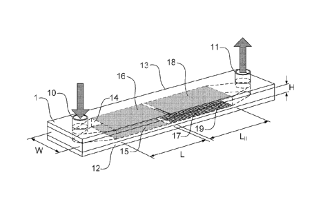
FIG. 1
shows a non-limiting example of the device, 1. It has a thin flow channel 14
which is
.. defined by the base plate 12 and top plate 13 separated by a thin spacer
with the
channel cut from it. Typically, the spacer is made of a dielectric material
which is slightly
deformable under an applied clamping pressure. The spacer thus defines the
side walls
of the channel, provides the fluid seal, and electrically insulates the top
and bottom
plates from each other. While the channel is disclosed in Figure 1 as being
formed
between two plates and laterally bound by a spacer layer, those skilled in the
art will
readily appreciate that a wide variety of channel geometries and assemblies
are
envisioned by the present embodiments. In a further non-limiting example, the
channel
may be formed as a recess within a substrate, where a top plate defines the
top channel
wall, and the recess defines both the lower channel wall and the lateral
channel walls.
The channel includes an inlet 10 through which fluids may be introduced such
as
the fluid sample to be analyzed and other liquids which may be required for
channel
washing or detection of the cellular contents. The device is also equipped
with an outlet
11 that can be in fluid communication with a collecting means such as a waste
chamber,
or, for example with an absorbent pad. Flow along the channel is provided by
means of
a pressure differential between inlet and outlet ports.
In one embodiment, the pressure differential may be generated by a pump
means such as external pump that is interfaced to the device through fluidic
fittings
known in the art, such as tubing and sealing fittings. While the sample may be
made to
flow directly from the inlet to the outlet port of the device, alternative
embodiments may
be used in which the sample is re-circulated within the channel, thereby
increasing the
likelihood that cellular analyte will be captured by adherent material in the
second zone
of the device. In yet another embodiment, the pump means may be configured to
produce an oscillatory flow of the sample in a longitudinal direction to
increase the
binding probability. In another embodiment, fluid may be introduced into the
sample
through a manual or automated pipettor configured to inject sample and/or
other
reagents or buffers into the inlet port.
The working section of the flow channel 14 is divided into two zones. The
first
zone is referred to as the "concentration zone" and has dimensions adapted to
produce
laminar flow. In a non-limiting example, dimensions H, W and LI may be
selected to be
18
CA 02769320 2012-01-27
WO 2011/014946
PCT/CA2010/001176
on the order of approximately 0.1x5x10 mm3. Two electrodes 15 and 16,
respectively at
the inner sides of the plates 12 and 13, are intended for inducing an electric
field across
the zone. The voltage is preferably applied by an external voltage source,
which is
preferably electrically connected to external contact pads on the device that
are
themselves connected to the plates 12 and 13.
The time dependent electric field exerts an effective force on cells, provided
that
they comprise a surface charge, and carries them to a thin region at the
immediate
vicinity of the anodic electrode 15. Details of the time dependent pulses are
provided
below. The second zone may also be referred to as "reaction zone" and contains
an
adherent material for capturing the concentrated analyte. In a non-limiting
example, the
second zone may have a longitudinal dimension L11 in the range of 10 mm.
As described above, the second zone contains an adherent material, which
preferably selectively binds to the cells. The adherent material is preferably
provided in a
horizontal stripe that is approximately perpendicular to the direction of
fluid flow within
the channel. In this manner, cells concentrated to the region just above the
channel
surface flows over the adherent material and the binding capability of the
device is
enhanced. Preferably, the adherent material is selective and provided in the
form of an
array 19 of stripes have been created to bind to more than one type of cells.
Those
skilled in the art will understand that a wide range of other geometries of
arrayed
immobilization regions and stripes are possible within the scope of the
present invention.
In one non-limiting example, the array may be a regular array of spots. The
arrayed
adherent material may further comprise additional molecular components to
improve the
performance of the adherent material, for example, excipients for non-specific
blocking,
shelf life stability, and hydrogel materials for improved porosity and/or
binding capacity.
In a preferred embodiment, each array element is a geometrically well defined
area over which an adherent material (e.g. capture ligands specific to a class
of analyte)
have been immobilized. As the cells, concentrated at the lower extremity of
the channel,
slowly move over the array of binding elements, they may bind with the
adherent
material and become captured onto the solid phase. In a preferred embodiment,
at least
a portion of the channel is transparent within the second zone, thereby
enabling the
direct optical probing of bound cells. For example, the presence of cells
bound to the
adherent material may be determined by many optical methods, such as, but not
limited
to, light scattering, fluorescence, chemiluminescence, imaging, and surface
plasmon
resonance.
19
CA 02769320 2012-01-27
WO 2011/014946 PCT/CA2010/001176
In a preferred embodiment, two electrodes 17 and 18 are additionally provided
at
the inner sides of the plates 12 and 13, and are intended for inducing an
electric field
across the second zone for the electroporation or electro-lysis of captured
membrane
bound or cellular analyte. The adherent material (either as a single line or
array) is
provided on the inner surface of one of the electrodes (the electrode 17).
Applying a brief
and large potential difference between the two electrodes 17 and 18
electroporates cells
and depending on the magnitude and duration of the resulting electric field
some
molecules inside the cell are released. These can be used for detecting the
cell's
presence. Preferably, one of the electrodes 17 and 18 is transparent, thereby
enabling
the direct optical detection of a signal from the interaction of the released
intercellular
material with one or more detection reagents flowed through the channel.
Concentration Module
The section of device 1 that constitutes the first zone is referred to as the
"concentration module". It is intended for separation of charged cells based
on
application of a non-Faradic electric field (i.e. no charge is transported
across the double
layer formed at the channel walls). When a sample containing charged cells
(for
example, bacteria) is injected through the inlet 10 into the device, it
develops a uniform
Poiseuille flow in longitudinal direction by the time it reaches to the
concentration zone.
There, the charged cells are subjected to a transverse electric field and is
concentrated
to one side of the channel under an electrophoretic force.
As mentioned above, in a preferred embodiment, the charged cells are
microorganisms such as bacteria or fungi. At physiological pH (5-7), most
microorganisms are negatively charged because the number of carboxyl and
phosphate
groups exceeds the number of amino groups at the cell surface. As charged
particles,
these cells experience an attractive force towards the anode 15, henceforth
termed the
"accumulation wall". As the cells approach the wall, their overall motion is
halted by
various repulsive forces, lift forces and diffusive forces associated with
Brownian motion
and are held at a small distance away from the wall. At regions close to the
exit of the
concentration zone a Guassian-type concentration profile of cells is formed in
the
proximity of the accumulation wall. The cells then slowly travel to the
reaction zone at
the velocity associated with the flow at the equilibrium distance from the
wall.
The main challenge for the successful operation of the concentration module is
establishing a transverse electric field with sufficient strength in the
central region of the
channel. It is well known that the application of a constant electric field in
a channel
CA 02769320 2012-01-27
WO 2011/014946 PCT/CA2010/001176
containing an aqueous solution results in formation of electric double layers
near the
electrodes and in some instances as much as 99% of the potential drop occurs
across
the double layers. Accordingly, the actual electric field experienced by the
charged
cellular analyte, referred to as the "effective field", is only a small
fraction of the
nominally applied field and the bulk of the liquid in the channel is shielded
from the
electrodes by polarization layers of ions and water molecules on the electrode
surfaces.
Unfortunately, clinical samples generally have high ionic strengths. For
example,
a common culture medium tryptic soy broth includes 5 g/L of sodium chloride
and 2.5 g/L
of dipotassium phosphate. These salts give rise to an ionic strength of about
100 mM. If
such a solution is introduced into a channel with at least one blocking
electrodes
connected to DC power supply, the non-Faradiac electric potential will drop by
37% at a
distance of about 1 nm from the electrode. This distance is the Debye length,
ko, related
to the ionic strength / by the following relation;
A, õ =0 .3041 .NIT (1)
where /and k0 have the units of mole/L and nm, respectively.
Application of an electric potential difference between two unblocked
electrodes
separated by an electrolytic solution can result in electrochemical reactions
at the
electrode¨electrolyte interface if the applied voltage exceeds a threshold
value. In that
case gas bubbles are generated at the electrodes due to electrolysis of water.
The gas
formation can rapidly obstruct the channel leading to electrophoretic failure.
In addition,
the pressure increase in the channel might cause mechanical damage of the
module.
The amount of lateral electric field that can be applied is therefore limited
by the
restriction that it should not result in generation of gases in amounts
exceeding the
solubility limit.
A common approach in the prior art involves suppressing the generation of
oxygen and hydrogen bubbles by adding a redox-couple to the sample flowing
along the
electrodes. As an example, quinhydrone, which is a complex between
hydroquinone
(H2Q) acting as an electron donor and p-benzoquinone (Q) acting as an electron
acceptor, can be added to the flow streams. Instead of water oxidation and
reduction
that generates oxygen and hydrogen, now H2Q is oxidized and Q is reduced
without any
bubble generation. Obviously, this method complicates sample introduction and
contradicts the goal of performing a low cost and rapid assay.
In contrast to known methods, both of the foretold issues, i.e. gas bubble
formation and the field shielding, may be alleviated by including at least one
electrical
21
CA 02769320 2012-01-27
WO 2011/014946 PCT/CA2010/001176
insulating layer to prevent a Faradic current from flowing in the channel. The
generation
of gas bubbles is avoided by insulating the anode from the sample with a thin
layer of
dielectric coating, which serves to eliminate any charge transfer processes
from
occurring across the electrode-electrolyte boundary. In another embodiment of
the
invention, the electrodes may be non-blocking, and the generation of a Faradic
current
may be suppressed by maintaining the applied voltage below the threshold
voltage.
Thus in a preferred embodiment of the invention, the device is non-Faradic and
comprises at least one blocking electrode, and the shielding of the electric
field at central
parts of the channel is partially avoided by applying the driving voltage in
two stages. In
the first step, termed as on-time, a potential difference is rapidly created
between the
two electrodes and is maintained over a time period of tõ. Over this time
period the
double layer is being developed on the electrode-electrolyte interface and
field strength
within the channel is still appreciable. In the second step, the applied
electric field is zero
or slightly negative for time toff, termed as off-time. This time is
sufficiently long to allow
the smaller ions, such as Ci, to diffuse back and rebuild their uniform
distribution. On the
other hand, toff should be sufficiently short that the average diffusive
displacement of the
cells during off-time does not exceed (preferably does not amount to more than
a few
percent of) the electrophoretic displacement they received during on-time. As
will be
shown below, the much higher diffusivity of ions relative to cells makes this
possible.
The construction and operation of an exemplary but non-limiting example of the
concentration module is now described by referring to its schematic cross-
sectional view
parallel to the flow that is illustrated in figure 2. In the preferred
embodiment, the
transparent electrode 16 is commonly prepared by chemically bonding a
conductive
metallic oxide coating to an optically transparent plate such as glass (13).
The preferred
oxide layer is a thin layer of ITO (Indium tin oxide), approximately 100 nm
thick. The
transparency of the electrode is essential for accessing the signal in the
reaction zone if
the reactions devised for detecting the cellular contents have been selected
to generate
optical signals. As it is known in the prior art, other transparent or
partially transparent
conductive layers, such as thin metallic films, can be used instead of the ITO
layer.
The electrode 15 is preferably mounted on a base plate 12. This electrode
preferably has a dielectric surface layer, 24 at the channel interface. The
dielectric layer
may be prepared by coating the plate with a thin layer of materials such as
polystyrene.
In the preferred embodiment, the conductive electrode 15 and the base plate 12
are
aluminum and the dielectric coating 24 is aluminum oxide (A1203). The surface
of
22
CA 02769320 2012-01-27
WO 2011/014946
PCT/CA2010/001176
aluminum oxide is preferably modified to create hydroxyl groups followed by
coating with
a heterobifunctional silane layer, creating functional groups to interact
covalently with the
capture ligands. In applications requiring long exposure to C1 the oxide layer
may not
provide enough corrosion protection. In this case the observation by B. F.
Shew et al ( J.
Electrochem. Soc.138: 3288 (1991)) can be utilized in preparation of the
electrode. The
addition of quite small quantities (5 mol % and less) of transition metals
(e.g., Ta. Mo,
and W) to Al can reduce the rate of corrosion of Al by up to about 100 times,
and the
time to breakdown under constant electric field across the protective oxide
layer may be
increased by about 10 times.
Using an external voltage source, 25. a potential difference is applied
between
the two electrodes, 15 and 16, with the bottom electrode having a positive
potential with
respect to the top electrode. The output of the voltage source 25 is
preferably a high
frequency train of pulses and the pulses are preferably substantially square.
The
frequency, the amplitude and the pulse shape of the applied electric waveform
may be
predetermined based on known properties of the sample liquid, or may be
selected
according to the feedback based on the current monitored by the meter 26.
Those
skilled in the art will appreciate that the waveform may be varied in order to
optimize the
performance of the device.
As schematically illustrated in the figure, the inflow 22 has a substantially
uniform
distribution of the suspended cells. As a result of the concentrating action
of the module
in the outflow 23 the cells are localized close to the anode surface. The
liquid convection
slowly carries them to the reaction zone.
The basic structure of the concentration module is analogous to the structure
of a
polarized electrolytic capacitor. In such capacitors the aluminum oxide
(A1203) dielectric
layer is formed by electrochemically oxidizing the aluminum. In order to
increase the
effective surface by as much as 100 times, and so increase the capacitance per
unit
nominal area, the electrode is etched with a dense network of microscopic
tunnels. The
thickness of the dielectric layer is determined by the applied voltage during
the
electrochemical forming (anodizing) process and is often chosen to be 2 nm per
each
volt that can be safely applied on the electrode. Since the required voltage
at the
concentration module does not exceed a couple of volts in many applications,
naturally
occurring A1203 layer (thickness about 5 nm) may be sufficient.
Circuit Model of Electrical Concentration Module
The concentration module can be modeled by the equivalent electrical circuit
23
CA 02769320 2012-01-27
WO 2011/014946 PCT/CA2010/001176
presented in figure 3a. The capacitance CDLi and CDL2 correspond to the
dynamic
double-layer capacitances at the interfaces of dielectric layer 24 and
electrode 16
respectively with the liquid in the channel. RDLi and RDL2are the parallel
resistances
corresponding to leakage current in the two capacitors. In general, values of
CDL for flat
metal surfaces fall in the range 5-50 F/cm2 depending on the type of metal,
ionic
strength and composition of the solution, surface roughness, temperature and
voltage.
Capacitance CDE is the capacitance of the dielectric layer whose value depends
on the layer thickness and the effective area of the electrode. For example,
roughness of
the surface can increase capacitance by a factor as high as 1000. Resistance
ROE is the
.. equivalent parallel resistance of the dielectric layer and accounts for
leakage current in
the capacitor. It decreases with increasing capacitance, temperature and
voltage.
Typical values for ROE are on the order of 100/00E WI with CDE in pF.
Rai represents the bulk solution resistance and CcH the bulk capacitance. The
value of CcH is so small that it can be approximated with open circuit. For a
channel with
.. a width of 100 m, the resistance RcH is about 100 Q./cm2 for an ionic
strength of 1 MM.
RLOAD is the sum of the power supply output resistance and the input
resistance
of the electrodes. All the electrical parameter values, with the exception of
RLoAD ,ROE
and CUE are dependent on the ionic strength of the carrier solution. The load
resistance
modifies the voltage division among the circuit components and becomes
particularly
important at higher ionic strengths.
Considering the typical values of the electrical parameters, the equivalent
circuit
can be simplified as presented in figure 3b. The resistances ROE, RDLi and
RDL2 are
sufficiently large that they can be approximated as open and the two double
layer
capacitances have been combined in series as CDL. The double layer charging
time,
according to this circuit model, is given by
¨ (RmAD )(C DEC DL I (C DE C DL)) (2)
Thus, the period ton over which the potential difference is maintained between
the
electrodes should be chosen to be in the order of tc. Bazant et al (Physical
Review E
70, 021506 (2004)) have suggested that the primary time scale for charge
relaxation is
given by
¨ 2201 (3)
were Doi is the diffusivity coefficient of the ions and XD is given by
relation (1).
Preferably, tott, the period over which the potential difference between the
electrodes is
24
CA 02769320 2012-01-27
WO 2011/014946 PCT/CA2010/001176
brought to zero, is chosen to be longer than TID
As it can be easily remarked both characteristic times of the concentration
module (T, and Tip of equations 2 and 3) depend on the ionic strength of the
aqueous
solution. This implies that optimum values of tõ and toff will vary for
samples with
different ionic strengths. While these value can be chosen empirically for a
given sample
type, or predicted if the sample ionic strength is known or can be measured, a
preferred
embodiment, employs a feedback loop, shown in Figure 2 at 27, comprising a
current
meter 26 and the controller unit 28.
In one embodiment, depending on the current measurement at some points in
time the lumped circuit parameters of the module can be estimated and optimum
values
of tõ and to determined and applied. This control scheme is based on the fact
that the
current flow is an indicator of the effective electric field experienced by
cells in the
channel. According to M. Marescaux et al. (PHYSICAL REVIEW E 79. 011502
(2009)),
there are two contributions to the current flow. Double layer charging is
initially the
dominant phenomenon, resulting in an exponentially decreasing transient
current. At the
second stage, termed as "delayed buildup", near the double layer, the
concentration of
positive and negative charges becomes lower than in the bulk. As a result,
positive and
negative charges diffuse toward the electrodes. The readjustment of the double
layer
leads to a measurable current. This transient current is negligible during the
initial double
layer charging, but it becomes dominant at longer times because it decreases
more
slowly than an exponential decay. The applied potential difference across the
two
electrodes should be turned off before the onset of the "delayed buildup" as
by then the
electric field will already be shielded from the channel center.
In another embodiment, the feedback means may comprise the measurement of
a circuit parameter, such as the current, and the time tõ may be determined to
be the
time interval following the initial application of the electric field and the
time at which the
measured current falls below a pre-determined threshold. In one embodiment,
the
threshold may be a pre-selected fraction of the current measured when the
electric field
is initially applied.
In a preferred embodiment. the threshold is determined by applying an initial
series of pulses to the electrodes and measuring the resulting current, and
fitting the
measured current to a known function. For example, the measured current may be
fitted
to an exponentially decaying function, and the threshold may be approximately
equal to
the current measured at a time approximately equal to a fitted time constant.
CA 02769320 2012-01-27
WO 2011/014946 PCT/CA2010/001176
Without intending to be limited by theory, the effectiveness of the
concentration
module is believed to be dependent on the fact that while electrophoretic
mobilities of
non-motile cells and smaller ions are numerically of the similar order of
magnitude, their
diffusivity coefficients vastly differ. In order to illustrate this principle,
a generic example
is provided.
We consider an electrolytic sample containing a suspension of non-motile
bacteria having spherical shapes with a radius of 1 Rm that flows into a
concentration
module. The channel height, H, is taken to be 100 Rm. The diffusivity
coefficient and
electrophoretic mobility of the bacteria is estimated to be DõIi=2.2x10⁻⁹
cm2/s and
licem=2.0x10⁻⁴ (cm/s)/(V/cm), respectively. Square pulses with ten=0.5 ms and
t0ff=2 ms
are applied to the electrodes. The amplitude of the pulses are adjusted such
that the
effective field during the "on" time is Eeff=100 V/cm. The average lateral
displacement of
the bacteria during on-time is A ¨Yeell=[icellEeffton =0.1 Rm. During the off-
time the cell
randomly diffuse over an average length of 8,, = =2.1x10⁻² Rm. The ratio
8ceii/Ayeeir is calculated to be 21%. Its smallness indicates that the
diffusion does not
severely disturb the trajectory of the bacteria that will reach the collecting
wall after
cell H/(2Av )=500 cycles, if it had started from the channel center. On the
other hand, for a
,
Cl- ion with diffusivity coefficient and mobility of D0=1 .86x10 cm2/s
andilioe=8.0x1 04
(CM/S)/(V/cm) the corresponding displacements are Ayi0n=0.4 Rm and 5=l .93 Rm.
Then, A /AV
ion =480%, indicating that when the external field is switched off, the ions
relax to a uniform density distribution, driven by diffusion.
In selected cases, the motility of bacteria can affect the performance of the
concentration module. In the absence of a force field and in a large container
motile cells
move by propelling themselves by means of long hairlike flagella with a
swimming
pattern that resembles a three-dimensional random walk. The usual Fickian
diffusion can
be used to describe their random motility as is done, for example, by P.
Lewus, R. M.
Ford (Biotechnology and Biosensing 75 292 (2001)) who showed that the motion
of E.
coli AW405 is similar to a particle with an diffusion rate of 3x 10⁻⁶ cm2/s.
This value is
close to the diffusivity coefficient of small ions. However, there are two
reasons that
suggest that, in the presence of cell motility, the concentration module
should remain
effective. In the presence of electric field bacteria cells align themselves
along the
electric field and will migrate toward one electrode depending on the nature
of the cell
surface, known as galvanotaxis. As a result of galvanotaxis the motion of the
cells is
26
CA 02769320 2012-01-27
WO 2011/014946 PCT/CA2010/001176
thus restricted to the lateral direction. Also, when a bacteria cell collides
with the channel
surface it tends to swim parallel to the surface and therefore will accumulate
near the
surface as described by G. Li and J. X. Tang PRL 103, 078101 (2009). The
pulsing
nature of the applied electric field will increases the number of collisions
to enhance this
effect.
The concentration module can operate over a wide range of ionic strengths.
However, high ionic strength lowers the performance of the module for three
reasons: 1)
the electrophoretic mobilities of the cells appreciably reduce as the ionic
strength
increases, which requires application of higher voltages for efficient
concentration; 2)
.. high ionic strengths are associated with shorter charging times, thus
requiring shorter ton
as a result of which the duty cycle, defined as ton/(ton + to). is reduced;
and 3) The
channel resistance, RcH, is inversely proportional to the ionic strength, and
the lower this
resistance becomes the more heat is generated in the electrodes and the
channel, which
may have deteriorating effects on the cells. Therefore, reducing the ionic
strength
generally results in the improved performance of the concentration module. The
task of
ion reduction in the sample can be performed by the sample pre-treatment
module that
may be integrated in the sample inlet 10 of the device (see Figure 1).
Sample Pre-Treatment
Figure 4 shows an example of a sample pre-treatment filter according to a
preferred embodiment of the invention. The sample pre-treatment module, 4,
consists of
inlet 40, outlet 41. pre-filter, 42, packed ion exchange resins, 43, and a
filter, 44. The
pre-filter 42 excludes large particles such as cationic exchange resins and
non-ionic
adsorbing resins used in some samples such as the culture media of the Becton
Dickinson system. The ion exchange resins (43) comprising mixed cationic and
anionic
resins serve to de-ionize the sample and to capture smaller ionic particles
(for example,
activated charcoal and fuller's earth powder, as employed in the culture media
of
bioMerieux). The filter 44 retains the ionic resins and bound ions and ionic
particles to
prevent them from entering the concentration module.
The pre-filter and the filter can be made of a non-woven polyalkylene porous
material such as polypropylene, polyethylene or polytetrafluoroethylene porous
frits with
an appropriate pore size of about 35-125 pm suitable for retaining the resins
and large
particles. More preferably, the porous material is a chemical and thermal
resistant
material such as high density polyethylene. A pre-filter and a filter may be
present at the
respective ends of a tube such as heat shrinkable low density polyethylene
tubing and
27
CA 2769320 2017-03-06
ion exchange resins will be packed in between. Preferably, a pre-filter, a
filter and a
tubing material will be a hydrophilic type or coated with a hydrophilic
polymer.
Hydrophilic high density polyethylene porous sheets to make pre-filters and
filters,
and low density polyethylene tubing materials are widely available from
commercial
sources.
To de-ionize the ions and ionic particles, mixed H+ form cation exchange resin
and OH- form anion resin may be used. Na + in the medium binds to the cation
resin
in exchange of H+ and Cl- binds to the anion resin in exchange of OH-. Removed
H+
and OH form H20 molecules. This method is widely applied in water
deionization.
Preferably, microporous gel resins with the pore size larger than the size of
bacteria
or other cellular analyte of interest are be used. In addition, as negatively
charged
bacteria can still bind to the surface of the anionic resin and
nonspecifically bind to
the surface of the resins, both types of resins will be treated with a non-
ionic
surfactant such as TritonX-100Tm. Examples of mixed resins are Amberlite MB-
150
from Rohm & Hass and Dowex-Marathon MR-3 from Dow Chemicals with particle
sizes ranging from 500-700 pm.
Reaction Zone
A cross section of the second zone is schematically presented in figure 5.
This section of the device is known as the reaction module. It can be
understood to
be an extension of the concentration module and during the sample
concentration
stage the optional additional electrode pair 18 and 17, like the two
electrodes 16 and
15 of the concentration module, may be driven by the power supply 25 of figure
2 to
further assist in the sample concentration. The inflow 57 is passed along from
the
concentration zone by fluid convection.
The cells, which have been localized at the lower extremity of the flow, do
not
diffuse into the central region of the channel by the simultaneous action of
the
concentration mechanism in both concentration and reaction modules. This
ensures
more efficient cell-capture by the adherent material as the cells spend long
times in
the vicinity of the surface. In the zoomed section of the figure, a non-
limiting
embodiment is shown in which the adherent material comprises capture ligands
which in this case have high affinity to the cells (e.g. high affinity to a
selected class
of cells represented by the black circles). In this embodiment, cells
belonging to other
classes will pass over to their respective immobilization regions without
being
retained. The capture ligands may be antibodies and are preferably immobilized
by
covalently binding to a layer of spacer molecules 55 at the immobilization
regions.
28
CA 02769320 2012-01-27
WO 2011/014946 PCT/CA2010/001176
While the aforementioned embodiments disclose a device comprising both a
concentration zone and a reaction zone, it is to be understood that devices
according to
different embodiments may comprises either one or both of the concentration
and
reaction zones. For example, in one embodiment where the sample contains a
relatively
high concentration of cells, in which case a concentration step may not be
necessary to
bind a sufficient number of cells at the reaction zone, a device may comprise
a reaction
zone without a concentration zone.
The dielectric coating in the reaction zone 54 is preferably substantially
thicker
than its counterpart 24 in the concentration module. The large thickness
ensures that the
dielectric layer will be able to withstand the high strength of the electric
field used during
electroporation, as discussed below. In the preferred embodiment the layer is
A1203 and
the thickness is 2 nm per each volt to be applied on the electrode 17.
Once the entire sample has passed the channel and the cells are retained, an
optional washing process can be performed by injecting a washing liquid into
the
channel. The flow carries away analyte that has been adhered on the channel
surface.
In the specific case of cells, they generally have little affinity to the non-
adherent surface
they may be displaced by shear force of the washing fluid. When more stringent
washing
is required, the washing action can be assisted by applying a weak repulsive
electric
force to the cells. This is done by reversing the polarity of the power supply
25 and
applying a sequence of pulses to the electrodes.
Electro-Lysis and Detection
In a preferred embodiment. the reaction zone is employed for the
electroporation
or electrolysis of bound cell. The first step of the reaction stage is filling
the channel with
an electroporation liquid or buffer. The composition of this liquid depends on
the nature
of the intended reaction. For example, if the intention is to detect the
presence of the
cells via their ATP content, as further described below, the appropriate
liquid should
contain reagents necessary for initiating and driving the oxidation of
luciferin under
catalysis by luciferase followed by emission of light. A typical buffer may
have the
following composition: luciferase, D-luciferin, Tricine buffer pH 7.8,
Magnesium sulfate,
EDTA, DTT, BSA.
The release of desired cellular contents is accomplished by applying a strong
electric field to the captured cells to make the cell membrane permeable to an
outside
medium. This method is known as electroporation. The electroporation of cell
membrane can be reversible or irreversible, depending on the electric field
strength.
29
CA 02769320 2012-01-27
WO 2011/014946 PCT/CA2010/001176
Preferably, the electric field is made sufficiently high to cause irreversible
breakdown of the cell wall. The irreversible breakdown of the membrane causes
cell
membranes to burst open, and then the osmotic pressure of the cytosol and the
external
medium become unbalanced and the cells are disrupted as a result of the
overswelling.
.. The irreversible electroporation is commonly known as cell lysis and is
desirable in
cases that the device is intended for the release of cellular molecules, such
as ATP,
nucleic acids and proteins.
Generally speaking, the internal field for electroporation or electro-lysis
depends
on many factors, including the size of and cell wall structure of the cell,
the applied
voltage, and the separation between the electrodes used to apply the field.
The electric
field strength required to achieve a trans-membrane potential of more than 1 V
is about
1 kV/cm. Preferably, the applied voltage is selected to provide an internal
electric field of
at least 1 kV/cm, although this threshold is known to vary for different cell
types and
species. Depending on the applied field, electroporation can be permanent, or
reversible.
The voltage may be applied in as DC or AC voltage, and may be continuous or
pulsed. In a preferred embodiment, an AC voltage is applied to limit the
formation of
bubbles due to electrolysis. Preferably, the voltage is AC, has a frequency
between 1
and about 10 MHz. In another preferred embodiment, the voltage is applied in
one or
more pulses, with each pulse lasting for at approximately 10 microseconds to
10
milliseconds. Those skilled in the art will readily appreciate that different
combinations of
voltage, frequency, pulse duration will be appropriate for different
materials, geometries,
and cell types.
In the previous art, two basic configurations have been suggested for
performing
.. electroporation or electro-lysis; axial-ohmic and transverse-ohmic. The
axial-ohmic
configuration has been utilized in the microfluidic devices (e.g. US6287831,
and Wang et
al., Biosensors and Bioelectronics 22:582-588, 2006). The required field is
generated by
the voltage drop as an electric current passes through a high resistance
aqueous
medium containing a suspension of cells. An electrical field is then
established along the
length of the device by inserting two wires into the sample inlet and outlet.
The transverse-ohmic configuration is utilized in commercial electroporation
vessels (e.g. US6074605). In the basic form, the device includes a hollow
housing
substantially rectangular in shape. Two electrodes are inserted into the
interior of the
housing directly opposite one another, flush against the housing walls. The
CA 02769320 2012-01-27
WO 2011/014946 PCT/CA2010/001176
electroporation is performed by applying a voltage difference between the two
electrodes. These configurations can be adopted in embodiments disclosed
herein.
Alternatively, for the embodiments for which the primary cell receptors are
immobilized
on a dielectric surface, the required field is generated by charging the
capacitor formed
by the two conductor electrodes, one attaching behind the dielectric surface
and the
other opposite to this surface. In this case the transit field is able to
electro-lysing the
cells.
While electrical lysis of cells in known, (e.g., Bioelectrochemistry, 2004,
64, 113-
124. Lab Chip, 2005, 5, 23-29. Anal. Bioanal. Chem., 2006, 385, 474-485.
US7418575), these methods teach that the cells should be suspended in a liquid
medium and that a large electric field is required to be above the threshold
strength in
the entire volume of the medium. Devices based on such an approach have
encountered challenges in achieving lysis due to the presence of the field
shielding by
the double layer formation.
In contrast, in the embodiments disclosed herein, the reaction module, the
cells
are surface bound. During the double layer charging process, while the field
strength
rapidly diminishes in the inner channel regions, it increases at the electrode
boundary
(Phys. Rev. E70, 021506 (2004)). Therefore, relatively low potential
differences are
sufficient to provide high electric field strengths in the vicinity of the
cells.
In order to illustrate the advantages of electroporation at the electrode
surface, a
non-limiting example is henceforth provided. Considering the case of a channel
with a
height (the dimension H in figure 1) of 100 gm, the intention is to lyse
bacterial cells by
irreversible electroporation. It has been reported that the required
electrical fields are
about 10 kV/cm. If the cells are suspended in the liquid, the power supply
must deliver a
potential difference of 100 V.
However, for the surface bound cells the voltage requirement can be
substantially lowered. Without intending to be limited by theory, this finding
may be
interpreted within the context of a qualitative model of charge transport in
an electric field
developed by Beunis et al (Physical Review E, 78, 011502 (20008)). Immediately
after
the application of a voltage VA over the blocking electrodes at the reference
time (t=O) a
positive surface charge builds up near the negative electrode and a negative
surface
charge builds up near the positive electrode. Adjacent to the electrodes,
space charge
regions with thicknesses A(t) occur where charges of one polarity are
completely
absent. For a sufficiently large value of the applied voltage drift is the
dominant charge
31
CA 02769320 2012-01-27
WO 2011/014946 PCT/CA2010/001176
transport mechanism and diffusion can be neglected. Therefore. the speed at
which the
space charge regions grow is equal to the speed of charges in the bulk:
d2. (t)
¨1-1E1,,,a (4),
dt
where, p is the mobility of ions (assumed to be the same for the positive and
negative
ions) and Ebuik is the electric field in the bulk. Beunis et al. show that
within reasonable
approximations, the field in the space charge region, Esc, can be calculated
using the
following equation:
sc bidk ________ IP ,
E E: +4171111 = -1+ As- ¨ r,i A
1
Eeo \ '( , ''' (5),
1 2
where, q is the ionic charge, n is the average ionic density in the medium, Eo
is the
dielectric permittivity of vacuum and E is the relative dielectric constant of
the medium,
and x is measured from the center of the channel.
To estimate the internal electric field based on the above analysis, a
microchannel having a height of 100 pm was filled with 170 pM NaCI solution,
and a
step voltage of amplitude VA=1 V was applied to the channel electrodes. The
measured
current as a function of time (tin seconds) could be approximated by
1 VIII
1(t)= , 0 , . Accordingly, it was inferred that E1õ.õ(t)= ,
.1
1+V16.5x10-4 )--
1+V16.5x10-4 y 2 '
Substituting this result in equation (4) with p=7.15x10⁻⁸ m2/V-s it was found
that about 8
ms after the application of the external field, the width of the space charge
region will be
comparable to the typical size of a bacterial cell, i.e. As0(t=8 ms)=1 pm. At
this time the
field strength in the center of the space charge region (0.5 pm from the
electrode)
reaches a magnitude of 1.5 kV/cm, which is 15 times higher than what was
expected if
the screening effect were not present.
The above analysis demonstrates that by selecting a dielectric layer having
thickness and a dielectric constant such that the electric field drop occurs
substantially
within the space charge layer of the channel, an amplified electric field is
obtained within
the channel proximal to the dielectric layer. As noted above, a preferred
thickness for the
dielectric layer is in the range of about 10 to 100 nm, and a preferred
dielectric constant
of the dielectric layer is in the range of approximately 3 to 10, and more
preferably above
10. Thus a separate high voltage power supply is not needed for the
electroporation and
32
CA 02769320 2012-01-27
WO 2011/014946 PCT/CA2010/001176
a single low-cost power supply can drive the concentration, washing and
electroporation
processes. Moreover, due to the lower value of the required applied voltage,
the
thickness of the dielectric layer (preferablyt A1203) layer, necessary for
electrode
blocking, can be kept low and the reduction in the electrode charging time,
and its
associated problems, be avoided.
As described above, in one embodiment the adherent material comprises a cell-
specific receptor, where cells bind specifically to the solid support directly
via the specific
binding of a cell surface with the receptor immobilized within an
immobilization region. In
a preferred embodiment, the adherent material that is provided for the binding
of the cell
to the solid support further includes immobilized secondary receptors that are
specific to
intracellular analyte released from the cells following the application of the
electric field.
The secondary receptors enable the capture of locally released intracellular
analyte from
a bound cell immediately following electroporation or electro-lysis.
Preferably, the
adherent material is provided in a spatial array, and the secondary receptors
are
provided within the array. The adherent material may be provided within an
array of
immobilization regions comprising primary receptors, with each immobilization
region in
the array comprising a receptor specific to a given type of cell or cells, and
where each
immobilization region further comprises a secondary receptor for detecting
intracellular
analyte post-lysis or post-electroporation. In another embodiment, the
secondary
receptors are provided within an immobilization region of the solid support
adjacent to a
given zone of adherent material.
The capture of cells by the adherent material and subsequent electroporation
or
electrolysis of cells on the solid support effectively concentrates the
intracellular analyte
near the secondary receptors, without the express need for thorough and
efficient
mixing. The subsequent addition of a detector reagent enables the detection
and/or
quantification of the presence of the intracellular analyte based on the
spatial location of
the signal in the array.
It is to be understood, however, that the present embodiment involving the
capture of cells via the adherent material in the immobilization zone, the
lysis of captured
cells to release their intracellular contents, and the subsequent detection of
the
intracellular contents via the binding of the intracellular contents to
secondary receptors
provided in the immobilization region, is not limited to embodiments involving
the electro-
lysis or electroporation of bound cells. As such, the adherent material and
secondary
receptors need not be bound on an electrode and dielectric layer, but may be
bound on
33
CA 02769320 2012-01-27
WO 2011/014946 PCT/CA2010/001176
any suitable solid support, as further described below. In one preferred
embodiment, the
adherent material and secondary receptors are provided within an
immobilization region
defined on a microplate well surface. To release the intracellular contents,
any suitable
lysis method may be employed, including, but not limited to, chemical lysis,
mechanical
lysis, and ultrasonic lysis.
It is to be further understood that while the preceding embodiments have been
described within the context of the binding of cells to the solid support via
the adherent
material in order to disrupt the cell membrane (e.g. by electro-lysis or
electroporation),
the capture and/or concentration of the cells in the vicinity of the
dielectric layer for
subsequent electroporation or electro-lysis may be achieved by electric field
mediated
concentration alone, without the need for the adherent material within an
immobilization
zone. In such an embodiment, the preceding method of applying unipolar voltage
pulses
may be performed to concentrate the cells in the region proximal to the
dielectric layer,
preferably within or adjacent to the aforementioned space charge region. Once
cells
have been accumulated in this region under application of the electric field
of the
unipolar voltage pulses. a one or more voltage pulses with an amplitude
sufficient for the
disruption of the cellular membrane may be applied. Accordingly, cells in a
cell-
containing liquid sample may be concentrated proximal to the dielectric
surface and
electroporated and/or electro-lysed. In a preferred embodiment, the dielectric
layer may
comprise an immobilization region having thereon only secondary receptors for
binding
intracellular analyte released from the cells.
This unique aspect of embodiments disclosed herein enables highly sensitive
detection of a wide range of intracellular analytes including proteins and
nucleic acids.
Furthermore, the local concentration of intracellular analyte in the spatial
vicinity of the
secondary receptors supports the detection of analyte with very low copy
number,
without resorting to complex mixing and concentration schemes. As will be
shown in
subsequent examples, these embodiments may be adapted to a wide range of assay
platforms, and is particularly suited to automated analyzer systems that
employ
microfluidic cartridges.
In a preferred embodiment. the secondary receptors are immobilized nucleic
acid
probes that specifically recognize and hybridize with nucleic acids released
following the
application of the electric field. This embodiment therefore provides a hybrid
two-stage
solid phase binding assay, with a first stage involving the capture of cells
onto an array
of immobilization regions on a solid support via the primary receptors, and
the second
34
CA 02769320 2012-01-27
WO 2011/014946 PCT/CA2010/001176
stage involving the capture of released nucleic acids by secondary receptors
immobilized on the same solid support. Following the application of the
electric field and
the release of intracellular analyte, the reaction vessel is incubated for a
period of time
sufficient to enable the binding of released nucleic acids to the immobilized
nucleic acid
probes. The assay may then proceed according to methods known in the art, in
which a
sandwich assay is performed by adding labeled detector probes that are
specific to the
nucleic acids comprising the released intracellular analyte.
In another preferred embodiment, the secondary receptors are antibodies that
specifically recognize intracellular analyte following the application of the
electric field.
As in the preceding embodiment involving the use of nucleic acid probes as
secondary
receptors, this embodiment also provides a hybrid two-stage solid phase
binding assay,
with a first stage involving the capture of cells onto an array of spots on a
solid support
via the adherent material (e.g. primary receptors), and the second stage
involving the
capture of released intracellular analyte by antibodies immobilized on the
same solid
.. support as the adherent material. Following the application of the electric
field and the
release of intracellular analyte, the reaction vessel is incubated for a
period of time
sufficient to enable the binding of released intracellular analyte to the
immobilized
antibodies. The assay may then proceed according to methods known in the art,
in
which a sandwich assay is performed by adding labeled antibodies that are
specific to
the intracellular analyte.
In a preferred embodiment, the signal from each spot in the preceding
embodiments (involving the detection of intracellular analyte via immobilized
secondary
receptors) is an optical signal that may include, but is not limited to,
signals produced by
chromogenic, fluorometric, luminescent, chemiluminescent, electro-luminescent,
or time-
resolved fluorometric labels. The optical signal may be produced or
facilitated by a single
label, such as a fluorophore, or may be produced or facilitated by two or more
labeled
moieties, or may require the addition of further reagents such as signal-
producing
substrates.
Exemplary methods for preparing a solid support with adherent material
and secondary receptors are henceforth described. Preferably, the adherent
material
comprises an antibody having an affinity for the cell surface, and the
secondary
receptors comprise nucleic acid probes (or synthetic analogs thereof) for
binding
intracellular analyte comprising nucleic acids (such as rRNA). The adherent
material and
the secondary receptors with appropriate functional groups can be immobilized
on any
CA 02769320 2012-01-27
WO 2011/014946 PCT/CA2010/001176
surface known in the prior art following any known surface preparation methods
to
introduce appropriate reactive functional groups on the solid support.
Examples of
surface preparations can be deposition of small molecules such as
organosilanes and
thiol linkers by covalent interaction or macromolecules such as poly-L-Lysine
and PEI by
physical adsorption.
In an exemplary, yet non-limiting embodiment, a hetrobifunctional silane layer
with functional groups, X-Si-X', can be deposited on any surface (Y) on which
a silane
layer can be applied to form Y-0-Si-X'. X' may be trimethoxy (-00 H3)3,
triethoxy (-
002H5) 3 or trichloro (013) and form Y-0-Si-X' chemistry upon hydrolysis. One
example of
such surface is hydroxylated surface of aluminum support, with a naturally or
artificially
processed oxide layer, and a hetrobifunctional silane layer is generated by A1-
0-Si-X'
formation. X may vary and covalently interacts with the respective functional
group of
capture ligand or cross-linker molecule to be attached to the silane layer via
any
appropriate chemistry. For example, X can be glycidyl functional group of
glycidyloxipropyl-trimethoxysilane (GOPTS) or amino functional group of 3-
aminopropyltriethoxysilane (APTS). Glycidyl functional group of GOPTS will
interact
readily with amino functional group of the molecule to be attached. However,
an
additional activation of amino functional group of APTS with any crosslinking
chemistry,
for example, 1-ethyl-3-(3-dimethylaminopropyl) carbodiimide) (EDC) and N-
hydroxysuccinimide (NHS), will be required for covalent interaction with
carboxyl
functional group of the molecule to be attached. Optionally, amino functional
group of
APTS can be pre-activated with any known chemistry, for example, with
glutaraldehyde
homobifunctional crosslinker, to interact readily with amino functional group
of the
capture ligand to be immobilized. Alternatively, protein molecules with high
affinity and
specificity such as avidin or streptavidin can be immobilized on the
functionalized
aluminum surface via any suitable chemistry and a biotinylated capture ligand
can be
readily immobilized on the surface by biotin-avidin affinity interaction.
Once the surface preparation is completed, the adherent materials and the
secondary receptors, which have been suspended in an appropriate buffer, can
be
dispensed on the desired region of the surface by liquid dispensing methods
known in
prior art. In the preferred embodiment, the adherent materials and the
secondary
receptors are suspended in a common buffer and therefore are dispensed
together. In
another embodiment, the secondary receptor is already bound on the adherent
material
by covalent or bio-affinity bonding. The resulting adherent material is
suspended in an
36
CA 02769320 2012-01-27
WO 2011/014946 PCT/CA2010/001176
appropriate buffer for dispensing. These embodiments provide better
consistency in
manufacturing. In the third embodiment the adherent materials and the
secondary
receptors are suspended in separate buffers and are dispensed sequentially.
After
dispensing, using either approach, the surface is kept in an appropriate
environment,
which has been optimized in terms of temperature and ambient atmosphere, to
allow for
the formation of covalent bonding and evaporation of excess buffer. Then, the
surface is
washed to remove excess materials. Finally, the unbound reactive sites are
blocked by
methods known in the prior art.
Preferably, prior to lysis, a lysis buffer is flowed into the microfluidic
channel to
support efficient lysis of the adhered cells. After lysis, the released
nucleic acids may be
concentrated to the surface and retained at the surface by the application of
voltage to
the electrodes 17 and 18. Preferably, the voltage is applied in a uni-polar AC
form, under
pulsed operation, as described above in the concentrating section, for
concentrating the
cells at the anode side of the cell. The lysis buffer may then be removed, and
a
hybridization buffer can be flowed into the chamber while retaining the
released nucleic
acids in close proximity to the immobilized probes (e.g. by maintaining the
pulsed
voltage). After providing the hybridization buffer and releasing the applied
electric field,
hybridization occurs. Preferably, the hybridization buffer further comprises
labeled
detector probes. Alternatively, an additional step may be provided in which
the
hybridization buffer is removed and a detector probe solution is flowed into
the chamber
to facilitate detection of the bound intracellular nucleic acids.
For example, a detector probe may constitute an oligonucleotide labeled with a
chemiluminescent enzyme such as horseradish peroxidase. In this exemplary
embodiment, the unbound labeled oligonucleotide is removed in an additional
wash
step, and the assay signal is optically detected following the detection of a
chemiluminescent substrate.
A wide range of alternative assays may be employed for the detection of the
bound nucleic acids, such as use of PNA labeled probes and molecular beacon
assays.
During target hybridization, high cationic concentration in the buffer
neutralizes the
negative charge on the single stranded DNA probe and accelerates hybridization
kinetics. Therefore, in the assay steps previously described, a low-ionic
lysis buffer is
replaced with a high-ionic hybridization buffer. The neutral charged backbone
of PNA
allows a low ionic strength buffer for target hybridization, which is
advantageous for lysis
immediately followed by hybridization in the same low ionic buffer. In another
approach
37
CA 02769320 2012-01-27
WO 2011/014946 PCT/CA2010/001176
using a molecular beacon assay, the fluorescent signal of molecular beacon is
detected
only upon target hybridization and therefore separation of hybridized probes
and
unbound probes is not necessary, eliminating a wash step following
hybridization.
Molecular beacons can be designed as either DNA or PNA backbone as know in the
art,
allowing the flexibility of ionic concentration selection for the lysis
buffer. Instead of using
as detector probes, molecular beacon probes can be immobilized to the solid
support as
the secondary receptors to capture released intracellular nucleic acids
following lysis,
thereby providing a hybrid two-stage solid phase binding assay without
requirements of
an additional detector reagent and wash steps.
In another preferred embodiment, each spot in the array on the solid support
further comprises an electrode, with each electrode electrically connected to
an
externally addressable contact pad. Preferably, a reference electrode is
additionally
provided in fluidic contact with the array electrodes. Accordingly, an
electrochemical
label and substrate may be employed for use in such an embodiment for the
spatially
resolved measurement of an electrically assay signal.
Devices according to various embodiments as disclosed herein may be in the
format of a kit enabling users to customize and/or select the adherent
material and/or
secondary receptors appropriate to target cells and intracellular analytes of
interest. The
kit preferably comprises the elements required for at least one of
concentration, lysis and
detection as described above and furthermore provide a means for the user to
provide
user-selected adherent material (for example, primary antibodies targeting a
cell
surface) and/or secondary receptors (e.g. antibodies or probes for binding
intracellular
analyte of interest) to the solid support.
In a preferred embodiment kit comprises a substrate containing an open
microfluidic channel, exposing the solid support, a separate top plate, and
optionally a
sealing adhesive or clamping structure for contacting the top plate with the
substrate and
enclosing the microfluidic channel for use with a liquid sample. The user may
apply
capture ligands to the prepared solid support by manual or automated
dispensing (e.g.
spotting) methods.
When the capture ligands have been applied and all excess material removed
the top plate will be applied to the channel and fixed there by means of, for
example,
pressure sensitive adhesive pre-applied to either the top or bottom plate. The
device, so
prepared, may then be employed to carry out the aforementioned method steps
according to various embodiments (e.g. concentration, lysis and detection) as
described
38
CA 02769320 2012-01-27
WO 2011/014946 PCT/CA2010/001176
elsewhere in this disclosure.
The open channel may comprise a surface prepared to bind capture ligands of
interest over the entirety of the surface providing the user with the
flexibility to select any
appropriate spatial configuration for the array. Tools and instruments are
available
commercially to aid in the application of arrays to solid supports. A
preferred
embodiment provides a pre-placed mask on the solid support which isolates
discrete
binding regions forming the array. The mask allows droplets of distinct
capture ligands
to be placed on each binding region while preventing these droplets from
spreading to
neighboring regions.
As an example, the mask may comprise a thin plastic film with holes defining
potential immobilization regions, where the plastic film is preferably fixed
to the solid
support by an adhesive such as a removable adhesive backing. The droplets of
desired
capture ligands would be applied to the binding regions by, for example, a pin
applicator
or a pipette. Following completion of the application of capture ligands
(which may
comprise either or both of the cell surface adherent material and secondary
receptors for
binding intracellular analyte) and removal of excess material, the mask may
optionally be
be removed from the solid support and the microfluidic channel may be sealed
by
providing the top plate as noted above. A more elaborate mask could be
envisaged
which would provide for enhanced control of droplets during application of the
capture
ligands for ease of use by the user.
The solid support of the foretold open platform kit can be prepared by
procedures
presented in the following two non-limiting examples. In the first example,
the polished
aluminum support plates are cleaned with water then rinsed twice with methanol
and air-
dried. 2% 3-Aminopropyl Triethoxysilane is prepared in 95% Methanol 5% water
and the
plates are immersed in silane for 5 min. Then, the plates are rinsed in
methanol twice,
air-dried and baked at 110 C for 10 min. After cooling, the plates are
immersed in 2.5%
glutaraldehyde homobifunctional crosslinker in phosphate buffered saline, pH
7.4 at
room temperature for 1 hour. The plates are rinsed thoroughly with water and
air-dried.
The reaction zone of the microfluidic channel is defined by applying a double-
sided
adhesive spacer on the treated surface of the plates. The user will immobilize
antibody
or amino-labeled oligonucleotide capture probe, or a mixture thereof as
illustrated in
Example 2 below, in a basic pH binding buffer such as carbonate buffer pH 9,
by manual
or automated spotting method, wash unbound materials and block non-specific
binding
sites by method of choice before applying the top plate.
39
CA 02769320 2012-01-27
WO 2011/014946 PCT/CA2010/001176
In the second example, 2.5% glutaraldehyde homobifunctional crosslinker in
phosphate buffered saline, pH 7.4 will be applied only at the pre-defined
areas of the
reaction zone with isolated binding areas created by removable mask with
adhesive
backing. The user will apply a droplet of antibody or amino-labeled
oligonucleotide
capture probe, or a mixture thereof, in a basic pH binding buffer such as
carbonate
buffer pH 9, over the pre-defined areas, allow immobilization of the capture
ligands,
wash unbound materials, remove the removable mask and block non-specific
binding
sites on the entire reaction zone by method of choice before applying the top
plate.
Devices according to various embodiments as disclosed herein may be
employed for the screening of biological samples for the detection of the
presence of
microorganisms above a certain threshold detection limit. This may be
accomplished by
flowing a sufficiently high quantity of sample through the device to obtain a
measurable
signal, while employing the above mentioned techniques for cell retention and
concentration. Assuming a retention efficiency of less than 100%, the volume
of the
sample may be adjusted to ensure a minimum sensitivity requirement is
established. In
other words, the amount of sample that is flowed through the device is
adjusted to
compensate for the sensitivity requirements of the particular clinical
application. As will
be apparent to those skilled in the art, this may be achieved in a calibration
step. In a
non-limiting example, if the clinical sample is blood and a detection limit of
100 CFU/ml
is required, then the total sample volume that is used for concentration and
detection
must be 100 times that of the case where the sample is urine and a detection
limit of
10^4 CFUu/mlis desired. In one embodiment, the device can be operated in a
"flow-
through" regime, where a volume of sample is employed that is substantially
larger than
the volume of the device, so as to improve the limit of detection to address
clinical
performance requirements.
As illustrated in the preceding example, the amount of sample required to
achieve a certain detection limit is variable across different clinical sample
and specimen
types. Selected embodiments support the continuous flowing of sample through
the
device until the appropriate cell concentration has been achieved by
monitoring, for
example, an optical signal such as auto fluorescence or light scattering, in
the
concentration zone.
In a particular embodiment in which the adherent material comprises primary
receptors, the primary and/or secondary receptors may be chemically attached
to a
hydrogel, such as a polyacrylamide based hydrogel (e.g., Yu et al.,
BioTechniques
,
CA 2769320 2017-03-06
34:1008-1022, 2003). Acrylamide monomers may be copolymerized with different
probes (e.g., oligonucleotides, DNA, proteins, aptamers, etc.) by photoinduced
polymerization of methacrylic modified monomers. The hydrogels may be attached
to
glass, silicone or other surfaces. Avidin-modified receptors may be attached
to
hydrogels containing biotin-modified monomers. The use of hydrogels improves
the
stability of receptors, such as proteins, and can maintain their binding
activity for six
months or longer (Yu et at., 2003). Hydrogel based microfluidic devices may be
utilized in combination with optical detection methods discussed above.
The primary and/or secondary receptors may be attached to the surface of
the network-like hydrogel or alternatively may be embedded within the hydrogel
to
increase their stability. In addition hydrophilic nature of hydrogel prevents
non-
specific binding to the solid support, resulting in a lower background signal.
The
three-dimensional structure of hydrogel provides larger surface area for
immobilization of receptors and as a result, assay signal intensity is
improved. Where
the receptors are embedded within the hydrogel, the aforementioned binding
assays
for the presence or absence of intracellular analyte may also be performed
within the
gel. The hydrogel may be used to confine the reaction and/or reagents in
localized
manner to improve the sensitivity of the assays. As noted above, such assays
may
be performed using, for example, nucleic acid detection or immunoassays. In a
preferred embodiment, the primary and/or secondary receptors may be attached
to
the surface of the brush-like hydrogel in which hydrogel polymers are
extending like a
brush from the surface, providing higher surface area for receptor
immobilization than
the aforementioned network-like hydrogel. In addition, the receptors are
entirely
exposed to the aqueous medium and therefore specific binding reactions between
the receptors and analytes are further enhanced. The use of the brush-like
hydrogel
for arrays has been disclosed in U.S. Patent No., 6,994,964.
The preceding embodiments involving the use of secondary receptors may
be adapted for the specific detection of cells in a number of ways. In a
preferred
embodiment, the adherent material comprises primary receptors, and both the
primary and secondary receptors immobilized at a given spot in the array are
specific
to a given cell or cell type (or cell genus, species, or strain). In another
embodiment,
the adherent material provided at each spot in the array bind with a wide
range of cell
types, and the secondary receptors at each spot are specific to intracellular
analyte
from a given cell or cell type. In another preferred embodiment, the adherent
material
comprises primary
41
CA 02769320 2012-01-27
WO 2011/014946 PCT/CA2010/001176
receptors that are specific to a given cell or cell type at each spot, and the
secondary
receptors bind with intracellular analyte from a wide range of cell types. In
embodiments
in which the secondary probes provided at each spot are specific to
intracellular analyte
from a given cell or cell type, and where different spots target different
cells or cell types,
the labeled detector reagents are preferably specific to the intracellular
analyte, and thus
provide an additional degree of specificity. Such labeled detector reagents
may be
provided as a multiplexed set of labeled reagents in a liquid form that is
added to the
reaction.
In contrast to the above embodiments involving solid-phase detection of the
.. intracellular analyte, alternative embodiments utilize a homogenous assay
for the
detection of intracellular analyte. The homogenous assay involves the addition
of one or
more reagents that react with released intracellular analyte to produce a
detectable
signal. Embodiments as disclosed below advantageously enable the use of a
homogenous assay for the local detection of intracellular analyte after lysis
or
electroporation, in close vicinity to the spot onto which a cell is bound.
The additional reagents required for the homogeneous assay may be provided to
the reaction vessel or chamber, (i.e. contacted with the solid support) prior
to the
application of the electric field and the release of the intracellular
analyte. Accordingly,
upon release of the intracellular analyte, the homogenous reaction is
initiated
immediately due to the presence of the additional reagents. This in turn
enables the
detection of an assay signal that, while originating from a homogeneous
reaction, is
generated locally in a spatial volume that is in close proximity to the spot
onto which the
cell was initially immobilized.
The reagents are preferably selected to produce an assay signal over a time
interval that is less than the time interval over which intracellular analyte
may diffuse to
an adjacent spot, thereby enabling spatially-resolved detection of the assay
signal. In
other words, by selecting homogeneous assay reagents that rapidly produce a
signal
relative to the timescale of lateral diffusion, the assay signals from each
individual spot
may be spatially resolved and detected in a multiplexed format.
Since the very nature of the homogeneous reaction requires that the same
homogeneous assay reagents are present at each spot, it is preferable that the
homogeneous assay reagents detect an intracellular analyte that may be common
to
cells bound at all spots in the array. Accordingly, the specificity for a
particular cell bound
at a particular spot is provided by the primary receptors that bind the cell
to the solid
42
CA 02769320 2012-01-27
WO 2011/014946 PCT/CA2010/001176
support prior to the application of the electric field.
In a preferred embodiment. the homogenous assay is an assay for endogenous
intracellular adenosine triphosphate (ATP), and the homogeneous reagents are
preferably luciferin and luciferase. The reaction of endogenous ATP (released
by bound
cells following the application of an electric field) with luciferin and
luciferase produces
luminescence that will initially be produced from a volume that is spatially
localized near
the array spot upon which the cell was initially bound. In a preferred
embodiment of the
invention, an optical imaging device such as a charge-coupled device (CCD)
camera is
employed to image the luminescence from the homogeneous reaction prior to the
loss of
spatial resolution resulting from lateral diffusion.
When a homogeneous assay is utilized for the detection of intracellular
analyte,
the sample may be pre-treated with a substance that inactivates extraneous
intracellular
analyte that may be originate in the sample from additional source such as
cell types
that are not intended to be assayed (for example, red blood cells in an assay
for bacteria
in a blood sample). In a preferred embodiment in which the homogeneous assay
is
provided to detect endogenous ATP, a sample pre-treatment step may include
providing
ATPase to the sample to inactivate any free ATP in the sample.
The preceding embodiments may also be combined with an additional binding
assay that is performed prior to the release of intracellular analyte (Le.
prior to the
application of the electric field) for the detection of identification of
cells bound to the
array. In this embodiment, additional labeled binding moieties comprising
labeled
receptors or ligands are included that specifically bind with cell surface
analyte or
receptors on the cell surface. During a subsequent wash step, unbound or non-
specifically bound labeled binding moieties are removed. As will be apparent
to those
skilled in the art, the labeled binding moieties preferably comprise a
multiplexed set of
labeled binding moieties, with each member in the set specifically binding to
a cell or cell
type targeted by the array.
The labeled binding moieties preferably produce optical signals that enables
the
detection and/or quantification of cells bound to the array by imaging or
microscopy. As
discussed above, the labeled binding moieties may include, but are not limited
to,
signals produced by chromogenic, fluorometric, luminescent, chemiluminescent,
electro-
luminescent, or time-resolved fluorometric labels. As will be appreciate by
those skilled
in the art, the optical signal may be produced or facilitated by a single
label, such as a
fluorophore, or may be produced or facilitated by two or more labeled
moieties, or may
43
CA 02769320 2012-01-27
WO 2011/014946 PCT/CA2010/001176
require the addition of further reagents such as signal-producing substrates.
The optical
signal is preferably imaged by an imaging device such as a CCD camera, and the
spatially-resolved multiplexed signal may be utilized to detect and/or
quantify the binding
of cells to the array spots prior to the application of the electric field. In
a preferred
embodiment, sufficient optical resolution is utilized to detect the cell
morphology.
In yet another preferred embodiment, cells bound to the primary receptors are
detector and/or quantified by the aforementioned method utilizing labeled
binding
moieties, and the viability of the bound cells is determined following the
release of
intracellular analyte according to the aforementioned embodiment of the
invention.
In an exemplary embodiment, the array is configured to specifically bind
multiple
types of cells, in which cells of unique genus, species, or strain are bound
to each spot
in the array. Initially, bound cells are identified by the aforementioned
binding assay.
Homogeneous assay reagents comprising luciferin and lucif erase are
subsequently
contacted with the array, and an electric field is applied to locally release
ATP from the
bound cells. The viability of the cells (e.g. a determination of whether the
cells are alive
or dead) is determined by correlating the signal obtained from the homogeneous
ATP
assay with the presence or amount of cells determined from the initial binding
assay.
This embodiment may be further adapted for use in multiplexed arrays for the
determination of antibiotic resistance , whereby the viability of
microorganisms exposed
to antibiotics can be determined and an indication of the susceptibility or
resistance of
the microorganisms to the antibiotics can be obtained.
Methods of Detection
Selected non-limiting examples are henceforth provided describing methods for
the detection of microorganisms in a sample such as blood, urine or a growth
medium
into which a biological sample suspected of containing microorganisms may have
been
inoculated and incubated. In a first ATP-based detection method, the following
steps are
preferably followed:
1) Sample filtering: The sample is optionally passed through a filter unit to
remove
eukaryotic cells, ATP, and particulate matter. The filtered sample may be
continuously pumped via an inlet port through a reaction chamber.
2) Concentrated layer formation: By applying an electric field, the
microorganism
cells may be optionally (if high sensitivity is desired) brought into a layer
at the
vicinity of the lower chamber wall.
44
CA 02769320 2012-01-27
WO 2011/014946 PCT/CA2010/001176
3) Solid phase cell retention: Microorganism cells are captured and retained
onto an
array of specific capture ligands.
4) Wash: The debris nonspecifically retained is preferably removed with a wash
step.
5) Detection reagent: The reaction chamber is filled with a solution
containing
Luciferin and Luciferase.
6) Cell lysis: The cells are lysed by briefly applying a large electrical
field to the
array.
7) Signal detection: The biosites are imaged and the bioluminescence signal is
measured.
These steps can be understood by referring to Figures 6-9 which schematically
describes the basic assay steps utilizing the ATP-based detection methods. It
should be
understood that in applications where high sensitivity is not required some of
the steps
may be omitted and thereby the assay method is simplified. For instance, when
the
microorganism concentration in a sample, such as positive post-culture growth
media, is
very high, the concentration step may be ignored. However, in the following
section, the
process will be described with all steps included.
A typical sample, represented in Figure 6 at 60. contains microorganism cells,
such as 62 and 64, eukaryotic cells 66 and the background ATP molecules 68.
The
eukaryotic cells have far more ATP than microorganism cells, so even a small
amount of
these cells, if not successfully removed from the reaction chamber during the
washing
step, may release large amounts of ATP resulting in unreliable assay results.
Devices for
removing these cells are known in the prior art (US 7419798) and they work by
filtering
out the eukaryotic cells with a filter that allows microorganism cells to pass
through.
Typically this is accomplished by having pores in the filter of a particular
nominal size.
For instance, filters of particular of relevance have pores sufficiently large
to allow
passage of microorganism but small enough to prevent passage of eukaryotic
cells
present in the fluid sample of interest. Microorganisms are typically smaller
than 1
micron in diameter; platelets are approximately 3 microns in diameter; and
nucleated
eukaryotic cells are typically 10-200 microns in diameter.
Preferably, a filtering unit 70 is employed, which comprises a filter 72. The
sample may contain ATPase enzymes, and the fitler may include immobilized
ATPase
enzymes 74 which remove the background ATP of the sample for further reducing
the
assay background and enhancing the assay sensitivity.
CA 02769320 2012-01-27
WO 2011/014946 PCT/CA2010/001176
Figure 7 schematically shows a portion of a concentration, lysis and detection
device 80 similar to that shown in Figures 1, 2 and 5, comprising a
concentration area 90
and a reaction area 100. The device includes lower and upper flat plates, 82
and 84,
separated by means of a spacing element (not shown), which forms and seals the
chamber. As described above, this type of chamber can be manufactured
according to
known methods, such as those disclosed in US Patents 5,240,618 and 6,180,906.
Typically, the spacer between the plates 82 and 84 is made from MYLAR or
similar
material which is slightly deformable under an applied clamping pressure. The
spacer
thus serves to define the side walls of the chamber, provides the fluid seal,
and
electrically insulates the plates from each other. The dimensions of length,
L, width, W,
and height, H, of the reaction area 100 in the chamber are in the order of 2
cm, 2 mm
and 100 Ilm, respectively.
The fluid is introduced into the chamber through the inlet port and is
conducted
into the waste chamber via outlet port (these ports are shown in Figure 1).
The chamber
elements are illustrated in greater detail in the cross sectional views of
figure 2 and 3.
The upper plate 84 preferably is made from a transparent material. A thin and
semitransparent layer of metal or other conductor material 86 is been coated
over the
inner surface of the upper plate (thickness exaggerated in the Figure).
Therefore the
conductor is in physical contact with the fluids. The conductor material is
shown as two
distinct sections 86 and 88 in figure 7 and 8, but in a second embodiment
there is no
physical separation between the sections and they form a single conductive
surface. The
bottom plate 82 can be made of a metal plate or a conductor coated on a
dielectric
substrate 88 in which the conductor consists of two distinct sections 92 and
94 located
opposite corresponding conductor sections on the upper plate as illustrated in
Figure 7.
In this case the bottom plate conductor is also in physical contact with the
fluid in the
chamber and is defined as the Top Conductor-Bottom Conductor (TC-BC)
configuration.
In another embodiment, designated as Top Conductor-Bottom Dielectric (TC-BD)
configuration, the bottom plate sections 92 and 94 are made of conductor or
semiconductor material (such as Al or Si) with the inner side of the plate
oxidized to
form, or coated with, a thin layer of dielectric 96. Under some circumstances
it may also
be desirable for the above configurations to consist of single continuous
conductive
sections on the upper and bottom surface respectively spanning the
electrically active
length of the chamber.
The voltage necessary at different stages of the assay are applied by the
voltage
46
CA 02769320 2012-01-27
WO 2011/014946
PCT/CA2010/001176
source 98. The source is connected to the electrically conductive surfaces of
the plates
by electrical leads. As it will be discussed in the following, electrical cell
lysis requires
brief fields of order 5 kV/cm. Therefore, the voltage source 98 should be able
to be
switched from 0 to about 100 Volts in millisecond timescales.
The concentration step may be necessary in applications requiring high assay
sensitivity of less than 10,000 CFU/m L. This step is included to remedy a key
limitation
on the assay sensitivity imposed by the dependency on the passive diffusion of
microorganism to the capture ligands. The diffusion rates of some
microorganisms are
extremely small such that they diffuse only about 1 [tm in one second. The
concentration
step of the present embdodiment can be understood by referring to figure 7.
When fluid
flows into the reaction chamber, a parabolic velocity profile (Poiseuille
flow) is
established across the thickness of the chamber due to the no-slip boundary
condition at
the chamber walls. Application of a voltage difference to the plates 86 and 92
establishes a transverse electric field, which induces a transverse
displacement of
microorganism cells across the chamber toward plate 92. As the cells approach
the wall,
their overall motion is eventually halted. The final equilibrium position or
steady state
distribution across the thin dimension of the chamber is determined by the
balance of the
primary driving force and the opposing forces, which are produced by
displacement or
hydrodynamic effects. The region 102, where the majority of the microorganism
resides,
is termed as the concentration layer.
If the dielectric layer 96 is not included, then DC operation is preferably
employed, and a sustainable field may be achieved with the addition of a red-
ox couple
to the sample. A well known red-ox couple is quinone/hydroquinone. Preferably,
dielectric layer 98 is provided on the conductive layers 92 and 94, and
concentration is
achieved by rapidly switching the voltage on a millisecond timescale to
achieve net drift
of the cells relative to the background ions, as described in the sections
above.
Microorganism cells transported to the vicinity of the array of immobilized
receptors 104 within the reaction area 100 may collide with a specific capture
ligand 106
and be specifically retained. The array spots may have arbitrary geometries,
but in a
preferred embodiment they are rectangular in shape with dimensions of around
0.5 mm
x 2 mm, with the longer dimension aligned perpendicular to the chamber's axial
flow
direction. The capture ligands are preferably antibodies that recognize cell
surface
antigens.
As will be known to those skilled in the art, the method by which the
antibodies
47
CA 02769320 2012-01-27
WO 2011/014946 PCT/CA2010/001176
are immobilized to form the array depends on the material surface properties.
In a
preferred embodiment, plate 82 is Al with a thin layer of A1203 forming
dielectric layer 96.
The surface of aluminum oxide may be modified to create hydroxyl groups
followed by
coating with a heterobifunctional silane layer, creating functional groups to
interact
covalently with the capture ligands. The non-specific binding of microorganism
and other
undesired materials to the surface is prevented by treating the surface with a
suitable
blocking buffer.
To further improve the assay sensitivity, the surface of plate 82 may be
coated
with a three-dimensional brush-like polymeric functionalized hydrogel layer.
The capture
ligands are immobilized on the hydrogel layer by covalent interaction with
functional
groups on the polymer brushes. The hydrophilic polymer inhibits non-specific
microorganism binding to the surface thereby reduces the background signal.
Polymer
brushes provide a much larger area of substrate for capture ligand
immobilization,
resulting in multiplicity of binding sites for the target microorganism cells
and
enhancement of the signal detection. In this method, the requirement for
treatment of the
surface with a blocking buffer is eliminated because of the inherent
inhibition of non-
specific binding on the hydrogel layer.
The specificity of the receptors (e.g. antibodies) employed to form the array
of
spots may be tailored depending on the application. For example, it may be
desirable to
have specific capture ligands for different strains of E. cot/ that will not
cross-react with
each other. In another non-limiting example, it may be desirable to have a
capture ligand
that binds to many or all E. coil strains, and another that binds to many or
all species or
strains of the Streptococcus genus.
The washing process, which is depicted in Figure 8, is performed to remove non-
specifically bound microorganism cells. These include cells 110 that had
retained
outside of their corresponding (specific) array spot. Flushing the reaction
chamber with a
washing buffer may not be very effective as the fluid velocity at the
proximity of the array
is close to zero under laminar flow conditions. For more effective washing,
electrophoretic forces may be used to provide discrimination force between
specifically
and nonspecifically bound cells. For this purpose, a slightly reverse biased
voltage is
applied to the electrodes within the reaction area 100 before and/or during
washing.
Figure 9 schematically presents the last two steps when the ATP-based
detection scheme is employed. Prior to the cell lysis the chamber is filled
with a solution
containing Luciferin 120 and Luciferase 122. Then, a high voltage pulse with a
short
48
CA 02769320 2012-01-27
WO 2011/014946
PCT/CA2010/001176
duration in the sub millisecond level is applied on the electrodes 88 and 94.
This places
the bound cells in a high field in the order of few kV/cm, which opens pores
in the cell
membrane and allows the cellular content, amongst which there are nucleotides
such as
ATP, to be released. In Figure 9 the lysed cells are indicted by 124 and 126
and the
released ATP by 128.
Theoretically, it is possible to measure a wide range of the nucleotides that
are
released by the cell lysis with sensitivity provided by use of one or more of
the many
enzyme based assay systems that are available in the art. However, in this
preferred
method, ATP is readily measurable by assay with a variety of enzyme/enzyme
substrate
combinations. For the rapid and efficient determination of levels of released
ATP it is
especially preferred to utilize enzyme reactions which result in production of
luminescence, most conveniently using luciferase (US4200493). The released ATP
is
quantifiable with commercially available reagents using bioluminescence
wherein it is
used to drive oxidation of luciferin under catalysis by luciferase resulting
in the emission
of light. The quantum efficiency of this reaction is extremely high and the
presence and
amount of light detected by optical system 130 (Figure 9) gives a measure of
ATP
released and thus of the presence and numbers of the target organisms.
One of the main advantages of the present method is that due to rapid
electrical
cell lysis and immediate onset of the enzymatic reaction the signal detection,
and
subsequent cell identification and quantification, is accomplished before the
released
ATP can diffuse to the adjacent array spots. This enables multiplexed assaying
of many
cells in a single reaction chamber. The characteristic distance, I, which a
particle with
diffusion coefficient D will diffuse in time, t, is
.. The diffusion coefficient of small molecules such as ATP is in the range of
5x10⁻⁶ cm2/s.
Therefore, the characteristic time to diffuse 500 juii, which is the typical
separation of the
immobilization regions, is estimated to be 500 s. Thus simultaneous detection
of multiple
cells is possible in such an array of immobilization regions is possible since
the
characteristic time is much longer than the combined lysing and detection
times.
As it is known in the art, designing nucleic acid probes is generally easier
than
preparing highly specific antibodies. Accordingly, and as described above, in
a second
example, antibodies are used to capture the microorganisms and the
intracellular nucleic
acid material of the cells can be used as the target for identification
through hybridization
49
CA 02769320 2012-01-27
WO 2011/014946 PCT/CA2010/001176
with specific nucleic acid probes. The specificity of the antibodies can be
relaxed in
accordance with the range of target microorganisms which are sought. In this
second
detection method involving the detection of intracellular nucleic acids, the
following steps
are preferably followed:
1) Sample filtering: The sample optionally is flowed through a filter unit to
remove
eukaryotic cells or other particulate matter.
2) The filtered sample is continuously pumped via an inlet port through a
reaction
chamber.
3) Wash: The debris nonspecifically retained is preferably removed with a wash
step.
4) Concentrated layer formation: By applying an electric field, the
microorganism
cells may be optionally (if high sensitivity is desired) brought into a layer
at the
vicinity of the lower chamber wall.
5) Solid phase cell retention: Microorganism cells are captured and retained
onto an
adherent material (preferably an array of specific capture ligands)
6) The reaction chamber is filled with a solution containing labeled nucleic
acid
detector probes.
7) Cell lysis: The cells are lysed by briefly applying a large electrical
field to the
immobilization regions.
8) Incubation: The released rRNA is allowed to be hybridized with both
immobilized
nucleic acid capture probes and the labeled nucleic acid detector probes.
9) Wash: The excess unbound labeled nucleic acid detector probes are removed
10) Signal generation and detection: Signals from the labeled probes are
measured
(preferably the array is imaged and an optical signal is measured).
An important aspect of this detection example is that it enables rapid and
sensitive identification of microorganisms for which a specific antibody is
not available or
not practical for any reason. In this case nucleic acid content of the
microorganism,
preferably rRNA, can be detected as an identifier since designing specific
probes for
rRNA is relatively achievable. The nucleic acid hybridization-based method is
intended
for this application. In this case a less specific antibody, or even a non-
specific yet cell
adherent surface may be used in the immobilization regions to capture all of
the target
species and strains.
An example is the case when the goal is detecting a given strain of a
microorganism and specific antibody is only available with adequate
specificity up to the
CA 02769320 2012-01-27
WO 2011/014946 PCT/CA2010/001176
species level. A remedy offered by the present method is the following. A
specific nucleic
acid capture probe for the strain specific rRNA is prepared and immobilized
alongside
with the available antibody at, within, or adjacent to the same array spot.
Then, the
assay proceeds via the sequences represented by Figures 7-8 and 10-11.
After the microorganism cells are captured by the onto the array and
optionally
washed, the reaction chamber is filled with a buffer that is selected to
support the lysis of
the adhered cells, and optionally to further support the hybridization of
released nucleic
acids to bound probes in the array with an appropriate stringency. The buffer
may further
comprise labeled detector probes for subsequent detection of hybridized
nucleic acids
bound to the array. Suitable labels include, but are not limited to, enzyme
indicators.
Preferably, as described above, the buffer is selected to provide efficient
lysis of
the adhered cells, and the released nucleic acids are subsequently drawn to
the surface
and retained at the surface by the application of voltage to the electrodes 88
and 94.
Preferably, the voltage is applied as described above for concentrating a
charged
species at the anode side of the cell, e.g. under pulsed operation. This
allows the lysis
buffer to be removed and replaced with a hybridization buffer while retaining
the
released nucleic acids in close proximity to the immobilized probes. After
providing the
hybridization buffer, the field may be released, allowing hybridization to
occur.
Preferably, the hybridization buffer further comprises labeled detector
probes.
Alternatively, an additional step may be provided in which the hybridization
buffer is
removed and a detector probe solution is flowed into the chamber to facilitate
detection
of the bound intracellular nucleic acids.
A high voltage pulse with a short duration in the sub millisecond level is
applied
on the electrodes. This places the bound cells in a high field in the order of
few kV/cm to
open cell membrane and allow the cellular content, including nucleic acids
such as
rRNA, to be released.
Figure 10 illustrates the binding of release rRNA 140 to immobilized probes
142
residing within the array spots. Also shown are detector probes 144 in
solution, which
bind to the released rRNA to facilitate detection in the form of a molecular
sandwich
assay.
The diffusion coefficient of the rRNA molecules is in the range of 10-8 cm2/s
(Biosensors and Bioelectronics 20 (2005) 2488-2503). Therefore, the
characteristic
diffusion distance in is is about 10 m. This indicates that for an
appreciable period
following the cell lysis, the concentration of the released of rRNA, 140, in
the vicinity of
51
CA 02769320 2012-01-27
WO 2011/014946 PCT/CA2010/001176
the immobilized nucleic capture probes 142, will be very high. This high
target
concentration minimizes the time needed for target-capture probe
hybridization. Thus an
appreciable fraction of the released rRNA may be hybridized to the nucleic
acid probes
142. These bound rRNA will also be hybridized to the labeled detector nucleic
acid
detector probes, comprising a sandwich assay as described above. The unbound
or
nonspecifically bound detector probes are removed by a washing step, also
described
above in the recited method steps of the example.
The detail of detecting the bound labeled nucleic acid detector probes is
determined by the type of the label. A non-limiting embodiment is shown in
Figure 11,
where the label is an enzyme-catalyzed reaction that generates
chemiluminescent
signal. The reaction is initiated by filling the reaction chamber with an
appropriate
substrate 146. The signal 148 is recorded by the optical system 150.
Additional Applications of Electroporation
One application of reversible electroporation is to release the smaller
molecules,
such as ATP, while maintaining the viability of the cell for the purpose of
cell activity
monitoring. In this case, a transmembrane potential of the order of 0.2-1.0 V
is
generated by applying a strong voltage difference to the electrodes. The
resulting
electric field generates pores in the cell membrane. These hydrophilic pores
enable
large and charged molecules, which are normally incapable of crossing the
membrane,
to leak out by diffusion.
The application of the reversible electroporation is not limited to the
release of
cellular contents, and other applications are considered within the scope of
embodiments of the present invention. Electroporation can be applied for any
cell type
including plant cells and cultured cells for the delivery of molecules such as
DNA, RNA,
proteins, drugs and dyes into the cells. One exemplary embodiment involves the
detection of only live cells, specifically retained by captured ligands, using
a dye which is
impermeant to live cells and which can access the interior of the cells only
upon
transient electroporation. Another approach for detecting intracellular
targets, is the
introduction of fluorescent molecular beacon probes for live cell RNA
detection as
described by Bao et al. (Annu Rev Biomed Eng. 2009; 11: 25-47).
Other applications of reversible electroporation include gene delivery of
recombinant gene or silencer gene such as RNA interference (RNAi) into a
specific
target cell for manipulation of a specific gene expression, and the
introduction of DNA
vaccine into a specific target cell for targeted antigen presentation in
immunology
52
CA 02769320 2012-01-27
WO 2011/014946 PCT/CA2010/001176
research. An example of application for small molecule delivery can be
preclinical
studies of electro-chemotherapy into a specific type of cells in cancer
research.
Another embodiment of the present invention offers more control over the rate
of
molecular transport across the membrane due to the narrow distribution of cell-
electrode
distances. The adhered cells are bound to the capture ligands thus are
separated from
the electrode surface by an average distance equal to the length of the
ligand. Following
the application of the voltage to the electrodes, all of the cells experience
similar time-
dependent electric field and will develop similar distribution of pores on
their surfaces.
This uniformity of the electric field can be further controlled by applying a
pulsed voltage
with a timescale sufficiently short to maintain a substantially uniform field
local to the cell.
In addition, the length of the spacers 55 can be advantageously used as a
multiplexing
parameter. The electroporation rate of similar cells hybridized at two
immobilization
regions differing in the spacer length are different. This provides a tool for
simultaneously studying the effect of a molecule on the cell behavior as a
function of
dosage.
In another embodiment, electroporation can be made to be occur at a specific
area or location of a cellular species. The cell is first bound to as
described in the above
embodiments, and the binding is performed using a cellular receptor that is
found or
concentrated at a specific region on the cellular surface. This provides an
orientation of
the cell relative to the channel wall. The electric field is then applied at
low voltage
(below a threshold for electroporation or lysis) for a timescale sufficient to
cause ionic
screening within the channel liquid, which produces a rapidly decaying and
spatially
inhomogeneous field profile at the channel wall. The subsequent application of
one or
more electroporation or electrolysis pulses produces preferential
electroporation or cell
rupture at the side of the cell closest to the channel wall due to the
increased electric
field strength in this region.
Antibiotic Susceptibility Testing
Embodiments of the present invention may also be adapted to address a major
drawback of current clinical bacteriology methods, namely the need to isolate
bacteria
on solid agar media when processing a clinical specimen. Even rapidly growing
bacteria
such as E. coli require at least 8 hours to form macroscopic colonies on agar
plates.
While many aspects of clinical laboratory workflow have been automated by
incorporating molecular methods, clinical bacteriology remains highly labor-
intensive.
Most laboratories currently automate identification and susceptibility testing
using either
53
CA 02769320 2012-01-27
WO 2011/014946
PCT/CA2010/001176
the Vitek (Biomerieux) or Phoenix (Becton-Dickenson) instruments. However,
these
systems depend on selection of appropriate colonies by expert personnel from
overnight
growth on agar plates. Several DNA-amplification approaches for clinical
bacteriology
have been commercialized; however, these efforts have achieved limited market
penetration due to high costs, the need for target purification (due to the
sensitivity of
DNA amplification technology to polymerase inhibitors), the failure to
automate the
methods, and most importantly the need to lyse the cells which in effect halts
their
growth and provides only discrete data regarding growth thereby overly
complicating
estimation of antibiotic susceptibility.
This embodiment of the present invention allows for detection of cell growth
in
the presence of culture media inoculated with the appropriate antibiotics by
detecting the
change in fluorescence signal from bacteria over a period of time. This is
achieved by
injecting the specimen (pre or post culture) through the channel of Figure 1,
whereby the
cells are further concentrated using an applied electrical field as already
described. The
cell concentration at the surface of the reaction zone can be monitored by
measuring the
fluorescence signal intensity (either due to auto fluorescence, or
fluorescence from a
labeled receptor bound to the analyte). At this point, the flow of sample is
interrupted
and the channel is washed. After cell concentration of a desired amount in the
reaction
zone has been achieved, culture media previously inoculated with the
appropriate
antibiotics, as determined by the species identification step, is inserted
into the channel
where it comes into contact with the previously retained cells. An initial
fluorescence
measurement is obtained to establish a baseline for "no growth". Measurement
of
subsequent fluorescence signal reveals growth and rate of growth from which
susceptibility data can be inferred, as illustrated in Figure 9.
The change in fluorescence signal can reveal growth which helps to determine
the
minimum inhibitory concentration (MIC). If auto-fluorescence is used, then the
MIC can
be determined by measuring the antibiotic dosage at which fluorescence signal
growth is
reduced beyond a certain level. This can be accomplished by two methods: 1)
increasing the antibiotic dosage during growth per one or two multiplication
cycles, or 2)
have multiple channels that have been previously inoculated with different
dosages.
However, it will not allow the determination of minimum bactericidal
concentration
(MBC) for antibiotics that actually kill the bacterial cells. That can be
accomplished by
staining the bacteria with an appropriate fluorescent dyes (i.e. vital and
mortal stains)
such that the signal from dead and live cells can be distinguished. These
stains differ in
54
CA 02769320 2012-01-27
WO 2011/014946 PCT/CA2010/001176
their ability to penetrate healthy bacterial cells. Using these types of
stains, and when
the fluorescence signal is monitored with appropriate filters, it is possible
to determine
MIC, MBC, or no growth.
Given that some growth media are highly fluorescent, it may be desirable to
use
fluorescent stains for common mode rejection. In other words, by using a stain
that
fluoresces in a certain part of the spectrum in the presence of dead bacteria
and a
different stain for live bacteria that is spectrally separated, it is possible
to null the effects
of background fluorescence that is omnipresent in culture media. Additionally,
and
owing to the fact that certain stains can inhibit bacterial growth, one stain
for dead
bacteria and scattering signal from the region of growth can be used to
accomplish the
same common mode rejection.
Lateral Flow Device with Electro Lysis for Rapid Detection
In another embodiment, a test device is provided for determining the presence
or
absence of cellular analyte in a fluid sample. The test device includes a
support and a
matrix defining an axial flow path. Typically, the matrix further includes a
sample
receiving zone and an observation area that contains a capture zone. In a
related
embodiment, the matrix further includes an absorbent zone disposed downstream
of the
observation area. Electrodes are provided contacting the capture zone for the
electroporation or electro-lysis of cellular analytes bound thereto. Such
electroporation or
electro-lysis enables the direct detection of cellular analyte by assaying for
endogenous
moieties such as ATP or enzymes that produce ATP. This embodiment
advantageously
provides a label-free cellular lateral flow device that overcomes many
limitations and
problems with prior art devices and methods. In particular, a lateral flow
test device and
method for the detection of cellular analyte with improved speed and
sensitivity are
provided.
In a preferred embodiment. the sample receiving zone accepts a fluid sample
that may contain cellular analyte. Further, an observation area is disposed
downstream
of the sample addition zone, and contains an immobilized capture reagent that
selectively binds with a cellular antigen. Thus, as the fluid sample flows
through the
matrix, cellular analyte will bind with the immobile capture reagent in the
capture zone of
the observation area.
In a particularly preferred embodiment, the cellular analyte of interest is
from the
group including, but not limited to. Escherichia coli, Streptococcus spp.,
Enterococcus
faecium, Enterococcus faecalis, Staphylococcus aureus, CoNS, Strep.
Pneumoniae,
CA 2769320 2017-03-06
Coagulase Negative Staphylococci (S.epidermidis, S.haemolyticus), Enterobacter
(cloacae/aerog.), Klebsiella (pneumoniae/oxytoca), Serratiamarcescens, Proteus
mirabilis, Pseudomonas aeruginosa, Acinetobacter baumannii, Stenotrophomonas
maltophilia, Candida albicans, Candida tropicalis, Candida parapsilosis,
Candida
glabrata, Candida krusei, and Aspergillus fumigatus.
In another preferred embodiment, the test device may detect the presence or
absence of more than a single cellular antigen. For example, the capture
reagent
may selectively bind to all members of the Candida family. In another example,
the
capture reagent may selectively bind to all members of the Enterococcus
family. This
may be achieved by a number of means or methods known in the art, such as
raising
antibodies against broader genus level species, or by mixing multiple
antibodies that
are each selective to one or more cellular analytes.
In another preferred embodiment, the fluid sample flows along a flow path
=
running from the sample receiving zone (upstream) to the observation area
(downstream). Optionally, the fluid may thereafter flow to the absorbent zone.
In a preferred embodiment, the sample receiving zone is made of an
absorbent application pad that permits the flow of cells of interest. Suitable
materials
for manufacturing absorbent application pads include, but are not limited to,
hydrophilic polyethylene materials or pads, glass fiber filter paper or pads,
desiccated
paper, paper pulp, fabric, and the like. In a related embodiment, the sample
receiving
zone is constructed from any material that absorbs water.
In a preferred embodiment, the absorbent application pad is made of any
material from which the fluid sample can pass containing cellular analyte.
Further, the
absorbent application pad may be constructed to act as a filter for cellular
components that are not of interest, hormones, particulate, and other certain
substances that may occur in the fluid sample. Application pad materials
suitable for
use in embodiments of the invention also include those application pad
materials
disclosed in U.S. Pat. No. 5,075,078.
In yet another preferred embodiment, the absorbent application pad may
incorporate other reagents such as ancillary specific binding members, fluid
sample
pretreatment reagents, and signal producing reagents.
In another preferred embodiment, the test device is configured to perform an
immunological analysis process. In yet another embodiment, the liquid
transport
along the matrix is based upon capillary action, whereby the liquid transport
path can
be
56
CA 2769320 2017-03-06
formed not only by one or more layers of absorbent material, for example paper
or
fleece, but also by a gap which is sucked full by capillary action.
In a preferred embodiment, the matrix is capable of non-bibulous lateral flow.
"Non-bibulous lateral flow" is meant liquid flow in which all of the dissolved
or
dispersed components of the liquid are carried at substantially equal rates
and with
relatively unimpaired flow laterally through the membrane, as opposed to
preferential
retention of one or more components as would occur, e.g., in materials capable
of
adsorbing or imbibing one or more components.
In a further preferred embodiment, the matrix is made of a typical non-
bibulous material such as high density polyethylene sheet material
manufactured by
Porex Technologies Corp. of Fairburn, Ga., USA. The sheet material has an open
pore structure with a typical density, at 40% void volume, of 0.57 gm/cc and
an
average pore diameter of 1 to 250 micrometers, the average generally being
from 3
to 100 micrometers. The optimum pore diameter for the membrane is about 10 to
about 50 pm. The membranes typically are from about 1 mil to about 15 mils in
thickness, typically in the range of from 5 or 10 mils, but may be up to 200
mils and
thicker. In a preferred embodiment, the matrix has a pore size distribution
that
minimizes non-specific trapping of cellular analyte.
In yet another preferred embodiment, the matrix is made of a material such as
untreated paper, cellulose blends, nitrocellulose, polyester, an acrylonitrile
copolymer, and the like. The matrix may be constructed to provide either
bibulous or
non-bibulous flow. In an especially preferred embodiment, the matrix is made
of a
nonwoven fabric such as Rayon or glass fiber. Other suitable materials include
those
chromatographic materials disclosed in U.S. Pat. No. 5,075,078,. In a
preferred
embodiment, all or part of the matrix material may be treated with solution
that
includes blocking and/or stabilizing agents. Blocking agents include bovine
serum
albumin (BSA), methylated BSA, casein, nonfat dry milk.
In prior art devices requiring a label zone, employment of the selected
blocking and stabilizing agents together with colored moieties in the labeling
zone
followed by the immobilization of the blocking and stabilizing agents on the
support
(by, e.g., a freeze-drying process, or a forced air heat drying process) is of
utmost
importance for achieving suitable performance of the device. It is well known
that
visible moieties, especially particles, aggregate upon air-drying and do not
readily
rehydrate in contact with a liquid sample. Therefore, absent conversion to the
nonbibulous surface, instead of being
57
CA 02769320 2012-01-27
WO 2011/014946 PCT/CA2010/001176
transported to the capture zone with the sample, the visible moieties will
remain trapped
in the labeling zone. In contrast, embodiments of the present invention, which
do not
require such labeling moieties and such blocking and/or stabilizing means,
provide a
dramatic improvement in the cost, manufacturing yield. long-term stability,
and
performance.
The observation area may be made of any of the materials listed above, or may
be made of a material that is opaque when in a dry state, and transparent when
in a
moistened state, examples of which include nitrocellulose, nylon, and
hydrophilic
polyvinylidene difluoride (PVDF). Hydrophilic polyvinylidene difluoride (PVDF)
is
commercially available form the firm Millipore, Bedford, U.S.A. under
trademark
Immobilon AV. However, on the basis of the present description, the expert can
also
select other materials and especially synthetic material membranes which
fulfill the
above-mentioned conditions. It is believed that the refractive index of the
synthetic
material is of major influence to this characteristic. It is to be assumed
that porous
materials, the refractive index of which is close to that of the sample
liquid, have the
property of becoming transparent in a moist state. In another embodiment, the
observation area is made of nylon.
In a preferred embodiment. the capture zone may be constructed from any of the
materials as listed above. In a particularly preferred embodiment, the capture
zone is
made of the same material as the observation zone. Embodiments of the present
invention comprise a test device with one or more capture zones.
Further embodiments include capture zones that include microporous materials
made from nitrocellulose, by which term is meant any nitric acid ester of
cellulose. Thus
suitable materials may include nitrocellulose in combination with carboxylic
acid esters of
cellulose. The pore size of nitrocellulose membranes may vary widely, but is
preferably
within 5 to 20 microns, preferably 8 to 15 microns. Again, it is optimal to
provide a
material with a pore size distribution that minimizes non-specific trapping of
cellular
analyte. To provide non-bibulous flow, these materials may be treated with
blocking
agents that can block the forces which account for the bibulous nature of
bibulous
membranes. Suitable blocking agents include bovine serum albumin, methylated
bovine
serum albumin, whole animal serum, casein, and non-fat dry milk.
In a preferred embodiment. the observation area further includes a procedural
control line, to verify that the sample flow is as expected. The control line
is a spatially
distinct region that includes an immobilized binding member which reacts with
a labeled
58
CA 02769320 2012-01-27
WO 2011/014946 PCT/CA2010/001176
reagent. In a preferred embodiment, the labeled reagent is provided in an
additional
control label zone that forms a part of the matrix and is located between and
in fluid-flow
contact with the sample addition zone and the capture zone. Preferably, the
control
reagent is freeze-dried in the control label zone. More preferably,
aforementioned
blocking and stabilizing reagents may further be added to the control label
zone or
sample receiving zone. In another embodiment, the procedural control line
contains an
authentic sample of the cellular analyte of interest, or a fragment thereof.
In another
preferred embodiment, the control line contains antibody that is specific for,
or otherwise
provides for the immobilization of, the labeled reagent. In operation, a
labeled reagent
binds to the control line, even when the analyte of interest is absent from
the test
sample.
In a related embodiment, a control conjugate is introduced into the flow
sample
upstream from the control line. For example, the control conjugate may be
added to the
fluid sample before the sample is applied to the assay device. Alternatively,
the control
conjugate may be diffusively bound in the sample receiving zone, or in the
control label
zone.
In a preferred embodiment, the control conjugate includes a control label and
a
control reagent. Typically, a control reagent is chosen to be different from
the reagent
that is recognized by the capture reagent. Further, the control agent is
generally not
specific for the analyte. In a preferred embodiment, the control reagent binds
to a control
capture partner that is immobilized on the procedural control line. Thus the
control
conjugate is directly detected in the control line.
The control label may include streptavidin, and the control capture partner
may
include biotin, which couples to the avidin specifically. In a particularly
preferred
embodiment, the control label includes biotin, and the control capture partner
includes
streptavidin. The artisan will appreciate that other "irrelevant" binding
pairs can also be
used-such as antigen/antibody reactions unrelated to analyte.
The use of a control line is helpful in that appearance of a signal in the
control
line indicates the time at which the test result can be read, even for a
negative result.
Thus, when the expected signal appears in the control line, the presence or
absence of
a signal in the capture zone can be noted.
In another preferred embodiment, the matrix may further incorporate an
absorbent zone. The absorbent zone can act to increase the amount of fluid
sample that
travels through the capture zone.
59
CA 02769320 2012-01-27
WO 2011/014946 PCT/CA2010/001176
In this embodiment, the absorbent zone is located downstream from the capture
zone and can be a means for removing excess sample and free label other than
the
analyte of interest from the matrix of the device. Generally, the absorbent
zone will
consist of an absorbent material such as filter paper, a glass fiber filter,
or the like.
The device may also contain an end of assay control zone indicator. The
control
zone indicator may consist of a pH indicating reagent (such as bromocresol
green)
impregnated in the absorbent zone or at a location downstream of the capture
zone.
Upon contact with the sample, a pH change occurs in the processed matrix. This
pH
shift converts the pH indicator to a different color (for instance,
bromocresol green may
be converted from yellow to blue) which is seen in an observation window over
the
control zone. This technology may also serve as an internal assay control.
The end of assay control zone may be constructed by applying a line of soluble
ink on the capture zone (at the interface with the absorbent zone). The liquid
front
moving through the capture zone will solubilize the ink and transfer it into
the absorbent.
The resulting color change will be seen in an observation window above the
absorbent
zone, signifying end of assay.
In a preferred embodiment. the capture reagent binds with the cellular
analyte.
The capture reagent can be chosen to directly bind the cellular analyte or
indirectly bind
the analyte by binding with an ancillary specific binding member which is
bound to the
cellular analyte. In addition, the capture reagent may be immobilized on the
solid phase
before or during the performance of the assay by means of any suitable
attachment
method. Typically, the capture site is a delimited or defined portion of the
solid phase
such that the specific binding reaction of the capture reagent and analyte is
localized or
concentrated in a limited site, thereby facilitating the detection of signal
local to the
capture site in contrast to other portions of the solid phase. In a related
embodiment, the
capture reagent can be applied to the solid phase by dipping, inscribing with
a pen,
dispensing through a capillary tube, or through the use of reagent jet-
printing or other
techniques. In addition, the capture zone can be marked, for example with a
dye, such
that the position of the capture zone upon the solid phase can be visually or
instrumentally determined even when there is no label immobilized at the site.
In another embodiment, the observation area includes a negative control area.
The purpose of this control area is to alert the user that the test device is
not working
properly. In a preferred embodiment, the negative control is that part of the
observation
area outside of the capture zone, and does not include any part of the
observation area
CA 02769320 2012-01-27
WO 2011/014946 PCT/CA2010/001176
located directly at or nearby the capture zone. When working properly, no
signal or mark
should be visible in the negative control area.
The test device preferably includes electrodes for the application of a
voltage
across the matrix in the capture zone after the sample has been added to the
test device
and cellular analyte has become bound at the capture zone. The electrodes
include a
lower electrode provided below the matrix and an upper electrode provided
above the
matrix. The application of a voltage between the electrodes results in the
creation of an
internal electric field within the matrix at the capture zone. If the voltage
is selected to
cause an internal electric field that exceeds the threshold for
electroporation of cellular
analyte bound at the capture zone, electroporation of the bound cellular
analyte will
occur. Similarly, if the voltage is selected to cause an internal electric
field that exceeds
the threshold for electro-lysis of cellular analyte bound at the capture zone,
electro-lysis
of the bound cellular analyte will occur.
In a preferred embodiment, the test device includes a hollow casing or housing
having an application aperture and an observation port. In this embodiment,
the flow
matrix is contained within the hollow casing, and the fluid sample is added to
the matrix
through the aperture, which is an opening located in an upstream location on
the
housing.
Typically, the aperture is located above the sample application pad. In a
related
embodiment, an aperture may be disposed in any location above the matrix that
would
provide for facile addition of fluid sample or reagent to the matrix.
Suitable electrode materials include metals such as copper, silver or gold,
and
other conductive materials. The upper and lower electrodes are electrically
connected to
contact pads or other suitable contact points on the test device. Exemplary
locations for
contact pads are on the outer surface of the casing, such as the top surface,
side
surfaces, or bottom surface. In a preferred embodiment, the contact pads are
accessible
to mating contact points provided in an automated analyzer or reader.
Preferably, the lower electrode directly contacts the bottom surface of the
matrix
in the capture zone, so as to be in direct fluidic contact with liquids
flowing through the
capture zone. The lower electrode preferably comprises a metal foil or a metal
sheet.
Alternatively, the lower electrode may be deposited onto the top surface of a
backing
material used to support the matrix in the housing. The lower electrode may
extend over
the full spatial range of the capture zone, or may extend only in the region
where
antibodies or other receptors are located.
61
CA 02769320 2012-01-27
WO 2011/014946
PCT/CA2010/001176
In a preferred embodiment. the membrane may be backed by a generally water
impervious layer, such as mylar, with the lower electrode sandwiched between
the layer
and membrane. When employed, the backing is generally fastened to the membrane
by
an adhesive, such as 3M 444 double-sided adhesive tape, with the electrode
positioned
between the adhesive and the layer. Typically, a water impervious backing is
used for
membranes of low thickness. A wide variety of polymers may be used provided
that they
do not bind nonspecifically to the assay components and do not interfere with
flow of the
sample. Illustrative polymers include polyethylene, polypropylene, polystyrene
and the
like. Alternatively, the membrane may be self supporting. Other non-bibulous
membranes, such as polyvinyl chloride, polyvinyl acetate, copolymers of vinyl
acetate
and vinyl chloride, polyamide, polycarbonate, polystyrene, and the like, can
also be
used.
The upper electrode is provided in contact with the top side of the capture
zone,
and is in fluid-flow contact with liquid flowing through the capture zone when
the capture
zone is moistened. The upper electrode may extend over the full spatial range
of the
capture zone, or may extend only in the region where antibodies or other
receptors are
located. The upper electrode may comprise an opaque conductive material or may
preferably comprise a transparent electrode that is optionally provided on a
transparent
substrate. In one embodiment, the transparent electrode is a layer of indium
tin oxide
coated on a glass substrate.
In another preferred embodiment, the upper and lower electrodes are further
used to detect the arrival of the sample fluid front at the capture zone by
changes in
electrical properties such as conductivity or resistivity of the material
between the two
electrodes. This provides an additional procedural control for the test
device.
The internal field for electroporation or electro-lysis depends on many
factors,
including the size of and cell wall structure of the cellular analyte, the
applied voltage,
and the separation between the electrodes. The electric field strength
required to
achieve a trans-membrane potential of more than 1 V is about 1 kV/cm.
Preferably, the
applied voltage is selected to provide an internal electric field of at least
1 kV/cm,
although this threshold is known to vary for different cell types and species.
In an
exemplary embodiment, the matrix has a thickness of 5 mil and the applied
voltage is at
least 12.5 V and is applied for at least 10 microseconds. Depending on the
applied field,
electroporation can be permanent, or reversible.
The voltage may be applied in as DC or AC voltage, and may be continuous or
62
CA 02769320 2012-01-27
WO 2011/014946 PCT/CA2010/001176
pulsed. In a preferred embodiment, an AC voltage is applied to limit the
formation of
bubbles due to electrolysis. Preferably, the voltage is AC, has a frequency
between 1
and about 10 MHz. In another preferred embodiment, the voltage is applied in
one or
more pulses, with each pulse lasting for at approximately 10 microseconds to
10
milliseconds. Those skilled in the art will readily appreciate that different
combinations of
voltage, frequency, pulse duration will be appropriate for different
materials, geometries,
and cell types.
The detection of a signal due to the presence of one or more cellular antigens
bound at the capture zone can be achieved in a number of different
embodiments, as
disclosed below. Generally, a signal is obtained by providing a signal
producing reagent
to the capture zone, where it reacts with a signal producing component
provided by the
electroporated or electro-lysed cellular analyte. In a preferred embodiment,
the signal
generating reagent is made to flow to the capture zone prior to the
application of the
voltage.
The signal producing reagent may contain an additional material that allows
for
the detection and/or confirmation of the presence of the signal producing
reagent at the
capture zone. Materials that may be included are, but not limited to,
chromogenic,
luminescent and fluorometric materials. In a preferred embodiment, detection
of the
presence of the signal producing reagent is provided by a detection system in
an
automated analyzer or reader.
The signal producing reagent may follow the same flow path as the sample, i.e.
it
may be applied to the sample port and flow through the label zone to the
capture zone.
In another embodiment, the signal producing reagent may be added to the
capture zone
from above, for example, by manual or automated pipettor, dropper or other
liquid
dispensing means. In another embodiment, the signal producing reagent may be
contained within the casing in a sealed compartment or chamber such as a foil
pouch,
which can be actuated (for example, by opening a valve) or ruptured to cause
the signal
producing reagent to flow onto the matrix or directly to the capture zone from
a lateral
direction. In a preferred embodiment, the actuation is provided by an
automated
analyzer or reader.
In a preferred embodiment, the signal is luminescence. In a preferred
embodiment, the signal producing component is adenosine-5'-triphosphate (ATP).
In this
embodiment, the signal producing reagent is one or more assay known reagents
for the
assaying of ATP, such as luciferase.
63
CA 02769320 2012-01-27
WO 2011/014946 PCT/CA2010/001176
In an alternative embodiment. the signal producing component is an enzyme that
generates ATP, for example, adenylate kinase. In this embodiment, the signal
producing
reagent includes ADP and one or more assay known reagents for the assaying of
ATP,
such as luciferase
In one preferred embodiment in which the signal is luminescence, the casing is
adapted to enable a test operator or automated device to remove the upper
electrode
from the capture zone, thereby enabling the detection of luminescence from the
capture
zone follow the application of a voltage between the electrodes. In one
preferred
embodiment, the upper electrode is removably attached to the device. In
another
.. preferred embodiment, the upper electrode may be moved axially along the
device, and
is preferably supporting by the casing. In yet another preferred embodiment,
the upper
electrode may reside externally as a permanent or disposal external electrode.
For
example, the upper electrode may be provided and physically applied and/or
translated
by an automated analyzer or reader.
The present embodiment improves over prior art electroporation devices,
particularly those involving closed fluidic cells, by providing an open
fluidic environment
in which any gas bubbles created by electrolytic processes are readily removed
into the
surrounding environment.
In another preferred embodiment, the upper electrode may be adapted to apply a
compressive force to the capture zone during the application of the voltage.
This
compressive force may be applied externally by an automated analyzer or
reader. or
manually. Alternatively, the compressive force may be applied by temporarily
or
permanently affixing the upper electrode relative to the casing. The
compressive force
preferably reduces the spacing between the electrodes, which reduces the
required
voltage for achieving electroporation or electro-lysis. In a preferred
embodiment, the
compressive force reducing the spacing between electrodes by a factor of two
or more.
From the foregoing, it is appreciated that the outer casing or housing of the
device may take various forms. Typically, it will include an elongate casing
and may
have a plurality of interfitting parts. In a particularly preferred
embodiment, the housing
includes a top cover and a bottom support. In one embodiment, the top cover
contains
an application aperture and an observation port. In another embodiment, the
housing
may also contain dividers between the matrix strips to inhibit flow of fluid
sample
between strips.
In a preferred embodiment. the housing is made of moisture impervious and non-
64
CA 02769320 2012-01-27
WO 2011/014946 PCT/CA2010/001176
conductive solid material, for example, a plastic material. It is contemplated
that a variety
of commercially available plastics, including, but not limited to, vinyl,
nylon, polyvinyl
chloride, polypropylene, polystyrene, polyethylene, polycarbonates,
polysulfanes,
polyesters, urethanes, and epoxies maybe used to construct a housing. The
housing
may be prepared by conventional methodologies, such as standard molding
technologies that are well known and used in the art. The housing may be
produced by
molding technologies which include, but are not limited to, injection molding,
compression molding, transfer molding, blow molding, extrusion molding, foam
molding,
and thermoform molding. The aforementioned molding technologies are well known
in
the art and so are not discussed in detail herein. See for example, Processes
And
Materials Of Manufacture, Third Edition, R. A. Lindsberg (1983) Allyn and
Baron pp.
393-431.
It will be appreciated by one skilled in the art that a test strip device can
be made
of more than one material (e.g., different zones or sites can be made of
different
materials) and a flow-through device can have more than one layer, wherein
different
layers can be made of different materials, so long as the multiple materials
or layers are
in fluid-flow contact with one another thereby enabling the passage of test
sample
between the materials or layers. Fluid-flow contact permits the passage of at
least some
components of the test sample between the zones or layers of the device. Fluid-
flow is
preferably uniform along the contact interface between the different zones or
layers.
Different reagents may be disposed on different materials, and different
reagents may
be disposed on different zones.
Embodiments of the present invention are particularly suitable for a test
device
as shown in the accompanying drawings, and described in detail as follows. It
is
understood that the drawings are provided for purposes of illustration and not
meant limit
the scope of the present invention.
Figure 13 shows a first embodiment of a test device 10 constructed in
accordance with a preferred embodiment of the present invention. The example
is
provided for the purpose of teaching a preferred embodiment and is not
intended to limit
the scope of the invention in any way.
Test device 210 has a bottom support 214, a flow matrix 218, a top cover 222,
and an optional desiccant 226. In its longitudinal direction, matrix 218 can
be subdivided
into a sample application zone 230, an optional control label zone 234, an
observation
area 238, and an absorbent zone 242. The Figure shows the device schematically
in an
CA 02769320 2012-01-27
WO 2011/014946 PCT/CA2010/001176
expanded view where the top cover 222 and bottom support 214 are vertically
displaced
for illustration purposes.
The sample application zone is located at an upstream location on matrix 218,
and is configured to receive the fluid sample. Control label zone 234 is
optionally located
downstream of application zone 230 and contains label reagent for use with an
optional
control line. The observation area is located downstream of the label zone,
and includes
a capture zone 240 that contains capture reagent. Absorbent pad 242 is located
downstream of observation area 38.
Top cover 222 has an application aperture 248 disposed above the sample
application pad, and an observation port 252 disposed above the observation
area. In
cooperation, the top cover and the bottom support are configured to provide a
housing
for matrix 218 and desiccant 226. As shown, the desiccant is typically
positioned
separately from the matrix. Upper 260 and lower 262 electrodes are provided
above and
below the capture zone, respectively. Capture zone 240 optionally includes an
immobilized control line that selectively binds the label reagent provided in
the optional
label zone.
In operation, the sample fluid is added through aperture 248, and on to
application pad 230. The fluid sample is transported from application pad 230
to the
optional label zone 234, where the fluid elutes labeled reagent. If the label
zone is not
provided, the fluid sample flows from the sample application pad to the
capture zone.
Next, the fluid sample is advanced to observation area 238, and then on to the
absorbent zone. Observation area 238, now moistened by the sample fluid, may
become
transparent. Cellular analyte binds to receptors immobilized in the capture
zone 240
within the observation zone 238. The fluid front progresses axially along the
matrix and
is absorbed by the absorbent pad 242.
In a preferred embodiment, prior to the application of a voltage to the upper
260
and lower 262 electrodes, a signal producing reagent is first transported to
the capture
zone. As described above, this may be achieved by many different methods that
will be
apparent to those skilled in the art. Exemplary methods including adding the
signal
producing reagent to the sample application pad 230, which will flow axially
along the
matrix to the capture zone, or directly adding the signal producing reagent to
the capture
zone from above the capture zone 240 (through the observation port 252).
The application of a voltage to the upper 260 and lower 262 electrodes causes
cellular analyte bound in the capture zone 240 to be electroporated or electro-
lysed
66
CA 02769320 2012-01-27
WO 2011/014946 PCT/CA2010/001176
(depending on the nature of the applied voltage). If signal producing reagent
has not
been added prior to the application of the voltage, the signal producing
reagent is
subsequently added by means including those described in the preceding
paragraph.
Preferably, the upper electrode applies a compression force to the capture
zone while
the voltage is applied, which reduces the spacing between the electrodes and
lowers the
threshold voltage that is required for electroporation or electro-lysis. More
preferably, the
compressive force is applied by an automated analyzer or reader.
Following the application of the voltage, cellular analyte bound at the
capture
zone 240 makes available signal producing component, which can react with the
signal
producing reagent to produce a signal. The signal producing component may be
made
available by electro-lysis, in which the signal producing component is
released into the
fluid in the capture zone 240, or it may be make available following
electroporation by
allowing signal producing reagent to enter the cellular analyte and react with
the signal
producing component internally, or both internally and externally, to the
cellular analyte
cell wall.
As described above, the signal is preferably an optical signal and is more
preferably luminescence, but it may also be chromogenic or fluorometric. The
signal
producing reagent is preferably luciferase and the signal producing component
is
preferably ATP. Optical emission comprising the signal may be detected by an
imager
such as a CCD or CMOS imager. Preferably, the imager is housed within an
automated
analyzer or reader. The analyzer or reader may include translation means such
as linear
motors to scan an area of the capture zone.
To enable the imaging of a test device in which the signal is optical
emission, the
upper electrode 260 may be removed prior to imaging. The electrode may be
removed
manually or may be removed by an automated analyzer. Preferably, the upper
electrode
is translated axially along the device to render the capture zone accessible
to the
imager. Alternatively, the upper electrode may form an external component of a
test
device kit, and may be applied and removed by an automated analyzer or reader.
More
preferably, the upper electrode 260 is a transparent conductor, such as indium
tin oxide,
which may be provided on a transparent substrate. In such an embodiment, the
optical
power comprising the signal may be directly imaged or detected without needed
to move
or remove the upper electrode 260.
After imaging or detecting the optical emission from the test device, the
signal is
related to a bacterial contcentration, or a positive/negative result, by
virtue of pre-
67
CA 02769320 2012-01-27
WO 2011/014946 PCT/CA2010/001176
established calibration data or a pre-established calibration curve. Such
calibration
information can be obtained by assaying samples containing known
concentrations of
cellular analyte, as is known in the art.
Embodiments of the invention are not intended to be limited to a single test
device, and embodiments further contemplate a multi-cellular-analyte device.
For
example, the test device may include more than one capture zone, wherein
capture
zones are located serially and axially along the matrix, and each capture zone
has
therein a unique receptor targeted at a unique cellular analyte. In another
preferred
embodiment, the test device may comprise multiple parallel flow devices in
fluid-flow
connection with one or more sample pads, with the set of devices contained in
a single
housing. In a more preferred embodiment, a multi-strip device comprising
multiple
parallel test devices is provided, with multiple capture zones per parallel
test device,
where each capture zone is in fluid contact with upper and lower electrodes
when
moistened.
In a further embodiment of the invention, cellular analyte that is
electroporated or
electro-lysed makes genetic molecules such as DNA or RNA available in the
capture
zone, where it may bind to additional molecular receptors located within the
capture
zone or downstream from the capture zone, in a supplemental molecular capture
zone
that is within the observation zone. Additional molecular labeled reagents,
for example.
labeled DNA or PNA oligonucleotides, may be provided to the capture zone to
enable
detection. For example, molecular labeled reagents may be provided in the
sample
addition zone, in the optional label zone, or added directly to the capture
zone via
external liquid dispensing means such as a pipettor.
The following examples are presented to enable those skilled in the art to
understand and to practice the present invention. They should not be
considered as a
limitation on the scope of the invention, but merely as being illustrative and
representative thereof.
EXAMPLES
Example 1: Preparation of Immobilization Regions Comprising Antibodies or
Nucleic Acid Probes
In one example, a solid support is prepared for the immobilization of either
an
antibody recognizing a cell surface or a nucleic acid probe for binding
intracellular
68
CA 2769320 2017-03-06
nucleic acids. Polished aluminum bottom plates were cleaned with water then
rinsed
twice with methanol and air-dried. 2% 3-Aminopropyl Triethoxysilane was
prepared in
95% Methanol 5% water and the plates were immersed in silane for 5 min. Then,
the
plates were rinsed in methanol twice, air-dried and baked at 110 C for 10 min.
After
cooling, the plates were immersed in 2.5% glutaraldehyde homobifunctional
crosslinker in phosphate buffered saline, pH 7.4 for 1 hour, thoroughly rinsed
in water
and air-dried. The reaction zone of the microfluidic channel was defined by
applying
a double-sided adhesive spacer on the treated surface of the aluminum plates.
Amino-labeled capture oligonucleotide probe of 1 uM final concentration which
recognizes 16S rRNA of E.coli or goat anti-E.coli antibody (Abcam) of 50 ug/mL
final
concentration in 10 mM carbonate buffer pH 9 was spotted on the reaction zone
of
prepared aluminum plates and incubated in a humidified chamber at room
temperature for 1 hr or at 4 C overnight. Unbound antibody or probe was washed
from the surface with water and the reaction zone was blocked with 0.2% bovine
serum albumin and 0.1% Tweeni' 20 in PBS pH 7.4 at room temperature for 1 hr.
After washing with water, the plates were air-dried and the microfluidic
channels were
assembled by applying the top plate on the adhesive spacer.
Example 2: Preparation of Immobilization Regions Comprising Antibodies and
Nucleic Acid Probes
In a second example, the above protocol was adapted to support the co-
immobilization of antibody and nucleic acid probes within a common
immobilization
region. The method is schematically illustrated in the flow chart shown in
Figure 14,
as henceforth described. 1 pM final concentration of biotin-labeled capture
oligonucleotide probe was mixed with 20 pg/mL of Streptavidin (SigmaTM) in 10
mM
carbonate buffer pH 9 for 5 min, and then goat anti-E.coli antibody (Abcam) of
50
ug/mL final concentration was added. The antibody and probe mixture was
spotted
on the reaction zone of prepared aluminum plates, therefore forming a common
immobilization region, and incubated in a humidified chamber at 4 C overnight.
Unbound materials were washed from the surface with water and the reaction
zone
was blocked with 0.2% bovine serum albumin and 0.1% Twee0 20 in PBS pH 7.4 at
room temperature for 1 hr. After washing with water, the plates were air-dried
and the
microfluidic channels were assembled by applying the top plate on the adhesive
spacer.
In order to demonstrate the effectiveness of the foretold receptor
immobilization method, two reaction channels were constructed. Each channel
has
three zones; the
69
CA 02769320 2012-01-27
WO 2011/014946 PCT/CA2010/001176
first zone, indicated by "Anti-Bacteria Antibodies" in Figure 15, has two
identical
immobilization regions with antibodies immobilized following the method of
Example 1.
The second zone, indicated by "rRNA capture probes" in Figure 15, has two
identical
immobilization regions with nucleic acid probes immobilized following the
method of
Example 1. The third zone, indicated by "Hybrid biosite" in Figure 15, has two
identical
immobilization regions with antibody and nucleic acid probes co-immobilized
following
the method of Example 2.
Escherichia coli DH5-a strain was re-suspended in PBS as 108 CFU/mL. The
bacteria suspension in 50 pL volume was flowed into the channel #1, indicated
by
"Whole bacteria sample" in Figure 15. After 10 minutes of incubation at room
temperature. the channel was washed through with 100 pL volume of water for 5
times.
Then, the channel was filled with 2 pg/m L of peroxidase-conjugated goat anti-
E.coli
antibody in the blocking buffer. After incubation for 10 min at room
temperature, the
channel was washed with water for 5 times and filled with TMB peroxidase
substrate for
membrane (sigma) to detect the captured bacteria. The result is shown in
figure 15.
The second channel, indicated by "Bacteria lysate" in Figure 15, was used to
detect 16S rRNA. Escherichia coli DH5-a strain was re-suspended in deionized
water as
108 CFU/mL and bacterial cell lysis was allowed for 10 min. The lysed bacteria
in 20 pL
volume was mixed with 20 pL volume of 0.5 pM FITC-labeled detector probe in
500 mM
phosphate buffer pH 7.4. The mixture was flowed into the channel and incubated
for 10
min at 46 C. The channel was washed with water for 5 times and filled with
peroxidase-
conjugated anti-FITC antibody (Chemicon), diluted 1:1000 in the blocking
buffer. After
incubation for 10 min at room temperature, the channel was washed with water
for 5
times and filled with TMB to detect the bound bacterial rRNA. The result is
shown in
figure 15. It can be easily noted that the performance of the hybrid
immobilization region
is similar to the biosite having individual receptors.
The foregoing description of the preferred embodiments of the invention has
been presented to illustrate the principles of the invention and not to limit
the invention to
the particular embodiment illustrated. It is intended that the scope of the
invention be
defined by all of the embodiments encompassed within the following claims and
their
equivalents.