Note: Descriptions are shown in the official language in which they were submitted.
CA 02769678 2014-06-18
EXTRACTS OF PORIA COCOS AND PHRAGMITES KHARKA
The present invention relates to an active agent complex of extracts of Poria
cocos
and Phragmites kharka (also Phragmites karka), formulations comprising said
active
agent complex as well as to the use of the formulations of the invention for
the
strengthening, maintenance and quicker restoration of epidermal integrity.
Poria cocos is a solid fungus (Polyporaceae) which is also known as Fu Ling,
Tuckahoe, Indian Bread or Hoelen. It grows preferably on pine roots where it
is
harvested between July and October and has a very hard white mycelium which
lead
to its name. Poria cocos has been used in diverse ways in Traditional Chinese
Medicine (TCM) and other schools of Far Eastern Medicine for a long time. It
is said
to have immunological, anti-inflammatory and anti-tumour effects.
Traditionally, it is
also used for the treatment of insomnia, as diuretic, for the balance of
electrolytes, for
"invigorating" the spleen and as tonic for the internal organs. It is also
referred to as
"medicine or mushroom of immortality".
Phragmites kharka belongs to the true grasses (Poaceae) and is generally
referred to
as reed. It is a tall grass growing in wetlands which is native in all
tropical regions of
the earth and it has been used in various manners. e.g. for thatching and for
the
purification of water. Its pharmacological properties have been known for a
long time.
Thus, Phragmites kharka is used in traditional medicine for the treatment of
fever,
cough and even of cancer. The North American Navajo Indians used Phragmites
for
the treatment of skin diseases.
Preparation of the active agent complex
The active agent complex of the invention comprises a combination of the
extracts of
Poria cocos and Phragmites kharka wherein the extracts comprise aqueous,
glycolic
or alcoholic extracts. In this context, the extraction with water or buffers
such as PBS
(phosphate-buffered saline) or Sorensen's buffer is preferred.
The extraction should take place at a pH of 2 to 9, preferably at a pH of 4.5,
and at a
temperature between 40 C and 100 C, preferably at 80 C. The extraction can be
carried out for 1 to 24 hours, preferably for 2 to 4 hours.
According to the invention, the extract of Poria cocos and the extract of
Phragmites
kharka can be produced by combined or separate extraction.
Preferably, the extract produced by combined extraction or the extracts
produced by
separate extraction, subsequent to their combination, are subjected to
CA 02769678 2012-01-31
2
Purification/separation by centrifugation, decantation, filtration and/or
particularly
preferred by ultrafiltration (preferably with a cut-off of 100 kDa). The
extracts may
preferably be subjected to conjoint maturation which lasts preferably between
2 to 10
days, more preferably 4 days.
In a particularly preferred embodiment Poria cocos and Phragmites kharka are
subjected to separate extraction under the above conditions, the resulting
extracts
are combined and then subjected to separation as described above, in
particular by
ultrafiltration (cut-off of 100 kDa).
For the component Poria cocos, the whole fungus is used for the extraction,
also with
respect to the component Phragmites kharka the entire plant, i.e. with rhizome
and
leaves, is used as starting material for the extraction. In this context, the
plant
material can be fresh, dried or freeze-dried.
The relation between plant material and extraction agent is preferably 1 to
10% (w/w)
and particularly preferred 2 to 5% (w/w).
Summary of the preparation of the active agent complex
- aqueous extraction in water, glycolic solution, buffer (PBS, Sorensen)
- pH between 2 to 9, preferably 4.5
- duration of extraction: 1 to 24 h, preferably 2 to 4 h
- temperature: 40 to 100 C, preferably 80 C
- preferably separate extraction of the components
- preferably separation by ultrafiltration (100 kDa)
In a further embodiment, the present invention provides formulations which
comprise
the active agent complex of the invention.
Preferably, the formulations of the invention are in form of formulations for
topical
application onto the skin in form of a cream (o/w or w/o), an ointment, a
paste, lotion
(o/w and w/o emulsion), multiple emulsion (w/o/w or o/w/o), a solution (oily,
alcoholic
or aqueous), a dispersion (hydrodispersion or lipodispersion), a stick, foam
or gel.
The formulations of the invention can be formulated in a manner which per se
is
known to the person skilled in the art with the common agents and excipients,
as
described e.g. in Bauer et al., Pharmazeutische Technologie, 5th
ea Govi-Verlag
Frankfurt, 1997; Rudolf Voigt, Pharmazeutische Technologie, 9th ed. Deutscher
Apotheker Verlag Stuttgart, 2000.
CA 02769678 2012-01-31
3
The formulations of the invention contain between 1% (w/w) to 10% (w/w), more
preferred 2% (w/w) to 5% (w/w) and particularly preferred between 3% (w/w) of
the
active agent complex of the invention.
According to the present invention, the active agent complex of the invention
can
also be combined in combination with further plant extracts or active agents
having
an anti-inflammatory effect or an effect protecting or restoring the skin
barrier.
The present invention further relates to the use of the formulations of the
invention for
the maintenance of the barrier function of the epidermis as well as in the
therapy and
prophylaxis of skin conditions requiring the strengthening and/or maintenance
of the
epidermal barrier and anti-inflammatory care and of skin conditions involving
skin
barrier dysfunction. Thus, the present invention in particular relates to
formulations
for topic use with skin conditions requiring the strengthening and/or
maintenance of
the epidermal barrier and anti-inflammatory care, such as:
= atopic skin (neurodermitis [atopic dermatitis], atopic eczema, endogenous
eczema),
= psoriasis
= ichthyosis,
= general dry skin conditions (xerodermia), for example caused by
o general skin ageing,
o hormonal as well as pathological alternations, such as diabetes,
o exogenous influences such as daily hygiene and the exposition to water,
sindets, soap, chemicals, cosmetics, disinfectants etc. associated therewith,
o climatic conditions (UV, dry air, sea water etc.), as well as
o side effects of medicaments.
The invention comprises further the use of the formulation for the treatment
of
inflammatory processes of the skin, in particular with allergic reactions,
phototoxic
reactions, sunburn and actinic keratosis, inflammations of the scalp
(pityriasis
simplex capitis, pityriasis oleosa), seborrheic dermatitis, rosacea and with
processes
inducing histamine release such as insect stings or bites and pruritus.
Only an intact skin barrier protects against excessive transepidermal loss of
water
and, thus, contributes to the resistance of the skin to irritant agents.
Damage of the epidermal barrier may result in elevated values of
transepidermal
water loss (TEWL-values), inflammatory reactions and an increased penetration
of
CA 02769678 2012-01-31
4
exogenous substances and/or organisms which cause the inflammatory process
(release of free radicals or endotoxins).
Moreover, basal keratinocytes express pro-inflammatory cytokines which lead to
further cell damage and, thus, further impair the epidermal barrier.
Thus, the aim of the present invention is to provide a product which
strengthens the
epidermis and at the same time has an anti-inflammatory effect and which
contributes to the resilience of the skin and, thus, avoids or minimises the
mentioned
conditions resulting from stressed skin.
The application of the formulations of the invention results inter alia in
improved skin
complexion as well as in a reduced TEWL value.
The TEWL value refers to the amount of water which is diffused via the stratum
corneum of the skin per hour and cm2. Thus, changes in the transepidermal
water
loss provide information on the efficacy of the skin barrier function.
Furthermore, after application of the formulations according to the invention,
skin
irritations, erythemas and pruritus were reduced.
Moreover, increased tolerance of the skin towards UV radiation and other
exogenous
stress factors such as osmotic stress and photoallergic reactions are achieved
in
vitro.
In vivo tests prove that the active agent complex has an advantageous effect
on
epidermal cells which were exposed to UV light. Furthermore, in vitro, it
reduces the
loss of energy and viability of the cells to a high degree and efficiently
down-
regulates the production of pro-inflammatory cytokines. In this context, the
active
agent complex of the invention showed significantly accelerated reduction of
inflammatory erythemas caused by UV radiation. Further, the active agent
complex in
vivo showed a significantly increased effect even in comparison to the
positive control
containing the antihistamine dimetindene maleate.
It was shown that the active agent complex of the invention is capable of
compensating the phototoxic effects of hypericin, a known photoallergen, on
cell
viability. After treatment with hypericin and UV radiation, the active agent
complex of
the invention reduces the production of TNF-a and IL-8, two of the most
important
pro-inflammatory mediators which are known to cause skin irritations.
There is both in vitro and in vivo evidence, that the active agent complex has
a
significant effect on epidermal homeostasis and the epidermal barrier
function. In in
CA 02769678 2012-01-31
. .
vivo studies the active agent complex of the invention shows even better
results than
the positive control, the potent anti-inflammatory pharmaceutical 5%
panthenole in
lanolin.
It was further observed that the active agent complex of the invention has a
positive
effect in keratinocytes which were exposed to osmotic stress by intense down-
regulation of the production of pro-inflammatory mediators.
As illustrated in Figures 1 to 7, the active agent complex of the invention
consisting of
two components shows a super-additive effect with respect to the
strengthening,
maintenance and quicker restoration of epidermal integrity.
Thus, it could be shown that the active agent complex of the invention in
vitro has an
effect on the following parameters of the cells after UV radiation:
In vitro experiments
In all the tests described and in Figures 1 to 7, R1 = Phragmites kharka
extract, R2 =
Poria cocos extract and R3 = active agent complex of Phragmites kharka and
Poria
cocos according to the invention, cells that were treated only with medium
served as
controls.
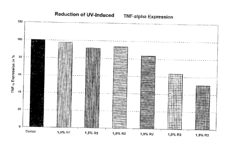
1. Reduction of UV-induced TN-alpha expression (TNF-alpha Assay)
UV light and other factors that can trigger a cutaneous inflammation lead to
the expression of TNF-alpha (tumour necrosis factor), the most important pro-
inflammatory cytokine. TNF-alpha controls both local and systemic
inflammatory processes by induction of cyclooxygenase-2 (COX-2) and
prostaglandin E2 (PGE2). This induces the expression of substance P (SP), a
sensory neuropeptide which is responsible for the sensation of pain and,
moreover, stimulates TNF-alpha again. This illustrates the necessity to
inhibit
or minimise TNF-alpha, which is at the basis of the cytokine cascade, during
an inflammatory process.
Apart from the sensory pain sensation, the consequences of a dermal
inflammation, however, are reddening and swelling, which weakens the barrier
function of the skin. With UV-induced cutaneous erythema, there is also
damage to the DNS induced directly by UVB radiation, which, on the one hand
leads to TNF-alpha expression and, on the other hand, also increases the
number of apoptotic cells. This, in turn, can lead to a drastic impairment of
the
CA 02769678 2012-01-31
6
differentiation process of the keratinocytes and, thus, to an impaired
formation
of the Stratum corneum.
Due to its ability to protect the epidermal homeostasis from such influences
in
an effective manner in combination with its very good tolerability, the active
agent complex according to the invention is ideally suited to strengthen,
maintain and restore the epidermal integrity.
TNF-alpha after UV radiation - procedure:
Human keratinocytes were incubated at 37 C and 5% CO2 in Dulbecco's
Modified Eagle Medium (DMEM); Biochrom, F0415) which was supplemented
with 5% FCS (Foetal Calf Serum; Biochrom, S 0115 - heat-inactivated) and
L-glutamine (Biochrom, K 0282).
Prior to reaching the stationary growth phase, the cells were trypsinated,
which included pretreatment with an EDTA solution (ethylene diamine tetra-
acetic acid, Biochrom, L2113, 1:20 in PBS). After determination of the number
of cells, a cell suspension was prepared and seeded into a 96 well microtitre
plate (MTP; TPP, 92696) with a cell number of 3 x 10⁴ cells/well.
The samples to be examined, R1 - R3, were diluted in medium and added to
the cells at the corresponding concentrations. The plates were incubated for
72 h at 37 C and 5% CO2.
After expiry of this time period, the medium was removed and the cells were
washed with PBS (phosphate-buffered saline solution, without Mg2+ and Ca2+;
Biochrom, L1825).
For the subsequent UV radiation, the cells were covered with 50 tl PBS/well
and radiated with 2 J/cm2 UVA + 0.2 J/cm2 UVB by means of a UV lamp
simulating the natural sun light spectrum (Dr. Honle, SOL 500).
After repeated incubation of the cells at 37 C and 5% CO2 for 18 h, the TNF-
alpha luminescence ELISA was carried out (R&D Systems, QTA00B).
The microtitre plates were centrifuged at 250 x g for 10 minutes and the media
supernatants were carefully transferred to the microtitre plate coated with
anti-
TNF-alpha + assay diluent, without taking up the precipitated cell debris. The
cell supernatants to be examined were incubated for 3 h at room temperature
while shaking. Then, the plates were incubated with the 2nd antibody (anti-
TNF-alpha-POD) for 2 h at room temperature on the shaker. After addition of
the Glo-reagent, a ten-minute incubation of the plate, protected from light,
took
place at room temperature.
CA 02769678 2012-01-31
7
Luminescence was measured in a microplate reader (Labsystems, Fluoscan
Ascent FL). The obtained RLU values (Relative Luminescence Units)
correspond to the amount of the expressed TNF-alpha. Cells which had not
been pre-treated with R1, R2, R3 served as controls. The RLU values of these
control cells were set as 100% value.
As illustrated in Figure 1, the active agent complex according to the
invention
(R3) had a significant reducing effect on TNF-alpha, which was dose-
dependent, whereas the individual components caused only an insignificant
reduction, if at all.
2. Reduction of UV-induced IL-8 expression
Interleukin-8 is one of the primary inflammatory cytokines stimulated mainly
by
TNF-alpha and interleukin-1, which, in turn, is expressed by exogenous factors
such as UV, infections, ischemia, wound healing after traumata, phototoxic
and photoallergic reactions, respectively, osmotic stress and others by a
plurality of cell types. It plays a crucial role in immune-related
inflammations
where it activates neutrophils and leads them to the source of the
inflammation
where they trigger intensification of the chemotactic recruitment of the
neutrophils by IL-8 secretion. Thus, it is a key cytokine for inflammatory
processes, which can lead to chronic inflammatory conditions in the case of
lack of intervention.
Interleukin-8 after UV radiation - procecure:
Human keratinocytes were incubated at 37 C and 5% CO2 in Dulbecco's
Modified Eagle Medium (DMEM); Biochrom, F0415) which was supplemented
with 5% FCS (Foetal Calf Serum; Biochrom, S 0115 - heat-inactivated) and
L-glutamine (Biochrom, K 0282).
Prior to reaching the stationary growth phase, the cells were trypsinated,
which included pretreatment with an EDTA solution (ethylene diamine tetra-
acetic acid, Biochrom, L2113, 1:20 in PBS). After determination of the number
of cells, a cell suspension was prepared and seeded into a 96 well microtitre
plate (MTP; TPP, 92696) with a cell number of 3 x 10⁴ cells/well.
The samples to be examined, R1 - R3, were diluted in medium and added to
the cells at the corresponding concentrations. The plates were incubated for
72 h at 37 C and 5% CO2.
CA 02769678 2012-01-31
8
After expiry of this time period, the medium was removed and the cells were
washed with PBS (phosphate-buffered saline solution, without Mg2+ and Ca2+;
Biochrom, L1825).
For the subsequent UV radiation, the cells were covered with 50 III PBS/well
and radiated with 2 J/cm2 UVA + 0.2 J/cm2 UVB by means of a UV lamp
simulating the natural sun light spectrum (Dr. Honle, SOL 500).
After further incubation of the cells at 37 C and 5% CO2 for 18 h, the
interleukin-8 luminescence ELISA was carried out (R&D Systems, Q8000B).
The microtitre plates were centrifuged at 250 x g for 10 minutes and the media
supernatants were carefully transferred to the microtitre plate coated with
anti-
IL-8 + assay diluent, without taking up the precipitated cell debris. The cell
supernatants to be examined were incubated for 2 h at room temperature
while shaking. Then, the plates were incubated with the 2nd antibody (anti-IL-
8-
POD) for 3 h at room temperature on the shaker. After addition of the Glo-
reagent, a ten-minute incubation of the plate, protected from light, took
place
at room temperature.
Luminescence was measured in a microplate reader (Labsystems, Fluoscan
Ascent FL). The obtained RLU values (Relative Luminescence Units)
correspond to the amount of the expressed Interleukin-8. Cells which were not
pre-treated with R1, R2, R3 serve as controls. The RLU values of these control
cells were set as 100% value.
The active agent complex according to the invention (R3) reduced the
expression of interleukin-8 in a significant and dose-dependent manner and to
a much larger extent than the individual components of the active agent
complex (see also Figure 2).
3. Reduction of IL-8 expression after hyperosmotic stress
In the case of hyperosmotic cell stress, the inflammation process takes also
place via the activation of the mitogen-activated protein kinases (MAPKs)
which includes ERK (extracellular signal-regulated kinase) and c-Jun N-
terminal kinase (JNK). These activated kinasea induce nuclear transcription
factors (NF-kappa B, AP-1) to secrete pro-inflammatory mediators.
Cell damages at DNS level which are caused by inflammatory mechanisms
due to osmotic stress are not subject to DNS repair mechanisms as it is the
case with UV-induced DNS damages. The possibility of repairing these
damages is more limited. Thus, there is a need for an effective active agent
which is also capable of preventing or reducing cell damages of this kind.
CA 02769678 2012-01-31
9
Interleukin-8 after hvperosmotic stress - procedure:
Human keratinocytes were incubated at 37 C and 5% CO2 in Dulbecco's
Modified Eagle Medium (DMEM; Biochrom, F0415) which was supplemented
with 5% FCS (fetal calf serum; Biochrom, S0115 - heat-inactivated) and L-
glutamine (Biochrom, K0282).
Prior to reaching the stationary growth phase, the cells were trypsinated,
which included a pretreatment with an EDTA solution (ethylene diamine tetra-
acetic acid, Biochrom, L2113; 1:20 in PBS). After determination of the cell
number, the cells were suspended and this cell suspension was seeded into a
96-well microtitre plate (MTP; TTP, 92696) with a cell number of 3 x 10⁴
cells/well.
The samples to be examined R1 - R3 were diluted in medium and added to
the cells at the corresponding concentrations. The plates were incubated for
72 h at 37 C and 5% CO2.
After expiry of this time period, the medium was removed and the cells were
washed with PBS (phosphate-buffered saline, without Mg2+ and Ca2+;
Biochrom, L1825).
Subsequently, the cells were incubated with different doses of R1 - R3 in
hyperosmolar medium for 30 min and for 12 h. The hyperosmolar medium was
adjusted by means of sodium chloride (Merck, 1064041000) to 400 mOsm
using an osmometer (Roebling, Digital Microosmometer Type 5R).
After the incubation periods of the cells at 37 C and 5% CO2 had elapsed, an
interleukin-8 luminescence ELISA (R&D Systems, Q8000B) was carried out.
The microtitre plates were centrifuged at 250 x g for 10 minutes and the media
supernatants were carefully transferred to the microtitre plate coated with
anti-
IL-8 + assay diluent, without taking up the precipitated cell debris. The cell
supernatants were incubated for 2 hours at room temperature while shaking.
Subsequently, the plates were incubated with the 2nd antibody (anti-IL-8-POD)
for 3 hours at room temperature on the shaker. After addition of the Glo
reagent, a ten-minute incubation of the plate, protected from light, took
place
at room temperature.
The luminescence was measured in a microplate reader (Labsystems,
Fluoscan Ascent FL). The obtained RLU values (Relative Luminescence Units)
correspond to the content of interleukin-8 expressed. Cells that were not
pretreated with R1, R2, R3 serve as control. The RLU values of these control
cells were set as 100% value.
CA 02769678 2012-01-31
. .
Also in these studies, the active agent complex of the invention reduced IL-8
expression significantly and in a dose-dependent manner. The individual
components inhibited IL-8 expression to a much smaller degree (see also
Figure 3).
4. Reduction of phototoxic-dependent IL-8 expression
Photoallergic or phototoxic reactions can be triggered by photosensitizing
substances. An example is hypericin from Hypericum perforatum (St John's
wort). The actually damaging components of these substances are free
radicals which form under the influence of light. These are the elicitors of
the
inflammatory condition which is manifest by the expression of the pro-
inflammatory cytokines (e.g. IL-8).
Interleukin-8 after phototoxic reaction - procedure:
Human keratinocytes were incubated at 37 C and 5% CO2 in Dulbecco's
Modified Eagle Medium (DMEM; Biochrom, F0415) supplemented with 5%
FCS (Fetal Calf Serum; Biochrom, S0115 - heat-inactivated) and L-glutamine
(Biochrome, K0282).
Prior to reaching the stationary growth phase, the cells were trypsinated,
which included a pretreatment with an EDTA solution (ethylene diamine
tetraacetic acid, Biochrom, L2113; 1:20 in PBS). After determination of the
cell
number, the cells were suspended and this cell suspension was seeded into a
96-well microtitre plate (MTP; TTP, 92696) with a cell number of 3 x 10⁴
cells/well.
The samples to be examined R1 - R3 were diluted in medium and added to
the cells at the corresponding concentrations. The plates were incubated for
72 h at 37 C and 5% CO2.
After expiry of this time period, the medium was removed and the cells were
washed with PBS (phosphate-buffered saline, without Mg2+ and Ca2 ;
Biochrom, L1825).
Subsequently, the cells were treated with different doses of R1 - R3 in the
medium with and without 0.5 pM hypericin (Sigma, 56690). This was followed
by UV radiation at 0.25 J/cm2 UVA + 0.025 J/cm2 UVB by means of a UV lamp
simulating the spectrum of natural sunlight (Dr. HOnle, SQL 500).
CA 02769678 2012-01-31
11
After a further incubation of the cells at 37 C and 5% CO2 for 18 h, the
interleukin-8 luminescence ELISA (R&D Systems, Q8000B) was carried out.
The microtitre plates were centrifuged at 250 x g for 10 minutes and the media
supernatants were carefully transferred to the anti-L-8-coated microtitre
plate +
assay diluent, without taking up the precipitated cell debris. The
supernatants
to be examined were incubated for 2 hours at room temperature while
shaking. Subsequently, the plates were incubated with the 2nd antibody (anti-
IL-8-POD) for 3 hours at room temperature on the shaker. After the addition of
the Glo reagent, a ten-minute incubation of the plate, protected from light,
took
place at room temperature.
Luminescence was measured in a microplate reader (Labsysems, Fluorscan
Ascent FL). The obtained RLU values (Relative Luminescence Units)
correspond to the content of interleukin-8 expressed. Cells that were not
pretreated with R1, R2, R3 serve as control. The RLU values of these control
cells were set as 100% value.
The active agent complex of the invention (R3) also inhibited the phototoxic-
dependent expression of interleukin-8 in a dose-dependent manner. The
individual components of the active agent complex cause only insignificant
inhibition of the expression (see also Figure 4).
5. Reduction of formation of soluble E-cadherin
Cadherins are calcium-dependant cell-cell adhesion molecules.
Epethelial cadherins (E-cadherins) belong to the group of classical cadherins
which are essential for the architecture of the epidermis since they occur in
desmosomes as well as in the "adhesion junctions".
E-cadherin functions as the transmembrane anchor which is linked to the actin
cytoskeleton of the cell (E-cadherin/catenin complex).
Different mechanisms regulate the adhesion strength of this complex. Thus, a
phosphorylation of beta-catenin (induced by MMPs [stromelysine-1,
matrilysine]) induces impairment of this complex and, thus, leads to loss of
epidermal integrity.
The destruction of the intercellular bond of this complex releases the soluble
E-cadherin ectodomain fragment of 80 kDa.
This E-cadherin fragment causes the separation of epithelial cells in vitro
and,
moreover, contributes to the development of epidermal skin cancers (tumour
progression).
CA 02769678 2012-01-31
12
The formation of the epidermal layers is an essential property of the dermis.
Thus, dysfunctions of the epidermal structure are serious impairments
regarding the barrier and protective function of the skin.
As demonstrated in the following, it was surprisingly found that the active
agent complex of the invention (R3) has a positive effect on the degradation
of
the E-cadherin/cateinin complex due to damage.
Formation of E-cadherin fragment - sE-cadherin after UV radiation -
procedure:
Human keratinocytes were incubated at 37 C and 5% CO2 in Dulbecco's
Modified Eagle Medium (DMEM; Biochrom, F0415) which was supplemented
with 5% FCS (Fetal Calf Serum); Biochrom, S0115 - heat-inactivated) and
L-glutamine (Biochrom, K0282).
Prior to reaching the stationary growth phase, the cells were trypsinated
which
included a pretreatment with an EDTA-solution (ethylene diamine tetra-acetic
acid; Biochrom, L2113, 1:20 in PBS). After the determination of the number of
cells, the cells were suspended and the cells suspension was seeded into a
95-well microtitre plate (MTP; TPP, 92696) with a cell number of 3 x 10⁴
cells/well.
The samples to be examined R1 - R3 were diluted in medium and added to
the cells at the corresponding concentrations. The plates were incubated for
72 h at 37 C and 5% CO2.
After the incubation period, the medium was removed and the cells were
washed with PBS (phosphate-buffered saline, without Mg2+ and Ca2+;
Biochrom, L1825).
For subsequent UV radiation, the cells were covered with 50 pl PBS per well
and radiated at 1 J/cm2 + 0.1 J/cm2 and 2 J/cm2 UVA + 0.2 J/cm2 UVB by
means of a UV lamp simulating the natural spectrum of sunlight (Dr. Honle,
SOL 500).
After a further incubation of the cells at 37 C and 5% CO2 for 18 h, the
sE-cadherin ELISA (R&D Systems, DCADEO) was carried out.
The microtitre plates were centrifuged at 250 x g for 10 minutes and the media
supernatants were carefully transferred to the microtitre plate coated with
anti-
sE-cadherin + assay diluent, without taking up the precipitated cell debris.
The
cell supernatants to be examined were incubated at room temperature for
2 hours while shaking. Subsequently, the plates were incubated with the 2nd
antibody (anti-sE-cadherin POD) at room temperature for 2 hours on the
CA 02769678 2012-01-31
13
shaker. After the addition of the Glo reagent, a ten-minute incubation of the
plate, protected from light, took place at room temperature.
Luminescence was measured in a microplate reader (Labsystems, Fluoscan
Ascent FL). The obtained RLU values (Relative Luminescence Units)
correspond to the content of interleukin-8 expressed. Cells that were not
pretreated with R1, R2, R3 serve as control. The RLU values of these controls
are set as 100% value.
The active agent complex of the invention (R3) significantly reduced the
expression of E-cadherin fragment - sE-cadherin in a dose-dependent
manner. The individual components of the active agent complex, however,
have, at best, an insignificant inhibitory effect (see also Figure 5).
6. Impedance after UV radiation
In order to evaluate epidermal integrity at cell level, the electrical cell-
substrate
impedance sensing method (ECIS) was used.
ECIS is a non-invasive method which allows to observe cell behaviour in real
time and to make predictions regarding growth behaviour, cell adhesion,
micro-movement of cells, morphological changes and, finally, barrier function
properties.
This method is based on the finding that cells represent electrical resistance
(impedance) and each change in volume, form and magnitude of the cell/cell
contacts have a measurable effect on the impedance. This resistance is called
impedance.
Healthy cells are seeded onto a chip on which they form a confluent
monolayer over an electrode. After the formation of desmosomes and zonulae
adherentes (adherent junctions), it is possible to determine changes in
impedance, which are not caused by a decrease in cell number but by
morphological changes (cell-cell-contacts), by means of damages, e.g.
damage by UV). This is possible by a real time observation period starting
prior to the start of damage to several hours after completion of damage.
Since, in case of moderate damage, it cannot be assumed that there are
necrotic cells in the first minutes up to 1 hour (this would also be clear
from the
absence of regeneration and stable impedance values), the changes in the
integrity of the cell layer (lawn) is clearly recognizable throughout this
early
observation period. Thus, this methodology is well suited to draw conclusions
with respect to the integrity of the metabolically active layers of the
epidermis.
As the experimental results show, after an observation period of approximately
20 h with a radiation of 3 J, it can be clearly observed that the values of
the
CA 02769678 2012-01-31
14
cells treated with the individual components of the active agent complex are
at
the level of the control cells (R2) and significantly above (R1),
respectively,
(however, below the initial value). Surprisingly, cells that had been treated
with
the active agent complex of the invention showed values slightly above the
initial value prior to radiation.
The high impedance loss of the control cells and of the cells treated with R1
and R2 may be explained by the possible initiation of apoptosis. Within a
comparable period of time, this damage cannot be observed in the cells
treated with the active agent complex. However, the enhancement of
epidermal integrity is particularly clear during the first 30 minutes after
the
damage.
The control cells as well as the cells treated with the individual components
R1
and R2 are almost at the same level, whereas the cells treated with the active
agent complex of the invention (R3) were capable of maintaining a higher
impedance level already during damaging.
Thus, with R3, the initial impedance value (100%) is reached again 30 minutes
after UV radiation.
In contrast, control, R1 and R2 are still approximately 25% below the initial
value at a comparable point in time.
The above results are evidence for the fact that the active agent complex of
the invention (R3) is capable of significantly enhancing the epidermal
integrity
of the skin (see Figures 6a and 6b).
Impedance measurement after UV radiation - procedure:
Human keratinocytes were incubated at 37 C and 5% CO2 in Dulbecco's
Modified Eagle Medium (DMEM; Biochrom, F0415) which was supplemented
with 5% FCS (Fetal Calf Serum); Biochrom, S0115 - heat-inactivated) and
L-glutamine (Biochrom, K0282).
Prior to reaching the stationary growth phase, the cells were trypsinated
which
included a pretreatment with an EDTA-solution (ethylene diamine tetra-acetic
acid; Biochrom, L2113, 1:20 in PBS). After the determination of the number of
cells, the cells were suspended and the cells suspension was seeded onto an
electrode chip (IBIDI, 8E 10) with a cell number of 3 x 10⁴ cells/well.
The assays were incubated at 37 C and 5% CO2 for 72 h until a confluent
monolayer was formed. Samples R1 - R3 to be analysed were diluted at a
concentration of 1.5 % in medium without FCS for 24 h and added to the cells.
CA 02769678 2012-01-31
After this period, the chip was connected to the ECIS measurement device.
After a relatively constant impedance curve was reached (5 h), UV radiation at
3 J/cm2 UVA + 0.3 J/cm2 UVB by means of a UV lamp simulating the natural
spectrum of sunlight (Dr. Hanle, SQL 500) took place.
The chip was connected to the measurement device also during the radiation
period so that a continuous registration of data was ensured. The data were
registered for further 20 h.
7. Avoidance of apoptotic cells after UV radiation
For the determination of apoptotic cells, a cytotoxicity test by Roche was
used.
This LDH test (Roche; 11644793) is based on the principle that the enzyme
LDH (lactate dehydrogenase), which is present in the cytosol of intact cells,
is
discharged into the extracellular space (supernatant).
The emitted amount of LDH due to the cell's entry into apoptosis can be
determined by means of this photometric test and is, thus, a measure for the
present damage of the cell membrane and, thus, also for the number of
apoptotic cells.
Human keratinocytes were incubated at 37 C and 5% CO2 in Dulbecco's
Modified Eagle Medium (DMEM); Biochrom, F0415) which was supplemented
with 5% FCS (Foetal Calf Serum; Biochrom, S 0115 ¨ heat-inactivated) and L-
glutamine (Biochrom, K 0282).
Prior to reaching the stationary growth phase, the cells were trypsinated,
which included pretreatment with an EDTA solution (ethylene diamine tetra-
acetic acid, Biochrom, L2113, 1:20 in PBS). After determination of the number
of cells, a cell suspension was prepared and seeded into a 96 well microtitre
plate (MTP; TPP, 92696) with a cell number of 3 x 10⁴ cells/well.
The samples to be examined, R1 - R3, were diluted in medium and added to
the cells at the corresponding concentrations. The plates were incubated for
72 h at 37 C and 5% CO2.
After expiry of this time period, the medium was removed and the cells were
washed with PBS (phosphate buffered saline solution, without Mg2+ and Ca2+;
Biochrom, L1825).
For the subsequent UV radiation, the cells were covered with 50 tl PBS/well
and radiated with 1 J/cm2 UVA + 0.1 J/cm2 UVB by means of a UV lamp
simulating the natural sun light spectrum (Dr. Honle, SQL 500).
After further incubation of the cells at 37 C and 5% CO2 for 18 h, the
interleukin-8 luminescence ELISA was carried out (R&D Systems, Q8000B).
CA 02769678 2012-01-31
16
The microtitre plates were centrifuged at 250 x g for 10 minutes and the media
supernatants were carefully transferred to the new microtitre plate, without
taking up the precipitated cell debris. After addition of the reaction mixture
(diaphorase/NAD + iodine tetrazolium/sodiunn lactate), incubation of the plate
for 30 minutes, protected from light, took place at room temperature.
Absorption was measured in a microplate reader (Fynex, MRX) at 480 nm,
reference wavelength 630 nm. The OD values obtained correspond to the
released amount of LDH enzyme and, thus, to the number of damaged and/or
apoptotic cells. Cells which had not been pre-treated with R1, R2, R3 served
as controls. The OD values of these control cells are fixed as 100% value.
The result of this test shows that both the treatment with the individual
components and with the active agent complex according to the invention
results in a reduction of apoptotic cells in comparison to the control cells.
With increased UV radiation dose, this result is still observable, even if the
number of apoptotic cells on the whole has increased.
In both experiments, however, the active agent complex according to the
invention (R3) achieved a clearly better effect than the individual components
(R1 and R2) alone, or the merely calculated addition of the result values of
R1
and R2 (see also Figure 7).
Thus, the active agent complex according to the invention from both extracts
shows an overeffect with regard to the reduction of the expression of pro-
inflammatory cytokines (IL-8, TNF-a) in various test models (osmotic stress,
UV radiation, photoallergic reactions) and strengthening of the epidermal
integrity, as could be determined due to the reduction of the E-cadherin
degradation and a more rapid regeneration of the epidermal integrity. Due to
its ability to protect the epidermal homeostasis in an effective manner in
combination with its good tolerance, the active agent complex according to the
invention is ideally suited to strengthen, maintain and restore the epidermal
integrity.