Note: Descriptions are shown in the official language in which they were submitted.
DEVICE FOR DETECTION OF ANTIGENS AND USES THEREOF
Field of the Invention
100021 The present invention is directed, in part, to a device and assay for
detecting
one or more antigens and methods of using the same.
Background of the Invention
100031 Detection of antigens is important for many areas of scientific
research,
diagnostic use and therapeutic uses. There are several ways in which antigens
can be
detected. Various methods are described in U.S. Patent: 5,160,701, U.S.
Patent: 5,141,850,
PCT Publication WO 91/12336, U.S. Patent: 5,451,504, U.S. Patent: 5,559,041,
European
Patent Application No.: 0505636A1, PCT Publication No. WO 88/08534, European
Patent
Application No. 0284 232A1, U.S. Patent Application Publication No.
20070020768 and
U.S. Patent No. RE39664. The methods and devices available prior to the
present invention
may still require improvements in sensitivity or speed at which results can be
obtained.
These factors can be important where time is of the essence when attempting to
determine the
presence or absence of an antigen.
100041 One such area is the area of detecting food borne pathogenic
contaminants.
Approximately, seventy-six million people in the United States become
afflicted with a food
borne illness. Of those seventy-six million, approximately, 325,000 will
become violently ill,
requiring hospitalization, and approximately 5,000 will die. The majority of
food-borne
illnesses are causes by Salmonella, E. colt, and Campylobacter costing
approximately $35
billion dollars.
100051 Current measures at ensuring a safe food supply involve a combination
of
local, state and federal authorities as well as an elaborate system of
inspectors and
surveillance networks. Food manufacturers are held to certain United States
Department of
Agriculture, United States Food and Drug Administration, and the National
Marine Fisheries
Service regulations that are enforceable by law. The USDA has created a system
of health
inspectors that are charged with performing daily meat, produce, and other
consumable
products inspections made or processed in manufacturing and processing
facilities. These
1
CA 2769747 2017-06-20
CA 02769747 2012-01-31
WO 2011/014763 PCT/US2010/043889
DOCKET NO.: 135958.00102
PATENT
inspections have been created to involve a detailed statistical analysis to
best ensure safety
and sterility of food before it reaches the consumer. Moreover, the majority
of the meat
industry has adopted irradiation techniques to further demonstrate sterility
of products. At a
lower level, local and municipal health departments work to ensure that local
distributors,
restaurants, and retailers follow strict guidelines to ensure a safe food
supply. However,
despite this elaborate network, food-borne infections are still common.
[0006] Once an outbreak is strongly suspected, an investigation begins. A
search is
made for more cases among persons who may have been exposed. The symptoms and
time of
onset and location of possible cases are determined, and a "case definition"
is developed that
describes these typical cases. The outbreak is systematically described by
time, place, and
person. A graph is drawn of the number of people who fell ill on each
successive day to show
pictorially when it occurred. Calculating the distribution of cases by age and
sex shows
whom is affected.
[0007] Often the causative microbe is not known, so samples of stool or blood
must
be collected from ill people and sent to the public health laboratory to make
a diagnosis. Each
collection and sampling can cost upwards of $500 per test and often takes 2-4
days for
analysis (CDC "Food-borne Infections").
[0008] Prior to the present invention, to identify the food or other source of
the
outbreak, the investigators first interview a few persons with the most
typical cases about
exposures they may have had in the few days before they got sick. In this way,
certain
potential exposures may be excluded while others that are mentioned repeatedly
emerge as
source possibilities. Combined with other information, such as likely sources
for the specific
microbe involved, hypotheses are then tested in a formal epidemiologic
investigation. The
investigators conduct systematic interviews about a list of possible exposures
with the ill
persons, and with a comparable group of people who are not ill. By comparing
how often an
exposure is reported by ill people and by well people, investigators can
measure the
association of the exposure with illness. Using probability statistics, the
probability of no
association is directly calculated.
[0009] As new food-borne problems emerge there is a need for novel devices and
methods for detecting food borne pathogens. The present invention provides a
device for the
detection of antigens, such as antigens from food-borne bacteria, and fulfills
the needs of
having a device and assay with increased sensitivity and/or speed of
detection. The present
invention fulfills other needs as well as will be discussed herein.
2
CA 02769747 2012-01-31
WO 2011/014763 PCT/US2010/043889
DOCKET NO.: 135958.00102
PATENT
Summary of the Invention
[0010] In some embodiments, the present invention provides devices for
detecting
an antigen. In some embodiments, the devices comprise a housing comprising a
first housing
member and a second housing member, wherein said housing comprises: an inlet
opening in
the second housing member; a force member attached to the first housing
member; a slidable
locking member contacting the first housing member and contacting the force
member; an
antigen detection membrane system comprising in the following order: a
conjugate pad; a
permeable membrane; a test membrane; and an absorbent member; and a flexible
attachment
member attached to the locking member and the conjugate pad; wherein at least
a portion of
each of the conjugate pad, permeable membrane, test membrane, and absorbent
member are
substantially parallel to each other; wherein the conjugate pad is capable of
being compressed
against the perimeter of the inlet opening in the second housing member: and
wherein the
force member contacts the absorbent member and is capable of applying pressure
substantially perpendicular to the antigen detection membrane system.
[0011] In some embodiments of the devices, the devices further comprise a
hydrophobic membrane located between the test membrane and the absorbent
member. In
some embodiments, the first housing member further comprises a sliding button
that
protrudes the outer surface of the first housing member, wherein the sliding
button is attached
to the locking member, wherein movement of the sliding button moves the
locking member.
[0012] In some embodiments, the conjugate pad comprises a first antigen-
specific
antibody.
[0013] In some embodiments, the antigen recognized by the first antigen-
specific
antibody is a food-borne pathogen antigen.
[0014] In some embodiments, the present invention provides systems comprising
a
device as described herein and a buffer container or a sample collector.
[0015] The present invention also provides methods of detecting an antigen.
Brief Description Of Drawings
[0016] Figure 1: Depicts a perspective view of a representative device
according
to some embodiments of the present invention.
[0017] Figure 2: Depicts some components of a representative device
according
to some embodiments of the present invention.
3
CA 02769747 2012-01-31
WO 2011/014763 PCT/US2010/043889
DOCKET NO.: 135958.00102
PATENT
[0018] Figure 3: Depicts some components of a representative device
according
to some embodiments of the present invention.
[0019] Figure 4: Depicts some components of a representative device
according
to some embodiments of the present invention.
[0020] Figure 5: Depicts some components of a representative device in
various
positions according to some embodiments of the present invention.
[0021] Figure 6: Depicts a lateral view of some components of a
representative
device according to some embodiments of the present invention.
[0022] Figure 7: Depicts a lateral view of some components of a
representative
device according to some embodiments of the present invention.
[0023] Figure 8: Depicts a lateral view of some components of a
representative
device according to some embodiments of the present invention.
[0024] Figure 9: Depicts a flexible attachment member attached to a
conjugate
pad.
[0025] Figure 10: Depicts membranes in a representative housing member.
Description of Embodiments
[0026] As used herein and unless otherwise indicated, the term "about" is
intended
to mean 5% of the value it modifies. Thus, about 100 means 95 to 105.
[0027] The present invention provides devices and methods for detecting
antigens
or other molecules. In some embodiments, devices in use chromatographic
assays. In some
embodiments, the assays use specifying binding assays to indicate the presence
or absence of
an antigen.
[0028] The term -capture reagent" refers to a reagent, for example an antibody
or
antigen binding protein, capable of binding a target molecule or analyte to be
detected in a
biological sample. A capture reagent may also be, for example, an
oligonucleotide or a
peptoid.
[0029] The term "detecting" or "detection" is used in the broadest sense to
include
qualitative and/or quantitative measurements of a target analyte.
[0030] The terms "attached" or "attachment" can include both direct attachment
or
indirect attachment. Two components that are directly attached to one another
are also in
physical contact with each other. Two components that are indirectly attached
to one another
are attached through an intermediate component. For example, Component A can
be
indirectly attached to Component B if Component A is directly attached to
Component C and
4
CA 02769747 2012-01-31
WO 2011/014763 PCT/US2010/043889
DOCKET NO.: 135958.00102
PATENT
Component C is directly attached to Component B. Therefore, in such an
example,
Component A would be said to be indirectly attached to Component B.
[0031] The term "isolated" refers to a molecule that is substantially
separated from
its natural environment. For instance, an isolated protein is one that is
substantially separated
from the cell or tissue source from which it is derived.
[0032] The term "purified" refers to a molecule that is substantially free of
other
material that associates with the molecule in its natural environment. For
instance, a purified
protein is substantially free of the cellular material or other proteins from
the cell or tissue
from which it is derived. The term refers to preparations where the isolated
protein is
sufficiently pure to be analyzed, or at least 70% to 80% (w/vv) pure, at least
80%-90% (w/w)
pure, 90-95% pure; and, at least 95%, 96%, 97%, 98%. 99%, or 100% (w/w) pure.
[0033] The terms -specific binding," -specifically binds," and the like, mean
that
two or more molecules form a complex that is measurable under physiologic or
assay
conditions and is selective. An antibody or antigen binding protein or other
molecule is said
to "specifically bind" to a protein, antigen, or epitope if, under
appropriately selected
conditions, such binding is not substantially inhibited, while at the same
time non-specific
binding is inhibited. Specific binding is characterized by a high affinity and
is selective for
the compound, protein, epitope, or antigen. Nonspecific binding usually has a
low affinity.
Binding in IgG antibodies for example is generally characterized by an
affinity of at least
about 10-7 M or higher, such as at least about 10-8 M or higher, or at least
about 10-9 M or
higher, or at least about 10-10 or higher, or at least about 10-11 M or
higher, or at least about
10-12 M or higher. The term is also applicable where, e.g., an antigen-binding
domain is
specific for a particular epitope that is not carried by numerous antigens, in
which case the
antibody or antigen binding protein carrying the antigen-binding domain will
generally not
bind other antigens. In some embodiments, the capture reagent has a Kd equal
or less than
10-9M, 10-1 M, or 10-11M for its binding partner (e.g. antigen). In some
embodiments, the
capture reagent has a Ka greater than or equal to 109M-1 for its binding
partner.
[0034] Capture reagent can also refer to, for example, antibodies. Intact
antibodies,
also known as immunoglobulins, are typically tetrameric glycosylated proteins
composed of
two light (L) chains of approximately 25 kDa each, and two heavy (H) chains of
approximately 50 kDa each. Two types of light chain, termed lambda and kappa,
exist in
antibodies. Depending on the amino acid sequence of the constant domain of
heavy chains,
immunoglobulins are assigned to five major classes: A, D, E, G, and M, and
several of these
CA 02769747 2012-01-31
WO 2011/014763 PCT/US2010/043889
DOCKET NO.: 135958.00102
PATENT
may be further divided into subclasses (isotypes), e.g., IgGl, IgG2, IgG3,
IgG4, IgAl, and
IgA2. Each light chain is composed of an N-terminal variable (V) domain (VL)
and a
constant (C) domain (CL). Each heavy chain is composed of an N-terminal V
domain (VH),
three or four C domains (CHs), and a hinge region. The CH domain most proximal
to VH is
designated CH 1. The VH and VL domains consist of four regions of relatively
conserved
sequences named framework regions (FR1, FR2, FR3, and FR4), which form a
scaffold for
three regions of hypervariable sequences (complementarity determining regions,
CDRs). The
CDRs contain most of the residues responsible for specific interactions of the
antibody or
antigen binding protein with the antigen. CDRs are referred to as CDR1, CDR2,
and CDR3.
Accordingly, CDR constituents on the heavy chain are referred to as H1, H2,
and H3, while
CDR constituents on the light chain are referred to as Li, L2, and L3. CDR3 is
the greatest
source of molecular diversity within the antibody or antigen binding protein-
binding site. H3,
for example, can be as short as two amino acid residues or greater than 26
amino acids. The
subunit structures and three-dimensional configurations of different classes
of
itnmunoglobulins are well known in the art. For a review of the antibody
structure, see
Antibodies: A Laboratory Manual, Cold Spring Harbor Laboratory, Eds. Harlow et
al., 1988.
One of skill in the art will recognize that each subunit structure, e.g., a
CH, VH, CL, VL,
CDR, and/or FR structure, comprises active fragments. For example, active
fragments may
consist of the portion of the VH, VL, or CDR subunit that binds the antigen,
i.e., the antigen-
binding fragment, or the portion of the CH subunit that binds to and/or
activates an Fc
receptor and/or complement.
[0035] Non-limiting examples of binding fragments encompassed within the term
-antigen-specific antibody" used herein include: (i) an Fab fragment, a
monovalent fragment
consisting of the VL, VH, CL and CH1 domains; (ii) an F(ab')2 fragment, a
bivalent fragment
comprising two Fab fragments linked by a disulfide bridge at the hinge region;
(iii) an Fd
fragment consisting of the VH and CH1 domains; (iv) an Fv fragment consisting
of the VL
and VH domains of a single arm of an antibody, (v) a dAb fragment, which
consists of a VH
domain; and (vi) an isolated CDR. Furthermore, although the two domains of the
Fv
fragment, VL and VH, are coded for by separate genes, they may be
recombinantly joined by
a synthetic linker, creating a single protein chain in which the VL and VH
domains pair to
form monovalent molecules (known as single chain Fv (scFv)). The most commonly
used
linker is a 15-residue (Gly4Ser)3 peptide, but other linkers are also known in
the art. Single
chain antibodies are also intended to be encompassed within the terms -
antibody or antigen
6
CA 02769747 2012-01-31
WO 2011/014763 PCT/US2010/043889
DOCKET NO.: 135958.00102
PATENT
binding protein," or -antigen-binding fragment" of an antibody. The antibody
can also be a
polyclonal antibody, monoclonal antibody, chimeric antibody, antigen-binding
fragment. Fc
fragment, single chain antibodies, or any derivatives thereof.
[0036] These antibodies are obtained using conventional techniques known to
those
skilled in the art, and the fragments are screened for utility in the same
manner as intact
antibodies. Antibody diversity is created by multiple germline genes encoding
variable
domains and a variety of somatic events. The somatic events include
recombination of
variable gene segments with diversity (D) and joining (J) gene segments to
make a complete
VH domain, and the recombination of variable and joining gene segments to make
a
complete VL domain. The recombination process itself is imprecise, resulting
in the loss or
addition of amino acids at the V(D)J junctions. These mechanisms of diversity
occur in the
developing B cell prior to antigen exposure. After antigenic stimulation, the
expressed
antibody genes in B cells undergo somatic mutation. Based on the estimated
number of
germline gene segments, the random recombination of these segments, and random
VH-VL
pairing, up to 1.6X10⁷ different antibodies may be produced (Fundamental
Immunology, 3rd
ed. (1993), ed. Paul, Raven Press, New York, N.Y.). When other processes that
contribute to
antibody diversity (such as somatic mutation) are taken into account, it is
thought that
upwards of 1X101 different antibodies may be generated (Immunoglobulin Genes.
2nd ed.
(1995), eds. Jonio et al., Academic Press, San Diego, Calif.). Because of the
many processes
involved in generating antibody diversity, it is unlikely that independently
derived
monoclonal antibodies with the same antigen specificity will have identical
amino acid
sequences.
[0037] Antibody or antigen binding protein molecules capable of specifically
interacting with the antigens, epitopes, or other molecules described herein
may be produced
by methods well known to those skilled in the art. For example, monoclonal
antibodies can
be produced by generation of hybridomas in accordance with known methods.
Hybridomas
formed in this manner can then be screened using standard methods, such as
enzyme-linked
immunosorbent assay (ELISA) and Biacore analysis, to identify one or more
hybridomas that
produce an antibody that specifically interacts with a molecule or compound of
interest.
[0038] As an alternative to preparing monoclonal antibody-secreting
hybridomas, a
monoclonal antibody to a polypeptide of the present invention may be
identified and isolated
by screening a recombinant combinatorial immunoglobulin library (e.g., an
antibody phage
display library) with a polypeptide of the present invention to thereby
isolate
7
CA 02769747 2012-01-31
WO 2011/014763 PCT/US2010/043889
DOCKET NO.: 135958.00102
PATENT
immunoglobulin library members that bind to the polypeptide. Techniques and
commercially
available kits for generating and screening phage display libraries are well
known to those
skilled in the art. Additionally, examples of methods and reagents
particularly amenable for
use in generating and screening antibody or antigen binding protein display
libraries can be
found in the literature.
[0039] The term "capture reagent" also includes chimeric antibodies, such as
humanized antibodies, as well as fully humanized antibodies. In some
embodiments the
capture reagent is a Goat anti-E. coli 0157:H7 antibody Cat #: 70-XG13
(Fitzgerald
Industries); E. coli 0157:H7 mono Cat #: 10-E13A(Fitzgerald Industries); E.
coli 0157:H7
Cat #: 10C-CR1295M3(Fitzgerald Industries); E. coli 0157:H7 mono Cat #: 10-
E12A(Fitzgerald Industries); or Goat anti-mouse IgG Cat #: ABSE-020 (DCN).
[0040] In some embodiments, the devices of the present invention comprise a
housing comprising a first housing member and a second housing member. In some
embodiments, the first and second housing members can be constructed as a
single unit. The
housing can comprise an inlet opening. The inlet opening allows the
introduction of a sample
onto the chromatographic assay. In some embodiments, the first housing member
comprises
the inlet opening. The inlet opening can be of sufficient size to handle an
appropriate amount
of volume of a solution that is added to the device. In some embodiments, the
size of the
opening is large enough to handle about 0.1 to 3 ml, about 0.1 to 2.5 ml,
about 0.5 to 2.0 ml,
about 0.1 to 1.0 ml, about 0.5 to 1.5 ml, 0.5 to 1.0 ml, and 1.0 to 2.0 ml.
[0041] In some embodiments, the housing comprises a conjugate pad, a permeable
membrane, a test membrane, and/or an absorbent member. In some embodiments,
the
housing comprises an antigen detection membrane system. In some embodiments,
the
antigen detection membrane system comprises a conjugate pad, a permeable
membrane, a
test membrane, and an absorbent member. In some embodiments, the antigen
detection
membrane system is free of a permeable membrane. In some embodiments, the
antigen
detection membrane system comprises in the following order: a conjugate pad, a
permeable
membrane, a test membrane, and an absorbent member.
[0042] As used herein, the term "conjugate pad" refers to a membrane or other
type
of material that can comprise a capture reagent. The conjugate pad can be a
cellulose acetate,
cellulose nitrate, polyamide, polycarbonate, glass fiber, membrane,
polyethersulfone,
regenerated cellulose (RC), polytetra-fluorethylene, (PTFE), Polyester (e.g.
Polyethylene
Terephthalate), Polycarbonate (e.g., 4, 4-hydroxy-dipheny1-2, 2'-propane),
Aluminum Oxide,
8
CA 02769747 2012-01-31
WO 2011/014763 PCT/US2010/043889
DOCKET NO.: 135958.00102
PATENT
Mixed Cellulose Ester (e.g., mixture of cellulose acetate and cellulose
nitrate), Nylon (e.g.,
Polyamide, Hexamethylene-di amine, and Nylon 66), Polypropylene, PVDFõHigh
Density
Polyethylene (HDPE) + nucleating agent "aluminum dibenzoate" (DBS) (e.g. 80u
0.024
HDPE DBS (Porex)), and HDPE. Examples of conjugate pads also include,
Cyclopore0
(Polyethylene terephthalate), Nucleopore0 (Polyethylene terephthalate), Membra-
Fil0
(Cellulose Acetate and Nitrate), Whatman (Cellulose Acetate and Nitrate),
Whatman #12-S
(rayon)), Anopore (Aluminum Oxide), Anodise (Aluminum Oxide), Sartorius
(cellulose
acetate, e.g. 5 p.m), and Whatman Standard 17 (bound glass).
[0043] In some embodiments, the conjugate pad or test membrane comprises a
capture reagent. In some embodiments, the conjugate pad or test membrane is
contacted with
the capture reagent and then allowed to dry. The conjugate pad or test
membrane can also
comprise other compositions to preserve the capture reagent such that it can
be stably stored
at room temperature or under refrigeration or freezing temperatures. In some
embodiments,
the conjugate pad or test membrane is soaked with a buffer prior to the
capture reagent being
applied. In some embodiments, the buffer is a blocking buffer that is used to
prevent non-
specific binding. In some embodiments, the buffer comprises Borate, BSA, PVP40
and/or
Tween-100. In some embodiments, the buffer is 10mM Borate, 3% BSA, 1% PVP40,
and
0.25% Tween-100. In some embodiments the capture reagent is applied to the pad
or
membrane in a solution comprising trehalose and sucrose. In some embodiments,
the capture
reagent is applied to the conjugate pad or test membrane in a solution
comprising trehalose,
sucrose and phosphate and/or BSA. In some embodiments, the capture reagent is
applied in a
solution that is 5% trehalose. 20% sucrose, 10 mM phosphate, and 1% BSA.
[0044] In some embodiments, the pad or membrane (e.g. conjugate pad or test
membrane) comprises about 0.5 to about 5.0 lig of a capture reagent, about 1
to about 3 lig of
a capture reagent, about 1 to about 2 tg of a capture reagent, about to 2 to
about 3 lig of a
capture reagent, about 1.5 jig of a capture reagent, 2.5 lig of a capture
reagent, or about 2.7
14 of a capture reagent.
[0045] In some embodiments, the permeable membrane is attached to or adhered
to
a test membrane. In some embodiments, the permeable membrane is laminated onto
the test
membrane. The permeable membrane can be a membrane of any material that allows
a
sample, such as a fluid sample, to flow through to the test membrane. Examples
of test
membrane include, but are not limited to, nitrocellulose, cellulose, glass
fiber, polyester,
9
CA 02769747 2012-01-31
WO 2011/014763 PCT/US2010/043889
DOCKET NO.: 135958.00102
PATENT
polypropylene, nylon, and the like. In some embodiments, the permeable
membrane
comprises an opening. The opening can be present to allow visualization or
detection of the
test membrane. In some embodiments, the opening in the permeable membrane is
substantially the same size as the inlet opening in the housing. Examples of
permeable
membranes include, but are not limited to, Protran BA83, Whatman, and the
like.
[0046] As used herein, the "test membrane" refers to a membrane where
detection
of a binding partner to a capture reagent occurs. Test membranes include, but
are not limited
to a nitrocellulose membrane, a nylon membrane, a polyvinylidene fluoride
membrane, a
polyethersulfone membrane, and the like. The test membrane can be any material
that can be
used by one of skill in the art to detect the presence of a capture reagent's
binding partner
(e.g. antigen or epitope). The test membrane can also comprise a capture
reagent. In some
embodiments, the test membrane is contacted with a capture reagent and the
capture reagent
is allowed to dry and adhere to the test membrane. Examples of test membranes
include, but
are not limited to Protran BA83, Whatman, Opitran BA-SA83, and 0.22 p.m white
plain
(Millipore Product No. SA3J036107). The test membrane can comprise a plurality
of capture
reagents. In some embodiments, the test membrane comprises 1, 2, 3, 4, 5, 6,
7, 8, 9, or 10
capture reagents. In some embodiments, the test membrane comprises a plurality
of areas
each with a different capture reagent. In some embodiments, the plurality of
areas do not
completely overlap or coincide with one another. By using a plurality of
capture reagents,
multiple binding partners (e.g. epitopes or antigens) can be detected.
[0047] In some embodiments, the housing also comprises an absorbent member.
The absorbent member can also be referred to as a "wick pad" or "wicking pad."
The
absorbent member absorbs the fluid that flows through the device when the
sample is applied
to the device and provides for the wicking force that aids in the flow of the
sample when it is
applied to the device.
[0048] The absorbent member can be any material that can facilitate the flow
of the
sample through the conjugate pad and to the test membrane. Examples of
absorbent
members include, but are not limited to cellulose, super absorbent polymers,
glass fiber pads
(e.g. C083 (Millipore)), and the like. In some embodiments, the housing
comprises a
plurality (e.g. 2 or more) of absorbent members. In some embodiments, the
housing
comprises 2, 3, 4, or 5 absorbent members. In some embodiments, the absorbent
member
comprises one or more membranes up to 10 individual membranes, and each
membrane may
be the same material or a different material.
CA 02769747 2012-01-31
WO 2011/014763 PCT/US2010/043889
DOCKET NO.: 135958.00102
PATENT
[0049] In some embodiments, the device comprises a force member. The force
member can be used to apply pressure or to compress the other components of
the antigen
detection membrane system against one another. In some embodiments, the force
member
can comprise a shaft and a head. The force member can have a mushroom type
shape where
the head is wider than the shaft. In some embodiments, the head is narrower
than the shaft.
The force member comprising a head and a shaft can be a single unit or can be
made up of
multiple parts that contact one another to form the force member. For example,
the head
could be one unit that can be separated from the shaft. Upon assembly the head
and shaft are
contacted with one another to make the force member. In another example, the
head and
shaft are one cohesive unit and are manufactured together and not as separate
parts that are
later assembled to form the force member. The force member allows the device
to work with
vertical flow as opposed to relying upon lateral flow.
[0050] The devices described herein can be used in assays to detect the
presence of
a capture reagent's binding partner. For example, an antigen can be detected
by an antibody
using the devices of the present invention. The devices of the present
invention employ
vertical flow. "Vertical flow" refers to the direction that the sample flows
across the different
membranes and members present in the device. Vertical flow refers to a sample
flowing
through the membrane (e.g. top to bottom) as opposed to lateral flow, which
refers to a
sample flowing across (e.g. side to side) a membrane, pad or absorbent member.
In a lateral
flow device the membranes and pads sit horizontally adjacent to one another
substantially on
the same plane. In a vertical flow device each membrane or pad is
substantially parallel or
completely parallel to each other and occupy substantially different spatial
planes in the
device. The membranes and pads may occupy similar planes when they are
compressed or
put under pressure. In some embodiments, at least a portion of each membrane
or pad is
layered on top of each other. In some embodiments, at least a portion of each
layer of
membrane or pad is substantially parallel to each other. In some embodiments,
at least a
portion of each layer is in a different spatial plane than each other layer.
[0051] To allow vertical flow to occur efficiently, in some embodiments, the
conjugate pad, permeable membrane, test membrane and the absorbent member are
substantially parallel to each other. In some embodiments, the conjugate pad,
permeable
membrane, test membrane and the absorbent member are present in different
spatial planes.
In some embodiments, the housing also comprises a hydrophobic membrane that
can slow or
stop the vertical flow of the sample. The hydrophobic membrane can be in
contact with the
11
CA 02769747 2012-01-31
WO 2011/014763 PCT/US2010/043889
DOCKET NO.: 135958.00102
PATENT
test membrane, which would allow the sample to dwell or rest upon the test
membrane. The
dwell can allow for increased sensitivity and detection. The vertical flow is
modulated by the
pressure that is applied to the membranes. In some embodiments, the pressure
is applied
perpendicular to the test membrane and/or the conjugate pad. The pressure can
be applied so
that the conjugate pad is compressed against the housing. The compression
against the
housing can be such that the conjugate is in direct contact with the housing,
0-ring, or collar,
or through an intermediate so that the conjugate pad and the test membrane are
compressed
against one another.
[0052] The force member can apply pressure that is substantially perpendicular
to
the test membrane. The pressure facilitates the vertical flow. The pressure
allows each layer
of the membrane stack to be in contact with another layer. The pressure can
also be relieved
to stop the flow so that the test sample can dwell or rest upon the test
membrane, which can
allow for greater sensitivity. The pressure can then be reapplied to allow the
vertical flow to
continue by allowing the sample to flow into the absorbent member(s). The
force member
can apply pressure such that the conjugate pad contacts a portion of the
housing. In some
embodiments, the conjugate pad contacts the housing when it is not under the
pressure being
exerted by the force member but upon the force member exerting pressure the
conjugate pad
is compressed against a portion of the housing.
[0053] In some embodiments, the conjugate pad contacts the perimeter of the
inlet
opening. The inlet opening can also comprise a collar or other similar
feature, such as an 0-
ring. In some embodiments, the conjugate pad contacts the perimeter of a
collar and/or an 0-
ring. In some embodiments, the conjugate pad is capable of being compressed
against the
perimeter of the inlet opening, which can include, in some embodiments, a
collar and/or an
0-ring.
[0054] "Capable of being compressed against the perimeter of the inlet
opening"
refers to a membrane or pad (e.g. conjugate pad) being compressed either
directly in contact
with the perimeter of the inlet opening or being compressed against another
layer or material
(e.g. membrane) that is in contact with the perimeter of the inlet opening.
[0055] In some embodiments, the conjugate pad is not in direct physical
contact
with the housing but is in fluid contact with the housing. "Fluid Contact"
means that if a
sample is applied to the device through the inlet opening or other opening the
fluid will
contact the conjugate pad. In some embodiments, the conjugate pad can be
separated from
the housing by another membrane, such as a permeable membrane, where the other
12
CA 02769747 2012-01-31
WO 2011/014763 PCT/US2010/043889
DOCKET NO.: 135958.00102
PATENT
membrane is in direct physical contact with the housing or in direct physical
contact with the
collar or 0-ring. When the sample is applied to the device the fluid can
contact the other
membrane first and then contact the conjugate pad. This is just one example of
the conjugate
pad being in fluid contact with the housing. There are numerous other
embodiments where
the conjugate pad is not in direct physical contact with the housing, the
collar, or the 0-ring,
but is in fluid contact with the housing.
[0056] The force member can apply any pressure that is sufficient to
facilitate
vertical flow across the different membrane layers. In some embodiments, the
layers of the
device (e.g. conjugate pad, permeable membrane, test membrane, and absorbent
member) are
compressed under a force chosen from about 5 lbf to 100 lbf, about 5 lbf to 50
lbf, about 10
lbf to 401bf, about 15 lbf to 40 lbf, about 15 lbf to 25 lbf, or about 301bf
to 40 lbf. The force
can also compress a hydrophobic or impermeable membrane as well if one is
present in the
device.
[0057] In some embodiments, the force member contacts a first surface of an
absorbent member. In some embodiments, a conjugate pad contacts a test
membrane. In
some embodiments, a first surface of a test membrane contacts a permeable
membrane. In
some embodiments, a second surface of the test membrane contacts a second
surface of the
absorbent pad. In some embodiments, the device comprises a hydrophobic
membrane, and,
for example, the hydrophobic membrane contact a second surface of the test
membrane. In
some embodiments, the hydrophobic membrane contact a first surface of the
absorbent pad.
[0058] In some embodiments, a first surface of the conjugate pad contacts the
housing and a second surface of the conjugate pad contacts a first surface of
the permeable
membrane, wherein the second surface of the permeable membrane contacts a
first surface of
the test membrane, wherein a second surface of the test membrane contacts a
first surface of
the absorbent pad, wherein a second surface of the absorbent pad contacts the
force member.
In some embodiments, the first surface of the conjugate pad contacts a
perimeter of the inlet
opening of said housing. In some embodiments, the first surface of the
conjugate pad
contacts a perimeter of a collar or an 0-ring.
[0059] The device can also comprise an attachment member. In some
embodiments, the attachment member is flexible or made from a flexible
material. The
flexible material can be, for example, an elastic or elastomer material. An
attachment
member can be, for example, attached to a conjugate pad and/or a hydrophobic
membrane.
The attachment member can also be attached to any membrane or member of the
device.
13
CA 02769747 2012-01-31
WO 2011/014763 PCT/US2010/043889
DOCKET NO.: 135958.00102
PATENT
Examples of attachment members include, but are not limited to, elastomer
band, rubber
band, spring, and the like. In some embodiments, the attachment member can be
made of a
shape memory material. The attachment member makes it possible to create a
delay between
moving the locking member and moving the conjugate pad or any other type of
membrane or
pad that the attachment member is attached to. The movement of the pad or
membrane does
not happen at the same time as the sliding button or locking member is moved.
Not being
bound to any particular theory, as the sliding button or locking member is
moved energy is
accumulated in the attachment member and this energy is used to pull on a pad
or membrane
that it is attached to the attachment member after the pressure has been
released. In some
embodiments, the locking member is moved away from the force member (i.e., the
force
member no longer contacts the locking member) before the conjugate pad is
moved or
removed. The conjugate pad, in some embodiments, is moved once the compression
or
pressure being exerted by the force member is completely removed.
[0060] The attachment member can also be attached to either a sliding button
or
locking member. The attachment member can be attached through any means such
as,
adhesives, staples, tying, and the like to the other components. In some
embodiments, the
membrane or pad has notches in the membrane or pad that allow the attachment
member to
attach to the membrane or pad. A non-limiting example can be seen in Figure 9.
[0061] In some embodiments, the housing comprises a locking member. The
locking member can be a slidable locking member that can move within the
device. The
locking member can be used to lock the force member in a position such that
the force
created by the force member upon the different layers is maintained. The
locking member is,
for example, locking the force member in place so that the pressure cannot be
relieved unless
the locking member is moved to allow the force member to change positions
(i.e. lowered).
The locking member, can for example, fit under the head of the force member,
which would
keep the force member in the raised position. The locking member can also be
situated so
that it keeps the force member in a particular position (e.g. raised or
lowered). The locking
member can be made of any material including, but not limited to, plastic and
the like. The
locking member can, for example, contact the force member either directly or
indirectly
through another component that prevents the force member from releasing the
pressure. In
some embodiments, the locking member contacts the force member to compress the
conjugate pad.
14
CA 02769747 2012-01-31
WO 2011/014763 PCT/US2010/043889
DOCKET NO.: 135958.00102
PATENT
[0062] The locking member can also contact the attachment member such that
movement of the locking member will move the attachment member, any other
membrane
(e.g. conjugate pad, hydrophobic membrane, test membrane, or absorbent member)
or other
component that is attached to the attachment member. For example, if the
locking member is
moved to relieve the pressure of the force member thereby allowing the force
member to
change positions (e.g. from raised to a lower position), the movement of the
locking member
will also deform/accumulate energy into the attachment member so it can move
the
membrane or pad once the pressure has been sufficiently reduced. When the
conjugate pad is
attached to the attachment member and the locking member is moved this will
also move the
conjugate pad once the pressure has been sufficiently reduced. In some
embodiments, the
pressure is completely removed. The conjugate pad can be, for example, moved
such that it
is removed from the device. In some embodiments, the conjugate pad is moved to
reveal the
test membrane through the inlet opening. The amount of the test membrane that
is revealed
will depend upon the type of detection that is used. For a visual detection
more of the test
membrane may need to be revealed in the inlet opening. For a non-visual, e.g.
fluorescent,
infrared, radioactive or chemiluminescent detection, less of the test membrane
may need to
be revealed. In some embodiments, the conjugate pad is moved so that it no
longer can be
seen or detected through the inlet opening. In some embodiments, the movement
of the
conjugate pad can create another opening other than the inlet opening to
visualize or detect
the test membrane.
[0063] In some embodiments, the attachment member is also attached to the
impermeable or hydrophobic membrane. When the attachment member is moved the
movement will also move or remove the impermeable or hydrophobic membrane. As
discussed herein, the presence of the impermeable or hydrophobic membrane can
allow the
test sample to dwell or rest upon the test membrane by slowing or stopping the
vertical flow.
When the impermeable or hydrophobic membrane is moved or removed, either by
its
attachment to the attachment member or through other means, the vertical flow
is no longer
impeded or inhibited.
[0064] In some embodiments, the housing comprises a sliding button. A sliding
button can also be referred to as a sliding member. The sliding button can
cross the inner and
outer surfaces of the housing. In some embodiments, the sliding button or
sliding member
protrudes to an outer surface of the housing. In some embodiments, the sliding
button is
attached either directly or indirectly to the locking member. When the sliding
button is
CA 02769747 2012-01-31
WO 2011/014763 PCT/US2010/043889
DOCKET NO.: 135958.00102
PATENT
attached (directly or indirectly) to the locking member the movement of the
sliding button
also moves the locking member. The attachment member in some embodiments can
be
attached to the sliding button. In some embodiments, the attachment member is
attached to
both the sliding button and the locking member. The sliding button and the
locking member
can also be constructed as a single unit.
[0065] In some embodiments, the inlet opening comprise an opening chosen from
a
range of about 0.2-20 cm2. In some embodiments, the inlet opening is about 1
to about 2 cm
in diameter. In some embodiments, the inlet opening is about 1 or about 1.5 cm
in diameter.
In some embodiments, the inlet opening is about 1, about 2, about 3, about 4,
or about 5 cm
in diameter.
[0066] As discussed herein, the conjugate pad can comprise an antigen specific
capture reagent. In some embodiments, the conjugate pad comprises a plurality
of antigen
specific capture reagents. In some embodiments, the conjugate pad comprises 1,
2, 3, 4, or 5
antigen specific capture reagents. The antigen can be any molecule that can be
specifically
recognized by a capture reagent. Examples of antigens include a polynucleotide
molecule
(e.g. DNA, RNA, siRNA, antisense oligonucleotide) a peptide, a protein, a
saccharide, a
polysaccharide, a carbohydrate, and the like. The antigen can also refer to
different epitopes
present on the same protein or polypeptide.
[0067] The capture reagent can also be, for example, protein A, protein G, and
the
like.
[0068] In some embodiments, the protein is a pathogen protein. A pathogen
protein
refers to a protein that is from a pathogen. Examples of pathogens include,
but are not
limited to, viruses, prokaryote and pathogenic eukaryotic organisms such as
unicellular
pathogenic organisms and multicellular parasites. Pathogens also can include
protozoan
pathogens which include a stage in the life cycle where they are intracellular
pathogens. As
used herein, the term "intracellular pathogen" is meant to refer to a virus or
pathogenic
organism that, at least part of its reproductive or life cycle, exists within
a host cell and
therein produces or causes to be produced, pathogen proteins.
[0069] Bacterial pathogens include, but are not limited to, such as bacterial
pathogenic gram-positive cocci, which include but are not limited to:
pneumococcal;
staphylococcal; and streptococcal. Pathogenic gram-negative cocci include:
meningococcal;
and gonococcal. Pathogenic enteric gram-negative bacilli include:
enterobacteriaceae;
pseudomonas, acinetobacteria and eikenella; melioidosis; salmonella;
shigellosis;
16
hemophilus; chancroid; brucellosis; tularemia; yersinia (pasteurella);
streptobacillus
moniliformis and spirilum; listeria monocytogenes; erysipelothrix
rhusiopathiae; diphtheria;
cholera; anthrax; donovanosis (granuloma inguinale); and bartonellosis.
Pathogenic
anaerobic bacteria include; tetanus; botulism; other clostridia; tuberculosis;
leprosy; and other
mycobacteria. Pathogenic spirochetal diseases include: syphilis;
treponematoses: yaws, pinta
and endemic syphilis; and leptospirosis. Other infections caused by higher
pathogen bacteria
and pathogenic fungi include: actinomycosis; nocardiosis; cryptococcosis,
blastomycosis,
histoplasmosis and coccidioidomycosis; candidiasis, aspergillosis, and
mucormycosis;
sporotrichosis; paracoccidiodomycosis, petriellicliosis, torulopsosis,
mycetoma and
chromomycosis; and dermatophytosis. Rickettsial infections include rickettsial
and
rickettsioses. Examples of mycoplasma and chlamydial infections include:
mycoplasma
pneumoniae; lymphogranuloma venereum; psittacosis; and perinatal chlamydial
infections.
Pathogenic protozoans and helminths and infections eukaryotes thereby include:
amebiasis;
malaria; leishmaniasis; trypanosomiasis; toxoplasmosis; pneumocystis carinii;
babesiosis;
giardiasis; trichinosis; filariasis; schistosomiasis; nematodes; trematodes or
flukes; and
cestode (tapeworm) infections. Bacteria also include E. colt, an
Campylobacter, and a
Salmonella.
100701 In some embodiments, E. Coll is E. coli 0157.
100711 Examples of viruses include, but are not limited to, HIV, Hepatitis A,
B, and
C, Fly, lentiviruses, pestiviruses, West Nile Virus, measles, smallpox,
cowpox, ebola,
coronavirus, and the like. Other pathogens are also disclosed in U.S. Patent
Application
Publication No. 20080139494.
100721 In some embodiments, the pathogen is a food borne pathogen. The antigen
can be present on a food borne pathogen. Food borne pathogens are pathogens
(e.g. viral or
bacterial) that cause illness after eating contaminated food. The food itself
does not directly
cause the illness, but it is rather the consumption of the food borne pathogen
that is present
on the food that causes the illness. In some embodiments, the food borne
pathogen is E. coli,
Campylobacter, or Salmonella. In some embodiments, the antigen is an antigen
chosen from
a food borne pathogen antigen. For example, the food borne pathogen antigen
can be, but is
not limited to, chosen from an E. coli antigen, a Campylobacter antigen, or a
Salmonella
antigen. In some embodiments, the antigen is the species specific 0-Antigen.
In some
embodiments, the 0-antigen is the E. coli and/or the Salmonella 0-antigen and
can be used
17
CA 2769747 2017-06-20
CA 02769747 2012-01-31
WO 2011/014763 PCT/US2010/043889
DOCKET NO.: 135958.00102
PATENT
for E. coli and Salmonella detection. In some embodiments, the antigen is a
flagellin antigen.
In some embodiments, the antigen is the Campylobacter flagellin antigen.
[0073] In some embodiments, the capture reagent comprises a detection reagent.
The detection reagent can be any reagent that can be used to detect the
presence of the
capture reagent binding to its specific binding partner. The capture reagent
can comprise a
detection reagent directly or the capture reagent can comprise a particle that
comprises the
detection reagent. In some embodiments, the capture reagent and/or particle
comprises a
color, colloidal gold, radioactive tag, fluorescent tag, or a chemiluminescent
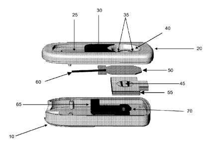
substrate. The
particle can be, for example, a viral particle, a latex particle, a lipid
particle, or a fluorescent
particle. In some embodiments, the colloidal gold has a diameter size of:
about 20 nm, about
30 nm, or about 40 nm or in the range of about 20-30 nm, about 20-40 nm, about
30-40 nm,
or about 35-40 nm.
[0074] In some embodiments, the test membrane also comprises one or more
capture reagents.
[0075] The capture reagents of the present invention can also include an anti-
antibody, i.e. an antibody that recognizes another antibody but is not
specific to an antigen,
such as, but not limited to, anti-IgG, anti-IgM, or ant-IgE antibody. Where
the test
membrane comprises an anti-antibody, such as anti-IgG, anti-IgM, or anti-IgE
antibody, this
non-specific antibody can be used as a positive control to detect whether the
conjugate has
been released from the conjugate pad. When the sample is applied to the device
it allows a
first capture reagent to be released from the conjugate pad. As the capture
reagent is released
and flows through the device, either attached to the antigen or not, it can
contact the anti-
antibody, such as anti-IgG or anti-IgM antibody, which can then be detected.
This detection
can be used to show that the device is working properly.
[0076] In some embodiments, the test membrane comprises a second antigen
specific capture reagent. In some embodiments, the test membrane comprises a
first area
comprising a first capture reagent comprising an anti-IgG capture reagent; and
a second area
comprising a second antigen specific capture reagent, wherein the first and
second areas do
not completely overlap or coincide with one another. This non-limiting
embodiment can be
used to demonstrate the device is working properly and be used to detect the
presence of the
antigen of interest.
[0077] In some embodiments, the conjugate pad comprises a first antigen
specific
capture reagent and the test membrane comprises a second antigen specific
capture reagent,
18
CA 02769747 2012-01-31
WO 2011/014763 PCT/US2010/043889
DOCKET NO.: 135958.00102
PATENT
wherein the first and second antigen specific capture reagents bind to non-
competitive
epitopes present on the antigen. The device can, for example, employ a
sandwich type assay
that occurs in two steps. The first step is the binding of the antigen to the
capture reagent
present in the conjugate pad. After binding to the first antigen specific
capture reagent the
antigen can flow through to or make contact with the test membrane where a
second antigen
specific capture reagent is present. Upon interaction with the test membrane
if the test
antigen can bind to the second antigen-specific capture reagent it will be
able to be detected
either through visualization or through the use of another detection device
such as, but not
limited to, a fluorescent reader. The test membrane and the conjugate pad can
comprise
additional antigen-specific capture reagents that recognize different antigens
or different
epitopes. In some embodiments, the test membrane or the conjugate pad
comprises 1, 2, 3, 4,
5. 6, 7, 8, 9, 10 antigen-specific capture reagents. In some embodiments, the
test membrane
or the conjugate pad comprises a plurality of antigen-specific capture
reagents. In some
embodiments, each antigen-specific capture reagent recognizes a different
antigen or a
different epitope on the same antigen.
[0078] "Different antigens" can also refer to the same protein but a protein
that is
from different strains of the same organism. Different antigens can also refer
to antigens
from different organisms. For example, there are any many strains of E. coll.
Not all strains
of E. coli cause a food-borne illness. The present invention can be used, for
example, to
detect an antigen from a pathogenic E. coli strain as opposed to detecting an
antigen from a
non-pathogenic E. coli strain. In some embodiments, the conjugate pad and/or
test
membrane comprises a first and a second antigen-specific capture reagents,
wherein the first
and said second capture reagents recognize different antigens. In some
embodiments, the test
membrane and/or conjugate pad comprises a plurality of areas comprising a
plurality of
antigen-specific capture reagents, wherein the plurality of antigen-specific
capture reagents
recognize different antigens. In some embodiments, the plurality of areas do
not completely
overlap or coincide with one another. In some embodiments, the plurality of
antigens are
each independently chosen from an E. coli antigen, an Campylobacter antigen,
and a
Salmonella antigen. In some embodiments of the present invention, the
plurality of antigens
is 2, 3, 4, 5, 6, 7, 8, 9, 10, or more than 10 antigens.
[0079] The devices may be housed singly, in pairs, or in multiple
configurations.
The housing can be watertight to prevent leakage and can be manufactured from
a variety of
inert materials, such as polymer materials. The inlet opening, in some
embodiments, can be
19
CA 02769747 2012-01-31
WO 2011/014763 PCT/US2010/043889
DOCKET NO.: 135958.00102
PATENT
of sufficient volume to contain any required amount of sample or reagents to
be used with the
invention.
[0080] Because the membranes or pads of the device is preferably chemically
inert,
it may have to be activated at any reaction site where it is desired to
immobilize a specific
binding reagent against solvent transport. Various methods may be required to
render the
reagent immobilized according to the particular chemical nature of the
reagent. Generally,
when the media is nitrocellulose or a mixed nitrocellulose ester, no special
chemical linkage
is required for the immobilization of reagents. Various techniques may be used
for other
materials and reagents which include functionalization with materials such as
carbonyldiimidazole, glutaraldehyde or succinic acid, or treatment with
materials such as
cyanogen bromide. Other suitable reactions include treatment with Schiff bases
and
borohydride for reduction of aldehyde, carbonyl and amino groups. DNA, RNA and
certain
antigens may be immobilized against solvent transport by baking onto the
chromatographic
material. Baking may be carried out at temperatures ranging from about 60 C to
about 120 C
for times varying from about five minutes to about 12 hours, and in some
embodiments, at
about 80 C for about two hours.
[0081] The present invention also provides systems comprising the devices
described herein and a buffer container. The buffer container can be any
buffer that the
sample that is being tested can be mixed with and then applied to the device.
For example,
the sample can be taken from a source and the sample can be mixed with the
buffer. The
buffer can be a lysis buffer that will lyse the cells or a buffer that
maintains the pH of the
sample so that the analysis can be done properly. The buffer container can be
any shape and
can be included outside or inside the housing of the device.
[0082] In some embodiments, the present invention provides a system that
comprises a sample collector. The sample collector can be any material that
can take a
sample from a source and allow the sample to be tested. For example, the
sample collector
can be a swab, such as a cotton-swab. In some embodiments, the sample
collector is an
innoculator. In some embodiments, the housing comprises the sample collector
and a portion
of the sample collector is in the inside of the housing. In some embodiments,
the sample
collector is partially outside and partially inside the housing. In some
embodiments, the
sample collector is completely outside the housing.
[0083] The present invention also provides for kits comprising the devices
described
herein. The kit can include a device as described herein, a sample collector,
a buffer
CA 02769747 2012-01-31
WO 2011/014763 PCT/US2010/043889
DOCKET NO.: 135958.00102
PATENT
container, an instruction manual, a positive control, a negative control, or
any combination
thereof. With respect to the kit, a positive control is a sample that is known
to contain the
antigen that can be detected with the device present in the kit. In contrast
the negative
control, would not contain an antigen that can be detected by the kit. The
negative control
when used in conjunction with the anti-antibody would be able to demonstrate
that the device
is working properly.
[0084] Buffers can also be included in the present invention. Examples of
buffers
include, but are not limited to, 1X PBS (10 mM Phosphate, 137 mM Sodium
Chloride, 2.7
mM Potassium Chloride), a wash buffer (e.g. 10mM Sodium Phosphate, 150mM NaC1,
0.5%
Tween-20, 0.05% Sodium Azide), a membrane buffer (e.g. 10mM Sodium Phosphate,
0.1%
Sucrose, 0.1% BSA, 0.2%, PVP-40 pH 7.21, filtered with 0.21im filter.),
Polyclonal
Conjugate Block Buffer (e.g. 50mM Borate, 10% BSA, pH 8.93); Polyclonal
Conjugate
Diluent (e.g. 50mM Borate, l % BSA, pH 9.09), or Blocking Buffers (e.g. 10mM
Sodium
Phosphate, 0.1% Sucrose, 0.025% Silwet pH 7.42; 10mM Sodium Phosphate, 1%
Sucrose,
1% Trehalose, 0.01% BSA, 0.025% Tween-20; 0.05% Sodium Azide, 0.025% Silwet pH
7.4;
10mM Sodium Phosphate, 0.1% Sucrose, 0.1% BSA, 0.2% PVP-40 pH 7.21). The
buffer
can also be, but is not limited to, a blocking buffer (e.g. 10% BSA in
deionized water, pH 7.4
or 1% BSA in deionized water, pH 7.4); 10mM Borate, 3% BSA, 1% PVP40, and
0.25%
Tween-100; and the like.
[0085] The conjugate pad and the test membrane can be contacted with any of
the
buffers described herein either in the presence or absence of a capture
reagent and, in some
embodiments, allowed to dry.
[0086] Examples of buffers that are lysis buffers include, for example, but
are not
limited to. 2% Tween (v/v) and 0.1% Triton(v/v); 2% Tween(v/v) and 0.1%
SDS(w/v); 2%
Tween(v/v) and 0.1% BSA(w/v); 2% Tween(v/v) and 1% BSA(w/v), 0.1% SDS(w/v), 1%
BSA(w/v), or any combination thereof.. The lysis buffers can also be, for
example, 5%
Tween/PBS; 2% Tween/PBS + 0.1% SDS; 2% Tween/PBS + 1% BSA. Other examples of
lysis buffers include, but are not limited to, 5% Tween-80(v/v); 5% Triton X-
100(v/v); 5%
NP40(v/v); 2% Tween-80(v/v): 2% Triton X-100(v/v); 2% NP40(v/v); 1% Tween-
80(v/v);
1% Triton X-100(v/v); and 1% NP40(v/v). The detergents and other components of
the
buffers can be made with any suitable buffer suitable for proteins, and
includes, but is not
limited to, water and phosphate buffered saline. The lysis buffers can be used
to prepare the
samples prior to the samples making contact with the devices described herein.
In some
21
CA 02769747 2012-01-31
WO 2011/014763 PCT/US2010/043889
DOCKET NO.: 135958.00102
PATENT
embodiments, a lysis buffer is not used. A lysis buffer is not used on a
sample when a
surface protein or surface antigen is desired to be detected. Accordingly, in
some
embodiments, the sample is not subject to lysis or conditions that would cause
a cell to be
lysed.
[0087] The present invention also provides for methods of detecting an antigen
comprising contacting a sample with a device as described herein, wherein the
sample
contacts the conjugate pad and the test membrane, wherein a positive reaction
with the test
membrane indicates the presence of the antigen, wherein the conjugate pad
comprises a first
antigen-specific capture reagent and the test membrane comprises a second
antigen-specific
capture reagent. A positive reaction is indicated by the capture reagent
present in the test
membrane binding to an antigen in the test sample. The capture reagent in the
test membrane
is applied to the test membrane so that it will indicate a positive reaction
when it binds to its
specific antigen. The specific capture reagent can be applied in any manner
such that when it
is detected it can form a line, a circle, a plus sign, a broken line. an "X"
or any other pattern.
In some embodiments, the control line indicating that the device is working
properly will
cross the antigen specific line and when the antigen specific capture reagent
binds to the
antigen the detectable label will form a plus sign.
[0088] In some embodiments, a sample contacts the device, which is then
followed
by a buffer being applied to the device after the sample has contacted the
device. For
example, a sample comprising an antigen can be contacted with the conjugate
pad such that
the sample is transferred to the conjugate pad. Following the contact with the
conjugate pad
a separate solution can be applied to the device to facilitate or initiate the
vertical flow
through the devices described herein.
[0089] In some embodiments as described herein the capture reagent is an
antibody.
In some embodiments, the sample that is tested is a solution but can also be a
mixture of
solution or buffer and solid material that can be applied to the device. The
solution will then
solubilize the antigen and allow the conjugate pad's capture reagent to come
into contact with
the antigens present in the sample. In some embodiments, the sample comprises
a cell lysate.
In some embodiments, the cell lysate has been clarified by centrifugation or
other means to
remove non-soluble materials.
[0090] In some embodiments, the methods comprise contacting a test sample with
a
sample collector and contacting the sample collector with the device. In some
embodiments,
the methods comprise contacting the sample collector with a solution or
buffer, wherein the
22
CA 02769747 2012-01-31
WO 2011/014763 PCT/US2010/043889
DOCKET NO.: 135958.00102
PATENT
solution or buffer is applied to the device. In some embodiments, the samples
are contacted
with the conjugate pad prior to the sample coming into contact with the test
membrane. In
some embodiments, the sample is contacted with the conjugate pad and the test
membrane
simultaneously.
[0091] In some embodiments, the method comprises moving the conjugate pad of
the devices described herein, wherein the movement of the devices exposes the
test
membrane for detection. In some embodiments, the locking member moves the
conjugate
pad. In some embodiments, the conjugate pad is attached to the locking member
and/or the
sliding button member. The antigen that the method can be used to detect can
be any
antigen. The antigen can be those that are discussed herein or any other
antigen that can be
detected using the methods and devices described herein. In some embodiments,
the method
comprises applying the sample to the device and allowing the sample to flow
through the
device via vertical flow.
[0092] In some embodiments the detection or indication of the presence or
absence
of an antigen occurs in less than 60 seconds. In some embodiments, the
detection or
indication of the presence or absence of an antigen occurs in about 30 to
about 60 seconds.
In some embodiments, the detection or indication of the presence or absence of
an antigen
occurs in less than 2 minutes. In some embodiments, the detection or
indication of the
presence or absence of an antigen occurs in about 30 seconds.
[0093] Referring to the drawings, in some embodiments, Figures 1 through 10,
depicts representative devices, components of a device, and various views of a
device.
Figure 1 depicts a device comprising a first housing member (10), a buffer
container (15), a
second housing member (20), a groove for the sliding button (25), a sliding
button (30), an
inlet opening (35), a collar (40), and a test membrane (45). Figure 1 depicts
a test membrane
(45) comprising two capture reagents. The first (10) and second (20) housing
members can
also be referred to as the lower and upper housing members, respectively. In
Figure 1, the
sample would be applied through the inlet opening (35) and can be allowed to
vertically flow
through to the test membrane (45). In Figure 1, the groove (25) allows the
sliding button to
move, which when attached to the locking member moves the locking member and
can, in
some embodiments, move the conjugate pad and change the position of the force
member.
[0094] Figure 2 depicts a device comprising a first housing member (10), a
second
housing member (20), a groove for the sliding button (25), a sliding button
(30), an inlet
opening (35), a collar (40), a test membrane (45). a conjugate pad (50), a
plurality of
23
absorbent members (e.g. pads) (55), an attachment member (60), a locking
member (65), and
a force member (70). Figure 2 depicts the conjugate pad (50), test membrane
(45) and
absorbent pad (55) arranged substantially parallel to one another. The force
member (70)
when in contact with the absorbent member would be applying pressure that is
substantially
perpendicular to the conjugate pad. As can be seen in Figure 2, a sample that
is contacted
with the device through the inlet opening (35) would flow vertically through
the conjugate
pad (50) to the test membrane (45). Not explicitly shown in Figure 2, but in
some
embodiments, a the permeable membrane is also substantially parallel to the
conjugate pad
(50) and to the test membrane (45), with a first surface of the permeable
membrane
contacting a surface of the conjugate pad (50) a second surface of the
permeable membrane
contacting a surface of the test membrane (45).
100951 Figure 3 depicts a conjugate pad (50), a permeable membrane (75), a
test
membrane (45), and a plurality of absorbent members (55). Figure 3 depicts the
components
being substantially parallel with one another. Figure 3 depicts the permeable
membrane (75)
comprising an opening. This opening can be used to allow visualization and
detection of the
test membrane's results.
[0096] Figure 4 depicts a device comprising a first housing member (10), a
buffer
container (15), a second housing member (20), a sliding button (30), a test
membrane (45), a
conjugate pad (50), a permeable membrane (75), a plurality of absorbent
members (e.g. pads)
(55), an attachment member (60), a locking member (65), and a force member
(70). Figure 4
also depicts the force member (70) comprising a shaft (72) and a head (71)
where the head
(71) is wider than the shaft (72).
[0097] Figure 5 depicts a partial view of a device comprising a first housing
member (10), a locking member (65), a sliding button (30), and force member
(70). Figure 5
depicts the locking member (65) in contact with the force member (70) such
that the force
member (70) is in a raised method. Figure 5 also depicts the movement of the
locking
member (65) and the sliding button (30) away from the force member (70)
allowing the force
member to change positions. In some embodiments, the change in position is
that the force
member is lowered.
[0098] Figure 6 depicts a side cut away view of a device comprising a first
housing
member (10), a second housing member (20), a sliding button (30), a locking
member (65), a
collar (40), an 0-ring (41), a force member (70), and a support for the force
member (73).
The support for the shaft can be, for example, part of the first housing
member (10) and is
24
CA 2769747 2017-06-20
shaded differently for example purposes only. Figure 6 depicts the button (30)
in contact
with the locking member (65) in such a way that movement of the button (30)
will move the
locking member (65). Movement of the locking member (65) will take away the
support
from the force member (70), which would allow the force member (70) to change
positions.
Figure 6 also depicts the shaft (72) and the head (71) of the force member.
The head (71)
creates a lip where the locking member (65) can slide under and support the
force member
(70).
[0099] Figure 7 depicts a partial view of a device comprising a first housing
member (10), a second housing member (20), an inlet opening (35), a test
membrane (45), a
conjugate pad (50), a plurality of absorbent members (55), an attachment
member (60), a
locking member (65), and a force member (70). Figure 8 depicts the attachment
member
(60) attached to the conjugate pad (50) and the locking member (65). Figure 8
also depicts
the conjugate pad being compressed against the second housing member (20) and
the
perimeter of the inlet opening (35). Figure 7 depicts the head of the force
member (71)
applying the pressure by contacting the plurality of absorbent members (55).
In Figure 7, a
sample can be applied to the device through the inlet opening (35) so that the
sample contacts
the conjugate pad (50) and because of the pressure the sample through vertical
flow contacts
the test membrane (45).
1001001 Figure 8 depicts a partial view of a device comprising a first housing
member (10), a second housing member (20), an inlet opening (35), a test
membrane (45), a
conjugate pad (50), a plurality of absorbent members (55), an attachment
member (60), a
locking member (65), and a force member (70). Figure 8 depicts the movement of
the
locking member (65), which is attached to the attachment member (60). The
movement of
the attachment member (60), which is attached to the conjugate pad (50) moves
the conjugate
pad. Figure 8 depicts the test force member (70) changing positions and a
lessening or
elimination of the pressure and/or compression of the test membrane (45).
Figure 9 also
depicts the movement of the conjugate pad (50) away from the inlet opening
(35) revealing
the test membrane (45) for visualization and/or detection.
100101] Figure 9 depicts an attachment member (60) attached to a conjugate pad
(50). Figure 9 depicts notches (51) in the conjugate pad (50) as locations for
the attachment
member (60) to attach to. The attachment member can also be attached through
other means
such as through adhesives, staples, and other forms of attachment.
CA 2769747 2017-06-20
CA 02769747 2012-01-31
WO 2011/014763 PCT/US2010/043889
DOCKET NO.: 135958.00102
PATENT
[00102] Figure 10 depicts a partial view of device comprising a second housing
member (20), a plurality of pads or membranes (80), wherein the plurality of
pads comprises
a test membrane, a permeable membrane, and one or more absorbent members, and
retaining
members (85) that can retain the plurality of pads or membranes (80). Figure
10 depicts the
structures that when the conjugate pad is moved the plurality of pads remains
in place. Any
means or other structure can be used to keep the plurality of pads in place.
[00103] The invention is now described with reference to the following
examples.
These examples are provided for the purpose of illustration only and the
invention should in
no way be construed as being limited to these examples, but rather should be
construed to
encompass any and all variations which become evident as a result of the
teaching provided
herein. Those of skill in the art will readily recognize a variety of non-
critical parameters that
could be changed or modified to yield essentially similar results.
Examples
[00104] Antibody specific for E. coli 0157:H7 conjugated to colloidal gold was
baked and dried onto the conjugate pad. A second antibody specific for E. Coli
0157:H7 and
an anti-antibody was striped onto a test membrane and assembled into an
antigen detection
device.
[00105] A sample containing LPS E. Coli 0157 was serially diluted in PBS to
concentrations of 100 p.g/ml, 50 jig/ml, 25 14/ml, 12.5 14/ml, 6.25 112/ml,
3.125 14/ml, 1.56
ps/ml, and 0.78 Kg/ml. The samples were applied to the device to detect the
presence of LPS
E. Coli 0157. The experiments were graded based upon signal intensity and the
results are
shown below. PBS was used as a negative control. TL refers to the test line
(antigen
specific) and CL refers to the control line (non-antigen specific). The
detection occurred
within 30 to 60 seconds of application of the sample onto the conjugate pad.
The device
could detect the presence of a food borne antigen.
Grade
Sample Concentration TL CL
0Oug/m1 6 8
26
DOCKET NO.: 135958.00102
PATENT
5Oug/m1 6 8
25ug/m1 4 8
12.5ug/m1 4 8
6.25ug/m1 3 8
3.125ug/m1 3 8
1.56ug/mL 1 8
0.78ug/m1 1 8
1XPBS (Negative Control) 1 8
[00107] While this invention has been disclosed with reference to specific
embodiments, it is apparent that other embodiments and variations of this
invention may be
devised by others skilled in the art without departing from the true spirit
and scope of the
invention. The appended claims are intended to be construed to include all
such embodiments
and equivalent variations.
27
CA 2769747 2017-06-20