Note: Descriptions are shown in the official language in which they were submitted.
CA 02770228 2012-03-02
SYSTEMS AND METHODS FOR IDENTIFYING TISSUE AND VESSELS
BACKGROUND
1. Technical Field
[0001] The present disclosure relates to in vivo systems and methods of
identifying tissue
parameters (e.g., tissue type) and assessing the conditions of the tissue
during a surgical
procedure. More specifically, the present disclosure relates to systems and
methods for
measuring the relative level of blood circulation in tissue with an energy-
based surgical
instrument for vessel sealing.
2. Background of Related Art
[0002] Correctly identifying tissue parameters including tissue type is
important for any
surgical operation. But it is especially important during laparoscopic
operations when a surgeon
can only view tissue through a camera. A camera, however, may provide a
surgeon with a
limited view of the tissue. As a result, several ex vivo and in vivo methods
have been proposed
to measure different characteristics of tissue in order to identify and assess
the tissue.
[0003] Publication number US 2008/0200843 describes a method and apparatus for
measuring human tissue properties in vivo. The method is based on sensing the
mechanical
response of tissue. The method includes applying a predetermined force to the
surface of a
patient with a probe and measuring the displacement of the probe as a function
of applied force.
Tissue properties are then determined based on the result of measuring the
displacement of the
probe.
1
CA 02770228 2012-03-02
[0004] Publication number US 2008/0154145 describes a method and apparatus for
determining characteristics of biological tissues. Tissue characteristics are
determined by
introducing a sound wave into the tissue and recording the response of the
tissue to the sound
wave.
[0005] Publication number US 2009/0124902 describes a method for classifying
tissue from
the lumbar region using a combination of ultrasonic and optical measurements.
[0006] In publication number US 2007/0276286, a miniature electrode array is
used to
stimulate tissue and to measure a tissue response in order to provide tissue
diagnosis and spatial
tissue mapping.
[0007] Publication number US 2005/0283091 describes a method and apparatus for
determining the conditions of biological tissue. The method includes exciting
tissue with
electrical signals at different frequencies and analyzing the cross-
correlation of response signals
with delayed excitation signals. Cross-correlation products are then auto-
correlated. Cross-
correlation products correspond to tissue conditions and auto-correlation
products correspond to
changes in the tissue conditions.
[0008] Publication number US 2003/0060696 discloses an apparatus for
recognizing tissue
type using multiple measurement techniques. For example, electrical signals
are applied to a
tissue via electrodes to measure impedance magnitude and phase at a plurality
of frequencies.
The phase information at the plurality of frequencies is compared with the
phase information of
known tissue types to identify the tissue type.
[0009] Publication number US 2002/0077627 describes a method for detecting and
treating
tumors using localized impedance measurements. The method includes providing
an impedance
measurement apparatus having a plurality of resilient members deployable to
sample tissue
2
CA 02770228 2012-03-02
impedance through a plurality of conductive pathways. Information from the
impedance
measurements is then used to determine the condition of the tissue.
[0010] Publication number US 2009/0253193 describes a device for
characterizing tissue ex
vivo. The device includes a set of independent electrodes that scan the tissue
by moving a
voltage gradient across the tissue surface and acquiring impedance
spectrographs that may be
mapped to an image.
[0011] U.S. Patent No. 5,769,791 describes a tool for nondestructive
interrogation of the
tissue including a light source emitter and detector, which may be mounted
directly on the
surgical tool in a tissue contacting surface or mounted remotely and guided to
the surgical field
with fiber optic cables.
[0012] Publication number US 2009/0054908 describes a system having a surgical
instrument with a sensor for generating a signal indicative of a property of a
patient's tissue. The
signal is converted into a current dataset and stored. A processor compares
the current dataset
with other previously stored datasets and uses the comparison to assess a
physical condition of
the tissue and/or to guide a procedure being performed on the tissue.
[0013] Although existing methods can provide various measurements of tissue
parameters,
these methods may be difficult to implement because of their complexity and
may provide
inaccurate measurements.
SUMMARY
[0014] The systems and methods according to embodiments of the present
disclosure provide
accurate information about tissue parameters and conditions. These systems and
methods also
provide a relatively quick and simple way to identify tissue parameters and
conditions during
3
CA 02770228 2012-03-02
laparoscopic procedures without requiring the introduction of additional
instruments or tools into
a patient's body.
[0015] According to one aspect, the present disclosure features a method of
determining a
tissue parameter. The method includes applying a probing signal to tissue,
monitoring a
response signal over an interval longer than an interval between two
successive cardiac
contractions, determining the amplitude of the response signal, determining
the level of blood
circulation in the tissue based upon the amplitude of the response signal, and
determining a tissue
parameter based upon the level of blood circulation. The probing signal is
configured to interact
with the tissue in a predetermined way.
[0016] In some embodiments, the tissue parameter is a tissue type, such as
connective tissue,
muscle tissue, nervous tissue, or epithelial tissue. In other embodiments, the
tissue type includes
a vessel type, such as a bile vessel, a lymph vessel, a blood vessel, an
artery, an arteriole, a
capillary, a venule, or a vein. In yet other embodiments, the tissue parameter
is the tissue
condition, such as whether the tissue is damaged.
[0017] In some embodiments, determining the amplitude of the response signal
includes
determining the amplitude of the response signal at the frequency of the
cardiac contractions or
at the harmonics of the frequency of the cardiac contractions. In other
embodiments, the method
of identifying tissue parameters may include applying the probing signal to
different portions of
the tissue, determining the amplitude of the resulting response signals to
determine the level of
blood circulation in the different portions of the tissue, and determining the
tissue parameter
based on the level of blood circulation in the different portions of the
tissue.
[0018] The probing signal may be an acoustical signal, an optical signal, or
an RF signal. In
the case where the probing signal is an RF signal, monitoring the response
signal includes
4
CA 02770228 2012-03-02
monitoring the response signal at a frequency within a range from 10 kHz to 10
MHz. In some
embodiments, monitoring the response signal includes monitoring the response
signal with an
energy-based tissue sealing instrument. In other embodiments, determining the
amplitude of the
response signal includes determining the amplitude and phase of the response
signal.
[0019] In another aspect, the present disclosure features another method of
determining a
tissue parameter. The method includes applying a probing signal to tissue,
monitoring a
response signal that has interacted with the tissue over an interval longer
than an interval
between two successive cardiac contractions, monitoring a cardiac signal
related to cardiac
contractions, correlating the response signal and the cardiac signal,
determining a level of blood
circulation in the tissue based upon the result of correlating the response
signal and the cardiac
signal, and determining a parameter of the tissue based upon the result of
determining the level
of blood circulation in the tissue. In some embodiments, the parameter of the
tissue is a type of
the tissue. The type of the tissue may be connective tissue, muscle tissue,
nervous tissue, or
epithelial tissue.
[0020] In yet another aspect, the present disclosure features a system for
determining a tissue
parameter. The system includes a probing signal source configured to apply a
probing signal to
tissue, a response signal monitor configured to monitor a response signal over
an interval longer
than an interval between two successive cardiac contractions, and a processor
configured to
analyze the amplitude of the response signal to determine a level of blood
circulation in the
tissue. The processor is further configured to determine a tissue parameter
based on the level of
blood circulation. In some embodiments, the system further includes an
electrosurgical energy
source configured to apply electrosurgical energy to tissue during an
electrosurgical procedure.
CA 02770228 2012-03-02
In these embodiments, the probing signal source is the same source as the
electrosurgical energy
source.
BRIEF DESCRIPTION OF THE DRAWINGS
[0021] The systems and methods of in vivo assessment of tissues and vessels
will now be
described with reference to the accompanying drawings, in which:
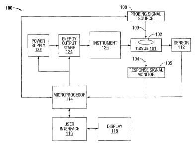
[0022] FIG. 1 is a block diagram of a system for recognizing tissue and
vessels based on
blood circulation according to embodiments of the present disclosure;
[0023] FIGS. 2A and 2B are cross-sectional side views of a portion of the
instrument of FIG.
1 having jaw members for grasping tissue and blood vessels according to
embodiments of the
present disclosure;
[0024] FIG. 3 is a graphical diagram showing impedance variations induced by
blood
circulation and measured with an RF-based tissue sealing device according to
embodiments of
the present disclosure;
[0025] FIG. 4 is a graphical diagram showing the frequency spectrum of the
impedance
variations illustrated in FIG. 3; and
(0026] FIGS. 5 and 6 are flow diagrams of methods for recognizing parameters
of tissue and
vessels according to embodiments of the present disclosure.
DETAILED DESCRIPTION
[0027] Different types of human and animal tissues have different densities of
blood vessels
(i.e., the number of blood vessels per unit area or volume of tissue) and
experience different
levels of blood circulation (i.e., the amount of blood flow per unit volume of
tissue). These
6
CA 02770228 2012-03-02
parameters can be used to identify different types of tissues during a
surgical procedure. For
example, when the tissue structure changes as a result of damage to the
tissue, the blood
circulation usually changes as well. This phenomenon allows one to distinguish
between
damaged and normal portions of tissue by comparing corresponding levels of
blood circulation.
As another example, when tumors form and grow in normal tissue, the density of
blood vessels
in the tissue increases because these tumors depend on the formation of new
blood vessels for
their growth. Thus, by measuring the density of blood vessels or the level of
blood circulation in
tissue, one can distinguish between a tumor and normal tissue.
[0028] For some surgical procedures, such as electrosurgical procedures, the
surgeon may
need to distinguish between blood vessels and other types of vessels, e.g.,
bile ducts. For blood
vessels, the surgeon may need to check for blood clots or other structural
changes in the blood
vessels. For vessel sealing procedures, the surgeon may need to confirm that
the vessel has been
properly sealed before it is cut. In all of these procedures, the tissue or
vessel can be examined
to assess blood circulation conditions. Information regarding blood
circulation conditions may
inform a surgeon regarding the type of the tissue or vessel and/or the
condition of the tissue or
vessel.
[0029] FIG.1 is a block diagram of an energy-based tissue-sealing system 100
for
recognizing tissue or vessels based upon blood circulation in the tissue or
vessels according to
embodiments of the present disclosure. The system 100 (and the methods
described below) may
use any type of energy to seal tissue including mechanical energy, acoustical
energy, thermal
energy, electric energy, or electromagnetic energy (e.g., optical energy or
radio frequency (RF)
energy).
7
CA 02770228 2012-03-02
[0030] The system 100 includes a power supply 122, an energy output stage 124,
and an
instrument 126. The power supply 122 supplies power to the energy output stage
124, which
generates energy and provides the energy to the instrument 126. The instrument
126, in turn,
applies the generated energy to the tissue 101, which includes at least one
vessel 102. For an
RF-based tissue-sealing system, the energy output stage 124 generates RF
energy and the
instrument 126 applies the RF energy to the tissue 101 through at least one
contact to seal the
tissue 101.
[0031] The system 100 also includes a sensor 112, a microprocessor 114, a user
interface
116, and a display 118. The sensor 112 senses various parameters and/or
properties of tissue 101
at the operating site and transmits sensor signals representing the sensed
parameters or properties
of the tissue 101 to the microprocessor 114. The microprocessor 114 processes
the sensor
signals and generates control signals based on the processed sensor signals to
control the power
supply 122 and/or the energy output stage 124. For example, the microprocessor
114 may
regulate the voltage or current output from the power supply 122 or the energy
output stage 124
based on the processed sensor signals.
[0032] The sensor 112 is configured to measure various electrical or
electromechanical
conditions at the operating site such as tissue impedance, changes in tissue
impedance, tissue
temperature, changes in tissue temperature, leakage current, applied voltage,
and applied current.
The sensor 112 continuously measures one or more of these conditions so that
the
microprocessor 114 can continually adjust the energy output from the power
supply 122 and/or
the energy output stage 124 during a sealing procedure. For example, in an RF-
based vessel
sealing instrument, the sensor 112 may measure tissue impedance and the
microprocessor 114
may adjust the voltage generated by the energy output stage 124.
8
CA 02770228 2012-03-02
[0033] The user interface 116 is coupled to the microprocessor 114 allowing a
user to control
various parameters of the energy applied to the tissue 101 during a surgical
procedure. For
example, the user interface 116 may allow a user to manually set, regulate
and/or control one or
more parameters of the energy delivered to the tissue, such as voltage,
current, power, frequency,
and/or pulse parameters, e.g., pulse width, duty cycle, crest factor, and/or
repetition rate.
[0034] The microprocessor 114 can execute software instructions for processing
data
received from the user interface 116 and for outputting control signals to the
power supply 122
and/or the energy output stage 124. The software instructions are stored in an
internal memory
of the microprocessor 114, an internal or external memory bank accessible by
the microprocessor
114 and/or an external memory, e.g., an external hard drive, floppy diskette,
or CD-ROM.
Control signals generated by the microprocessor 114 may be converted to analog
signals by a
digital-to-analog converter (DAC) (not shown) before being applied to the
power supply 122
and/or energy output stage 124.
[0035] For some embodiments of an RF-based tissue-sealing system, the power
supply 122
is a high-voltage DC power supply that produces RF current. In these
embodiments, the
microprocessor 114 transmits control signals to the power supply to control
the magnitudes of
the RF voltage and current output from the power supply 122. The energy output
stage 124
receives the RF current and generates one or more pulses of RF energy. The
microprocessor 114
generates control signals to regulate the pulse parameters of the RF energy,
such as pulse width,
duty cycle, crest factor, and repetition rate. In other embodiments, the power
supply 122 is an
AC power supply, and the energy output stage 124 may vary the waveform of the
AC signal
generated by the power supply 122 to achieve a desired waveform.
9
CA 02770228 2012-03-02
[0036] As described above, the energy-based tissue-sealing system 100 includes
a user
interface 116. The user interface 116 includes an input device, such as a
keyboard or touch
screen, through which a user enters data and commands. The data may include
the type of
instrument, the type of procedure, and/or the type of tissue. The commands may
include target
effective voltage, current, or power level, or other commands for controlling
parameters of the
energy that is delivered from the energy output stage 124 to the instrument
126.
[0037] The system 100 also includes a probing signal source 108 and a response
signal
monitor 105. The probing signal source 108 applies a probing signal 109 to the
tissue 101 and
the response signal monitor 105 senses a response signal 104. The response
signal 104 is the
probing signal 109 that has been transmitted and/or scattered by the tissue
101 and vessel 102.
The probing signal 109 and the response signal 104 may be acoustical signals,
optical signals,
RF signals, or any combination of these signals. In some embodiments, the
probing signal
source 108 is the energy output stage 124. The energy output stage 124 may
generate a probing
signal 109 that is the same as the electrosurgical energy applied to the
tissue 101 to perform an
electrosurgical procedure (e.g., vessel sealing). Alternatively, the energy
output stage 124 may
generate a probing signal 109 that has parameters that are different from the
parameters of the
electrosurgical energy applied to the tissue 101.
[0038] The response signal monitor 105 generates a sensor signal or sensor
data based on the
response signal 104 and transmits the sensor signal or sensor data to the
microprocessor 114.
The microprocessor 114 processes the sensor signal or sensor data to determine
the level of
blood circulation in the tissue 101 or vessel 102. For example, the
microprocessor 114 may
determine the level of blood circulation based on the magnitude of the sensor
signal or the
response signal 104.
i
CA 02770228 2012-03-02
[00391 The response signal 104 may provide information about the tissue type.
For example,
the response signal 104 may identify the tissue as connective tissue, muscle
tissue, nervous
tissue, epithelial tissue, or any combination of these tissue types. The
response signal 104 may
also identify the vessel type within the tissue 101. The vessel types include
bile vessels, lymph
vessels, and blood vessels. The response signal 104 may distinguish the type
of blood vessel that
resides in a given portion of tissue. The types of blood vessels include
arteries, arterioles,
capillaries, venules, and veins. The response signal 104 may also be used to
identify the
condition of the tissue, such as whether the tissue is damaged.
[00401 The system 100 may determine the level of blood circulation by sensing
tissue
parameters or properties that depend on the level of blood circulation during
a period exceeding
one cardiac cycle. In some embodiments, the system 100 may sample tissue
parameters or
properties for multiple cardiac cycles to more accurately determine the level
of blood circulation.
In other embodiments, a cardiac signal, which is related to heart contractions
(e.g., an
electrocardiographic signal), can be used to evaluate the correlation between
the parameters of
the sensor signal and the cardiac signal to more accurately assess the level
of blood circulation.
[00411 FIGS. 2A and 2B show portions of an embodiment of the energy-based
instrument
126 of FIG. 1 having jaw members 203, 204 configured to grasp and compress
tissue 101 and
vessels 102. The jaw members 203, 204 include electrodes or contacts 205, 206
that are
electrically coupled to the energy output stage 124. The electrodes 205, 206
receive energy from
the energy output stage 124 and apply it to the tissue 101 and vessels 102 to
seal the tissue 101
and vessels 102.
[00421 As described above, the energy-based instrument 126 is also configured
to evaluate
blood circulation in a given volume of tissue 101. To evaluate blood
circulation, the given
11
i
CA 02770228 2012-03-02
volume of tissue 101 is first grasped between the jaw members 203, 204 of the
energy-based
instrument 126. The pressure that is applied to the tissue 101 by the jaw
members 203, 204 is
selected to provide electrical contact between the electrodes 205, 206 and the
tissue 101.
However, the amount of pressure applied to the tissue 101 may be lower than
the amount of
pressure used to compress the tissue 101 during tissue sealing. Then, a
probing signal 109 (e.g.,
an RF signal) is applied to the tissue 101 by the electrodes 205, 206 and a
response signal 104
(e.g., tissue impedance) is measured during one or more cardiac cycles.
[00431 During the cardiac cycles, the pressure of the blood flowing in the
blood vessels 102
varies and, as a result, the relative amount of blood in a given volume of
tissue 101 also varies.
For example, as shown in FIG. 2A, during a first portion of the cardiac cycle,
the pressure of the
blood flowing within the blood vessels 102 is at a low level and the volume of
blood within the
given volume of tissue 101 is at a low level. On the other hand, as shown in
FIG. 1 B, during a
second portion of the cardiac cycle, the pressure of the blood flowing within
the blood vessels
102 is at a high level and the volume of blood within the given volume of
tissue 101 is at a high
level. The volume of blood within the given volume of tissue 101 may be
measured by
measuring the impedance of the tissue 101. The impedance may be measured by
applying the
probing signal 109 to the tissue 101 and sensing the response signal 104.
[0044] During a cardiac cycle, as the volume of blood in a given volume of
tissue increases,
a force is applied to the jaw members 203, 204 to increase the distance
between the jaw members
203, 204. In some embodiments, the system 100 includes a motion sensor
configured to sense
the change in distance between the jaw members 203, 204. This distance
information may be
used together with the response signal 104 to evaluate the level of blood
circulation within a
given volume of tissue 101.
12
CA 02770228 2012-03-02
[0045] As described above, a probing signal 109 is applied to a vessel and a
response signal
104 is measured over time to identify tissue 101 and/or vessels 102 or to
determine parameters of
the tissue 101 and/or the vessels 102. The response signal 104 may include the
frequency and
amplitude of an electrical impedance of the tissue 101. If the frequency of
the electrical
impedance correlates to the frequency of cardiac contractions, then the vessel
102 is identified as
a blood vessel. If the vessel is identified as a blood vessel, the amplitude
of the electrical
impedance would indicate the level of blood circulation.
[0046] FIG. 3 is a graph showing experimentally-measured impedance of tissue
302 versus
time. The graph has a y-axis 311 that indicates the tissue impedance in ohms
and an x-axis 312
that indicates the time in seconds. As shown in FIG. 3, the measured impedance
302 continually
varies according to the cardiac cycles where a cardiac cycle is the distance
between the peaks of
the measured impedance 302. In this case, the measured impedance 302 has a
peak-to-peak
amplitude of approximately 0.1 ohms and a period of approximately 0.8 seconds
(which
corresponds to a heart rate of 75 beats per minute). The measured impedance
302 varies
according to the cardiac cycles because the volume of blood within a given
volume of tissue 101
varies according to the cardiac cycles. In other words, the measured impedance
302 correlates
with the volume of blood within a given volume of tissue 101. Depending on the
design of the
instrument, it is also possible that an increase in blood pressure can expand
the grasped tissue
and, as a result, the tissue volume between the jaw members changes. This
effect may also
contribute to variations in measured impedance.
[0047] FIG. 4 is a graph showing the frequency spectrum of experimentally-
measured
impedance variations in tissue corresponding to FIG. 3. The graph has a y-axis
411 that
indicates the spectral power density of the experimentally-measured impedance
variations in
13
CA 02770228 2012-03-02
tissue and an x-axis 412 that indicates the frequency in Hertz. The graph
shows modulation
variations related to the fundamental frequency of cardiac contractions 402
and its harmonics
403, 404. In this case, the fundamental frequency of cardiac contractions 402
is approximately
1.25 Hz, which corresponds to a cardiac cycle of approximately 0.8 seconds in
FIG. 3. The
measured impedance also includes variations related to breathing 401 and the
inter-modulation
products between the variations due to heart contraction and the variations
due to breathing.
[0048] FIG. 5 is a flow diagram of a process for identifying parameters of
tissue and vessels
according to embodiments of the present disclosure. After the process starts
in step 501, a
probing signal 109 is applied to tissue in step 502. The probing signal 109
interacts with the
tissue to create a response signal 104. In step 504, the response signal 104
is monitored over an
interval equal to or longer than an interval between two successive cardiac
contractions. The
response signal may be monitored at a frequency within a range between 10 kHz
and 10 MHz
using, e.g., an energy-based tissue sealing instrument.
[0049] Next, in step 506, the amplitude of the response signal 104 is
determined. The
amplitude of the response signal 104 may be determined at a frequency of the
cardiac
contractions or at the harmonics of the frequency of the cardiac contractions.
Then, in step 508,
the level of blood circulation in the tissue is determined based on the
amplitude of the response
signal 104. In other embodiments, the amplitude and phase of the response
signal 104 are
analyzed to determine the level of blood circulation in the tissue. Finally,
before the process
ends in step 511, a tissue parameter is determined in step 510 based on the
level of blood
circulation.
[0050] In some embodiments, the probing signal source 108 of FIG. 1 applies a
probing
signal to different portions of the tissue. The response signal monitor 105
then monitors
14
CA 02770228 2012-03-02
parameters of the response signals and the microprocessor 114 determines the
level of blood
circulation in different portions of the tissue based on the response signals.
The microprocessor
114 may also determine parameters of the tissue based on the level of blood
circulation in
different portions of the tissue.
[00511 FIG. 6 is a flow diagram of a process for identifying parameters of
tissues and vessels
according to other embodiments of the present disclosure. As in FIG. 5, after
the process starts
in step 601, a probing signal 109 is applied to tissue in step 602. The
probing signal 109
interacts with the tissue to create a response signal 104. In step 604, the
response signal 104 is
monitored over an interval equal to or longer than an interval between two
successive cardiac
contractions. In addition, a cardiac signal related to cardiac contractions is
monitored in step
606. In step 608, the response signal 104 and the cardiac signal are
correlated. Then, in step
610, the level of blood circulation in the tissue is determined based upon the
result of correlating
the response signal 104 and the cardiac signal. Finally, before the process
ends in step 613, a
tissue parameter is determined in step 612 based upon the result of
determining the level of blood
circulation in the tissue. As described above, the tissue parameter may
include the tissue type,
such as connective tissue, muscle tissue, nervous tissue, or epithelial
tissue.
[00521 Although the illustrative embodiments of the present disclosure have
been described
herein with reference to the accompanying drawings, it is to be understood
that the disclosure is
not limited to those precise embodiments, and that various other changes and
modifications may
be effected therein by one skilled in the art without departing from the scope
or spirit of the
disclosure.