Note: Descriptions are shown in the official language in which they were submitted.
CA 02770243 2012-02-06
WO 2011/026640 PCT/EP2010/005437
ES-MS of glycopeptides for analysis of glycosylation
Herein is reported a mass spectrometric method for the analysis of the
glycosylation of an immunoglobulin which does not require a chromatographic
separation step.
Background of the Invention
The glycosylation of a polypeptide is an important characteristic for many
recombinantly produced therapeutic polypeptides. Glycosylated polypeptides,
also
termed glycoproteins, mediate many essential functions in eukaryotic
organisms,
e.g. humans, and some prokaryotes, including catalysis, signaling, cell-cell
communication, activities of the immune system, molecular recognition and
association. Glycoproteins account for the majority of non-cytosolic proteins
in
eukaryotic organisms (Lis, H., et al., Eur. J. Biochem. 218 (1993) 1-27). The
introduction of the glycosylation is a cotranslational and posttranslational
modification and, thus, is not genetically controlled. The biosynthesis of
oligosaccharides is a multistep process involving several enzymes, which
compete
with each other for the substrate. Consequently, glycosylated polypeptides
comprise a microheterogeneous array of oligosaccharides, giving rise to a set
of
different glycoforms containing the same amino acid backbone. Terminal
sialylation of glycosylated polypeptides for example has been reported to
increase
serum-half life of therapeutics, and glycosylated polypeptides containing
oligosaccharide structures with terminal galactose residues show increased
clearance from circulation (Smith, P.L., et al., J. Biol. Chem. 268 (1993) 795-
802).
Thus, in the biotechnological production of therapeutic polypeptides, e. g. of
immunoglobulins, the assessment of oligosaccharide microheterogeniety and its
batch-to-batch consistency are important tasks.
Immunoglobulins differ significantly from other recombinant polypeptides in
their
glycosylation. Immunoglobulin G (IgG) e. g. is a symmetrical, multifunctional
glycosylated polypeptide of an approximate molecular mass of 150 kDa. It is
consisting of two identical Fab parts responsible for antigen binding and the
Fc part
responsible for effector function. Glycosylation tends to be highly conserved
in
IgG molecules at Asn-297, which is buried between the CH2 domains of the heavy
chains, forming extensive contacts with the amino acid residues within the CH2
domain (Sutton, B.J. and Phillips, D.C., Biochem. Soc. Trans. 11 (1983) 130-
132).
The Asn-297 linked core oligosaccharide structures are heterogeneously
processed,
CA 02770243 2012-02-06
WO 2011/026640 PCT/EP2010/005437
-2-
such that a specific IgG exists in multiple glycoforms. Variations exist in
the site
occupancy of the Asn-297 site (macroheterogeniety) or by variation in the
oligosaccharide structure at the glycosylation site (microheterogeniety), see
for
example Jenkins, N., et al., Nature Biotechnol. 14 (1996) 975-981. Generally,
the
more abundant oligosaccharide groups in IgG mAb are asialo biantennary complex
type glycans, primarily agalactosylated (GO), mono-galactosylated (G1), or bi-
galactosylated (G2) types (Ghirlandaio, R., et al., Immunol. Lett. 68 (1999)
47-52).
Given the importance of glycosylation on functional properties of recombinant
glycosylated polypeptides and the necessity of a well-defined and consistent
product production process, an on-line or ad-line analysis of the
glycosylation
profile of recombinantly produced glycosylated polypeptides during the
fermentation process is highly desirable.
Kuhlmann (Kuhlmann, F.E., et al., J. Am. Soc. Mass Spec. 6 (1995) 1221-1225)
reported the post reverse-phase high-performance liquid chromatography column
addition of a solution of 75 % propionic acid and 25 % 2-propanol in a ratio
1:2 to
the column flow. High-performance liquid chromatography with electrospray
ionization mass spectrometry (LCIMS) and liquid chromatography with tandem
mass spectrometry (LC/MS/MS) were applied to the analysis of the site-specific
carbohydrate heterogeneity in erythropoietin (EPO) (Kawasaki, N., et al.,
Anal.
Biochem. 285 (2000) 82-91).
In US 2006/0269979 a high throughput glycan analysis for diagnosing and
monitoring rheumatoid arthritis and other autoimmune diseases is reported. An
identification method of glycoproteins is reported in WO 2009/048196. In US
7,351,540 protein isolation and analysis is reported. Development of an
immunofluorometric assay for human kallikrein 15 is reported by Shaw et al.
(Clin.
Biochem. 40 (2007) 104-110).
Summary of the Invention
Herein is reported as one aspect a method for the determination of the
glycosylation of an immunoglobulin comprising
- enzymatically digesting the immunoglobulin,
- absorbing the immunoglobulin fragments to Sepharose beads,
- washing the Sepharose beads with the absorbed immunoglobulin
fragments with a solution comprising trifluoroacetic acid,
CA 02770243 2012-02-06
WO 2011/026640 PCT/EP2010/005437
-3-
- recovering the immunoglobulin fragments from the Sepharose beads,
- performing an electrospray mass spectrometry of the recovered
immunoglobulin fragments, and
- determining the glycosylation of the immunoglobulin from the mass
spectrometric data.
Detailed Description of the Invention
The current invention is directed to a method for the determination of the
glycosylation of an immunoglobulin with ES-MS without the need for a
chromatographic purification step after the enzymatic digestion of the
immunoglobulin and prior to the mass spectrometric analysis.
Human immunoglobulins are mainly glycosylated at the asparagine residue at
position 297 (Asn297) with a core fucosylated biantennary complex
oligosaccharide (numbering according to Kabat). Asn297 refers to the
asparagine
residue located at about position 297 in the Fc region (Eu numbering of Fc
region
residues) of an immunoglobulin. However, Asn297 may also be located about 3
amino acids upstream or downstream of position 297, i.e., between positions
294
and 300, due to minor sequence variations occurring in immunoglobulins.
Immunoglobulins produced by mammalian cells typically comprise a branched,
biantennary oligosaccharide that is generally attached by an N-linkage to
Asn297
of the CH2 domain of the Fc region (see, e.g., Wright, A. and Morrison, S.L.,
Trend. Biotechnol. 15 (1997) 26-32). The oligosaccharide may include various
carbohydrates, e.g., mannose, N-acetyl glucosamine (G1cNAc), galactose, and
sialic acid, as well as a fucose attached to a G1cNAc in the "stem" of the
biantennary oligosaccharide structure. The biantennary glycostructure, i.e.
the
biantennary oligosaccharide, is terminated by up to two galactose residues in
each
arm. The arms are denoted (1,6) and (1,3) according to the bond to the central
mannose residue. The glycostructure denoted as GO comprises no terminal
galactose residue. The glycostructure denoted as G1 contains one or more
galactose
residues in one arm. The glycostructure denoted as G2 contains one or more
galactose residues in each arm (Raju, T.S., Bioprocess Int. 1 (2003) 44-53).
Human
constant heavy chain regions are reported in detail by Kabat, E.A., et al.,
Sequences
of Proteins of Immunological Interest, 5th Ed. Public Health Service, National
Institutes of Health, Bethesda, MD. (1991); by Brueggemann, M., et al., J.
Exp.
Med. 166 (1987) 1351-1361; and by Love, T.W., et al., Methods Enzymol. 158
CA 02770243 2012-02-06
WO 2011/026640 PCT/EP2010/005437
-4-
(1989) 515-527. CHO type glycosylation of antibody Fc parts is e.g. described
by
Routier, F.H., Glycoconjugate J. 14 (1997) 201-207.
The term "immunoglobulin" encompasses the various forms of immunoglobulins
such as human immunoglobulins, humanized immunoglobulins, chimeric
immunoglobulins, or T cell antigen depleted immunoglobulins (see e.g.
WO 98/33523, WO 98/52976, and WO 00/34317). Genetic engineering of
immunoglobulins is e.g. described in Morrison, S.L., et al., Proc. Natl. Acad
Sci.
USA 81 (1984) 6851-6855; US 5,202,238 and US 5,204,244; Riechmann, L., et al.,
Nature 332 (1988) 323-327; Neuberger, M.S., et al., Nature 314 (1985) 268-270;
Lonberg, N., Nat. Biotechnol. 23 (2005) 1117-1125.
An immunoglobulin in general comprises two so called full length light chain
polypeptides (light chain) and two so called full length heavy chain
polypeptides
(heavy chain). Each of the full length heavy and light chain polypeptides
contains a
variable domain (variable region) (generally the amino terminal portion of the
full
length polypeptide chain) comprising binding regions which can interact with
an
antigen. Each of the full length heavy and light chain polypeptides comprises
a
constant region (generally the carboxyl terminal portion). The constant region
of
the full length heavy chain mediates the binding of the antibody i) to cells
bearing a
Fc gamma receptor (FcyR), such as phagocytic cells, or ii) to cells bearing
the
neonatal Fc receptor (FcRn) also known as Brambell receptor. It also mediates
the
binding to some factors including factors of the classical complement system
such
as component (C 1 q). The variable domain of a full length immunoglobulin's
light
or heavy chain in turn comprises different segments, i.e. four framework
regions
(FR) and three hypervariable regions (CDR). A "full length antibody heavy
chain"
is a polypeptide consisting in N-terminal to C-terminal direction of an
antibody
heavy chain variable domain (VH), an antibody constant domain I (CHI), an
antibody hinge region, an antibody constant domain 2 (CH2), an antibody
constant
domain 3 (CH3), and optionally an antibody constant domain 4 (CH4) in case of
an
antibody of the subclass IgE. A "full length antibody light chain" is a
polypeptide
consisting in N-terminal to C-terminal direction of an antibody light chain
variable
domain (VL), and an antibody light chain constant domain (CL). The full length
antibody chains are linked together via inter-chain disulfide bonds between
the CL-
domain and the CH 1 domain and between the hinge regions of the full length
antibody heavy chains.
CA 02770243 2012-02-06
WO 2011/026640 PCT/EP2010/005437
-5-
It has been reported in recent years that the glycosylation of
immunoglobulins, i.e.
the saccharide composition and multitude of attached glycostructures, has a
strong
influence on the biological properties (see e.g. Jefferis, R., Biotechnol.
Prog. 21
(2005) 11-16). Immunoglobulins produced by mammalian cells contain 2-3 % by
mass oligosaccharides (Taniguchi, T., et al., Biochem. 24 (1985) 5551-5557).
This
is equivalent e.g. in an immunoglobulin of class G (IgG) to 2.3
oligosaccharide
chains in an IgG of mouse origin (Mizuochi, T., et al., Arch. Biochem.
Biophys.
257 (1987) 387-394) and to 2.8 oligosaccharide chains in an IgG of human
origin
(Parekh, R.B., et al., Nature 316 (1985) 452-457), whereof generally two are
located in the Fc-region at Asn297 and the remaining in the variable region
(Saba,
J.A., et al., Anal. Biochem. 305 (2002) 16-31).
The term "glycosylation" denotes the sum of all oligosaccharides which are
attached to all amino acid residues of an immunoglobulin. Due to the
glycosylation
heterogeneity of a cell, a recombinantly produced immunoglobulin comprises not
only a single, defined N- or O-linked oligosaccharide at a specified amino
acid
residue, but is a mixture of polypeptides each having the same amino acid
sequence
but comprising differently composed oligosaccharides at the respective
specified
amino acid position. Thus, the above term denotes a group of oligosaccharides
that
are attached to specified amino acid positions of a recombinantly produced
immunoglobulin, i.e. the heterogeneity of the attached oligosaccharide. The
term
"oligosaccharide" as used within this application denotes a polymeric
saccharide
comprising two or more covalently linked monosaccharide units.
For the notation of the different N- or O-linked oligosaccharides the
individual
sugar residues are listed from the non-reducing end to the reducing end of the
oligosaccharide residue. The longest sugar chain was chosen as basic chain for
the
notation. The reducing end of an N- or O-linked oligosaccharide is the
monosaccharide residue, which is directly bound to the amino acid of the amino
acid backbone of the immunoglobulin, whereas the end of an N- or O-linked
oligosaccharide, which is located at the opposite terminus as the reducing end
of
the basic chain, is termed non-reducing end.
An aspect as reported herein is a method for the determination of the
glycosylation
of an immunoglobulin comprising
- enzymatically digesting the immunoglobulin,
- absorbing the immunoglobulin fragments to Sepharose beads,
CA 02770243 2012-02-06
WO 2011/026640 PCT/EP2010/005437
-6-
- washing the Sepharose beads with the absorbed immunoglobulin
fragments with a solution comprising trifluoroacetic acid,
- recovering the immunoglobulin fragments from the Sepharose beads,
- performing an electrospray mass spectrometry of the recovered
immunoglobulin fragments, and
- determining the glycosylation of the immunoglobulin from the mass
spectrometric data.
It has been found that by washing the adsorbed immunoglobulin fragments with a
solution comprising trifluoroacetic acid an improved electrospray mass
spectrometric determination of the glycosylation of the immunoglobulin can be
achieved. In one embodiment the concentration of the trifluoroacetic acid is
of
from 0.01 % to 1 % (v/v). In another embodiment the concentration of the
trifluoroacetic acid is of from 0.05 % to 0.5 % (v/v). In still another
embodiment
the concentration of the trifluoroacetic acid is about 0.1 % (v/v).
Additionally a
chromatographic purification step can be performed after the enzymatic
digestion
but is not necessary. As can be seen from the following Table 1 the washing
with
trifluoroacetic acid clearly improves the accuracy of the quantitative
determination
and concomitantly reduces the standard deviation (SD) and variation
coefficient
(VK) of the analysis results.
Table 1: Comparison of exemplary results for the determination of the
glycosylation of an exemplary anti-CCR5 antibody. The
determinations have been made in triplicate. The reference values
have been determined by ion exchange chromatography with pulsed
amperometric detection (Fuc = fucose).
glyco- reference without trifluoroacetic with trifluoroacetic acid
structure value acid washing washing
[%] 1%] [%]
value SD variation value SD variation
coefficient coefficient
Man-Fuc 23.4 0.6 2.7 22.4 0.3 1.3
28.4
G(0)-Fuc 10.9 1.0 9.3 10.8 1.0 9.3
G(0) 44.3 40.1 3.0 7.5 43.4 0.4 1.0
G(1) 25.2 19.4 1.7 9.0 19.9 0.2 1.1
G(2) 2.1 6.2 3.8 61.1 3.4 0.7 21.9
CA 02770243 2012-02-06
WO 2011/026640 PCT/EP2010/005437
-7-
The term "Sepharose" denotes a crosslinked form of agarose. Agarose is a
linear
polysaccharide comprising as monomeric building blocks agarobiose, which in
turn
is a disaccharide consisting of glycosidically linked D-galactose and 3,6-
anhydro-
L-galactopyranose.
In one embodiment the enzymatically digesting is by incubating with an enzyme
selected from trypsin, chymotrypsin, papain, IdeS, and the endoproteinases Arg
C,
Lys C and Glu C. In another embodiment the enzymatically digesting is by
incubating with trypsin.
It has further been found that it is advantageous to use a solution in the
washing
step with an acetonitrile concentration of from 78 % to 88 % (v/v). In one
embodiment the acetonitrile concentration is of from 80 % to 85 % (v/v). In
another embodiment the acetonitrile concentration is about 83 % (v/v). The
term
"about" denotes that the thereafter following value is the center of a range
of +/-
10 % of the value. Values beside that range have a negative influence on the
quantitative determination. Therefore, in one embodiment the solution in the
washing step comprises about 0.1 % (v/v) trifluoroacetic acid and about 83 %
(v/v)
acetonitrile. In one embodiment comprises the method the step of washing the
Sepharose beads with a solution consisting of 78 % to 88 % (v/v) acetonitrile
and
water. In one embodiment the method comprises the step of washing the
Sepharose
beads with a solution consisting of 80 % to 85 % (v/v) acetonitrile and water.
In
one embodiment the washing is with a solution consisting of about 83 % (v/v)
acetonitrile and water. In a further embodiment the method comprises the step
of
adjusting the solution of the enzymatic digest to 78 % to 88 % (v/v)
acetonitrile. In
a further embodiment the method comprises the step of adjusting the solution
of the
enzymatic digest to 80 % to 85 % (v/v) acetonitrile. In one embodiment the
adjusting is to about 83 % (v/v) acetonitrile. In another embodiment the
method
comprises a second washing step with 78 % to 88 % (v/v) acetonitrile. In one
embodiment the method comprises a second washing step with 80 % to 85 % (v/v)
acetonitrile. In another embodiment the second washing is with about 83% (v/v)
acetonitrile.
If a reference is made to a volumetric ratio (v/v) within this application the
following applies:
CA 02770243 2012-02-06
WO 2011/026640 PCT/EP2010/005437
-8-
- depending on the intended final volume the relative volume of the
acetonitrile fraction, e.g. 83 %, is calculated from the intended final
volume,
- the calculated relative volume of acetonitrile is provided and water is
added until the intended final volume is obtained,
- thereafter the relative volume fraction of trifluoroacetic acid is added,
calculated based on the intended final volume.
For example, one liter (1000 ml) of a solution consisting of 0.1 % (v/v)
trifluoroacetic acid, 83 % (v/v) acetonitrile and water is obtained by
providing
830 ml acetonitrile (83 % of 1000 ml), adding water thereto until a volume of
1000 ml is reached, and thereafter adding 1 ml (0.1 % (v/v) of 1000 ml)
trifluoroacetic acid.
In one embodiment the method comprises as first step denaturating the
immunoglobulin with a denaturing agent. In another embodiment the denaturing
is
at pH 8.5. In one embodiment the solution consists of 0.1 % (v/v) trifluoro
acetic
acid, 83 % (v/v) acetonitrile and water. In another embodiment the Sepharose
beads are sepharose CL-4B beads. In one embodiment the applying to Sepharose
beads is for 5 minutes.
In another embodiment the method comprises the step of washing the Sepharose
beads with water. In this step the immunoglobulin fragments are recovered from
the Sepharose beads.
It has further been found that without adding the solution containing 25 %/75
%
(v/v) 2-propanol/propionic acid the ionization of the purified glycopeptides
is very
poor. Thus, although the method works without the addition it can be further
improved by additionally adding a solution containing 25 %/75 % (v/v)
2-propanol/propionic acid. Additionally, the higher charge states of the
glycopeptides, which can be used for a correct quantitation of the different
glycopeptides species of glycopeptides, are increasingly present if a solution
containing 25 %/75 % (v/v) 2-propanol/propionic acid is added. This can be
seen
for the fucosylated and G(2) forms from Table 2. Therefore, in one embodiment
the
method comprises the step of mixing the immunoglobulin fragments with a
solution consisting of 25 % (v/v) 2-propanol and 75 % (v/v) propionic acid.
CA 02770243 2012-02-06
WO 2011/026640 PCT/EP2010/005437
-9-
Table 2: Comparison of exemplary results for the determination of the
glycosylation of an exemplary anti-CCR5 antibody. The
determinations have been made in triplicate. The reference values
have been determined by ion exchange chromatography with pulsed
amperometric detection (Fuc = fucose).
glyco- reference with trifluoroacetic with trifluoroacetic
structure value acid washing acid washing
[%] without addition of with addition of 2-propanol
2-propanol/propionic acid and propionic acid
[%] [%]
value SD variation value SD variation
coefficient coefficient
Man-Fuc 28.4 22.4 0.3 1.3 22.2 0.0 0.0
G(0)-Fuc 10.8 1.0 9.3 8.5 0.2 2.1
G(0) 44.3 43.4 0.4 1.0 44.3 0.8 1.8
G(l) 25.2 19.9 0.2 1.1 21.6 0.7 3.1
G(2) 2.1 3.4 0.7 21.9 3.5 0.3 8.4
Signal intensity 3139.0 1326.4 42.3 7775.0 796.7 10.2
[area units]
The following examples and figures are provided to aid the understanding of
the
present invention, the true scope of which is set forth in the appended
claims. It is
understood that modifications can be made in the procedures set forth without
departing from the spirit of the invention.
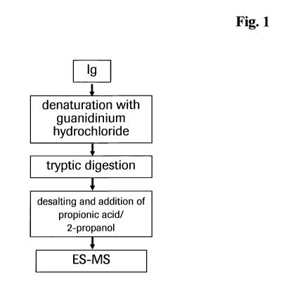
Description of the Figure
Figure 1 Schematic method diagram.
Materials
Tris (hydroxy aminomethane) hydrochloride (TRIS-HC1) and guanidinium-
hydrochloride were purchased from Merck. Acetonitril (ACN), trifluoroacetic
acid
(TFA), hydrochloric acid, 2-propanol and propionic acid were obtained from VWR
International Baker.
Trypsin was obtained from Roche Diagnostics GmbH, Mannheim, Germany.
NAP5-Sephadex columns were obtained from GE Healthcare. CL-4B Sepharose
beads were purchased form Amersham Bioscience. Multiscreen Solvinert 96 well
CA 02770243 2012-02-06
WO 2011/026640 PCT/EP2010/005437
-10-
0.45 pm pore-size low-binding hydrophilic PTFE Filter Plates were obtained
from
Millipore.
The invention is exemplified with an anti-CCR5 antibody. The production
thereof
and the coding sequences thereof are reported e.g. in WO 2006/103100 and
WO 2009/090032.
Example 1
Digestion
300 g of purified anti-CCR5 antibody were incubated for some minutes with
guanidinium-hydrochloride at pH 8.5. After buffer exchange to TRIS-HC1 pH 8.5
using a Sephadex column the antibody was digested without prior reduction with
trypsin at 37 C over night (16 hours).
Example 2
Purification
I ml of Sepharose CL-4B beads were washed three times with water. 15 l of
cleaned Sepharose beads were dissipated in 200 l water and thereafter
assigned to
the wells of a 96-well Multiscreen filter plate. The beads were washed two
times
each with 200 l of water and conditioned two times each with 200 Al of an 83
%
acetonitrile/water solution on a vacuum manifold using vacuum at < 0.1 inch.
Hg.
40 gl of the tryptic digest were adjusted to 83 % (v/v) acetonitrile. The
digest
solution was thereafter applied to the conditioned Sepharose beads and
incubated
for five minutes with gentle shaking. The 96 well plate was covered with a
suitable
lid to prevent acetonitrile from evaporating. The beads were washed two times
each
with 200 pl 0.1 % TFA-83 %ACN (v/v) and two times each with 200 l 83 % (v/v)
acetonitrile. During the washing steps the beads must be kept always wet to
prevent
the glycopeptides form eluting. The glycopeptides were recovered from the
beads
with three times 30 gl of water in a 96 well v-bottom plate.
Example 3
Sample preparation for mass spectrometry
For MS nanospray analysis the glycopeptides were mixed with 30 l of a
solution
containing 25 %/75 % (v/v) 2-propanol/propionic acid. The prepared sample was
directly infused to the mass spectrometer by means of a nanospray (NanoMate).
CA 02770243 2012-02-06
WO 2011/026640 PCT/EP2010/005437
-11-
Example 4
Mass spectrometry
For the measurement a calibrated q-TOF Ultima from waters with a NanoMate
source from Advion instead of the normal ultima nanospray source was used. 96
samples can be measured within 288 minutes completely automated.