Note: Descriptions are shown in the official language in which they were submitted.
CA 02770545 2012-02-09
WO 2011/020612
PCT/EP2010/005085
Use of a bis-maleic anhydride cross-linking agent
for fixation of a cell or tissue sample
Background of the Invention
The present invention relates to novel bis-maleic anhydrides. It especially
relates to
the discovery that bis-maleic anhydride cross-linking agents can be used for
preservation/fixation of a cell or tissue sample. With great advantage a bis-
maleic
anhydride cross-linking agent can be used in methods requiring fixation of a
cell or
tissue sample and at the same time requiring that the fixative has little
impact on
the later detection of a protein or a nucleic acid in procedures like
immunohistochemistry, fluorescence in situ hybridization or RT-PCR. It is also
demonstrated that the use of a bis-maleic anhydride cross-linking agent as a
fixative greatly facilitates later detection of an analyte of interest in a
previously
fixed cell or tissue sample.
To date there is no generally applicable, "ideal" way to prepare a cell or
tissue
sample, e.g. for immunohistochemistry or detection of a nucleic acid of
interest,
respectively. Fixation and the reversibility of negative effects introduced by
the
fixation have a major impact on the detectability of polypeptide antigens and
nucleic acids, respectively, and on the reproducibility of the results
obtained
thereupon.
For successful immunostaining of an antigen in a cell or tissue sample at
least three
criteria have to be met: a) retention of the antigen at its original site, b)
accessibility
of the antigen and c) correct conformation/preservation of the antigen/epitope
of
interest. It would appear that at present no fixation and/or detection
procedure fully
meets all these three criteria. For the procedures known in the art, best
performance
for one or two of these criteria goes to the expense of a reduced performance
in at
least one other criterion.
Several fixatives are available and used in the routine of a clinical
pathology
laboratory, like glutardialdehyde, formaldehyde and acetone, or other organic
solvents. The vast majority of fixation procedures, however, are based on the
use of
cross-linking agents, like formaldehyde. The fixative solution usually is an
aqueous
formaldehyde solution that contains sodium phosphates, contrived to provide
buffering (minimal pH change following addition of a small amount of strong
acid
or base) to pH 7.2-7.6 and an approximately isotonic solution (one whose
osmotic
CA 02770545 2012-02-09
WO 2011/020612
PCT/EP2010/005085
- 2 -
pressure is the same as that of mammalian extracellular fluids, often based on
physiological saline).
With state-of-the-art procedures fixation has to be just right.
If fixation is too short, instead of fixation merely coagulation of proteins
by the
alcohols used to dehydrate the sample occurs. This may e.g. negatively impact
the
preservation of tissue morphology or impair long term storage stability.
With prolonged formaldehyde fixation the cross-linked protein molecules form a
dense network that can impair the penetration of paraffin wax or/and the
access of
antibody molecules. As a result an antigen of interest may be reversibly or
even
irreversibly masked. Further an epitope may be chemically modified
("destroyed")
e.g., by reaction with formaldehyde.
In addition, it is known that the activity of most enzymes is impaired after
formaldehyde fixation.
As mentioned before, fixation in formaldehyde is most widely used in clinical
pathology. The major reason most likely is that by fixation with formaldehyde
the
antigen of interest is trapped at the sites it occupied in the living
organism. By way
of methylene bridges introduced upon formaldehyde fixation also the morphology
of a cell or tissue sample is well preserved. These positive effects, however,
go to
the expense of permeability of the sample and to the fixation causing changes
in the
accessibility and/or conformation of an antigen/epitope of interest, damage in
nucleic acids and inactivation of enzyme activity.
Cross-linking due to formaldehyde fixation is likely to mask or to destroy
epitopes,
leading to a false negative immunostaining. This failure is even more likely
to
occur when the primary immunoreagent is a monoclonal antibody than when a
polyclonal antiserum is used. This is why many, many attempts have been made
and are found in the relevant literature dealing with reversing the effects of
formaldehyde fixation.
For long term storage a fixed cell or tissue sample usually has to be de-
hydrated
and embedded in an appropriate embedding medium. Paraffin embedding is usually
preferable to either plastic embedding or cutting un-embedded specimens with a
vibrating microtome or in a cryostat.
CA 02770545 2012-02-09
WO 2011/020612
PCT/EP2010/005085
- 3 -
As illustrated above, all fixation procedures to a certain extent represent
compromises of various kinds. Often optimal preservation of morphology goes to
the expense of accessibility for an antibody or destruction of an antigen or
of an
epitope thereon.
However and also important to the present invention, not only is there a high
variability introduced during preparation of a specimen, like its fixation or
further
processing like embedding with paraffin, probably even more variability is
caused
by the various modes and routes of regaining immunological reactivity or
accessibility in detection of nucleic acids, i.e. in procedures known as
antigen
retrieval.
Despite the broad use and great utility of e.g. immunohistochemical methods or
methods for detecting a nucleic acid of interest in a cell or tissue sample
there is
great need for further improvements. Such improvements may for example relate
to
more gentle fixation of a cell or tissue sample, to improvements in antigen
retrieval
or/and to better comparability and reproducibility of results and may be even
to the
possibility to use antibodies for which the corresponding antigen or epitope
is
destroyed in standard procedures, like formaldehyde fixation.
The inventors of the present invention have surprisingly found that the use of
bis-
maleic anhydrides as a cross-linking agent in the preparation/fixation of a
cell or
tissue sample is of tremendous advantage and can and will lead to significant
improvements regarding at least one or even several of the problems known in
the
art.
Summary of the Invention
The present invention relates to a method for fixation of a cell or a tissue
sample in
vitro wherein said cell or said tissue sample is incubated with a bis-maleic
anhydride cross-linking agent, whereby said cell or said tissue sample is
fixed.
Further disclosed is a method of preserving a cell or a tissue sample the
method
comprising the steps of fixing a tissue sample with a bis-maleic anhydride
cross-
linking agent and of embedding said fixed sample in paraffin.
The present invention also discloses a method for performing
immunohistochemistry on a cell or a tissue sample the method comprising the
steps
of fixing a cell or tissue sample with a bis-maleic anhydride cross-linking
agent,
CA 02770545 2012-02-09
WO 2011/020612
PCT/EP2010/005085
- 4 -
embedding said fixed sample in paraffin, de-paraffinizing said sample,
removing
the bis-maleic amide cross-link and immunologically detecting an epitope of
interest.
Also described is a method for detecting in vitro a nucleic acid of interest
by in situ
hybridization on a cell or a tissue sample the method comprising the steps of
fixing
a cell or tissue sample with a bis-maleic anhydride cross-linking agent,
embedding
said fixed sample in paraffin, de-paraffinizing said sample, removing the bis-
maleic
amide cross-link and detecting a nucleic acid of interest by in situ
hybridization.
Further a method for detecting in vitro a nucleic acid of interest by RT-PCR
in a
cell or a tissue sample the method comprising the steps of fixing a cell or
tissue
sample with a bis-maleic anhydride cross-linking agent, embedding said fixed
sample in paraffin, de-paraffinizing said sample, removing the bis-maleic
amide
cross-link and detecting a nucleic acid of interest by performing RT-PCR is
given.
It is also shown that based on a method according to the present invention
both a
polypeptide of interest and a nucleic acid of interest can be detected in the
same
specimen prepared from a cell or tissue sample. The invention relates to a
method
for detecting in vitro at least one polypeptide of interest by
immunohistochemistry
and at least one nucleic acid of interest in one test sample comprising a cell
or a
tissue sample, the method comprising the steps of fixing a cell or tissue
sample
with a bis-maleic anhydride cross-linking agent, embedding said fixed sample
in
paraffin, de-paraffinizing said sample, removing the bis-maleic amide cross-
link
and immunologically detecting the at least one polypeptide of interest and
detecting
the at least one nucleic acid of interest by performing RT-PCR or fluorescence
in
situ hybridization.
The invention further relates to the use of a bis-maleic anhydride cross-
linking
agent for fixation of a cell or a tissue sample as well as to the use of a bis-
maleic
anhydride cross-linking agent in the manufacturing of a fixative for fixation
of a
cell or tissue sample.
Detailed Description of the Invention
In a first embodiment the present invention relates to a method for fixation
of a cell
or a tissue sample in vitro wherein said cell or said tissue sample is
incubated with
a bis-maleic anhydride according to Formula I,
CA 02770545 2012-02-09
WO 2011/020612
PCT/EP2010/005085
- 5 -
Formula I
0 0
0 1 I 0
R 1 F X
0 0
wherein R1 and R2 independently are selected from the group consisting of
hydrogen, methyl, ethyl, propyl, isopropyl and butyl, wherein X is a linker
with
between 1 and 30 atoms in length and whereby said cell or said tissue sample
is
fixed.
The compound according to Formula I sometimes will simply be referred to as
"bis-maleic anhydride cross-linking agent" or "bis-maleic anhydride".
It will be understood that "a method for fixation of' in the sense of the
present
invention is equivalent to "a method of treating", that "fixing" is equivalent
to
"cross-linking", "is fixed" could alternatively phrased as "is cross-linked"
and that
"fixed" by and in a method of the present invention relates to "comprising a
bis-
maleic amide cross-link" or "comprise(s) a bis-maleic amide cross-link". For
the sake
of convenience and in light of the fact that the skilled artisan is fully
aware of the
meanings attached to terms like fixation, fixative or fixed, and for the sake
of
convenience, only these terms will generally be used throughout the
description.
It will also be appreciated that in a scientifically correct sense it is not a
cell or a
tissue sample that is fixed or cross-linked but rather it is the biomolecules
contained in such sample that are cross-linked or fixed in a fixation method
as
disclosed in the present invention. The cross-links in these biomolecules may
be
intra-molecular as well as intermolecular cross-links.
If at least two maleic anhydrides, linked to each other by a linker X (at
least a bis-
maleic anhydride), are reacted with at least two primary amines at least two
amide
bounds are formed and the at least two primary amines are cross-linked via the
at
least two amide bonds and via the linker X. For the sake of convenience this
type
of cross-link in the following will be referred to as "bis-maleic amide cross-
link".
The articles "a" and "an" are used herein to refer to one or to more than one
(i.e., to
at least one) of the grammatical object of the article.
CA 02770545 2012-02-09
WO 2011/020612
PCT/EP2010/005085
- 6 -
The expression "one or more" or "at least one" denotes 1 to 20, preferably 1
to 15
also preferred 1, 2, 3,4, 5, 6, 7, 8,9, 10, or 12.
The present invention is based on the surprising and striking discovery that a
bis-
maleic anhydride can be used to gently and reversible fix a cell or a tissue
sample.
Without being wanted to be bound to this theory the great advantages are
believed
to be due to the facts that a) a bis-maleic anhydride cross-linking agent
rapidly and
effectively forms bis maleic amide cross-links, that b) the cross-linking
agent bis-
maleic acid can be easily removed whenever desired and/or as may also be that
c)
by using a linker X of an appropriate length less negative effects like
distortion or
destruction of epitopes are likely as compared to the relatively short cross-
linker
formaldehyde.
To fully appreciate the tremendous advantages of the method according to the
present application it will be helpful to discuss in more detail those tissue
fixation
procedures that are most frequently used, i.e. procedures based on the use of
formaldehyde as a fixative.
A formaldehyde-based fixative is usually derived from formalin, which is a
solution containing 37% w/w (= 40% w/v) formaldehyde in water. The working
fixative is a ten-fold dilution of formalin (4 grams per 100 m1). A solution
of
almost identical composition may be made with paraformaldehyde as the starting
material. Paraformaldehyde is a solid polymer that changes into formaldehyde
when heated (in slightly alkaline water) to 60 C. Though the phrase "fixed in
4%
paraformaldehyde" is often used in the literature it is not fully correct.
Most of the
formaldehyde in a diluted aqueous solution is present as methylene glycol,
which is
formed by addition of a molecule of water to one formaldehyde molecule:
HCOH + H20 4- H2C(OH)2
The concentration of free formaldehyde in the fixative solution is very low.
Nevertheless, it is free formaldehyde, rather than methylene glycol, that
enters the
chemical reactions of fixation.
The chemical reactions of fixation by formaldehyde are primarily with primary
amines, as e.g. present in polypeptides.
Formaldehyde fixation does appear to be a two-step process. In a first step
formaldehyde is rapidly bound. By this rapid binding formaldehyde probably
stops
CA 02770545 2012-02-09
WO 2011/020612
PCT/EP2010/005085
- 7 -
autolysis but it does little to stabilize the fine structure of the tissue,
and does not
provide effective protection against disruptive effects of later treatments
such as
paraffin embedding.
In the first stage (hours), formaldehyde molecules combine with various parts
of
protein molecules, especially the side-chain amino group of lysine and the
nitrogen
atoms of peptide linkages:
Protein ¨NH2 + H2C(OH)2 4¨ protein ¨ NHCH2OH + H20.
In the second step the bound hydroxymethyl groups react with other nitrogen
atoms
of the same or adjacent protein molecules, thereby generating a methylene
cross-
link or bridge. These methylene (¨CH2 ¨) bridges, are stable and account for
the
insolubility and rigidity of protein-containing tissues that have been fixed
by
formaldehyde. One possible reaction is:
Protein-NH-CH2OH + NH2-protein ¨> protein-NH-CH2-NH-protein + H20
In addition to the reactions with proteins, formaldehyde may also combine with
some basic lipids.
It is likely that the above discussed two steps of chemical reactions required
for
"full" formaldehyde fixation account for some of the difficulties encountered
when
working with formaldehyde-fixed material. Brief exposure to formaldehyde does
not cause sufficient cross-linking to immobilize small proteins or other small
analytes. A too long fixation can cause irreversible damages especially due to
formation of an excess of methylene bridges. It is generally accepted that for
reasonable structural preservation a specimen should remain in a formaldehyde
solution at least over night or even for about 24 hours.
In immunohistochemistry the epitopes of an antigen of interest must be
accessible
to the primary antibody. An epitope is a small part of a large molecule, such
as an
amino acid sequence of about 5 to about 10 amino acids that specifically binds
to
the binding site of an antibody molecule. A monoclonal antibody recognizes
only
one epitope. A polyclonal antiserum, on the other hand may recognize several
different epitopes.
Reversing the unwanted negative side effects of e.g. formaldehyde fixation is
known in the literature under headings like antigen retrieval or epitope
retrieval.
CA 02770545 2012-02-09
WO 2011/020612
PCT/EP2010/005085
- 8 -
At least three different routes for antigen retrieval are broadly used alone
or on
combination: partial enzymatic digestion, heat and/or different chemicals
supposed
to reverse formaldehyde effects.
For partial proteolytic digestion e.g. an inexpensive grade of porcine trypsin
(containing some chymotrypsin) is used. The rationale of using a proteolytic
enzyme is that breaking some peptide bonds will make holes in the matrix of
cross-
linked proteins, allowing the entry of e.g. antibody molecules. However,
proteolytic enzymes invariably attack all proteins, including the antigen of
interest.
The tight-rope walk is to digest just long enough and as one can easily
appreciate
the conditions for digestion will vary from tissue to tissue and/or from
antigen to
antigen.
Most of the formaldehyde bound to a fixed tissue can be removed by heat
induced
antigen retrieval. However, heat can also cause the irreversible destruction
of target
analytes. Heat is e.g. known to destroy heat-labile epitopes or heat-labile
enzymes.
Heat induced antigen retrieval is often combined with use of special
"retrieval" or
"extraction" buffers. However, success of these procedures can no way be
predicted and the unpredictable outcome of such procedure is one of the great
mysteries in the field of e.g. immunohistochemistry.
Many different buffers and pH-values (e.g. citrate; glycine/HC1 ¨ mainly for
acidic
pH-values ranging from about 2.5 to about 6 and alkaline buffer on basis of
Tris in
the pH-range of about 9 to 10) have been used, either alone or in combination
with
chemicals believed to reverse at least partially the negative effects of the
methylene
cross-links introduced by formaldehyde. Chemicals used either alone dissolved
in
water, or dissolved in buffer e.g. are EDTA, citraconic acid, lead thiocyante,
aluminum chloride, or zinc sulfate.
The large variety of ingredients used in solutions for high temperature
antigen
retrieval indicates that more than one mechanism is probably involved. Most
antigens can be retrieved at near neutral pH, but a more alkaline medium is
needed
for some and acidic conditions for others. In certain cases bonds to tissue-
bound
calcium ions may mask epitopes, necessitating removal of the metal ions by
chelation. Other ingredients of retrieval solutions include heavy metal ions,
which
may expose epitopes by a coagulation-like action on proteins, and chaotropic
CA 02770545 2012-02-09
WO 2011/020612 PCT/EP2010/005085
- 9 -
substances which may modify the shapes of proteins by changing the structures
of
clusters of water molecules.
For analysis of nucleic acids, e.g. from formaldehyde-fixed paraffin-embedded
(FFPE) tissue various methods of antigen (analyte) retrieval, often quite
different to
the ones required for immunological detection of an epitope of interest, are
recommended and used in the art. Nonetheless it is also accepted that
formaldehyde
fixation may have a negative impact on nucleic acids. For example, it is known
that
messenger RNA (m-RNA) in FFPE tissue is at least partially destroyed,
rendering
the detection of m-RNAs of more the 100 nucleotides in length a quite
challenging
task.
Coming now to the properties and advantages of a bis-maleic anhydride cross-
linking agent as shown in the present application: The reaction of maleic
anhydride
with a primary amine can be depicted by the following reaction scheme:
0 0
X¨ R ).
¨
R-NH2 + 0 I -OH N
- I _________________________________________________________ X
+ HO
)
.R1
0 R1 2
0
It is important that on the one hand the amide bond formed is rather stable
during
(long term) storage of a sample fixed on the basis of such reagent and that on
the
other hand the amide bonds can be easily broken and that the primary amines
can
be regained easily and under gentle conditions.
Amide bond formation between a primary amine group and a maleic anhydride
group occurs rapidly at neutral and alkaline pH. Preferably the incubation of
a cell
or tissue sample with a bis-maleic anhydride according to Formula I is
performed
at a pH of 7.0 or above. Also preferred the pH is below pH 12. Further
preferred
the pH used for fixation is in the range of pH 7.0 to pH 11.0, including the
boarders. Also preferred the incubation is performed at a pH of 7.5 to 10.0,
the
boarders inclusive. For obvious reasons a buffer substance comprising a
primary
amine should be avoided.
CA 02770545 2012-02-09
WO 2011/020612
PCT/EP2010/005085
- 10 -
Preferably the fixative will not only be buffered to stabilize the pH during
fixation,
but the buffer will also have a physiological salt concentration. It is
further
advantageous and preferred, if the bis-maleic anhydride cross-linking agent is
provided in a buffer also comprising a water-miscible organic solvent.
Preferred
organic solvents in this context are ethanol, N,N'-dimethylformamide (DMF) and
dimethyl sulfoxide (DMSO).
The amide bound formed by reaction of maleic anhydride with a primary amine is
stable at alkaline pH. If stored, a sample fixed with a bis-maleic anhydride
according to Formula I preferably is kept at neutral or an alkaline pH, with
the
same preferred ranges as given above for the fixation or incubation step.
A sample comprising cells in suspension or a small tissue sample of 1 AfrI3 or
less
can be fixed within short time, like, e.g. within 10 to 60 min. In clinical
routine a
tissue sample, however, will often be much larger than 1 m3. For routine
purposes
it is therefore preferred that a cell or tissue sample is incubated for 1 to
72 hours
with the bis-maleic anhydride cross-linking agent. As the artisan would say
the cell
or tissue sample is fixed for 1 to 72 hours. It is also preferred that in a
method
according to the present invention the incubation of the cell or tissue sample
in the
fixative comprising a bis-maleic anhydride according to Formula I is performed
for
2 to 48 hours.
A bis-maleic anhydride cross-linking agent may e.g. be dissolved in a organic
solvent at a high concentration and can the be diluted to yield an appropriate
working or final concentration. The final concentration of the bis-maleic
anhydride
cross-linking agent used in the incubation/fixation step may vary to some
extend.
Preferably the final concentration is between 0.1 and 20 %. Further preferred
the
final concentration will be between 0,25 and 10 % (% in weight per volume).
The amide bond formed between a primary amine group and a maleic anhydride
group in a fixation method as disclosed in the present application can rapidly
be
broken under acidic buffer conditions. Upon incubation in acidic pH-buffers
the
amide bond is broken, the bis-maleic amide cross-linking is removed and the
bis-
maleic anhydride can be washed away. At the same time the primary amine
previously part of an amide bond with the cross-linking agent is regained and
present again. Preferably a bis-maleic amide cross-link is removed by
incubating a
fixed sample, i.e. a sample fixed by use of a bis-maleic anhydride according
to
Formula I, under acidic pH. Preferably a bis-maleic amide cross-link is
removed by
CA 02770545 2012-02-09
WO 2011/020612
PCT/EP2010/005085
- 11 -
incubation in a buffer with a pH from 2.0 to 6.5. Also preferred the buffer
used for
removing a bis-maleic amide cross-link will have a pH from 2.5 to 6.5, or from
3.0
to 6.0, wherein each of the boarders is inclusive. Removal occurs rapidly, the
more
rapid, the more acidic the buffer. Preferably the fixed sample is incubated
for 2 min
to 2 hours, also preferred from 5 min to 1 hour in order to remove a bis-
maleic
amide cross-link.
It has been found that the residues R1 and R2 have to be selected from
hydrogen,
methyl, ethyl, propyl, isopropyl or butyl for use of the bis-maleic anhydride
cross-
linking agent in a method disclosed in the present application.
Further preferred, the residues R1 and or R2 according to Formula I are
selected
from hydrogen, methyl or ethyl. Also preferred the bis-maleic anhydride for
use in
a method according to the present invention is a bis-maleic anhydride
according to
Formula I, wherein R1 is hydrogen or methyl and R2 is hydrogen or methyl.
If R1 and R2 are the same the amide bonds formed during fixation of a cell or
tissue sample can be reversed under the same conditions. This will be
advantageous
whenever as much as possible cross-linker shall be removed and at the same
time
as much as possible primary amine shall be regained. In a preferred embodiment
the bis-maleic anhydride for use in a method according to the present
invention
therefore is a bis-maleic anhydride according to Formula I, wherein R1 and R2
are
the same.
As the skilled artisan will appreciate, now that it has been found that a bis-
maleic
anhydride can be used for fixation of a cell or tissue sample many, many
compounds comprising at least two maleic anhydrides linked by a linker can be
designed and used.
The linker X in the bis-maleic anhydride for use in a method according to the
present invention has a length of between 1 and 30 atoms. The term length must
understood as consisting of the number of atoms given. A linker of 30 atoms in
lengths has a backbone that consists of 30 atoms.
Obviously the linker X can be designed in many ways as the specific
application
may require and no undue limitation or restriction would be appropriate.
Nonetheless some preferred examples of such linkers shall be given.
CA 02770545 2012-02-09
WO 2011/020612
PCT/EP2010/005085
- 12 -
In one preferred embodiment the bis-maleic anhydride according to Formula I
for
use in a method according to the present invention will have a linker X with a
backbone consisting of carbon atoms and optionally one or more heteroatom(s)
selected from 0, N and S. Also preferred the heteroatoms comprised in the
backbone of the linker X will be either 0 or N or both.
The linker X may carry side chains. In a preferred embodiment the bis-maleic
anhydride according to Formula I for use in a method according to the present
invention will have a linker X having one or more side chains designed to
carry one
or more further maleic anhydride group or groups, respectively. Preferably the
bis-
maleic anhydride according to Formula I for use in a method according to the
present invention will have a linker X with one to three maleic acid groups
attached
to one or more side chain, resulting in a compound according to Formula 1 with
3
to five maleic anhydride groups.
Preferably the linker X in a compound according to Formula I for use in a
method
according to the present invention will have a molecular weight of 10 kD or
below.
Also preferred, the linker X will have a molecular weight of 5 kD or below, of
3
kD or below, of 2 kD or below, or of 1 kD or below. In one preferred
embodiment
the bis-maleic anhydride cross-linking agent for use in a method according to
the
present application will have a molecular weight of 1 kD or below.
While it is possible to design and use maleic anhydride cross-linking agents
comprising three, four, five or even more maleic anhydride groups, it is
preferred to
use a bis-maleic anyhydride cross-linking agent having exactly two maleic
anhydride groups that are linked by the linker X.
Whereas in some applications side chains in the linker X will be of utility in
other
applications, a linker X having a backbone without side chains will be
preferred. A
linker without side chains is a linker having only the atoms of the backbone
and
atoms that are directly bound to the atoms of the backbone.
In one preferred embodiment the bis-maleic anhydride according to Formula I
for
use in a method according to the present invention will have a linker X of 1
to 20
atoms in length.
In one preferred embodiment the linker X in the bis-maleic anhydride according
to
Formula I for use in a method according to the present invention is selected
from
the group of linkers consisting of a linker of 1 to 30 atoms in length with a
CA 02770545 2012-02-09
WO 2011/020612
PCT/EP2010/005085
- 13 -
backbone consisting of carbon atoms and optionally one or more heteroatom(s)
selected from 0, N and S; a linker of 1 to 20 methylene (-CH2-) units; a
linker of
between 3 and 30 atoms consisting of methylene (-CH2-), ethylene (-C2H4-)
and/or propylene (-C3H6-) units and oxygen, wherein the number of ether bonds
with oxygen is from 1 to 8, a linker of a backbone of 5 to 30 atoms comprising
methylene groups and 1 to 4 carbonyl units bound via ester or amide bond, or a
linker of between 11 to 30 atoms comprising 6 to 25 methylene groups, 2
carbonyl
units bound via ester or amide bond and in addition 1 to 6 oxygen atoms linked
by
ether bond.
In one preferred embodiment the linker X in the bis-maleic anhydride according
to
Formula I for use in a method according to the present invention is selected
from
the group of linkers consisting of a linker of 1 to 30 atoms in length with a
backbone consisting of carbon atoms and optionally one or more heteroatom(s)
selected from 0, N and S; a linker of 1 to 6 methylene (-CH2-) units; a linker
of
between 3 and 30 atoms consisting of methylene (-CH2-), ethylene (-C2H4-)
and/or propylene (-C3H6-) units and oxygen, wherein the number of ether bonds
with oxygen is from 1 to 8, a linker of a backbone of 5 to 30 atoms comprising
methylene groups and 1 to 4 carbonyl units bound via ester or amide bond, or a
linker of between 11 to 30 atoms comprising 6 to 25 methylene groups, 2
carbonyl
units bound via ester or amide bond and in addition 1 to 6 oxygen atoms linked
by
ether bond.
In one preferred embodiment the bis-maleic anhydride according to Formula I
for
use in a method according to the present invention will have a linker X of 1
to 20
methylene (-CH2-) units. Preferably such type of linker will have 1 to 8
methylene
units and also preferred 2 to 6 methylene units.
In a preferred embodiment the present invention relates to a bis-maleic
anhydride
according to formula I, wherein the linker X consists of five methylene units.
In one preferred embodiment the bis-maleic anhydride according to Formula I
for
use in a method according to the present invention will have a linker X of
between
3 and 30 atoms consisting of methylene (-CH2-), ethylene (-C2H4-) and/or
propylene (-C3H6-) units and oxygen, wherein the number of ether bonds with
oxygen is from 1 to 6.
An example of such linker is given in Formula II below.
CA 02770545 2012-02-09
WO 2011/020612 PCT/EP2010/005085
- 14 -
Formula II
0 o____tO
0 0
0 V OC)0()0
Preferably such linker will comprise from 4 to 8 methylene, ethylene (-C2H4-)
and/or propylene (-C3H6-)units and from 1 to 8 ether bonds. Also preferred the
linker will have 4 to 6 methylene units and 1 or 2 ether bonds.
In one preferred embodiment the bis-maleic anhydride according to Formula I
for
use in a method according to the present invention will have a linker X with a
backbone of 5 to 30 atoms comprising methylene groups and 1 to 4 carbonyl
units
bound via ester or amide bond. Also preferred the linker X will have 8 to 20
atoms
in length, comprising 4 to 16 methylene groups and 2 to 4 carbonyl units bound
via
ester or amide bond into the backbone of the linker X. Also preferred the
linker X
will comprising 4 to 12 methylene groups and 2 to 4 carbonyl units bound via
ester
or amide bond into the backbone of the linker. In another preferred embodiment
the
linker X will comprise 4 to 8 methylene groups and 2 carbonyl units bound via
ester or amide bond into the backbone of the linker. Examples of such bis-
maleic
anhydrides are given in Formulas III and IV below.
Formula III
0 0
0
/ 0
0 HN
/ N
H 0
0 0
Formula IV
0 H
0 0
0 / 0
/ 0
0 0
H 0
CA 02770545 2012-02-09
WO 2011/020612
PCT/EP2010/005085
- 15 -
In one preferred embodiment the bis-maleic anhydride according to Formula I
for
use in a method according to the present invention will have a linker X of
between
11 to 30 atoms comprising 6 to 25 methylene groups, 2 carbonyl units bound via
ester or amide bond into the backbone of the linker X and in addition 1 to 6
oxygen
atoms linked by ether bond. Preferred examples are depicted in Formulas V to
VII
below.
Formula V
0 H
0 N 0
H .V0.r
0
0
0
0
0
Formula VI
0 0
0
H / 0
H 0
0 0
Formula VII
0
0
20)
0 I 0
0
ON
0 N H 0
H
The cell or tissue sample may comprise cells derived from an in vitro cell or
tissue
culture or may represent a sample as available in clinical routine. In a
preferred
CA 02770545 2012-02-09
WO 2011/020612
PCT/EP2010/005085
- 16 -
embodiment the cell or tissue sample will contain cells of interest as
investigated in
clinical routine. In the field of oncology such cell or tissue sample may e.g.
comprise circulating tumor cells or be a tissue suspected or known to contain
tumor
cells. Preferred samples are whole blood and tissue samples, like specimen
obtained by surgery or biopsy.
As the skilled artisan will appreciate, any method according to the present
invention is practiced in vitro. The patient sample is stored or discarded
after the
analysis. The sample is not transferred back into the patient's body.
Cells as e.g. contained in blood are usually at least partially isolated from
blood
plasma and may be fixed either in suspension or embedded into agar. A sample
of
tissue, as e.g. obtained by resection or biopsy, usually is either briefly
washed in a
physiological buffer or directly transferred into a solution containing an
appropriate
fixative.
As mentioned further above, formaldehyde fixation is a two-step process and
therefore not easy to control. The bis-maleic anhydrides as used in a method
according to the present invention have the tremendous advantage that they
require
only one type of reaction to occur, the formation of an amide bond. Once at
least
two amide bonds are formed between at least two primary amines and maleic
anhydride groups, linked to each other via the linker X, thereby cross-linking
has
occurred. No formation of a methylene bridge in a second type of chemical
reaction
is required.
Fixation of a cell or tissue sample ¨ despite being probably the most critical
step ¨
is in most cases only one out of several potentially critical steps in
clinical routine.
Usually the cell or tissue sample is microscopically investigated. To that end
a
sample has to be prepared that has the appropriate thickness for staining and
microscopic investigation. In case of a tissue sample usually a frozen sample
or a
so-called paraffin block (see below) is cut with a microtome in so-called thin
sections. Thin sections usually are 2 to 10 pm thick. In case the sample is a
tissue
sample the analytic method used in the investigation of such sample preferably
is
performed on a thin section of the tissue.
In clinical pathology it is routine to take measures that allow for long term
storage
of a cell or tissue sample. While e.g. tissue preservation can also be
obtained by
CA 02770545 2012-02-09
WO 2011/020612
PCT/EP2010/005085
- 17 -
low temperature storing (e.g. at about -70 C), routine storage conditions are
either
storage at 4-8 C or even storage at ambient temperature.
After fixation of a cell or tissue sample, in a method as disclosed herein
above,
direct use of such sample for removal of the bis-maleic anhydride cross-
linking
agent and for further analysis is possible and represents a preferred
embodiment.
The sample may be analyzed using any of the methods given below in more detail
for FFPE material.
While after fixation of a cell or tissue sample, in a method as disclosed
herein
above, embedding the fixed sample in paraffin is only one out of several
options it
is the procedure used most broadly in clinical routine. Paraffin-embedding
represents an intermediate step between fixation and analysis.
For long term storage at e.g. ambient temperature it is standard practice that
a cell
or a tissue sample is dehydrated and embedded in an appropriate medium. The
skilled artisan is fully aware of procedural details and these need not to be
given
here. It is also preferred to practice the methods disclosed in the present
application
with machines for automatic tissue processing, like embedding, deparaffinizing
and/or staining.
In clinical routine paraffin is most widely used to embed and preserve a
sample for
e.g. later histopathology, immunohistochemistry and so on. The method
according
to the present invention is compatible with routine methods for embedding in
paraffin. Therefore in a preferred embodiment the present invention relates to
a
method of preserving a cell or a tissue sample the method comprising the steps
of
a) fixing a tissue sample with a bis-maleic anhydride cross-linking agent
and
b) embedding said fixed sample of step (a) in paraffin.
By this process a paraffin block is obtained that can easily be cut into thin
sections.
Once embedded, the cell or tissue sample may be stored till an analysis fall
due.
The analysis can be performed within hours or days or as the case may be after
several years. Before a later analysis can be performed, on e.g. a section of
paraffin
embedded tissue, it is necessary to remove the paraffin and to rehydrate the
sample
of interest. Various methods for removal of paraffin are available and the
skilled
artisan will have no difficulty to remove paraffin in an appropriate method.
CA 02770545 2012-02-09
WO 2011/020612
PCT/EP2010/005085
- 18 -
As mentioned above, various types of analyses may be performed on a cell or a
tissue sample. Usually morphology, enzymatic activity, immunoreactivity and/or
nucleic acids is/are assessed. As will be appreciated each of these types of
assessments will to a large extend depend on the degree of structural and
functional
preservation of the sample to be investigated.
Investigations on enzymatic properties usually are not at stake if
formaldehyde is
used as a fixative, because of the often observed negative effects of
formaldehyde
on enzymatic activity. Due to the gentle fixation in a method as disclosed in
the
present invention it is more likely that enzymatic activity is less or as the
case may
be even not affected. In a preferred embodiment the present invention relates
to a
method for analyzing enzymatic activity in a cell or tissue sample the method
comprising the steps of fixing the sample with a bis-maleic anhydride cross-
linking
agent, removing the bis-maleic amide cross-link and analyzing the enzymatic
activity.
The method of fixation with a bis-maleic cross-linking agent according to the
present application can be used with great advantage in routine procedures of
immunohistochemistry. In a preferred embodiment the present invention thus
relates to a method for performing immunohistochemistry on a cell or a tissue
sample the method comprising the steps of
a) fixing a cell or tissue sample with a bis-maleic anhydride cross-linking
agent,
b) embedding said fixed sample of step (a) in paraffin
c) de-paraffinizing said sample,
d) removing the bis-maleic amide cross-link and
e) immunologically detecting an epitope of interest.
In one embodiment the invention relates to a method comprising fixing a cell
or
tissue sample as disclosed in the present invention the method further
comprising
the steps of
a) embedding the fixed sample in paraffin
b) de-paraffinizing said sample,
c) removing the bis-maleic amide cross-link and
e) immunologically detecting an epitope of interest.
CA 02770545 2012-02-09
WO 2011/020612
PCT/EP2010/005085
- 19 -
If a cell or tissue sample is fixed with a bis-maleic cross-linking agent and
the bis-
maleic amide cross-link is released before analysis, the primary amine as
originally
present in the sample becomes available again. It is expected that this will
represent
a major advantage over other fixatives, like formaldehyde, that are known for
their
often detrimental and irreversible effects on many epitopes. This negative may
be
especially critical if the single epitope recognized by a monoclonal antibody
is
affected or destroyed. Many of these potentially extremely valuable monoclonal
antibodies have gained little attention and market penetration because they do
not
work in standard immunohistochemistry based on formalin-fixed paraffin
embedded tissue (FFPET). It is quite likely that many monoclonal antibodies
failing with FFPET will work on a sample fixed with a bis-maleic anhydride
cross-
linking agent. In a preferred embodiment the present invention therefore
relates to
an immunohistochemistry method essentially as described in the previous
paragraph with the additional feature that the epitope of interest is an
epitope that is
masked or destroyed when said cell or tissue sample comprising said epitope is
fixed with a formaldehyde fixative. In other words, the method is practiced
using
an antibody not working with FFPET.
As decribed above the method disclosed in the present application works well
with
polypeptides having e.g. enzymatic or antigenic properties. Surprisingly the
method disclosed herein also is of advantage in the detection of a nucleic
acid of
interest.
In one preferred embodiment the nucleic acid is a deoxyribonucleic acid (DNA)
as
for example present in the nucleus of a eukaryotic cell. DNA in one preferred
embodiment is analyzed by an in situ hybridization method. Methods for in situ
hybridization (ISH) are well-known to the skilled artisan. Gene amplification
can
e.g. be measured with in situ hybridization methods, like fluorescence in situ
hybridization techniques (FISH), chromogenic in situ hybridization techniques
(CISH) or silver in situ hybridization techniques (SISH). In a preferred
embodiment the present invention relates to a method for detecting in vitro a
nucleic acid of interest by in situ hybridization on a cell or a tissue sample
the
method comprising the steps of
a) fixing a cell or tissue sample with a bis-maleic anhydride cross-linking
agent,
b) embedding said fixed sample of step (a) in paraffin,
c) de-paraffinizing said sample,
d) removing the bis-maleic amide cross-link and
CA 02770545 2012-02-09
WO 2011/020612
PCT/EP2010/005085
- 20 -
e) detecting a nucleic acid of interest by in situ hybridization.
In one embodiment the invention relates to a method comprising fixing a cell
or
tissue sample as disclosed in the present invention the method further
comprising
the steps of
a) embedding the fixed sample of step (a) in paraffin
b) de-paraffinizing said sample,
c) removing the bis-maleic amide cross-link and
d) detecting a nucleic acid of interest by in situ hybridization.
Surprisingly the fixation method as described in the present invention also is
of
advantage in the detection of m-RNA in a cell or tissue sample prepared used a
bis-
maleic anhydride cross-linking agent. The expression level of an m-RNA of
interest may be determined by appropriate techniques, such as Northern Blot,
real
time polymerase chain reaction (RT-PCR) and the like. All these detection
techniques are well known in the art and can be deduced from standard text
books,
such as Lottspeich (Bioanalytik, Spektrum Akademischer Verlag, 1998) or
Sambrook and Russell (2001, Molecular Cloning: A Laboratory Manual, CSH
Press, Cold Spring Harbor, NY, USA). Preferably m-RNA is detected using real
time polymerase chain reaction (RT-PCR). In a preferred embodiment the present
invention therefore relates to a method for detecting in vitro a nucleic acid
of
interest by RT-PCR in a cell or a tissue sample the method comprising the
steps of
a) fixing a cell or tissue sample with a bis-maleic anhydride cross-
linking agent
by a method as described herein above,
b) embedding said fixed sample of step (a) in paraffin
c) de-paraffinizing said sample,
d) removing the bis-maleic amide cross-link and
e) detecting a nucleic acid of interest by performing RT-PCR.
In one embodiment the invention relates to a method comprising fixing a cell
or
tissue sample as disclosed in the present invention the method further
comprising
the steps of
a) embedding the fixed sample in paraffin
b) de-paraffinizing said sample,
c) removing the bis-maleic amide cross-link and
CA 02770545 2012-02-09
WO 2011/020612
PCT/EP2010/005085
- 21 -
d) detecting a nucleic acid of interest by performing RT-PCR.
In a further preferred embodiment a nucleic acid is isolated from a cell or
tissue
sample that had been fixed using a bis-maleic anhydride cross-linking agent
and the
isolated nucleic acid is further analyzed. Further preferred such isolated
nucleic
acid is used for mutation analysis. In a preferred embodiment the present
invention
therefore relates to a method for performing a mutation analysis in vitro on
an
nucleic acid sample isolated from a cell or a tissue sample the method
comprising
the steps of
a) fixing a cell or tissue sample with a bis-maleic anhydride cross-linking
agent,
b) embedding said fixed sample of step (a) in paraffin,
c) de-paraffinizing said sample,
d) removing the bis-maleic amide cross-link,
e) isolating the nucleic acid and
0 performing mutation analysis using the nucleic acid isolated in step (e).
In one embodiment the invention relates to a method comprising fixing a cell
or
tissue sample as disclosed in the present invention the method further
comprising
the steps of
a) embedding the fixed sample in paraffin,
b) de-paraffinizing said sample,
c) removing the bis-maleic amide cross-link,
d) isolating the nucleic acid and
e) performing mutation analysis using the nucleic acid isolated in step
(d).
Determining the presence or absence of a particular mutation can be performed
in a
variety of ways. Such methods including but not limited are PCR, hybridization
with allele-specific probes, enzymatic mutation detection, chemical cleavage
of
mismatches, mass spectrometry or DNA sequencing, including minisequencing. In
particular embodiments, hybridization with allele specific probes can be
conducted
in two formats: (1) allele specific oligonucleotides bound to a solid phase
(glass,
silicon, nylon membranes) and the labeled sample in solution, as in many DNA
chip applications, or (2) bound sample (often cloned DNA or PCR amplified DNA)
and labeled oligonucleotides in solution (either allele specific or short so
as to
allow sequencing by hybridization). Preferably, the determination of the
presence
CA 02770545 2012-02-09
WO 2011/020612
PCT/EP2010/005085
- 22 -
or absence of a mutation involves determining an appropriate nucleotide
sequence
comprising the site of mutation by methods such as polymerase chain reaction
(PCR), DNA sequencing, oligonucleotide hybridization or mass spectrometry.
With the method now at hand by the disclosure provided in the present
application
it is even possible to detect a nucleic acid of interest and a polypeptide of
interest in
the same sample. In a further preferred embodiment the present invention
relates to
a method for detecting in vitro at least one polypeptide of interest by
immunohistochemistry and at least one nucleic acid of interest in one test
sample
comprising a cell or a tissue sample, the method comprising the steps of
a) fixing a cell or tissue sample with a bis-maleic anhydride cross-linking
agent,
b) embedding said fixed sample of step (a) in paraffin,
c) de-paraffinizing said sample,
d) removing the bis-maleic amide cross-link and
e) immunologically detecting the at least one polypeptide of interest and
detecting the at least one nucleic acid of interest by performing RT-PCR or
fluorescence in situ hybridization.
In one embodiment the invention relates to a method comprising fixing a cell
or
tissue sample as disclosed in the present invention the method further
comprising
the steps of
a) embedding the fixed sample in paraffin,
b) de-paraffinizing said sample,
c) removing the bis-maleic amide cross-link and
d) immunologically detecting the at least one polypeptide of interest and
detecting the at least one nucleic acid of interest by performing RT-PCR or
fluorescence in situ hybridization.
As discussed above and as further illustrated in the Examples given, the use
of a
bis-maleic anhydride cross-linking agent has great advantages in many respect
over
other fixatives and procedures used in the routine of an up-to-date pathology
laboratory. In a very preferred embodiment the present invention thus relates
to the
use of a bis-maleic anhydride cross-linking agent for fixation of a cell or a
tissue
sample.
CA 02770545 2012-02-09
WO 2011/020612
PCT/EP2010/005085
- 23 -
In yet a further preferred embodiment a bis-maleic anhydride cross-linking
agent is
used in the preparation of a ready-to-use fixative. Preferably the present
invention
thus relates to the use of a bis-maleic anhydride in the manufacturing of a
fixative
for fixation of a cell or tissue sample.
Preferably the bis maleic anhydride cross-linking agent used for fixation of a
cell or
a tissue sample or in the manufacturing of a fixative for fixation of a cell
or tissue
sample will be a bis-maleic cross-linking agent as defined in Formula I.
Preferably
the linker X of Formula I will be selected from the group of linkers disclosed
as
preferred when practicing the fixation method disclosed in the present
invention. In
a further embodiment a cross-linking agent will be selected from a compound as
described in Formula II, III, IV, V, VI, VII, VIII and IX.
Now that the tremendous advantages of using a bis-maleic anhydride cross-
linking
agent, especially its easy to accomplish reversibility are known, it can
easily be
imagined that such reagent can be combined with other fixatives, e.g. with a
fixative bringing about a permanent fixation. This way it may be possible to
further
improve the long term preservation of a tissue, but yet to have the
possibility to
regain at least a relevant portion of a nucleic acid or an antigen of interest
easily.
In another preferred embodiment the present application relates to a method of
fixing a cell or a tissue sample wherein a mixture comprising a bis-maleic
acid
anhydride cross-linking agent and a second cross-linking agent selected from
formaldehyde and/or glutardialdehyde is used. Preferably such mixture is one
as
described hereinafter. In yet another preferred embodiment the present
application
relates to a fixative comprising a bis-maleic acid anhydride cross-linking
agent and
a fixative selected from formaldehyde and/or glutardialdehyde. The other
components of such fixative will be selected from the preferred embodiments
given
for a bis-maleic anhydride fixative above. Preferably the volume-based ratio
of the
bis-maleic anhydride cross-linking agent to either formaldehyde or
glutardialdehyde or to the sum of both if a mixture is used will be from 1:10
to 10:1
(weight/weight). The final concentration of the sum of fixatives in such
mixture
will be as outlined above for a fixative containing only a bis-maleic
anhydride
cross-linking agent. Preferably such fixative comprising a bis-maleic
anhydride
cross-linking agent and either formaldehyde or glutardialdehyde or both will
have a
pH in the range of from pH 8.0 to 11Ø
CA 02770545 2012-02-09
WO 2011/020612
PCT/EP2010/005085
- 24 -
The following examples, sequence listing and figures are provided to aid the
understanding of the present invention, the true scope of which is set forth
in the
appended claims. It is understood that modifications can be made in the
procedures
set forth without departing from the spirit of the invention.
Description of the Figures
Figure 1 shows stained 4 gm thin sections obtained from H322M
xenogaft tumor bearing SCID beige mice stained with
monoclonal antibody 5G11 that specifically binds to IGF-1R.
Tissue fixation had been performed with different fixatives using
4% formaldehyde ("formalin"), 4% or 8% bis-citraconic acid,
respectively. Immunohistochemical localization of IGF-1R after
heat-induced antigen retrieval for formaldehyde-fixed tissue and
removal of bis-citraconic acid from tissue fixed with bis-
citraconic acid by use of an acidic buffer, respectively,
demonstrated comparable staining intensity or quality between
the fixatives formalin (Fig. 1; A) or bis-citraconic acid used at 4%
(Fig. 1: B) and 8% (Fig. 1: C), respectively.
Figure 2 shows stained 4 gm thin sections obtained from H322M
xenograft tumor bearing SCID beige mice stained with
monoclonal antibody 3C6 that specifically binds to EGFR. Tissue
fixation had been performed with different fixatives using 4%
formaldehyde ("formalin"), 4% or 8% bis-citraconic acid,
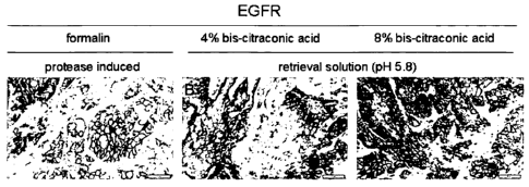
respectively. Immunohistochemical localization of EGFR after
protease-assisted antigen retrieval for formaldehyde-fixed tissue
and removal of bis-citraconic acid from tissue fixed with bis-
citraconic acid by use of an acidic buffer, respectivelyõ
demonstrated comparable staining intensity or quality between
the fixatives formalin (Fig. 2; A) or bis-citraconic acid used at 4%
(Fig. 2: B) and 8% (Fig. 2: C), respectively.
Figure 3 shows the PCR-amplification of the EGFR-gene using mRNA
isolated from MDA468 cells that had been subjected to different
pre-treatment/fixation protocols. Shown are EGFR-mRNA
amplifications from MDA-MB468 cells (fresh and stored for 4
hours in RPMI both not fixed, fresh MDA-MB468 cells
treated/fixed in 10% DMSO, 80% DMSO, 10% DMSO with 1%
CA 02770545 2012-02-09
WO 2011/020612
PCT/EP2010/005085
- 25 -
bis-citraconic acid, 80% DMSO with 1% bis-citraconic acid and
water "Wasser" as negative control, respectively). As obvious
from the amplification curves and the inserted table, mRNA from
all samples is amplified in a rather similar manner.
Figure 4 shows the PCR-amplification of the HER2-gene using mRNA
isolated from MDA468 cells that had been subjected to different
pre-treatment/fixation protocols. Shown are HER2-mRNA
amplifications from MDA-MB468 cells (fresh and stored for 4
hours in RPM both not fixed, fresh MDA-MB468 cells
treated/fixed in 10% DMSO, 80% DMSO, 10% DMSO with 1%
bis-citraconic acid, 80% DMSO with 1% bis-citraconic acid and
water "Wasser" as negative control, respectively. As obvious
from the amplification curves and the inserted table, mRNA from
all samples is amplified in a rather similar manner.
Example 1:
Synthesis of "bis-citraconic acid" of Formula VIII
Formula VIII
0
0
0 I 0
0
0
Synthesis of methyl-3-tolylcarbamoyl-acrylic acid
To a solution of 3,2 ml 3-methyl-furan-2,5-dione (citraconic anhydride) in 25
ml
ethyl ether a solution of 3,74 g p-tolylamine in 25 ml ethyl ether was added
dropwise over a period of 15 min.. The yellow suspension was stirred for 1 h
and
filtrated. The residue was washed with ethyl ether and dried under vacuum.
Yield: 7,22 g, 94%
Synthesis of 3-methyl-1-p-tolyl-pyrrole-2,5-dione
7,22 g of methyl-3-tolylcarbamoyl-acrylic acid were suspended in 60 ml acetic
anhydride. The suspension was heated under reflux for 3 h . After cooling to
room
CA 02770545 2013-12-20
- 26 -
temperature the solvent was removed under vacuum. The residue was
recrystallized from ethanol.
Yield: 4,32 g, 65 %
Synthesis of 3-(5-(4-methy1-2,5-dione-l-p-toly1-2,5-dihydro-1H-pyrrol-3-y1)-
penty1+4-
methyl-1-p-tolyl-pyrrole-2,5-dione
12 g 3-methyl-1-p-tolyl-pyrrole-2,5-dione and 15,6 g triphenyl-phosphane are
dissolved in 155
ml acetic acid and 2,15 ml pentanedial are added. The reaction mixture was
refluxed for 20 h.
The acetic acid was removed by distillation and the residue heated up to 150-
160 C for 6 h.
The crude product was purified by column chromatography on silica gel,
petrolether:acetic acid
ethyl ester 7:3. The product was further purified by digestion in methanol,
filtrated and dried.
Yield: 2 g, 35%
Synthesis of 3-(5-(4-methy1-2,5-dioxo-2,5-dihydro-furan-3-y1)-penty1)-4-methyl-
furan-2,5-
dione ("bis-citraconic acid")
1,07 g of 3-(5-(4-methy1-2,5-dione-1-p-toly1-2,5-dihydro-1H-pyrrol-3-y1)-
penty1+4-methyl-1-p-
tolyl-pyrrole-2,5-dione were dissolved in 30 ml of a 1:1 mixture of
tetrahydrofuran and methanol.
After the addition of 3,48 g potassium hydroxide dissolved in water the
mixture was heated under
reflux for 3 h. The solvent was removed by distillation and the residue was
purified by column
chromatography on silica gel, petrolether:acedic acid ethyl ester 7:3. The
product was dried under
vacuum.
Yield: 402 mg, 60%
CA 02770545 2012-02-09
WO 2011/020612
PCT/EP2010/005085
- 27 -
Example 2:
Synthesis of 3-(5-(4-methy1-2,5-dioxo-2,5-dihydro-furan-3-y1)-3-oxa-penty1)-4-
methyl-furan-2,5-dione (Formula IX):
Formula IX
0
0 0
The cross-linking agent 3-(5-(4-methy1-2,5-dioxo-2,5-dihydro-furan-3-y1)-3-oxa-
penty1)-4-methyl-furan-2,5-dione (Formula IX) is synthesized analogous to the
procedure described in Example 1 using 3-oxa-1,5-pentanedial instead of 1,5-
pentanedial. The starting material 3-oxa-1,5-pentanedial is described by
Bowers, S.
et al., Bioorganic & Medicinal Chemistry Letters 19 (2009) 6952-6956.
Example 3:
Staining with an antibody to IGF-1R
H322M xenograft tumor bearing SCID beige mice were sacrificed. The tumors
were removed and were cut into 3 pieces of approximately the same size. The
tissue samples were subsequently transferred into the respective fixation
solutions.
For fixation with bis-citraconic acid this substance was resolved in DMSO to a
final concentration of 80 % by repeated pipetting at room temperature. After
complete dissolution this solution was either used directly or diluted 1:1 in
DMSO
and further diluted in 1 x PBS pH 7,4, resulting in PBS with a final
concentration
of 10% DMSO and 8 % or 4 % of bis-citraconic acid, respectively. To test the
impact of different concentrations of bis-citraconic acid on fixation
efficacy,
solutions containing 4% or 8% bis-citraconic acid (w/v) were prepared. For
preparation of the formaldehyde fixative, formalin (40% (w/v) paraformaldehyde
in
H20) was diluted 1:10 in 1 x PBS pH 7.4.
All tumor samples were fixed over night for 12 h at room temperature. The next
day the tissue samples were washed with H20 for 1 h. Afterwards the fixed
tumor
tissues were embedded in paraffin.
Sections of the paraffin embedded tissue samples fixed with different
fixatives (4%
formalin, 4% or 8% bis-citraconic acid) were cut at 4 gm using a conventional
CA 02770545 2013-12-20
=
- 28 -
rotation microtome. For immunohistochemical localization of IGF-1R the cut
tissue sections
were mounted on glass slides. Deparaffinization of the tissue samples was
performed on the
Ventana Benchmark XT automated IHC stainer (Ventana, Tucson). For localization
of IGF-1R
with the <IGF-1R> 5G11 monoclonal antibody (Ventana, Tucson) in FFPE tissue by
immunohistochemistry, a heat induced antigen retrieval has to be performed
prior to staining
of the formalin fixed sample. Heat induced antigen retrieval for
immunohistochemical
detection of IGF-1R was performed by incubating the tissue sections on the
Ventana
Benchmark XT for 1 h at 95 C in buffer CC1 (Ventana, Tucson). Antigen
retrieval in thin
sections previously fixed with bis-citraconic acid and after paraffin can be
performed by a
simple incubation of the tissue sections in a buffer with an acidic pH. Thin
sections were
incubated in buffer of pH 5.8 for 2 h. After antigen retrieval, all slides
were placed on the
Ventana Benchmark and were stained for IGF-1R with a primary antibody
incubation time of
16 min. The bound primary antibody was detected using Ventana iview DAB
detection kit.
Examination of the stained sections revealed that fixation of tissues with bis-
citraconic acid
conserved the morphology of the tissue (Fig. 1 B and C). Furthermore, the bis-
citraconic acid
could be retrieved by a simple incubation in an acidic buffer solution.
Immunohistochemical
localization of IGF-1R did not reveal great differences in staining intensity
or morphological
quality between formalin (Fig. 1; A) or bis-citraconic acid fixed tissues
(Fig. 1; B and C).
The results obtained in this Example demonstrate that fixation with bis-
citraconic acid enables
conservation as well as easy and gentle retrieval of epitopes that in formalin-
fixed tissues only
become accessible after tissue treatment with a method known as heat-induced
antigen
retrieval.
Example 4:
Staining with an antibody to EGFR
By a procedure similar to Example 2, formalin or bis-citraconic acid fixed
tissues were
prepared for staining for EGFR using the antibody 3C6 (<EGFR> mAB 3C6;
Ventana,
Tucson). This antibody is known to depend on a protease pretreatment of FFPE-
derived tissue
sections in order to regain access to its epitope in such FFPE-sample. As
shown in Figure 2, no
differences in immunohistochemical localization of EGFR between formalin
fixation and
protease-assisted epitope retrieval or bis-citraconic acid fixation and
retrieval by incubation in
an acidic buffer were found.
CA 02770545 2012-02-09
WO 2011/020612
PCT/EP2010/005085
- 29 -
The results obtained in this Example demonstrate that fixation with bis-
citraconic
acid enables conservation as well as easy and gentle retrieval of epitopes
that in
formalin-fixed tissues only become accessible after tissue treatment with a
protease.
As demonstrated in Examples 2 and 3, collectively, bis-citraconic acid
fixation and
retrieval is the method for the detection of different epitopes which so far
in
formalin-fixed tissue have to be retrieved by one or more different retrieval
methods (heat or protease induced). Bis-citraconic acid, as a prototype for
other
bis-maleic anhydrides, works without harsh retrieval methods for different
antibodies otherwise requiring quite different methods of retrieval. Whereas
heat-
induced or protease-assisted retrieval is not easy to standardize and
reproduce, it
will be possible to obtain a more reproducible accessibility/reactivity of
antigens/epitopes by use of a gentle and easy to remove fixative based on a
bis-
maleic anhydride cross-linking agent as shown above.
Example 5:
DNA isolation and qPCR for gene amplification analysis
MDA-MB468 cells were first fixed in different fixation reagents for 10 min and
then neutralized in citrate buffer (pH 4.4). The different samples given in
Figure 3
are MDA-MB468 cells (fresh and stored for 4 hours in RPM both not fixed, fresh
MDA-MB468 cells treated/fixed in 10% DMSO, 80% DMSO, 10% DMSO with
1% bis-citraconic acid and 80% DMSO with 1% bis-citraconic acid, respectively.
After the different fixation procedures DNA isolation was performed using 1 x
107
MDA-MB468 cells. To isolate the DNA the High Pure Template Preparation Kit
(Roche Diagnostics GmbH, Cat. No.: 11796828) was used according to the
manufacturer's instructions. Isolated DNA was stored at -20 C.
Amplification status of the EGFR and HER2 was measured in MDA-MB468 cells.
Therefore a quantitative PCR based on the use of hydrolysis probes (Taqman
probes) was performed using gene specific primers and probes (see Table 1).
The
probes for the target genes were labeled with Fam at the 5'end and with BHQ-2
at
the 3'end.
CA 02770545 2012-02-09
WO 2011/020612
PCT/EP2010/005085
- 30 -
Table 1: Primer and Probes for the target genes HER2 and EGFR
HER2 EGFR
Forward Primer: CTCAGCGTACCCTTGTCC TGAAAACACCGCAGCATGTCAA
SEQ ID NO:1 SEQ ID NO:4
Reverse Primer: TGTCAGGCAGATGCCCAGA CTCCTTCTGCATGGTATTCTTTCTCT
SEQ ID NO:2 SEQ ID NO:5
Probes: TGGTGTGGGCTCCCCATATGTCTCCC TTTGGGCTGGCCAAACTGCTGGGTG
SEQ ID NO:3 SEQ ID NO:6
For each gene an individual, verified PCR- mix was used (see Table 2).
Table 2: PCR-Mix composition for the qPCR assays
PCR-Mix for EGFR, HER2:
component lx (p1)
Nuclease-Free H20 5.5
5x RNA MasterMix 4.0
Forward Primer (500 nM) 1.0
Reverse Primer (500 nM) 1.0
TaqMan Probe (100 nM) 0.2
DMSO 100% 0.8
MgAc (25 mM) 2.2
template 5.0
Each mix is composed of 5 M forward and reverse primer and 2 laM probe. The
applied Z05 polymerase was included in the COBAS Taqman RNA Reaction Mix
(LUO M/N 58004938) purchased by Roche Molecular Diagnostics (Branchburg,
USA) and Magnesium Acetate [25 mM] (Fluka, Cat. No.: 63049) was added in
different concentrations according to the different oligo mixes. 2 1 DNA-
template
was used and filled up to 5 IA with nuclease free water. Each sample was
measured
in triplicates. The LightCycler 480 (Roche Diagnostics GmbH) and appropriate
96-
well plates and sealing foils were applied for the measurements. The following
thermocycling profile was used on the cycler (Table 3).
CA 02770545 2012-02-09
WO 2011/020612
PCT/EP2010/005085
- 31 -
Table 3: LightCycler thermocycling profile
Program Name !Cycles jAnalysis Mode_
Decontamination. 1 Quantification
Amplification 47 Quantification
Cooling 1 None _
Target [ C] Acquisition Mode 'Hold 'Ramp Rate [ C/s]
50 None . 5 min 4,4
95 None 1 min 4,4
Amplification
92 None 15s 4,4
, _
60 Single 50s 2,2
Cooling
40 I None I 30s I 2,2