Note: Descriptions are shown in the official language in which they were submitted.
CA 02770716 2012-02-09
WO 2011/019704 PCT/US2010/044997
METHODS, PRIMERS, PROBES AND KITS USEFUL FOR THE DETECTION
OF BRAF MUTATIONS
RELATED APPLICATIONS
This application claims priority to 61/233,054 (filed August 11, 2009);
61/237,078 (filed August 26, 2009) and 61/301,790(filed February 5, 2010).
FIELD OF THE INVENTION
The present invention relates to methods, primers and probes for detecting the
presence of mutant BRAF sequences in a sample, specifically for detecting the
presence
of the BRAF V600E, V600D, V600K, V600M, and V600A mutations.
BACKGROUND
Cancer arises when a normal cell undergoes neoplastic transformation and
becomes a malignant cell. Transformed (malignant) cells escape normal
physiologic
controls specifying cell phenotype and restraining cell proliferation.
Transformed cells in
an individual's body thus proliferate, forming a tumor. When a tumor is found,
the
clinical objective is to destroy malignant cells selectively while mitigating
any harm
caused to normal cells in the individual undergoing treatment.
B-raf (or BRAF) encodes a protein that belongs to the Serine/Threonine protein
kinases. BRAF is a part of the Ras/Raf/MEK/MAP signal transduction pathway and
plays a role in regulating the MAP Kinse/ERK signaling pathway. Mutations in
this gene
have been associated with various cancers such as colorectal cancer (CRC), non
small
cell lung cancer (NSCLC), malignant melanomas and adenocarcinomas. Oncogenic
mutations in BRAF, nearly all of which are the V600E mutation, have been
reported in
colon cancer (Davies H, et al. Nature 2002;417:949-54; Rajagopalan H, et al.,
Nature
2002;418:934.). The V600E mutation has been observed in over half of all
microsatellite-unstable carcinomas and in a much smaller subset of stable
colon tumors
(Wang L, et al., Cancer Res 2003;63:5209-12). The V600E (formerly V599E)
mutation
is located on exon 15 of the B-raf gene (Accession number NM04333.4) at
position
1
CA 02770716 2012-02-09
WO 2011/019704 PCT/US2010/044997
1860 (1799 of the coding sequence). At position 1799 of the coding sequence, a
thymidine is changed to an adenosine, which results in the change from a
valine (V) in
the wildtype/non mutant B-rag gene to a Glutamine (E) in the mutated gene. In
addition,
a rare (<1%) V600K (1798-1799 GT>AA) mutation also exists. Furthermore, the
V600D
mutation exists in 4.6% of cases, the V600A mutation exists in <I% of cases,
and the
V600M mutation exists in <1% of cases. In addition, there are V600R and K601E
BRAF
mutations.
The V600E BRAF mutation is found in a number of tissue/tumor types including:
nervous system, thyroid, skin, gastrointestinal tract, large intestine,
biliary tract, ovary,
eye, prostate, central nervous system, liver, small intestine, breast,
pancreas, soft tissue,
upper, aerodigestive tract, adrenal gland, autonomic ganglia, haematopoietic
and
lymphoid tissue, lung, esophagus, pituitary, and stomach. DNA or RNA extracted
from
samples of any of these types of tissues can be utilized in assays of the
present invention.
In both stable and unstable cancers, >90% of tumors with BRAF mutations have
widespread methylation of CpG islands or what is known as the CpG island
methylator
phenotype (CIMP). Improved survival associated with microsatellite instability
(MSI) in
sporadic colon cancers has been reported (Samowitz WS, et al., Cancer
Epidemiol
Biomarkers Prev 2001;10:917-23; Halling KC, et al., J Natl Cancer Inst
1999;91:1295-
303), and because sporadic unstable tumors commonly show both CIMP (Toyota M,
et
al., Proc Natl Acad Sci U S A 1999;96:8681-6; Toyota M, et al., Proc Natl Acad
Sci U S
A 2000;97:710-5) and BRAF mutations (Kambara T, et al. , Gut 2004;53:1137-44;
Nagasaka T, et al., J Clin Oncol 2004;22:4584-94), one would expect that these
features
would also show a relationship to improved survival in unstable tumors.
Samowitz has
studied the relationship between CIMP and survival in microsatellite-stable
tumors and
has evaluated the relationship between BRAF mutations and survival in
microsatellite-
stable colon cancers. See Samowitz, Wade S., et al., Cancer Research 65, 6063-
6069,
July 15, 2005. Samowitz has evaluated a large population-based sample of
individuals
with colon cancer to determine its relationship to survival and other
clinicopathologic
variables. The V600E BRAF mutation was seen in 5% of microsatellite-stable
tumors
and 51.8% of microsatellite-unstable tumors. In microsatellite-stable tumors,
this
2
CA 02770716 2012-02-09
WO 2011/019704 PCT/US2010/044997
mutation was related to poor survival, CIMP high, advanced American Joint
Committee
on Cancer (AJCC) stage, and family history of colorectal cancer. The poor
survival was
observed in a univariate analysis of 5-year survival (16.7% versus 60.0%); in
an analysis
adjusted for age, stage, and tumor site; in stage-specific, age-adjusted
analyses for AJCC
stages 2 to 4 (HRR, 4.88, 3.60, and 2.04, respectively); and in Kaplan-Meier
survival
estimates for AJCC stages 2 to 4. Microsatellite-unstable tumors were
associated with an
excellent 5-year survival whether the V600E mutation was present or absent
(76.2% and
75.0%, respectively). Samowitz has concluded that the BRAF V600E mutation in
microsatellite-stable colon cancer is associated with a significantly poorer
survival in
stages 2 to 4 colon cancer but has no effect on the excellent prognosis of
microsatellite-
unstable tumors.
Moreover, BRAF mutations proved to be absent in tumors from hereditary
nonpolyposis colorectal cancer syndrome (HNPCC) families with germline
mutations in
the MMR genes MLH1 and MSH2. These data suggest that the oncogenic activation
of
BRAF is involved only in sporadic colorectal tumorigenesis. The detection of a
positive
BRAF-V600E mutation in a colorectal cancer suggests a sporadic origin of the
disease
and the absence of germline alterations of MLH1, MSH2 and also of MSH6. These
findings have a potential impact in the genetic testing for HNPCC diagnostics
and
suggest a potential use of BRAF as exclusion criteria for HNPCC or as a
molecular
marker of sporadic cancer. See Domingo et al., Oncogene (2005) 24, 3995-3998.
Solit's group have found, using small-molecule inhibitors of MEK and an
integrated genetic and pharmacologic analysis, that mutation of BRAF is
associated with
enhanced and selective sensitivity to MEK inhibition when compared to either
'wild-type'
cells or cells harboring a RAS mutation. This MEK dependency was observed in
BRAF
mutant cells regardless of tissue lineage, and correlated with both down
regulation of
cyclin Dl protein expression and the induction of G1 arrest. Pharmacological
MEK
inhibition completely abrogated tumor growth in BRAF mutant xenografts,
whereas RAS
mutant tumors were only partially inhibited. These data suggest an exquisite
dependency
on MEK activity in BRAF mutant tumors, and offer a rational therapeutic
strategy for
3
CA 02770716 2012-02-09
WO 2011/019704 PCT/US2010/044997
this genetically defined tumor subtype. See Solit, David B., et al., Nature
439, 358-362
(19 January 2006).
In addition, a model of human melanocyte transformation has emerged based on
the results of genetic studies, cell biology, molecular pathology and mouse
modeling.
Studies have identified involvement of various factors including basic
fibroblast growth
factor production, ERK activation, and frequent BRAF mutations in melanoma
tissues.
BRAF acts downstream of RAS, and studies have demonstrated that simultaneous
mutations in RAS and BRAF are extremely rare in melanoma, suggesting that BRAF
mutations substitute for at least some of the oncogenic function of mutant
RAS.
The development of tumor markers to better stratify patients for their risk of
developing metastases is under active investigation. Although assessment of
tumor
markers and selection of treatment based on the results has been part of the
standard of
care in colon and breast cancer management for several years, no such markers
exist for
melanoma. Many studies have shown promise, but none have moved past the
preliminary
stages of development into a clinically useful assay.
Despite recent advances in the study of melanoma biology, the development of
molecular tools useful for diagnosing and/or monitoring patients with melanoma
is still
relatively new. Few advances have been made in protocols designed to monitor
patients
for disease recurrence, or to select patients at high risk for the development
of metastases.
Tumor stage, the best predictor of survival from melanoma, is based on
conventional
clinicopathologic variables such as thickness and ulceration of the primary
tumor, and the
presence of metastatic disease in regional lymph nodes or at distant sites.
Two patients
with primary tumors of intermediate thickness that appear microscopically
identical can,
however, have dramatically different survivals. The absence of improved
prognostic
tools for such assessments makes it difficult for attending physicians to
determine the
best treatment strategies.
Mutations in the BRAF oncogene have been discovered in up to 80% of
melanoma tissues, frequencies strikingly higher than any other molecular
alteration in
this disease. BRAF mutations have also been detected in tumor tissues from
other types
of cancer. Experimental studies have demonstrated that several BRAF mutations,
4
CA 02770716 2012-02-09
WO 2011/019704 PCT/US2010/044997
especially the T1799A (formerly designated T1796A) hotspot mutation, which
accounts
for 90% of BRAF mutations in melanoma, can transform fibroblasts in culture.
Most
recently, experiments blocking the expression of mutant BRAF in melanoma cell
culture
were shown to inhibit cell growth and promote cell death, suggesting that BRAF
inhibitors could bolster melanoma treatment significantly.
SUMMARY OF THE INVENTION
The present invention discloses methods of detecting BRAF V600E, V600D,
V600K, V600M, and V600A mutations in a sample. The present invention discloses
compositions comprising primer and probe sequences used in the amplification
and
detection of V600E, V600D, V600K, V600M, or V600A mutant BRAF sequences
present in samples. Particular primer combinations as disclosed herein are
used in
amplifying particular BRAF mutations. It will be appreciated by those skilled
in the art,
one may also design primers specific to the 1798-1799 GT>AA double mutation.
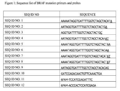
BRIEF DESCRIPTION OF THE DRAWINGS
Figure 1 shows the primers and probes used in the amplification and detection
of
BRAF mutations.
DETAILED DESCRIPTION OF THE INVENTION
The present invention provides methods, primers, probes and kits useful for
the
detection of BRAF mutations. The methods, primers, probes and kits of the
present
invention can be used for detecting the BRAF V600E, V600D, V600K, V600M, and
V600A mutations in many different cell types and thus can be used for the
diagnosis of
many different cancers, such as, but not limited to, melanoma, colorectal
cancer, lung
cancer and thyroid cancer. The methods of the invention may be useful as a
predictor of
outcome for cancer patients. One of the key factors that contribute to
improved outcome
for a patient with any disease and in particular cancer, due to its
progressive and invasive
nature, is early and accurate diagnosis. The method of the present invention
addresses
the desperate need for a rapid, non-invasive, and accurate screening assay for
detecting
mutant BRAF sequences, the presence of which is a positive indicator of
metastasizing
CA 02770716 2012-02-09
WO 2011/019704 PCT/US2010/044997
disease. As such, it identifies those patients who need to be treated with
more aggressive
treatment regimens. Moreover, since the invention can be used for either DNA
or RNA,
sample preparation is facile, thereby reducing assay variability that can
result from
differences in the expertise level of laboratory technicians involved in
sample
preparation.
As a non limiting example, the method of the present invention may be used to
monitor patients with advanced, metastatic melanoma (Stages III/IV). These
patients are
at the highest risk for disease progression, and early detection of an
increase in disease
activity would lead to earlier treatment and improvement in outcome. The
method of the
present invention may also be directed to testing patients with earlier stages
of disease
(Stages I/II), who are at risk for metastatic spread of their disease. Again,
early
intervention with additional diagnostic tests and treatments would lead to
improved
patient survival.
The present invention provides a method for detecting the presence of a BRAF
mutation in a sample, said method comprising: (a) isolating nucleic acid from
said sample
wherein the sample comprises nucleic acid sequences; (b) performing an
amplification
reaction of said nucleic acid sequences of said sample, wherein said
amplification
reaction comprises a first primer capable of annealing specifically to a BRAF
mutant
sequence at a first position in a BRAF sequence wherein said first primer is
SEQ ID
NO:1 , SEQ ID NO: 2, SEQ ID NO: 3, SEQ ID NO: 4, SEQ ID NO: 5, SEQ ID NO: 6,
SEQ ID NO: 7, SEQ ID NO: 8, or SEQ ID NO: 9 and a second primer capable of
annealing specifically at a second position in a BRAF sequence wherein said
second
primer is SEQ ID NO: 10, wherein said first and second primers anneal to
different
strands of double stranded BRAF sequence, wherein the amplification reaction
is capable
of producing a BRAF mutant specific amplification product when the sequences
of the
sample comprise a BRAF sequence comprising a mutant sequence at said first
position of
said BRAF sequence; and (c) visualizing amplification products produced by
said
amplification reaction, wherein detection of a BRAF mutant specific
amplification
product is a positive indicator of the presence of a BRAF mutation in said
sample.
6
CA 02770716 2012-02-09
WO 2011/019704 PCT/US2010/044997
The present invention also provides a method for detecting the presence of
metastatic melanoma in a sample, said method comprising: (a) isolating nucleic
acid from
said sample wherein the sample comprises nucleic acid sequences; (b)
performing an
amplification reaction of said nucleic acid sequences of said sample, wherein
said
amplification reaction comprises a first primer capable of annealing
specifically to a
BRAF mutant sequence at a first position in a BRAF sequence wherein said first
primer
is SEQ ID NO:1 , SEQ ID NO: 2, SEQ ID NO: 3, SEQ ID NO: 4, SEQ ID NO: 5, SEQ
ID NO: 6, SEQ ID NO: 7, SEQ ID NO: 8, or SEQ ID NO: 9 and a second primer
capable
of annealing specifically at a second position in a BRAF sequence wherein said
second
primer is SEQ ID NO: 10, wherein said first and second primers anneal to
different
strands of double stranded BRAF sequence, wherein the amplification reaction
is capable
of producing a BRAF mutant specific amplification product when the sequences
of the
sample comprise a BRAF sequence comprising a mutant sequence at said first
position of
said BRAF sequence; and (c) visualizing amplification products produced by
said
amplification reaction, wherein detection of a BRAF mutant specific
amplification
product is a positive indicator of metastatic melanoma in said sample.
Embodiments of the present invention comprise BRAF V600E, V600D, V600K,
V600M, and V600A mutant specific primers. Exemplary BRAF V600 E mutant
specific
primer pairs include SEQ ID NO: 1, SEQ ID NO: 2, or SEQ ID NO: 9 and SEQ ID
NO:
10. Exemplary BRAF V600D mutant specific primer pairs include SEQ ID NO: 1,
SEQ
ID NO: 2, SEQ ID NO: 4, or SEQ ID NO: 7 and SEQ ID NO: 10. Exemplary BRAF
V600K mutant specific primer pairs include SEQ ID NO: 1, SEQ ID NO: 4, SEQ ID
NO:
5, or SEQ ID NO: 6 and SEQ ID NO: 10. Exemplary BRAF V600M mutant specific
primer pairs include SEQ ID NO: 6 or SEQ ID NO: 8 and SEQ ID NO: 10. Exemplary
BRAF V600A specific primers include SEQ ID NO: 3 and SEQ ID NO: 10. These
primers were designed to avoid any known BRAF polymorphisms. As described
herein,
such oligonucleotides can be detectably labeled.
BRAF V600 mutant specific primers (SEQ ID NO:1; SEQ ID NO:2; SEQ ID
NO:3, SEQ ID NO: 4, SEQ ID NO: 5, SEQ ID NO: 6, SEQ ID NO: 7, SEQ ID NO: 8,
SEQ ID NO: 9, SEQ ID NO: 10) or appropriate BRAF mutant specific primer pairs
may
7
CA 02770716 2012-02-09
WO 2011/019704 PCT/US2010/044997
be components of compositions comprising biologically compatible salt
solutions and/or
other buffers or components.
Embodiments of the present invention comprise oligonucleotide probe sequences,
SEQ ID NO: 11 and SEQ ID NO: 12, wherein the oligonucleotide is used as a
probe for
the detection of BRAF mutant sequences. This probe was designed to avoid any
known
BRAF polymorphisms. Optionally, the oligonucleotide is detestably labeled.
The present invention also provides a kit comprising at least one of SEQ ID
NO:
1-12.
Embodiments of the present invention can be utilized to detect the V600E,
V600D, V600K, V600M, and V600A BRAF mutations.
Samples
The method comprises obtaining a sample of a tissue or a body fluid from the
subject (e.g., a mammal) wherein the sample contains nucleic acid. Non-
limiting
examples of tissue or body fluids that can be used include blood, plasma,
lymph, tumor
biopsies, and body tissue. In one embodiment, the tissue sample comprises
paraffin
embedded tissue specimens. In some embodiments, the nucleic acid is
deoxyribonucleic
acid (DNA). In some embodiments, the nucleic acid is ribonucleic acid (RNA).
The present method can be applied to any type of tissue from a patient.
Sources of
such tissue include but are not limited to nervous system, thyroid, skin,
gastrointestinal
tract, large intestine, biliary tract, ovary, eye, prostate, central nervous
system, liver,
small intestine, breast, pancreas, soft tissue, upper, aerodigestive tract,
adrenal gland,
autonomic ganglia, haematopoietic and lymphoid tissue, lung, esophagus,
pituitary, and
stomach. For examination of resistance of tumor tissue, it is preferable to
examine the
tumor tissue. In a preferred embodiment, a portion of normal tissue from the
patient from
which the tumor is obtained is also examined.
The methods of the present invention can be applied over a wide range of tumor
types. This allows for the preparation of individual "tumor expression
profiles" whereby
expression levels of BRAF V600E, V600D, V600K, V600M, or V600A mutant
sequences are determined in individual patient samples and response to various
8
CA 02770716 2012-02-09
WO 2011/019704 PCT/US2010/044997
chemotherapeutics is predicted. In certain embodiments, the methods of the
invention are
applied to colon cancer or melanoma tumors.
Isolating nucleic acid
Embodiments of the present invention utilize methods of DNA isolation known to
those skilled in the art. In general, the aim is to separate DNA present in
the nucleus of
the cell from other cellular components. The isolation of DNA usually begins
with lysis,
or breakdown, of tissue or cells. This process is essential for the
destruction of protein
structures and allows for release of nucleic acids from the nucleus. Lysis is
carried out in
a salt solution, containing detergents to denature proteins or proteases
(enzymes digesting
proteins), such as Proteinase K, or in some cases both. It results in the
breakdown of cells
and dissolving of membranes. Methods of DNA isolation include, but are not
limited to,
phenol: chloroform extraction, high salt precipitation, alkaline denaturation,
ion exchange
column chromatography, resin binding, and paramagnetic bead binding.
Embodiments of the present invention utilize methods of RNA isolation known to
those skilled in the art. RNA may be isolated and prepared for hybridization
by a variety
of methods including, but not limited to, Trizol and Guanidinium thiocyanate-
phenol-
chloroform extraction. The principle of RNA isolation is based on cell/tissue
lysis,
followed by extraction, precipitation, and washing. It will be understood by
those skilled
in the art the selection of RNA isolation will depend on sample type.
Incorporated by
reference is US 12/144,388 directed to a method of RNA isolation from paraffin
embedded tissue, a common source for oncogene marker testing.
Amplification
Embodiments of the present invention utilize thermal and isothermal
amplification methods including, but not limited to, polymerase chain reaction
(PCR),
reverse transcriptase polymerase chain reaction (RT-PCR), ligase chain
reaction (LCR),
helicase dependent amplification (HDA) and Nucleic Acid Sequence Based
Amplification (NASBA) and Amplification Refractory Mutation System (ARMS). In
a
preferred embodiment, the primers and probes are used in ARMS.
9
CA 02770716 2012-02-09
WO 2011/019704 PCT/US2010/044997
Detection
Embodiments of the present invention utilize detection methods including, but
not
limited to, labeling primers used during the amplification step such that the
amplification
products are labeled with a detectable marker and hybridizing the
amplification product
to oligonucleotide probes labeled with a detectable marker. Detectable markers
include
but are not limited to chemiluminescent tags, fluorescent tags, and
radioactive tags.
Labeled amplification product can be directly measured using methods
corresponding to
the type of label used according to methods would be known to one skilled in
the art.
Labeled probe can be hybridized to the amplification product according to
methods
known to one skilled in the art.
In performing the method of the present invention BRAF V600E mutant
expression levels are assayed in patient tumor samples to prognosticate the
efficacy a
treatment regimen. In performing the method of the present invention BRAF
V600E
mutant expression levels are assayed in patient tumor samples to predict the
efficacy a
treatment regimen. In performing the method of the present invention BRAF
V600D
mutant expression levels are assayed in patient tumor samples to predict the
efficacy a
treatment regimen. In performing the method of the present invention BRAF
V600K
mutant expression levels are assayed in patient tumor samples to predict the
efficacy a
treatment regimen. In performing the method of the present invention BRAF
V600M
mutant expression levels are assayed in patient tumor samples to predict the
efficacy a
treatment regimen. In performing the method of the present invention BRAF
V600A
mutant expression levels are assayed in patient tumor samples to predict the
efficacy a
treatment regimen.
In performing the method of this embodiment of the present invention, tumor
cells are preferably isolated from the patient. Solid or lymphoid tumors or
portions
thereof are surgically resected from the patient or obtained by routine
biopsy. RNA
isolated from frozen or fresh samples is extracted from the cells by any of
the methods
typical in the art, for example, Sambrook, Fischer and Maniatis, Molecular
Cloning, a
laboratory manual, (2nd ed.), Cold Spring Harbor Laboratory Press, New York,
(1989).
Preferably, care is taken to avoid degradation of the RNA during the
extraction process.
CA 02770716 2012-02-09
WO 2011/019704 PCT/US2010/044997
Table 1
Predicted Mutation
Primer Name Sequence Detection
Braf_1799A_1GT-F AAAAATAGGTGATTTTGGTCTAGCTACA"A 600E, 600D and 600K
Braf 1799A 2AT-F AATAGGTGATTTTGGTCTAGCTAC'GA 600E and 600D
Braf_V600A_2AT-F AGGTGATTTTGGTCTAGCTACTGC 600A Only
600D and 600K
Braf V600D 2GA-F AATAGGTGATTTTGGTCTAGCTACAAAT (weak)
Braf V600K 2AT-F AAAATAGGTGATTTTGGTCTAGCTAC`AA 600K Only
Braf V600M 2CG-F AAAATAGGTGATTTTGGTCTAGCTAAA 600K and 600M
Braf V600D 2GC-F3 AAATAGGTGATTTTGGTCTAGCTACA'AT 600D Only
Braf V600M 2GT-F2 AAAATAGGTGATTTTGGTCTAGCTACTAT 600M Only
Braf 1799A 2GC-F AATAGGTGATTTTGGTCTAGCTACACAG V600E
Common Reverse Primer (used with ALL above Forward primers
2Braf C600-R GATCCAGACAACTGTTCAAACTGA
Common Probe (used with ALL primer combinations)
Braf C600-Mc2 6FAM-TCCATCGAGATTTC
Braf C600-Mc3 6FAM-ACCCACTCCATCGAGA
X base = secondary mutation
XX base = mutation of interest
EXAMPLES
Example 1: Testing of the primers of the present invention
A synthetic V600E construct was made to test the ability of the primers and
the
probes of the present invention to specifically amplify a nucleic acid
containing a BRAF
V600E mutation. Two set of Primers/probes for BRAF V600E mutation were used
for
the validation. The V600E synthetic construct was serially diluted (1:2) 17
times in a
background of gDNA (0.67ng/uL, 5ng/PCR). The mutation concentration ranged
from
11
CA 02770716 2012-02-09
WO 2011/019704 PCT/US2010/044997
lOfM to 0.l5aM. Each diluted sample was assayed 6x in duplicate (12 total) for
the
control (Exonl3) and the V600E mutation.
Example 2: Detection of V600K BRAF mutation
The rare V600K BRAF mutation can be detected utilizing the same pair of
primers designed for the V600E mutation. The V600K mutation is a 1798-1799 GT>
AA
double mutation. SEQ ID NO:2 comprises a highly specific primer that will only
result
in amplified product in the presence of the single 1799 T>A mutation. Thus,
when
performing amplification reactions utilizing primer pairs SEQ ID NO:1 and SEQ
ID NO:
10, and another reaction on the same sample utilizing primer pairs SEQ ID NO:2
and
SEQ ID NO: 10, the first reaction will provide amplified product (positive)
whereas the
second reaction will not provide product (negative). This combination of
positive and
negative results indicates the presence of the V600K mutation.
Example 3: BRAF T1799A/GT1798-1799AA/TG1799-1800AT mutation Exclusivity
Test
A) Test material
1. DNA synthetic fragments were generated that contained the BRAF V600 D, E,
and K mutations
a. BRAF V600D :
AGTAAAAATAGGTGATTTTGGTCTAGCTACAGATAAATCTCGAT
GGAGTGGGTCCCATCAGTTTGAACAGTTGTCTGGATCCATTT
b. BRAF V600E
c. BRAF V600K:
ACAGTAAAAATAGGTGATTTTGGTCTAGCTACAAAGAAATCTC
GATGGAGTGGGTCCCATCAGTTTGAACAGTTGTCTGGATCCATT
TT
2. Sequence specific primers/probes for BRAF mutations V600 D, E and K
mutations (sequence listed in Table 1)
a. 1799A1 GT (specific for 1799 T to A base pair change (V600E, V600D
and V600K)
b. V600D_2GA (specific for V600D)
c. V600K2AT (specific for V600K)
12
CA 02770716 2012-02-09
WO 2011/019704 PCT/US2010/044997
B. Procedure
1. Dilute each of three synthetic fragments to a concentration of 200 aM in
0.667
ng/ul of Genomic DNA (Promega Corp. - human genomic DNA, 100ug)
2. PCR amplify the synthetic specific fragments for each mutation with all
three
primer sets (specific for each mutation
3. Analyze exclusivity
C. Analysis of exclusivity
The following two primer/probe sets have been designed to amplify specific
mutations. These primers were tested with each of the synthetic fragments to
test
for exclusivity
a. 1799A1GT (Amplifies V600E, V600D and V600K) This primer/probe
set has been described in the assay development section ( 7) to amplify a
T to A change at base 1799.
b. V600D_2GA (specific for V600D) This primer/probe set has been
described in the assay development section (7) to amplify a TG change to
AT at base 1799-1800.
c. V600K2AT (specific for V600K) This primer/probe set has been
described in the assay development section (7) to amplify a CT change to
AA at base 1798-1799.
All primers and probes were used in the exclusivity testing
Results: The following table describes fragments that were successfully
amplified
with specific primer probe sets. A plus (+) signifies that a specific fragment
was
amplified.
Table 2: The combination of primers to define each mutant type are the
following
1799A_1GT V600K_2AT V600D_2GA Mutant
+ - - V600E
+ - + V600D
+ + - V600K
wild type
invalid
13
CA 02770716 2012-02-09
WO 2011/019704 PCT/US2010/044997
Results:
We collected Ct data for each synthetic and primer/probe combination
V600D V600E V600K
Synthetic Synthetic Synthetic
Ct Ct Ct
V600D_2GA
(amplifies
only V600D)
30.39 36.87 33.79
1 799A 1 GT
(amplifies
V600D,
V600E, and
V600K)
30.34 30.65 29.61
V600K 2AT
(amplifies
only V600K)
39.06 38.72 29.67
Exclusivity was determined by subtracting Cts
of the PCR amplification of each template using
primer/probe sets designed to be specific and
non-specific for each template
V600D V600E V600K
Synthetic Synthetic Synthetic
Delta Ct Delta Ct Delta Ct
V600D_2GA
(amplifies
only V600D)
0 6.22 4.12
1 799A 1 GT
(amplifies
V600D,
V600E, and
V600K)
-0.05 0 -0.06
V600K 2AT 8.67 8.07 0
14
CA 02770716 2012-02-09
WO 2011/019704 PCT/US2010/044997
(amplifies
only V600K)
The delta Cts were determined as follows:
Example:
Exclusivity of the V600D_2GA for V600D synthetic fragment = 0
Using V600 D fragment as the template amplification was performed with
V600D_2GA
primer/probes
dCt = 30.39 - 30.39 = 0 (exclusive)
Using V600 D fragment as the template amplification was performed with
1799A_1GT
primer/probes
dCT = 30.34 - 30.39 = - 0.05 (exclusive)
Using V600 D fragment as the template amplification was performed with
V600K_2AT
primer/probes
dCT = 39.06 - 30.39 = 8.67 (non-specific)
Acceptance criteria
dCT (Ct using primers specific for the fragment - Ct using primers non-
specific for the
fragment) < 4
Preset Acceptance criteria described in section 7 (assay development - see
following
table) that primer/probe 1799_1 GT would detect a T to A base pair change at
1799 and,
therefore, would detect all mutations.
Results:
V600K2AT is exclusive for V600K mutation
V600D2GA is exclusive for V600D mutation
1799A 1 GT is exclusive for 1799 T to A change which is contained in all three
mutations (V600E, V600E and V600K)
Table 3: The combination of primers to define each mutant t e
BrafExl3 1799A1GT 1799A2AT V600K_2AT V600D_2GA Mutant
+ + + - - V600E
+ + - - + V600D
+ + - + - V600K
+ - - - - wild type
>30 Ct - - - - invalid