Note: Descriptions are shown in the official language in which they were submitted.
CA 02771120 2016-07-28
ENDOPROSTHESIS WITH FILAMENT
REPOSITIONING OR RETRIEVAL MEMBER AND
GUARD STRUCTURE
FIELD OF THE INVENTION
The present invention relates generally to an endoprosthesis and, more
specifically, to
an endoprosthesis including a stent structure and a guard structure secured
thereto for
obstructing tangling of a suture structure with the stent structure.
BACKGROUND OF THE INVENTION
An endoprosthesis is implantable in the body of the patient, such as a blood
vessel, or
other body cavity, such as non-vascular orifices and/or lumens. The
endoprosthesis includes
a medical structure, such as a stent, which may be braided. A braided stent
may include loop
structures and adjacent crossover structures. The crossover structures include
longitudinal
portions of the elongate members of the braided stent which overlap one
another and may be
in direct contact. A suture structure may extend through the loop structures
to, for example,
provide for the cinching of the stent structure. Cinching of the stent
structure may be desired
to reduce the profile thereof for insertion in and displacement through a
vessel in the body of
a patient.
Manipulation of the suture structure in the stent structure may cause the
suture
structure to approach the crossover structures and directly contact the
longitudinal portions of
the elongate members which overlap. The direct contact of the suture structure
with the
overlapping longitudinal portions of the elongate members may result in the
suture structure
becoming tangled therewith. Alternatively, the suture structure may be drawn
into direct
contact with the longitudinal portions of the elongate members with sufficient
force and the
elongate members may be sufficiently resilient such that the direct contact of
the suture
structure forces the elongate members apart to provide a path for the suture
structure to
translate through the crossover structure. Neither of the scenarios is
typically desirable since
the suture structure is normally intended to remain within the loop structures
through which
1
CA 02771120 2012-02-14
WO 2011/031587
PCT/US2010/047292
the suture structure initially extends. Accordingly, displacement of the
suture structure
through the crossover structure is normally not desirable. Also, tangling of
the suture
structure with the stent structure is normally undesirable since substantially
unimpeded
displacement of the suture structure relative to the stent structure is
typically preferred.
SUMMARY OF THE INVENTION
The endoprosthesis of the present invention includes a stent structure having
loop
structures through which a suture structure extends. The stent structure has
crossover
structures which are contiguous to the loop structures. The stent structure
has an elongate
member longitudinal portions of which overlap to define the crossover
structures. A guard
structure is secured to the stent structure and located adjacent to the
crossover structures to
obstruct displacement of the suture structure between the longitudinal
portions of the elongate
member of the crossover structures. A method for operating the endoprosthesis
includes
displacing the loop structures toward one another along the suture structure
to displaced
positions relative to the suture structure for reducing the respective
profiles of the stent
structure and guard structure. The loop structures are secured in the
displaced positions to
retain the profiles of the stent structure and guard structure which are
reduced.
The guard structure and location thereof adjacent to the crossover structure
obstructs
the suture structure from directly contacting the overlapping portions of the
elongate member
in the crossover structure. The obstruction provided by the guard structure
reduces the
likelihood of the suture structure becoming entangled with the overlapping
portions of the
elongate member. Preventing the suture structure from becoming tangled with
the elongate
member of the braided stent structure is normally desirable. Also, the
obstruction provided
by the guard structure reduces the likelihood of the suture structure forcing
apart the
overlapping portions of the elongate member and translating through the
crossover structure.
Preventing the suture structure from displacing through the crossover
structure is normally
desirable.
These and other features of the invention will be more fully understood from
the
following description of specific embodiments of the invention taken together
with the
accompanying drawings.
2
CA 02771120 2016-07-28
BRIEF DESCRIPTION OF THE DRAWINGS
In the drawings:
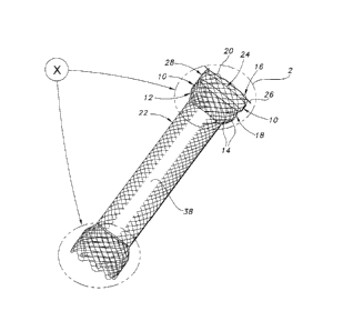
FIG. 1 is a perspective view of the endoprosthesis of the present invention,
the
endoprosthesis being shown as including a braided stent structure which is
partially covered,
and a ring structure secured to the stent structure;
FIG. 2 is an enlarged view of the circled portion 2 of FIG. 1 showing the ring
structure
located adjacent to the crossover structures of the stent structure, the stent
structure being
shown as including loop structures through which a suture structure extends;
FIG. 3 is a graph showing the test results of the diametrical expansion of a
stent
structure to which was secured silicone ring structures having different
longitudinal
dimensions;
FIG. 4A is a partial cross-sectional view of the stent structure of FIG. 2
taken along
the 4A-4A axis;
FIG. 4B is a partial cross-sectional view of the stent structure of FIG. 2
taken along
the 4B-4B axis;
FIG. 5 is a partial planar view of the stent of the present invention showing
an
interwoven ribbon guard structure;
FIG. 6A is a partial planar view of the stent of the present invention showing
an
embodiment of a guard structure disposed at wire crossings;
FIG. 6B is a partial cross-sectional view of the stent of the present
invention showing
the guard structure of FIG. 6A disposed at a wire crossing;
FIG. 6C is a partial cross-sectional view of the stent of the present
invention showing
a wire crossing without a guard structure; and
FIG. 7 is a partial planar view of the stent of the present invention
depicting a patch-
like guard structure.
Corresponding reference characters indicate corresponding parts throughout the
several views of the drawings.
3
CA 02771120 2012-02-14
WO 2011/031587
PCT/US2010/047292
DETAILED DESCRIPTION OF THE INVENTION
Referring to the drawings and more specifically to FIGS. 1 and 2, the
endoprosthesis
includes a stent structure 12 having elongate members 14 which are braided
into a tubular
structure. In an alternative embodiment, the tubular structure is braided from
a single
5 elongate member 14. The stent structure 12 may include a one over and a
one under braided
pattern of elongate members or filaments 14. Even so, the invention is
applicable to any type
of stent structure comprising at least one crossover point where one or more
elongate
members intersect. For example, stent having at least one crossover point may
include
without limitation, a braided stent, a wound stent, a helically wound stent, a
knitted stent, a
10 woven stent, and the like. Furthermore, the invention is not limited to
stents; it pertains to
any endoprosthesis comprising elongate members and a crossover point where one
or more
elongate members intersect.
The stent structure 12 may include loop structures 16 which are defined by
longitudinal portions of the elongate members 14. The portions of the elongate
members 14
which define the loop structures 16 are welded to adjacent portions of the
elongate members
14, as shown in FIG. 2. In an alternative embodiment, the loop structures 16
may be defined
by longitudinal portions of a single elongate member 14 which are continuous.
The stent structure 12 has an end portion 18 which includes end surfaces 20
which are
defined by sections of the loop structures 16. The end portion 18 has a
transverse dimension
which is generally constant along the longitudinal axis of the stent structure
12. The
transverse dimension of the end portion 18 corresponds to the diameter thereof
from the
tubular shape of the stent structure 12.
The stent structure 12 includes an intermediate portion 22 having a transverse
dimension, i.e., diameter, which is generally constant along the longitudinal
axis of the stent
structure 12. The transverse dimension of the intermediate portion 22
corresponds to the
diameter thereof from the tubular shape of the stent structure 12. The
diameter of the
intermediate portion 22 is smaller than the diameter of the end portion 18.
The portion of the
stent structure 12 between the intemediate portion 22 and end portion 18 is
flared to
accommodate the different diameters of the end portion 18 and intermediate
portion 22.
4
CA 02771120 2012-02-14
WO 2011/031587
PCT/US2010/047292
The stent structure 12 includes crossover structures 24 which are located
longitudinally relative to the stent structure between the end surface 20 and
intermediate
portion 22. The crossover structures 24 include longitudinal portions of the
elongate
members 14 which overlap one another and may be in direct contact. The
overlapping
portions of the elongate members 14 extend in respective directions which are
different and
thereby cause the overlapping portions to appear to intersect as viewed from
the perspective
of FIG. 2.
The overlapping relation of the elongate members 14 in the crossover
structures 24
al lows relative movement between the elongate members therein. The relative
movement
and flexibility of the elongate members 14 allows transverse expansion and
contraction of the
stent structure 12. The transverse expansion and contraction of the stent
structure 12
corresponds to radial expansion and contraction from the tubular shape of the
stent structure
12.
The flexibility and resiliency of the elongate members 14 may result in the
radial
separation thereof in the crossover structures 24 when the elongate members
are subjected to
forces which are sufficiently large and directed to the respective elongate
members 14 in
suitably opposite directions.
The stent structure 12 may be formed of any suitable implantable material,
including
without limitation nitinol, stainless steel, cobalt-based alloy such as
Elgiloy , platinum, gold,
titanium, titanium alloys, tantalum, niobium, polymeric materials and
combinations thereof.
Useful polymeric materials may include, for example, polyesters, including
polyethylene
terephthal ate (PET) polyesters, polypropylenes, polyethylenes, polyurethanes,
polyolefins,
polyvinyls, polymethylacetates, polyam ides, naphthalane dicarboxylene
derivatives, natural
silk, polyvinyl chloride, polytetrafluoroethylene, including expanded
polytetrafluoroethylene
(ePTFE), fluorinated ethylene propylene copolymer, polyvinyl acetate,
polystyrene,
poly(ethylene terephthalate), naphthalene dicarboxylate derivatives, such as
polyethylene
naphthalate, polybutylene naphthalate, polytrimethylene naphthalate and
trimethylenediol
naphthalate, polyurethane, polyurea, silicone rubbers, polyamides,
polycarbonates,
polyaldehydes, natural rubbers, polyester copolymers, styrene-butadiene
copolymers,
polyethers, such as fully or partially halogenated polyethers, and copolymers
and
combinations thereof. Further, useful and nonlimiting examples of polymeric
stent materials
5
CA 02771120 2016-07-28
include poly(L-lactide) (PLLA), poly(D,L-lactide) (PLA), poly(glycolide)
(PGA), poly(L-
lactide-co-D,L-lactide) (PLLA/PLA), poly(L-lactide-co-glycolide) (PLLA/PGA),
poly(D,L-
lactide-co-glycolide) (PLA/PGA), poly(glycolide-co-trimethylene carbonate)
(PGA/PTMC),
polydioxanone (PDS), Polycaprolactone (PCL), polyhydroxybutyrate (PHBT),
poly(phosphazene) poly(D,L-lactide-co-caprolactone) PLA/PCL), poly(glycolide-
co-
caprolactone) (PGA/PCL), poly(phosphate ester) and the like. Wires made from
polymeric
materials may also include radiopaque materials, such as metallic-based
powders, particulates
or pastes which may be incorporated into the polymeric material. For example
the
radiopaque material may be blended with the polymer composition from which the
polymeric
wire is formed, and subsequently fashioned into the stent as described herein.
Alternatively,
the radiopaque material and/or radiopaque markers may be applied to the
surface of the metal
or polymer stent. In either embodiment, various radiopaque materials and their
salts and
derivatives may be used including, without limitation, bismuth, barium and its
salts such as
barium sulphate, tantulaum, tungsten, gold, platinum and titanium, to name a
few. Additional
useful radiopaque materials may be found in U.S. Patent No. 6,626,936.
Metallic complexes
useful as radiopaque materials are also contemplated. The stent may be
selectively made
radiopaque at desired areas along the wire or made be fully radiopaque,
depending on the
desired end-product and application. Further, the stent filaments may have an
inner core of
tantalum, gold, platinum, iridium or combination of thereof and an outer
member or layer of
nitinol to provide a composite wire for improved radiocapicity or visibility.
Desirably, the
inner core is platinum and the outer layer is nitinol. More desirably, the
inner core of
platinum represents about at least 10% of the wire based on the overall cross-
sectional
percentage. Moreover, nitinol that has not been treated for shape memory such
as by healing,
shaping and cooling the nitinol at its martensitic and austenitic phases, is
also useful as the
outer layer. Further details of such composite wires may be found in U.S.
Patent Application
Publication 2002/0035396 Al. Preferably, the stent filaments are made from
nitinol, or a
composite wire having a central core of platinum and an outer layer of
nitinol.
The stent 12 may be capable of radially expanding by radial or circumferential
distension or deformation. The stent 12 may self-expand at one or more
specific
temperatures as a result of the memory properties of the material included in
the stent for a
specific configuration. Nitinol is a material which may be included in the
stent 12 for
6
CA 02771120 2016-07-28
providing radial expansion thereof by the memory properties of the nitinol
based on one or
more specific temperatures or the superelastic properties of nitinol.
The endoprosthesis may include a cover or liner 38. The cover or liner 38 may
be
disposed over portions of the stent 12. As depicted in FIG. 1, the cover or
liner 38 may be
disposed along the intermediate portion 22 of the stent 12 or along a portion
or portions of the
intermediate portion 22. The cover or liner 38 may be a coating of a polymeric
material. For
example, the stent wires may be partially or fully covered with a biologically
active material
which is elutably disposed with the polymeric material. Further, the polymeric
coating may
extend over or through the interstitial spaces between the stent wires so as
to provide a
hollow tubular liner or cover over the interior or the exterior surface of the
stent, thereby
providing a stent-graft device. The polymeric material may be selected from
the group
consisting of polyester, polypropylene, polyethylene, polyurethane,
polynaphthalene,
polytetrafluoroethylene, expanded polytetrafluoroethylene, silicone, and
combinations
thereof. The covering may be in the form of a tubular structure. The silicone
covering may
be suitably formed by dip coating the stent. Details of such dip coating may
be found in U.S.
Patent No. 5,875,448. The present invention is not limited to forming the
silicone film by dip
coating, and other techniques, such as spraying, may suitably be used. After
applying the
silicone coating or film to the stent, the silicone may be cured. Desirably,
the curing is low
temperature curing, for example from about room temperature to about 90 C for
a short
period of time, for example from about 10 minutes or more to about 16 hours.
The cured
silicone covering may also be sterilized by electronic beam radiation, gamma
radiation
ethylene oxide treatment and the like. Further details of the curing and/or
sterilization
techniques may be found in U.S. Patent Application No. 6,099,562. Argon plasma
treatment
of the cured silicone may also be used. Argon plasma treatment of the cured
silicone
modifies the surface to the cured silicone to, among other things, make the
surface less sticky.
The invention, however, is not limited to stent-graft devices having polymeric
coatings. The
graft portion may suitably be formed from polymeric films, polymeric tapes,
polymeric tubes,
polymeric sheets and textile materials. Textile material may be woven,
knitted, braided
and/or filament wound to provide a suitable graft. Various biocompatible
polymeric
materials may be used as textile materials to form the textile structures,
including
polyethylene terephthalate (PET), naphthalene dicarboxylate derivatives such
as polyethylene
naphthalate, polybutylene naphthalate, polytrimethylene naphthalate,
7
CA 02771120 2012-02-14
WO 2011/031587
PCT/US2010/047292
trimethylenediol naphthalate, ePTFE, natural silk, polyethylene and
polypropylene, among
others. Moreover, textile materials and stent materials may be co-formed, for
example co-
braided, to fonn a stent-graft device.
The endoprosthesis 10 includes a suture structure 26 which may be defined by a
filament which extends through the loop structures 16, as shown in FIG. 2. The
extension of
the suture structure or filament 26 through the loop structures 16 provides
for the cinching of
the end portion 18 by displacing the loop structures 16 toward one another
along the suture
structure or filament 26. The cinching reduces the diameter of the end portion
18 which
reduces the profile thereof. The cinching of the end portion 18 is allowed by
the resilience of
the elongate members 14. Also, the resilience of the elongate members 14
resists the
contraction caused by the cinching such that the release thereof results in
the elongate
members 14 urging the end portion 18 to expand to the diameter thereof before
the cinching.
Return of the end portion 18 to the diameter thereof before the cinching is
possible in the
absence of any obstructions to the expansion of the end portion.
The reduction of the profile of the end portion 18 facilitates insertion of
the end
surfaces 20 into a vessel in the body of a patient, and the subsequent
displacement of the stent
structure 12 through the vessel. Following the positioning of the stent
structure 12 at the
desired location within the vessel, the cinching of the end portion 18 is
released which results
in the elongate members 14 urging the end portion 18 to expand to the diameter
thereof
before the cinching. Obstructions located in the vessel may prevent the end
portion 18 from
expanding to the diameter thereof before the cinching. The complete return of
the end
portion 18 to the diameter thereof before the cinching is possible in the
absence of
obstructions in the vessel.
The insertion and displacement of the stent structure 12 into and through the
vessel
may be provided by inserting the suture structure or filament 26 into the
vessel before the
insertion of the stent structure 12 therein. Subsequently, the suture
structure or filament 26
may be translated through the vessel sufficiently to draw the end surfaces 20
into and through
the vessel. The translation of the suture structure or filament 26 through the
vessel may be
continued to draw the stent structure 12 to the desired location in the
vessel.
8
CA 02771120 2012-02-14
WO 2011/031587
PCT/US2010/047292
Various biocompatible polymeric materials may be used for the suture structure
or
filament 26, including polyethylene tereplithalate (PET), naphthalene
dicarboxylate
derivatives such as polyethylene naphthalate, polybutylene naphthalate,
polytrimethylene
naphthalate, trimethylenediol naphthalate, ePTFE, natural silk, polyethylene
and
polypropylene, among others. Moreover, the filament 26 may be or may include a
metallic
strand or strands. The metallic strands or strands may include any suitable
implantable
metallic material, including without limitation nitinol, stainless steel,
cobalt-based alloy such
as Elgiloy , platinum, gold, titanium, titanium alloys, tantalum, niobium and
combinations or
alloys thereof The suture structure or filament 26 may be monofilament,
multifilament or
combinations thereof. Further, the suture structure or filament 26 may include
twisted or
non-twisted filaments.
The endoprosthesis 10 includes a guard structure 28 having outer and inner
edges 30,
32, as shown in FIG. 2. As depicted in FIG. 2, the guard structure 28 is in a
form of a ring or
circular band. The guard or ring structure 28 is secured to the inner surface
of the stent
structure 12 such that the guard or ring structure has a transverse
orientation relative to the
stent structure. The guard or ring structure 28 is oriented relative to the
stent structure 12
such that the inner edge 32 is located between the intermediate portion 22 and
outer edge 30.
Also, the orientation of the guard or ring structure 28 relative to the stent
structure 12
provides for the outer edge 30 to intersect the crossover structures 24, as
viewed from the
perspective of FIG. 2.
The location of the guard or ring structure 28 adjacent to the crossover
structures 24
obstructs displacement of the suture structure 26 to the longitudinal portions
of the elongate
members 14 which overlap in the crossover structures 24. The displacement
begins with the
suture structure 26 being located within the loop structure 16 and translated
in the direction
toward the crossover structures 24 and intermediate portion 22. The guard or
ring structure
28 obstructs the suture structure 26 from becoming lodged between the
overlapping portions
of the elongate member 14 as a result of displacement of the suture structure
in a direction
toward the intermediate portion 22. Also, the guard or ring structure 28
obstructs the suture
structure 26 from becoming tangled with or translating through the region
between the
overlapping portions of the elongate member 14 in the crossover structures 24.
Translation
of the suture structure 26 between the overlapping portions of the elongate
members 14 in the
crossover structures 24 may result from radial separation of the overlapping
portions which
9
CA 02771120 2016-07-28
may be provided by the resilience thereof and radial separation forces
provided by the forced
displacement of the suture structure 26 in the axial direction relative to the
stent structure 12.
The guard or ring structure 28 may have a resilience which is sufficiently
limited to
resist transverse compression thereof. Consequently, the guard or ring
structure 28 resists the
cinching of the end portion 18 by the suture structure 26. The resilience of
the guard or ring
structure 28 is sufficient to allow the cinching of the end portion 18.
Consequently, the
cinching provides for the transverse compression of the end portion 18 and
guard or ring
structure 28 from an original transverse dimension to a reduced transverse
dimension thereof.
When the cinching of the end portion 18 is released, the guard or ring
structure 28 urges the
end portion 18 to return to the original transverse dimension.
The guard structure 28 may be formed of the same material as the covering or
liner
38. The guard structure 28 may be formed in conjunction with the cover or
liner 38, for
example by selective coating and or coating followed by removal of certain
portions of the
coating to form the separate and spaced apart liner and guard structures. In
one aspect of the
present invention, the guard structure 28 may include a polymeric ribbon
formed of silicone
material or a ring of such material. The silicone material may have a Modulus
of Elasticity or
Young's Modulus from about 200 pounds-force per square inch (lbsf/in2) to
about 400
lbsf/in2, desirably from about 250 lbsf/in2 to about 350 lbsf/in2. These
values are non-limiting
and any suitable elasticity modulus may be used. Further, the silicone
material may have a
Tensile Strength from about 500 lbsf/in2 to about 1,200 lbsf/in2, desirably
from about 650
lbsf/in2 to about 970 lbsf/in2. These values are non-limiting and any suitable
tensile strength
may be used. Additional materials and creep compliances, which is the inverse
of the
modulus of elasticity, may be found U.S. Patent No. 6,656,216.
The guard structure 28 may have a resilience which is sufficiently limited to
resist
transverse compression thereof Consequently, when the guard structure 28 and
stent
structure 12 are transversely compressed from an original diameter to a
reduced diameter, the
ring structure urges the stent structure to return to the original diameter.
The urging by the
guard structure 28 to return the stent structure to the original diameter is
indicated by FIG. 3.
FIG. 3 shows the results of tests in which a stent structure corresponding to
the stent structure
12, and a ring structure corresponding to the guard structure 28 which was
secured to the
CA 02771120 2012-02-14
WO 2011/031587
PCT/US2010/047292
stent structure were subjected to a radial compression force which reduced the
diameter
thereof. The ring structure was formed of silicone material. Both the stent
structure 12 and
the guard structure 28 had a nominal diameter of 18 mm in FIG. 3.
Subsequently, the radial
compression force was removed resulting in an increase in the diameter of the
stent structure
and a ring structure. The tests were performed for ring structures having
longitudinal
dimensions (i.e., annulus width) of 1 mm, 2 mm, 3 mm, and 3.5 mm. For each
assembly of
stent structure and ring structure, measurements of the diameter of the
portion of the stent
structure to which the ring structure was secured were made 30 seconds, 2.5
minutes, and 1
hour following the removal of the radial compression force from the stent
structure and ring
structure. FIG. 3 indicates that the stent structures, to which were assembled
the ring
structures having the increased longitudinal dimensions, radially expanded to
the increased
diameters. This indicates that the radial expansion of the stcnt structures
was enhanced by
the ring structures since the ring structures having the increased
longitudinal dimensions, and
consequently increased sizes, were assembled to the stent structures which
radially expanded
to the larger diameters. The proximal flare diameter, as used in FIG. 3,
refers the diameter of
the flared end of the stent which is proximal to a practitioner during
delivery if the stent 12.
FIGS. 4A and 4B depict additional details of the guard or ring structure 28 as
a
coating. As depicted in FIG. 4A, the elongate members or stcnt filaments 14
may be fully or
substantially coated with the coating material of the guard or ring structure
28. A substantial
or significant portion of the guard material may be disposed towards the
interior portion 34 of
the stent structure 12. As depicted in FIG. 4B, the elongate members or stent
filaments 14 at
stent crossover structures 24 may also be fully or substantially coated with
the coating
material of the guard or ring structure 28.
In one aspect of the present invention as depicted in Fig. 5, the guard
structure 28A
may be a ribbon of biocompatible material which is interwoven or inter-
disposed between the
stent wire crossings 24 or the stent wire interstices thereat. For example,
the guard structure
or ribbon 28A may be disposed under stent wire crossings 24a and over stent
wire crossings
24b. Although an alternating pattern of under stent wire crossings 24a and
over stent wire
crossings 24b is depicted in FIG. 5, the present invention is not so limited.
For example, any
suitable pattern or interweaving may be used, such as but not limited to
crossing over and/or
under multiple wire crossings 24. Guard structure 28A need not be a coating,
but rather a
11
CA 02771120 2012-02-14
WO 2011/031587
PCT/US2010/047292
separate ribbon on material may be used. The ribbon may be disposed among the
wire
crossings and/or stent wire interstices in any suitable manner.
In another aspect of the present invention, the guard structure 28 may have a
minimal
width along the longitudinal dimension of the stent 12. Indeed, the guard
structure 28 may
just disposed at the wire crossings to secure the wire crossings to each
other, thereby
preventing the suture structure 26 from becoming entangled thereat. For
example, as
depicted in FIGS. 6A and 6B, the guard structure 28B may be represented by a
small amount
of material, such as a bead or the like, disposed at selected wire crossings
24c. As depicted in
FIG. 6C, wire crossing 24d not having the guard structure 28B may be free of
such guard
structure material.
As depicted in FIG. 7, in yet another aspect or yet another alternative
embodiment of
the present invention, the ring structure 28 provides for a patch structure
28C to be secured to
the stent structure 12 at a location which is adjacent to the crossover
structures 24. The patch
structure 28C may have an arcuate or other shape and is not required to
encircle the entire
circumference of the end portion 18. The patch structure 28C obstructs
displacement of the
suture structure 26 between the overlapping portions of the elongate members
14 which
overlap in the crossover structures 24 to which the patch structure 28C is
adjacent. The
obstruction provided by the patch structure corresponds to the obstruction
provided by the
ring structure 28 and, consequently, reduces the likelihood of the suture
structure 26
becoming entangled with or translating between the overlapping portions of the
elongate
members 14 in the crossover structures 24. The patch structure may be made
from the same
materials as the cover or liner 38 or made be made from different material.
Although the
patch structure 28C is depicted as being rectangular in shape in a planar
view, the present
invention is not so limited. Any suitable shape for the patch structure 28C
may be used.
The guard structure 28 and/or the suture structure 26 may be formed of
biocoinpatible
materials, such as biocompatible polymers including those which are known.
Such polymers
may include fillers such as metals, carbon fibers, glass fibers or ceramics.
Also, such
polymers may include olefin polymers, polyethylene, polypropylene, polyvinyl
chloride,
polytetrafluoroethylene which is not expanded, expanded
polytetrafluoroethylene (ePTFE),
fluorinated ethylene propylene copolymer, polyvinyl acetate, polystyrene,
poly(ethylene
terephthalate), naphthalene dicarboxylate derivatives, such as polyethylene
naphthalate,
12
CA 02771120 2012-02-14
WO 2011/031587
PCT/US2010/047292
polybutylene naphthalate, polytrimethylene naphthalate and trimethylenediol
naphthalate,
polyurethane, polyurea, silicone rubbers, polyamides, polycarbonates,
polyaldehydes, natural
rubbers, polyester copolymers, styrene-butadiene copolymers, polyethers, such
as fully or
partially halogenated polyethers, copolymers, and combinations thereof. Also,
polyesters,
including polyethylene terephthalate (PET) polyesters, polypropylenes,
polyethylenes,
polyurethanes, polyolefins, polyvinyl s, polymethylacetates, polyamides,
naphthalane
dicarboxylene derivatives, and natural silk may be included in the guard
structure 28 and/or
the suture structure 26.
The stent structure 12, guard structure 28, and/or the suture structure 26 may
be
treated with a therapeutic agent or agents. The therapeutic agent may be any
suitable
biologically acceptable agent such as a non-genetic therapeutic agent, a
biomolecule, a small
molecule, or cells.
Exemplary non-genetic therapeutic agents include anti-thrombogenic agents such
as
heparin, heparin derivatives, prostaglandin (including micellar prostaglandin
El), urokinase,
and PPack (dextrophenylalanine proline arginine chloromethyl ketone); anti-
proliferative
agents such as enoxaparin, angiopeptin, sirolimus (rapamycin), tacrolimus,
everolimus,
zotarolimus, biolimus, monoclonal antibodies capable of blocking smooth muscle
cell
proliferation, hirudin, and acetylsalicylic acid; anti-inflammatory agents
such as
dexamethasone, rosiglitazone, prednisolone, corticosterone, budesonide,
estrogen, estradiol,
sulfasalazine, acetylsalicylic acid, mycophenolic acid, and mesalamine; anti-
neoplastic/anti-
proliferative/anti-mitotic agents such as paclitaxel, epothilone, cladribine,
5-fluorouracil,
methotrexate, doxorubicin, daunorubicin, cyclosporine, cisplatin, vinblastine,
vincristine,
epothilones, endostatin, trapidil, halofuginone, and angiostatin; anti-cancer
agents such as
antisense inhibitors of c-myc-oncogene; anti-microbial agents such as
triclosan,
cephalosporins, aminoglycosides, nitrofurantoin, silver ions, compounds, or
salts; biofilm
synthesis inhibitors such as non-steroidal anti-inflammatory agents and
chelating agents such
as ethylenediaminetetraacetic acid, 0,0'-bis(2-aminoethyl) ethyleneglycol-
N,N,N',N'-
tetraacetic acid and mixtures thereof; antibiotics such as gentamicin,
rifampin, minocycline,
and ciprofloxacin; antibodies including chimeric antibodies and antibody
fragments;
anesthetic agents such as lidocaine, bupivacaine, and ropivacaine; nitric
oxide; nitric oxide
(NO) donors such as linsidomine, molsidomine, L-arginine, NO-carbohydrate
adducts,
polymeric or oligomeric NO adducts; anti-coagulants such as D-Phe-Pro-Arg
chloromethyl
CA 02771120 2012-02-14
WO 2011/031587
PCT/US2010/047292
ketone, an RGD peptide-containing compound, heparin, antithrombin compounds
including
anti-thrombin antibodies, platelet receptor antagonists, anti-platelet
receptor antibodies,
enoxaparin, hirudin, warfarin sodium, dicumarol, aspirin, prostaglandin
inhibitors, platelet
aggregation inhibitors such as cilostazol and tick antiplatelet factors;
vascular cell growth
promoters such as growth factors, transcriptional activators, and
translational promoters;
vascular cell growth inhibitors such as growth factor inhibitors, growth
factor receptor
antagonists, transcriptional repressors, translational repressors, replication
inhibitors,
inhibitory antibodies, antibodies directed against growth factors,
bifunctional molecules
consisting of a growth factor and a cytotoxin, bifunctional molecules
consisting of an
antibody and a cytotoxin; cholesterol-lowering agents; vasodilating agents;
agents which
interfere with endogenous vasoactive mechanisms; inhibitors of heat shock
proteins such as
geldanamycin; angiotensin converting enzyme (ACE) inhibitors; beta-blockers;
i3AR kinase
(OARK) inhibitors; phospholamban inhibitors; protein-bound particle drugs such
as
ABRAXANETM; and any combinations and prodrugs of the above.
Exemplary biomolecules include peptides, polypeptides and proteins;
oligonucleotides; nucleic acids such as double or single stranded DNA
(including naked and
cDNA), RNA, antisense nucleic acids such as antisense DNA and RNA, small
interfering
RNA (siRNA), and ribozymes; genes; carbohydrates; angiogenic factors including
growth
factors; cell cycle inhibitors; and anti-restenosis agents. Nucleic acids may
be incorporated
into delivery systems such as, for example, vectors (including viral vectors),
plasmids or
liposomes.
Non-limiting examples of proteins include SERCA 2 protein, monocyte
chemoattractant proteins (-MCP-1") and bone morphogenic proteins ("BMPs"),
such as, for
example, BMP-2, BMP-3, BMP-4, BMP-5, BMP-6 (VGR-1), BMP-7 (0P-1), BMP-8, BMP-
9, BMP-10, BMP-11, BMP-12, BMP-13, BMP-14, BMP-15. Preferred BMPs are any of
BMP-2, BMP-3, BMP-4, BMP-5, BMP-6, and BMP-7. These BMPs can be provided as
homodimers, heterodimers, or combinations thereof, alone or together with
other molecules.
Alternatively, or in addition, molecules capable of inducing an upstream or
downstream
effect of a BMP can be provided. Such molecules include any of the "hedgehog"
proteins, or
the DNAs encoding them. Non-limiting examples of genes include survival genes
that
protect against cell death, such as anti-apoptotic Bc1-2 family factors and
Akt kinase; serca 2
gene; and combinations thereof. Non-limiting examples of angiogenic factors
include acidic
14
CA 02771120 2012-02-14
WO 2011/031587
PCT/US2010/047292
and basic fibroblast growth factors, vascular endothelial growth factor,
epidermal growth
factor, transforming growth factors a and 0, platelet-derived endothelial
growth factor,
platelet-derived growth factor, tumor necrosis factor a, hepatocyte growth
factor, and insulin-
like growth factor. A non-limiting example of a cell cycle inhibitor is a
cathepsin D (CD)
inhibitor. Non-limiting examples of anti-restenosis agents include p15, p16,
p1 8, p19, p21,
p27, p53, p57, Rb, nEkB and E2F decoys, thymidine kinase and combinations
thereof and
other agents useful for interfering with cell proliferation.
Exemplary small molecules include hormones, nucleotides, amino acids, sugars,
and
lipids and compounds that have a molecular weight of less than 100kD.
Exemplary cells include stem cells, progenitor cells, endothelial cells, adult
cardiomyocytes, and smooth muscle cells. Cells can be of human origin
(autologous or
allogeneic) or from an animal source (xenogeneic), or genetically engineered.
Non-limiting
examples of cells include side population (SP) cells, lineage negative (Lin-)
cells including
Lin-CD34-, Lin-CD34+, Lin-c Kit 4-, mesenchymal stem cells including
mesenchymal stem
cells with 5-aza, cord blood cells, cardiac or other tissue derived stem
cells, whole bone
marrow, bone marrow mononuclear cells, endothelial progenitor cells, skeletal
myoblasts or
satellite cells, muscle derived cells, Go cells, endothelial cells, adult
cardiomyocytes,
fibroblasts, smooth muscle cells, adult cardiac fibroblasts + 5-aza,
genetically modified cells,
tissue engineered grafts, MyoD scar fibroblasts, pacing cells, embryonic stem
cell clones,
embryonic stem cells, fetal or neonatal cells, immunologically masked cells,
and teratoma
derived cells.
Any of the therapeutic agents may be combined to the extent such combination
is
biologically compatible.
Any of the above mentioned therapeutic agents may also be incorporated into a
polymeric coating on the medical device or a portion of the medical device, or
may also be
applied onto a polymeric coating on a medical device or a portion of the
medical device. The
polymers of the polymeric coatings may be biodegradable or non-biodegradable.
Non-
limiting examples of suitable non-biodegradable polymers include polystyrene;
polystyrene
maleic anhydride; polyisobutylene copolymers such as styrene-isobutylene-
styrene block
CA 02771120 2012-02-14
WO 2011/031587
PCT/US2010/047292
copolymers (SIBS) and styrene-ethylene/butylene-styrene (SEBS) block
copolymers;
polyvinylpyrrolidone including cross-linked polyvinylpyrrolidone; polyvinyl
alcohols,
copolymers of vinyl monomers such as EVA; polyvinyl ethers; polyvinyl
aromatics;
polyethylene oxides; polyesters including polyethylene terephthalate;
polyamides;
polyacrylamides including poly(methylmethacrylate-butylacetate-
methylmethacrylate) block
copolymers; polyethers including polyether sulfone; polyalkylenes including
polypropylene,
polyethylene and high molecular weight polyethylene; polyurethanes;
polycarbonates,
silicones; siloxane polymers; cellulosic polymers such as cellulose acetate;
polymer
dispersions such as polyurethane dispersions (BAYHYDROLg); squalene emulsions;
and
mixtures and copolymers of any of the foregoing.
Non-limiting examples of suitable biodegradable polymers include
polycarboxylic
acid, polyanhydrides including maleic anhydride polymers; polyorthoesters;
poly-amino
acids; polyethylene oxide; polyphosphazenes; polylactic acid, polyglycolic
acid and
copolymers and mixtures thereof such as poly(L-lactic acid) (PLLA), poly(D,L-
lactide),
poly(lactic acid-co-glycolic acid), 50/50 (DL-lactide-co-glycolide);
polydioxanone;
polypropylene fumarate; polydepsipeptides; polycaprolactone and co-polymers
and mixtures
thereof such as poly(D,L-lactide-co-caprolactone) and polycaprolactone co-
butyl acrylate;
polyhydroxybutyrate valerate and blends; polycarbonates such as tyrosine-
derived
polycarbonates and acrylates, polyiminocarbonates, and
polydimethyltrimethylcarbonates;
cyanoacrylate; calcium phosphates; polyglycosaminoglycans; macromolecules such
as
polysaccharides (including hyaluronic acid; cellulose, and hydroxypropyl
methyl cellulose;
gelatin; starches; dextrans; alginates and derivatives thereof), proteins and
polypeptides; and
mixtures and copolymers of any of the foregoing. The biodegradable polymer may
also be a
surface erodable polymer such as polyhydroxybutyrate and its copolymers,
polycaprolactone,
polyanhydrides (both crystalline and amorphous), maleic anhydride copolymers,
and zinc
calciurn phosphate.
Such coatings used with the present invention may be formed by any method
known
to one in the art. For example, an initial polymer/solvent mixture can be
formed and then the
therapeutic agent added to the polymer/solvent mixture. Alternatively, the
polymer, solvent,
and therapeutic agent can be added simultaneously to form the mixture. The
polymer/solvent/therapeutic agent mixture may be a dispersion, suspension or a
solution.
The therapeutic agent may also be mixed with the polymer in the absence of a
solvent. The
16
CA 02771120 2012-02-14
WO 2011/031587
PCT/US2010/047292
therapeutic agent may be dissolved in the polymer/solvent mixture or in the
polymer to be in
a true solution with the mixture or polymer, dispersed into fine or micronized
particles in the
mixture or polymer, suspended in the mixture or polymer based on its
solubility profile, or
combined with micelle-forming compounds such as surfactants or adsorbed onto
small carrier
particles to create a suspension in the mixture or polymer. The coating may
comprise
multiple polymers and/or multiple therapeutic agents.
The coating can be applied to the medical device by any known method in the
art
including dipping, spraying, rolling, brushing, electrostatic plating or
spinning, vapor
deposition, air spraying including atomized spray coating, and spray coating
using an
ultrasonic nozzle.
The coating is typically from about 1 to about 50 microns thick. In the case
of
balloon catheters, the thickness is preferably from about 1 to about 10
microns, and more
preferably from about 2 to about 5 microns. Very thin polymer coatings, such
as about 0.2-
0.3 microns and much thicker coatings, such as more than 10 microns, are also
possible. It is
also within the scope of the present invention to apply multiple layers of
polymer coatings
onto the medical device. Such multiple layers may contain the same or
different therapeutic
agents and/or the same or different polymers. Methods of choosing the type,
thickness and
other properties of the polymer and/or therapeutic agent to create different
release kinetics are
well known to one in the art.
With any embodiment, the endoprosthesis 10 and/or stent 12 may be used for a
number of purposes including to maintain patency of a body lumen, vessel or
conduit, such as
in the coronary or peripheral vasculature, esophagus, trachea, bronchi colon,
biliary tract,
urinary tract, prostate, brain, and the like. The devices of the present
invention may also be
used to support a weakened body lumen or to provide a fluid-tight conduit for
a body lumen.
While the invention has been described by reference to certain preferred
embodiments, it should be understood that numerous changes could be made
within the spirit
and scope of the inventive concept described. Accordingly, it is intended that
the invention
not be limited to the disclosed embodiments, but that it have the full scope
permitted by the
language of the following claims.
17