Note: Descriptions are shown in the official language in which they were submitted.
CA 02771212 2016-11-22
- 1 -
= MULTI-BUTTON BIOPSY DEVICE
John A. Hibner
BACKGROUND
100011 Biopsy samples have been obtained in a variety of ways in various
medical
= procedures using a variety of devices. Biopsy devices may be used under
stereotactic guidance, ultrasound guidance, MRl guidance, PEM guidance,
BSG1 guidance, or otherwise. For instance, some biopsy devices may be fully
operable by a user using a single hand, and with a single insertion, to
capture
one or more biopsy samples from a patient. In addition, some biopsy devices
may be tethered to a vacuum module and/or control module, such as for
communication of fluids (e.g., pressurized air, saline, atmospheric air,
vacuum, etc.), for communication of power, and/or for communication of
commands and the like. Other biopsy devices may be fully or at least partially
operable without being tethered or otherwise connected with another device.
[0002] Merely exemplary biopsy devices are disclosed in U.S. Pat. No.
5,526,822,
entitled -Method and Apparatus for Automated Biopsy and Collection of Soft
Tissue," issued June 18, 1996; U.S. Pat. No. 6,086,544, entitled "Control
Apparatus for an Automated Surgical Biopsy Device," issued July 11, 2000;
= U.S. Pub. No. 2003/0109803, entitled -MRI Compatible Surgical Biopsy
Device," published June 12, 2003; U.S. Pub. No. 2006/0074345, entitled
"Biopsy Apparatus and Method," published April 6, 2006; U.S. Pub. No.
2007/0118048, entitled "Remote Thumbwheel for a Surgical Biopsy Device,"
published May 24, 2007; -U.S. Pub. No. 2008/0214955, entitled "Presentation
of Biopsy Sample by Biopsy Device," published September 4, 2008; U.S.
Non-Provisional Pat. App. No. 11/964,811, entitled "Clutch and Valvine
System for Tetherless Biopsy Device," filed December 27, 2007; U.S. Non-
.
CA 02771212 2016-11-22
- 2 -
Provisional Pat. App. No. 12/335,578, entitled "Hand Actuated Tetherless
Biopsy Device with Pistol Grip," filed December 16, 2008; and U.S. Non-
Provisional Pat. App. No. 12/337,942, entitled "Biopsy Device with Central
Thumbwheel," filed December 18, 2008, '
[0003] While, several systems and methods have been made and used for
obtaining a
biopsy sample, it is believed that no one prior to the inventors has made or
used the invention described in the appended claims.
BRIEF DESCRIPTION OF THE DRAWINGS
[0004] While the specification concludes with claims which particularly
point out and
distinctly claim the invention, it is believed the present invention will be
better
understood from the following description of certain examples taken in
conjunction with the accompanying drawings, in which like reference
numerals identify the same elements and in which:
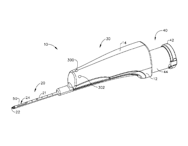
[0005] FIG. 1 depicts a perspective view of an exemplary biopsy device;
[0006] FIG. 2 depicts a perspective view of the biopsy device of FIG. 1,
with a probe
portion separated from a holster portion;
[0007] FIG. 3 depicts a side cross-sectional view of the biopsy device of
FIG. 1, with
the probe portion separated from the holster portion;
[0008] FIG. 4 depicts an exploded view of the biopsy device of FIG. 1, with
portions
shown in cross-section, and with housing components, a battery, and a circuit
. board removed;
[0009] FIG. 5 depicts a perspective cross-sectional view of a needle
portion of the
biopsy device of FIG. 1, with the cutter in a partially retracted position;
CA 02771212 2012-02-10
WO 2011/022122
PCT/US2010/040315
-3-
100101 FIG. 6 depicts a perspective cross-sectional view of cutter
actuation
mechanism and valve mechanism components of the biopsy device of FIG. 1,
with the cutter in the partially retracted position of FIG. 5;
[0011] FIG. 7 depicts a side cross-sectional view of the components of FIG.
6, with
the cutter in the partially retracted position of FIG. 5;
[0012] FIG. 8 depicts a schematic view of the biopsy device of FIG. 1,
showing
exemplary control components; and
[0013] FIG. 9 depicts a perspective view of an exemplary alternative biopsy
device.
[0014] The drawings are not intended to be limiting in any way, and it is
contemplated that various embodiments of the invention may be carried out in
a variety of other ways, including those not necessarily depicted in the
drawings. The accompanying drawings incorporated in and forming a part of
the specification illustrate several aspects of the present invention, and
together with the description serve to explain the principles of the
invention; it
being understood, however, that this invention is not limited to the precise
arrangements shown.
DETAILED DESCRIPTION
[0015] The following description of certain examples of the invention
should not be
used to limit the scope of the present invention. Other examples, features,
aspects, embodiments, and advantages of the invention will become apparent
to those skilled in the art from the following description, which is by way of
illustration, one of the best modes contemplated for carrying out the
invention.
As will be realized, the invention is capable of other different and obvious
aspects, all without departing from the invention. Accordingly, the drawings
and descriptions should be regarded as illustrative in nature and not
restrictive.
100161 Overview
CA 02771212 2012-02-10
WO 2011/022122
PCT/US2010/040315
-4-
100171 As shown
in FIGS. 1-3 (among others), an exemplary biopsy device (10)
comprises a needle (20), a body (30), and a tissue sample holder (40). In
particular, needle (20) extends distally from the distal portion of body (30),
while tissue sample holder (40) extends proximally from the proximal portion
of body (30). Body (30) is sized and configured such that biopsy device (10)
may be operated by a single hand of a user. In particular, and as described in
greater detail below, a user may grasp body (30) with a single hand, insert
needle (20) into a patient's breast, and collect one or a plurality of tissue
samples from within the patient's breast, all with just using a single hand.
Alternatively, a user may grasp body (30) with more than one hand and/or
with any desired assistance. In some settings, the user may capture a
plurality
of tissue samples with just a single insertion of needle (20) in the patient's
breast. Such tissue samples may be pneumatically deposited in tissue sample
holder (40), as described in greater detail below, then retrieved from tissue
sample holder (40) for analysis.
[0018] Body
(30) of the present example comprises a probe (12) and a holster (14).
As shown in FIGS. 2-3, and as described in greater detail below, probe (12) is
separable from holster (14). In particular, probe (12) and holster (14) may be
removably coupled using bayonet mounts (not shown) or any other suitable
structures or features. Use of the term "holster" herein should not be read as
requiring any portion of probe (12) to be inserted into any portion of holster
(14). Indeed, in some variations of biopsy device (10), probe (12) may simply
sit on holster (14). In some other variations, a portion of holster (14) may
be
inserted into probe (12). Furthermore, in some biopsy devices (10), probe (12)
and holster (14) may be of unitary or integral construction, such that the two
components cannot be separated. By way of example only, in versions where
probe (12) and holster (14) are provided as separable components, probe (12)
may be provided as a disposable component, while holster (14) may be
provided as a reusable component. In some versions, holster (14) is
CA 02771212 2012-02-10
WO 2011/022122
PCT/US2010/040315
- 5 -
"activated" or powered up when probe (12) is coupled therewith. In some
other versions, holster (14) is "activated" or powered up when holster (14) is
removed from a charging base (not shown). Still other suitable structural and
functional relationships between probe (12) and holster (14), as well as
various
ways in which holster (14) may be activated, will be apparent to those of
ordinary skill in the art in view of the teachings herein.
[0019] While examples described herein refer to the acquisition of biopsy
samples
from a patient's breast, it should be understood that biopsy device (10) may
be
used in a variety of other procedures for a variety of other purposes and in a
variety of other parts of a patient's anatomy.
[0020] Exemplary Needle
[0021] As shown in FIG. 5 (among others), needle (20) of the present
example
comprises a cannula (21) with a tissue piercing tip (22), a lateral aperture
(24),
a first lumen (26), and a second lumen (28). Tissue piercing tip (22) is
configured to pierce and penetrate tissue, without requiring a high amount of
force, and without requiring an opening to be pre-formed in the tissue prior
to
insertion of tip (22). A cutter (50) is disposed in first lumen (26), and is
operable to rotate and translate within first lumen (26) as will be described
in
greater detail below. Lateral aperture (24) is located proximal to tip (22),
is in
fluid communication with first lumen (26), and is configured to receive tissue
when needle (20) is inserted in a breast and when a cutter (50) is retracted
as
will also be described in greater detail below. A plurality of openings (27)
provide fluid communication between first and second lumens (26, 28).
Needle (20) of the present example further comprises a hub (200), as shown in
FIGS. 6-7. Hub (200) may be formed of plastic that is overmolded about
needle (20) or otherwise secured to needle (20), such that hub (200) is
unitarily secured to needle (20). Alternatively, hub (200) may be formed of
CA 02771212 2012-02-10
WO 2011/022122
PCT/US2010/040315
- 6 -
any other suitable material through any suitable process and may have any
other suitable relationship with needle (20).
[0022] Hub
(200) of the present example comprises a sleeve portion (204), which
extends integrally into probe portion (12) of body (30). As shown in FIGS. 6-
7, sleeve portion (204) defines a hollow interior (206), which is in fluid
communication with second lumen (28) of needle (20). Sleeve portion (204)
also defines a plurality of openings (208), which are radially spaced about
the
perimeter of sleeve portion (204) at a common longitudinal position, and
which are in fluid communication with hollow interior (206). Openings (208)
are exposed to ambient air, such that openings (208) provide a vent in the
present example. Openings (208) are selectively fluidly coupled with second
lumen (28) of needle (20) in this example, as will be described in greater
detail
below. A pair of o-rings (210) are positioned about a shuttle valve slider
(152), to substantially seal second lumen (28) relative to openings (208) when
second lumen (28) is not to be vented, depending on the longitudinal position
of slider (152) as will be described in greater detail below. A seal (212) is
also
provided at the proximal end of sleeve (204), at the interface of cutter (50)
and
sleeve (204).
[0023] It
should be understood that, as with other components described herein,
needle (20) may be varied, modified, substituted, or supplemented in a variety
of ways; and that needle (20) may have a variety of alternative features,
components, configurations, and functionalities. By way of example only,
needle (20) may simply lack second lumen (28) altogether in some versions,
such that first lumen (26) is the only lumen defined by needle (20). As
another merely exemplary alternative, biopsy device (10) may be configured
such that needle (20) may be fired distally relative to body (30), such as to
assist in penetration of tissue. Such firing may be provided by one or more
actuators (e.g., solenoid, pneumatic cylinder/piston, etc.), by one or more
CA 02771212 2012-02-10
WO 2011/022122
PCT/US2010/040315
- 7 -
springs, or in any other suitable fashion. Other suitable alternative
versions,
features, components, configurations, and functionalities of needle (20) will
be
apparent to those of ordinary skill in the art in view of the teachings
herein.
Similarly, other suitable modifications to other components of biopsy device
(10) that may be made in accordance with variations of needle (20) (e.g.,
modifying or omitting valve mechanism (150) in versions where second lumen
(28) is omitted from needle (20), etc.) will be apparent to those of ordinary
skill in the art in view of the teachings herein.
[0024] Exemplary Body
[0025] As noted above, body (30) of the present example comprises a probe
portion
(12) and a holster portion (14). As shown in FIGS. 3-4, a motor (36), a
vacuum pump (38), a battery (320), and a control module (330) are provided
within holster portion (14). Battery (320) may be coupled with motor (36) via
control module (330) and one or more trigger buttons (300, 302, 304), as will
be described in greater detail below.
[0026] As shown in FIGS. 3-4, motor (36) of the present example is in
mechanical
communication with vacuum pump (38) and a cutter actuation mechanism
(60). In particular, motor (36) is operable to simultaneously activate vacuum
pump (38) and cutter actuation mechanism (60) when motor (36) is activated.
Alternatively, vacuum pump (38) and cutter rotation mechanism (60) may be
activated in any other suitable fashion. By way of example only, vacuum
pump (38) and/or cutter rotation mechanism (60) may be activated manually
and/or by separate motors and/or in any other suitable fashion. Motor (36) of
the present example comprises a conventional DC motor. However, it should
be understood that motor (36) may alternatively comprise a pneumatic motor
(e.g., with impeller, etc.), a pneumatic linear actuator, an electromechanical
linear actuator, or a variety of other types of movement-inducing devices.
Various suitable ways in which other types of movement-inducing devices
CA 02771212 2012-02-10
WO 2011/022122
PCT/US2010/040315
- 8 -
may be incorporated into biopsy device (10) will be apparent to those of
ordinary skill in the art in view of the teachings herein.
[0027] As shown in FIGS. 3-4, a drive shaft (62) extends from motor (36),
and is
rotationally driven by motor (36). A pair of bearings (70) and a drive gear
(72) are positioned about drive shaft (62). Bearings (70) support drive shaft
(62), while drive gear (72) rotates unitarily with drive shaft (62). Drive
gear
(72) meshes with a second gear (74), which is unitarily secured to a second
shaft (64). Second shaft (64) also includes associated bearings (70) and a
third
gear (76).
[0028] Vacuum pump (38) of the present example comprises a conventional
diaphragm pump, which is driven by a second shaft (64), which is in turn
rotationally driven by motor (36) as described above. Vacuum pump (38) of
the present example operates in the same way regardless of which direction
motor (36) rotates. Of course, any other suitable type of vacuum pump may
be used. Vacuum pump (38) of the present example is operable to induce a
vacuum in tissue sample holder (40) when vacuum pump (38) is activated, as
will be described in greater detail below.
[0029] It should be understood that, as with other components described
herein, body
(30) may be varied, modified, substituted, or supplemented in a variety of
ways; and that body (30) may have a variety of alternative features,
components, configurations, and functionalities. Suitable alternative
versions,
features, components, configurations, and functionalities of body (30) will be
apparent to those of ordinary skill in the art in view of the teachings
herein.
[0030] Exemplary Valve Mechanism
[0031] As shown in FIGS. 4 and 6-7, biopsy device (10) also includes a
valve
mechanism (150) in the present example. Valve mechanism (150) of this
example comprises shuttle valve slider (152), o-rings (210), and sleeve (204)
CA 02771212 2016-11-22
- 9 -
of needle hub (200). Shuttle valve slider (152) is positioned coaxially about
cutter (50), longitudinally between the distal end of sleeve (250) and the
proximal cnd of a stop member (55), and is configured to translate relative to
sleeve (204) and relative to cutter (50). 0-rings (210) are configured to seal
the exterior of shuttle valve slider (152) against the interior sidewall of
sleeve
(204). A gap provides longitudinal fluid communication (e.g., atmospheric
air, etc.) between the outer diameter of cutter (50) and the inner diameter of
shuttle valve slider (152). Notches (153) are configured to provide fluid
communication to the interior of Shuttle. valve slider (152), even as the
distal
end of smooth portion (254) of sleeve (250) engages the proximal end of
shuttle valve slider (152).
100321 As described in greater detail below, cutter (50) is configured to
rotate and
translate relative to body (30), while sleeve (204) remains substantially
stationary relative to body (30). Sleeve (250) and stop member (55) translate
unitarily with cutter (50). In addition, stop member (55) may push shuttle
valve slider (152) proximally; while sleeve (250) may push shuttle valve
slider
(152) distally. Shuttle valve slider (152) may thus translate within sleeve
(250) in accordance with translation of cutter (50) relative to body (30).
However, the distance between the distal end of sleeve (250) and the proximal
end of stop member (55) is greater than the length of shuttle valve slider
(152),
such that there is a degree of "lost motion" between shuttle valve slider
(152)
and cutter (50) as cutter (50) translates in the present example. An example
of
such action of shuttle valve slider (152) is disclosed in U.S. Non-Provisional
Pat. App. No. 12/483,305, entitled "Tetherless Biopsy Device with Reusable
Portion," filed June 12, 2009..
100331 In the present example, shuttle valve slider (152) remains distal to
openings
(208), thereby venting second lumen (28), when cutter (50) is at a distal-most
CA 02771212 2012-02-10
WO 2011/022122
PCT/US2010/040315
- 10 -
position; when cutter (50) is transitioning between the distal-most position
and
the proximal-most position (see, e.g., FIGS. 5-7); and at latter stages of
cutter
(50) transitioning from the proximal-most position to the distal-most
position.
However, when cutter (50) moves to the proximal position, stop member (55)
pushes shuttle valve slider (152) proximally such that openings (208) are
longitudinally positioned between o-rings (210), thus substantially sealing
second lumen (28) until the distal end of sleeve (250) engages the proximal
end of shuttle valve slider (152) and begins pushing shuttle valve slider
(152)
distally to the point where the proximal-most o-ring (210) is moved distal to
openings (208). Second lumen (28) is thereby again vented as noted above.
Thus, valve mechanism (150) of the present example substantially seals off
second lumen (28) relative to atmosphere when cutter (50) is at a proximal
position and when cutter (50) is at initial stages of advancement; while
venting
second lumen (28) to atmosphere when cutter (50) is at other positions.
[0034] It should be understood that, as with other components described
herein, valve
mechanism (150) may be varied, modified, substituted, or supplemented in a
variety of ways; and that valve mechanism (150) may have a variety of
alternative features, components, configurations, and functionalities.
Suitable
alternative versions, features, components, configurations, and
functionalities
of valve mechanism (150) will be apparent to those of ordinary skill in the
art
in view of the teachings herein.
[0035] Exemplary Tissue Sample Holder
[0036] As shown in FIGS. 1-4, tissue sample holder (40) of the present
example
comprises a cap (42), an outer cup (44), and a filter tray (46). Cup (44) is
secured to probe (12) in the present example. Such engagement may be
provided in any suitable fashion. Outer cup (44) of the present example is
substantially transparent, allowing the user to view tissue samples on filter
tray
(46), though outer cup (44) may have any other suitable properties if desired.
CA 02771212 2012-02-10
WO 2011/022122
PCT/US2010/040315
-11-
100371 Outer
cup (44) is in fluid communication with cutter lumen (52) and with
vacuum pump (38) in the present example. In particular, outer cup (44) is in
fluid communication with cutter lumen (52) via a first port (45); and is in
fluid
communication with vacuum pump (38) via a second port (47). A conduit
(not shown) couples port (41) of vacuum pump (38) with second port (47) of
outer cup (44). In the present example, second port (47) is further coupled
with a hydrophobic filter (48), which is in fluid communication with the
interior space defined by outer cup (44). In addition to or in lieu of having
hydrophobic filter (48) a highly absorbent material may be provided in tissue
sample holder (40) to soak up liquids. Alternatively, liquids may be dealt
with
in any other suitable fashion. In the present example, vacuum pump (38) may
thus be used to induce a vacuum in cutter lumen (52); with such a vacuum
being communicated through conduit (39), ports (41, 45, 47), and the interior
of outer cup (44).
[0038] Filter
tray (46) of the present example has a basket-like configuration, and has
plurality of openings (47) formed therethrough. Openings (47) are sized and
configured to permit the passage of fluids therethrough while preventing the
passage of tissue samples therethrough. Filter tray (46) is thus configured to
receive tissue samples that are communicated proximally through cutter (50)
as will be described in greater detail below. It should be understood that
filter
tray (46) may take a variety of alternate forms.
[0039] Cap (42)
is removably coupled with outer cup (44) in the present example. An
o-ring (53) provides a seal when cap (42) is engaged with outer cup (44). A
vacuum may thus be maintained within outer cup (44) when cap (42) is
secured to outer cup (44). In operation, a user may remove cap (42) to access
tissue samples that have gathered on filter tray (46) during a biopsy process.
[0040] Tissue
sample holder (40) of the present example is configured to hold at least
ten tissue samples. Alternatively, tissue sample holder (40) may be configured
CA 02771212 2016-11-22
- 12 -
to hold any other suitable number of tissue samples. It should be understood
that, as with other components described herein, tissue sample holder (40)
may be varied, modified, substituted, or supplemented in a variety of ways;
and that tissue sample holder (40) may have a variety of alternative features,
components, configurations, and functionalities. For instance, tissue sample
holder (40) may be alternatively configured such that it has a plurality of
discrete tissue sample compartments that may be selectively indexed to cutter
lumen (52). Such indexing may be. provided automatically or manually. By
way of example only, tissue sample holder (40) may he configured and
operable in accordance with the teachings of U.S. Pub. No. 2008/0195066,
entitled "Revolving Tissue Sample Holder for Biopsy Device," published
August 14, 2008; I;
U.S. Non-Provisional Patent Application Serial No. 12/337,997, entitled
"Tissue Biopsy Device with Rotatably Linked Thumbwheel and Tissue
Sample Holder," filed December 18, 2008; -U.S. Non-Provisional Patent
Application Serial No. 12/337,911, entitled -Biopsy Device with Discrete
Tissue Chambers," filed December 18, 2008,
i; or U.S. Non-Provisional Patent Application
Serial No. 12/337,874, entitled "Mechanical Tissue Sample Holder Indexing
Device," filed December 18, 2008.
Other suitable alternative versions, features, components,
configurations, and functionalities of tissue sample holder (40) will be
apparent to those of ordinary skill in the art in view of the teachings
herein.
Alternatively, tissue sample holder (40) may simply be omitted, if desired.
10041] Exemplary Cutter
10042] As shown in FIG. 5, cutter (50) of the present example is
substantially hollow,
such that cutter (50) defines a cutter lumen (52). Cutter (50) also has a
substantially sharp distal edge (51), such that cutter (50) is operable to
sever a
CA 02771212 2012-02-10
WO 2011/022122
PCT/US2010/040315
- 13 -
biopsy sample from tissue protruding through lateral aperture (24) of needle
(20). Alternatively, the distal end of cutter (50) may have any other suitable
configuration. As shown in FIGS. 3-4, a proximal portion of cutter (50)
extends into tissue sample holder (40). A vacuum created in tissue sample
holder (40) by vacuum pump (38) is thus communicated to cutter lumen (52).
A seal (54) is provided at the interface of cutter (50) and outer cup (44).
Seal
(54) is configured to substantially seal the interface of cutter (50) and
mount
(42). Furthermore, cutter (50) is configured such that it remains in sealed
fluid
communication with the interior of tissue sample holder (40) even when cutter
(50) is in a distal most position. Of course, cutter (50) may have any other
suitable alternative features or configurations. Similarly, cutter (50) may
have
any other suitable alternative relationships with tissue sample holder (40).
[0043] It should be understood that, as with other components described
herein, cutter
(50) may be varied, modified, substituted, or supplemented in a variety of
ways; and that cutter (50) may have a variety of alternative features,
components, configurations, and functionalities. Suitable alternative
versions,
features, components, configurations, and functionalities of cutter (50) will
be
apparent to those of ordinary skill in the art in view of the teachings
herein.
[0044] Exemplary Cutter Actuation Mechanism
[0045] As shown in FIGS. 3-7, cutter actuation mechanism (60) of the
present
example comprises motor (36), shafts (62, 64), and gears (72, 74, 76). It
should be understood that activation of motor (36) will rotate gears (82, 84)
in
the present example. As shown in FIGS. 3 and 6, motor (36), shafts (62, 64,
68, 69), gears (72, 74, 76, 78, 80, 82, 84), and bearings (70) are all
contained
within holster (14) in the present example. As shown in FIG. 2, gears (82, 84)
are partially exposed by an opening formed in a cover plate (18) of holster
(14) in the present example.
CA 02771212 2012-02-10
WO 2011/022122
PCT/US2010/040315
- 14 -
[0046] Cutter
actuation mechanism (60) of the present example further comprises a
hex nut (100) and a worm nut (120). Hex nut (100) includes a unitary gear
(86). Worm nut (120) also includes a unitary gear (88). Gear (86) is
configured to mesh with gear (82) when probe (12) and holster (14) are
coupled together; while gear (88) is configured to mesh with gear (84) when
probe (12) and holster (14) are coupled together. In particular, and as shown
in FIG. 2, gears (86, 88) are partially exposed by an opening formed in a
cover
plate (16) of probe (12) in the present example. Motor (36) is thus operable
to
rotatingly drive gears (86, 88) in the present example when probe (12) and
holster (14) are coupled together. As described in greater detail below, such
rotation of gears (86, 88) will cause cutter (50) to rotate and translate in
the
present example.
[0047] A sleeve
(250) is unitarily secured to cutter (50). As best seen in FIGS. 6-7,
sleeve (250) comprises a hex portion (252), a smooth portion (254), and a
flange (256) separating hex portion (252) from smooth portion (254). Hex nut
(100) is slidably positioned over hex portion (252) of sleeve (250). The
engagement between sleeve (250) and hex nut (100) is such that rotation of
hex nut (100) provides corresponding rotation of sleeve (250), yet hex nut
(100) may slide longitudinally relative to sleeve (250). It
should be
understood that sleeve (250) and hex nut (100) may have a variety of other
configurations (e.g., complementary key and keyway instead of hex features,
etc.) and relationships.
[0048] As noted
above, gear (86) of hex nut (100) is configured to mesh with gear
(82), such that rotation of gear (82) causes rotation of hex nut (100). Such
rotation of hex nut (100) will cause corresponding rotation of cutter (50) as
noted above. It will therefore be understood that cutter actuation mechanism
(60) may cause rotation of cutter (50) in response to activation of motor
(36),
with rotation of motor (36) being communicated to cutter (50) through shafts
CA 02771212 2012-02-10
WO 2011/022122
PCT/US2010/040315
- 15 -
(62, 64, 68, 69), gears (72, 74, 76, 78, 80, 82, 84, 86), hex nut (100), and
sleeve (250). Of
course, any other suitable structures, components,
configurations, or techniques may be used to provide rotation of cutter (50).
[0049] Cutter
actuation mechanism (60) of the present example further comprises a
lead screw (122). Lead screw (122) is positioned about hex portion (252) of
sleeve (250), and is configured to rotate unitarily therewith. Lead screw
(122)
defines a hexagonal opening with six flat faces that are configured to
complement the flat faces of sleeve (250), such that rotation of cutter (50)
and
sleeve (250) provides corresponding rotation of lead screw (122). Lead screw
(122) is further secured to hex portion (252) of sleeve (250) by a clip (124),
and a washer (126) is positioned between clip (124) and lead screw (122). A
first coil spring (128) is positioned between the proximal end of lead screw
(122) and washer (126). A second coil spring (130) is positioned between the
distal end of lead screw (122) and flange (256) of sleeve (250). The spacing
between flange (256) and washer (126) permit some freedom of movement for
lead screw (122) along a portion of the length of sleeve (250) between flange
(256) and washer (126); while springs (128, 130) bias lead screw (122) to be
substantially centered between flange (256) and washer (126). It should be
understood that any other suitable type of resilient member may be used in
addition to or in lieu of coil springs (128, 130).
[0050] Lead
screw (122) has external threads (132) that are engaged with internal
threads (134) of worm nut (120). Accordingly, lead screw (122) translates
relative to worm nut (120) when lead screw (122) rotates relative to worm nut
(120) when threads (132, 134) are engaged. However, the interior length of
worm nut (120) also includes smooth sections (136) that are distal to and
proximal to internal threads (134). Thus, lead screw (122) may not translate
relative to worm nut (120) when lead screw (122) rotates relative to worm nut
(120) when threads (132) are located at smooth sections (136) (e.g., when
CA 02771212 2012-02-10
WO 2011/022122
PCT/US2010/040315
- 16 -
threads (132, 134) are not engaged). Worm nut (120) is further supported by a
bushing (138) in the present example. It should be understood that, due to
engagement of lead screw (122) with flange (256) and washer (126), and due
to engagement of sleeve (250) with cutter (250), translation of lead screw
(122) relative to worm nut (120) in the present example also results in
translation of cutter (50) relative to body (30) in the present example. It
should also be understood that sleeve (250), lead screw (122), and worm nut
(120) may have a variety of other configurations and relationships. Similarly,
a variety of other structures or components may be used in addition to or in
lieu of sleeve (250) and/or worm nut (120).
[0051] As noted
above, gears (82, 84) of holster (14) rotate simultaneously when
motor (36) is activated. As further noted above, gears (82, 84) mesh with
gears (86, 88) of probe (12) when probe (12) is coupled with holster (14),
such
that activated motor (36) rotates gears (86, 88) simultaneously. Activated
motor (36) will thus rotate hex nut (100) and worm nut (120) simultaneously.
It should therefore be understood that sleeve (250), cutter (50), lead screw
(122), and worm nut (120) will all rotate simultaneously when motor (36) is
activated. It is also noted that gears (82, 84) have different pitch
diameters.
Gears (86, 88) also have different pitch diameters. Accordingly, even with
motor (36) rotating at one rotational speed, hex nut (100) and worm nut (120)
rotate simultaneously in the same direction at different rotational speeds.
Since rotation of lead screw (122) is driven by rotation of hex nut (100),
lead
screw (122) and worm nut (120) also rotate simultaneously in the same
direction at different rotational speeds. Even though lead screw (122) and
worm nut (120) rotate simultaneously in the same direction, the difference
between rotational speeds of lead screw (122) and worm nut (120) provide a
net result of lead screw (122) rotating relative to worm nut (120), and such
relative rotation provides translation of cutter (50) as cutter (50) rotates.
By
way of example only, with motor (36) providing an output speed of
CA 02771212 2016-11-22
- 17 -
approximately 8,000 rpm, the above-described configuration may provide
rotation of cutter (50) at a speed of approximately 1,000 rpm and rotation of
worm nut (120) at a speed of approximately 850 rpm, resulting in a net
rotation of cutter (50) relative to worm nut (120) at approximately 150 rpm.
Of course, any other suitable differential may be provided.
100521 In the present example, cutter (50) is retracted proximally when
motor (36) is
activated to rotate cutter (50) counterclockwise (viewed from tissue sample
holder (40) toward needle (20)); while cutter (50) is advanced distally When
motor (36) is activated to rotate cutter (50) clockwise (viewed from tissue
sample holder (40) toward needle (20)). The direction of motor (36) rotation
may thus be reversed to transition between distal and proximal translation of
cutter (50). Alternatively, cutter (50) may instead be rotated clockwise
during
retraction of cutter (50) and counterclockwise during advancement of cutter
(50). Furthermore, cutter actuation mechanism (60) may be configured to be
self-reversing, such that cutter (50) may be translated distally and
proximally
without reversing the direction of motor (36) rotation.
[0053] Further exemplary details of cutter actuation mechanism (100) arc
disclosed in
U.S. Non-Provisional Pat. App. No. 12/483,305, entitled "Tetherless Biopsy
= Device with Reusable Portion," filed June 12, 2009,.
Of course, any other suitable structures,
components, configurations, or techniques may be used to provide translation
and/or rotation of cutter (50). It should therefore be understood that, as
with
other components described herein, cutter actuation mechanism (60) may be
varied, modified, substituted, or supplemented in a variety of ways; and that
cutter actuation mechanism (60) may have a variety of alternative features,
= components, configurations, and functionalities. By way of example only,
biopsy device (10) may be configured such that cutter (50) does not translate
(e.g., such that cutter (50) merely rotates, etc.); or such that cutter (50)
does
CA 02771212 2012-02-10
WO 2011/022122
PCT/US2010/040315
- 18 -
not rotate (e.g., such that cutter (50) merely translates, etc.). Other
suitable
alternative versions, features, components, configurations, and
functionalities
of cutter actuation mechanism (60) will be apparent to those of ordinary skill
in the art in view of the teachings herein.
[0054] Exemplary Pneumatic Operation
[0055] As noted above, vacuum pump (38) may start building a vacuum in
cutter
lumen (52) as soon as motor (36) is activated; and such a vacuum may
continue to build or be maintained as cutter (50) starts moving proximally
toward the retraced position. At this stage, second lumen (28) is vented to
atmosphere. As cutter (50) moves toward retracted position, such that lateral
aperture (24) of needle (20) is "partially open" as shown in FIG. 5, a vacuum
in cutter lumen (52) may be further communicated through first lumen (26),
which may draw tissue into lateral aperture (24). At this stage, second lumen
(28) is still vented to atmosphere. In the present example, second lumen (28)
is substantially sealed when cutter (50) reaches a longitudinal position that
is
proximal to the position shown in FIG. 5, and before cutter (50) reaches the
fully retracted position.
[0056] When cutter (50) reaches the fully retracted position, such that
lateral aperture
(24) of needle (20) is "open", a vacuum in cutter lumen (52) may continue to
be further communicated through first lumen (26), which may continue to
draw tissue into lateral aperture (24). Of course, some amount of tissue may
naturally prolapse into lateral aperture (24) without the assistance of
vacuum,
such that vacuum may not even be needed to draw tissue into lateral aperture
(24). At this stage, second lumen (28) is substantially sealed relative to
atmosphere. In particular, stop member (55) has pushed shuttle valve slider
(152) to a proximal position, such that o-rings (210) "straddle" openings
(208)
and seal against the interior sidewall of sleeve portion (204) to prevent
CA 02771212 2012-02-10
WO 2011/022122
PCT/US2010/040315
- 19 -
atmospheric air from being communicated from openings (208) to second
lumen (28) via hollow interior (206) of sleeve portion (204).
[0057] As motor
(36) is reversed and cutter (50) is advanced to sever tissue
protruding through lateral aperture (24), vacuum pump (38) may continue to
induce a vacuum in cutter lumen (52), and second lumen (28) may eventually
be vented to atmosphere. However, in the initial stages of advancement of
cutter (50) from the proximal-most position to the distal-most position, the
"lost motion" between cutter (50) and shuttle valve slider (152) leaves
shuttle
valve slider (152) in the proximal position (thereby sealing second lumen
(28))
until cutter (50) advances far enough for the distal end of sleeve (250) to
engage the proximal end of shuttle valve slider (152). After the distal end of
sleeve (250) engages the proximal end of shuttle valve slider (152), and after
cutter (50) has continued to move distally to a sufficient degree, the distal
end
of sleeve (250) eventually pushes shuttle valve slider (152) distally, such
that
the proximal-most o-ring (210) is eventually moved distal to openings (208)
(thereby venting second lumen (28)). As cutter (50) again finally reaches the
distal-most position, cutter (50) may completely sever the tissue protruding
through lateral aperture (24), with second lumen (28) being vented.
[0058] With the
severed tissue sample residing in cutter lumen (52), with vacuum
pump (38) drawing a vacuum at the proximal face of the severed tissue
sample, and with the venting being provided at the distal face of the severed
tissue sample (via openings (208), second lumen (28), and openings (27)), the
pressure differential applied to the severed tissue sample may cause the
severed tissue sample to be drawn proximally through cutter lumen (52) and
into tissue sample holder (40). The severed tissue sample may thus be
deposited on filter tray (46) of tissue sample holder (40).
[0059] Further
exemplary details of pneumatic operation are disclosed in U.S. Non-
Provisional Pat. App. No. 12/483,305, entitled "Tetherless Biopsy Device with
CA 02771212 2016-11-22
- 20 -
Reusable Portion," filed June 12, 2009..
Of course, any other suitable structures, components,
configurations, or techniques may be used to provide selective sealing and/or
venting of second lumen (28). Furthermore, in some variations of biopsy
device (10), a vacuum, saline, pressurized air, atmospheric air, and/or any
other medium may be communicated to second lumen (28) at any suitable
stage of operation of biopsy device (10) (e.g., applying vacuum or venting to
second lumen (28) during and/or upon retraction of cutter (50) and/or during
advancement of cutter (50), sealing second lumen during advancement of
cutter (50), etc.). Other suitable alternative structures, components,
configurations, or techniques for communicating severed tissue samples
proximally through cutter lumen (52) to reach tissue sample holder (40) will
be apparent to those of ordinary skill in the art in view of the teachings
herein.
[0060] But ton Activation
[0061] As shown in FIGS. 1 and 8, biopsy device (10) has a plurality of
buttons (300,
302, 304), which may be used to control operation of biopsy device (10). For
instance, either of buttons (302, 304) may bc used as trigger buttons. That
is,
an operator may actuate a selected one of trigger buttons (302, 304) to
initiate
a cutting stroke (e.g., by activating motor (36), etc.). Trigger button (302)
may thus provide the same functionality as trigger button (304). As described
in greater detail below, biopsy device (10) may be configured such that either
trigger button (302, 304) may be used to initiate a cutting stroke; or such
that
one particular trigger button (302, 304) must be selected to initiate a
cutting
stroke while the non-selected trigger button (302, 304) is left disabled. It
should be noted that the term "trigger button" is intended to encompass a
variety of types of structures and devices, and is not limited to just a
standard
electromechanical button. By way of example only, trigger buttons (302, 304)
may include pneumatic buttons, capacitive buttons, inductive buttons, etc.
CA 02771212 2012-02-10
WO 2011/022122
PCT/US2010/040315
- 21 -
Still other suitable components that may be used to initiate a cutting stroke
will be apparent to those of ordinary skill in the art in view of the
teachings
herein.
[0062] As shown
in FIG. 8, trigger buttons (302, 304) are in communication with
control module (330), which is in communication with battery (320) and
motor (36). Battery (320) is also in communication with motor (36). Control
module (330) is configured to selectively complete a circuit between battery
(320) and motor (36), such that motor (36) may be selectively powered in
accordance with an operator's input via trigger buttons (302, 304). Control
module (330) may also include a control logic such that control module (330)
is operable to selectively reverse the direction of rotation of motor (36)
(e.g.,
to transition between proximal and distal movement of cutter (50), etc.). As
used herein, the term "control module" should not be read as necessarily
requiring all of the components of control module (330) to be integrated into
a
single self-contained unit. Indeed, control module (330) may in fact comprise
several components that are positioned at various locations within biopsy
device (10), with such separate components being in communication with each
other to some degree. Suitable components of a control module (330) may
include one or more processors and associated circuitry, etc., as will be
apparent to those of ordinary skill in the art in view of the teachings
herein.
[0063] In some
versions, the operator need only tap one of trigger buttons (302, 304),
or hold one of trigger buttons (302, 304) down for a predetermined period of
time, in order to initiate and complete a cutting stroke. In some other
versions,
the operator needs to hold down one of trigger buttons (302, 304) during the
entire cutting stroke. In some such versions, where the selected trigger
button
(302, 304) is released in the middle of a cutting stroke, the cutting stroke
may
be paused until the selected trigger button (302, 304) is again actuated.
CA 02771212 2012-02-10
WO 2011/022122
PCT/US2010/040315
- 22 -
[0064] As shown
in FIGS. 1 and 8, one trigger button (302) is on the left-hand side of
body (30); while the other trigger button (304) is on the other side of body
(30). Those of ordinary skill in the art will understand that any given
operator
of biopsy device (10) may prefer one trigger button (302) over the other
trigger button (304) due to the location of trigger buttons (302, 304). By way
of example only, such preference may be dictated at least in part by whether
the operator is right-handed or left-handed, the way in which the operator
prefers to grip biopsy device (10), etc. Having two trigger buttons (302, 304)
will thus allow the operator to select one particular trigger button (302,
304)
for operation of biopsy device (10) in accordance with the operator's
preference. In some versions, trigger buttons (302, 304) and control module
(330) are configured such that the operator may use either or both trigger
buttons (302, 304) at any time during a biopsy procedure. In other words,
both trigger buttons (302, 304) may be considered "active," and therefore
immediately operable/enabled, at all times. The operator may thus switch
from using one trigger button (302, 304) to the other trigger button (302,
304),
without having to do anything additional in order to switch from using one
trigger button (302, 304) to the other trigger button (302, 304).
[0065] In some
other versions, however, trigger buttons (302, 304) and control
module (330) may be configured such that only one trigger button (302, 304)
can be enabled at a given time. For instance, trigger buttons (302, 304) and
control module (330) may be configured such that a user must select one
particular trigger button (302, 304) for activation/enablement; and once such
a
selection has been made, the other trigger button (302, 304) will be
deactivated/disabled for the rest of the biopsy procedure. Such
deactivation/disablement may prevent inadvertent activation of biopsy device
(10) by inadvertent actuation of the non-selected trigger button (302, 304).
CA 02771212 2012-02-10
WO 2011/022122
PCT/US2010/040315
-23-
100661 It
should be understood that there are a variety of ways in which one particular
trigger button (302, 304) may be activated/enabled while the other trigger
button (302, 304) is deactivated/disabled. For instance, activation/enablement
of a particular trigger button (302, 304) and deactivation/disablement of the
other trigger button (302, 304) may be provided electronically. By way of
example only, activation/enablement of a particular trigger button (302, 304)
may be provided as soon as the operator first actuates the particular trigger
button (302, 304). In some such versions, control module (330) may include a
logic that is configured to sense which particular trigger button (302, 304)
was
first actuated, and that may designate that particular trigger button (302,
304)
as the activated/enabled trigger button (302, 304). Such a logic in control
module (330) may also designate or treat the other trigger button (302, 304)
as
the deactivated/disabled trigger button (302, 304). In other words, as soon as
a
first trigger button (302, 304) is actuated, control module (330) may permit
biopsy device (10) to be controlled by that first trigger button (302, 304)
while
preventing biopsy device (10) from being controlled by the other trigger
button (302, 304).
[0067] As
another merely illustrative example, electronic activation/enablement and
deactivation/disablement may be provided by logic in control module (330) in
response to the operator actuating a selected trigger button (302, 304) in a
particular way. For instance, control module (330) may be programmed with
logic to sense when a particular trigger button (302, 304) is "double
clicked."
That is, biopsy device (10) may be configured such that the operator may
activate/enable a selected trigger button (302, 304) by "double clicking" it.
Once the first trigger button (302, 304) has been double clicked, logic in
control module (330) may designate or treat the other trigger button (302,
304)
as the deactivated/disabled trigger button (302, 304), such that biopsy device
(10) cannot be controlled by the deactivated/disabled trigger button (302,
304).
Alternatively, control module (330) may be programmed with logic to sense
CA 02771212 2012-02-10
WO 2011/022122
PCT/US2010/040315
- 24 -
when a particular trigger button (302, 304) has been actuated in some other
predetermined pattern or number of times (e.g., three times within a certain
period of time, once for a duration of three seconds, etc.); and may designate
the active/enabled trigger button (302, 304) in response to such operator
input.
[0068] As yet
another merely illustrative example, electronic activation/enablement
and deactivation/disablement may be provided in response to the operator
actuating a selection button or switch. For instance, in the present example,
button (300) may be used to turn on biopsy device (10) and to select which
trigger button (302, 304) to activate/enable. In some such versions, button
(300) is operable to turn biopsy device (10) on or off when button (300) is
actuated for a certain period of time (e.g., three seconds, etc.). The
required
actuation duration may differ for turning biopsy device (10) on versus turning
biopsy device (10) off For instance, biopsy device (10) may be turned on
when button (300) is merely tapped; while button (300) must be held down for
at least three seconds in order to turn biopsy device (10) off Once biopsy
device (10) is turned on, the operator may tap button (300) to select a
particular trigger button (302, 304) for activation/enablement.
[0069] In some
versions where button (300) may be used to select a particular trigger
button (302, 304) for activation/enablement, each trigger button (302, 304)
may have an associated LED (306, 308) indicating whether the trigger button
(302, 304) is activated/enabled; and tapping button (300) may toggle between
trigger buttons (302, 304) and their associated LED (306, 308). In other
words, button (300) may be used to program control module (300) to select
which trigger button (302, 304) is to be activated/enabled and which trigger
button (302, 304) is to be deactivated/disabled; and control module (300) may
also a logic configured to illuminate the LED (306, 308) that is associated
with
the activated/enabled trigger button (302, 304) to provide visual feedback to
the operator. To the extent that such LEDs (306, 308) are used, such LEDs
CA 02771212 2012-02-10
WO 2011/022122
PCT/US2010/040315
- 25 -
may be positioned at any suitable location, including but not limited to in or
on body (30) next to their associated trigger button (302, 304). In addition
or
in the alternative, the activated/enabled trigger button (302, 304) may itself
be
illuminated.
[0070] In some
versions where visual feedback is provided to indicate which
particular trigger button (302, 304) is activated/enabled (e.g., through LEDs
(306, 308) and/or through illumination of activated/enabled trigger button
(302, 304), etc.), visual feedback may also be provided to indicate that a
trigger button (302, 304) needs to be selected for activation. For instance,
as
soon as biopsy device (10) is turned on, LEDs (306, 308) or trigger buttons
(302, 304) may both flash, indicating to the operator that the operator must
select one particular trigger button (302, 304) for activation/enablement. As
soon as the operator selects a preferred trigger button (302, 304) for
activation/enablement, the LEDs (306, 308) or trigger buttons (302, 304) may
stop flashing, the selected LED (306, 308) or trigger button (302, 304) may
illuminate, and the non-selected LED (306, 308) or trigger button (302, 304)
may go dark. Of course, a variety of other types of visual indication may be
provided to indicate which trigger button (302, 304) has been selected for
activation/enablement. Other suitable ways in which such visual indication
may be provided will be apparent to those of ordinary skill in the art in view
of
the teachings herein. Similarly, other suitable ways in which electronic
activation/enablement and deactivation/disablement of trigger buttons (302,
304) may be provided will be apparent to those of ordinary skill in the art in
view of the teachings herein.
[0071] In versions where electronic
activation/enablement and
deactivation/disablement of trigger buttons (302, 304) is provided, an
operator's selection of a preferred trigger button (302, 304) may be stored in
a
non-volatile memory within body (30). In other words, if the operator turns
CA 02771212 2012-02-10
WO 2011/022122
PCT/US2010/040315
- 26 -
off biopsy device (10) and later turns biopsy device (10) back on, such
memory may recall the operator's preference of trigger button (302, 304), such
that the operator does not need to re-designate the preferred trigger button
(302, 304). Alternatively, an operator's selection of a preferred trigger
button
(302, 304) may be stored in a volatile memory within body (30), such that an
operator must re-designate the preferred trigger button (302, 304) after
biopsy
device (10) is turned off and then turned back on. As yet another merely
illustrative variation, control module (330) may be configured such that the
operator must designate the preferred trigger button (302, 304) before each
cutting stroke is initiated, even if biopsy device (10) has not been turned
off/on
between cutting strokes.
[0072] Still
another merely illustrative example is illustrated in FIG. 9, in which
activation/enablement of a particular trigger button (302, 304) and
deactivation/disablement of the other trigger button (302, 304) may be
provided mechanically. In particular, FIG. 9 shows a sliding cover (350),
which is positioned adjacent to trigger button (302), and which is slidable
along rails (352). Rails (352) are integral with body (300) in the present
example. It should be understood that trigger button (304) may also have a
sliding cover (350). Cover (350) is shown in FIG. 9 as leaving trigger button
(302) exposed, thereby permitting trigger button (302) to be actuated. In this
example, a cover (350) may be slid over trigger button (304), such that
actuation of trigger button (302) is permitted while actuation of trigger
button
(304) is prevented. The operator may thus select which trigger button (302,
304) should be enabled and which trigger button (302, 304) should be disabled
simply by sliding covers (350) to selectively expose or cover trigger buttons
(302, 304) in accordance with the operator's preference. While sliding covers
(350) are shown as providing selective exposure/covering of trigger buttons
(302, 304), it should be understood that a variety of other structures may be
used to provide selective exposure/covering of trigger buttons (302, 304)
(e.g.,
CA 02771212 2012-02-10
WO 2011/022122
PCT/US2010/040315
-27 -
pivoting cover, swinging cover, snap-on cover, etc.). Still other suitable
ways
in which trigger buttons (302, 304) may be mechanically selectively covered
or exposed will be apparent to those of ordinary skill in the art in view of
the
teachings herein.
[0073] While
button (300) is described above as being usable to turn biopsy device
(10) on and off, and/or to provide selection of which trigger button (302,
304)
is to be activated, it should be understood that button (300) may be used as
yet
another trigger button (300, 302, 304). For instance, button (300) may be
provided as another available trigger button, just like trigger buttons (302,
304), in addition to or as an alternative to button (300) having the other
functionality described above. Button (300) may thus be selected by an
operator who prefers the location of button (300) at the top of body (30) over
the location of trigger buttons (302, 304) on the sides of body (30). In such
versions, when button (300) is selected as the preferred trigger button,
actuation of button (300) may initiate a cutting stroke, etc. Similarly, while
two trigger buttons (302, 304) and three trigger buttons (300, 302, 304) are
discussed above, it should be understood that any suitable number of trigger
buttons (302, 304) may be provided and available for selection. Such
additional trigger buttons (302, 304) may be provided in any suitable location
¨ on body (30) (e.g., on probe (12) or holster (14)), remotely via an
electrical
cable (e.g., using footswitch and/or using a vacuum control module, etc.),
remotely via an wireless activation (e.g., using footswitch and/or using a
vacuum control module, etc.), or elsewhere.
[0074] In some
versions, some other switch is used to select a particular trigger button
(302, 304) for activation/enablement. For instance, biopsy device (10) may
include a slider switch or toggle switch to provide selection of which trigger
button (302, 304) is to be activated/enabled. As described above, control
module (330) may include a logic that is configured to deactivate/disable
CA 02771212 2012-02-10
WO 2011/022122
PCT/US2010/040315
- 28 -
whichever trigger button (302, 304) is not selected, such that biopsy device
(10) will simply not respond to actuation of the non-selected trigger button
(302, 304). Still other ways in which selection of a trigger button (302, 304)
¨ electronic, mechanical, both, or otherwise ¨ may be provided will be
apparent to those of ordinary skill in the art in view of the teachings
herein.
[0075] It should also be understood that the concepts of selectively
enabled buttons
(302, 304) described herein may be applied to a variety of other devices. By
way of example only, the concepts of selectively enabled buttons (302, 304)
described herein may be applied to any biopsy device that is disclosed in any
patent, patent publication, or patent application that is cited herein.
Various
ways in which the concepts of selectively enabled buttons (302, 304)
described herein may be applied to such biopsy devices will be apparent to
those of ordinary skill in the art in view of the teachings herein.
Furthermore,
the concepts of selectively enabled buttons (302, 304) described herein may be
applied to virtually any type of device ¨ whether a biopsy device or some
other type of device. It is therefore contemplated that the concepts of
selectively enabled buttons (302, 304) described herein are certainly not
limited to the particular biopsy device (10) described herein.
[0076] Exemplary Method of Operation
[0077] In a merely exemplary use of biopsy device (10), a user first
inserts tissue
piercing tip (22) into the breast of a patient. During such insertion, cutter
(50)
may be advanced to the distal-most position, such that lateral aperture (24)
of
needle (20) is closed. As also noted herein, such insertion may be performed
under visual guidance, stereotactic guidance, ultrasound guidance, MRI
guidance, PEM guidance, BSGI guidance, palpatory guidance, some other
type of guidance, or otherwise. With needle (20) sufficiently inserted into
the
patient's breast, the user may select which particular trigger button (302,
304)
to use, such as by double-clicking on the selected trigger button (302, 304),
CA 02771212 2012-02-10
WO 2011/022122
PCT/US2010/040315
- 29 -
sliding a cover (350) to reveal the selected trigger button (302, 304), etc.
The
user may then activate motor (36), which may in turn activate vacuum pump
(38) and cutter actuation mechanism (100), by actuating the selected trigger
button (302, 304). Such activation of vacuum pump (38) may induce a
vacuum in tissue sample holder (40) and cutter lumen (52) as described above.
Such activation of cutter actuation mechanism (60) may cause cutter (50) to
rotate counterclockwise and translate proximally, as shown in FIGS. 5-7. As
cutter (50) starts retracting, and when cutter (50) reaches the retracted
position, vacuum from vacuum pump (38) (as communicated through tissue
sample holder (40) and cutter lumen (52)) may draw tissue into lateral
aperture
(24) of needle (20). During this time, second lumen (28) may be vented by
valve mechanism (150).
[0078] Once
cutter (50) reaches a proximal-most position, vacuum may still be
communicated through vacuum lumen (52) and first lumen (26), drawing
tissue into lateral aperture (24) of needle (20). Second lumen (28) is
substantially sealed by valve assembly (150) at this time. In addition, lead
screw (122) freewheels yet is biased distally by spring (128) as cutter (50)
continues to rotate counterclockwise. Lateral aperture (24) is fully open at
this
stage, with tissue prolapsed therein.
[0079] The
rotation direction of motor (36) is then reversed and cutter (50) begins to
advance distally until again reaching the distal-most position. As cutter (50)
advances distally, vacuum is still being communicated through vacuum lumen
(52), helping to hold tissue in place as sharp distal edge (51) of cutter (50)
begins to sever the tissue. Second lumen (28) is initially substantially
sealed
by valve assembly (150) at this time, but is eventually vented. Cutter (50)
then reaches the distal-most position, thereby "closing" lateral aperture
(24),
and such that sharp distal edge (51) of cutter (50) completely severs the
tissue.
Vacuum is still being communicated through cutter lumen (52) at this time,
CA 02771212 2012-02-10
WO 2011/022122
PCT/US2010/040315
- 30 -
and valve assembly (150) vents second lumen (28). As described above, this
combination of vacuum and venting provides communication of the severed
tissue sample proximally through cutter lumen (52) and into tissue sample
holder (40). Motor (36) may continue to operate at the end of the cutting
stroke, thereby continuing to drive vacuum pump (38) to maintain a vacuum in
tissue sample holder (40). In addition, spring (130) biases lead screw (122)
proximally to engage threads (132), while allowing cutter (50) to continue
rotating at the distal-most position. A cutting stroke will thus be complete,
and may be initiated as many times as desired to acquire additional tissue
samples.
[0080] As noted
above, several cutting strokes may be performed to acquire several
tissue samples without the user having to withdraw needle (20) from the
patient's breast. The user may adjust the orientation of lateral aperture (24)
about the axis defined by needle (20) by rotating the entire biopsy device
(10)
between cutting strokes for multiple sample acquisition. Alternatively, biopsy
device (10) may be configured such that needle (20) is rotatable relative to
body (30), such that needle (20) may be rotated via a thumbwheel or other
feature. Once the desired number of tissue samples have been obtained, the
user may withdraw needle (20) from the patient's breast. The user may then
remove cap (42) from cup (44) and retrieve the tissue samples from filter tray
(46).
[0081] At the
end of a procedure, the user may separate probe (12) from holster (14).
Holster (14) may then be cleaned and/or sterilized for subsequent use. Probe
(12) may be disposed of Alternatively, as noted above, biopsy device (10)
may alternatively be formed as a unitary construction, such that there is no
probe (12) separable from a holster (14).
[0082] It
should be understood that any of a variety of operations may occur at the
end of a cutting stroke. For instance, biopsy device (10) may provide a
variety
CA 02771212 2012-02-10
WO 2011/022122
PCT/US2010/040315
-31 -
of forms of feedback to inform the user that a cutting stroke as been
completed. By way of example only, biopsy device (10) may provide an
electronic beep or other audible indication, a mechanical audible indication
(e.g., a loud click), a visual indication (e.g., a light illuminating or
flashing), or
some other type of audible and/or visual indication. Alternatively, and
particularly in versions where cup (44) is transparent, the user may know that
a cutting stroke is complete by simply watching tissue sample holder (40)
until
the user sees a tissue sample being deposited on filter tray (46).
Alternatively,
control module (330) may automatically deactivate motor (36) as soon as a
cutting stroke is complete, even if the user continues to hold a trigger
button
(302, 304) down. The user may then initiate another cutting stroke by
releasing and then re-pressing the trigger button (302, 304). As yet another
merely illustrative example, control module (330) may initiate a cutting
stroke
in response to the user briefly pressing or tapping a trigger button (302,
304),
and may automatically deactivate motor (36) as soon as the cutting stroke is
complete. The user may then initiate another cutting stroke by briefly
pressing
or tapping the trigger button (302, 304) again. Still other suitable ways in
which biopsy device (10) may operate at the end of a cutting stroke and/or to
initiate a subsequent cutting stroke will be apparent to those of ordinary
skill
in the art in view of the teachings herein.
[0083] It
should also be understood that control module (330) may include circuitry
that is configured to automatically cause the rotational direction of motor
(36)
to reverse as soon as cutter (50) reaches the proximal-most position. For
instance, one or more sensors (e.g., hall effect sensor, etc.) may track or
otherwise sense the longitudinal position of cutter (50). In addition or in
the
alternative, one or more sensors (e.g., encoder with encoder wheel, etc.) may
track or otherwise sense the number of rotations of cutter (50), and control
module (330) may understand the longitudinal position of cutter (50) as a
function of the number of rotations of cutter (50). As yet another
alternative,
CA 02771212 2012-02-10
WO 2011/022122
PCT/US2010/040315
-32 -
motor reversal may be essentially manual (e.g., such that biopsy device (10)
includes a "forward" button and a "reverse" button, etc.). Still other
suitable
ways in which the rotational direction of motor (38) may be manually or
automatically reversed will be apparent to those of ordinary skill in the art
in
view of the teachings herein. It should also be understood that control module
(330) may continue to operate motor (36) at least temporarily (e.g., for a few
seconds, etc.) at the end of each cutter (50) stroke (e.g., while cutter (50)
remains at the distal-most position and/or at the proximal-most position),
such
as to continue to operate vacuum pump (38).
[0084] In
versions of biopsy device (10) where an electronic based audible and/or
visual indication of the end of a cutting stroke is provided, as well as
versions
of biopsy device (10) where control module (330) automatically deactivates
motor (36) or disengages a clutch or provides some other type of automated
response, there are a variety of ways in which the end of a cutting stroke may
be sensed. For instance, a portion of cutter (50) may include a magnet, and a
hall effect sensor may be positioned in body (30) to sense the presence of the
magnet when cutter (50) reaches the distal-most position at the end of a
cutting stroke. As another merely illustrative example, an encoder wheel may
be coupled with cutter (50) or a rotating component of cutter rotation
mechanism (60), such that the longitudinal position of cutter (50) may be
determined based on a number of rotations. Other suitable ways in which the
end of a cutting stroke may be sensed (e.g., electronically, mechanically,
electro-mechanically, manually, etc.) will be apparent to those of ordinary
skill in the art in view of the teachings herein.
[0085] Of
course, the above examples of use of biopsy device (10) are merely
illustrative. Other suitable ways in which biopsy device (10) may be used will
be apparent to those of ordinary skill in the art in view of the teachings
herein.
It should also be understood that at least a portion of biopsy device (10) may
CA 02771212 2016-11-22
- 33 -
be configured and/or usable in accordance with the teachings of U.S. Non-
Provisional Pat. App. No. 12/483,305, entitled "Tetherless Biopsy Device with
Reusable Portion," filed June 12, 2009,.
As additional merely illustrative alternatives, at least a
portion of biopsy device (10) may be configured and/or usable in accordance
with the teachings of any patent, patent publication, or patent application
that
is cited herein.
[00861
100871 Embodiments of the present invention have application in
conventional
endoscopic and open surgical instrumentation as well as application in robotic-
assisted surgery.
100881 Embodiments of the devices disclosed herein can be designed to be
disposed
of after a single use, or they can be designed to be used multiple times.
Embodiments may, in either or both cases, be reconditioned for reuse after at
least one use. Reconditioning may include any combination of the steps of
disassembly of the device, followed by cleaning or replacement of particular
pieces, and subsequent reassembly. In particular, embodiments of the device
may be disassembled, and any number of the particular pieces or parts of the
CA 02771212 2012-02-10
WO 2011/022122
PCT/US2010/040315
- 34 -
device may be selectively replaced or removed in any combination. Upon
cleaning and/or replacement of particular parts, embodiments of the device
may be reassembled for subsequent use either at a reconditioning facility, or
by a surgical team immediately prior to a surgical procedure. Those skilled in
the art will appreciate that reconditioning of a device may utilize a variety
of
techniques for disassembly, cleaning/replacement, and reassembly. Use of
such techniques, and the resulting reconditioned device, are all within the
scope of the present application.
[0089] By way
of example only, embodiments described herein may be processed
before surgery. First, a new or used instrument may be obtained and if
necessary cleaned. The instrument may then be sterilized. In one sterilization
technique, the instrument is placed in a closed and sealed container, such as
a
plastic or TYVEK bag. The container and instrument may then be placed in a
field of radiation that can penetrate the container, such as gamma radiation,
x-
rays, or high-energy electrons. The radiation may kill bacteria on the
instrument and in the container. The sterilized instrument may then be stored
in the sterile container. The sealed container may keep the instrument sterile
until it is opened in a medical facility. A device may also be sterilized
using
any other technique known in the art, including but not limited to beta or
gamma radiation, ethylene oxide, or steam.
[0090] Having
shown and described various embodiments of the present invention,
further adaptations of the methods and systems described herein may be
accomplished by appropriate modifications by one of ordinary skill in the art
without departing from the scope of the present invention. Several of such
potential modifications have been mentioned, and others will be apparent to
those skilled in the art. For instance, the examples, embodiments, geometrics,
materials, dimensions, ratios, steps, and the like discussed above are
illustrative and are not required. Accordingly, the scope of the present
CA 02771212 2012-02-10
WO 2011/022122
PCT/US2010/040315
-35 -
invention should be considered in terms of the following claims and is
understood not to be limited to the details of structure and operation shown
and described in the specification and drawings.