Note: Descriptions are shown in the official language in which they were submitted.
CA 02771324 2014-04-04
HIGH-SPEED CELLULAR CROSS SECTIONAL IMAGING
CROSS-REFERENCE TO RELATED APPLICATIONS
[0001] The present application claims the benefit of U.S. Provisional
Application No.
61/235,608, filed on August 20, 2009 and titled "High-speed Cellular Cross
Sectional
Imaging".
BACKGROUND OF THE INVENTION
[0002] Cytometry is a technical specialty concerned with the counting and
characterization
of biological cells. Figure 1 shows a simplified diagram of one technique
known as flow
cytometry. In a basic form of flow cytometry, cells 101 are suspended in a
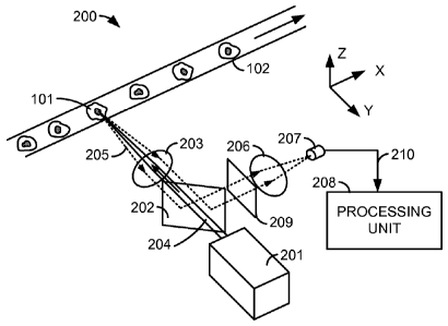
fluid and
entrained single-file in a narrow transparent tube 102. The entrainment can be
accomplished
by any of several methods, including hydrodynamic focusing. A light source 103
illuminates
each cell 101 as it passes a measurement location 104. Light source 103 may
be, for
example, a laser. Light from light source 103 is scattered by the cell 101
being measured.
Some light 105 is scattered generally in the same direction as it traveled to
reach the cell 101.
Light 105 is sometimes called "forward scatter", and may be collected by a
forward sensor
106. Some light may be scattered in other directions as well. This light may
be called "side
scatter", and some of the side scattered light 107 may be collected by one or
more other
sensors 108. Output signals from sensors 106 and 108 are sent to a computer
109, which may
store and analyze the signals. By analyzing the amount and distribution of the
scattered light,
it is possible to discern information about each cell, for example its size
and some limited
information about its internal structure.
[0003] Flow cytometry may measure the scattered light directly, or may make
use of
fluorescence. In fluorescence cytometry, the cells may be marked with one or
more
fluorophores, which are excited by light from source 103 to produce light by
fluorescence.
The nature of the emitted light may reveal additional information about the
cells.
[0004] The technique shown in Figure 1 relies entirely on measurements of
scattered light
to infer information about the cell structure, but does not produce an image
of any particular
cell. In another technique, called "image cytometry", an image of an
individual cell may be
recorded by a camera or scanning microscope. Image cytometry may provide
detailed
information about the cell's structure, but results in much more data than
techniques that use
1
CA 02771324 2012-02-16
WO 2011/022686 PCT/US2010/046215
only scattered light. Consequently, image cytometry may be relatively slow,
and require the
storage and analysis of large quantities of data.
BRIEF SUMMARY OF THE INVENTION
[0005] A cross sectional imaging system performs high-resolution, high-speed
partial
imaging of cells. Such a system may provide much of the information available
from full
imaging cytometry, but can be performed much more quickly, in part because the
data
analysis is greatly reduced in comparison with full image cytometry.
BRIEF DESCRIPTION OF THE DRAWINGS
[0006] Figure 1 shows a simplified diagram of a technique known as flow
cytometry.
[0007] Figure 2 shows a simplified conceptual diagram of a high-speed cross
sectional cell
imaging system in accordance with an embodiment.
[0008] Figure 3 illustrates an example data set representing the readings
taken from one
cell.
[0009] Figure 4 illustrates a system for performing simultaneous multicolor
cross section
cytometry, in accordance with an embodiment.
[0010] Figure 5 illustrates semi-confocal imaging.
[0011] Figure 6 shows an example system that provides rotational movement of
the cells
being studied, and a translational movement of the sensing system.
[0012] Figure 7 illustrates a system for performing simultaneous multi-line
cross sectional
imaging, in accordance with another embodiment.
[0013] Figure 8 illustrates a system for performing multi-line cross sectional
imaging, in
accordance with another embodiment.
DETAILED DESCRIPTION OF THE INVENTION
[0014] Some applications for cytometry require more information than may be
available
from techniques based purely on scattered light, but may not require all of
the information
available from full image cytometry. For example, a researcher may wish to
investigate
whether specific biological activity occurs at the surface or nucleus of a
cell, or in the cell's
cytoplasm. Certain molecules may be labeled with fluorescent tags and
incorporated into the
2
CA 02771324 2012-02-16
WO 2011/022686 PCT/US2010/046215
cells to be studied. Many different tagging compounds, sometimes called
fluorophores, are
available, including the ALEXA FLUORTM series of fluorophores available from
Life
Technologies Corporation of Carlsbad, California, USA.
[0015] Figure 2 shows a simplified conceptual diagram of a high-speed cross
sectional cell
imaging system 200 in accordance with an embodiment. The system of Figure 2 is
a flow
cytometry system, although one of skill in the art will recognize that
embodiments of the
invention may be utilized in other kinds of cytometry as well, including
embodiments
described below.
[0016] In system 200, cells 101 are entrained in fluid to progress through
tube 102 in single
file. The system may be used to characterize cells of many different kinds,
but in a typical
application, cells 101 may be, for example, about 10 to 20 micrometers across,
and may
progress through tube 102 at a speed of, for example, 5 to 50 millimeters per
second. A light
source 201 illuminates a cell through partially-reflective mirror 202 and a
first lens 203.
Light source 201 may be a laser that emits coherent light, or may be another
kind of light
source, for example a light emitting diode (LED), an arc lamp, an incandescent
lamp, or
another kind of light source. Light source 201 may emit coherent or non-
coherent light, and
may produce light continuously or may be pulsed. Partially-reflective mirror
202 may be
configured, for example, to reflect the majority of light falling on it, but
to transmit a portion
as well. The partial reflectivity may be neutral density, in that all
wavelengths are affected
generally equally. Alternatively, mirror 202 may be a dichroic mirror
configured to pass
substantially all light at the wavelength of light source 201, and to reflect
substantially all
light of other wavelengths. In another embodiment, mirror 202 may simply have
a hole
through it to allow light from light source 201 to pass through.
[0017] Light source 201 produces a beam 204, a portion of which passes through
partially-
reflective mirror 202 and is focused by first lens 203 onto a small area of
cell 101. (Some of
the light may be reflected away from mirror 202, but is not indicated in
Figure 2.) The
illuminated area of cell 101 may be, for example, only a few microns across,
as compared
with the unfocused diameter of beam 204, which may be hundreds or thousands of
microns in
diameter.
[0018] Some of the light from light source 201 may be reflected from cell 101.
Additionally, a one or more fluorophore markers in cell 101 may be excited by
beam 204,
and may emanate light by fluorescence. Typically, the light emitted by
fluorescence will be
at longer wavelengths than the laser excitation light. The light 205 emanating
from cell 101,
3
CA 02771324 2012-02-16
WO 2011/022686 PCT/US2010/046215
whether by reflection or fluorescence, is shown in broken line in Figure 2.
Some of the
emanated light 205 is gathered by first lens 203 and at least partially
collimated. After
passing through first lens 203, the emanated light encounters partially-
reflective mirror 202,
where most of it is reflected toward second lens 206. (Some of the emanated
light may also
pass through mirror 202, but this is not indicated in Figure 2). An optional
filter 209 may
further condition light 205, for example by preferentially excluding the
reflected light in
favor of passing the light wavelengths emanated by fluorescence from cell 101.
Second lens
206 redirects the emanated light toward a light sensor 207. Lenses 203 and 206
may form an
infinity corrected optical system, allowing for the insertion of other
components such as
mirror 202 and filter 209 into the optical path between the lenses. Light
sensor 207 converts
the received light into an electrical signal 210 representing the intensity of
light falling on
sensor 207. The signal may be digitized and sent to a processing unit 208 for
recording an
analysis. In some applications, sensor 207 may be sampled, for example, at a
rate of 20 to
200 kHz, resulting in a large number of samples per cell. Processing unit 208
may be a
computer system, and may be stand-alone or integrated in to a testing station
including the
other system components.
[0019] The resolution of the system depends on the sample rate, the speed of
transport of
the cells past the scan location, and the light spot size on cell 101. The
nominal resolution in
the X direction is equal to rdt, where v is the sample delivery speed and dt
is the sampling
frequency. The resolution may be more limited if the light spot size is large
in relation to the
nominal resolution. Preferably, v is a known parameter, either pre-determined
before a
particular flow experiment or measured during the course of a cell's passage
through the
system. Ideally, a cell being scanned should be rotation-free and jittering-
free during its
passage of the scan line.
[0020] Light sensor 207 may be, for example, a photodiode, a photomultiplier
tube, an
avalanche photodiode, a silicon photodiode, or any other suitable kind of
sensor. When
signal 210 is repeatedly sampled and correlated with the movement of cell 101,
the resulting
data provides a high-resolution view of the light emanated from locations on
or in cell 101
along a single substantially linear path. Figure 3 illustrates an example data
set representing
the readings taken from one cell, along path 301, as compared with the
readings that might be
taken using conventional scattering-base cytometry. As is evident, the
readings taken
according to an embodiment of the invention, labeled "cross section imaging"
in Figure 3,
provide a much more detailed view of the activity within cell 101 than is
available from the
data based on scattering alone.
4
CA 02771324 2012-02-16
WO 2011/022686 PCT/US2010/046215
[0021] In some experiments, the data shown in Figure 3 may be sufficient to
enable the
researcher to answer the question of interest. Cross section imaging according
to an
embodiment of the invention may performed very rapidly, for example at a rate
of thousands
of cells per second. In other experiments, the cross section image data may be
useful for
sorting cells so that some of particular interest may be further analyzed
using full imaging
cytometry. The trace of Figure 3 may be thought of as a single row of pixels
from a full
image scan. Such a single-row image may provide much of the information
available from a
full two-dimensional image of a cell, and can be acquired and processed much
more quickly.
[0022] In other embodiments, the system may be configured to perform multi-
color
cytometry, either using multiple excitation sources having different
wavelengths, by sensing
different wavelength bands of reflected or fluorescent light, or both. Figure
4 illustrates a
system 400 for performing simultaneous multicolor cross-section cytometry, in
accordance
with an embodiment.
[0023] In system 400, three different light sources 401a, 401b, 401c produce
beams 402a,
402b, 402c, each in a narrow band of light wavelengths different from the
others. For
example, light sources 401a, 401b, and 401c may be different lasers. Beams
402a, 402b, and
402c reflect respectively from mirrors 403a, 403b, and 403c. In one
embodiment, mirror
403c is a simple reflective mirror, while mirrors 403a and 403b are dichroic
mirrors,
configured to reflect substantially all of the light wavelengths produced by
their respective
light sources 401a and 401b, while passing substantially all of the
wavelengths produced by
the other light sources. The resulting composite laser beam 404 thus contains
three narrow
bands of wavelengths produced by the three light sources, 401a, 401b, and
401c. Composite
beam 404 passes substantially through mirror 202, and is focused by lens 203
onto cell 101.
The system thus has a light source that includes multiple wavelength bands.
Light 205
emanating from cell 101, whether by reflection from cell 101 or as the result
of fluorescence,
is collected by lens 203 and at least partially collimated. The emanated light
205 mostly
reflects from mirror 202 toward mirrors 405a, 405b, 405c. One or more optional
filters 209
may be placed in the optical path as shown, or in another location.
[0024] Mirrors 405a and 405b are preferably dichroic mirrors, configured to
preferentially
reflect certain wavelength components of emanated light 205. For example,
mirror 405a may
reflect light in wavelengths produced by fluorescence of a first fluorophore
excited by light
source 401a, while mirror 405b may reflect light in wavelengths produced by
fluorescence of
a second fluorophore excited by light source 401b. The light reflected by
mirrors 405a,
405b, and 405c passes through lenses 406a, 406b, and 406c, and reaches sensors
407a, 407b,
5
CA 02771324 2012-02-16
WO 2011/022686 PCT/US2010/046215
and 407c respectively. The sensors 407a, 407b, and 407c thus receive light in
different
wavelength bands. For the purposes of this disclosure, wavelength bands that
are different
may overlap, so that some wavelengths are contained in both bands. The outputs
of sensors
407a, 407b, 407c are preferably digitized and sent to processing unit 208 for
storage,
analysis, and display. For the purposes of this disclosure, a particular
sensor and its
associated components will be referred to as a "channel".
[0025] While example system 400 has been shown having three light sources
401a, 401b,
401c and three sensors 407a, 407b, 407c, one of skill in the art will
recognize that other
numbers of light sources, sensors, or both may be used. A system such as
system 400 may be
configured in many different ways. For example, mirrors 405a, 405b, and 405c
may be
configured to reflect light in the wavelength bands of light sources 401a,
401b, 401c, so that
light reflected from cell 101 is measured. Or one or more of mirrors 405a,
405b, 405c may
be configured to reflect light in the wavelength bands of respective light
sources, while one or
more other mirrors may be configured to preferentially reflect light in
wavelengths emitted
by fluorophores excited by respective light sources, so that fluorescence
cross section
imaging is performed. Or all three mirrors 405a, 405b, 405c may be configured
to
preferentially reflect light emanated from cell 101 by fluorescence. Any
combination is
possible. For example, one channel sensing reflected light may be used to
measure the
geometric limits of a particular cell, and data from channels reading light
produced by
fluorescence may be correlated with the geometry data to indicate where
specific biologic
activity is occurring. In another example, a single light source may be used
to excite multiple
fluorophores, so that fewer light sources are used than sensors. Other
embodiments may be
envisioned that include fewer sensors than light sources.
[0026] Any or all of the channels may optionally be configured to be confocal
or semi-
confocal. A confocal optical system uses an aperture placed near the sensor to
preferentially
exclude light emanating from locations other than in the focal plane of the
system. Apertures
408a, 408b, and 408c are shown in Figure 4, and Figure 5 illustrates their
function, in the
context of a simplified optical system 500. In system 500, a first pencil of
rays 501,
illustrated in solid lines, emanates from focal plane 502 at cell 101. The
rays are gathered by
lens 504 and focused at sensor 505, after passing through aperture 506. A
second pencil of
rays 503, illustrated in broken lines, emanates from a point 507, removed from
focal plane
502 and farther from lens 504. The rays in pencil 503 will focus at a point
508 in front of
sensor 505, so that by the time the rays reach aperture 506, they are already
diverged and a
significant portion of the rays in pencil 503 are excluded by aperture 506
from reaching
6
CA 02771324 2012-02-16
WO 2011/022686 PCT/US2010/046215
sensor 505. In this way, the system preferentially excludes rays that emanate
from other than
the focal plane of the system. This kind of system may produce images with
higher contrast
than a system without an aperture such as aperture 506. Aperture 506 may be
sized or shaped
in any of a variety of ways. The larger the aperture, the fewer rays are
excluded, and the
system may be considered to be "semi-confocal". The aperture need not be
circular. For
example, it could be oblong, so that its performance is different between two
orthogonal axes.
[0027] In the embodiments shown thus far, relative motion is provided between
the cell
being evaluated and the sensing system by moving the cell past a fixed
scanning location. In
alternative embodiments, relative motion may be provided by moving the sensing
system and
holding the cell fixed, or both the sensing system and the cell may be moving,
so long as
relative motion between them is occurring.
[0028] In another embodiment, the relative motion is provided by a rotational
scanning
system. Figure 6 shows an example system 600 that provides rotational movement
of the
cells being studied, and a translational movement of the sensing system. In
system 600, cells
such as cell 101 are adhered to a rotating platen or substrate 601. Platen 601
may be, for
example, a disk about 100 to 150 millimeters in diameter and made of a polymer
such as
polycarbonate or acrylic. Other sizes and materials may also be used. Platen
601 may be
clear.
[0029] Platen 601 is rotated so that the cells are passed under an optical
system such as
optical system 200 shown in Figure 2, although an optical system according to
any
embodiment could be used. Optical system 200 is also translatable along a
generally radial
path 602, so that a large portion of the surface of platen 601 is accessible
for scanning by
optical system 200. In this embodiment, the scanning path across any
particular cell will be
an arc of a circle. However, the radius of the arc is very large in comparison
with the
dimensions of any particular cell, and the path across a single cell may be
considered
essentially linear. Using a system such as system 600, a large number of cells
may be
characterized by systematically scanning the platen with coordinated
translation of the optical
system and rotation of the platen, in much the same way that a compact disc
(CD) or digital
versatile disc (DVD) is read by an audio or video system. The platen 601 may
be "read"
from the top, as by optical system 200, or from the bottom by alternate
optical system 200a.
[0030] In some embodiments, platen 601 may be scanned or otherwise accessed
from both
sides in a coordinated manner. For example, optical systems 200 and 200a may
both scan the
7
CA 02771324 2012-02-16
WO 2011/022686 PCT/US2010/046215
cells adhered to plate 601. The two optical systems may scan using different
excitation
wavelengths, different filtration of the light emanated from the cells being
measured, or both.
[0031] In another embodiment, if a measurement of a particular cell made from
the top of
platen 601 indicates that the cell may exhibit an activity or characteristic
that warrants further
study, the cell may be liberated from the platen 601 by a burst of light
delivered from the
bottom of platen 601. As is shown in the detail view in Figure 6, the top
surface of platen
601 may include a wavelength-selective fusible surface 602, that releases a
cell adhered to it
when subjected to light in a particular wavelength. The liberated cells may be
washed from
the surface of platen 601 and collected for more detailed analysis, for
example by full
imaging cytometry or microscopic examination.
[0032] Figure 7 illustrates a system for performing simultaneous multi-line
cross sectional
imaging, in accordance with another embodiment. In some applications, it may
be helpful to
scan multiple traces across each cell. For example, one cross section image
may be gathered
across the approximate center of each cell, and another image gathered near
the periphery of
the cell. If the two images are gathered with the same resolution, this
nominally doubles the
amount of information gathered from each cell, but still results in much less
data than full
imaging cytometry. Other numbers of traces may be gathered, for example,
three, four, or
any another number, but preferably five or fewer. Similarly, the traces need
not all be of the
same resolution.
[0033] In the example of Figure 7, a light source 201 generates a collimated
beam 204,
which passes substantially through partially reflective mirror 202 and
encounters lens 701. In
contrast to previous embodiments, lens 701 is a multifaceted "fly's eye" lens,
a diffractive
lens, a holographic lens, or another kind of optical system that splits beam
204, focusing
portions of beam 204 on two different spots at cell 101. The two spots are
displaced from
each other, so that they trace displaced parallel paths 702, 703 across cell
101 as cell 101 is
transported by the scanning area. The displacement of the spots is in a
direction oriented at
an angle 0 from the direction of travel of cell 101. Preferably, 0 is greater
than zero degrees,
and may be about 90 degrees. (If 0=0, no additional information is gathered
about cell 101 as
compared with single line cross sectional image, but cell 101 may be double
sampled,
enabling construction of a cross section image with reduced noise.)
[0034] Light 205 emanates from the illuminated spots on cell 101, whether by
reflection or
fluorescence or both, and is gathered and at least partially collimated by
lens 701. The light
substantially reflects from mirror 202, may encounter a filter 209 and mirrors
such as mirrors
8
CA 02771324 2012-02-16
WO 2011/022686 PCT/US2010/046215
704 and 705, and eventually reaches lenses 706 and 707. (The path of light 205
between lens
701 and lenses 705 and 707 is simplified in Figure 7.) Mirrors 704 and 705 may
be, for
example, dichroic mirrors that preferentially separate different bands of
wavelengths from
light 205 to be directed to lenses 706 and 707. Lenses 706 and 707 preferably
cooperate with
lens 701 to form an infinity corrected optical system, allowing for the
insertion of mirrors,
filters, or other components between them. Like lens 701, lenses 706 and 707
may be
multifaceted "fly's eye" lenses, diffractive lenses, holographic lenses, or
other optical
systems that displace portions of the light reaching them, directing images to
two sets of
sensors 708a and 708b, and 709a and 709b. The respective images correspond to
the two
spots illuminated on cell 101. The signals produced by sensors 708a, 708b,
709a, and 709b
are passed to processing unit 208 for storage, analysis, display or the like.
[0035] The system of Figure 7 thus scans cross section images along two
separated paths
702, 703 on cell 101. For each path, images are scanned in two wavelength
bands. One of
skill in the art will recognize that system 700 could be adapted to use
multiple sources of
illumination, such as multiple lasers emitting light in different wavelength
bands, and could
be adapted to scan pairs of images in fewer or more wavelength bands.
[0036] Figure 8 shows a system 800 for performing multi-line cross sectional
imaging, in
accordance with another embodiment. In this embodiment, rather than using
optical means to
split the illumination light and light emanating from the measured cell, two
cross section
imaging systems are placed along the flow path with a lateral displacement, so
that one path
across a cell is imaged by the first optical system at a first time, and a
different path across
the cell is imaged by a different optical system at a later time. In system
800, two optical
systems like those of Figure 2 are shown providing signals to a processing
unit 208 for
storage, analysis, display, or the like. More than two systems could be used.
While the
system of Figure 2 utilizes only one excitation source 201 and one sensor 207,
one of skill in
the art will recognize that system 800 could easily be adapted to use more
excitation sources,
sensors, or both. For example, system 800 could be adapted to use two or more
of the optical
system of Figure 4.
[0037] The first optical system in system 800 illuminates cell 101 with light
source 201,
eventually resulting in light that is sensed by sensor 207. The system is
aligned to scan cell
101 along a path 801, which in this example is near the center of the cell.
The second optical
system (denoted with primed reference numbers) illuminates passing cells in a
second
scanning location with light source 201', resulting in light that is sensed by
sensor 207'. The
second optical system is aligned to scan cells along path 802, which in this
example is near
9
CA 02771324 2012-02-16
WO 2011/022686 PCT/US2010/046215
the edge of each passing cell. Any particular cell 101 is scanned by both
optical systems, but
not simultaneously. Processing unit 208 may store the results the first cross
sectional
imaging and correlate them with the results of the second imaging, to create a
set of cross
section images of each cell.
[0038] While embodiments of the invention have been illustrated as scanning
cells
confined in a linear tube or adhered to a rotating substrate, one of skill in
the art will
recognize that embodiments of the invention may be utilized in systems using
any of a wide
range of cell delivery techniques, including electrophoresis, pressure driven
flow, optical
tweezers, motorized translation stage, and others. Cells may be conveyed as a
payload in an
oil emulsion, in an electrowetting-actuated droplet, or via magnetic transport
assisted by
magnetic bead tagging. It is intended that the claims not be limited by the
cell delivery
method utilized.
[0039] In the claims appended hereto, the term "a" or "an" is intended to mean
"one or
more." The term "comprise" and variations thereof such as "comprises" and"
comprising,"
when preceding the recitation of a step or an element, are intended to mean
that the addition
of further steps or elements is optional and not excluded. The invention has
now been
described in detail for the purposes of clarity and understanding. However,
those skilled in
the art will appreciate that certain changes and modifications may be
practiced within the
scope of the appended claims.