Note: Descriptions are shown in the official language in which they were submitted.
CA 02771630 2012-02-20
WO 2011/022710 PCT/US2010/046248
THERAPEUTIC METHODS AND COMPOSITIONS
CROSS REFERENCE TO RELATED APPLICATIONS
This application claims the benefit of United States provisional patent
application No.
61/235,852, filed August 21, 2009, the disclosure of which is hereby
incorporated by reference in
its entirety for all purposes.
This application is related to United States provisional patent application
No. 61/235,846
(filed August 21, 2009) and to United States provisional patent application
No. 61/235,796 (filed
August 21, 2009), the disclosures of which are hereby incorporated by
reference in their
entireties for all purposes.
This application is also related to co-owned United States patent application
entitled "In
vivo Screening Assays," Attorney Docket No.ARBS-012, Client Ref. No. A12-US 1;
and to co-
owned United States patent application entitled "In vitro Screening Assays,"
Attorney Docket
No. ARBS-013, Client Ref. No. A13-US1; each of which is filed even date
herewith; and the
disclosures of which are incorporated by reference in their entireties for all
purposes.
STATEMENT REGARDING FEDERAL SUPPORT
Not applicable.
FIELD
The present application is in the fields of cancer, oncology and fibrotic
diseases.
BACKGROUND
Extensive clinical evidence and mouse models of tumorigenesis support the
critical role
of the microenvironment in promoting tumor growth and metastasis. The
recruitment and
activation of fibroblasts, vascular cells and inflammatory cells by tumor
cells has been shown to
facilitate metastatic potential and can impact the outcome of therapy.
Epithelial malignancies of
the pancreas, breast, prostate, colon, lung and uterus often contain a
desmoplastic stroma
composed of tumor-associated fibroblasts (TAFs) and accumulated extracellular
matrix, which
has been associated with a poorer prognosis. These TAFs are thought to
contribute to
1
CA 02771630 2012-02-20
WO 2011/022710 PCT/US2010/046248
tumorigenesis in part by stimulation of tumor angiogenesis. TAFs exhibit the
smooth muscle-
like contractile properties of myofibroblasts, which play a significant role
in the pathologic
remodeling of organs leading to fibrosis. Providing further evidence of the
role that factors
which modify the microenvironment play in disease progression, recent studies
have shown that
changes in mechanical tension of the extracellular matrix can lead to
significant changes in cell
morphology, activation of signaling pathways, tissue remodeling, and
pathogenesis. These
findings underscore the potential for new therapeutic strategies in oncology
and fibrosis,
targeting proteins that regulate the composition and mechanical properties of
the extracellular
matrix.
Lysyl oxidase-type enzymes (LOX/Ls) comprise a family of 5 enzymes sharing a
conserved C-terminal enzymatic domain with divergent N-termini. LOX/Ls are
copper-
containing enzymes that catalyze the oxidative deamination of the epsilon-
amine group in
particular lysine residues to promote the covalent cross-linking of proteins
such as fibrillar
collagen I, a major component of desmoplastic stroma. There is some evidence
that certain
LOX/Ls play a role in initiation and progression of both oncologic and
fibrotic diseases, and
lysyl oxidase (LOX) has been shown to play a role in the development of
metastasis and
metastatic niche formation. See, for example, co-owned United States Patent
Application
Publication No. US 2009/0104201 (Apr. 23, 2009), entitled "Methods and
compositions for
treatment and diagnosis of fibrosis, tumor invasion, angiogenesis &
metastasis," the disclosure of
which is incorporated by reference in its entirety for the purposes of
describing various aspects
of the biology of the lysyl oxidase-type enzymes.
Lysyl-oxidase like 2 (LOXL2) mRNA is highly expressed in a number of different
solid
tumors and tumor cell lines. LOXL2 has been reported to enhance the in vivo
accumulation and
deposition of collagen in breast tumors and gliomas formed by LOXL2-expres
sing cancer cells.
Expression of LOXL2 protein has been described previously in breast and
esophageal tumors,
and squamous carcinomas, primarily with an intracellular localization, while a
recent report
supports a role for secreted LOXL2 in promoting tumor cell invasion in stomach
cancer.
Increased LOXL2 levels have also been associated with degenerative and
fibrotic diseases, for
example, in hepatocytes from patients with Wilson's disease or primary biliary
cirrhosis and in
renal tubulointerstitial fibrosis.
2
CA 02771630 2012-02-20
WO 2011/022710 PCT/US2010/046248
SUMMARY
In the present disclosure, the inventors have identified roles for LOXL2 in
(1) creation of
the tumor microenvironment and (2) fibroblast activation.
Accordingly, the present disclosure provides methods and compositions for
reducing
desmoplasia and fibroblast activation in tumors and fibrotic disease,
including but not limited to
the following embodiments:
1. A method for inhibiting fibroblast activation in a tumor environment, the
method
comprising inhibiting the activity of lysyl oxidase-like 2 (LOXL2).
2. The method of embodiment 1, wherein the fibroblast activation is mediated
by
transforming growth factor-beta (TGF-(3) signaling.
3. The method of embodiment 1, wherein inhibition of LOXL2 activity results in
disorganization of the extracellular matrix.
4. The method of embodiment 3, wherein disorganization of the extracellular
matrix
results in disruption of the cytoskeleton of cells in the tumor stroma.
5. The method of embodiment 1, wherein the fibroblasts are tumor-associated
fibroblasts (TAFs).
6. The method of embodiment 1, wherein the fibroblasts are myofibroblasts.
7. A method for inhibiting desmoplasia in a tumor environment, the method
comprising inhibiting the activity of lysyl oxidase-like 2 (LOXL2).
8. The method of embodiment 7, wherein the tumor is a metastatic tumor.
9. A method for inhibiting vasculogenesis in a tumor environment, the method
comprising inhibiting the activity of lysyl oxidase-like 2 (LOXL2).
10. The method of embodiment 9, wherein vasculogenesis comprises recruitment
of
vascular cells or vascular cell progenitors to a tumor environment.
11. The method of embodiment 9, wherein vasculogenesis comprises vascular
branching.
12. The method of embodiment 9, wherein vasculogenesis comprises increase in
vessel length.
13. The method of embodiment 9, wherein vasculogenesis comprises an increase
in
the number of vessels.
3
CA 02771630 2012-02-20
WO 2011/022710 PCT/US2010/046248
14. A method for reducing the number of tumor-associated fibroblasts (TAFs) in
a
tumor stroma, the method comprising inhibiting the activity of lysyl oxidase-
like 2 (LOXL2).
15. A method for inhibiting collagen deposition in a tumor environment, the
method
comprising inhibiting the activity of lysyl oxidase-like 2 (LOXL2).
16. A method for modulating a tumor environment, the method comprising
inhibiting
the activity of lysyl oxidase-like 2 (LOXL2).
17. The method of embodiment 16, wherein modulation comprises a reduction in
desmoplasia.
18. The method of embodiment 16, wherein modulation comprises a reduction in
the
number of tumor-associated fibroblasts (TAFs).
19. The method of embodiment 16, wherein modulation comprises a reduction in
the
number of myofibroblasts.
20. The method of embodiment 16, wherein modulation comprises remodeling of
the
cytoskeleton of a cell.
21. The method of embodiment 20, wherein the cell is a tumor cell.
22. The method of embodiment 20, wherein the cell is a fibroblast.
23. The method of embodiment 20, wherein the cell is an endothelial cell.
24. The method of embodiment 16, wherein modulation comprises a reduction in
tumor vasculature.
25. The method of embodiment 16, wherein modulation comprises a reduction in
collagen production.
26. The method of embodiment 16, wherein modulation comprises a reduction in
fibroblast activation.
27. The method of embodiment 16, wherein modulation comprises inhibition of
recruitment of fibroblasts to the tumor environment.
28. The method of embodiment 16, wherein modulation comprises a reduction in
expression of a gene encoding a stromal component.
29. The method of embodiment 28, wherein the stromal component is selected
from
the group consisting of alpha-smooth muscle actin, Type I collagen, vimentin,
matrix
metalloprotease 9, and fibronectin.
4
CA 02771630 2012-02-20
WO 2011/022710 PCT/US2010/046248
30. A method for modulating the production of growth factors in a tumor
environment, the method comprising inhibiting the activity of lysyl oxidase-
like 2 (LOXL2).
31. The method of embodiment 30, wherein the growth factor is selected from
the
group consisting of vascular endothelial growth factor (VEGF) and stromal cell-
derived factor-1
(SDF-1).
32. A method for increasing necrosis in a tumor, the method comprising
inhibiting the
activity of lysyl oxidase-like 2 (LOXL2).
33. A method for increasing pyknosis in a tumor, the method comprising
inhibiting
the activity of lysyl oxidase-like 2 (LOXL2).
34. The method of any of embodiments 1, 7, 9, 14, 15, 16, 30, 32 or 33,
wherein the
activity of LOXL2 is inhibited using an anti-LOXL2 antibody.
35. The method of embodiment 34, wherein the antibody comprises heavy chain
sequences as set forth in SEQ ID NO:1 and light chain sequences as set forth
in SEQ ID NO:2.
36. The method of embodiment 34, wherein the antibody is a humanized antibody.
37. The method of embodiment 36, wherein the antibody comprises heavy chain
sequences as set forth in SEQ ID NO:3 and light chain sequences as set forth
in SEQ ID NO:4.
38. The method of any of embodiments 1, 7, 9, 14, 15, 16, 30, 32 or 33,
wherein the
activity of LOXL2 is inhibited using a nucleic acid.
39. The method of embodiment 38, wherein the nucleic acid is a siRNA.
40. A method for identifying an inhibitor of LOXL2, the method comprising
assaying
a test molecule for its ability to modulate a tumor environment.
41. The method of embodiment 40, wherein modulation comprises a reduction in
desmoplasia.
42. The method of embodiment 40, wherein modulation comprises a reduction in
the
number of tumor-associated fibroblasts (TAFs).
43. The method of embodiment 40, wherein modulation comprises a reduction in
the
number of myofibroblasts.
44. The method of embodiment 40, wherein modulation comprises remodeling of
the
cytoskeleton of a cell.
45. The method of embodiment 44, wherein the cell is a tumor cell.
46. The method of embodiment 44, wherein the cell is a fibroblast.
5
CA 02771630 2012-02-20
WO 2011/022710 PCT/US2010/046248
47. The method of embodiment 44, wherein the cell is an endothelial cell.
48. The method of embodiment 40, wherein modulation comprises a reduction in
tumor vasculature.
49. The method of embodiment 48, wherein reduction in tumor vasculature is
evidenced by reduction in the levels of CD31and/or vascular endothelial growth
factor (VEGF).
50. The method of embodiment 40, wherein modulation comprises a reduction in
collagen production and/or a reduction in degree of collagen crosslinking.
51. The method of embodiment 40, wherein modulation comprises a reduction in
fibroblast activation.
52. The method of embodiment 40, wherein modulation comprises inhibition of
recruitment of fibroblasts to the tumor environment.
53. The method of embodiment 40, wherein modulation comprises a reduction in
expression of a gene encoding a stromal component.
54. The method of embodiment 53, wherein the stromal component is selected
from
the group consisting of alpha-smooth muscle actin, Type I collagen, vimentin,
matrix
metalloprotease 9, and fibronectin.
55. The method of embodiment 40, wherein modulation comprises reduction in the
levels of stromal cell-derived factor-1 (SDF-1) in the tumor environment.
56. The method of embodiment 40, wherein modulation comprises an increase in
the
incidence of necrosis and/or pyknosis in cells of the tumor.
57. The method of embodiment 40, wherein the test molecule is a small organic
molecule with a molecular weight les than 1 kD.
58. The method of embodiment 40, wherein the test molecule is a polypeptide.
59. The method of embodiment 58,wherein the polypeptide is an antibody.
60. The method of embodiment 40, wherein the test molecule is a nucleic acid.
61. The method of embodiment 60, wherein the nucleic acid is a siRNA.
62. An inhibitor of LOXL2 for use in inhibiting fibroblast activation in a
tumor
environment.
63. An inhibitor of LOXL2 for use in inhibiting desmoplasia in a tumor
environment.
64. An inhibitor of LOXL2 for use in inhibiting vasculogenesis in a tumor
environment.
6
CA 02771630 2012-02-20
WO 2011/022710 PCT/US2010/046248
65. An inhibitor of LOXL2 for use in reducing the number of tumor-associated
fibroblasts (TAFs) in a tumor stroma.
66. An inhibitor of LOXL2 for use in inhibiting collagen deposition in a tumor
environment.
67. An inhibitor of LOXL2 for use in modulating a tumor environment.
68. An inhibitor of LOXL2 for use in modulating the production of growth
factors in
a tumor environment.
69. An inhibitor of LOXL2 for use in increasing necrosis in a tumor.
70. An inhibitor of LOXL2 for use in increasing pyknosis in a tumor.
71. The inhibitor of any of claims 62-70, wherein the inhibitor of LOXL2 is an
anti-
LOXL2 antibody.
72. The inhibitor of embodiment 71, wherein the antibody comprises heavy chain
sequences as set forth in SEQ ID NO:1 and light chain sequences as set forth
in SEQ ID NO:2.
73. The inhibitor of embodiment 71, wherein the antibody is a humanized
antibody.
74. The inhibitor of embodiment 73, wherein the antibody comprises heavy chain
sequences as set forth in SEQ ID NO:3 and light chain sequences as set forth
in SEQ ID NO:4.
75. The inhibitor of any of embodiments 62-70, wherein the inhibitor is a
nucleic
acid.
76. The inhibitor of embodiment 75, wherein the nucleic acid is a siRNA.
77. A pharmaceutical composition for use in inhibiting fibroblast activation
in a
tumor environment, wherein the composition comprises an inhibitor of LOXL2 and
a
pharmaceutically acceptable excipient.
78. A pharmaceutical composition for use in inhibiting desmoplasia in a tumor
environment, wherein the composition comprises an inhibitor of LOXL2 and a
pharmaceutically
acceptable excipient.
79. A pharmaceutical composition for use in inhibiting vasculogenesis in a
tumor
environment, wherein the composition comprises an inhibitor of LOXL2 and a
pharmaceutically
acceptable excipient.
80. A pharmaceutical composition for use in reducing the number of tumor-
associated
fibroblasts (TAFs) in a tumor stroma, wherein the composition comprises an
inhibitor of LOXL2
and a pharmaceutically acceptable excipient.
7
CA 02771630 2012-02-20
WO 2011/022710 PCT/US2010/046248
81. A pharmaceutical composition for use in inhibiting collagen deposition in
a tumor
environment, wherein the composition comprises an inhibitor of LOXL2 and a
pharmaceutically
acceptable excipient.
82. A pharmaceutical composition for use in modulating a tumor environment,
wherein the composition comprises an inhibitor of LOXL2 and a pharmaceutically
acceptable
excipient.
83. A pharmaceutical composition for use in modulating the production of
growth
factors in a tumor environment, wherein the composition comprises an inhibitor
of LOXL2 and a
pharmaceutically acceptable excipient.
84. A pharmaceutical composition for use in increasing necrosis in a tumor,
wherein
the composition comprises an inhibitor of LOXL2 and a pharmaceutically
acceptable excipient.
85. A pharmaceutical composition for use in increasing pyknosis in a tumor,
wherein
the composition comprises an inhibitor of LOXL2 and a pharmaceutically
acceptable excipient.
86. The composition of any of embodiments 77-85, wherein the inhibitor of
LOXL2
is an anti-LOXL2 antibody.
87. The composition of embodiment 86, wherein the antibody comprises heavy
chain
sequences as set forth in SEQ ID NO:1 and light chain sequences as set forth
in SEQ ID NO:2.
88. The composition of embodiment 86, wherein the antibody is a humanized
antibody.
89. The composition of embodiment 88, wherein the antibody comprises heavy
chain
sequences as set forth in SEQ ID NO:3 and light chain sequences as set forth
in SEQ ID NO:4.
90. The composition of any of embodiments 77-85, wherein the inhibitor is a
nucleic
acid.
91. The composition of embodiment 90, wherein the nucleic acid is a siRNA.
BRIEF DESCRIPTION OF THE DRAWINGS
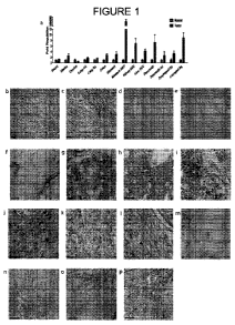
Figure 1, Panels a-p shows that LOXL2 is highly expressed and secreted in
solid tumors
and in liver fibrosis. Figure 1, Panel a shows qRT-PCR analysis of LOXL2
transcripts in solid
tumors as compared to non-neoplastic tissues. Figures 1, Panel b and 1, Panel
c show
immunohistochemistry (IHC) of laryngeal squamous cell carcinoma for collagen I
(Figure 1b)
and LOXL2 (Figure 1c) expression in matched tumor sections. Figures 1, Panel d
and 1, Panel e
8
CA 02771630 2012-02-20
WO 2011/022710 PCT/US2010/046248
show IHC analysis of sections from a lung squamous cell carcinoma (grade 2)
testing for
expression of collagen I (Figure 1, Panel d) and LOXL2 (Figure 1, Panel e).
Figures 1, Panel f
and 1, Panel g show IHC analysis of LOXL2 expression in sections from a
pancreatic
adenocarcinoma (grade 3). Figure 1, Panel f shows LOXL2 expression in the
matrix and the
tumor-stroma boundary; while LOXL2 expression on glomeruloid structures was
also apparent
in Figure 1, Panel f and Figure 1, Panel g. Figures 1, Panel h and 1, Panel i
show IHC analysis
of LOXL2 expression in an omental metastasis of an ovarian carcinoma. Figure
1, Panel h
shows tumor cell expression, and Figure 1, Panel i shows LOXL2 expression in
glomeruloid
structures. Figures 1, Panel j and 1, Panel k show IHC of sections from a
pancreatic
adenocarcinoma. Figure 1, Panel j shows LOXL2 expression, and Figure 1, Panel
k shows LOX
expression. Figure 1, Panel l shows LOXL2 expression in a section from a renal
cell clear cell
carcinoma. Figures 1, Panel m and In show IHC for LOXL2 expression in active
Hepatitis C-
induced liver fibrosis (Figure 1, Panel m: 5X magnification; Figure 1, Panel
n: 40X
magnification). Figures 1, Panel o and 1, Panel p shows IHC for LOXL2 and LOX
expression,
respectively, in sections from a steatohepatitic liver (40X magnification).
Figure 2, Panels a-f shows that secreted LOXL2 promotes invasion of tumor
cells in
vitro. Figure 2, Panels a and b show immunoflorescence analysis of cultures of
Hs578t tumor
cells co-stained for LOXL2 (Figure 2, Panel a) and collagen I (Figure 2, Panel
b). Expression of
collagen I and LOXL2 is co-localized in the extracellular matrix in these
cultures. Figures 2,
Panels c-f show rhodamine-phalloidin staining of cultures of MCF-7 cells,
after treatment of the
cultured MCF-7 cells with: MCF7 conditioned medium (Figure 2, Panel c), MDA-
MB231
conditioned medium (Figure 2, Panel d), MDA-MB231 conditioned medium that was
pre-
incubated with 4ug anti-IgG antibody (Figure 2, Panel e), or MDA-MB231
conditioned medium
that was pre-incubated with 4ug of anti-LOXL2 antibody AB0023 (Figure 2, Panel
f).
Figure 3, Panels a-k show that LOXL2 promotes fibroblast activation in vitro
and in
vivo. Figure 3, Panel a shows a protein ("Western") blot analysis, testing for
effects of tension
on the expression level of LOXL2 in human foreskin fibroblasts (HFFs). Cells
were grown on a
tissue culture plate (lanes labeled 1), a 0.2% bis-acrylamide cross-linked
collagen coated gel
(lanes labeled 2), or a 0.8% bis-acrylamide cross-linked collagen coated gel
(lanes labeled 3).
Figures 3, Panel b and 3, Panel c show photographs of HFF cells transfected
with a non-targeting
siRNA (Figure 3, Panel b) or a LOXL2 siRNA (Figure 3, Panel c), and stained
for collagen I at
9
CA 02771630 2012-02-20
WO 2011/022710 PCT/US2010/046248
days post transfection. Figure 3, Panels d and e show photographs of HFF cells
transfected
with a non-targeting siRNA (Figure 3, Panel d) or a LOXL2 siRNA (Figure 3,
Panel e), and
stained with rhodamine phalloidin at 10 days post transfection. Figure 3,
Panels f and g show
photographs of HFF cells grown under low tension (Figure 3f, Panel) or high
tension (Figure 3,
5 Panel g), then stained with rhodamine-phalloidin. Figure 3, Panel h shows a
protein ("Western")
blot of lysates from HFF cells from transwell cultures with MDA-MD-231 or MCF7-
LOXL2
cells. Figure 3, Panel i shows quantitation, by densitometry, of the results
shown in Figure 3,
Panel h, indicating AB0023-specific effects on pSMAD2 and VEGF expression.
Figure 3,
Panel j shows a comparison of the size of xenografts generated in the sub-
renal capsule of nu/nu
10 mice implanted with MCF7 cells (MCF7-control) or with MCF7 cells stably
transfected with a
LOXL2 expression vector (MCF7-LOXL2). Figure 3, Panel k shows analysis of the
xenografts
by quantitative RT-PCR, to examine the relative induction of various stromal
components in the
LOXL2-expressing tumors. Mouse-specific primers were used, to distinguish
stromal expression
from expression in the implanted (human) cells. aSMA = alpha smooth muscle
actin; COL1A1
= Type I collagen; MMP9 = matrix metalloprotease 9; FN1 =f ibronectin type 1;
VIM =
vimentin. Fold activation in the stroma of MCF7-LOXL2-induced tumors, compared
to MCF7-
induced tumors, is shown by the numeral above the bar representing each gene.
Figure 4, Panels a-o show examples of inhibition of angiogenesis and
vasculogenesis by
the anti-LOXL2 antibody AB0023, in vitro and in vivo. Figure 4, Panels a and b
show
rhodamine-phalloidin staining of HUVEC cells transfected with either a non-
targeting siRNA
(Figure 4, Panel a) or a siRNA targeted to LOXL2 (Figure 4, Panel b), then
cultured for 10 days.
Figure 4, Panels c-i show results of in vitro tube formation assays, in which
human umbilical
vein endothelial cells (HUVEC) in culture were treated with increasing
concentrations of
AB0023, followed by staining for the endothelial marker CD31. The four panels
show HUVEC
cultured in the absence of antibody (Figure 4, Panel c) or in the presence of
lug/ml (Figure 4,
Panel d), 10ug/ml (Figure 4, Panel e) or 50ug/ml (Figure 4, Panel f) of
AB0023. Quantitation of
the mean number of branching points (Figure 4, Panel g), mean number of
vessels (Figure 4,
Panel h) and mean total tubule length (Figure 4, Panel i) was also conducted.
Figure 4,
Panels j-m show effects of the anti-LOXL2 antibody AB0023 on vasculogenesis in
a MatrigelTm
plug assay. Balb/C mice were implanted in the flank with a MatrigelTm plug
containing bFGF,
then treated with either AB0023 or vehicle (PBST). Histology (H&E staining) of
the plug in
CA 02771630 2012-02-20
WO 2011/022710 PCT/US2010/046248
animals treated with vehicle only, at day 10 after implantation, showed
evidence of branching
and invading vasculature (Figure 4, Panel j), which is virtually absent in the
plug from AB0023-
treated animals (Figure 4, Panel k). CD31 staining of plugs from animals
treated with vehicle
only (Figure 4, Panel 1) and AB0023 (Figure 4, Panel m) provided similar
results; i.e., lack of
vasculogenesis in plugs from AB0023-treated animals. Figure 4, Panel n
provides a quantitative
analysis of the average number of vessels in plugs from vehicle-treated and
AB0023-treated
animals, indicating a --7-fold decrease of vasculogenesis in the AB0023-
treated mice (p=0.0319).
Figure 4, Panel o shows quantitation of CD31-positive cells in the plugs from
vehicle-treated and
AB0023-treated mice, corroborating the decrease in vasculogenesis (p=0.0168).
Figure 5, Panels a-u show that the anti-LOXL2 antibody AB0023 is effective in
reducing stromal activation and inhibiting generation of a tumor environment
in vivo in both
primary tumors and metastatic xenograft models of cancer. For the results
shown in Figure 5,
Panels a and b, approximately 106 MDA-MB231 cells were injected into mice (in
the left
ventricle) to generate a disseminated bone metastasis model and, 28 days after
injection, the
tumor burden was assessed. Injected animals were treated with the anti-LOX
antibody M64, the
anti-LOXL2 antibody AB0023, Taxotere or a vehicle control. Figure 5, Panel a
shows the day
28 tumor cell burden in the femur (AB0023 p=0.0021, M64 p=0.5262); Figure 5,
Panel b shows
the 28 day tumor cell burden in total ventral bone (AB0023 p=0.0197, M64
p=0.5153).
For the results shown in Figure 5, Panels c-m, primary tumors were generated
using the
MDA-MB-435 cell line and treated as described. Sections from tumors generated
in this model
system, in which the host animals were treated only with vehicle were stained
for the expression
of LOXL2 (Figure 5, Panel c) and for the expression of LOX (Figure 5, Panel
d). Figure 5,
Panel e shows measurements of tumor volumes in mice treated with vehicle only,
taxotere
(positive control for reduction of tumor volume), anti-LOXL2 antibody AB0023
and anti-LOX
antibody M64. AB0023-treated mice maintained a significant decrease in tumor
volume (45% at
week 3, p=0.001; 33% at week 5, p=0.0240) while the M64 treated mice did not
(27% at week 3,
p=0.040; not significant at week 5). Figure 5, Panels f-i show examples of
Sirius Red staining of
tumors from the vehicle-treated (Figure 5, Panel f), AB0023-treated (Figure 5,
Panel g), M64-
treated (Figure 5, Panel h) and taxotere-treated (Figure 5, Panel i) animals.
Figure 5, Panels j-m
show IHC analyses of alpha-smooth muscle actin (a-SMA) expression in sections
from tumors
obtained from animals that had been treated with vehicle only (Figure 5, Panel
j), AB0023
11
CA 02771630 2012-02-20
WO 2011/022710 PCT/US2010/046248
(Figure 5, Panel k), M64 (Figure 5, Panel 1) and taxotere (Figure 5, Panel m).
Figure 5n shows
quantitation of Sirius Red staining, a-SMA expression and CD31 expression in
the tumor
environment of the MDA-MB-435-induced tumors. The results indicate a 61%
reduction in
crosslinked collagen in the AB0023 treated mice (p=0.0027) as determined by
Sirius Red
staining, an 88% reduction in the presence of TAFs (p=0.011) assessed by a-SMA
expression,
and a 74% reduction in tumor vasculature as assessed by CD31 expression
(p=0.0002).
Figure 5, Panel o shows results of a separate study of tumor volume in MDA-MB-
435-
induced primary tumors in AB0023- and BAPN-treated mice; indicating a
statistically significant
reduction in tumor volume following treatment with the anti-LOXL2 antibody.
Figure 5, Panel p
presents a quantitative analysis of Sirius Red staining (collagen production),
CD-31 expression
(vasculogenesis), and a-SMA expression (fibroblast activation) in MDA-MB-435-
induced
tumors from AB0023- and BAPN-treated mice; showing a reduction in all three
markers in
AB0023-treated mice. Figure 5, Panel q shows analysis of expression of LOXL2,
VEGF and
SDF-1 in MDA-MB-435-induced tumors from AB0023-treated and control (vehicle-
treated)
mice; showing 76% reduction of VEGF levels (p=0.0001), 80% reduction of SDF1
levels
(p=0.0200), and 55% reduction in LOXL2 levels (p=0.0005) in AB0023-treated MDA-
MB-435
tumors.
Figure 5, Panels r and s provide evidence of necrosis in AB0023-treated MDA-MB-
435
tumors. Figure 5, Panel r shows IHC analysis for Tumor Necrosis Factor alpha
(TNF-a) in a
section from an AB0023-treated MDA-MB-435 tumor. Figure 5, Panel s shows
hematoxylin
and eosin (H&E) staining of a section from an AB0023-treated MDA-MB-435 tumor.
Figures 5t
and 5u provide evidence for pyknosis in AB0023-treated MDA-MB-435 tumors.
While nuclei in
sections of vehicle-treated tumors were well defined (Figure 5, Panel t),
those in sections of
AB0023-treated tumor appeared pyknotic (Figure 5, Panel u).
Figure 6, Panels a-e show AB0023-mediated inhibition of CCl4-induced liver
fibrosis
and myofibroblast activation. Figure 6, Panel a shows a Kaplan Meier survival
analysis of CCl4-
treated mice also treated with anti-LOXL2 antibody AB0023, anti-LOX antibody
M64 or
vehicle. A significant increase in survival was apparent in the AB0023
treatment arm (p=0.0029
in log rank test, or p=0.0064 in the Mantel-Cox test). Figure 6, Panel b shows
a significant
decrease in the amount of bridging fibrosis in the livers of AB0023 treated
mice (p=0.0020).
Figure 6, Panels c and d show IHC analysis for a-SMA in sections of the porto-
portal region of a
12
CA 02771630 2012-02-20
WO 2011/022710 PCT/US2010/046248
liver from a vehicle treated mouse (Figure 6, Panel c), compared to a liver
from an AB0023
treated mouse (Figure 6, Panel d). Figure 6, Panel e provides a quantitative
analysis of a-SMA
signal, demonstrating that lack of bridging fibrosis in the livers of AB0023-
treated animals was
accompanied by a significant reduction in the number of alpha-SMA positive
myofibroblasts
(p=0.0260).
Figure 7, Panels a-z shows evidence of LOXL2 expression in various human
tumors and
normal tissues. (Panels a-f) Quantitative RT-PCR analysis of LOXL2 transcripts
was performed
on human colon adenocarcinoma (Panel a), pancreatic adenocarcinoma (Panel b),
uterine
adenocarcinoma (Panel c), renal cell carcinoma (Panel d), stomach
adenocarcinoma (Panel e),
and laryngeal squamous cell carcinoma (Panel f),; a trend for increased LOXL2
transcript with
increasing tumor grade was observed. (Panels g-y) A Western blot analysis of
various LOX/L
species shows the polyclonal antibody used for IHC of human and mouse tissue
sections is
specific for LOXL2 (Panel g, cLOX = mature LOX, propeptide cleaved; MCD =
catalytic
domain of protein only; FL = full length protein; this specificity was also
confirmed by ELISA
(data not shown)). Additional examples of LOXL2 expression in: breast
infiltrative ductal
carcinoma (Panel h), uterine endometrial carcinoma (Panel i), colon
adenocarcinoma (Panel j),
hepatocellular carcinoma (Panel k, also stained for LOX expression (Panel 1)),
neurendocrine
carcinoma of the pancreas (Panel m, also stained for LOX expression (Panel
n)), melanoma
(Panel o), normal heart (Panel p, also stained with CD31 (Panel q)), normal
liver (Panel r),
normal lung (Panel s, also stained with CD31 (Panel t)), normal ovary (Panel
u), normal spleen
Panel v), normal smooth muscle (Panel x, also stained for LOX expression
(Panel w)), and
normal artery (z, also stained for LOX expression (Panel y)). Table 1
presented in Figure 7
summarizes LOXL2 expression in human healthy tissues. Human normal tissues
were stained
with the anti-LOXL2 polyclonal antibody and a qualitative assessment of the
relative LOXL2
expression levels was compiled.
Figure 8, Panels a-t shows that secreted LOXL2 promotes remodeling and
invasion of
tumor cells in vitro (Panel a) A qRT-PCR analysis (Ct values) of LOXL2
transcripts in various
tumor and fibroblast cell lines (normoxic conditions, RPL19 used for
reference). (Panel b) A
western analysis of LOXL2 expression in human tumor and fibroblast cell lines
(whole cell
pellet = cell; conditioned media= CM). (Panel c) An Amplex Red assay using
purified
recombinant human LOXL2 showed both the 87kD and 55kD forms of LOXL2 to be
active and
13
CA 02771630 2012-02-20
WO 2011/022710 PCT/US2010/046248
inhibited by BAPN (mixture = 50:50 mixture of both forms). (Panel d) The dose
response
curve for BAPN inhibition of purified recombinant human LOXL2 (Amplex Red
assay; data
normalized to control). (Panels e-g) HS578t were transfected with a non-
targeting siRNA (siNT)
or a LOXL2 siRNA and then stained for expression of LOXL2 or collagen I. LOXL2
expression
co-localized with collagen I (siNT stained for LOXL2 (Panel e), LOXL2 siRNA
stained for
LOXL2 (Panel f) and collagen I (Panel g)). (Panel h) Secretion of LOX in
MC3T3E1 (CM
concentrated --20X). (Panels i,j) LOX expression in tumor or fibroblast cell
lines under
normoxic (Panel i) or hypoxic (Panel j) conditions showed no detectable
secretion of LOX (CM
concentrated --20X). (k,l) MDA-MB-231 cells transfected with non-targeting
shRNA (Panel k)
and stained with rhodamine-phalloidin retained their mesenchymal phenotype
while those
transfected with a LOXL2 shRNA (Panel 1) adopted a more epithelial phenotype.
(Panels m,n)
A western blot analysis and ELISA (Panel n) both show AB0023 is specific for
LOXL2.
(Panel o) A dose response curve for AB0023 inhibition of LOXL2 enzymatic
activity (Amplex
Red assay). (Panel p) AB0023 cross reacts with mouse LOXL2. (Panels q-t) The
growth media
of SW620 cells was supplemented with the following conditioned medias: MDA-MB-
231 CM
(Panel r) or HEK293 CM transfected with an empty vector (Panel q), LOXL2
(Panel s) or
LOXL2 Y689F (Panel t). The cells were stained with rhodamine-phalloidin.
Figure 9, Panels a-b shows LOXL2 expression in HFF cells under varying tension
and
confirmation of LOXL2 knockdown. (Panel a) HFF cells were grown in tissue
culture plates
(Plastic) or collagen I gels containing 2mg/ml (2) or 3mg/ml (3) collagen I.
The gels were either
detached (Floating) or anchored to the culture dish (Attached). The
conditioned media was
analyzed by Western analysis and probed for LOXL2 expression. (Panel b) HFF
cells were
transfected with non-targeting siRNA (siNT) of LOXL2 siRNA (siLOXL2) and the
conditioned
media probed for LOXL2 expression via western blot analysis.
Figure 10, Panels a-b shows LOXL2 expression in infiltrating cells in an in
vivo
matrigel plug (Panels a,b) IHC analysis of endothelial cell infiltrates in a
matrigel plug confirms
LOXL2 expression (Panel a). The section was also stained with CD31 (Panel b)
to confirm
presence of endothelial cells.
Figure 11, Panels a-o shows AB0023 efficacy in vivo in primary tumor and
metastatic
xenograft models of cancer (Panel a) A qRT-PCR analysis of MDA-MB-231 cells
confirms the
transcription of all LOX/L proteins (RPL-19 used as a reference). (Panels b-e)
CD31 staining of
14
CA 02771630 2012-02-20
WO 2011/022710 PCT/US2010/046248
MDA-MB-435 established primary tumors harvested from mice treated with a
vehicle (Panel b),
anti-LOXL2 antibody AB0023 (Panel c), anti-LOX antibody M64 (Panel d), or
Taxotere (Panel
e) showed a 74% reduction in CD31 staining in the AB0023 treatment relative to
vehicle
(p=0.0002). (Panels f, g) A human breast adinocarcinoma stained for expression
of VEGF
(Panel f) and LOXL2 (Panel g) shows similarities in TAF expression. (Panel h-
o) MDA-MB-
435 established primary tumors from vehicle and AB0023 treated mice were
stained for
expression of LOXL2 (Panel h, vehicle treatment; Panel i, AB0023 treatment),
VEGF (Panel j,
vehicle; Panel k, AB0023), and SDF-1 (Panel 1, vehicle; Panel m, AB0023), as
well as with H&E
(Panel n, vehicle, Panel o, AB0023).
Figures 12, Panels a-d shows fibrogenesis in murine livers from a CC14-induced
fibrosis
model. (Panels a-d) A murine CC14-induced liver fibrosis model showed early
evidence of liver
damage and fibrosis, as evidenced by collagen I staining (Sirius Red) of a
liver from an early-
death animal (day 11) (Panel a) compared to a healthy liver (Panel b). Example
of livers used in
analysis of bridging fibrosis: the AB0023 treated mice (Panel d) had
significantly less complete
bridging fibrosis (p=0.002) as compared to the vehicle (Panel c).
DETAILED DESCRIPTION
Practice of the present disclosure employs, unless otherwise indicated,
standard methods
and conventional techniques in the fields of cell biology, toxicology,
molecular biology,
biochemistry, cell culture, immunology, oncology, recombinant DNA and related
fields as are
within the skill of the art. Such techniques are described in the literature
and thereby available to
those of skill in the art. See, for example, Alberts, B. et al., "Molecular
Biology of the Cell," 5d'
edition, Garland Science, New York, NY, 2008; Voet, D. et al. "Fundamentals of
Biochemistry:
Life at the Molecular Level," 3rd edition, John Wiley & Sons, Hoboken, NJ,
2008; Sambrook, J.
et al., "Molecular Cloning: A Laboratory Manual," 3rd edition, Cold Spring
Harbor Laboratory
Press, 2001; Ausubel, F. et al., "Current Protocols in Molecular Biology,"
John Wiley & Sons,
New York, 1987 and periodic updates; Freshney, R.I., "Culture of Animal Cells:
A Manual of
Basic Technique," 4d' edition, John Wiley & Sons, Somerset, NJ, 2000; and the
series "Methods
in Enzymology," Academic Press, San Diego, CA.
The present inventors have identified a role for matrix enzyme lysyl oxidase-
like-2
(LOXL2) in the creation of the pathologic microenvironment of oncologic and
fibrotic diseases.
CA 02771630 2012-02-20
WO 2011/022710 PCT/US2010/046248
Analysis of human tumors and liver fibrosis revealed widespread and conserved
expression of
LOXL2 by activated fibroblasts and neovasculature. The inhibition of LOXL2
with an anti-
LOXL2 monoclonal antibody was efficacious in both primary and metastatic
xenograft models
of cancer, as well as CC14-induced liver fibrosis. Inhibition of LOXL2
resulted not only in a
substantial reduction in fibroblast activation, fibroblast recruitment,
desmoplasia, and
vascularization, but also in significantly decreased production of pro-
angiogenic growth factors
and cytokines such as VEGF and SDF1. Inhibition of lysyl oxidase (LOX) had
little, if any such
effects.
The small molecule beta-aminoproprionitrile (BAPN) has been used to explore
the
effects of inhibition of LOX/L activity in vitro and in vivo. BAPN covalently
modifies the
lysine-tyrosine quinone in the enzymatic domain and thus acts as an
irreversible inhibitor.
BAPN lacks specificity as it inhibits not only the potentially diverse
activities of different
LOX/Ls, but similar domains in other amine oxidases as well. The anti-LOXL2
antibody
outperformed the small molecule pan-lysyl oxidase inhibitor beta-
aminoproprionitrile (BAPN).
The anti-LOXL2 antibody acts as a specific inhibitor of LOXL2, and represents
a new
therapeutic approach with broad applicability in oncologic and fibrotic
diseases.
The present inventors have uncovered a role for LOXL2 in establishing the
pathologic
microenvironment of tumors and fibrotic disease, and have demonstrated it is a
target for
therapy. LOXL2 protein expression and secretion, by TAFs and tumor
vasculature, is
widespread among solid tumors, and is particularly evident at the tumor-stroma
interface.
LOXL2 expression is also pronounced in regions of desmoplasia and glomeruloid
microvascular
proliferation, both of which are associated with poor outcome in several
cancers. In active liver
fibrosis, LOXL2 was similarly expressed at the hepatocyte-myofibroblast
interface and
associated neovasculature.
The inventors have further determined that expression of LOXL2 results in
remodeling of
the actin cytoskeleton in multiple cells types, including tumor cells of
epithelial origin,
endothelial cells, and fibroblasts. One contribution of LOXL2 to disease
progression is the
activation and recruitment of disease-associated fibroblasts, most likely
through its
enzymatically-catalyzed cross-linking of fibrillar collagen and corresponding
changes in local
matrix tension. In tumors and in liver fibrosis, increases in tension can lead
to disease-associated
cellular differentiation. Beyond the production of fibrillar collagens and the
creation of tension
16
CA 02771630 2012-02-20
WO 2011/022710 PCT/US2010/046248
within tissue, TAFs (and potentially also myofibroblasts) secrete many of the
angiogenic,
vasculogenic and chemotactic growth factors and cytokines that support ongoing
tumorigenesis
and fibrosis.
It is disclosed herein that specific inhibition of activity of secreted LOXL2,
in models of
both cancer and fibrosis, resulted in significant reduction of disease as
assessed by a variety of
parameters. Inhibition of LOXL2 is capable of directly affecting angiogenesis,
as well as
invasion and differentiation of disease-associated epithelia. However,
inhibition of angiogenesis
alone is not completely responsible for the effects observed following
inhibition of LOXL2,
inasmuch as potent anti-angiogenics directed at the VEGFR and P1GF pathways do
not affect the
number of aSMA positive cells in tumors, as does inhibition of LOXL2.
It is also disclosed herein that inhibition of LOXL2 in vivo resulted in
inhibition of
fibroblast activation and recruitment, the consequences of which include
substantial reduction of
desmoplasia and the expression of pro-angiogenic growth factors and cyotkines,
lack of
formation of tumor vasculature, and increased necrosis and autophagy of tumor
cells.
Production of fibrillar collagen, a hallmark of fibrosis, was also greatly
reduced by inhibition of
LOXL2, not due to direct regulation of collagen expression but rather due to
the substantial
reduction in the number of activated myofibroblasts (the cell type responsible
for the majority of
collagen production).
Many potential sources of disease-associated activated fibroblasts have been
proposed,
including fibrocytes and other bone-marrow derived cells, resident fibroblasts
or other
precursors, and epithelial-to-mesenchymal transition (EMT) of epithelial
cells. In the work
disclosed herein, therapeutic benefits were obtained in three very different
mouse models
involving different sites of disease, and in the 2 models amenable for further
analysis, the
mechanism appeared conserved, wherein fibroblast activation was substantially
reduced. These
results suggest that LOXL2 is important for the ultimate differentiation and
activation of
fibroblasts, independent of their origin.
The inventors show herein that inhibition of LOXL2 alone was sufficient to
obtain
therapeutic efficacy, despite the use of model systems containing cells that
make multiple lysyl
oxidase-type enzymes, including LOX. In comparison, the use of a particular
LOX-specific
monoclonal antibody targeting a peptide previously identified as generating a
polyclonal
17
CA 02771630 2012-02-20
WO 2011/022710 PCT/US2010/046248
antiserum capable of inhibiting LOX enzymatic activity provided little
therapeutic benefit in
models of oncology and fibrosis.
The differential expression of LOXL2 in diseased versus healthy tissues
provides a
functional therapeutic window. In support of the safety of anti-LOXL2 antibody
AB0023, the
inventors found AB0023 to be well-tolerated at a dosage of 50 mg/kg twice per
week for 14
weeks in mice, with no impact on weight or behavior and no drug-related
observations upon
necropsy, hematology, clinical chemistry and histopathology. Pilot studies in
cynomolgus
monkeys with a humanized anti-LOXL2 variant (AB0024) provided further support
that anti-
LOXL2 antibody therapy was well tolerated upon repeat dosing at 100 mg/kg.
Antibody therapeutics provide one example of a highly specific mechanism for
inhibition. Indeed, specific targeting of secreted LOXL2 with an antibody
(AB0023) that
inhibits its enzymatic activity outperformed the less-specific cell-permeable
pan-inhibitor BAPN,
in cell based assays and in vivo. (Note that contrary to previous reports,
find LOXL2 was found
to be readily inhibited by BAPN in vitro, with a low nanomolar IC50, similar
to that observed for
LOX; Figure 8, panel D and Rodriguez et al. (2010) J. Biol. Chem. 285:20964-
20974). Apart
from specificity, this therapeutic mode provides an additional advantage: as
non-competitive
allosteric inhibitors of LOXL2, AB0023 and AB0024 act independently of
substrate
concentration, or of the state of association between LOXL2 and its substrate,
whereas the
irreversible inhibitor BAPN behaves as a competitive inhibitor and is less
effective at high
substrate concentrations or under conditions where LOXL2 is bound to its
substrate. This
alternative mechanism of inhibition represents a novel therapeutic approach
that has broad
applicability for matrix enzymes functioning within a dynamic complex cellular
milieu
containing a local high concentration of substrate, such as fibrillar
collagen, in active disease.
Allosteric inhibition of LOXL2, as described herein, represents a new approach
to
inhibiting the growth and progression of tumors and fibrotic diseases, by
targeting fundamental
shared features of disease progression, e.g., the creation of the stromal
compartment or matrix
microenvironment or metastatic niche. That is, inhibition of a single target
(LOXL2) has
multiple effects on a number of different drivers of desmoplasia, Targeting of
LOXL2 can be
made highly specific through use of a monoclonal antibody. In addition,
targeting the
genetically more stable stromal cells of the tumor microenvironment offers the
potential for
reduced likelihood of drug resistance.
18
CA 02771630 2012-02-20
WO 2011/022710 PCT/US2010/046248
Definitions
"Tumor environment" refers to a tumor and its surrounding tissue. A subset of
the tumor
environment is the tumor-stroma interface; i.e., the periphery of the tumor
(e.g., the tumor
capsule) along with the adjacent stromal tissue. Another subset is the tumor
itself; yet another
subset is the stromal tissue outside of a tumor.
"Fibroblast activation" refers to a process by which normal fibroblasts are
converted to
tumor-associated fibroblasts (TAFs) in response to signals (e.g., growth
factors, cytokines)
released by tumor cells. One example of such a growth factor is Transforming
Growth Factor-
beta (TGF-0). Exemplary consequences of fibroblast activation are increased
expression of
alpha-smooth muscle actin (aSMA) and increased expression of vascular
endothelial growth
factor (VEGF) in the activated fibroblasts.
"Tumor-associated fibroblasts (TAFs)" are fibroblasts that have undergone
fibroblast
activation and are characterized, inter alia, by increased expression of alpha-
smooth muscle actin
(aSMA) and vascular endothelial growth factor (VEGF).
"Myofibroblasts" are cells with characteristics of both fibroblasts and smooth
muscle
cells. They can be present in fibrotic tissue and are characterized, inter
alia, by expression of
alpha-smooth muscle actin.
"Desmoplasia" refers to the growth of fibrous or connective tissue. Some
tumors elicit a
desmoplastic reaction, i.e., the pervasive growth of dense fibrous tissue
around the tumor.
"Angiogenesis" refers to the formation of new blood vessels from pre-existing
vessels.
"Vasculogenesis" refers to the formation of new blood vessels in the absence
of pre-
existing vessels.
Tumor Stroma
Growth and development of a tumor rely on interactions between the tumor and
its
surrounding stromal tissue. Tumors grow within a stromal framework containing
connective
tissue, fibroblasts, myofibroblasts, white blood cells, endothelial cells,
pericytes and smooth
muscle cells. The growing tumor influences the surrounding stroma by, inter
alia, secreting
growth factors (that influence the behavior of the stromal cells) and
secreting proteases (that
remodel stromal extracellular matrix). Stromal cells, in return, secrete
growth factors that
19
CA 02771630 2012-02-20
WO 2011/022710 PCT/US2010/046248
stimulate growth and division of the tumor cells; and secrete proteases that
further modify the
matrix. In this fashion, a tumor and its surrounding stromal tissue form a
tumor environment that
supports further growth of the tumor. For example, research has shown that
certain carcinomas
depend on the presence of tumor-associated fibroblasts for continued growth,
and will not grow
at a detectable or appreciable level in the presence of normal fibroblasts. It
has also been shown
that robust growth of certain tumors requires a particular matrix
metalloprotease normally
secreted by mast cells, which acts by releasing angiogenic factors from the
extracellular matrix.
Lysyl Oxidase-type Enzymes
As used herein, the terms "lysyl oxidase-type enzyme" and "LOX/L" refer to a
member
of a family of proteins that, inter alia, catalyzes oxidative deamination of c-
amino groups of
lysine and hydroxylysine residues, resulting in conversion of peptidyl lysine
to peptidyl-a-
aminoadipic-8-semialdehyde (allysine) and the release of stoichiometric
quantities of ammonia
and hydrogen peroxide:
I I
C=O C=O
I I
CH-CH2-CH2-CH2-CH2-NH2 +H20 - CH-CH2-CH2-CH2-CH=O +NH3
I +02 I +H2O2
NH NH
I I
peptidyl lysine peptidyl allysine
This reaction most often occurs extracellularly, on lysine residues in
collagen and elastin.
The aldehyde residues of allysine are reactive and can spontaneously condense
with other
allysine and lysine residues, resulting in crosslinking of collagen molecules
to form collagen
fibrils.
Lysyl oxidase-type enzymes have been purified from chicken, rat, mouse,
bovines and
humans. All lysyl oxidase-type enzymes contain a common catalytic domain,
approximately 205
amino acids in length, located in the carboxy-terminal portion of the protein
and containing the
active site of the enzyme. The active site contains a copper-binding site
which includes a
conserved amino acid sequence containing four histidine residues which
coordinate a Cu(II)
atom. The active site also contains a lysyltyrosyl quinone (LTQ) cofactor,
formed by
CA 02771630 2012-02-20
WO 2011/022710 PCT/US2010/046248
intramolecular covalent linkage between a lysine and a tyrosine residue
(corresponding to lys314
and tyr349 in rat lysyl oxidase, and to 1ys320 and tyr355 in human lysyl
oxidase). The sequence
surrounding the tyrosine residue that forms the LTQ cofactor is also conserved
among lysyl
oxidase-type enzymes. The catalytic domain also contains ten conserved
cysteine residues,
which participate in the formation of five disulfide bonds. The catalytic
domain also includes a
fibronectin binding domain. Finally, an amino acid sequence similar to a
growth factor and
cytokine receptor domain, containing four cysteine residues, is present in the
catalytic domain.
Despite the presence of these conserved regions, the different lysyl oxidase-
type enzymes can be
distinguished from one another, both within and outside their catalytic
domains, by virtue of
regions of divergent nucleotide and amino acid sequence.
The first member of this family of enzymes to be isolated and characterized
was lysyl
oxidase (EC 1.4.3.13); also known as protein-lysine 6-oxidase, protein-L-
lysine:oxygen 6-
oxidoreductase (deaminating), or LOX. See, e.g., Harris et al., Biochim.
Biophys. Acta 341:332-
344 (1974); Rayton et al., J. Biol. Chem. 254:621-626 (1979); Stassen,
Biophys. Acta 438:49-60
(1976).
Additional lysyl oxidase-type enzymes were subsequently discovered. These
proteins
have been dubbed "LOX-like," or "LOXL." They all contain the common catalytic
domain
described above and have similar enzymatic activity. Currently, five different
lysyl oxidase-type
enzymes are known to exist in both humans and mice: LOX and the four LOX
related, or LOX-
like proteins LOXL1 (also denoted "lysyl oxidase-like," "LOXL" or "LOL"),
LOXL2 (also
denoted "LOR-1"), LOXL3 (also denoted "LOR-2"), and LOXL4. Each of the genes
encoding
the five different lysyl oxidase-type enzymes resides on a different
chromosome. See, for
example, Molnar et al., Biochim Biophys Acta. 1647:220-24 (2003); Csiszar,
Prog. Nucl. Acid
Res. 70:1-32 (2001); WO 01/83702 published on Nov. 8, 2001, and U.S. Patent
No. 6,300,092,
all of which are incorporated by reference herein. A LOX-like protein termed
LOXC, with some
similarity to LOXL4 but with a different expression pattern, has been isolated
from a murine EC
cell line. Ito et al. (2001) J. Biol. Chem. 276:24023-24029. Two lysyl oxidase-
type enzymes,
DmLOXL-1 and DmLOXL-2, have been isolated from Drosophila.
Although all lysyl oxidase-type enzymes share a common catalytic domain, they
also
differ from one another, particularly in their amino-terminal regions. The
four LOXL proteins
have amino-terminal extensions, compared to LOX. Thus, while human preproLOX
(i.e., the
21
CA 02771630 2012-02-20
WO 2011/022710 PCT/US2010/046248
primary translation product prior to signal sequence cleavage, see below)
contains 417 amino
acid residues; LOXL1 contains 574, LOXL2 contains 638, LOXL3 contains 753 and
LOXL4
contains 756.
Within their amino-terminal regions, LOXL2, LOXL3 and LOXL4 contain four
repeats
of the scavenger receptor cysteine-rich (SRCR) domain. These domains are not
present in LOX
or LOXL1. SRCR domains are found in secreted, transmembrane, or extracellular
matrix
proteins, and are known to mediate ligand binding in a number of secreted and
receptor proteins.
Hoheneste et al. (1999) Nat. Struct. Biol. 6:228-232; Sasaki et al. (1998)
EMBO J. 17:1606-
1613. In addition to its SRCR domains, LOXL3 contains a nuclear localization
signal in its
amino-terminal region. A proline-rich domain appears to be unique to LOXL1.
Molnar et al.
(2003) Biochim. Biophys. Acta 1647:220-224. The various lysyl oxidase-type
enzymes also
differ in their glycosylation patterns.
Tissue distribution also differs among the lysyl oxidase-type enzymes. Human
LOX
mRNA is highly expressed in the heart, placenta, testis, lung, kidney and
uterus, but marginally
in the brain and liver. mRNA for human LOXL1 is expressed in the placenta,
kidney, muscle,
heart, lung, and pancreas and, similar to LOX, is expressed at much lower
levels in the brain and
liver. Kim et al. (1995) J. Biol. Chem. 270:7176-7182. High levels of LOXL2
mRNA are
expressed in the uterus, placenta, and other organs, but as with LOX and
LOXL1, low levels are
expressed in the brain and liver. Jourdan Le-Saux et al.(1999) J. Biol. Chem.
274:12939:12944.
LOXL3 mRNA is highly expressed in the testis, spleen, and prostate, moderately
expressed in
placenta, and not expressed in the liver, whereas high levels of LOXL4 mRNA
are observed in
the liver. Huang et al. (2001) Matrix Biol. 20:153-157; Maki and Kivirikko
(2001) Biochem. J.
355:381-387; Jourdan Le-Saux et al. (2001) Genomics 74:211-218; Asuncion et
al. (2001)
Matrix Biol. 20:487-491.
The expression and/or involvement of the different lysyl oxidase-type enzymes
in
diseases also varies. See, for example, Kagen (1994) Pathol. Res. Pract.
190:910-919;
Murawaki et al. (1991) Hepatology 14:1167-1173; Siegel et al. (1978) Proc.
Natl. Acad. Sci.
USA 75:2945-2949; Jourdan Le-Saux et al. (1994) Biochem. Biophys. Res. Comm.
199:587-592;
and Kim et al. (1999) J. Cell Biochem. 72:181-188. Lysyl oxidase-type enzymes
have also been
implicated in a number of cancers, including head and neck cancer, bladder
cancer, colon cancer,
esophageal cancer and breast cancer. See, for example, Wu et al. (2007) Cancer
Res. 67:4123-
22
CA 02771630 2012-02-20
WO 2011/022710 PCT/US2010/046248
4129; Gorough et al. (2007) J. Pathol. 212:74-82; Csiszar (2001) Prog. Nucl.
Acid Res. 70:1-32
and Kirschmann et al. (2002) Cancer Res. 62:4478-4483.
Thus, although the lysyl oxidase-type enzymes exhibit some overlap in
structure and
function, each has distinct structure and functions as well. With respect to
structure, for
example, certain antibodies raised against the catalytic domain of the human
LOX protein do not
bind to human LOXL2. With respect to function, it has been reported that
targeted deletion of
LOX appears to be lethal at parturition in mice, whereas LOXL1 deficiency
causes no severe
developmental phenotype. Hornstra et al. (2003) J. Biol. Chem. 278:14387-
14393; Bronson et
al. (2005) Neurosci. Lett. 390:118-122.
Although the most widely documented activity of lysyl oxidase-type enzymes is
the
oxidation of specific lysine residues in collagen and elastin outside of the
cell, there is evidence
that lysyl oxidase-type enzymes also participate in a number of intracellular
processes. For
example, there are reports that some lysyl oxidase-type enzymes regulate gene
expression. Li et
al. (1997) Proc. Natl. Acad. Sci. USA 94:12817-12822; Giampuzzi et al. (2000)
J. Biol. Chem.
275:36341-36349. In addition, LOX has been reported to oxidize lysine residues
in histone HE
Additional extracellular activities of LOX include the induction of chemotaxis
of monocytes,
fibroblasts and smooth muscle cells. Lazarus et al. (1995) Matrix Biol. 14:727-
731; Nelson et
al. (1988) Proc. Soc. Exp. Biol. Med. 188:346-352. Expression of LOX itself is
induced by a
number of growth factors and steroids such as TGF-(3, TNF-a and interferon.
Csiszar (2001)
Prog. Nucl. Acid Res. 70:1-32. Recent studies have attributed other roles to
LOX in diverse
biological functions such as developmental regulation, tumor suppression, cell
motility, and
cellular senescence.
Examples of lysyl oxidase (LOX) proteins from various sources include enzymes
having
an amino acid sequence substantially identical to a polypeptide expressed or
translated from one
of the following sequences: EMBL/GenBank accessions: M94054; AAA59525.1 --
mRNA;
S45875; AAB23549.1-mRNA; S78694; AAB21243.1-mRNA; AF039291; AAD02130.1-
mRNA; BC074820; AAH74820.1-mRNA; BC074872; AAH74872.1- mRNA; M84150;
AAA59541.1--Genomic DNA. One embodiment of LOX is human lysyl oxidase (hLOX)
preproprotein.
Exemplary disclosures of sequences encoding lysyl oxidase-like enzymes are as
follows:
LOXL1 is encoded by mRNA deposited at GenBank/EMBL BC015090; AAH15090.1; LOXL2
23
CA 02771630 2012-02-20
WO 2011/022710 PCT/US2010/046248
is encoded by mRNA deposited at GenBank/EMBL U89942; LOXL3 is encoded by mRNA
deposited at GenBank/EMBL AF282619; AAK51671.1; and LOXL4 is encoded by mRNA
deposited at GenBank/EMBL AF338441; AAK71934.1.
The primary translation product of the LOX protein, known as the
prepropeptide,
contains a signal sequence extending from amino acids 1-21. This signal
sequence is released
intracellularly by cleavage between Cys21 and A1a22, in both mouse and human
LOX, to
generate a 46-48 kDa propeptide form of LOX, also referred to herein as the
full-length form.
The propeptide is N-glycosylated during passage through the Golgi apparatus to
yield a 50 kDa
protein, then secreted into the extracellular environment. At this stage, the
protein is
catalytically inactive. A further cleavage, between G1y168 and Asp169 in mouse
LOX, and
between G1y174 and Asp 175 in human LOX, generates the mature, catalytically
active, 30-32
kDA enzyme, releasing a 18 kDa propeptide. This final cleavage event is
catalyzed by the
metalloendoprotease procollagen C-proteinase, also known as bone morphogenetic
protein-1
(BMP-1). Interestingly, this enzyme also functions in the processing of LOX's
substrate,
collagen. The N-glycosyl units are subsequently removed.
Potential signal peptide cleavage sites have been predicted at the amino
termini of
LOXL1, LOXL2, LOXL3, and LOXL4. The predicted signal cleavage sites are
between G1y25
and G1n26 for LOXL1, between A1a25 and G1n26, for LOXL2, between G1y25 and
Ser26 for
LOXL3 and between Arg23 and Pro24 for LOXL4.
A BMP-1 cleavage site in the LOXL1 protein has been identified between Ser354
and
Asp355. Borel et al. (2001) J. Biol. Chem. 276:48944-48949. Potential BMP-1
cleavage sites in
other lysyl oxidase-type enzymes have been predicted, based on the consensus
sequence for
BMP-1 cleavage in procollagens and pro-LOX being at an Ala/Gly-Asp sequence,
often
followed by an acidic or charged residue. A predicted BMP-1 cleavage site in
LOXL3 is located
between G1y447 and Asp448; processing at this site may yield a mature peptide
of similar size to
mature LOX. A potential cleavage site for BMP-1 was also identified within
LOXL4, between
residues A1a569 and Asp570. Kim et al. (2003) J. Biol. Chem. 278:52071-52074.
LOXL2 may
also be proteolytically cleaved analogously to the other members of the LOXL
family and
secreted. Akiri et al.(2003) Cancer Res. 63:1657-1666.
As expected from the existence of a common catalytic domain in the lysyl
oxidase-type
enzymes, the sequence of the C-terminal 30 kDa region of the proenzyme in
which the active site
24
CA 02771630 2012-02-20
WO 2011/022710 PCT/US2010/046248
is located is highly conserved (approximately 95%). A more moderate degree of
conservation
(approximately 60-70%) is observed in the propeptide domain.
For the purposes of the present disclosure, the term "lysyl oxidase-type
enzyme"
encompasses all five of the lysine oxidizing enzymes discussed above (LOX,
LOXL1, LOXL2,
LOXL3 and LOXL4), and also encompasses functional fragments and/or derivatives
of LOX,
LOXL1, LOXL2, LOXL3 and LOXL4 that substantially retain enzymatic activity;
e.g., the
ability to catalyze deamination of lysyl residues. Typically, a functional
fragment or derivative
retains at least 50% of its lysine oxidation activity. In some embodiments, a
functional fragment
or derivative retains at least 60%, at least 70%, at least 80%, at least 90%,
at least 95%, at least
99% or 100% of its lysine oxidation activity.
It is also intended that a functional fragment of a lysyl oxidase-type enzyme
can include
conservative amino acid substitutions (with respect to the native polypeptide
sequence) that do
not substantially alter catalytic activity. The term "conservative amino acid
substitution" refers
to grouping of amino acids on the basis of certain common structures and/or
properties. With
respect to common structures, amino acids can be grouped into those with non-
polar side chains
(glycine, alanine, valine, leucine, isoleucine, methionine, proline,
phenylalanine and tryptophan),
those with uncharged polar side chains (serine, threonine, asparagine,
glutamine, tyrosine and
cysteine) and those with charged polar side chains (lysine, arginine, aspartic
acid, glutamic acid
and histidine). A group of amino acids containing aromatic side chains
includes phenylalanine,
tryptophan and tyrosine. Heterocyclic side chains are present in proline,
tryptophan and
histidine. Within the group of amino acids containing non-polar side chains,
those with short
hydrocarbon side chains (glycine, alanine, valine, leucine, isoleucine) can be
distinguished from
those with longer, non-hydrocarbon side chains (methionine, proline,
phenylalanine, tryptophan).
Within the group of amino acids with charged polar side chains, the acidic
amino acids (aspartic
acid, glutamic acid) can be distinguished from those with basic side chains
(lysine, arginine and
histidine).
A functional method for defining common properties of individual amino acids
is to
analyze the normalized frequencies of amino acid changes between corresponding
proteins of
homologous organisms (Schulz, G. E. and R. H. Schirmer, Principles of Protein
Structure,
Springer-Verlag, 1979). According to such analyses, groups of amino acids can
be defined in
which amino acids within a group are preferentially substituted for one
another in homologous
CA 02771630 2012-02-20
WO 2011/022710 PCT/US2010/046248
proteins, and therefore have similar impact on overall protein structure
(Schulz, G. E. and R. H.
Schirmer, Principles of Protein Structure, Springer-Verlag, 1979). According
to this type of
analysis, the following groups of amino acids that can be conservatively
substituted for one
another can be identified:
(i) amino acids containing a charged group, consisting of Glu, Asp, Lys, Arg
and His,
(ii) amino acids containing a positively-charged group, consisting of Lys, Arg
and His,
(iii) amino acids containing a negatively-charged group, consisting of Glu and
Asp,
(iv) amino acids containing an aromatic group, consisting of Phe, Tyr and Trp,
(v) amino acids containing a nitrogen ring group, consisting of His and Trp,
(vi) amino acids containing a large aliphatic non-polar group, consisting of
Val, Leu and
Ile,
(vii) amino acids containing a slightly-polar group, consisting of Met and
Cys,
(viii) amino acids containing a small-residue group, consisting of Ser, Thr,
Asp, Asn,
Gly, Ala, Glu, Gln and Pro,
(ix) amino acids containing an aliphatic group consisting of Val, Leu, Ile,
Met and Cys,
and
(x) amino acids containing a hydroxyl group consisting of Ser and Thr.
Thus, as exemplified above, conservative substitutions of amino acids are
known to those
of skill in this art and can be made generally without altering the biological
activity of the
resulting molecule. Those of skill in this art also recognize that, in
general, single amino acid
substitutions in non-essential regions of a polypeptide do not substantially
alter biological
activity. See, e.g., Watson, et al., "Molecular Biology of the Gene," 4th
Edition, 1987, The
Benjamin/Cummings Pub. Co., Menlo Park, CA, p. 224.
For additional information regarding lysyl oxidase-type enzymes, see, e.g.,
Rucker et al.
(1998) Am. J. Clin. Nutr. 67:996S-1002S and Kagan et al. (2003) J. Cell.
Biochem 88:660-672.
See also co-owned United States patent application publication Nos.
2009/0053224 (Feb. 26,
2009) and 2009/0104201 (April 23, 2009); the disclosures of which are
incorporated by
reference herein.
26
CA 02771630 2012-02-20
WO 2011/022710 PCT/US2010/046248
Modulators of the activity of lysyl oxidase-type enzymes
Modulators of the activity of lysyl oxidase-type enzymes include both
activators
(agonists) and inhibitors (antagonists), and can be selected by using a
variety of screening assays.
In one embodiment, modulators can be identified by determining if a test
compound binds to a
lysyl oxidase-type enzyme; wherein, if binding has occurred, the compound is a
candidate
modulator. Optionally, additional tests can be carried out on such a candidate
modulator.
Alternatively, a candidate compound can be contacted with a lysyl oxidase-type
enzyme, and a
biological activity of the lysyl oxidase-type enzyme assayed; a compound that
alters the
biological activity of the lysyl oxidase-type enzyme is a modulator of a lysyl
oxidase-type
enzyme. Generally, a compound that reduces a biological activity of a lysyl
oxidase-type
enzyme is an inhibitor of the enzyme.
Other methods of identifying modulators of the activity of lysyl oxidase-type
enzymes
include incubating a candidate compound in a cell culture containing one or
more lysyl oxidase-
type enzymes and assaying one or more biological activities or characteristics
of the cells.
Compounds that alter the biological activity or characteristic of the cells in
the culture are
potential modulators of the activity of a lysyl oxidase-type enzyme.
Biological activities that can
be assayed include, for example, lysine oxidation, peroxide production,
ammonia production,
levels of lysyl oxidase-type enzyme, levels of mRNA encoding a lysyl oxidase-
type enzyme,
and/or one or more functions specific to a lysyl oxidase-type enzyme. In
additional
embodiments of the aforementioned assay, in the absence of contact with the
candidate
compound, the one or more biological activities or cell characteristics are
correlated with levels
or activity of one or more lysyl oxidase-type enzymes. For example, the
biological activity can
be a cellular function such as migration, chemotaxis, epithelial-to-
mesenchymal transition, or
mesenchymal-to-epithelial transition, and the change is detected by comparison
with one or more
control or reference sample(s). For example, negative control samples can
include a culture with
decreased levels of a lysyl oxidase-type enzyme to which the candidate
compound is added; or a
culture with the same amount of lysyl oxidase-type enzyme as the test culture,
but without
addition of candidate compound. In some embodiments, separate cultures
containing different
levels of a lysyl oxidase-type enzyme are contacted with a candidate compound.
If a change in
biological activity is observed, and if the change is greater in the culture
having higher levels of
lysyl oxidase-type enzyme, the compound is identified as a modulator of the
activity of a lysyl
27
CA 02771630 2012-02-20
WO 2011/022710 PCT/US2010/046248
oxidase-type enzyme. Determination of whether the compound is an activator or
an inhibitor of
a lysyl oxidase-type enzyme may be apparent from the phenotype induced by the
compound, or
may require further assay, such as a test of the effect of the compound on the
enzymatic activity
of one or more lysyl oxidase-type enzymes.
Methods for obtaining lysysl oxidase-type enzymes, either biochemically or
recombinantly, as well as methods for cell culture and enzymatic assay to
identify modulators of
the activity of lysyl oxidase-type enzymes as described above, are known in
the art.
The enzymatic activity of a lysyl oxidase-type enzyme can be assayed by a
number of
different methods. For example, lysyl oxidase enzymatic activity can be
assessed by detecting
and/or quantitating production of hydrogen peroxide, ammonium ion, and/or
aldehyde, by
assaying lysine oxidation and/or collagen crosslinking, or by measuring
cellular invasive
capacity, cell adhesion, cell growth or metastatic growth. See, for example,
Trackman et al.
(1981) Anal. Biochem. 113:336-342; Kagan et al. (1982) Meth. Enzymol. 82A:637-
649;
Palamakumbura et al. (2002) Anal. Biochem. 300:245-251; Albini et al. (1987)
Cancer Res.
47:3239-3245; Kamath et al. (2001) Cancer Res. 61:5933-5940; U.S. Patent No.
4,997,854 and
U.S. patent application publication No. 2004/0248871.
Test compounds include, but are not limited to, small organic compounds (e.g.,
organic
molecules having a molecular weight between about 50 and about 2,500 Da),
nucleic acids or
proteins, for example. The compound or plurality of compounds can be
chemically synthesized
or microbiologically produced and/or comprised in, for example, samples, e.g.,
cell extracts
from, e.g., plants, animals or microorganisms. Furthermore, the compound(s)
can be known in
the art but hitherto not known to be capable of modulating the activity of a
lysyl oxidase-type
enzyme. The reaction mixture for assaying for a modulator of a lysyl oxidase-
type enzyme can
be a cell-free extract or can comprise a cell culture or tissue culture. A
plurality of compounds
can be, e.g., added to a reaction mixture, added to a culture medium, injected
into a cell or
administered to a transgenic animal. The cell or tissue employed in the assay
can be, for
example, a bacterial cell, a fungal cell, an insect cell, a vertebrate cell, a
mammalian cell, a
primate cell, a human cell or can comprise or be obtained from a non-human
transgenic animal.
Several methods are known to the person skilled in the art for producing and
screening
large libraries to identify compounds having specific affinity for a target,
such as a lysyl oxidase-
type enzyme. These methods include phage display method in which randomized
peptides are
28
CA 02771630 2012-02-20
WO 2011/022710 PCT/US2010/046248
displayed from phage and screened by affinity chromatography using an
immobilized receptor.
See, e.g., WO 91/17271, WO 92/01047, and U.S. Patent No. 5,223,409. In another
approach,
combinatorial libraries of polymers immobilized on a solid support (e.g., a
"chip") are
synthesized using photolithography. See, e.g., U.S. Patent No. 5,143,854, WO
90/15070 and
WO 92/10092. The immobilized polymers are contacted with a labeled receptor
(e.g., a lysyl
oxidase-type enzyme) and the support is scanned to determine the location of
label, to thereby
identify polymers binding to the receptor.
The synthesis and screening of peptide libraries on continuous cellulose
membrane
supports that can be used for identifying binding ligands of a polypeptide of
interest (e.g., a lysyl
oxidase-type enzyme) is described, for example, in Kramer (1998) Methods Mol.
Biol. 87: 25-39.
Ligands identified by such an assay are candidate modulators of the protein of
interest, and can
be selected for further testing. This method can also be used, for example,
for determining the
binding sites and the recognition motifs in a protein of interest. See, for
example Rudiger (1997)
EMBO J. 16:1501-1507 and Weiergraber (1996) FEBS Lett. 379:122-126.
WO 98/25146 describes additional methods for screening libraries of complexes
for
compounds having a desired property, e.g., the capacity to agonize, bind to,
or antagonize a
polypeptide or its cellular receptor. The complexes in such libraries comprise
a compound under
test, a tag recording at least one step in synthesis of the compound, and a
tether susceptible to
modification by a reporter molecule. Modification of the tether is used to
signify that a complex
contains a compound having a desired property. The tag can be decoded to
reveal at least one
step in the synthesis of such a compound. Other methods for identifying
compounds which
interact with a lysyl oxidase-type enzyme are, for example, in vitro screening
with a phage
display system, filter binding assays, and "real time" measuring of
interaction using, for
example, the BlAcore apparatus (Pharmacia).
All these methods can be used in accordance with the present disclosure to
identify
activators/agonists and inhibitors/antagonists of lysyl oxidase-type enzymes
or related
polypeptides.
Another approach to the synthesis of modulators of lysyl oxidase-type enzymes
is to use
mimetic analogs of peptides. Mimetic peptide analogues can be generated by,
for example,
substituting stereoisomers, i.e. D-amino acids, for naturally-occurring amino
acids; see e.g.,
Tsukida (1997) J. Med. Chem. 40:3534-3541. Furthermore, pro-mimetic components
can be
29
CA 02771630 2012-02-20
WO 2011/022710 PCT/US2010/046248
incorporated into a peptide to reestablish conformational properties that may
be lost upon
removal of part of the original polypeptide. See, e.g., Nachman (1995) Regul.
Pept. 57:359-370.
Another method for constructing peptide mimetics is to incorporate achiral o-
amino acid
residues into a peptide, resulting in the substitution of amide bonds by
polymethylene units of an
aliphatic chain. Banerjee (1996) Biopolymers 39:769-777. Superactive
peptidomimetic
analogues of small peptide hormones in other systems have been described.
Zhang (1996)
Biochem. Biophys. Res. Commun. 224:327-331.
Peptide mimetics of a modulator of a lysyl oxidase-type enzyme can also be
identified by
the synthesis of peptide mimetic combinatorial libraries through successive
amide alkylation,
followed by testing of the resulting compounds, e.g., for their binding and
immunological
properties. Methods for the generation and use of peptidomimetic combinatorial
libraries have
been described. See, for example, Ostresh, (1996) Methods in Enzymology
267:220-234 and
Dorner (1996) Bioorg. Med. Chem. 4:709-715. Furthermore, a three-dimensional
and/or
crystallographic structure of one or more lysyl oxidase-type enzymes can be
used for the design
of peptide mimetic inhibitors of the activity of one or more lysyl oxidase-
type enzymes. Rose
(1996) Biochemistry 35:12933-12944; Rutenber (1996) Bioorg. Med. Chem. 4:1545-
1558.
The structure-based design and synthesis of low-molecular-weight synthetic
molecules
that mimic the activity of native biological polypeptides is further described
in, e.g., Dowd
(1998) Nature Biotechnol. 16:190-195; Kieber-Emmons (1997) Current Opinion
Biotechnol.
8:435-441; Moore (1997) Proc. West Pharmacol. Soc. 40:115-119; Mathews (1997)
Proc. West
Pharmacol. Soc. 40:121-125; and Mukhija (1998) European J. Biochem. 254:433-
438.
It is also well known to the person skilled in the art that it is possible to
design,
synthesize and evaluate mimetics of small organic compounds that, for example,
can act as a
substrate or ligand of a lysyl oxidase-type enzyme. For example, it has been
described that D-
glucose mimetics of hapalosin exhibited similar efficiency as hapalosin in
antagonizing
multidrug resistance assistance-associated protein in cytotoxicity. Dinh
(1998) J. Med. Chem.
41:981-987.
The structure of the lysyl oxidase-type enzymes can be investigated to guide
the selection
of modulators such as, for example, small molecules, peptides, peptide
mimetics and antibodies.
Structural properties of a lysyl oxidase-type enzyme can help to identify
natural or synthetic
molecules that bind to, or function as a ligand, substrate, binding partner or
the receptor of, the
CA 02771630 2012-02-20
WO 2011/022710 PCT/US2010/046248
lysyl oxidase-type enzyme. See, e.g., Engleman (1997) J. Clin. Invest. 99:2284-
2292. For
example, folding simulations and computer redesign of structural motifs of
lysyl oxidase-type
enzymes can be performed using appropriate computer programs. Olszewski (1996)
Proteins
25:286-299; Hoffman (1995) Comput. Appl. Biosci. 11:675-679. Computer modeling
of protein
folding can be used for the conformational and energetic analysis of detailed
peptide and protein
structure. Monge (1995) J. Mol. Biol. 247:995-1012; Renouf (1995) Adv. Exp.
Med. Biol.
376:37-45. Appropriate programs can be used for the identification of sites,
on lysyl oxidase-
type enzymes, that interact with ligands and binding partners, using computer
assisted searches
for complementary peptide sequences. Fassina (1994) Immunomethods 5:114-120.
Additional
systems for the design of protein and peptides are described, for example in
Berry (1994)
Biochem. Soc. Trans. 22:1033-1036; Wodak (1987), Ann. N.Y. Acad. Sci. 501:1-
13; and Pabo
(1986) Biochemistry 25:5987-5991. The results obtained from the above-
described structural
analyses can be used for, e.g., the preparation of organic molecules, peptides
and peptide
mimetics that function as modulators of the activity of one or more lysyl
oxidase-type enzymes.
An inhibitor of a lysyl oxidase-type enzyme can be a competitive inhibitor, an
uncompetitive inhibitor, a mixed inhibitor or a non-competitive inhibitor.
Competitive inhibitors
often bear a structural similarity to substrate, usually bind to the active
site, and are more
effective at lower substrate concentrations. The apparent KM is increased in
the presence of a
competitive inhibitor. Uncompetitive inhibitors generally bind to the enzyme-
substrate complex
or to a site that becomes available after substrate is bound at the active
site and may distort the
active site. Both the apparent KM and the Vmax are decreased in the presence
of an uncompetitive
inhibitor, and substrate concentration has little or no effect on inhibition.
Mixed inhibitors are
capable of binding both to free enzyme and to the enzyme-substrate complex and
thus affect both
substrate binding and catalytic activity. Non-competitive inhibition is a
special case of mixed
inhibition in which the inhibitor binds enzyme and enzyme-substrate complex
with equal avidity,
and inhibition is not affected by substrate concentration. Non-competitive
inhibitors generally
bind to enzyme at a region outside the active site. For additional details on
enzyme inhibition
see, for example, Voet et al. (2008) supra. For enzymes such as the lysyl
oxidase-type enzymes,
whose natural substrates (e.g., collagen, elastin) are normally present in
vast excess in vivo
(compared to the concentration of any inhibitor that can be achieved in vivo),
noncompetitive
inhibitors are advantageous, since inhibition is independent of substrate
concentration.
31
CA 02771630 2012-02-20
WO 2011/022710 PCT/US2010/046248
Antibodies
In certain embodiments, a modulator of a lysyl oxidase-type enzyme is an
antibody. In
additional embodiments, an antibody is an inhibitor of the activity of a lysyl
oxidase-type
enzyme.
As used herein, the term "antibody" means an isolated or recombinant
polypeptide
binding agent that comprises peptide sequences (e.g., variable region
sequences) that specifically
bind an antigenic epitope. The term is used in its broadest sense and
specifically covers
monoclonal antibodies (including full-length monoclonal antibodies),
polyclonal antibodies,
human antibodies, humanized antibodies, chimeric antibodies, nanobodies,
diabodies,
multispecific antibodies (e.g., bispecific antibodies), and antibody fragments
including but not
limited to Fv, scFv, Fab, Fab' F(ab')2 and Fab2, so long as they exhibit the
desired biological
activity. The term "human antibody" refers to antibodies containing sequences
of human origin,
except for possible non-human CDR regions, and does not imply that the full
structure of an
immunoglobulin molecule be present, only that the antibody has minimal
immunogenic effect in
a human (i.e., does not induce clinically significant production of antibodies
to itself).
An "antibody fragment" comprises a portion of a full-length antibody, for
example, the
antigen binding or variable region of a full-length antibody. Examples of
antibody fragments
include Fab, Fab', F(ab')2, and Fv fragments; diabodies; linear antibodies
(Zapata et al. (1995)
Protein Eng. 8(10):1057-1062); single-chain antibody molecules; and
multispecific antibodies
formed from antibody fragments. Papain digestion of antibodies produces two
identical antigen-
binding fragments, called "Fab" fragments, each with a single antigen-binding
site, and a residual
"Fc" fragment, a designation reflecting the ability to crystallize readily.
Pepsin treatment yields
an F(ab')2fragment that has two antigen combining sites and is still capable
of cross-linking
antigen.
"Fv" is the minimum antibody fragment which contains a complete antigen-
recognition
and -binding site. This region consists of a dimer of one heavy- and one light-
chain variable
domain in tight, non-covalent association. It is in this configuration that
the three CDRS of each
variable domain interact to define an antigen-binding site on the surface of
the VH-VL dimer.
Collectively, the six CDRs confer antigen-binding specificity to the antibody.
However, even a
single variable domain (or an isolated VH or VL region comprising only three
of the six CDRs
32
CA 02771630 2012-02-20
WO 2011/022710 PCT/US2010/046248
specific for an antigen) has the ability to recognize and bind antigen,
although generally at a
lower affinity than does the entire Fõ fragment.
The "Fab" fragment also contains, in addition to heavy and light chain
variable regions,
the constant domain of the light chain and the first constant domain (CHI) of
the heavy chain.
Fab fragments were originally observed following papain digestion of an
antibody. Fab'
fragments differ from Fab fragments in that F(ab') fragments contain several
additional residues
at the carboxy terminus of the heavy chain CHI domain, including one or more
cysteines from
the antibody hinge region. F(ab')2 fragments contain two Fab fragments joined,
near the hinge
region, by disulfide bonds, and were originally observed following pepsin
digestion of an
antibody. Fab'-SH is the designation herein for Fab' fragments in which the
cysteine residue(s)
of the constant domains bear a free thiol group. Other chemical couplings of
antibody fragments
are also known.
The "light chains" of antibodies (immunoglobulins) from any vertebrate species
can be
assigned to one of two clearly distinct types, called kappa and lambda, based
on the amino acid
sequences of their constant domains. Depending on the amino acid sequence of
the constant
domain of their heavy chains, immunoglobulins can be assigned to five major
classes: IgA, IgD,
IgE, IgG, and IgM, and several of these may be further divided into subclasses
(isotypes), e.g.,
IgG1, IgG2, IgG3, IgG4, IgAl, and IgA2.
"Single-chain Fv" or "sFv" or "scFv" antibody fragments comprise the VH and VL
domains of antibody, wherein these domains are present in a single polypeptide
chain. In some
embodiments, the Fv polypeptide further comprises a polypeptide linker between
the VH and VL
domains, which enables the sFv to form the desired structure for antigen
binding. For a review
of sFv, see Pluckthun, in The Pharmacology of Monoclonal Antibodies, vol. 113
(Rosenburg and
Moore eds.) Springer-Verlag, New York, pp. 269-315 (1994).
The term "dabodies" refers to small antibody fragments with two antigen-
binding sites,
which fragments comprise a heavy-chain variable domain (VH) connected to a
light-chain
variable domain (VL) in the same polypeptide chain (VH-VL). By using a linker
that is too short
to allow pairing between the two domains on the same chain, the domains are
forced to pair with
the complementary domains of another chain, thereby creating two antigen-
binding sites.
Diabodies are additionally described, for example, in EP 404,097; WO 93/11161
and Hollinger
et al. (1993) Proc. Natl. Acad. Sci. USA 90:6444-6448.
33
CA 02771630 2012-02-20
WO 2011/022710 PCT/US2010/046248
An "isolated" antibody is one that has been identified and separated and/or
recovered
from a component of its natural environment. Components of its natural
environment may
include enzymes, hormones, and other proteinaceous or nonproteinaceous
solutes. In some
embodiments, an isolated antibody is purified (1) to greater than 95% by
weight of antibody as
determined by the Lowry method, for example, more than 99% by weight, (2) to a
degree
sufficient to obtain at least 15 residues of N-terminal or internal amino acid
sequence, e.g., by
use of a spinning cup sequenator, or (3) to homogeneity by gel electrophoresis
(e.g., SDS-PAGE)
under reducing or nonreducing conditions, with detection by Coomassie blue or
silver stain. The
term "isolated antibody" includes an antibody in situ within recombinant
cells, since at least one
component of the antibody's natural environment will not be present. In
certain embodiments,
isolated antibody is prepared by at least one purification step.
In some embodiments, an antibody is a humanized antibody or a human antibody.
Humanized antibodies include human immununoglobulins (recipient antibody) in
which residues
from a complementary determining region (CDR) of the recipient are replaced by
residues from
a CDR of a non-human species (donor antibody) such as mouse, rat or rabbit
having the desired
specificity, affinity and capacity. Thus, humanized forms of non-human (e.g.,
murine)
antibodies are chimeric immunoglobulins which contain minimal sequence derived
from non-
human immunoglobulin. The non-human sequences are located primarily in the
variable
regions, particularly in the complementarity-determining regions (CDRs). In
some
embodiments, Fv framework residues of the human immunoglobulin are replaced by
corresponding non-human residues. Humanized antibodies can also comprise
residues that are
found neither in the recipient antibody nor in the imported CDR or framework
sequences. In
certain embodiments, a humanized antibody comprises substantially all of at
least one, and
typically two, variable domains, in which all or substantially all of the CDRs
correspond to those
of a non-human immunoglobulin and all or substantially all of the framework
regions are those
of a human immunoglobulin consensus sequence. For the purposes of the present
disclosure,
humanized antibodies can also include immunoglobulin fragments, such as Fv,
Fab, Fab', F(ab')2
or other antigen-binding subsequences of antibodies.
The humanized antibody can also comprise at least a portion of an
immunoglobulin
constant region (Fc), typically that of a human immunoglobulin. See, for
example, Jones et al.
34
CA 02771630 2012-02-20
WO 2011/022710 PCT/US2010/046248
(1986) Nature 321:522-525; Riechmann et al. (1988) Nature 332:323-329; and
Presta (1992)
Curr. Op. Struct. Biol. 2:593-596.
Methods for humanizing non-human antibodies are known in the art. Generally, a
humanized antibody has one or more amino acid residues introduced into it from
a source that is
non-human. These non-human amino acid residues are often referred to as
"import" or "donor"
residues, which are typically obtained from an "import" or "donor" variable
domain. For
example, humanization can be performed essentially according to the method of
Winter and co-
workers , by substituting rodent CDRs or CDR sequences for the corresponding
sequences of a
human antibody. See, for example, Jones et al., supra; Riechmann et al., supra
and Verhoeyen
et al. (1988) Science 239:1534-1536. Accordingly, such "humanized" antibodies
include
chimeric antibodies (U.S. Patent No. 4,816,567), wherein substantially less
than an intact human
variable domain has been substituted by the corresponding sequence from a non-
human species.
In certain embodiments, humanized antibodies are human antibodies in which
some CDR
residues and optionally some framework region residues are substituted by
residues from
analogous sites in rodent antibodies (e.g., murine monoclonal antibodies).
Human antibodies can also be produced, for example, by using phage display
libraries.
Hoogenboom et al. (1991) J. Mol. Biol, 227:381; Marks et al. (1991) J. Mol.
Biol. 222:581.
Other methods for preparing human monoclonal antibodies are described by Cole
et al. (1985)
"Monoclonal Antibodies and Cancer Therapy," Alan R. Liss, p. 77 and Boerner et
al. (1991) J.
Immunol. 147:86-95.
Human antibodies can be made by introducing human immunoglobulin loci into
transgenic animals (e.g., mice) in which the endogenous immunoglobulin genes
have been
partially or completely inactivated. Upon immunological challenge, human
antibody production
is observed, which closely resembles that seen in humans in all respects,
including gene
rearrangement, assembly, and antibody repertoire. This approach is described,
for example, in
U.S. Patent Nos. 5,545,807; 5,545,806; 5,569,825; 5,625,126; 5,633,425;
5,661,016, and in the
following scientific publications: Marks et al. (1992) BiolTechnology 10:779-
783 (1992);
Lonberg et al. (1994) Nature 368: 856-859; Morrison (1994) Nature 368:812-813;
Fishwald et
al. (1996) Nature Biotechnology 14:845-851; Neuberger (1996) Nature
Biotechnology 14:826;
and Lonberg et al. (1995) Intern. Rev. Immunol. 13:65-93.
CA 02771630 2012-02-20
WO 2011/022710 PCT/US2010/046248
Antibodies can be affinity matured using known selection and/or mutagenesis
methods as
described above. In some embodiments, affinity matured antibodies have an
affinity which is
five times or more, ten times or more, twenty times or more, or thirty times
or more than that of
the starting antibody (generally murine, rabbit, chicken, humanized or human)
from which the
matured antibody is prepared.
An antibody can also be a bispecific antibody. Bispecific antibodies are
monoclonal, and
may be human or humanized antibodies that have binding specificities for at
least two different
antigens. In the present case, the two different binding specificities can be
directed to two
different lysyl oxidase-type enzymes, or to two different epitopes on a single
lysyl oxidase-type
enzyme.
An antibody as disclosed herein can also be an immunoconjugate. Such
immunoconjugates comprise an antibody (e.g., to a lysyl oxidase-type enzyme)
conjugated to a
second molecule, such as a reporter An immunoconjugate can also comprise an
antibody
conjugated to a cytotoxic agent such as a chemotherapeutic agent, a toxin
(e.g., an enzymatically
active toxin of bacterial, fungal, plant, or animal origin, or fragments
thereof), or a radioactive
isotope (e.g., to provide a radioconjugate).
An antibody that "specifically binds to" or is "specific for" a particular
polypeptide or an
epitope on a particular polypeptide is one that binds to that particular
polypeptide or epitope
without substantially binding to any other polypeptide or polypeptide epitope.
In some
embodiments, an antibody of the present disclosure specifically binds to its
target with a
dissociation constant (Kd) equal to or lower than 100 nM, optionally lower
than 10 nM,
optionally lower than 1 nM, optionally lower than 0.5 nM, optionally lower
than 0.1 nM,
optionally lower than 0.01 nM, or optionally lower than 0.005 nM; in the form
of monoclonal
antibody, scFv, Fab, or other form of antibody measured at a temperature of
about 4 C, 25 C,
37 C or 42 C.
In certain embodiments, an antibody of the present disclosure binds to one or
more
processing sites (e.g., sites of proteolytic cleavage) in a lysyl oxidase-type
enzyme, thereby
effectively blocking processing of the proenzyme or preproenzyme to the
catalytically active
enzyme, thereby reducing the activity of the lysyl oxidase-type enzyme.
In certain embodiments, an antibody according to the present disclosure binds
to human
LOX and/or human LOXL2, with a greater binding affinity, for example, 10
times, at least 100
36
CA 02771630 2012-02-20
WO 2011/022710 PCT/US2010/046248
times, or even at least 1000 times greater, than its binding affinity to other
lysyl oxidase-type
enzymes, e.g., LOXL1, LOXL3, and LOXL4.
In certain embodiments, an antibody according to the present disclosure is a
non-
competitive inhibitor of the catalytic activity of a lysyl oxidase-type
enzyme. In certain
embodiments, an antibody according to the present disclosure binds outside the
catalytic domain
of a lysyl oxidase-type enzyme. In certain embodiments, an antibody according
to the present
disclosure binds to the SRCR4 domain of LOXL2. In certain embodiments, an anti-
LOXL2
antibody that binds to the SRCR4 domain of LOXL2 and functions as a non-
competitive
inhibitor is the AB0023 antibody, described herein and in co-owned U.S. Patent
Application
Publications No. US 2009/0053224 and US 2009/0104201. In certain embodiments,
an anti-
LOXL2 antibody that binds to the SRCR4 domain of LOXL2 and functions as a non-
competitive
inhibitor is the AB0024 antibody (a human version of the AB0023 antibody),
described herein
and in co-owned U.S. Patent Application Publications No. US 2009/0053224 and
US
2009/0104201.
Optionally, an antibody according to the present disclosure not only binds to
a lysyl
oxidase-type enzyme but also reduces or inhibits uptake or internalization of
the lysyl oxidase-
type enzyme, e.g., via integrin beta 1 or other cellular receptors or
proteins. Such an antibody
could, for example, bind to extracellular matrix proteins, cellular receptors,
and/or integrins.
Exemplary antibodies that recognize lysyl oxidase-type enzymes, and additional
disclosure relating to antibodies to lysyl oxidase-type enzymes, is provided
in co-owned U.S.
Patent Application Publications No. US 2009/0053224 and US 2009/0104201, the
disclosures of
which are incorporated by reference for the purposes of describing antibodies
to lysyl oxidase-
type enzymes, their manufacture, and their use.
Polynucleotides for modulating expression of lysyl oxidase-type enzymes
Antisense
Modulation (e.g., inhibition) of a lysyl oxidase-type enzyme can be effected
by down-
regulating expression of the lysyl oxidase enzyme at either the
transcriptional or translational
level. One such method of modulation involves the use of antisense oligo- or
polynucleotides
capable of sequence-specific binding with a mRNA transcript encoding a lysyl
oxidase-type
enzyme.
37
CA 02771630 2012-02-20
WO 2011/022710 PCT/US2010/046248
Binding of an antisense oligonucleotide (or antisense oligonucleotide
analogue) to a
target mRNA molecule can lead to the enzymatic cleavage of the hybrid by
intracellular RNase
H. In certain cases, formation of an antisense RNA-mRNA hybrid can interfere
with correct
splicing. In both cases, the number of intact, functional target mRNAs,
suitable for translation, is
reduced or eliminated. In other cases, binding of an antisense oligonucleotide
or oligonucleotide
analogue to a target mRNA can prevent (e.g., by steric hindrance) ribosome
binding, thereby
preventing translation of the mRNA.
Antisense oligonucleotides can comprise any type of nucleotide subunit, e.g.,
they can be
DNA, RNA, analogues such as peptide nucleic acids (PNA), or mixtures of the
preceding. RNA
oligonucleotides form a more stable duplex with a target mRNA molecule, but
the unhybridized
oligonucleotides are less stable intracellularly than other types of
oligonucleotides and
oligonucleotide analogues. This can be counteracted by expressing RNA
oligonucleotides inside
a cell using vectors designed for this purpose. This approach may be used, for
example, when
attempting to target a mRNA that encodes an abundant and long-lived protein.
Additional considerations can be taken into account when designing antisense
oligonucleotides, including: (i) sufficient specificity in binding to the
target sequence; (ii)
solubility; (iii) stability against intra- and extracellular nucleases; (iv)
ability to penetrate the cell
membrane; and (v) when used to treat an organism, low toxicity.
Algorithms for identifying oligonucleotide sequences with the highest
predicted binding
affinity for their target mRNA, based on a thermodynamic cycle that accounts
for the energy of
structural alterations in both the target mRNA and the oligonucleotide, are
available. For
example, Walton et al. (1999) Biotechnol. Bioeng. 65:1-9 used such a method to
design antisense
oligonucleotides directed to rabbit (3-globin (RBG) and mouse tumor necrosis
factor-a (TNF (x)
transcripts. The same research group has also reported that the antisense
activity of rationally
selected oligonucleotides against three model target mRNAs (human lactate
dehydrogenase A
and B and rat gpl30) in cell culture proved effective in almost all cases.
This included tests
against three different targets in two cell types using oligonucleotides made
by both
phosphodiester and phosphorothioate chemistries.
In addition, several approaches for designing and predicting efficiency of
specific
oligonucleotides using an in vitro system are available. See, e.g., Matveeva
et al. (1998) Nature
Biotechnology 16:1374-1375.
38
CA 02771630 2012-02-20
WO 2011/022710 PCT/US2010/046248
An antisense oligonucleotide according to the present disclosure includes a
polynucleotide or a polynucleotide analogue of at least 10 nucleotides, for
example, between 10
and 15, between 15 and 20, at least 17, at least 18, at least 19, at least 20,
at least 22, at least 25,
at least 30, or even at least 40 nucleotides. Such a polynucleotide or
polynucleotide analogue is
able to anneal or hybridize (i.e., form a double-stranded structure on the
basis of base
complementarity) in vivo, under physiological conditions, with a mRNA encoding
a lysyl
oxidase-type enzyme, e.g., LOX or LOXL2.
Antisense oligonucleotides according to the present disclosure can be
expressed from a
nucleic acid construct administered to a cell or tissue. Optionally,
expression of the antisense
sequences is controlled by an inducible promoter, such that expression of
antisense sequences
can be switched on and off in a cell or tissue. Alternatively antisense
oligonucleotides can be
chemically synthesized and administered directly to a cell or tissue, as part
of, for example, a
pharmaceutical composition.
Antisense technology has led to the generation of highly accurate antisense
design
algorithms and a wide variety of oligonucleotide delivery systems, thereby
enabling those of
ordinary skill in the art to design and implement antisense approaches
suitable for
downregulating expression of known sequences. For additional information
relating to antisense
technology, see, for example, Lichtenstein et al., "Antisense Technology: A
Practical
Approach," Oxford University Press, 1998.
Small RNA and RNAi
Another method for inhibition of the activity of a lysyl oxidase-type enzyme
is RNA
interference (RNAi), an approach which utilizes double-stranded small
interfering RNA (siRNA)
molecules that are homologous to a target mRNA and lead to its degradation.
Carthew (2001)
Curr. Opin. Cell. Biol. 13:244-248.
RNA interference is typically a two-step process. In the first step, which is
termed as the
initiation step, input dsRNA is digested into 21-23 nucleotide (nt) small
interfering RNAs
(siRNAs), probably by the action of Dicer, a member of the RNase III family of
double-strand-
specific ribonucleases, which cleaves double-stranded RNA in an ATP-dependent
manner. Input
RNA can be delivered, e.g., directly or via a transgene or a virus. Successive
cleavage events
degrade the RNA to 19-21 bp duplexes (siRNA), each with 2-nucleotide 3'
overhangs.
39
CA 02771630 2012-02-20
WO 2011/022710 PCT/US2010/046248
Hutvagner et al. (2002) Curr. Opin. Genet. Dev. 12:225-232; Bernstein (2001)
Nature 409:363-
366.
In the second, effector step, siRNA duplexes bind to a nuclease complex to
form the
RNA-induced silencing complex (RISC). An ATP-dependent unwinding of the siRNA
duplex is
required for activation of the RISC. The active RISC (containing a single
siRNA and an RNase)
then targets the homologous transcript by base pairing interactions and
typically cleaves the
mRNA into fragments of approximately 12 nucleotides, starting from the 3'
terminus of the
siRNA. Hutvagner et al., supra; Hammond et al. (2001) Nat. Rev. Gen. 2:110-
119; Sharp (2001)
Genes. Dev. 15:485-490.
RNAi and associated methods are also described in Tuschl (2001) Chem. Biochem.
2:239-245; Cullen (2002) Nat. Immunol. 3:597-599; and Brantl (2002) Biochem.
Biophys. Acta.
1575:15-25.
An exemplary strategy for synthesis of RNAi molecules suitable for use with
the present
disclosure, as inhibitors of the activity of a lysyl oxidase-type enzyme, is
to scan the appropriate
mRNA sequence downstream of the start codon for AA dinucleotide sequences.
Each AA, plus
the downstream (i.e., 3' adjacent) 19 nucleotides, is recorded as a potential
siRNA target site.
Target sites in coding regions are preferred, since proteins that bind in
untranslated regions
(UTRs) of a mRNA, and/or translation initiation complexes, may interfere with
binding of the
siRNA endonuclease complex. Tuschl (2001) supra. It will be appreciated
though, that siRNAs
directed at untranslated regions can also be effective, as has been
demonstrated in the case
wherein siRNA directed at the 5' UTR of the GAPDH gene mediated about 90%
decrease in
cellular GAPDH mRNA and completely abolished protein level
(www.ambion.com/techlib/tn/91/912.html). Once a set of potential target sites
is obtained, as
described above, the sequences of the potential targets are compared to an
appropriate genomic
database (e.g., human, mouse, rat etc.) using a sequence alignment software,
(such as the BLAST
software available from NCBI at www.ncbi.nlm.nih.gov/BLAST/). Potential target
sites that
exhibit significant homology to other coding sequences are rejected.
Qualifying target sequences are selected as templates for siRNA synthesis.
Selected
sequences can include those with low G/C content as these have been shown to
be more effective
in mediating gene silencing, compared to those with G/C content higher than
55%. Several
target sites can be selected along the length of the target gene for
evaluation. For better
CA 02771630 2012-02-20
WO 2011/022710 PCT/US2010/046248
evaluation of the selected siRNAs, a negative control is used in conjunction.
Negative control
siRNA can include a sequence with the same nucleotide composition as a test
siRNA, but
lacking significant homology to the genome. Thus, for example, a scrambled
nucleotide
sequence of the siRNA may be used, provided it does not display any
significant homology to
any other gene.
The siRNA molecules of the present disclosure can be transcribed from
expression
vectors which can facilitate stable expression of the siRNA transcripts once
introduced into a
host cell. These vectors are engineered to express small hairpin RNAs
(shRNAs), which are
processed in vivo into siRNA molecules capable of carrying out gene-specific
silencing. See, for
example, Brummelkamp et al. (2002) Science 296:550-553; Paddison et al (2002)
Genes Dev.
16:948-958; Paul et al. (2002) Nature Biotech. 20:505-508; Yu et al. (2002)
Proc. Natl. Acad.
Sci. USA 99:6047-6052.
Small hairpin RNAs (shRNAs) are single-stranded polynucleotides that form a
double-
stranded, hairpin loop structure. The double-stranded region is formed from a
first sequence that
is hybridizable to a target sequence, such as a polynucleotide encoding a
lysyl oxidase-type
enzyme (e.g., a LOX or LOXL2 mRNA) and a second sequence that is complementary
to the
first sequence. The first and second sequences form a double stranded region;
while the un-base-
paired linker nucleotides that lie between the first and second sequences form
a hairpin loop
structure. The double-stranded region (stem) of the shRNA can comprise a
restriction
endonuclease recognition site.
A shRNA molecule can have optional nucleotide overhangs, such as 2-bp
overhangs, for
example, 3' UU-overhangs. While there may be variation, stem length typically
ranges from
approximately 15 to 49, approximately 15 to 35, approximately 19 to 35,
approximately 21 to 31
bp, or approximately 21 to 29 bp, and the size of the loop can range from
approximately 4 to 30
bp, for example, about 4 to 23 bp.
For expression of shRNAs within cells, plasmid vectors can be employed that
contain a
promoter (e.g., the RNA Polymerase III H1-RNA promoter or the U6 RNA
promoter), a cloning
site for insertion of sequences encoding the shRNA, and a transcription
termination signal (e.g., a
stretch of 4-5 adenine-thymidine base pairs). Polymerase III promoters
generally have well-
defined transcriptional initiation and termination sites, and their
transcripts lack poly(A) tails.
The termination signal for these promoters is defined by the polythymidine
tract, and the
41
CA 02771630 2012-02-20
WO 2011/022710 PCT/US2010/046248
transcript is typically cleaved after the second encoded uridine. Cleavage at
this position
generates a 3' UU overhang in the expressed shRNA, which is similar to the 3'
overhangs of
synthetic siRNAs. Additional methods for expressing shRNA in mammalian cells
are described
in the references cited above.
An example of a suitable shRNA expression vector is pSUPERTM (Oligoengine,
Inc.,
Seattle, WA), which includes the polymerase-Ill H1-RNA gene promoter with a
well defined
transcriptional startsite and a termination signal consisting of five
consecutive adenine-thymidine
pairs. Brummelkamp et al., supra. The transcription product is cleaved at a
site following the
second uridine (of the five encoded by the termination sequence), yielding a
transcript which
resembles the ends of synthetic siRNAs, which also contain nucleotide
overhangs. Sequences to
be transcribed into shRNA are cloned into such a vector such that they will
generate a transcript
comprising a first sequence complementary to a portion of a mRNA target (e.g.,
a mRNA
encoding a lysyl oxidase-type enzyme), separated by a short spacer from a
second sequence
comprising the reverse complement of the first sequence. The resulting
transcript folds back on
itself to form a stem-loop structure, which mediates RNA interference (RNAi).
Another suitable siRNA expression vector encodes sense and antisense siRNA
under the
regulation of separate pol III promoters. Miyagishi et al. (2002) Nature
Biotech. 20:497-500.
The siRNA generated by this vector also includes a five thymidine (T5)
termination signal.
siRNAs, shRNAs and/or vectors encoding them can be introduced into cells by a
variety
of methods, e.g., lipofection. Vector-mediated methods have also been
developed. For example,
siRNA molecules can be delivered into cells using retroviruses. Delivery of
siRNA using
retroviruses can provide advantages in certain situations, since retroviral
delivery can be
efficient, uniform and immediately selects for stable "knock-down" cells.
Devroe et al. (2002)
BMC Biotechnol. 2:15.
Recent scientific publications have validated the efficacy of such short
double stranded
RNA molecules in inhibiting target mRNA expression and thus have clearly
demonstrated the
therapeutic potential of such molecules. For example, RNAi has been utilized
for inhibition in
cells infected with hepatitis C virus (McCaffrey et al. (2002) Nature 418:38-
39), HIV-1 infected
cells (Jacque et al. (2002) Nature 418:435-438), cervical cancer cells (Jiang
et al. (2002)
Oncogene 21:6041-6048) and leukemic cells (Wilda et al. (2002) Oncogene
21:5716-5724).
42
CA 02771630 2012-02-20
WO 2011/022710 PCT/US2010/046248
Methods for modulating expression of lysyl oxidase-type enzymes
Another method for modulating the activity of a lysyl oxidase-type enzyme is
to
modulate the expression of its encoding gene, leading to lower levels of
activity if gene
expression is repressed, and higher levels if gene expression is activated.
Modulation of gene
expression in a cell can be achieved by a number of methods.
For example, oligonucleotides that bind genomic DNA (e.g., regulatory regions
of a lysyl
oxidase-type gene) by strand displacement or by triple-helix formation can
block transcription,
thereby preventing expression of a lysyl oxidase-type enzyme. In this regard,
the use of so-
called "switch back" chemical linking, in which an oligonucleotide recognizes
a polypurine
stretch on one strand on one strand of its target and a homopurine sequence on
the other strand,
has been described. Triple-helix formation can also be obtained using
oligonucleotides
containing artificial bases, thereby extending binding conditions with regard
to ionic strength and
pH.
Modulation of transcription of a gene encoding a lysyl oxidase-type enzyme can
also be
achieved, for example, by introducing into cell a fusion protein comprising a
functional domain
and a DNA-binding domain, or a nucleic acid encoding such a fusion protein. A
functional
domain can be, for example, a transcriptional activation domain or a
transcriptional repression
domain. Exemplary transcriptional activation domains include VP16, VP64 and
the p65 subunit
of NF-KB; exemplary transcriptional repression domains include KRAB, KOX and v-
erbA.
In certain embodiments, the DNA-binding domain portion of such a fusion
protein is a
sequence-specific DNA-binding domain that binds in or near a gene encoding a
lysyl oxidase-
type enzyme, or in a regulatory region of such a gene. The DNA-binding domain
can either
naturally bind to a sequence at or near the gene or regulatory region, or can
be engineered to so
bind. For example, the DNA-binding domain can be obtained from a naturally-
occurring protein
that regulates expression of a gene encoding a lysyl oxidase-type enzyme.
Alternatively, the
DNA-binding domain can be engineered to bind to a sequence of choice in or
near a gene
encoding a lysyl oxidase-type enzyme or in a regulatory region of such a gene.
In this regard, the zinc finger DNA-binding domain is useful, inasmuch as it
is possible to
engineer zinc finger proteins to bind to any DNA sequence of choice. A zinc
finger binding
domain comprises one or more zinc finger structures. Miller et al. (1985) EMBO
J 4:1609-1614;
Rhodes (1993) Scientific American, February: 56-65; U.S. Patent No. 6,453,242.
Typically, a
43
CA 02771630 2012-02-20
WO 2011/022710 PCT/US2010/046248
single zinc finger is about 30 amino acids in length and contains four zinc-
coordinating amino
acid residues. Structural studies have demonstrated that the canonical (C2H2)
zinc finger motif
contains two beta sheets (held in a beta turn which generally contains two
zinc-coordinating
cysteine residues) packed against an alpha helix (generally containing two
zinc coordinating
histidine residues).
Zinc fingers include both canonical C2H2 zinc fingers (i.e., those in which
the zinc ion is
coordinated by two cysteine and two histidine residues) and non-canonical zinc
fingers such as,
for example, C3H zinc fingers (those in which the zinc ion is coordinated by
three cysteine
residues and one histidine residue) and C4 zinc fingers (those in which the
zinc ion is coordinated
by four cysteine residues). Non-canonical zinc fingers can also include those
in which an amino
acid other than cysteine or histidine is substituted for one of these zinc-
coordinating residues.
See e.g., WO 02/057293 (July 25, 2002) and US 2003/0108880 (June 12, 2003).
Zinc finger binding domains can be engineered to have a novel binding
specificity,
compared to a naturally-occurring zinc finger protein; thereby allowing the
construction of zinc
finger binding domains engineered to bind to a sequence of choice. See, for
example, Beerli et
al. (2002) Nature Biotechnol. 20:135-141; Pabo et al. (2001) Ann. Rev.
Biochem. 70:313-340;
Isalan et al. (2001) Nature Biotechnol. 19:656-660; Segal et al. (2001) Curr.
Opin. Biotechnol.
12:632-637; Choo et al. (2000) Curr. Opin. Struct. Biol. 10:411-416.
Engineering methods
include, but are not limited to, rational design and various types of
empirical selection methods.
Rational design includes, for example, using databases comprising triplet (or
quadruplet)
nucleotide sequences and individual zinc finger amino acid sequences, in which
each triplet or
quadruplet nucleotide sequence is associated with one or more amino acid
sequences of zinc
fingers which bind the particular triplet or quadruplet sequence. See, for
example, U.S. Patent
Nos. 6, 140,081; 6,453,242; 6,534,261; 6,610,512; 6,746,838; 6,866,997;
7,030,215;
7,067,617; U.S. Patent Application Publication Nos. 2002/0165356;
2004/0197892;
2007/0154989; 2007/0213269; and International Patent Application Publication
Nos. WO
98/53059 and WO 2003/016496.
Exemplary selection methods, including phage display, interaction trap, hybrid
selection
and two-hybrid systems, are disclosed in U.S. Patent Nos. 5,789,538;
5,925,523; 6,007,988;
6,013,453; 6,140,466; 6,200,759; 6,242,568; 6,410,248; 6,733,970; 6,790,941;
7,029,847
44
CA 02771630 2012-02-20
WO 2011/022710 PCT/US2010/046248
and 7,297,491; as well as U.S. Patent Application Publication Nos.
2007/0009948 and
2007/0009962; WO 98/37186; WO 01/60970 and GB 2,338,237.
Enhancement of binding specificity for zinc finger binding domains has been
described,
for example, in U.S. Patent No. 6,794,136 (Sept. 21, 2004). Additional aspects
of zinc finger
engineering, with respect to inter-finger linker sequences, are disclosed in
U.S. Patent No.
6,479,626 and U.S. Patent Application Publication No. 2003/0119023. See also
Moore et al.
(2001a) Proc. Natl. Acad. Sci. USA 98:1432-1436; Moore et al. (2001b) Proc.
Natl. Acad. Sci.
USA 98:1437-1441 and WO 01/53480.
Further details on the use of fusion proteins comprising engineered zinc
finger DNA-
binding domains are found, for example, in U.S. Patents 6,534,261; 6,607,882;
6,824,978;
6,933,113; 6,979,539; 7,013,219; 7,070,934; 7,163,824 and 7,220,719.
Additional methods for modulating the expression of a lysyl oxidase-type
enzyme
include targeted mutagenesis, either of the gene or of a regulatory region
that controls expression
of the gene. Exemplary methods for targeted mutagenesis using fusion proteins
comprising a
nuclease domain and an engineered DNA-binding domain are provided, for
example, in U.S.
patent application publications 2005/0064474; 2007/0134796; and 2007/0218528.
Formulations, kits and routes of administration
Therapeutic compositions comprising compounds identified as modulators of the
activity
of a lysyl oxidase-type enzyme (e.g., inhibitors or activators of a lysyl
oxidase-type enzyme) are
also provided. Such compositions typically comprise the modulator and a
pharmaceutically
acceptable carrier. Supplementary active compounds can also be incorporated
into the
compositions. Modulators, particularly inhibitors, of the activity of a lysyl
oxidase-type enzyme
can be used, for example, in combination with a chemotherapeutic or anti-
neoplastic agent to
reduce or eliminate desmoplasia and/or fibroblast activation, for example.
Accordingly,
therapeutic compositions as disclosed herein can contain both a modulator of
the activity of a
lysyl oxidase-type enzyme and one or more chemotherapeutic or anti-neoplastic
agents. In
additional embodiments, therapeutic compositions comprise a therapeutically
effective amount
of a modulator of the activity of a lysyl oxidase-type enzyme, but do not
contain a
chemotherapeutic or anti-neoplastic agent, and the compositions are
administered separately
from the chemotherapeutic or anti-neoplastic agent.
CA 02771630 2012-02-20
WO 2011/022710 PCT/US2010/046248
As used herein, the term "therapeutically effective amount" or "effective
amount" refers
to an amount of a therapeutic agent that when administered alone or in
combination with another
therapeutic agent to a cell, tissue, or subject (e.g., a mammal such as a
human or a non-human
animal such as a primate, rodent, cow, horse, pig, sheep, etc.) is effective
to prevent or
ameliorate the disease condition or the progression of the disease. A
therapeutically effective
dose further refers to that amount of the compound sufficient to result in
full or partial
amelioration of symptoms, e.g., treatment, healing, prevention or amelioration
of the relevant
medical condition, or an increase in rate of treatment, healing, prevention or
amelioration of such
conditions. A therapeutically effective amount of, for example, an inhibitor
of the activity of a
lysyl oxidase-type enzyme varies with the type of disease or disorder,
extensiveness of the
disease or disorder, and size of the organism suffering from the disease or
disorder.
The therapeutic compositions disclosed herein are useful for, inter alia,
reducing
desmoplasia resulting from tumor growth and/or fibrosis. Accordingly, a
"therapeutically
effective amount" of a modulator (e.g., inhibitor) of the activity of a lysyl
oxidase-type enzyme
(e.g., LOXL2) is an amount that results in reduction of desmoplasia and/or
symptoms associated
with desmoplasia. For example, when the inhibitor of a lysyl oxidase enzyme is
an antibody and
the antibody is administered in vivo, normal dosage amounts may vary from
about 10 ng/kg to up
to 100 mg/kg of mammal body weight or more per day, for example, about 1
g/kg/day to 50
mg/kg/day, e.g., about 30 mg/kg/day, optionally about 100 g/kg/day to 20
mg/kg/day, 500
g/kg/day to 10 mg/kg/day, or 1 mg/kg/day to 10 mg/kg/day, depending upon,
e.g., body weight,
route of administration, severity of disease, etc. Dosage amounts can also be
administered rather
than daily on a schedule of, for example, once a week, twice per week, three
times per week,
once every 10 days, once every two weeks, or once a month. Dosages can be in
an amount of, for
example, from about 10 ng/kg to up to 100 mg/kg of mammal body weight or more
per dose, for
example, about 1 g/kg/dose to 50 mg/kg/dose, e.g., a bout 30 mg/kg/dose,
optionally about 100
g/kg/dose to 20 mg/kg/dose, 500 g/kg/dose to 10 mg/kg/dose, or 1 mg/kg/dose
to 10
mg/kg/dose, or about 15 mg/kg/dose. In one example, the dose is about 15/mg/kg
administered
twice weekly. The periods of treatment can range from, for example, 2 weeks, 3
weeks, 4 weeks,
5 weeks, 6 weeks,7 weeks, 8 weeks, 9 weeks, 10 weeks, 11 weeks, 12 weeks, 13
weeks or 14
weeks, or more. Dosage regimen can include administration of a dose (e.g.,
from about 10 ng/kg
to up to 100 mg/kg of mammal body weight or more per dose, for example, about
1 g/kg/dose
46
CA 02771630 2012-02-20
WO 2011/022710 PCT/US2010/046248
to 50 mg/kg/dose, e.g., a bout 30 mg/kg/dose, optionally about 100 g/kg/dose
to 20
mg/kg/dose, 500 g/kg/dose to 10 mg/kg/dose, or 1 mg/kg/dose to 10 mg/kg/dose,
or about
15 mg/kg/dose) every two weeks.
When a modulator of the activity of a lysyl oxidase-type enzyme is used in
combination
with a chemotherapeutic or anti-neoplastic agent, one can also refer to the
therapeutically
effective dose of the combination, which is the combined amounts of the
modulator and the
chemotherapeutic or anti-neoplastic agent that result in reduction of
desmoplasia, whether
administered in combination, serially or simultaneously. More than one
combination of
concentrations can be therapeutically effective.
Various pharmaceutical compositions and techniques for their preparation and
use are
known to those of skill in the art in light of the present disclosure. For a
detailed listing of
suitable pharmacological compositions and techniques for their administration
one may refer to
the detailed teachings herein, which may be further supplemented by texts such
as Remington's
Pharmaceutical Sciences, 17th ed. 1985; Brunton et al., "Goodman and Gilman's
The
Pharmacological Basis of Therapeutics," McGraw-Hill, 2005; University of the
Sciences in
Philadelphia (eds.), "Remington: The Science and Practice of Pharmacy,"
Lippincott Williams &
Wilkins, 2005; and University of the Sciences in Philadelphia (eds.),
"Remington: The Principles
of Pharmacy Practice," Lippincott Williams & Wilkins, 2008.
The disclosed therapeutic compositions further include pharmaceutically
acceptable
materials, compositions or vehicle, such as a liquid or solid filler, diluent,
excipient, solvent or
encapsulating material, i.e., carriers. These carriers are involved in
transporting the subject
modulator from one organ, or region of the body, to another organ, or region
of the body. Each
carrier should be "acceptable" in the sense of being compatible with the other
ingredients of the
formulation and not injurious to the patient. Some examples of materials which
can serve as
pharmaceutically-acceptable carriers include: sugars, such as lactose, glucose
and sucrose;
starches, such as corn starch and potato starch; cellulose and its
derivatives, such as sodium
carboxymethyl cellulose, ethyl cellulose and cellulose acetate; powdered
tragacanth; malt;
gelatin; talc; excipients, such as cocoa butter and suppository waxes; oils,
such as peanut oil,
cottonseed oil, safflower oil, sesame oil, olive oil, corn oil and soybean
oil; glycols, such as
propylene glycol; polyols, such as glycerin, sorbitol, mannitol and
polyethylene glycol; esters,
such as ethyl oleate and ethyl laurate; agar; buffering agents, such as
magnesium hydroxide and
47
CA 02771630 2012-02-20
WO 2011/022710 PCT/US2010/046248
aluminum hydroxide; alginic acid; pyrogen-free water; isotonic saline;
Ringer's solution; ethyl
alcohol; phosphate buffer solutions; and other non-toxic compatible substances
employed in
pharmaceutical formulations. Wetting agents, emulsifiers and lubricants, such
as sodium lauryl
sulfate and magnesium stearate, as well as coloring agents, release agents,
coating agents,
sweetening, flavoring and perfuming agents, preservatives and antioxidants can
also be present
in the compositions.
Another aspect of the present disclosure relates to kits for carrying out the
administration
of a modulator of the activity of a lysyl oxidase-type enzyme. Another aspect
of the present
disclosure relates to kits for carrying out the combined administration of a
modulator of the
activity of a lysyl oxidase-type enzyme and a chemotherapeutic or anti-
neoplastic agent. In one
embodiment, a kit comprises an inhibitor of the activity of a lysyl oxidase-
type enzyme (e.g. an
inhibitor of LOXL2, e.g., an anti-LOXL2 antibody) formulated in a
pharmaceutical carrier,
optionally containing at least one chemotherapeutic or anti-neoplastic agent,
formulated as
appropriate, in one or more separate pharmaceutical preparations.
The formulation and delivery methods can be adapted according to the site(s)
and degree
of desmoplasia. Exemplary formulations include, but are not limited to, those
suitable for
parenteral administration, e.g., intravenous, intra-arterial, intra-ocular, or
subcutaneous
administration, including formulations encapsulated in micelles, liposomes or
drug-release
capsules (active agents incorporated within a biocompatible coating designed
for slow-release);
ingestible formulations; formulations for topical use, such as eye drops,
creams, ointments and
gels; and other formulations such as inhalants, aerosols and sprays. The
dosage of the
compounds of the disclosure will vary according to the extent and severity of
the need for
treatment, the activity of the administered composition, the general health of
the subject, and
other considerations well known to the skilled artisan.
Therapeutic compositions can be administered to reduce desmoplasia resulting
from
tumor growth by any suitable route that provides for delivery of the
composition to the tumor-
stroma interface (i.e., the periphery of the tumor (e.g., the tumor capsule))
along with the
adjacent stromal tissue and/or to stromal tissue outside of a tumor.
In additional embodiments, the compositions described herein are delivered
locally.
Localized delivery allows for the delivery of the composition non-
systemically, for example, to a
wound or fibrotic area, reducing the body burden of the composition as
compared to systemic
48
CA 02771630 2012-02-20
WO 2011/022710 PCT/US2010/046248
delivery. Such local delivery can be achieved, for example, through the use of
various medically
implanted devices including, but not limited to, stents and catheters, or can
be achieved by
injection or surgery. Methods for coating, implanting, embedding, and
otherwise attaching
desired agents to medical devices such as stents and catheters are established
in the art and
contemplated herein.
Anti-LOXL2 Antibodies
A monoclonal antibody directed against LOXL2 has been described in co-owned
United
States Patent Application Publication No. US 2009/0053224 (Feb. 26, 2009).
This antibody is
designated AB0023. Antibodies having a heavy chain having the CDRs (CDR1,
CDR2, and
CDR3) of AB0023 and having a light chain having the CDRs (CDR1, CDR2, and
CDR3) of
AB0023 are of interest. The sequence of the CDRs and intervening framework
regions of the
variable region of its heavy chain is as follows (the sequences of CDR1, CDR2,
and CDR3 are
underlined):
MEWSRVFIFLLSVTAGVHSQVQLQQSGAELVRPGTSVKVSCKASGYAFTYYLIEWVKQRPGQGL
EWIGVINPGSGGTNYNEKFKGKATLTADKSSSTAYMQLSSLTSDDSAVYFCARNWMNFDYWGQG
TTLTVSS (SEQ ID NO:1)
Additional heavy chain variable region amino acid sequences having 75% or
more, 80% or more,
90% or more, 95% or more, or 99% or more homology to SEQ ID NO:1 are also
provided.
The sequence of the CDRs and intervening framework regions of the variable
region of
the light chain of the AB0023 antibody is (the sequences of CDR1, CDR2, and
CDR3 are
underlined):
MRCLAEFLGLLVLWIPGAIGDIVMTQAAPSVSVTPGESVSISCRSSKSLLHSNGNTYLYWFLQR
PGQSPQFLIYRMSNLASGVPDRFSGSGSGTAFTLRISRVEAEDVGVYYCMQHLEYPYTFGGGTK
LEIK (SEQ ID NO:2)
Additional light chain variable region amino acid sequences having 75% or
more, 80% or more,
90% or more, 95% or more, or 99% or more homology to SEQ ID NO:2 are also
provided.
Humanized versions of the above-mentioned anti-LOXL2 monoclonal antibody have
been described in co-owned United States Patent Application Publication No. US
2009/0053224
(Feb. 26, 2009). An exemplary humanized antibody is designated AB0024.
Humanized
antibodies having a heavy chain having the CDRs (CDR1, CDR2, and CDR3) of
AB0024 and
49
CA 02771630 2012-02-20
WO 2011/022710 PCT/US2010/046248
having a light chain having the CDRs (CDR1, CDR2, and CDR3) of AB0024 are of
interest.
The sequence of the CDRs and intervening framework regions of the variable
region of its heavy
chain is as follows (the sequences of CDR1, CDR2, and CDR3 are underlined):
QVQLVQSGAEVKKPGASVKVSCKASGYAFTYYLIEWVRQAPGQGLEWIGVINPGSGGTNYNEKF
KGRATITADKSTSTAYMELSSLRSEDTAVYFCARNWMNFDYWGQGTTVTVSS
(SEQ ID NO:3)
Additional heavy chain variable region amino acid sequences having 75% or
more, 80% or more,
90% or more, 95% or more, or 99% or more homology to SEQ ID NO:3 are also
provided.
The sequence of the CDRs and intervening framework regions of the variable
region of
the light chain of the AB0024 antibody is (the sequenced of CDR1, CDR2, and
CDR3 are
underlined):
DIVMTQTPLSLSVTPGQPASISCRSSKSLLHSNGNTYLYWFLQKPGQSPQFLIYRMSNLASGVP
DRFSGSGSGTDFTLKISRVEAEDVGVYYCMQHLEYPYTFGGGTKVEIK
(SEQ ID NO:4)
Additional light chain variable region amino acid sequences having 75% or
more, 80% or more,
90% or more, 95% or more, or 99% or more homology to SEQ ID NO:4 are also
provided.
Additional anti-LOXL2 antibody sequences, including additional humanized
variants of
the variable regions, framework region amino acid sequences and the amino acid
sequences of
the complementarity-determining regions, are disclosed in co-owned United
States Patent
Application Publication No. US 2009/0053224 (Feb. 26, 2009), the disclosure of
which is
incorporated by reference in its entirety herein for the purpose of providing
the amino acid
sequences of various anti-LOXL2 antibodies.
EXAMPLES
Example 1: Tissues and Cell Lines
Cell lines were obtained from ATCC (Manassas, VA) and were maintained in DMEM
+
10% FBS or serum-free DMEM, depending on the experiment.
Example 2: Constructs and expression vectors
Generation of human, rat, and cynomolgus monkey LOXL2 expression vectors
CA 02771630 2012-02-20
WO 2011/022710 PCT/US2010/046248
Rat LOXL2 was cloned from normal rat cDNA (a mixture of heart, kidney,
skeletal
muscle and colon cDNA, Biochain Institute) by PCR using Platinum Pfx DNA
polymerase
(Invitrogen) and primers 5'atggagatcccttttggctc 3' (SEQ ID NO:5) and
5'ttactgcacagagagctgatta3' (SEQ ID NO:6). 30 PCR cycles were run at 94 C for
15 seconds,
55 C for 30 seconds, and 68 C for 2.5 minutes after an initial incubation at
94 C for 4 minutes.
PCR fragments were gel purified (Gel Extraction Kit, Qiagen) and sequence
verified (MCLab,
South San Francisco, CA). A correct clone was amplified by PCR using primers
5'
atagctagcgccaccatggagatcccttttggctc 3' (SEQ ID NO:7) and
5'tatactcgagtctgcacagagagctgattatttag3' (SEQ ID NO:8) (as previously
described, but for 18
cycles), cloned into pSecTag2hygro-B (Invitrogen) at the Nhel and Xhol sites,
and sequence
verified. Cynomolgus monkey LOXL2 was cloned by PCR (as described previously)
from
normal cDNA (a mixture of stomach, kidney, colon, penis and skeletal muscle
cDNA, Biochain
Institute) using primers 5'cctgtcccccctgagcctggcacag3' (SEQ ID NO:9) and
5'ttactgcggggagagctggttgttcaagag3' (SEQ ID NO:10) (generates a fragment with
an incomplete
signal peptide) and the correct ORF sequence determined by comparison of
multiple PCR
reactions. This was cloned into the pSecTag2hygro vector using primers
5'tataggcccagccggcccagtatgacagctggccc3' (SEQ ID NO: 11) and
5'tatagcggccgcctgcggggagagctggttg3' (SEQ ID NO:12) at the SfiI and Nod sites
(excises
endogenous signal peptide), and sequence verified (MCLab). Human LOXL2 was
assembled by
Genecopoeia (Germantown, MD) into the pReceiverM08 vector and contains
hemagglutinin
(HA) and his6 tags. The catalytically inactive LOXL2, with a Y689F mutation in
the lysine
tyrosylquinone (LTQ) region was a gift from the Gera Neufeld lab (Technion,
the Israel Institute
of Technology).
Example 3: Antibody production and purification
Hybridoma cells were cultured in low IgG DMEM, 10% fetal bovine serum,
containing
penicillin/streptomycin, 5% hybridoma cloning factor, and HT media supplement.
Ascites fluid
was produced in BALB/c mice and antibody was purified by packed bed
chromatography with
MabSelect resin (GE Health). After batch binding, flow-through was collected
and the resin was
washed with 10 column volumes of PBS, pH 7.4. The antibody was eluted with
0.1M citric acid
51
CA 02771630 2012-02-20
WO 2011/022710 PCT/US2010/046248
pH 3. The eluate was neutralized with 1:10 volume 0.1M Tris pH 8.0 and
dialyzed overnight at
4 C in 0.01% Tween 20/PBS.
A mouse anti-human LOXL2 antibody (AB0023) was obtained by immunization with
full-length LOXL2 protein and purified by SEC-HPLC (Tosoh TSKGEL G3000SWXL
7.8X300). Analytical size exclusion chromatography was used to assess antibody
stability and
purity. Purified AB0023 was run on SDS-PAGE Coomassie (Invitrogen BT 4-12%
Gels)
reducing and non-reducing gels to assess purity. Potency was examined by ELISA
to determine
Kd, and effect on LOXL2 enzymatic activity was determined using an Amplex Red
Assay
(Molecular Probes/Invitrogen, Carlsbad, CA). Antibody concentration was
measured by
absorbance at 280 nm using an Extinction Coefficient Abs 0.1% of 1.4. Identity
was assayed by
isoelectric focusing (Invitrogen pH 3-10 IEF Gels). For safety analysis,
endotoxin levels were
measured at a sensitivity range of 0.01-1 EU/ml (Charles River EndoSafe PTS).
An anti-LOX antibody was obtained by immunization with a peptide having the
amino
acid sequence DTYERPRPGGRYRPGC (SEQ ID NO:13).
Example 4: Purification of LOXL2 and LOXL2 fragments
Ni-Sepharose (GE Healthcare) resin was equilibrated with 0.1M Tris-HCL pH 8Ø
Conditioned medium was loaded onto equilibrated resin. After loading, the
nickel affinity
column was washed with 0.1M Tris-HCL pH 8.0, 0.25M NaCl, 0.02M Imidazole.
Elution was
carried out with 0.1M Tris pH 8.0, 0.150M NaCl, 0.3M Imidazole. SDS-PAGE was
performed
with 4-12% BisTris (Invitrogen) gels on reduced samples to determine purity.
Purified protein
was then dialyzed overnight at 4 C in 0.05M Borate pH 8Ø
Example 5: Immunofluoresence Assays
Rhodamine Phalloidin Staining
Cells were seeded at 80% confluency, in an 8-chambered slide, 24 hours prior
to the day
of staining. After 24 hours, media was aspirated and the chambers were washed
with PBS. Cells
were then fixed with 4% Parafomaldehyde (PFA) for 20 minutes at room
temperature and then
permeabilized with 0.5 % Saponin (JT Baker, Phillipsburg, NJ) for 5 minutes at
room
temperature. Cells were then stained for 15 minutes at room temperature with a
1:100 dilution of
52
CA 02771630 2012-02-20
WO 2011/022710 PCT/US2010/046248
rhodamine phalloidin (Invitrogen, Carlsbad, CA). Slides were mounted with
Vectashield
(Vector Laboratories, Burlingame, CA).
Co localization of LOX, LOXL2 and Collagen Type 1: Immunofluorescence
Hs578t cells were seeded in an 8 chamber glass slide (BD Falcon, Franklin
Lakes, NJ)
and incubated overnight. For low confluency, cells were seeded at 30-40,000
cells per slide.
Low confluency conditions were used for detection of LOX in the cytosol 24
hours after seeding.
For high confluency, cells were seeded at 100,000 cells per slide. High
confluency conditions
were used for detection of matrix-associated LOX, and collagen, approximately
48-72 hours
after seeding.
After the cells were incubated for 24 hours, anti-LOX or anti-LOXL2 mAbs were
added
to the slides, in regular growth medium, to a final concentration of lug/ml,
and the slides were
incubated for approximately 24 hours. After 24 hours, medium was removed and
the cells were
rinsed with 1xPBS. The cells were then fixed in 4% paraformaldehyde (PFA) at
room
temperature for 20 minutes. For collagen detection, anti-collagen antibody
(1:50 anti-collagen
type I rabbit polyclonal, Calbiochem. Gibbstown, NJ) was added one hour prior
to fixing the
cells with 4% PFA and was detected using anti-rabbit Cy3 (ImmunoJackson Labs,
West Grove,
PA) as the secondary Ab.
Cells were permeabilized by addition of saponin buffer (0.5% Saponin/1% BSA in
PBS)
to the cells at room temperature and incubation for 20 minutes. The secondary
antibody (Alexa
Fluor 488 donkey anti-mouse IgG, Invitrogen, Carlsbad, CA) was added in
saponin buffer at
room temperature and incubated for 30-45 minutes. The cells were washed three
times in
saponin buffer and then mounted with Vectashield (Vector Laboratories,
Burlingame, CA).
Example 6: Cell-based assays
Preparation of cell lysate and conditioned medium samples for protein blotting
analysis
Hs578T, MDA-MB-231, MCF7, A549, and HFF cell lines were grown in Dulbecco's
modified Eagle's medium (DMEM, Mediatech, Manasas, VA), supplemented with 10%
FBS
(PAA, Etobicoke, Ontario, Canada) and L-glutamine (Mediatech, Manasas, VA).
Cells were
cultured under normoxic (95% air, 5% C02) or hypoxic (2% 02, 5% C02, balanced
with N2)
conditions at 37 C. Conditioned medium (DMEM without FBS) was collected and
concentrated
using an Amicon Ultra-4 (Millipore, Billerica, MA). Cells were scraped,
vortexed and sonicated
53
CA 02771630 2012-02-20
WO 2011/022710 PCT/US2010/046248
in 8 M urea (in 16 mM Na2HPO4). Then the cell lysate was concentrated using an
Amicon
Ultra-15 (Millipore, Billerica, NIA). Concentrated conditioned media and cell
lysates were
mixed with SDS sample buffer (Boston Bioproducts, Worcester, MA) and boiled at
95 C for 5
min.
Induction of EMT-like phenotype by LOXL2-containing media:
MCF7 or SW620 cells were seeded at 50,000 cells per well of an 8-chambered
culture
slide in HGDMEM (high-glucose Dulbecco's modified Eagle's medium containing
4.5 g/l
glucose) + 10% FBS, 2mM L-glutamine, 24 hours prior to being exposed to
conditioned medium
(CM). 500u1s of fresh conditioned medium from MDA MB 231 cells was added to
the chambers
containing MCF7 cells. The cells were incubated with the CM for 48-96 hours.
Conditioned
medium from MCF7 or SW620 cells was used as a negative control. After 48-96
hours
incubation with CM, the cells were stained with rhodamine-phalloidin as
described above.
LOXL2 Catalytic Activity is required for EMT-like change in SW620 Cells
treated with
LOXL2 CM
Rat, cynomolgous monkey and human LOXL2 (wild-type and the Y689F mutant) were
individually transfected into HEK293 cells in T175 flasks using Lipofectamine
2000 (Invitrogen,
Carlsbad, CA) according to the manufacturer's instructions. The transfection
medium was
aspirated four hours after transfection and replaced with 30m1 DMEM + 0.5%FBS,
and the cells
were grown at 37 C, 5% CO2 for 72 hours. The conditioned medium was collected
and
concentrated -10X in a 10,000MW cutoff column (Millipore) and filtered through
0.2um filter
(Aerodisk).
Three-Dimensional Collagen I Gels
HFF cells were grown in 2mg/ml or 3mg/ml Collagen I gel in 6 well plates at a
density of
2 x 10⁵ cells/well. Wozniak MA & Keely PJ (2005) Biological Procedures Online
7(1):144-161.
Briefly Collagen 1 (BD Biosciences) was mixed with neutralizing solution (100
mM HEPES in
PBS, pH 7.3) and HFF cells in 1 ml of RPMI (Mediatech) containing 10% FBS and
2mM L-
glutamine were added to a well. The plates were incubated at 37 C for 30 min,
then 2m1/well of
RPMU10%FBS/2mM glutamine medium was added. Half of the gels were floated in
the well by
dislodging the gel with a small pipette tip while other half were left
attached. Aliquots of the
conditioned media were collected on Day 4, Day 7 and Day 8, resolved on a SDS-
PAGE gel, and
proteins in the gel were transferred to a nitrocellulose membrane by blotting.
Membranes were
54
CA 02771630 2012-02-20
WO 2011/022710 PCT/US2010/046248
incubated with a mouse anti-LOXL2 monoclonal antibody, then with a HRP-
conjugated goat
anti-mouse antibody (GE-Healthcare). Signal was developed with a
chemiluminescent solution
(Alphalnnotech) and analyzed using UVP imaging.
Two-dimensional polyacrylamide gel electrophoresis
Polyacrylamide gels were cast and placed into 6-well plates as previously
described
(Schlunck et al 2007). Cells were seeded into 6-well plates at 1x10⁵
cells/well and were cultured
without media changes for 7-8 days. Subsequently, medium was removed for
analysis. Cells on
gels were then lysed for protein blot ("Western") analysis or fixed with 4%
paraformaldehyde
and stained with rhodamine phalloidin (Invitrogen, Carlsbad, CA) according to
manufacturer's
recommendations.
siRNA knockdown of Lox and Loxl2 in HFF cells
siRNA sequences for inhibition of LOXL2 expression were as follows:
5'-UAU GCU UUC CGG AAU CUC GAG GGU C- 3' (SEQ ID NO:14) (double-
stranded oligo)
5'-UGG AGU AAU CGG AUU CUG CAA CCU C- 3' (SEQ ID NO:15) (double-
stranded oligo)
5'- UCA ACG AAU UGU CAA AUU UGA ACC C- 3' (SEQ ID NO:16) (double-
stranded oligo)
siRNA sequence for inhibition of LOX expression was as follows:
5'-AUA ACA GCC AGG ACU CAA UCC CUG U- 3' (SEQ ID NO:17) (double-
stranded oligo)
Sixty microliters of 20 uM siRNA was mixed into 1 ml of OptiMEM (final siRNA
concentration was 100 nM); and 30 ul of Dharmafect 3 transfection reagent
(Thermo Scientific,
Chicago, IL) was mixed into 1 ml of OptiMEM . The two mixtures were combined
and
incubated for 20 minutes at room temperature.
HFF cells were cultured in 10 cm2 tissue culture plates until they reached
approximately
75% confluency, then they were trypsinized and resuspended in 10 ml of
complete medium.
Two ml of the transfection mixture (described in the previous paragraph) was
added to the cells
and the resultant mixture was plated in a 10 cm2 culture dish. Cells cultures
were harvested after
5 days for measurement of protein levels.
CA 02771630 2012-02-20
WO 2011/022710 PCT/US2010/046248
In-Vitro HUVEC Assay
Human umbilical vein endothelial cells (HUVECs) were plated on a feeder layer
of
fibroblasts and cultured in 24-well plates in Lonza EBM-2 medium (a basal
medium developed
for normal human endothelial cells in a low-serum environment) supplemented
with hEGF,
Hydrocortisone, GA-1000 (Gentamicin, Amphotericin-B), FBS (Fetal Bovine Serum)
10 ml (2%
final), VEGF, hFGF-B, R3-IGF-1, Ascorbic Acid and heparin. Cells were grown
until the
cultures demonstrated the earliest stages of tubule formation. At this point
(day 1), 0.5 ml of
fresh endothelial cell growth medium containing no additions (control), anti-
LOXL2 antibody
AB0023, or suramin was added to the wells. The plate was then cultured at 37 C
and 5% CO2.
On days 4, 7 and 9 the medium was removed from all wells and carefully
replaced with 0.5m1 of
fresh medium containing the additions listed above. On day 11, the plates were
fixed and tubules
were assayed for CD31 expression as follows. Wells were washed with 1 ml PBS
and fixed with
1 ml ice cold 70% ethanol for 30 minutes at room temperature. Cells were then
incubated, for 60
minutes at 37 C, with 0.5 ml mouse anti-human CD31 antibody diluted 1:400 in
PBS containing
1% BSA. Wells were then washed three times with 1 ml PBS, followed by
incubation for 60
minutes at 37 C with 0.5 ml alkaline phosphatase-conjugated goat anti-mouse
IgG secondary
antibody diluted 1:500 in PBS containing 1% BSA. Wells were washed a further
three times
with 1 ml PBS prior to incubation for 10 minutes at 37 C with 0.5 ml freshly
prepared and
filtered BCIP/NBT substrate. Wells were then carefully washed three times with
lml distilled
water and left to air dry overnight.
Digital images of each well were taken using a Nikon Coolpix camera on a Leica
inverted microscope at 10x magnification. Four random fields per well were
imaged, producing
96 images in total. Images were then converted to BMP files and imported into
an image
analysis package to measure number of vessel branches, number of vessels and
total vessel
length. These measurements were performed by thresholding the image so as to
detect only
CD31-stained vessels, which the software then skeletonises to produce single
pixel width
vessels, from which it is able to measure individual vessel length, total
vessel length and number
of vessel branch points. The data is then exported to Excel to calculate mean
and standard
deviation for each data set. Basic statistics were carried out using a one way
ANOVA.
56
CA 02771630 2012-02-20
WO 2011/022710 PCT/US2010/046248
Example 7: Xenograft model of metastasis and primary tumorigenesis
To provide growing tumor tissue stock for subsequent orthotopic implantation,
five- to
six-week-old nude mice (NCr nu/nu) were injected subcutaneously with 2 x 10⁶
MDA-MB-435-
GFP cells (Anticancer, Inc., San Diego, CA) on the right flank. For this
purpose, cultures of
MDA-MB-435-GFP cells were harvested and dissociated by trypsinization, washed
three times
with cold serum-containing medium, and then kept on ice until injection. Cells
were injected
into the subcutaneous space of the flank of the animal in a total volume of
0.1ml, within 30 min
of harvesting. The nude mice were sacrificed to harvest tumor tissue 4 to 6
weeks after tumor
cell injection for surgical orthotopic implantation (SOI) of tumor fragments.
Tumor pieces (-1mm3), extracted from subcutaneously-growing GFP-expressing
breast
tumors, were implanted by surgical orthotopic implantation (SOI) on the breast
of female nude
mice (NCr nu/nu). Treatments with AB0023 (anti-LOXL2 monoclonal antibody), M64
(anti-
LOX monoclonal antibody) and vehicle (all via intraperitoneal injection) and
with Taxotere (by
intravenous injection) were initiated when the average primary tumor volume
reached 75 mm3.
Mice were administered the antibodies at a dose of 30 mg/kg twice a week for
28 days and
Taxotere, at 10 mg/kg, was administered once a week for 3 weeks.
Body weight and tumor size were recorded weekly. At the conclusion of the
study, mice
were sacrificed by cervical dislocation after being anesthetized with carbon
dioxide. Primary
breast tumors were imaged, harvested, cut in half symmetrically and snap-
frozen for histological
and immunohistochemical analyses.
Example 8: CCl4-induced liver fibrosis
Male BALB/c mice (10-12 weeks old) were obtained from Aragen Biosciences
(Morgan
Hill, CA). Mice were distributed into 4 groups. Mice in 3 of the groups were
injected with CC14
(Sigma-Aldrich, St. Louis, MO), and mice in the remaining group were injected
with saline.
CC14 was intraperitoneally administered to mice at 1 ml/kg body weight (CC14:
mineral
oil in 1:1 (v/v) ratio) twice weekly for 4 weeks. In the control group, 0.9%
saline (saline:mineral
oil in 1:1 (v/v) ratio) was administered intraperitoneally using the same
dosing regimen.
In the CC14-treated groups, the first group was treated with AB0023 (diluted
in
PBS/0.01% Tween-20), the second group was treated with pep4 M64 (diluted in 10
ml-histidine
buffer) and the third group was treated with vehicle (PBST). Antibodies and
vehicle were
57
CA 02771630 2012-02-20
WO 2011/022710 PCT/US2010/046248
injected intraperioneally at a dose of 30 mg/kg twice a week. The treatment
started a day prior to
the first administration of CC14 and continued until the end of the study. The
study was
terminated after 4 weeks of CC14 and antibody administration. Mice were
euthanized and
sacrificed humanely and the livers were harvested 96 hours after completion of
dosing. The
livers were snap-frozen for histological and immunohistochemical analyses.
Example 9: In-Vivo Matrigel Plug Angiogensis Assay
Athymic female Ncr:Nu/Nu mice were injected subcutaneously in the flank with
0.5 ml
high-concentration Matrigel (BD Biosciences, San Jose, CA) supplemented with
100 ng/ml FGF
and 60 U heparin. Matrigel injections were conducted one week after initiation
of treatments
with antibodies). Antibodies (or PBST, as a control) were administered by
intraperitoneal
injection of 30mg/kg twice weekly. Matrigel plugs were harvested 10 days after
implantation by
excising the plug together with attached skin, and were fixed in 10% neutral
buffered formalin
and embedded in paraffin. 5 um sections were cut and stained with hematoxylin
and eosin, anti-
CD31 or anti-CD34 antibodies to assess degree of vessel formation.
Example 10: Immunohistochemistry
The solutions used for the immunohistochemistry (IHC) protocols were obtained
from
Biocare Medical (Concord, CA) unless otherwise stated. All procedures were
performed at room
temperature. Slides were fixed with 4% PFA for 10 minutes and were
subsequently treated with
Peroxidazed-1 (Biocare Medical, Concord, CA) for 5 minutes. Then, the slides
were background
blocked with SNIPER (Biocare Medical, Concord, CA) for 10 minutes. Primary
antibody (2-5
ug/ml final concentration) was diluted in Da Vinci Green Universal Diluent and
applied to slides
for 30 minutes. Slides were then rinsed in PBS-Tween-20. The Mach2 polymer kit
was used for
antigen detection by adding rabbit probe for 30 minutes. DAB chromagen was
added to the
slides for 3-5 minutes, then slides were rinsed once with deionized water. The
slides were then
counter-stained with hematoxylin, followed by dehydration with graded alcohol.
The slides were
mounted with entellan mounting medium (Electron Microscopy Sciences, Hatfield,
PA).
58
CA 02771630 2012-02-20
WO 2011/022710 PCT/US2010/046248
Example 11: Quantitative analysis of IHC images
Liver fibrosis
Ten fields (or areas or lobes) for each treatment regimen were randomly
selected and
stained with Sirius Red. The area used for scoring was 1.7 mm x 1.3 mm and
contained at least
8 portal triads.
Triads and areas of complete bridging fibrosis were counted. The number of
areas of
complete bridging fibrosis was divided by total number of triad areas and the
percentage of
complete bridging fibrosis was obtained from each field. The percentages from
10 fields (per
treatment) were averaged and standard error was calculated.
Fibroblast activation by staining for aSMA expression
For each treatment, five fields were selected and stained with an anti-alpha-
smooth
muscle actin (aSMA) antibody. aSMA-positive signal in the porto-portal region
(threshold area
%) was analyzed by Metamorph (Molecular Devices, Downingtown, PA). aSMA-
positive signal
in sections from animals undergoing AB0023 -treatment was compared to signal
obtained in
sections from animals that had been treated with vehicle (PBS).
Example 12: RT-PCR analysis
Total RNA Isolation and Quantitative Real-time PCR
RNA was extracted from pieces of frozen tissue using a RNeasy Mini Kit
(Qiagen,
Valencia, CA)according to the manufacturer's instructions. Briefly, tissues
were homogenized
in RLT lysis buffer with a Polytron hand-held electric homogenizer, and eluted
with nuclease-
free water (Ambion, Austin, TX). Residual genomic DNA contamination was
removed using
recombinant DNase I (Ambion, Austin, TX). One hundred nanograms of total RNA
per reaction
was used for reverse transcriptase-mediated cDNA synthesis and subsequent PCR
with the
Brilliantll qPCR one-step core reagent kit (Agilent, Santa Clara, CA).
Reactions were conducted
in duplicate on a Mx3000P instrument (Agilent,Santa Clara, CA). Gene
expression was analyzed
by real-time PCR (TagMan ), using species-specific primer and probe sets
designed by Beacon
Designer software for human (h) and mouse (m):
59
CA 02771630 2012-02-20
WO 2011/022710 PCT/US2010/046248
hLOX:
Forward 5' CTTGACTGGGGAAGGGTCTG 3' (SEQ ID NO:18),
Reverse 5' AAAACGGGGCTCAAATCACG 3' (SEQ ID NO:19),
Probe 5' ATCCCACCCTTGGCATTGCTTGGT 3' (SEQ ID NO:20)
hLOXL1:
Forward 5' AGCAGACTTCCTCCCCAACC 3' (SEQ ID NO:21),
Reverse 5' CAGTAGGTCGTAGTGGCTGAAC 3' (SEQ ID NO:22)
Probe 5' CACGGCACACCTGGGAGTGGCAC 3' (SEQ ID NO:23)
hLOXL2:
Forward 5' GGGGTTTGTCCACAGAGCTG 3' (SEQ ID NO:24),
Reverse 5' ACGTGTCACTGGAGAAGAGC 3' (SEQ ID NO:25),
Probe 5' TGGAGCAGCACCAAGAGCCAGTCT 3' (SEQ ID NO:26)
hLOXL3:
Forward 5' GTGTGCGACAAAGGCTGGAG 3' (SEQ ID NO:27),
Reverse 5' CCGCGTTGACCCTCTTTTCG 3' (SEQ ID NO:28),
Probe 5' AAGCCCAGCATCCCGCAGACCAC 3' (SEQ ID NO:29)
hLOXL4:
Forward 5' CTTACCACACACATGGGTGTTTC 3' (SEQ ID NO:30),
Reverse 5' TCAAGCACTCCGTAACTGTTGG 3' (SEQ ID NO:31),
Probe 5' CCTTGGAAGCACAGACCTCGGGCA 3' (SEQ ID NO:32)
hACTA2: (alpha-smooth muscle actin)
Forward 5' CTATCCAGGCGGTGCTGTC 3' (SEQ ID NO:33),
Reverse 5' ATGATGGCATGGGGCAAGG 3' (SEQ ID NO:34),
Probe 5' CCTCTGGACGCACAACTGGCATCG 3' (SEQ ID NO:35)
hFN1: (fibronectin)
Forward 5' TGGGAGTTTCCTGAGGGTTTTC 3' (SEQ ID NO:36),
Reverse 5' GCATCTTGGTTGGCTGCATATG 3' (SEQ ID NO:37),
Probe 5' AGGGCTGCACATTGCCTGTTCTGC 3' (SEQ ID NO:38)
CA 02771630 2012-02-20
WO 2011/022710 PCT/US2010/046248
hVIM: (vimentin)
Forward 5' CAGGCAAAGCAGGAGTCCAC 3' (SEQ ID NO:39),
Reverse 5' CTTCAACGGCAAAGTTCTCTTCC 3' (SEQ ID NO:40),
Probe 5' ACCGGAGACAGGTGCAGTCCCTCA 3' (SEQ ID NO:41)
hSNAI1: (Snail)
Forward 5' TCAAGATGCACATCCGAAGCC 3' (SEQ ID NO:42),
Reverse 5' CAGTGGGGACAGGAGAAGGG 3' (SEQ ID NO:43),
Probe 5' CCTGCGTCTGCGGAACCTGCGG 3' (SEQ ID NO:44)
hCOL1A1: (Type I Collagen)
Forward 5' ACAGAACGGCCTCAGGTACC 3' (SEQ ID NO:45),
Reverse 5' TTCTTGGTCTCGTCACAGATCAC 3' (SEQ ID NO:46),
Probe 5' CGTGTGGAAACCCGAGCCCTGCC 3' (SEQ ID NO:47)
hRPL]9: (Ribosomal protein L19)
Forward 5' CCGGCTGCTCAGAAGATAC 3' (SEQ ID NO:48),
Reverse 5' TTCAGGTACAGGCTGTGATACAT 3' (SEQ ID NO:49),
Probe 5' TGGCGATCGATCTTCTTAGATTCACG 3' (SEQ ID NO:50)
mLOX:
Forward 5' CAAGAGGGAAGCAGAGCCTTC 3' (SEQ ID NO:51),
Reverse 5' GCACCTTCTGAATGTAAGAGTCTC 3' (SEQ ID NO:52),
Probe 5' ACCAAGGAGCACGCACCACAACGA 3' (SEQ ID NO:53)
mLOXL] :
Forward 5' GGCCTTCGCCACCACCTATC 3' (SEQ ID NO:54),
Reverse 5' GTAGTACACGTAGCCCTGTTCG 3' (SEQ ID NO:55),
Probe 5' CCAGCCATCCTCCTACCCGCAGCA 3' (SEQ ID NO:56)
mLOXL2:
Forward 5' GCTATGTAGAGGCCAAGTCCTG 3' (SEQ ID NO:57),
Reverse 5' CAGTGACACCCCAGCCATTG 3' (SEQ ID NO:58),
Probe 5' TCCTCCTACGGTCCAGGCGAAGGC 3' (SEQ ID NO:59)
61
CA 02771630 2012-02-20
WO 2011/022710 PCT/US2010/046248
mLOXL3:
Forward 5' AACGGCAAGCTGTCTGGAAG 3' (SEQ ID NO:60),
Reverse 5' AGCCAACATTGACCTAGCACTG 3' (SEQ ID NO:61),
Probe 5' TCCCGCCCATTCCCACCCATCTCG 3' (SEQ ID NO:62)
mLOXL4:
Forward 5' CAAGACAGGTCCAGTAGAGTTAGG 3' (SEQ ID NO:63),
Reverse 5' AGGTCTTATACCACCTGAGCAAG 3' (SEQ ID NO:64),
Probe 5' ACAGAGCACAGCCGCCTCACTGGA 3' (SEQ ID NO:65)
mACTA2: : (alpha-smooth muscle actin)
Forward 5' TCTGCCTCTAGCACACAACTG 3' (SEQ ID NO:66),
Reverse 5' AAACCACGAGTAACAAATCAAAGC 3' (SEQ ID NO:67),
Probe 5' TGTGGATCAGCGCCTCCAGTTCCT 3' (SEQ ID NO:68)
mFN1: (fibronectin)
Forward 5' CACCTCTGCTTTCTTTTGCCATC 3' (SEQ ID NO:69),
Reverse 5' CTGTGGGAGGGGTGTTTGAAC 3' (SEQ ID NO:70),
Probe 5' TGCAGCACTGTCAGGACATGGCCT 3' (SEQ ID NO:71)
mVIM: (vimentin)
Forward 5' CGCCCTCATTCCCTTGTTGC 3' (SEQ ID NO:72),
Reverse 5' GGAGGACGAGGACACAGACC 3' (SEQ ID NO:73),
Probe 5' TTCCAGCCGCAGCAAGCCAGCC 3' (SEQ ID NO:74)
mCOL1A1: (Type I Collagen)
Forward 5' CGGCTGTGTGCGATGACG 3' (SEQ ID NO:75),
Reverse 5' ACGTATTCTTCCGGGCAGAAAG 3' (SEQ ID NO:76),
Probe 5' CAGCACTCGCCCTCCCGTCTTTGG 3' (SEQ ID NO:77)
mRPL19: (Ribosomal protein L19)
Forward 5' AGAAGGTGACCTGGATGAGAA 3' (SEQ ID NO:78),
Reverse 5' TGATACATATGGCGGTCAATCT 3' (SEQ ID NO:79),
Probe 5' CTTCTCAGGAGATACCGGGAATCCAAG 3' (SEQ ID NO:80)
Average fold-changes in transcript levels were calculated by differences in
threshold
cycles (Cr) between tumor and normal samples. Expression levels were
normalized to those of
the RPL19 gene.
62
CA 02771630 2012-02-20
WO 2011/022710 PCT/US2010/046248
Example 13: LOXL2 is strongly expressed by the stroma of diverse tumor types
and by pathogenic cells in liver fibrosis
Analysis of LOXL2 transcript in tumors revealed elevated expression in most
major solid
tumors when compared to non-neoplastic tissues (summarized in Figure 1, Panel
A). In several
tumor types, LOXL2 transcript showed a trend of increased expression with
increasing stage or
grade (such as colon, pancreatic, uterine, renal cell, stomach and head and
neck cancers, Figure
7, panels A, B, C, D, E, F; also elevated transcript in grade III lung
adenocarcinoma (not
shown)). The distribution and localization of LOXL2 protein in tumors was
further investigated
by immunohistochemistry using a LOXL2-specific polyclonal antibody (Figure 7,
panel G) and
minimally-processed fresh-frozen tissues. In addition to some cytoplasmic
signal, LOXL2 was
abundantly secreted in tumors and often associated with regions of collagenous
matrix (Figure 1,
Panels B, C, D, E, F; Figure 7, panels H, I, J, K, M, 0). A similar pattern of
expression was
observed across diverse solid tumor types, with LOXL2 expression by stromal
fibroblasts and
vasculature, and by some regions of tumor cells (Figure 1, Panels C, E, F, G,
H, I, J; Figure 7,
panels H-O). Figure 1, Panel k shows LOX expression. The stromal fibroblasts
expressing
LOXL2 were csSMA positive (not shown). Significant secreted LOXL2 signal was
detected at
active disease interfaces such as the tumor-stroma boundary (Figure 1, Panels
E, F, G; Figure 7,
Panel H), and strong LOXL2 signal was associated with glomeruloid
microvascular structures
indicative of tumor-associated angiogenesis (Figure 1, Panels F, I). LOXL2 was
also strongly
expressed in highly angiogenic tumors such as clear cell renal cell carcinomas
(Figure 1,
Panel L). In comparison, little LOXL2 protein was detected in most non-
neoplastic tissues and
major organs such as the heart, liver and lungs (Figure 7, Panels P, R, S;
summarized in Table 1
of Figure 7). Some signal was observed in reproductive organs such as ovary
and uterus,
consistent with previous reports, as well as reticular fibers in spleen
(examples in Figure 7,
Panels U, V), and some regions of non-neoplastic kidney. Despite strong
expression on tumor-
associated vasculature, little LOXL2 protein was detected in the vasculature
of healthy tissues
(Figure 7, panels P, Q, S, T). The differential expression, secretion and
association with active
disease support the targeting of LOXL2 in oncology. While tumor cell
expression of LOXL2
has been reported previously (primarily cytoplasmic), this analysis revealed
widespread
63
CA 02771630 2012-02-20
WO 2011/022710 PCT/US2010/046248
expression by tumor-associated stromal cells such as TAFs and neovasculature.
This pattern of
localization for LOXL2 was conserved among solid tumors of different origins.
In comparison, the predominant pattern of localization detected for LOX
protein in
neoplastic tissue was cytoplasmic staining of fibroblasts and endothelial
cells, and some tumor
cells, with less evidence for secretion and less association with the
collagenous matrix in tumors.
This pattern of expression was also conserved among different tumor types
(Figure 1, Panel K,
Figure 7, panels L, N). In contrast to LOXL2, high levels of LOX protein were
detected in
normal tissues such as artery and vascular and non-vascular smooth muscle
(Figure 7, Panels W,
X, Y, Z). Significant LOX expression in artery is consistent with literature
describing bovine
aorta as a primary source of cleaved, enzymatically active LOX protein.
Expression of LOXL2 and LOX was also evaluated in fibrotic liver. LOXL2 was
highly
expressed and secreted at the disease interface comprised of fibroblasts,
hepatocytes, blood
vessels and inflammatory cells (Figure 1, Panels M, N, 0). LOX protein was
also detected, but
with a predominantly cytoplasmic cellular localization in fibroblasts (Figure
1, Panel P). Despite
the different etiologies for these diseases, similar patterns of expression
and localization for LOX
and LOXL2 were observed for both tumors and active fibrotic liver.
Example 14: Secreted LOXL2 promotes remodeling and invasion of tumor cells in
vitro
LOXL2 was expressed by a number of different tumor cell lines under normoxic
conditions (Figure 8, panels A, B). LOXL2 protein was detected in conditioned
media as both
full-length (--80 kDa) and cleaved proteins (- 55 KDa). Analysis of purified
LOXL2 protein
revealed that both these forms of LOXL2 were enzymatically active and were
inhibited by
BAPN in vitro (Figure 8, panels C, D), contrary to previous reports. Use of
immunoflourescence
and a LOXL2-specific monoclonal antibody (AB0023) indicated that LOXL2 was co-
localized
with its substrate collagen I, in the extracellular matrix of tumor cells,
consistent with the results
obtained for tumor tissues (Figure 2. Panels A, B; Figure 8, panels E, F, G).
In comparison,
while secreted processed LOX was present in conditioned media from an
osteoblast cell line
(Figure 8, panel H), LOX was not detected reproducibly in the conditioned
media isolated from
tumor cell lines or fibroblasts under normoxic or hypoxic conditions (Figure
8, panels I, J) but
was found instead in the cell pellet fraction.
64
CA 02771630 2012-02-20
WO 2011/022710 PCT/US2010/046248
LOXL2 has been described as playing a role in the epithelial-to-mesenchymal
transition
(EMT) via direct interaction with the EMT-associated transcription factor
SNAIL. Depletion of
LOXL2 using shRNA knockdown of LOXL2 in breast tumor cell line MDA-MB-231,
which
expresses all 5 lysyl oxidase-type enzymes, resulted in remodeling of the
actin cytoskeleton to
produce a more epithelial phenotype with an associated reduction in actin
stress fibers
(visualized by phalloidin staining, Figure 8, panels K, L). However, a full
mesenchymal-to-
epithelial transition (MET)-like change was not observed as a result of LOXL2
inhibition, since
the cells remained negative for E-cadherin.
We explored the role of extracellular LOXL2 in remodeling of tumor cells by
treatment
of MCF7 cells (which express little LOXL2 under normoxic conditions) with
conditioned media
from MDA-MB231 or Hs-578t tumor cells, which contains endogenously secreted
LOXL2. The
conditioned media was treated with either an IgG control antibody or AB0023,
an inhibitory
LOXL2 monoclonal antibody. AB0023 binds to the SRCR3-4 region of LOXL2,
demonstrates
no cross-reactivity with other lysyl oxidase-type enzymes (Figure 8, panels M,
N), and inhibits
the lysyl oxidase enzymatic activity of LOXL2 (Figure 8, panel 0). AB0023
binds human and
mouse LOXL2 with similar affinity (Figure 8, panel P). The LOXL2-containing
conditioned
media induced remodeling of the actin cytoskeleton resulting in elongated cell
morphology and
increased actin stress fibers, and this remodeling was abrogated by addition
of AB0023 (Figure
2, Panels C, D, E, F). However, this change in phenotype was not inhibited by
pre-incubation of
the conditioned media with BAPN, even at high concentrations (2 mM, data not
shown).
Purified, enzymatically active LOXL2 alone was not capable of inducing the
cellular
remodeling, suggesting that the LOXL2 secreted by cells induces these changes
in concert with
other protein/s. To further investigate the domains required for phenotypic
remodeling by
secreted LOXL2, truncated and mutated versions of the protein were expressed
and evaluated for
their ability to induce this change, including the N-terminal SRCR domains and
a secreted but
enzymatically-inactive variant of LOXL2 generated by mutation of the lysine
residue required
for LTQ formation in the enzymatic domain. When expressed in conditioned
media, both the
SRCR domains alone (data not shown) and enzmatically-inactive LOXL2 were
incapable of
inducing remodeling (Figure 8, panels Q, R, S, T), indicating that both the
enzymatic domain and
enzymatic activity are required for this process. Overall, these data support
a role for secreted,
CA 02771630 2012-02-20
WO 2011/022710 PCT/US2010/046248
enzymatically-active LOXL2 in remodeling the actin cytoskeleton of tumor cells
toward a more
mesenchymal phenotype.
Example 15: LOXL2 promotes fibroblast activation in vitro and in vivo
To investigate the regulation of secretion of LOXL2 by fibroblasts, in vitro
models of
variable tension were established using bis-acrylamide cross-linked gels
coated with a collagen
matrix, and floating or attached collagen gels. At low tension (0.2% bis, and
floating collagen
gels), LOXL2 protein was detected intracellularly with very limited secreted
protein apparent in
conditioned media. At higher tension (0.8% bis, attached collagen gels, and
standard tissue-
culture plates), LOXL2 was abundantly secreted by fibroblasts (Figure 3, Panel
A, Figure 9,
panel A). These data suggest that LOXL2 secretion could be induced by changes
in local tension
(for example, associated with inflammation or matrix production or
crosslinking). Significant
levels of secreted LOX were not detected from HFF cells grown on either the
0.2 or 0.8% bis-
acrylamide collagen gels or on floating or attached collagen gels, but only
for HFF cells grown
on tissue-culture treated plastic.
Given the strong expression of LOXL2 by TAFs in human tumors and the effects
of
LOXL2 in tumor cell remodeling, a role for LOXL2 in fibroblast morphology was
examined
using siRNA knockdown. Depletion of LOXL2 in HFF resulted in reduced
intercellular
organization when compared to control-transfected cells. siLOXL2 cells still
secreted collagen I,
but the collagen was disorganized and lacked the fibrillar structural
organization apparent in
control transfected cells (Figure 3, Panels B, C; Figure 9, panel B). Staining
of siLOXL2
knockdown cells using phalloidin revealed dramatic alterations to the actin
cytoskeleton: cells
became less elongated and more rounded, with a reduction in either central or
peripheral actin
stress fibers (Figure 3. Panels D, E). Consistent with this change in
phenotype, fibroblasts grown
under low-tension conditions (0.2% bis), which secrete little LOXL2,
demonstrated a more
rounded, epithelioid structure with reduced actin stress fibers, whereas cells
grown under higher
tension (0.8% bis), which secrete significantly more LOXL2, adopted a more
elongated,
fibroblastoid phenotype (Figure 3, Panels F, G). These findings support a role
for LOXL2 in
maintenance of an activated fibroblastic morphology and intercellular
organization via the
collagenous extracellular matrix.
66
CA 02771630 2012-02-20
WO 2011/022710 PCT/US2010/046248
The involvement of LOXL2 with pathways associated with fibroblast activation,
fibrosis
and desmoplasia was examined. No significant effects on AKT phosphorylation
were observed
upon treatment of cells with PDGF-BB in fibroblasts or tumor cell lines in the
presence or
absence of AB0023 over 10 - 60 minute time periods (not shown). Investigation
of TGFb
signaling revealed evidence of modest inhibition (about 10 - 20%) of SMAD2
phosphorylation
by AB0023 in a 10 - 60 minute time frame. However, more dramatic effects were
apparent upon
treatment of HFF cells with LOXL2-containing conditioned media from tumor cell
lines by
transwell co-culture for a more prolonged period of time. After 72 hours, co-
culture in the
presence of AB0023 resulted in a relative decrease in pSMAD2 phosphporylation
of 56 - 94%
(Figure 3< Panels H, I). A reduction of 41% in a-SMA levels was also observed.
Evaluation of
VEGF protein expression, a characteristic of tumor-associated fibroblasts,
revealed a 39 - 46%
reduction in the presence of AB0023. Overall, these data suggest that LOXL2
does not act by
directly potentiating signaling pathways driven by growth factors, as more
profound effects
might be expected in experiments performed over short incubation times.
Rather, these results
may indicate that LOXL2 can mediate TGFb signaling and associated fibroblast
activation by its
activity on the extracellular matrix. Activation of TGFb signaling from the
latent complex in the
extracellular matrix has been shown to be induced by increases in tension.
These findings are
therefore consistent with the modulation of signaling via LOXL2-induced
changes in the
extracellular matrix, either directly or possibly through integrin-mediated
sensing of cross-linked
fibrillar collagen.
The consequences of LOXL2 expression were evaluated in vivo by comparing tumor
formation of MCF7 control cells with MCF7 cells stably transfected with an
expression vector
encoding LOXL2 (MCF7-LOXL2). The proliferation rate of the transfected cells
in vitro was
less than that observed for MCF7-control cells. However, upon generation of
tumors in the sub-
renal capsule of nu/nu mice, MCF7-LOXL2 cells yielded larger tumors (3.5X
increased volume)
compared to control MCF7 cells (Figure 3, Panel J). Analysis of the stromal
components of
these tumors using qRT-PCR with mouse-specific primers indicated that,
compared to controls,
MCF7-LOXL2 cells had induced activation of the stroma with increases in aSMA,
collagen I,
vimentin, MMP9 and fibronectin transcripts (Figure 3, Panel K). These results
support a role for
LOXL2 in activation and remodeling of tumor-associated stroma in vivo.
67
CA 02771630 2012-02-20
WO 2011/022710 PCT/US2010/046248
Overall, these results suggest that disruption of LOXL2 can lead to
perturbation of the
interaction between cells and their environment, likely through disruption of
collagen/matrix
mediated signaling, resulting in disruption of both intracellular organization
of the actin
cytoskeleton and intercellular organization of fibroblasts. These data also
indicated that LOXL2
could play an auto-stimulatory role in maintenance of a more highly activated
state, and thus be
important for the ongoing activation of disease-associated fibroblasts such as
TAFs and
myofibroblasts.
Example 16: Anti-LOXL2 antibody AB0023 inhibits angiogenesis in vitro and in
vivo
Analysis of human tumors revealed striking expression of LOXL2 by endothelial
cells of
glomeruloid vessels and other neovasculature. LOXL2 is known to be expressed
by cultured
primary endothelial cell and has been described as important for vascular
elastogenesis in this
context. HUVEC cells were depleted for LOXL2 using siRNA knockdown and
examined for
changes in morphology. Compared to control-transfected cells, siLOXL2 HUVEC
cells
demonstrated a reduction in actin stress fibers (Figures 4, Panels A, B),
similar to the effects of
LOXL2 inhibition on fibroblasts and tumor cells, described above.
To further assess the role of secreted LOXL2, the ability of anti-LOXL2
antibody
AB0023 to inhibit angiogenesis was investigated using an in vitro HUVEC tube
formation assay.
AB0023 inhibited vessel branching, vessel length and the total number of
vessels formed in a
dose-dependent manner with complete inhibition of all processes at 50 ug/ml
(Figures 4, Panels
C, D, E, F, G, H, I). The calculated IC50 for inhibition of each of these
processes by AB0023
(Figure 4, Panels G, H, I; 22.2 nM, 19.9 nM, 33.2 nM, respectively) is
consistent with the
apparent IC50 observed in vitro for inhibition of purified LOXL2 enzymatic
activity by AB0023
(--30 nM).
The ability of AB0023 to inhibit angiogenesis in vivo was assessed using
Matrigel plugs,
containing bFGF, inserted in the flank of Balb/C mice. Plugs isolated from
vehicle-treated
animals contained evidence of invading and branching vasculature comprised of
CD31-positive
cells (Figures 4, Panels J, L), whereas plugs isolated from animals treated
with AB0023 by
intraperitoneal injection displayed limited evidence of vasculature and far
fewer CD31-positive
cells (Figures 4, Panels K, M). LOXL2 expression by infiltrating endothelial
cells was
68
CA 02771630 2012-02-20
WO 2011/022710 PCT/US2010/046248
confirmed by IHC (Figure 10, panels A, B). Quantitative analysis of the
average number of
vessels from independent plugs yielded a --7-fold reduction for AB0023 treated
animals (p =
0.0319; Figure 4, Panel N). A separate analysis quantifying CD31 positive
cells in the matrigel
plugs also revealed a significant decrease in AB0023-treated animals (p =
0.0168; Figure 4,
Panel 0). These results indicate that secreted LOXL2 plays an important role
in multiple aspects
of angiogenesis, and that angiogenesis is inhibited directly by AB0023 both in
vitro and in vivo.
Example 17: Inhibition of LOXL2 provides therapeutic benefits in vivo in both
primary tumor and metastatic xenograft models of cancer
The therapeutic consequences of inhibiting either LOXL2 or LOX were assessed
in a
model of disseminated bone metastasis. Specific antibodies targeting LOXL2 or
LOX were used
to treat mice injected in the left ventricle with -1 million labeled MDA-MB-
231 cells. The
breast tumor cell line MDA-MB-231 has been widely used as a model to study
LOX, and
expresses all 5 lysyl oxidase-type enzymes (Figure 11, panel A). The ability
of LOXL2
inhibitory antibody AB0023 to reduce tumor burden was compared to LOX-specific
antibody
M64, which is a monoclonal antibody targeting the same peptide sequence in the
LOX
enzymatic domain previously described as generating an inhibitory polyclonal
antiserum. After
28 days, a significant reduction in tumor cell burden in the femurs and in
total ventral bone was
observed for the anti-LOXL2 AB0023-treated tumors (femurs 127-fold by median
value, p =
0.0021; total ventral bone 28-fold by median value, p = 0.0197; Figure 5,
Panels A, B), but not
for anti-LOX antibody M64 treated tumors (p = 0.5262 and 0.5153 respectively;
Figure 5,
Panels A, B; high-dose taxotere (20 mg/kg) was also used as a positive
control). In a separate
study, a significant survival benefit (p=0.025) was observed in animals
treated with 30 mg/kg
AB0023 twice weekly in combination with 5 mg/kg Paclitaxel once per week.
Our analysis of human tumors revealed a hitherto unrecognized strong
expression of
LOXL2 by stromal cells among different cancers. Xenograft models of primary
tumorigenesis
are typically poor models for the tumor microenvironment and desmoplasia
apparent in human
tumors, thus a number of different cell lines were evaluated to identify a
model yielding tumor
formation representative of LOXL2 expression in human tumors. MDA-MB-435 was
chosen as
a primary tumor model for analysis of anti-LOXL2 antibody AB0023, as tumors
formed by these
cells generated a desmoplastic reaction and share similarities with human
tumors with respect to
69
CA 02771630 2012-02-20
WO 2011/022710 PCT/US2010/046248
the localization of LOXL2, with secreted LOXL2 protein at the tumor-stroma
interface and
collagenous matrix; and are similar in that LOXL2 is expressed by fibroblasts,
blood vessels and
some tumor cells (Figure 5, Panel Q. LOX localization in MDA-MB-435-generated
tumors was
also consistent with the patterns detected in human tumors, with cytoplasmic
staining of
fibroblasts, a subset of tumor cells and blood vessels, and some evidence of
secreted LOX
associated with the matrix (Figure 5, Panel D).
Tumors were propagated initially in the flanks of nu/nu mice then implanted
into the
mammary fat pad and allowed to establish. Treatment of established primary
tumors with anti-
LOXL2 AB0023 resulted in a 45% decrease in tumor volume across a 3 week period
(p = 0.001).
Weaker inhibition was also observed for anti-LOX M64, with a 27% reduction in
tumor volume
(p = 0.04). Extension of the study for an additional 2 weeks resulted in
continued tumor growth.
However, a statistically significant decrease in tumor volume was maintained
by treatment with
AB0023 (p = 0.024; 33% reduction in volume; Figure 5, Panel E) but not by
treatment with anti-
LOX antibody M64. Overall, these results indicate that anti-LOXL2 AB0023 was
effective in
reducing tumor burden for established primary tumors over a 5 week period.
Example 18: Inhibition of LOXL2 significantly reduces stromal activation and
inhibits generation of the tumor microenvironment
To further investigate the mechanism by which anti-LOXL2 AB0023 reduced
primary
tumor volume, tumors covering a matched range of relative size were harvested
from vehicle-
treated controls, as well as from anti-LOXL2 AB0023-treated, anti-LOX M64-
treated, and
taxotere-treated groups at day 39 in the MDA-MB-435 established primary tumor
study. Tumors
were sectioned for histology and immunohistochemistry, and analyzed using a
variety of
antibodies for specific cellular markers. Strikingly, the composition of
AB0023-treated tumors
was different when compared to all other groups, including the very small
tumors isolated from
high-dose taxotere-treated animals (positive control group), which despite
their small size were
similar in composition to the much larger vehicle-treated tumors. AB0023
treated tumors lacked
many significant features of the tumor microenvironment. Specifically, there
was a significant
reduction in collagenous matrix or desmoplasia, as demonstrated by a 61%
reduction in Sirius
red staining (p = 0.0027; Figure 5, Panels G, N). Associated with this was an
88% reduction (p =
0.011) in the presence of activated TAFs as assessed by c SMA signal (Figures
5, Panels K, N).
CA 02771630 2012-02-20
WO 2011/022710 PCT/US2010/046248
No significant differences were observed for either of these markers in the
anti-LOX M64 or
taxotere treated tumors (Figures 5, Panels F- N). Tumor vasculature was also
significantly
reduced in the anti-LOXL2 AB0023 treated tumors (74% reduction in CD31 signal,
p = 0.0002;
Figure 5, Panel N). While less effective, taxotere treatment also reduced the
relative tumor
vasculature (43% reduction in CD31 signal, p = 0.023; Figure 5, Panel N)
consistent with
previous reports (Figure 5, Panel N; Figure 11, panels B, C, D, E).
In an independent study using the MDA-MB-435 primary tumor model, AB0023 (5
mg/kg twice per week) was compared directly with BAPN (100 mg/kg daily). A
similar
reduction in tumor volume was observed for AB0023-treated animals after 46
days (38.4%; p =
0.04; Figure 5, Panel 0). In comparison, a 19% reduction in tumor volume (not
statistically
significant) was observed for mice treated daily with BAPN (Figure 5, Panel
0). Importantly,
analysis of the stroma and matrix in these tumors revealed that AB0023 was
significantly more
effective in inhibiting stromal activation and generation of tumor
infrastructure. Treatment with
AB0023 again resulted in reduced collagen production (47% reduction, p =
0.0193) as assessed
by Sirius red staining, greatly reduced fibroblast activation as determined by
aSMA positivity
(>90% reduction, p = 0.0161), similar to the first study, whereas tumors
treated with BAPN
contained desmoplastic matrix and activated fibroblasts, similar to vehicle-
treated controls
(Figure 5, Panel P). Formation of vasculature was again significantly
inhibited in AB0023
treated tumors (52% reduction in CD31 signal, p = 0.0307) whereas there was no
reduction in
tumor vasculature resulting from BAPN treatment (Figure 5, Panel P). Overall,
these results
confirm the effectiveness of AB0023 in inhibiting LOXL2-mediated generation of
the tumor
microenvironment. In comparison, the pan-LOX/L inhibitor BAPN was ineffective
in inhibiting
fibroblast activation, desmoplasia or angiogenesis.
Given the emerging important role of activated, tumor-associated fibroblasts
in
promoting tumor growth through angiogenesis, vasculogenesis and other
processes, and the
substantial reduction in activated fibroblasts in anti-LOXL2 AB0023 treated
tumors, the effect of
AB0023 treatment on expression other key factors associated with tumorigenesis
was
investigated. TAFs are responsible for significant VEGF production in tumors,
and LOXL2 and
VEGF expression patterns in human tumors share similarities in TAF-associated
expression
(Figure 11, panels F, G). Analysis of MDA-MB-435 tumors revealed a 76%
reduction in VEGF
signal in AB0023-treated tumors compared to vehicle-treated tumors (p =
0.0001; Figure 5,
71
CA 02771630 2012-02-20
WO 2011/022710 PCT/US2010/046248
Panel Q). Analysis of SDF-1/CXCL12, a pro-angiogenic and pro-tumorigenic
cytokine
expressed by TAFs, revealed a similar reduction in signal (80%, p = 0.0205;
Figure 5, Panel Q).
Levels of connective tissue growth factor (CTGF) were also reduced in tissue
from AB0023-
treated animals. LOXL2 signal itself was reduced by 55% (p = 0.0005) in AB0023-
treated
tumors (Figure 5, Panel Q; Figure 11, panels H, I, J, K, L, M). Reductions in
the levels of these
growth factors, or in the level of LOXL2, were not observed in animals treated
with anti-LOX
antibody.
Tissue-based ELISA was used to measure the levels of transforming growth
factor betal
(TGF- (31) and of phosphorylated SMAD2 (PSMAD2) a downstream marker of TGF-(3
signaling.
Levels of both proteins were reduced in both fibroblasts and tumor cells from
AB0023-treated
animals. A comparable reduction was not observed in anti-LOX treated animals
or in controls
that did not receive antibody. These results indicate that inhibition of LOXL2
blocks TGF-(3
signaling pathways in tumor tissue, leading to slower tumor growth and/or
death of tumor cells.
Analysis of tumors using H&E staining and other markers indicated that
fibroblasts were
present in AB0023 treated tumors, although less abundant overall than in the
vehicle-treated
control (Figure 11, panels N, 0). This indicates that the ongoing recruitment
of fibroblasts was
probably also impacted by anti-LOXL2 treatment, in addition to fibroblast
activation.
Altogether, these data show that the inhibition of LOXL2 by AB0023 results in
substantial
reduction of TAF activation and fibroblast recruitment, with corresponding
significant reduction
in levels of key angiogenic, vasculogenic and tumor-associated factors such as
VEGF and SDF-
1, as well as LOXL2 itself.
Tumor cells in AB0023-treated tumors also showed differences compared to
vehicle-
treated tumor cells. Several tumors in the AB0023-treated group contained
significant regions of
necrosis (Figures 5, Panels R, S) whereas little necrosis was apparent in
other treatment groups.
Furthermore, AB0023-treated tumors showed other evidence of reduced viability,
with pyknosis
and increased cytoplasmic condensation of nuclei consistent with early tumor
necrosis, compared
to the well-defined nuclei of vehicle-treated tumors (Figures 5, Panels T, U).
Levels of Beclin-1,
a protein associated with autophagy, were increased in tumor cells from AB0023-
treated animals
and from taxotere-treated animals, but not in tumor cells from animals treated
with anti-LOX
antibody or in vehicle controls. These results are consistent with the idea
that tumor cells in
AB0023-treated animals underwent necrotic and type II autophagic cell death,
resulting from the
72
CA 02771630 2012-02-20
WO 2011/022710 PCT/US2010/046248
deprivation of growth factors secreted by TAFs, whose numbers are reduced in
AB0023-treated
animals as described supra. These analyses revealed that inhibition of LOXL2
by AB0023 was
significantly more effective in reducing tumor burden, and the establishment
of the tumor
microenvironment, than was treatment with anti-LOX antibody M64 or the pan-
LOX/L inhibitor
BAPN. Inhibition of LOXL2 with a specific monoclonal antibody provides an
example of a
novel therapeutic strategy that targets the stromal microenvironment, which is
genetically more
stable than a tumor cell. The conservation of stromal LOXL2 expression
patterns among
multiple tumor types suggests broad applicability for the use of LOXL2
inhibitors in cancer
therapy. The consequences of inhibiting LOXL2 activity extend beyond
alterations of tumor
infrastructure, but also include effects on the production of growth factors,
pro-angiogenic
proteins, and pro-vasculogenic proteins by TAFs. Thus, anti-LOXL2 therapy,
while highly
target-specific, has a broad spectrum of therapeutic effects that negatively
impact tumor
development.
Example 19: Anti-LOXL2 AB0023 inhibits liver fibrosis and myofibroblast
activation in vivo
The effectiveness of anti-LOXL2 and anti-LOX antibody treatments were assessed
in the
context of CC14-induced liver fibrosis in Balb/C mice. A significant degree of
mortality resulting
from injection of animals with CC14, which was associated with liver damage
and evidence of
fibrogenesis (Figure 12, panels A, B), was prevented by anti-LOXL2 antibody
AB0023 but not
by anti-LOX specific antibody M64 (AB0023 survival benefit p = 0.0029 by log
rank test and p
= 0.0064 by Mantel-Cox test, Figure 6, Panel A). Analysis of the livers of
surviving animals
from all groups revealed that AB0023 had significantly inhibited bridging
fibrosis (p = 0.002,
Figure 6, Panel B; Figure 12, Panels C, D) whereas treatment with anti-LOX
M64, while
showing a trend for reduction in bridging fibrosis, did not meet statistical
significance (p =
0.127). The porto-portal septa of vehicle-treated (Figure 6, Panel C) and M64-
treated animals
contained significant populations of csSMA-positive myofibroblasts associated
with bridging
fibrosis. In keeping with the lack of bridging fibrosis in AB0023-treated
animals, there was
substantial reduction in c SMA positive myofibroblasts in porto-portal septa
(Figures 6, Panels
C, D) of livers from AB0023-treated animals, indicating that AB0023 had
inhibited the CC14-
induced activation of disease-associated fibroblasts. Figure 6, Panel e
provides a quantitative
73
CA 02771630 2012-02-20
WO 2011/022710 PCT/US2010/046248
analysis of a-SMA signal, demonstrating that lack of bridging fibrosis in the
livers of AB0023-
treated animals was accompanied by a significant reduction in the number of
alpha-SMA
positive myofibroblasts (p=0.0260). These results are consistent with a
requirement for LOXL2
for the activation of myofibroblasts in vivo, similar to the observations
presented above with
respect to stromal TAFs.
74
CA 02771630 2012-02-20
WO 2011/022710 PCT/US2010/046248
REFERENCES
Akagawa, H., A. Narita, et al. (2007). "Systematic screening of lysyl oxidase-
like (LOXL)
family genes demonstrates that LOXL2 is a susceptibility gene to intracranial
aneurysms." Hum
Genet 121(3-4): 377-87.
Akiri, G., E. Sabo, et al. (2003). "Lysyl oxidase-related protein-1 promotes
tumor fibrosis and
tumor progression in vivo." Cancer Res 63(7): 1657-66.
Asuncion, L., B. Fogelgren, et al. (2001). "A novel human lysyl oxidase-like
gene (LOXL4) on
chromosome 10g24 has an altered scavenger receptor cysteine rich domain."
Matrix Biol 20(7):
487-91.
Atsawasuwan, P., Y. Mochida, et al. (2008). "Lysyl oxidase binds transforming
growth factor-
beta and regulates its signaling via amine oxidase activity." J Biol Chem
283(49): 34229-40.
Atsawasuwan, P., Y. Mochida, et al. (2005). "Expression of lysyl oxidase
isoforms in MC3T3-
El osteoblastic cells." Biochem Biophys Res Commun 327(4): 1042-6.
Bhowmick, N. A., E. G. Neilson, et al. (2004). "Stromal fibroblasts in cancer
initiation and
progression." Nature 432(7015): 332-7.
Cardone, A., A. Tolino, et al. (1997). "Prognostic value of desmoplastic
reaction and
lymphocytic infiltration in the management of breast cancer." Panminerva Med
39(3): 174-7.
Chang, Y. C., C. B. Liao, et al. (2008). "Expression of tumor suppressor p53
facilitates DNA
repair but not UV-induced G2/M arrest or apoptosis in Chinese hamster ovary
CHO-K1 cells." J
Cell Biochem 103(2): 528-37.
Chen, W., X. Wang, et al. (2008). "Blockage of NF-kappaB by IKKbeta- or Re1A-
siRNA rather
than the NF-kappaB super-suppressor IkappaBalpha mutant potentiates adriamycin-
induced
cytotoxicity in lung cancer cells." J Cell Biochem 105(2): 554-61.
Chichester, C. 0., K. C. Palmer, et al. (1981). "Lung lysyl oxidase and prolyl
hydroxylase:
increases induced by cadmium chloride inhalation and the effect of beta-
aminopropionitrile in
rats." Am Rev Respir Dis 124(6): 709-13.
Chioza, B. A., A. Ujfalusy, et al. (2001). "Mutations in the lysyl oxidase
gene are not associated
with amyotrophic lateral sclerosis." Amyotroph Lateral Scler Other Motor
Neuron Disord 2(2):
93-7.
Chu, T. J. and D. G. Peters (2008). "Serial analysis of the vascular
endothelial transcriptome
under static and shear stress conditions." Physiol Genomics 34(2): 185-92.
CA 02771630 2012-02-20
WO 2011/022710 PCT/US2010/046248
Chu, W. K., P. M. Dai, et al. (2008). "Glycogen synthase kinase-3beta
regulates DeltaNp63 gene
transcription through the beta-catenin signaling pathway." J Cell Biochem
105(2): 447-53.
Conti, J. A., T. J. Kendall, et al. (2008). The desmoplastic reaction
surrounding hepatic
colorectal adenocarcinoma metastases aids tumor growth and survival via alphav
integrin
ligation." Clin Cancer Res 14(20): 6405-13.
Copeland, R. A. (2005). Evaluation of enzyme inhibitors in drug discovery : a
guide for
medicinal chemists and pharmacologists. Hoboken, N.J., Wiley-Interscience.
Csiszar, K. (2001). "Lysyl oxidases: a novel multifunctional amine oxidase
family." Prog
Nucleic Acid Res Mol Biol 70: 1-32.
Csiszar, K., I. Entersz, et al. (1996). "Functional analysis of the promoter
and first intron of the
human lysyl oxidase gene." Mol Biol Rep 23(2): 97-108.
Csiszar, K., S. F. Fong, et al. (2002). "Somatic mutations of the lysyl
oxidase gene on
chromosome 5q23.1 in colorectal tumors." Int J Cancer 97(5): 636-42.
Decitre, M., C. Gleyzal, et al. (1998). "Lysyl oxidase-like protein localizes
to sites of de novo
fibrinogenesis in fibrosis and in the early stromal reaction of ductal breast
carcinomas." Lab
Invest 78(2): 143-51.
Di, L. J., L. Wang, et al. (2008). "Identification of long range regulatory
elements of mouse
alpha-globin gene cluster by quantitative associated chromatin trap (QACT)." J
Cell Biochem
105(1): 301-12.
El Mabrouk, M., H. Y. Qureshi, et al. (2008). "Interleukin-4 antagonizes
oncostatin M and
transforming growth factor beta-induced responses in articular chondrocytes."
J Cell Biochem
103(2): 588-97.
Erler, J. T., K. L. Bennewith, et al. (2009). "Hypoxia-induced lysyl oxidase
is a Critical mediator
of bone marrow cell recruitment to form the premetastatic niche." Cancer Cell
15(1): 35-44.
Erler, J. T., K. L. Bennewith, et al. (2006). "Lysyl oxidase is essential for
hypoxia-induced
metastasis." Nature 440(7088): 1222-6.
Erler, J. T. and A. J. Giaccia (2006). "Lysyl oxidase mediates hypoxic control
of metastasis."
Cancer Res 66(21): 10238-41.
Fogelgren, B., N. Polgar, et al. (2005). "Cellular fibronectin binds to lysyl
oxidase with high
affinity and is critical for its proteolytic activation." J Biol Chem 280(26):
24690-7.
76
CA 02771630 2012-02-20
WO 2011/022710 PCT/US2010/046248
Fong, S. F., E. Dietzsch, et al. (2007). "Lysyl oxidase-like 2 expression is
increased in colon and
esophageal tumors and associated with less differentiated colon tumors." Genes
Chromosomes
Cancer 46(7): 644-55.
Gacheru, S. N., K. M. Thomas, et al. (1997). "Transcriptional and post-
transcriptional control of
lysyl oxidase expression in vascular smooth muscle cells: effects of TGF-beta
1 and serum
deprivation." J Cell Biochem 65(3): 395-407.
Gorogh, T., C. Holtmeier, et al. (2008). "Functional analysis of the 5'
flanking domain of the
LOXL4 gene in head and neck squamous cell carcinoma cells." Int J Oncol 33(5):
1091-8.
Gorogh, T., J. B. Weise, et al. (2007). "Selective upregulation and
amplification of the lysyl
oxidase like-4 (LOXL4) gene in head and neck squamous cell carcinoma." J
Pathol 212(1): 74-
82.
Han, C., K. Lim, et al. (2008). "Regulation of Wnt/beta-catenin pathway by
cPLA2alpha and
PPARdelta." J Cell Biochem 105(2): 534-45.
Hayashi, K., K. S. Fong, et al. (2004). "Comparative immunocytochemical
localization of lysyl
oxidase (LOX) and the lysyl oxidase-like (LOXL) proteins: changes in the
expression of LOXL
during development and growth of mouse tissues." J Mol Histol 35(8-9): 845-55.
He, S., K. L. Dunn, et al. (2008). "Chromatin organization and nuclear
microenvironments in
cancer cells." J Cell Biochem 104(6): 2004-15.
Hein, S., S. Y. Yamamoto, et al. (2001). "Lysyl oxidases: expression in the
fetal membranes and
placenta." Placenta 22(1): 49-57.
Ho, C. Y., C. H. Wong, et al. (2008). "Perturbation of the chromosomal binding
of RCC1, Mad2
and survivin causes spindle assembly defects and mitotic catastrophe." J Cell
Biochem 105(3):
835-46.
Hollosi, P., J. K. Yakushiji, et al. (2009). "Lysyl oxidase-like 2 promotes
migration in
noninvasive breast cancer cells but not in normal breast epithelial cells."
Int J Cancer.
Jansen, M. K. and K. Csiszar (2007). "Intracellular localization of the matrix
enzyme lysyl
oxidase in polarized epithelial cells." Matrix Biol 26(2): 136-9.
Jeong, J. H., J. Y. An, et al. (2008). "Quercetin-induced ubiquitination and
down-regulation of
Her-2/neu." J Cell Biochem 105(2): 585-95.
Jin, C. X., W. L. Li, et al. (2008). "Conversion of immortal liver progenitor
cells into pancreatic
endocrine progenitor cells by persistent expression of Pdx-1." J Cell Biochem
104(1): 224-36.
77
CA 02771630 2012-02-20
WO 2011/022710 PCT/US2010/046248
Jourdan-Le Saux, C., O. Le Saux, et al. (1998). "The human lysyl oxidase-
related gene (LOXL2)
maps between markers D8S280 and D8S278 on chromosome 8p21.2-p21.3." Genomics
51(2):
305-7.
Jourdan-Le Saux, C., O. Le Saux, et al. (2000). The mouse lysyl oxidase-like 2
gene
(mLOXL2) maps to chromosome 14 and is highly expressed in skin, lung and
thymus." Matrix
Biol 19(2): 179-83.
Jourdan-Le Saux, C., H. Tronecker, et al. (1999). The LOXL2 gene encodes a new
lysyl
oxidase-like protein and is expressed at high levels in reproductive tissues."
J Biol Chem
274(18): 12939-44.
Jung, S. T., M. S. Kim, et al. (2003). "Purification of enzymatically active
human lysyl oxidase
and lysyl oxidase-like protein from Escherichia coli inclusion bodies."
Protein Expr Purif 31(2):
240-6.
Kagan, H. M., V. B. Reddy, et al. (1995). "Catalytic properties and structural
components of
lysyl oxidase." Ciba Found Symp 192: 100-15; discussion 115-21.
Kaku, M., Y. Mochida, et al. (2007). "Post-translational modifications of
collagen upon BMP-
induced osteoblast differentiation." Biochem Biophys Res Commun 359(3): 463-8.
Kim, M. S., S. S. Kim, et al. (2003). "Expression and purification of
enzymatically active forms
of the human lysyl oxidase-like protein 4." J Biol Chem 278(52): 52071-4.
Kim, Y., C. D. Boyd, et al. (1995). "A new gene with sequence and structural
similarity to the
gene encoding human lysyl oxidase." J Biol Chem 270(13): 7176-82.
Kim, Y., C. D. Boyd, et al. (1997). "A highly polymorphic (CA) repeat sequence
in the human
lysyl oxidase-like gene." Clin Genet 51(2): 131-2.
Kim, Y., S. Peyrol, et al. (1999). "Coexpression of the lysyl oxidase-like
gene (LOXL) and the
gene encoding type III procollagen in induced liver fibrosis." J Cell Biochem
72(2): 181-8.
Kirschmann, D. A., E. A. Seftor, et al. (2002). "A molecular role for lysyl
oxidase in breast
cancer invasion." Cancer Res 62(15): 4478-83.
Klutke, J., Q. Ji, et al. (2008). "Decreased endopelvic fascia elastin content
in uterine prolapse."
Acta Obstet Gynecol Scand 87(1): 111-5.
Kresse, S. H., M. Skarn, et al. (2008). "DNA copy number changes in high-grade
malignant
peripheral nerve sheath tumors by array CGH." Mol Cancer 7: 48.
78
CA 02771630 2012-02-20
WO 2011/022710 PCT/US2010/046248
Laczko, R., K. M. Szauter, et al. (2007). "Active lysyl oxidase (LOX)
correlates with focal
adhesion kinase (FAK)/paxillin activation and migration in invasive
astrocytes." Neuropathol
Appl Neurobiol 33(6): 631-43.
Lalancette, C., D. Miller, et al. (2008). "Paternal contributions: new
functional insights for
spermatozoal RNA." J Cell Biochem 104(5): 1570-9.
Lelievre, E., A. Hinek, et al. (2008). WE-statin/egfl7 regulates vascular
elastogenesis by
interacting with lysyl oxidases." EMBO J 27(12): 1658-70.
Leskovac, V. (2003). Comprehensive enzyme kinetics. New York, Kluwer
Academic/Plenum
Pub.
Li, H., X. Fan, et al. (2007). "Tumor microenvironment: the role of the tumor
stroma in cancer."
J Cell Biochem 101(4): 805-15.
Liang, S., B. Moghimi, et al. (2008). "Locus control region mediated
regulation of adult beta-
globin gene expression." J Cell Biochem 105(1): 9-16.
Liao, P. C., S. K. Tan, et al. (2008). "Involvement of endoplasmic reticulum
in paclitaxel-
induced apoptosis." J Cell Biochem 104(4): 1509-23.
Liao, Q. C., Y. L. Li, et al. (2008). "Inhibition of adipocyte differentiation
by phytoestrogen
genistein through a potential downregulation of extracellular signal-regulated
kinases 1/2
activity." J Cell Biochem 104(5): 1853-64.
Liao, R., J. Sun, et al. (2008). "MicroRNAs play a role in the development of
human
hematopoietic stem cells." J Cell Biochem 104(3): 805-17.
Liu, B. F. and J. J. Liang (2008). "Confocal fluorescence microscopy study of
interaction
between lens MIP26/AQPO and crystallins in living cells." J Cell Biochem
104(1): 51-8.
Lu, H. T., Y. C. Liang, et al. (2008). "Disease-modifying effects of
glucosamine HCl involving
regulation of metalloproteinases and chemokines activated by interleukin-lbeta
in human
primary synovial fibroblasts." J Cell Biochem 104(1): 38-50.
Lucero, H. A. and H. M. Kagan (2006). "Lysyl oxidase: an oxidative enzyme and
effector of cell
function." Cell Mol Life Sci 63(19-20): 2304-16.
Ma, J., F. Zeng, et al. (2008). "Characterization and functional studies of a
FYVE domain-
containing phosphatase in C. elegans." J Cell Biochem 104(5): 1843-52.
79
CA 02771630 2012-02-20
WO 2011/022710 PCT/US2010/046248
Macartney-Coxson, D. P., K. A. Hood, et al. (2008). "Metastatic susceptibility
locus, an 8p hot-
spot for tumour progression disrupted in colorectal liver metastases: 13
candidate genes
examined at the DNA, mRNA and protein level." BMC Cancer 8: 187.
Maeshima, A. M., T. Niki, et al. (2002). "Modified scar grade: a prognostic
indicator in small
peripheral lung adenocarcinoma." Cancer 95(12): 2546-54.
Maki, J. M. and K. I. Kivirikko (2001). "Cloning and characterization of a
fourth human lysyl
oxidase isoenzyme." Biochem J 355(Pt 2): 381-7.
Maki, J. M., H. Tikkanen, et al. (2001). "Cloning and characterization of a
fifth human lysyl
oxidase isoenzyme: the third member of the lysyl oxidase-related subfamily
with four scavenger
receptor cysteine-rich domains." Matrix Biol 20(7): 493-6.
McElroy, K. E., P. J. Bouchard, et al. (2000). "Implementation of a
continuous, enzyme-coupled
fluorescence assay for high-throughput analysis of glutamate-producing
enzymes." Anal
Biochem 284(2): 382-7.
Molnar, J., K. S. Fong, et al. (2003). "Structural and functional diversity of
lysyl oxidase and the
LOX-like proteins." Biochim Biophys Acta 1647(1-2): 220-4.
Molnar, J., Z. Ujfaludi, et al. (2005). "Drosophila lysyl oxidases Dmloxl-1
and Dmloxl-2 are
differentially expressed and the active DmLOXL-1 influences gene expression
and
development." J Biol Chem 280(24): 22977-85.
Monticone, M., Y. Liu, et al. (2004). "Gene expression profile of human bone
marrow stromal
cells determined by restriction fragment differential display analysis." J
Cell Biochem 92(4):
733-44.
Muller, K. C., L. Welker, et al. (2006). "Lung fibroblasts from patients with
emphysema show
markers of senescence in vitro." Respir Res 7: 32.
Nagaoka, H., Y. Mochida, et al. (2008). "1,25(OH)2D3 regulates collagen
quality in an
osteoblastic cell culture system." Biochem Biophys Res Commun 377(2): 674-8.
Nakken, K. E., S. Nygard, et al. (2007). "Multiple inflammatory-, tissue
remodelling- and
fibrosis genes are differentially transcribed in the livers of Abcb4 (-/ - )
mice harbouring chronic
cholangitis." Scand J Gastroenterol 42(10): 1245-55.
Orimo, A., Y. Tomioka, et al. (2001). "Cancer-associated myofibroblasts
possess various factors
to promote endometrial tumor progression." Clin Cancer Res 7(10): 3097-105.
CA 02771630 2012-02-20
WO 2011/022710 PCT/US2010/046248
Orimo, A. and R. A. Weinberg (2006). "Stromal fibroblasts in cancer: a novel
tumor-promoting
cell type." Cell Cycle 5(15): 1597-601.
Palamakumbura, A. H. and P. C. Trackman (2002). "A fluorometric assay for
detection of lysyl
oxidase enzyme activity in biological samples." Anal Biochem 300(2): 245-51.
Pascal, T., F. Debacq-Chainiaux, et al. (2005). "Comparison of replicative
senescence and stress-
induced premature senescence combining differential display and low-density
DNA arrays."
FEBS Lett 579(17): 3651-9.
Payne, S. L., B. Fogelgren, et al. (2005). "Lysyl oxidase regulates breast
cancer cell migration
and adhesion through a hydrogen peroxide-mediated mechanism." Cancer Res
65(24): 11429-36.
Payne, S. L., M. J. Hendrix, et al. (2007). "Paradoxical roles for lysyl
oxidases in cancer--a
prospect." J Cell Biochem 101(6): 1338-54.
Peinado, H., M. Del Carmen Iglesias-de la Cruz, et al. (2005). "A molecular
role for lysyl
oxidase-like 2 enzyme in snail regulation and tumor progression." EMBO J
24(19): 3446-58.
Peinado, H., G. Moreno-Bueno, et al. (2008). "Lysyl oxidase-like 2 as a new
poor prognosis
marker of squamous cell carcinomas." Cancer Res 68(12): 4541-50.
Peinado, H., F. Portillo, et al. (2005). "Switching on-off Snail: LOXL2 versus
GSK3beta." Cell
Cycle 4(12): 1749-52.
Pires Martins, R., R. E. Leach, et al. (2001). "Whole-body gene expression by
data mining."
Genomics 72(1): 34-42.
Polgar, N., B. Fogelgren, et al. (2007). "Lysyl oxidase interacts with hormone
placental lactogen
and synergistically promotes breast epithelial cell proliferation and
migration." J Biol Chem
282(5): 3262-72.
Postovit, L. M., D. E. Abbott, et al. (2008). "Hypoxia/reoxygenation: a
dynamic regulator of
lysyl oxidase-facilitated breast cancer migration." J Cell Biochem 103(5):
1369-78.
Qi, Y. J., Q. Y. He, et al. (2008). "Proteomic identification of malignant
transformation-related
proteins in esophageal squamous cell carcinoma." J Cell Biochem 104(5): 1625-
35.
Qiu, J., H. Q. Gao, et al. (2008). "Proteomics investigation of protein
expression changes in
ouabain induced apoptosis in human umbilical vein endothelial cells." J Cell
Biochem 104(3):
1054-64.
Rost, T., V. Pyritz, et al. (2003). "Reduction of LOX- and LOXL2-mRNA
expression in head
and neck squamous cell carcinomas." Anticancer Res 23(2B): 1565-73.
81
CA 02771630 2012-02-20
WO 2011/022710 PCT/US2010/046248
Salnikow, K., O. Aprelikova, et al. (2008). "Regulation of hypoxia-inducible
genes by ETS1
transcription factor." Carcinogenesis 29(8): 1493-9.
Schlotzer-Schrehardt, U., F. Pasutto, et al. (2008). "Genotype-correlated
expression of lysyl
oxidase-like 1 in ocular tissues of patients with pseudoexfoliation
syndrome/glaucoma and
normal patients." Am J Pathol 173(6): 1724-35.
Schmidt, H., A. Semjonow, et al. (2007). "[Mapping of a deletion interval on
8p2l-22 in prostate
cancer by gene dosage PCR]." Verh Dtsch Ges Pathol 91: 302-7.
Sebban, S., B. Davidson, et al. (2009). "Lysyl oxidase-like 4 is alternatively
spliced in an
anatomic site-specific manner in tumors involving the serosal cavities."
Virchows Arch 454(1):
71-9.
Shen, W., K. Liu, et al. (2008). "Protective effects of R-alpha-lipoic acid
and acetyl-L-carnitine
in MIN6 and isolated rat islet cells chronically exposed to oleic acid." J
Cell Biochem 104(4):
1232-43.
Stein, G. S., S. K. Zaidi, et al. (2008). "Genetic and epigenetic regulation
in nuclear
microenvironments for biological control in cancer." J Cell Biochem 104(6):
2016-26.
Sun, J., G. Watkins, et al. (2008). "Deregulation of cofactor of BRCA1
expression in breast
cancer cells." J Cell Biochem 103(6): 1798-807.
Szabo, Z., E. Light, et al. (1997). The human lysyl oxidase-like gene maps
between STS
markers D15S215 and GHLC.GCT7CO9 on chromosome 15." Hum Genet 101(2): 198-200.
Szauter, K. M., T. Cao, et al. (2005). "Lysyl oxidase in development, aging
and pathologies of
the skin." Pathol Biol (Paris) 53(7): 448-56.
Tang, S. S., D. E. Simpson, et al. (1984). "Beta-substituted ethylamine
derivatives as suicide
inhibitors of lysyl oxidase." J Biol Chem 259(2): 975-9.
Tang, S. S., P. C. Trackman, et al. (1983). "Reaction of aortic lysyl oxidase
with beta-
aminopropionitrile." J Biol Chem 258(7): 4331-8.
Trackman, P. C. and H. M. Kagan (1979). "Nonpeptidyl amine inhibitors are
substrates of lysyl
oxidase." J Biol Chem 254(16): 7831-6.
Trackman, P. C., C. G. Zoski, et al. (1981). "Development of a peroxidase-
coupled fluorometric
assay for lysyl oxidase." Anal Biochem 113(2): 336-42.
82
CA 02771630 2012-02-20
WO 2011/022710 PCT/US2010/046248
Tsai, C. S., F. Y. Lin, et al. (2008). "Cilostazol attenuates MCP-1 and MMP-9
expression in vivo
in LPS-administrated balloon-injured rabbit aorta and in vitro in LPS-treated
monocytic THP-1
cells." J Cell Biochem 103(1): 54-66.
Urban, Z., O. Agapova, et al. (2007). "Population differences in elastin
maturation in optic nerve
head tissue and astrocytes." Invest Ophthalmol Vis Sci 48(7): 3209-15.
Vadasz, Z., O. Kessler, et al. (2005). "Abnormal deposition of collagen around
hepatocytes in
Wilson's disease is associated with hepatocyte specific expression of lysyl
oxidase and lysyl
oxidase like protein-2." J Hepatol 43(3): 499-507.
Wang, Q. R., B. H. Wang, et al. (2008). "Purification and growth of
endothelial progenitor cells
from murine bone marrow mononuclear cells." J Cell Biochem 103(1): 21-9.
Wang, X. M., J. Li, et al. (2008). "Involvement of the role of Chkl in lithium-
induced G2/M
phase cell cycle arrest in hepatocellular carcinoma cells." J Cell Biochem
104(4): 1181-91.
Weise, J. B., P. Rudolph, et al. (2008). "LOXL4 is a selectively expressed
candidate diagnostic
antigen in head and neck cancer." Eur J Cancer 44(9): 1323-31.
Wu, M., Q. Chen, et al. (2008). "LRRC4 inhibits human glioblastoma cells
proliferation,
invasion, and proMMP-2 activation by reducing SDF-1 alpha/CXCR4-mediated
ERK1/2 and
Akt signaling pathways." J Cell Biochem 103(1): 245-55.
Yang, H., K. R. Landis-Piwowar, et al. (2008). "Pristimerin induces apoptosis
by targeting the
proteasome in prostate cancer cells." J Cell Biochem 103(1): 234-44.
Yang, Y. L., S. Y. Chang, et al. (2008). "Safflower extract: a novel renal
fibrosis antagonist that
functions by suppressing autocrine TGF-beta." J Cell Biochem 104(3): 908-19.
Yu, Z., W. Li, et al. (2008). "p21 is required for atRA-mediated growth
inhibition of MEPM
cells, which involves RAR." J Cell Biochem 104(6): 2185-92.
Zhang, Y., M. Q. Hassan, et al. (2008). "Intricate gene regulatory networks of
helix-loop-helix
(HLH) proteins support regulation of bone-tissue related genes during
osteoblast differentiation."
J Cell Biochem 105(2): 487-96.
Zhao, H., Y. Liang, et al. (2008). "N-glycosylation affects the adhesive
function of E-Cadherin
through modifying the composition of adherens junctions (AJs) in human breast
carcinoma cell
line MDA-MB-435." J Cell Biochem 104(1): 162-75.
Zheng, M., X. Gu, et al. (2008). "UBEIDCI, an ubiquitin-activating enzyme,
activates two
different ubiquitin-like proteins." J Cell Biochem 104(6): 2324-34.
83