Note: Descriptions are shown in the official language in which they were submitted.
CA 02771771 2012-02-21
WO 2011/031438 PCT/US2010/046330
PORCINE TORQUE TENO VIRUS VACCINES AND DIAGNOSIS
REFERENCE TO RELATED APPLICATION
[0001] This patent application claims the benefit of U.S. Provisional
Patent Application No.
61/235,833, filed on August 21, 2009, and U.S. Provisional Patent Application
61/316,519, filed
on March 23, 2010, whose disclosures are hereby incorporated by reference in
their its entirety
into the present disclosure.
FIELD OF INVENTION
[0002] The present invention relates to vaccines for protecting against
porcine Torque teno
virus (TTV) infection, and infectious DNA clones of porcine TTV (PTTV) and
their uses
thereof. The present invention also relates to diagnosis of porcine Torque
teno virus (PTTV)
infection, particularly diagnosis of species- or type-specific PTTV infection,
and simultaneous
infection of multiple strains from different genotypes.
BACKGROUND OF THE INVENTION
[0003] Torque teno virus (TTV) was first discovered in a Japanese patient
with post-
transfusion non-A-E hepatitis in 1997 (Nishizawa, T., Okamoto, H., Konishi,
K., Yoshizawa, H.,
Miyakawa, Y., and Mayumi, M. (1997). A novel DNA virus (TTV) associated with
elevated
transaminase levels in posttransfusion hepatitis of unknown etiology. Biochem
Biophys Res
Commun 241(1), 92-7.). Since then, a large number of human TTV strains and two
groups of
TTV-related viruses, designated subsequently as Torque teno mini virus (TTMV)
and Torque
teno midi virus (TTMDV), have been identified with high prevalence in serum
and other tissues
from healthy humans (Hino, S., and Miyata, H. (2007). Torque teno virus (TTV):
current status.
Rev Med Viral 17(1), 45-57; Okamoto, H. (2009a). History of discoveries and
pathogenicity of
TT viruses. Curr Top Microbiol Immunol 331, 1-20). Human TTV, TTMV and TTMDV
are
CA 02771771 2012-02-21
WO 2011/031438 PCT/US2010/046330
non-enveloped spherical viruses with circular single-stranded DNA (ssDNA)
genomes of 3.6-
3.9, 2.8-2.9 and 3.2 kb in length, respectively, and they are currently
classified into a newly-
established family Anelloviridae by the International Committee on Taxonomy of
Viruses
(ICTV; http://www.ictvonline.orgivirusTaxonomy.asp?bhcp=1) (Biagini, P.
(2009).
Classification of TTV and related viruses (anelloviruses). Curr Top Microbial
Immunol 331, 21-
33). These three groups of TTV-related viruses exhibit a high degree of
genetic heterogeneity,
each consisting of many genogroups and genotypes (Biagini, P., Gallian, P.,
Cantaloube, J. F.,
Attoui, H., de Micco, P., and de Lamballerie, X. (2006). Distribution and
genetic analysis of
TTV and TTMV major phylogenetic groups in French blood donors. J Med Virol
78(2), 298-
304; Jelcic, L, Hotz-Wagenblatt, A., Hunziker, A., Zur Hausen, H., and de
Villiers, E. M. (2004).
Isolation of multiple TT virus genotypes from spleen biopsy tissue from a
Hodgkin's disease
patient: genome reorganization and diversity in the hypervariable region. J
Virol 78(14), 7498-
507). The prevalence of multiple infections of TTV with different genotypes as
well as dual or
triple infections of TTV, TTMV and TTMDV have been documented in humans, and
are
considered to be a common event in healthy human adults (Niel, C., Saback, F.
L., and Lampe,
E. (2000). Coinfection with multiple TT virus strains belonging to different
genotypes is a
common event in healthy Brazilian adults. J Clin Microbiol 38(5), 1926-30;
Ninomiya, M.,
Takahashi, M., Hoshino, Y., Ichiyama, K., Simmonds, P., and Okamoto, H.
(2009). Analysis of
the entire genomes of torque teno midi virus variants in chimpanzees:
infrequent cross-species
infection between humans and chimpanzees. J Gen Virol 90(Pt 2), 347-58;
Okamoto, H. (2009a).
History of discoveries and pathogenicity of TT viruses. Curr Top Microbiol
Immunol 331, 1-20;
Takayama, S., Miura, T., Matsu , S., Taki, M., and Sugii, S. (1999).
Prevalence and persistence
2
CA 02771771 2012-02-21
WO 2011/031438 PCT/US2010/046330
of a novel DNA TT virus (TTV) infection in Japanese haemophiliacs. Br J
Haematol 104(3),
626-9).
[0004] TTV infects not only humans but also various other animal species as
well including
non-human primates, tupaias, pigs, cattle, cats, dogs and sea lions (Biagini,
P., Uch, R.,
Belhouchet, M., Attoui, H., Cantaloube, J. F., Brisban-e, N., and de Micco, P.
(2007). Circular
genomes related to anelloviruses identified in human and animal samples by
using a combined
rolling-circle amplification/sequence-independent single primer amplification
approach. J Gen
Virol 88(Pt 10), 2696-701; Inami, T., Obara, T., Moriyama, M., Arakawa, Y.,
and Abe, K.
(2000). Full-length nucleotide sequence of a simian TT virus isolate obtained
from a
chimpanzee: evidence for a new TT virus-like species. Virology 277(2), 330-5;
Ng, T. F.,
Suedmeyer, W. K., Wheeler, E., Gulland, F., and Breitbart, M. (2009). Novel
anellovirus
discovered from a mortality event of captive California sea lions. J Gen Viral
90(Pt 5), 1256-61;
Okamoto, H. (2009b). TT viruses in animals. Curr Top Microbial Immunol 331, 35-
52;
Okamoto, H., Nishizawa, T., Takahashi, M., Tawara, A., Peng, Y., Kishimoto,
J., and Wang, Y.
(2001). Genomic and evolutionary characterization of TT virus (TTV) in tupaias
and comparison
with species-specific TTVs in humans and non-human primates. J Gen Viral 82(Pt
9), 2041-50;
Okamoto, H., Nishizawa, T., Tawara, A., Peng, Y., Takahashi, M., Kishimoto,
J., Tanaka, T.,
Miyakawa, Y., and Mayoral, M. (2000a). Species-specific TT viruses in humans
and nonhuman
primates and their phylogenetic relatedness. Virology 277(2), 368-78; Okamoto,
H., Takahashi,
M., Nishizawa, T., Tawara, A., Fukai, K., Muramatsu, U., Naito, Y., and
Yoshikawa, A. (2002).
Genomic characterization of TT viruses (TTVs) in pigs, cats and dogs and their
relatedness with
species-specific TTVs in primates and tupaias. J Gen Virol 83(Pt 6), 1291-7).
In addition,
chimpanzees are also infected with TTMV and TTMDV (Ninomiya, M., Takahashi,
M.,
3
CA 02771771 2012-02-21
WO 2011/031438 PCT/US2010/046330
Hoshino, Y., lchiyama, K., Simmonds, P., and Okamoto, H. (2009). Analysis of
the entire
genomes of torque teno midi virus variants in chimpanzees: infrequent cross-
species infection
between humans and chimpanzees. J Gen Virol 90(Pt 2), 347-58; Okamoto et al.,
2000a, supra).
Although the genomic sizes of the identified animal TTV strains, especially
non-primate animal
TTV, are relatively smaller than that of human TTV, they share the same
genomic structure with
a minimum of two partially overlapping open reading frames (ORF1 and ORF2)
translated from
the negative ssDNA as well as a short stretch of untranslated region (UTR)
with high GC content
(-90%) (Okamoto, 2009b, supra). The arrangement of TTV ORFs is quite similar
to that of
chicken anemia virus (CAV) belonging to the genus Gyrovirus in the family
Circoviridae but is
different from porcine circovirus (PCV) types 1 (PCV1) and 2 (PCV2), which are
also classified
into the same family ( Davidson, I., and Shulman, L. M. (2008). Unraveling the
puzzle of human
anellovirus infections by comparison with avian infections with the chicken
anemia virus. Virus
Res 137(1), 1-15; Hino, S., and Prasetyo, A. A. (2009). Relationship of Torque
teno virus to
chicken anemia virus. Curt- Top Microbiol immunol 331, 117-30). The genomes of
PCV1 and
PCV2 are ambisense, in which the ORF1 is coded for by the genomic strand and
the ORF2 is
coded for by the antigenomic strand (Hino and Miyata, 2007, supra), Recently,
the transcription
pattern and translated products of both human TTV genotypes 1 and 6 have been
identified by
transfection of the respective TTV infectious DNA clones into cultured cells
(Mueller, B.,
Maerz, A., Doberstein, K., Finsterbusch, T., and Mankertz, A. (2008). Gene
expression of the
human Torque Teno Virus isolate P/1C1 . Virology 381(1), 36-45; Qiu, J.,
Kakkola, L., Cheng,
F., Ye, C., Soderlund-Venermo, M., Hedman, K., and Pintel, D. J. (2005). Human
circovirus TT
virus genotype 6 expresses six proteins following transfection of a full-
length clone. J Virol
79(10), 6505-10). Expression of at least six proteins, designated ORF1, ORF2,
ORF1/1, ORF2/2,
4
CA 02771771 2012-02-21
WO 2011/031438 PCT/US2010/046330
ORF1/2 and ORF2/3, from three or more spliced mRNAs, have been reported
(Kakkola, L.,
Hedman, K., Qiu, J., Pintel, D., and Soderlund-Venermo, M. (2009). Replication
of and protein
synthesis by TT viruses. Curr Top Microbial Immunol 331, 53-64; Mueller et
al., 2008, supra;
Qiu et al., 2005, supra). Accordingly, it is likely that, when more data
regarding the animal TTV
become available, the presumed genome structure of animal TTV will need to be
modified.
[0005] Although TTV was first identified in a cryptogenic hepatitis
patient, subsequent
studies were not able to produce evidence of a significant role of TTV in the
pathogenesis of
hepatitis or other diseases (Hino and Miyata, 2007, supra; Maggi, F., and
Bendinelli, M. (2009).
Immunobiology of the Torque teno viruses and other anelloviruses. Curr Top
Microbiol
Immunol 331, 65-90; Okamoto, 2009a, supra). While human TTV is not considered
to be
directly associated with a disease, porcine TTV (PTTV) was recently shown to
partially
contribute to the experimental induction of porcine dermatitis and nephropathy
syndrome
(PDNS) combined with porcine reproductive and respiratory syndrome virus
(PRRSV) infection
(Krakowka, S., Hartunian, C., Hamberg, A., Shoup, D., Rings, M., Zhang, Y.,
Allan, G., and
Ellis, J. A. (2008). Evaluation of induction of porcine dermatitis and
nephropathy syndrome in
gnotobiotic pigs with negative results for porcine circovirus type 2. Am J Vet
Res 69(12), 1615-
22), and also to the experimental induction of postweaning multisystemic
wasting syndrome
(PMWS) combined with PCV2 infection in a gnotobiotic pig model (Ellis, J. A.,
Allan, G., and
Krakowka, S. (2008). Effect of coinfection with genogroup 1 porcine torque
teno virus on
porcine circovirus type 2-associated postweaning multisystemie wasting
syndrome in gnotobiotic
pigs. Am .1 Vet Res 69(12), 1608-14). The data suggested that porcine TTV is
pathogenic in pigs.
However, further in-depth studies with a biologically pure form of PTTV virus
to definitively
characterize the diseases and lesions associated with PTTV infection are
needed.
CA 02771771 2012-02-21
WO 2011/031438 PCT/US2010/046330
100061 Compared to human TTV, the genomic information of PTTV is very
limited.
Currently, only one full-length and two near full-length genomic sequences of
PTTV are
reported from pigs in Japan and Brazil, respectively (Niel, C., Diniz-Mendes,
L., and Devalle, S.
(2005). Rolling-circle amplification of Torque teno virus (TTV) complete
genomes from human
and swine sera and identification of a novel swine TTV genogroup. J Gen Viral
86(Pt 5), 1343-7;
Okamoto et al., 2002, supra). Among the three known PTTV strains, the Sd-TTV31
and TTV-lp
stains were clustered together into the genogroup 1 (PTTV1), whereas TTV-2p
was the sole
strain classified into the genogroup 2 (PTTV2) (Niel et al., 2005, supra).
However, genogroup
classification is a vague concept in the taxonomy of virology, and further and
more accurate
classification of PTTV is needed but can only be performed when more full-
length genomic
sequences of new PTTV strains representing multiple genotypes become
available.
100071 It was previously showed that PTTV infections were widespread in
pigs from six
different countries including the United States, Canada, Spain, China, Korea
and Thailand
(McKeown, N. E., Fenaux, M., Halbur, P. G., and Meng, X. J. (2004). Molecular
characterization of porcine TT virus, an orphan virus, in pigs from six
different countries. Vet
Microbiol 104(1-2), 113-7).
100081 Whether porcine TTVs play a significant role in pathogenesis of
specific swine
diseases is still debatable. In a gnotobiotic pig model, it was shown that
PTTV I infection alone
did not develop any clinical diseases but induced mild histological lesions
(Krakowka, S. and
Ellis, J.A., 2008. Evaluation of the effects of porcine genogroup 1 torque
teno virus in
gnotobiotic swine. Am J Vet Res 69, 1623-9). Gnotobiotic pigs that were
experimentally
inoculated with both PTTV I and porcine reproductive and respiratory syndrome
virus (PRRSV)
developed clinical porcine dermatitis and nephropathy syndrome (PDNS) (
Krakowka, S.,
6
CA 02771771 2012-02-21
WO 2011/031438 PCT/US2010/046330
Hartunian, C., Ramberg, A., Shoup, D., Rings, M., Zhang, Y., Allan, G. and
Ellis, J.A., 2008.
Evaluation of induction of porcine dermatitis and nephropathy syndrome in
gnotobiotic pigs with
negative results for porcine cireovirus type 2. Am J Vet Res 69, 1615-22),
whereas pigs
inoculated with both PTTV1 and porcine circovirus type 2 (PCV2) developed
acute postweaning
multisysternic wasting syndrome (PMWS) (Ellis et al., 2008, supra). Although
PCV2 is
considered as the primary causative agent for clinical PMWS or PCV-associated
diseases
(PCVAD), a higher prevalence of PTTV2 infection in PMWS-affected pigs with low
or no
PCV2 than that in non-PMWS-affected pigs was observed in Spain (Kekarainen et
al., 2006,
supra). The data collectively suggest that porcine TTVs may serve as co-
factors involved in
triggering or exacerbating diseases in pigs.
[00091 Porcine TTV has been detected in porcine serum, fecal, saliva, semen
and tissue
samples of infected pigs, indicating its diverse transmission routes including
both horizontal and
vertical transmissions (Kekarainen et al., 2007, supra; Pozzuto, T., Mueller,
B., Meehan, B.,
Ringler, S.S., McIntosh, K.A., Ellis, J.A., Mankertz, A. and Krakowka, S.,
2009. In utero
transmission of porcine torque teno viruses. Vet Microbial 137, 375-9; Sibila,
M., Martinez-
Guino, L., Huerta, E., Llorens, A., Mora, M., Grau-Roma, L., Kekarainen, T.
and Segales, J.,
2009. Swine torque teno virus (TTV) infection and excretion dynamics in
conventional pig
farms. Vet Microbial 139, 213-8). However, current detection of porcine TTV
infection was
mainly based upon conventional PCR assays. Thus far, neither serological assay
nor viral culture
system has been established. In particular, nested PCR amplifications of the
conserved regions in
the U fR of PTTVI and PTTV2, respectively, developed by a Spanish group, have
become
widely used (Kekarainen et al., 2006, supra). Since the amount of virus is
likely associated with
the severity of clinical diseases, as demonstrated for PCV2-induced PCVAD
(Opriessnig, T.,
7
CA 02771771 2012-02-21
WO 2011/031438 PCT/US2010/046330
Meng, X.J. and Halbur, P.G., 2007. Porcine circovirus type 2 associated
disease: update on
current terminology, clinical manifestations, pathogenesis, diagnosis, and
intervention strategies.
J Vet Diagn Invest 19, 591-615), it will be important to determine the viral
load of porcine TTV
by quantitative real-time PCR than the presence of TTV DNA by conventional
PCR. In addition,
real-time PCR is more reliable, rapid and less expensive than conventional
PCR. Recently, two
TaqMan probe-based real-time PCR assays were described for detection and
quantification of
two porcine TTV species (Brassard, J., Gagne, M,J., Houde, A., Poitras, E. and
Ward, P., 2009.
Development of a real-time TaqMan PCR assay for the detection of porcine and
bovine Torque
teno virus. J Appl Microbial. Nov 14, 2009, Epub ahead of print; Gallei, A.,
Pesch, S., Esking,
W.S., Keller, C. and Ohlinger, V.F., 2009. Porcine Torque teno virus:
Determination of viral
genomic loads by genogroup-specific multiplex rt-PCR, detection of frequent
multiple infections
with genogroups 1 or 2, and establishment of viral full-length sequences. Vet
Microbial. Dec 21,
2009, Epub ahead of print). A main drawback of probe-based assays is that the
false-negative
results may be obtained if the probe-binding sequences contain mutations
(Anderson, T,P.,
Werno, A.M., Beynon, K.A. and Murdoch, D.R., 2003. Failure to genotype herpes
simplex virus
by real-time PCR assay and melting curve analysis due to sequence variation
within probe
binding sites. J Clin Microbial 41, 2135-7). Considering the high degree of
heterogeneity among
the sequences of known porcine TTV strains, variations in the probe-binding
sequences are
expected for field strains of PTTVs. The SYBR green-based real-time PCR is an
alternative
method avoiding this potential problem, in spite of its relatively lower
specificity, which
provides a universal way to detect and quantify the potential porcine TTV
variants. Moreover,
melting curve analysis (MCA) following SYBR green real-time PCR ensures
reaction specificity
and also allows multiplex detection of distinct types of virus (Ririe, K.M,,
Rasmussen, R.P. and
8
CA 02771771 2012-02-21
WO 2011/031438 PCT/US2010/046330
Wittwer, C.T., 1997. Product differentiation by analysis of DNA melting curves
during the
polymerase chain reaction. Anal Biochem 245, 154-60). MCA-based SYBR green
real-time PCR
methods have been successfully applied to various human and veterinary viruses
(Gibellini, D.,
Gardini, F., Vitone, F., Schiavone, P., Furlini, G. and Re, M.C., 2006.
Simultaneous detection of
HCV and HIV-1 by SYBR Green real time multiplex RT-PCR technique in plasma
samples. Mol
Cell Probes 20, 223-9; Martinez, E., Riera, P., Sitja, M., Fang, Y., Oliveira,
S. and Maldonado,
J., 2008. Simultaneous detection and genotyping of porcine reproductive and
respiratory
syndrome virus (PRRSV) by real-time RT-PCR and amplicon melting curve analysis
using
SYBR Green. Res Vet Sci 85, 184-93; Mouillesseaux, K.P., Klimpel, K.R. and
Dhar, A.K., 2003.
Improvement in the specificity and sensitivity of detection for the Taura
syndrome virus and
yellow head virus of penaeid shrimp by increasing the amplicon size in SYBR
Green real-time
RT-PCR. J Viral Methods 111, 121-7; Wilhelm, S., Zimmermann, P., Selbitz, H.J.
and Truyen,
U., 2006. Real-time PCR protocol for the detection of porcine parvovirus in
field samples. J
Viral Methods 134, 257-60).
100101 Currently, little is known about PTTV-specific humoral response.
Since PCR-based
assays do not reflect the course of PTTV infection in pigs, an efficient
enzyme-linked
immunosorbent assay (ELISA) =for detection of PTTV serum antibody is necessary
to evaluate
seroprevalence of PTTV and help characterize the role of PTTV in porcine
diseases.
[0011] Thus far, no subunit, killed and live vaccines for porcine TTVs are
available. It will
be desirable and advantageous to express recombinant PTTV capsid proteins from
different
genotypes for development of PTTV subunit vaccines, and to construct
infectious PTTV
molecular DNA clones from different genotypes for propagating biological pure
form of PTTVs
in cell culture system that are used for killed and live vaccines development.
9
CA 02771771 2012-02-21
WO 2011/031438 PCT/US2010/046330
SUMMARY OF THE INVENTION
100121 The present invention provides an infectious nucleic acid molecule
("infectious DNA
clone") of porcine Torque teno virus (PTTV) comprising a nucleic acid molecule
encoding an
infectious PTTV which contains at least one copy of genomic sequence having at
least 80%
homology to a genomic sequence selected from the group consisting of genotypes
of PTTV1a-
VA, PTTV I b-VA, PTTV2b-VA, and PTTV2c-VA.
100131 According to one aspect of the present invention, the infectious DNA
clones of PTTV
of set forth in claim 1, wherein the genomic sequence is selected from
sequences set forth in
SEQ ID NO:9, SEQ ID NO:10, SEQ ID NO:11, and SEQ ID NO:12.
100141 The present invention provides a biologically functional plasmid or
viral vector
containing the infectious PTTV genomes.
[0015] The present invention provides a suitable host cell transfected with
the infectious
clone DNA plasmid or viral vector.
[0016] The present invention provides an infectious PTTV produced by cells
transfected
with the PTTV infectious DNA clones.
[0017] The present invention also provides a viral vaccine comprising a
nontoxic,
physiologically acceptable carrier and an immunogenic amount of a member
selected from the
group consisting of (a) a nucleic acid molecule containing at least one copy
of genomic sequence
having at least 80% homology to a genomic sequence selected from the group
consisting of
PTTV genotypes or subtypes PTTV1a-VA, PTTV1b-VA, PTTV2b-VA, and PTTV2c-VA, or
its
complementary strand, (b) a biologically functional plasmid or viral vector
containing a nucleic
acid molecule containing at least one copy of genomic sequence having at least
80% homology
CA 02771771 2012-02-21
WO 2011/031438 PCT/US2010/046330
to a genomic sequence selected from the group consisting of PTTV genotypes or
subtypes
PTTV1a-VA, PTTV1b-VA, PTTV2b-VA, and PTTV2c-VA, or its complementary strand,
and
(c) an avirulent, infectious nonpathogenic PTTV which contains at least one
copy of genomic
sequence having at least 80% homology to a genomic sequence selected from the
group
consisting of PTTV genotypes or subtypes PTTV1a-VA, PTTV1b-VA, PTTV2b-VA, and
PTTV2c-VA.
[0018] According to one aspect of the present invention, the vaccine
contains live PTTV
virus derived from the PTTV infectious clones. According to another aspect of
the present
invention, the vaccine contains killed PTTV virus derived from the PTTV
infectious clones.
[0019] The present invention provides purified recombinant proteins
expressed from the
ORF1 capsid genes of PTTV genotypes or subtypes PTTV1a-VA, PTTV1b-VA, and
PTTV2c-
VA in bacterial expression system, and the use of these recombinant capsid
proteins as subunit
vaccines against PTTV infections. In one embodiment of the present invention,
the recombinant
capsid proteins for the use as subunit vaccines are expressed in baculovirus
expression system
and other expression vector systems,
100201 According to a further aspect of the present invention, further
contains an adjuvant.
[0021] The present invention further provides a method of immunizing a pig
against PTTV
viral infection, comprising administering to a pig an immunologically
effective amount of the
viral vaccine.
[00221 According to one aspect of the present invention, the method
comprising
administering the recombinant subunit capsid protein, the infectious nucleic
acid molecule or
live PTTV virus to the pig.
11
CA 02771771 2012-02-21
WO 2011/031438 PCT/US2010/046330
100231 According to another aspect of the present invention, the method
comprising
administering the vaccine parenterally, intranasally, intradermally, or
transdermally to the pig.
According a further aspect of the present invention, the method comprising
administering the
vaccine intralymphoidly or intramuscularly to the pig.
[0024] The present invention also provides an isolated polynucleotide
consisting of the
sequence of the nucleotide sequence of PTTV1 a-VA set forth in SEQ ID NO:9.
[0025] The present invention also provides an isolated polynucleotide
consisting of the
sequence of the nucleotide sequence of PTTV1b-VA set forth in SEQ ID No:10.
[0026] The present invention also provides an isolated polynucleotide
consisting of the
sequence of the nucleotide sequence of PTTV2b-VA set forth in SEQ ID No:11.
10027] The present invention also provides an isolated polynucleotide
consisting of the
sequence of the nucleotide sequence of PTTV2e-VA set forth in SEQ ID No:12.
100281 The present invention further provides a subunit vaccine comprising
an
immunogentic fragment of a polypeptide sequence or a complete protein
translated according to
a polynucleotide sequence selected from the group consisting of ORF1, ORF2,
ORF1 /1, and
ORF2/2 of PTTV genotypes or subtypes PTTV1a-VA, PTTV1b-VA, PTTV2b-VA, and
PTTV2e-VA, particularly the ORF1 encoding the capsid protein.
10029] According to one aspect of the present invention, the polynucleotide
sequence is
selected from the group consisting of ORF1 of PTTV genotypes or subtypes
PTTV1a-VA,
PTTV1b-VA, PTTV2b-VA, and PTTV2c-VA.
[0030] According to another aspect of the present invention, the
polynucleotide sequence is
ORF1 of PTTV genotype PTTV1a-VA. According to a further aspect of the present
invention,
12
CA 02771771 2012-02-21
WO 2011/031438 PCT/US2010/046330
the polynucleotide sequence is ORF I of PTTV genotype PTTV1b-VA. According to
yet another
aspect of the present invention, the polynucleotide sequence is ORF1 of PTTV
subtype PTTV2c-
VA.
100311 According to one aspect of the present invention, the polypeptide
sequence is selected
from the group consisting of sequence set forth in SEQ ID No:13, SEQ ID No:14,
SEQ ID
No:15, SEQ ID No:16, SEQ ID No:17, SEQ ID No:18, SEQ ID No:19, SEQ ID No:20,
SEQ ID
No:21, SEQ ID No:22, SEQ ID No:23, SEQ ID No:24, SEQ ID No:25, SEQ ID No:26,
SEQ ID
No:27, and SEQ ID No:28.
100321 According to another aspect of the present invention, the
polypeptide sequence is set
forth in SEQ ID No:13. According to another aspect of the present invention,
the polypeptide
sequence is set forth in SEQ ID No:14. According to a further aspect of the
present invention, the
polypeptide sequence is set forth in SEQ ID No:16. In one specific embodiment
of the present
invention, the polypeptide sequence is C-terminal region (aa 310-625) of SEQ
ID No:16.
According to yet another aspect of the present invention, the polypeptide
sequence is set forth in
SEQ ID No:20.
[0033] According to an additional aspect of the present invention, the
vaccine further
contains an adjuvant.
100341 The present invention further provides method of immunizing a pig
against PTTV
viral infection, comprising administering to a pig an immunologically
effective amount of the
vaccine comprising an immunogentic fragment of a polypeptide sequence or a
complete protein
translated according to a polynucleotide sequence selected from the group
consisting of ORF1,
ORF2, ORF1/1, and ORF2/2 of PTTV genotypes or subtypes PTTV1a-VA, PTTV1b-VA,
PTTV2b-VA, and PTTV2c-VA.
13
CA 02771771 2012-02-21
WO 2011/031438 PCT/US2010/046330
100351 According to one aspect of the present invention, the method
comprises administering
the immunogentic fragment or recombinant capsid protein to the pig.
100361 According to another aspect of the present invention, the method
comprises
administering the vaccine parenterally, intranasally, intradermally, or
transdermally to the pig.
According to a further aspect of the present invention, the method comprises
administering the
vaccine intralymphoidly or intramuscularly to the pig,
[00371 The present invention additionally provides a method for diagnosing
PTTV1 infection
and quantification of PTTV1 load, comprising extracting DNA from a sample
suspected of
PTTV1 infection, performing polymerase chain reaction (PCR) using primers
comprising the
sequences set forth in SEQ ID NO:29 and SEQ ID NO:30, and detecting PTTV1
specific
amplification. According to one aspect of the present invention, the
polymerase chain reaction is
a SYBR green real-time PCR.
[00381 The present invention further provides a method for diagnosing PTTV2
infection and
quantification of PTTV2 load, comprising extracting DNA from a sample
suspected of PTTV2
infection, performing polymerase chain reaction (PCR) using primers comprising
the sequences
set forth in SEQ ID NO:29, SEQ ID NO:30, SEQ ID NO:31 and SEQ ID NO:32, and
detecting
PTTV2 specific amplification. According to one aspect of the present
invention, the polymerase
chain reaction is a SYBR green real-time PCR.
100391 The present invention also provides a method for simultaneously
detecting and
diagnosing PTTV1 and PTTV2 infection, comprising extracting DNA from a sample
suspected
of PTTV infection, performing polymerase chain reaction (PCR) using primers
comprising the
sequences set forth in SEQ ID NO:31 and SEQ ID NO:32, and detecting PTTV1 and
PTTV2
14
CA 02771771 2012-02-21
WO 2011/031438 PCT/US2010/046330
specific amplification. According to one aspect of the present invention, the
polymerase chain
reaction is a SYBR green real-time PCR.
100401 The present invention, in addition, provides a method for
simultaneously detecting
and diagnosing PTTV 1 a and PTTV lb infection, comprising extracting DNA from
a sample
suspected of PTTV1 infection, performing a first polymerase chain reaction
(PCR) using primers
comprising the sequences set forth in SEQ ID NO:33 and SEQ ID NO:34,
performing a second
PCR using primers comprising the sequences set forth in SEQ ID NO:35, SEQ ID
NO:36, SEQ
ID NO:37, and SEQ ID NO:38, and detecting PTTVla and PTTVlb specific
amplification.
10041] The present invention provides a method for diagnosing PTTV
infection, comprising
immobilizing an immunogentic fragment of a polypeptide sequence translated
according to a
polynucleotide sequence selected from the group consisting of ORF1, ORF2,
ORF1/1, and
ORF2/2 of PTTV genotypes or subtypes PTTV1a-VA, PTTV1b-VA, PTTV2b-VA, and
PTTV2c-VA; contacting a serum sample from a pig suspected of PTTV infection
with the
immobilized immunogentic fragment, and detecting captured antibody specific to
the
immunogentic fragment.
[00421 According to one aspect of the present invention, the polynucleotide
sequence is
selected from the group consisting of ORF1 of PTTV genotypes or subtypes PTTV
1 a-VA,
PTTV1b-VA, PTTV2b-VA, and PTTV2c-VA.
[0043] According to one embodiment of the present invention, the
polynucleotide sequence
is ORF1 of PTTV genotype PTTV1a-VA. According to another embodiment of the
present
invention, the polynucleotide sequence is ORF1 of PTTV genotype PTTV lb-VA.
According to
a further embodiment of the present invention, the polynucleotide sequence is
ORF1 of PTTV
subtype PTTV2c-VA.
CA 02771771 2012-02-21
WO 2011/031438 PCT/US2010/046330
[0044]
According to another aspect of the present invention, the polypeptide sequence
is
selected from the group consisting of sequence set forth in SEQ ID No:13, SEQ
ID No:14, SEQ
ID No:15, SEQ ID No:16, SEQ ID No:17, SEQ ID No:18, SEQ ID No:19, SEQ ID
No:20, SEQ
ID No:21, SEQ ID No:22, SEQ ID No:23, SEQ ID No:24, SEQ ID No:25, SEQ ID
No:26, SEQ
ID No:27, and SEQ ID No:28.
[0045]
According to one embodiment of the present invention, the polypeptide sequence
is
set forth in SEQ ID No:13. According to another aspect of the present
invention, the polypeptide
sequence is set forth in SEQ ID No:14. According to another embodiment of the
present
invention, the polypeptide sequence is set forth in SEQ ID No:16. According to
a further
embodiment of the present invention, the immunogentie fragment is C-terminal
region (aa 310-
625) of SEQ ID No:
According to yet another embodiment of the present invention, the
polypeptide sequence is set forth in SEQ ID No:20.
[0046]
The present invention provides three standardized enzyme-linked irnmunosorbent
assays (ELISA) to diagnose PTTV infections and detect antibodies in serum of
pigs infected by
PTTV genotypes PTTV1a-VA, PTTV1b-VA, and all known subtypes in PTTV species 2.
[0047]
The ELISA diagnostic tests are based on the bacterial-expressed or baculovirus-
expressed recombinant ORF1 capsid protein of PTTV genotypes PTTV1a-VA, PTTV1b-
VA,
and PTTV2c-VA.
[0048]
According to another aspect of the present invention, the detecting captured
antibody
is via Western blot. According to yet another aspect of the present invention,
the detecting
captured antibody is via enzyme-linked immunosorbent assay (ELISA).
16
CA 02771771 2012-02-21
WO 2011/031438 PCT/US2010/046330
BRIEF DESCRIPTION OF THE DRAWINGS
(00491 The above-rnentioned features of the invention will become more
clearly understood
from the following detailed description of the invention read together with
the drawings in
which:
100501 Figure 1 A and 1B represent the schematic diagram of genomic
structures, strategies
for genomic cloning and assemblies of four prototype U.S. strains of porcine
TTV virus group 1
(species 1) and group 2 (species 2) strains;
100511 Figure 2 represents PASC (pairwise sequence comparisons)
distribution of nucleotide
sequence comparisons of 121 TTV strains available in GenBank database. The
genus, species,
types, subtypes and variants and their corresponding percentage of nucleotide
sequence identities
are displayed;
100521 Figure 3A illustrates a phylogenetic tree constructed by the
neighbor-joining method
based upon the fulf-length genomic nucleotide sequences;
[0053] Figure and 3B illustrates a phylogenetic trees constructed based
upon deduced amino
acid sequences of ORF1 among seven porcine TTV strains;
100541 Figure and 3C illustrates a phylogenetic trees constructed based
upon deduced amino
acid sequences of ORF1/1 among seven porcine TTV strains;
[0055] Figure 3D illustrates a phylogenetic trees constructed based upon
deduced amino acid
sequences of ORF2 among seven porcine TTV strains;
100561 Figure and 3E illustrates a phylogenetic trees constructed based
upon deduced amino
acid sequences of ORF2/2 among seven porcine TTV strains;
17
CA 02771771 2012-02-21
WO 2011/031438 PCT/US2010/046330
[0057] Figure 4 represents an alignment of the full-length amino acid
sequences of ORF1
among seven PTTV strains;
100581 Figure 5 represents an alignment of the full-length amino acid
sequences of ORF2
among seven PTTV strains;
[0059] Figure 6A illustrates melting curves of PTTV1 real-time PCR products
after 40 cycles
of amplifications of respective standard template (indicated in blue) and 20
porcine serum
samples;
[0060] Figure 6B illustrates melting curves of PTTV2 real-time PCR products
after 40 cycles
of amplifications of respective standard template and 20 porcine serum
samples;
[0061] Figures 7A-7E illustrate melting curve analysis (MCA) of PTTV1/PTTV2
SYBR
green-based duplex real-time PCR;
[0062] Figure 8 represents an alignment of nucleotide sequences located at
the N-terminal
part of the putative ORF1 among seven PTTV strains;
[0063] Figures 9A and 9B represent hydrophilicity profiles and conserved
regions of the four
known porcine TTV2;
[0064] Figures 10A-10C illustrate the expression and purification of
recombinant PTTV2c
ORF1 capsid protein;
[0065] Figures 11A-11C show representative results of Western blot analyses
of selected
porcine serum samples;
[0066] Figure 12 illustrates the consistency of PTTV2c-ORF1-based Western
blot and
ELISA;
18
CA 02771771 2012-02-21
WO 2011/031438 PCT/US2010/046330
[0067] Figure 13 shows Box-and-Whisker-plots of PTTV2 serum antibody level
by viral
load in 138 pigs from different sources;
[0068] Figure 14A illustrates a retrospective evaluation of the viral load
of PTTV2;
[0069] Figure 14B illustrates antibody level to PTTV2 ORF1 capsid protein
in 10 pigs
growing from arrival to two months after arrival;
[0070] Figures 15A-15C illustrate the expression and purification of PTTVla
and PTTVlb
recombinant ORF1 capsid protein; and
[0071] Figure 16 shows examples of PTTV1a-ORF1-based Western blot analyses
of selected
porcine serum samples from a farm of Wisconsin.
19
CA 02771771 2012-02-21
WO 2011/031438 PCT/US2010/046330
DETAILED DESCRIPTION OF THE INVENTION
[0072] In accordance with the present invention, in one specific example,
the aforementioned
four novel porcine TTV subtypes are isolated from a single boar in Virginia.
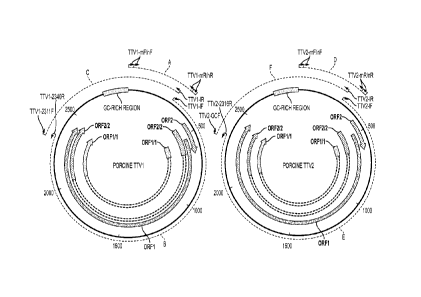
100731 In Fig. 1A, both the PTTV1 and PTTV2 genomes are shown in bold and
the sizes and
directions of the four putative ORFs (ORF1, ORF2, ORF1/1 and ORF2/2) are
indicated by
arrows. The GC-rich regions are also shown. Dashed-line arcs A and D represent
the regions
used for detection of PTTV1 and PTTV2 from serum and semen samples by nested
PCR,
respectively. Dashed-line arcs B and C represent the two overlapping PCR
fragments for
genomic cloning of PTTV I whereas dashed-line arcs E and F represent the two
overlapping PCR
fragments for genomic cloning of PTTV2. The locations of the primers used in
the study (see
Table 1) are also shown in the corresponding positions.
100741 One boar serum sample (SR#5) that was shown to be positive for both
PTTV1 and
PTTV2 in the first-round PCR, thus indicative of higher virus load, was used
for subsequent full-
length genomic cloning of PTTV. Surprisingly, initial attempts to utilize two
primer sets
(NG372/NG373 and NG384/NG385) of an inverse PCR (Okamoto et al., 2002, supra)
designed
for cloning of the first PTTV strain Sd-TTV31 to amplify the virus genomic DNA
were not
successful. No PCR product was obtained after several trials. Based upon the
initial sequence of
the region A of PTTV1 and the region D of PTTV2, two new pairs of primers
(TTV1-If(SEQ ID
NO:1)/TTV1-2340R(SEQ ID NO:2) and TTV1-2311F(SEQ ID NO:3)/TTV1-IR(SEQ ID
NO:4))
were subsequently designed to amplify regions B and C spanning the assumed
PTTV1 genome,
and two additional pairs of primers (TTV2-IF(SEQ ID NO:5)/TTV2.-2316R(SEQ ID
NO:6) and
TTV2-GCF(SEQ ID NO:7)/TTV2-1R(SEQ ID NO:8)) to amplify regions E and F
spanning the
assumed PTTV2 genome, respectively (Fig. IA and Table 1). Primers TTV1-
2340R(SEQ ID
CA 02771771 2012-02-21
WO 2011/031438 PCT/US2010/046330
NO:2) and TTV1-2311F(SEQ ID NO:3) were deduced from a common sequence in PTTVI
stains Sd-TTV31 (Okamoto et al., 2002, supra) and TTV-lp (Niel et al., 2005)
that is absent in
PTTV2 strain TTV-2p (Niel et al., 2005, supra), whereas primers TTV2-2316R(SEQ
ID NO:6)
and TTV2-GCF(SEQ ID NO:7) were deduced from a sequence of strain TTV-2p that
is absent in
the two PTTV I strains. The resulting four different PCR products with
expected sizes were each
inserted into a blunt-end cloning vector, and the resulting recombinant
plasmids were
transformed into Escherichia coll. Eight to fifteen positive (with white
color) bacterial clones for
each construct representing fragments B, C, E and F were identified and
subsequently sequenced.
21
CA 02771771 2012-02-21
WO 2011/031438 PCT/US2010/046330
Table 1. Oligonucleotide primers used for nested PCR and genomic PCR
amplifications of
porcine TT viruses
Primer ID
Sequence (5' to 3') Used for:
TTV1-mF
TACACTTCCGGGTTCAGGAGGCT Detection of porcine
TTV1
(SEQ ID NO:45)
TTV1-mR
ACTCAGCCATTCGGAACCTCAC Detection of porcine
TTV1
(SEQ ID NO:46)
TTV I -nF
CAATTTGGCTCGCTTCGCTCGC Detection of porcine TTV
I
(SEQ ID NO:47)
TTVI-nR
TACTTATATTCGCTTTCGTGGGAAC Detection of porcine
TTV1
(SEQ ID NO;48)
TTV2-mF
AGTTACACATAACCACCAAACC Detection of porcine
TTV2
(SEQ ID NO:49)
TTV2-mR
ATTACCGCCTGCCCGATAGGC Detection of porcine
TTV2
(SEQ ID NO:50)
TTV2-nF
CCAAACCACAGGAAACTGTGC Detection of porcine
TTV2
(SEQ ID NO:5 l)
TTV2-nR
CTTGACTCCGCTCTCAGGAG Detection of porcine
TTV2
(SEQ ID NO:52)
TTV I -IF
CATAGGGTGTAACCAATCAGATTTAAGGCGTT Genomic cloning (fragment 13)
(SEQ ID NO:1)
TTV1-2340R
GGTCATCAGACGATCCATCTCCCTCAG Genomic cloning
(fragment B)
(SEQ ID NO:2)
TTV1-2311F
CTTCTGAGGGAGATGGATCGTCTGATGA Genomic cloning
(fragment C)
(SEQ ID NO:3)
TTV1-1R
TTGAGCTCCCGACCAATCAGAATTGACT Genomic cloning
(fragment C)
(SEQ ID NO:4)
TTV2-IF
TTGTGCCGGAGCTCCTGAGAGC Genomic cloning
(fragment E)
(SEQ ID NO:5)
TTV2-2316R
AGGTGCTTGAGGAGTCGTCGCTTG Genomic cloning
(fragment E)
(SEQ ID NO:6)
TTV2-GCF
CTCAAGCACGAGCAGTGGATCCTCTCA Genomic cloning
(fragment F)
(SEQ ID NO:7)
TTV2-IR
TACCCAGGCGGTTAGACACTCAGCTCT Genomic cloning
(fragment F)
(SEQ ID NO:8)
[0075] Unexpectedly, two groups of sequence data from each construct were
identified,
indicating that there exist two types of PTTVs in genogroup 1 and genogroup 2
from the same
pig. In order to differentiate and assemble the four PTTV strains, sequence
comparisons were
performed together with the three known PTTV strains, Sd-TTV31, TTV-1 p and
TTV-2p (Fig.
1B and IC).
22
CA 02771771 2012-02-21
WO 2011/031438 PCT/US2010/046330
[00761 Fig. 113 illustrates differentiation and assembly of full-length
genomic sequences of
PTTV1 strains PTTV1a-VA and PTTV1b-VA with PCR fragments B and C that were
subsequently cloned. The initiation codons of ORF1 and ORF2 in the fragment B
as well as the
termination codons of ORF1 in the fragment C are marked by "^" or "*". The
corresponding
sequences of two known PTTV1 strains, Sd-TTV31 and TTV-1p, are also shown.
Conserved
sequences are shaded, and dashes indicate nucleotide deletions.
[0077] For PTTV1, the initiation codon ATG and the termination codon TGA of
the putative
ORF1 were located in fragments B and C, respectively (Fig. 1B). The positions
of the codons
were differed in two PTTV1 groups, the first one identical to Sd-TTV31 and the
second one
identical to TTV-1p (Fig. 1B). In addition, the ORF2 initiation codons in the
two groups were
also located at different positions consistent with that of ORF1. Moreover,
phylogenetic analyses
using four different sequences of the region B (two from the sequencing data
and two from
strains Sd-TTV31 and TTV-1p) and four different sequences of the region C
supported that the
first sequence was clustered with Sd-TTV31 and the second was clustered with
TTV-lp (data not
shown). Therefore, we were able to differentiate and assemble two groups of
sequence data from
both fragments B and C into two full-length PTTV1 genomes that were designated
as strains
PTTV1a-VA (SEQ ID NO:9) and PTTV1b-VA (SEQ ID NO:10), respectively (Fig. 1B).
[0078] Fig. 1C illustrates differentiation and assembly of full-length
genomic sequences of
PTTV2 strains PTTV2b-VA and PTIV2e-VA with PCR fragments E and F that were
subsequently cloned. The corresponding sequence of TTV-2p strain is included
and the
conserved sequences are shaded. Dashes indicate nucleotide deletions. The
unique nucleotides
within the overlapping region (boxed with dashed-line) for each strain (a
continuous "AG"
23
CA 02771771 2012-02-21
WO 2011/031438 PCT/US2010/046330
nucleotides for PTTV2b-VA (SEQ ID NO:11) and two single "A" and "G"
nucleotides for
PTIV2c-VA (SEQ ID NO:12)) are shown, respectively.
100791 Differentiation of the two PTTV2 strains was easier. A unique
continuous "AG"
nucleotides located in the overlapping region of two PCR fragments was shared
by two groups of
sequence data from fragments E and F, respectively (Fig. IC). The assembled
full-length
genomic sequence represented a PTTV2 strain and was designated as PTTV2b-VA
(SEQ ID
NO:11). Similarly, the complete genomic sequence of a second strain designated
as PTTV2c-VA
(SEQ ID NO:12)was assembled based upon two unique single -A" and "G"
nucleotides shared
in the overlapping region by another set of sequence data from fragments E and
F, respectively
(Fig. 1C). Phylogenetic analyses using four sequences from fragments E and F
together with the
two corresponding sequences from TTV-2p also supported this assignment (data
not shown).
100801 The present invention provides four isolated porcine TTV virus
genotypes or
subtypes that are associated with viral infections in pigs. This invention
includes. but is not
limited to. porcine TTV virus genotypes or subtypes PTTV la-VA, PTTV lb-VA,
PTTV2b-VA,
and PTTV2c-VA, the virus genotypes or subtypes which have nucleotide sequences
set forth in
SEQ ID NO:9 (PTTV1a-VA), SEQ ID NO:10 (PTTV1b-VA), SEQ ID NO:11 (PTTV2b-VA),
and SEC) 11) NO:12 (PTTV2e-VA). their functional equivalent or complementary
strand. It will
be understood that the specific nucleotide sequence derived from any porcine
TTV will have
slight variations that exist naturally between individual viruses. These
variations in sequences
may be seen in deletions, substitutions, insertions and the like.
[0081] The proposed genomic structure for each of the four PTTV strains was
analyzed in
detail and summarized in Table 2, together with the three known PTTV strains,
Sd-TTV31,
TTV-Ip and TTV-2p. All the four U.S. strains of PTTV have a similar genomic
size of 2,878 bp
24
CA 02771771 2012-02-21
WO 2011/031438 PCT/US2010/046330
(PTTV1a-VA SEQ ID NO:9), 2,875 bp (PTTV1b-VA SEQ ID NO:10), 2,750 bp (PTTV2b-
VA
SEQ ID NO:11), and 2,803 bp (PTTV2c-VA SEQ ID NO:12), respectively. Both
PTTV1a-VA
(SEQ ID NO:9)and Sd-TTV31 have the same genomic length. The published
sequences of the
strains TTV-lp and TTV-2p all have many undetermined nucleotides in the GC-
rich region of
the UTR. After artificial filling of these nucleotides with the consensus
sequences corresponding
to PTTV1 and PTTV2, it was shown that the TTV-lp is more closely-related to
PTTV1b-VA
(SEQ ID NO:10), and that TTV-2p is more closely-related to PTTV2b-VA (SEQ ID
NO:11) in
genomic length, respectively (data not shown).
100821
The assembled genomic sequences of porcine TTV virus genotypes or subtypes
PTTV1a-VA (SEQ II) NO:9). P
(SE() ID NO:10). IY1TV2b-VA (SA) 11) NO:11).
and PTTV2c-VA(SEQ ID NO:12) are submitted to Genbank (Nucleic Acids Research,
2008
Jan; 36(Database issue):D25-30) with accession numbers GU456383, GU456384,
GU456385,
and GU456386, respectively.
CA 02771771 2012-02-21
WO 2011/031438
PCT/US2010/046330
Table 2. Comparison of the genontic organization and ORFs of the seven porcine
TTV
strains
Porcine TTV species 1 Porcine TTV species 2
Vinis
Type la Type 1 b Subtype 2a Subtype
2b Subtype 2c
Strain PTIVIa-VA Sd-TTV31 PTTV I b-VA TTV- I p TTV-2p
PTTV2b-VA PTTV2c-VA
Country USA Japan USA Brazil Brazil USA
USA
Full-length (nt) 2878 2878 2875 Uncompleted Uncompleted 2750
2803
GenBank accession # GU456383 AB07600 I GU456384 AY823990
AY823991 0U456385 GU456386
TATA box 288-291 288-291 288-291 288-291 233-236 233-236
285-288
Putative mRNA 5'-end 316 316 316 316 261 261
313
ORF1
Size (aa) 635 635 639 637 624 625
625
Exon # l 1 1 1 1 1 1
Initiation 534 534 517 517 476 476
528
Termination 2441 2441 2436 2430 2350 2353
2405
ORF2
Size (aa) 73 73 72 72 68 68 68
Exon # 1 1 I 1 1 1 1
Initiation 430 430 428 428 393 393
445
Termination 651 651 646 646 599 599
651
ORF1/1
Size (aa) 174 174 182 182 178 178
178
Exort # 2 2 2 2 2 . 1,
2
Initiation 534 534 517 517 476 476
528
Splicing 647/648 647/648 642/643 642/643 595/596 595/596
647/648
2030/2031 2030/2031 2013/2014 2007/2008
1933/1934 1936/1937 1988/1989
Termination 2441 2441 2436 2430 2350 2353
2405
ORF2/2 (ORF3)
Size (aa) 224 224 228 228 199 199
199
Exon # 2 2 2 ? 2 2 2
Initiation 430 430 428 428 393 395
445
Splicing 647/648 647/648 642/643 642/643 595/596 595/596
647/648
2030/2031 2030/2031 2013/2014 2007/2008
1933/1934 1936/1937 1988/1989
Termination 2487 2487 2485 2479 2330 2333
2385
Polyadenylation signal 2458-2463 2458-2463 2462-2467 2456-2461
2473-2478 2476-2481 2528-2533
(AATAAA)
The numbers (except sizes of the full-length genome. ORFs and the exon
numbers) indicate the
nucleotide (nt) positions on the genorne of respective PTTV strains.
26
CA 02771771 2012-02-21
WO 2011/031438 PCT/US2010/046330
100831 Two recent studies have identified the transcribed viral mRNAs and
the expression of
at least six viral proteins during human TTV replication (Mueller et al.,
2008, supra; Qiu et al.,
2005, supra), which is more than the predicted number of ORFs encoded by human
TTV (
Okamoto, H., Nishizawa, T., Tawara, A., Takahashi, M., Kishimoto, J., Sai, T.,
and Sugai, Y.
(2000b). TT virus mRNAs detected in the bone marrow cells from an infected
individual.
Biochem Biophys Res Cornmun 279(2), 700-7), therefore we included the new
human TTV
genomic information for comparison with the PTTV sequences. The 5'-ends of the
mRNA
transcripts of human TTV strain P/1C1 were mapped to an "A" that is 25 nt
downstream of the
TATA-box (Mueller et al., 2008, supra). This starting point, its adjacent
sequence
(CGAATGGCTGAGTTTATGCCGC (SEQ ID NO:39); the starting point was underlined) and
the distance to the upstream TATA-box (24 nt; Table 2) are very conserved in
all seven PTTV
strains, suggesting that PTTV and human TTV may utilize a common 5'-end of
mRNA for
translation.
100841 Five additional completely-conserved regions were identified in the
vicinity of the
TATA-box among all seven PTTV strains. Two regions of 11 nt each (AGTCCTCATTT
(SEQ
ID NO:40) and AACCAATCAGA (SEQ ID NO:41)) are located in the upstream of the
TATA-
box, whereas the remaining three regions (CTGGGCGGGTGCCGGAG of 17 nt (SEQ ID
NO:42); CGGAGTCAAGGGGC of 14 nt (SEQ ID NO:43); TATCGGGCAGG of 11 nt (SEQ
ID NO:44)) are located between the proposed 5'-end of mRNA and the initiation
codon of
ORF2. These conserved PTTV-specific sequences may contain the common elements
regulating
the viral gene expression.
[0085] Previously, three ORFs (ORFs 1-3) were proposed in the genome of the
three known
PTTV strains, respectively (Niel et al., 2005, supra; Okamoto et al., 2002,
supra). The four
27
CA 02771771 2012-02-21
WO 2011/031438 PCT/US2010/046330
prototype U.S. strains of PTTV identified in this study possess this
structure. The corresponding
ORF3 in human TTV has been renamed as ORF2/2 since it initiates at the same
ATG in ORF2
and remains in the same ORF (extending ORF2) after the splicing (Fig. 1A)
(Mueller et al.,
2008, supra; Qiu et al., 2005, supra). We follow the nomenclature of human TTV
for revising
PTTV classification in this study. Human TTV ORF1/1 is a newly identified
viral protein that is
encoded by two exons in ORF1 (Qiu et at., 2005, supra). ORF1/1 share the
identical N- and C-
terminal part with ORF1. The PTTV ORF1/1 counterpart was readily identified in
all seven
PTTV strains (Fig. IA and Table 2).
[0086] The ORF1 and ORF2 are encoded by a ¨2.8 kb viral mRNA whereas the
ORF1/1 and
ORF2/2 are encoded by a spliced viral mRNA with ¨1.2 kb in human TTV (Mueller
et al., 2008,
supra; Qiu et al., 2005, supra). Since these four ORFs were also deduced in
PTTV genomes, and
since the sequences and positions of the putative splice donor and acceptor
sites in the seven
PTTV strains are very conserved (Table 2), it is speculated that porcine TTV
probably also
encodes the two corresponding mRNAs.
[0087] Most of the human TTV strains share a genetic similarity with the
CAV, encoding a
TTV apoptosis-inducing protein (TAIP) in which its CAV counterpart was named
apoptin (de
Smit, M. H., and Noteborn, M. H. (2009). Apoptosis-inducing proteins in
chicken anemia virus
and TT virus. Curr Top Microbiol Immunol 331, 131-49). The ORF of TAIP is
embedded within
the ORF2. However, the corresponding TAIP does not exist in porcine TTV. A
recent study
showed that the expression of apoptin or TAIP was required for CAV replication
in cultured
cells (Prasetyo, A. A., Kamahora, T., Kuroishi, A., Murakami, K., and Hino, S.
(2009).
Replication of chicken anemia virus (CAV) requires apoptin and is complemented
by VP3 of
human torque teno virus (TTV). Virology 385(1), 85-92).
28
CA 02771771 2012-02-21
WO 2011/031438 PCT/US2010/046330
100881 Pairwise sequence comparisons (PASC) is a useful method that plots
the frequency
distribution of pairwise nucleotide sequence identity percentages from all
available genomic
sequence of viruses in the same family (Bao, Y., Kapustin, Y., and Tatusova,
T. (2008). Virus
Classification by Pairwise Sequence Comparison (PASC). In ''Encyclopedia of
Virology, 5
vols." (B. W. J. Mahy, and M. H. V. Van Regenmortel, Eds.), Vol. 5, pp. 342-8.
Elsevier,
Oxford). The different peaks generated by the PASC program usually represent
groups of virus
genera, species, types, subtypes and strains (Fig. 2). In this study, we
performed PASC analyses
of TTV using 121 full-length genomic sequences of human and animal TTV-related
strains
available in GenBank database (Fig. 2). Assuming that TTV members are
classified into a
separate family, Anelloviridae, the two major peaks, at 36-55% and 55-67%
nucleotide sequence
identities, represent groups of genera and species, respectively (Fig. 2),
Accordingly, a TTV type
is defined as a group of TTV having 67-85% nucleotide sequence identity
whereas a TTV
subtype may be defined as a group of TTV sequences sharing 85-95% nucleotide
sequence
identity. TTV strains sharing more than 95% nucleotide sequence identity may
be further
classified into variants (Fig. 2). A similar classification has been proposed
using sequences of
103 TTV isolates by Jelcic et al (Jelcic, I., Hotz-Wagenblatt, A., Hunziker,
A., Zur Hausen, H.,
and de Villiers, E. M. (2004). Isolation of multiple TT virus genotypes from
spleen biopsy tissue
from a Hodgkin's disease patient: genome reorganization and diversity in the
hypervariable
region. J Viral 78(14), 7498-507).
100891 This proposed criteria of TTV classification were applied to
phylogenetic analyses of
the genomic sequences of the 4 prototype U.S. strains of PTTV and the 3 other
known PTTV
strains. Pairwise comparison of full-length nucleotide sequences among these
strains showed that
the four PTTV1 strains have 54.0-56.4% nucleotide sequence identity compared
to the three
29
CA 02771771 2012-02-21
WO 2011/031438 PCT/US2010/046330
PTTV2 strains (Table 3). Therefore, the previously designated "genogroup" of
PTTV in the
literature will probably be more appropriate to designate as "species", and
PTTV1 and PTTV2
probably should represent porcine TTV species 1 and species 2, respectively.
PTTV species 1
consists of two types of viruses designated as type la (including Sd-TTV31 and
PTTV 1 a-VA
(SEQ ID NO:9)) and type lb (including TTV-1p and PTTV I b-VA (SEQ ID NO:10)),
respectively, since the nucleotide sequence identity between these two types
of viruses is
between 69.8-70.7% (Table 3). Sd-TTV31 and TTV la-VA (SEQ ID NO:9) are
recognized as
variant strains of the same species due to their higher sequence identity
(95.1%). However, the
two type lb strains, TTV-lp and PTTV1b-VA (SEQ ID NO:10), may belong to two
different
subtypes (nucleotide sequence identity-86.4%). For PTTV species 2, three
strains are likely to
be classified into separate subtypes (TTV-2p for subtype 2a, PTTV2b-VA (SEQ ID
NO:11) for
subtype 2b, and PTTV2c-VA (SEQ ID NO:12) for subtype 2c, respectively) based
upon their
86.5-90.9% nucleotide sequence identity. This proposed new classification
system for PTTV was
clearly evident in the phylogenetic tree (Fig. 3A). Phylogenetic trees
constructed based upon the
deduced amino acid sequences of ORF1, ORF1/1, ORF2 and ORF2/2 of PTTV were
also
consistent with this proposed classification (Figs. 3B to 3E).
CA 02771771 2012-02-21
WO 2011/031438 PCT/US2010/046330
Table 3. Painvise sequence comparison of the full-length genomic sequence of
the seven
porcine TTV strains
Porcine TTV species 1 Porcine TTV species 2
Type la Type lb Subtype 2a Subtype 2b Subtype 2c
PTTV1a-VA Sd-TTV31 PTTV1b-VA TTV-lp ITV-2p PT"TV2b-VA PTTV2c-VA
Type la
PTTV1a-VA ¨ 95.1 70.5 69.8 55.7 55.1 56.2
Sd-TTV31 70.7 70.1 55,9 56.0 56,4
Type 1 b
PTTV1b-VA 86.4 54.0 54.7 55.2
TTV- I p 55.2 54.7 55.4
Subtype 2a
TTV-2p 86.5 86.8
Subtype 2b
PTTV2b-VA 90,9
Subtype 2c
PTTV2c-VA
The data were generated by using the PASC program, and the values indicate %
nucleotide
sequence identities.
[0090] Unique mutations and deletions and/or insertions are scattered
throughout the
genomes between PTTV species, types and subtypes. For example, the location of
ORF1
initiation and termination codons and the ORF2 initiation codons between PTTV
type la and lb,
which was shown in Fig. 1B as mentioned above, are different. The two PTTV lb
strains also
have a 2-codon deletion after the ORF2 initiation compared to PTTVla (Fig.
1B).
[0091] Remarkably, both TTV-2p and PTTV2b-VA have a large 52-nt deletion,
which is 39
nt upstream of the first 11-nt conserved sequence (AGTCCTCATTT (SEQ ID NO:40))
in the
UTR, compared to PTIV2c-V A. Due to this deletion, the genomic size of PTTV2b-
VA
(probably TTV-2p as well) was significantly smaller than that of PTTV2c-VA
(Table 2). A
number of "subviral" human TTV clones have been isolated from serum samples
that are
31
CA 02771771 2012-02-21
WO 2011/031438 PCT/US2010/046330
considered as full-length TTV genomes since the ORFs in a majority of these
subviral molecules
usually remain intact (de Villiers et al., 2009; Leppik et al., 2007). They
have variable lengths in
the UTR that are completely or partially deleted. The situation of TTV-2p and
PTTV2b-VA
appears to resemble that of the human TTV subviral molecules, implying that
subtypes PTTV2a
and PTTV2b might be the subviral molecules derived from subtype PTTV2c. Of
note, the 3'-
terminal sequence of a nested-PCR primer TTV2-nF (Table 1) that is commonly
used for
detection of the PTTV2 from field samples by other groups (Ellis et al., 2008,
supra; Kekarainen
et al., 2007, supra; Kekarainen et al., 2006, supra; Krakowka et al., 2008,
supra) is located at
both sides of the deletion. Therefore, the current nested-PCR assay for PTTV2
detection is likely
not sufficient to identify the genetically diverse strains of PTTV2c subtype.
[0092] The source of the isolated virus strain is serum, fecal, saliva,
semen and tissue
samples of pigs having the porcine TTV viral infection. However, it is
contemplated that
recombinant DNA technology can be used to duplicate and chemically synthesize
the nucleotide
sequence. Therefore, the scope of the present invention encompasses the
isolated polynucleotide
which comprises, but is not limited to, a nucleotide sequence set forth in SEQ
ID NO:9, SEQ ID
NO:10, SEQ ID NO:11, and SEQ ID NO:12, or its complementary strand; a
polynucleotide
which hybridizes to and which is at least 67% complementary to the nucleotide
sequence set
forth in SEQ ID NO:9, SEQ ID NO:10, SEQ ID NO:11, and SEQ ID NO:12, preferably
85%
complementary, or more preferably 95% complementary; or an immunogenic
fragment selected
from the group consisting of an amino acid sequence of ORF1 protein set forth
in SEQ ID
NO:13 (PTTV1a-VA), SEQ ID NO:14 (PTTV113-VA), SEQ ID NO:15 (PTTV2b-VA), SEQ ID
NO:16 (PTTV2c-VA), an amino acid sequence of ORF2 protein set forth in SEQ ID
NO:17
(PTTV1a-VA), SEQ ID NO:18 (PTTV lb-VA), SEQ ID NO:19 (PTTV2h-VA), SEQ ID NO:20
32
CA 02771771 2012-02-21
WO 2011/031438 PCT/US2010/046330
(PTTV2c-VA), an amino acid sequence of ORF1/1 protein set forth in SEQ ID
NO:21 (PTTV I a-
VA), SEQ ID NO:22 (PTTV lb-VA), SEQ ID NO:23 (PTTV2b-VA), SEQ ID NO:24 (PTTV2c-
VA), an amino acid sequence of ORF2/2 protein set forth in SEQ ID NO:25
(PTTV1a-VA), SEQ
ID NO:26 (PTTV1b-VA), SEQ ID NO:27 (PTTV2b-VA), SEQ ID NO:28 (PTTV2c-VA). The
immunogenic or antigenic coding regions or fragments can be determined by
techniques known
in the art and then used to make monoclonal or polyclonal antibodies for
immunoreactivity
screening or other diagnostic purposes. The invention further encompasses the
purified,
immunogenic protein encoded by the isolated polynucleotides. Desirably, the
protein may be an
isolated or recombinant ORF1 protein or an ORF2 protein of at least one of the
above isolated
porcine TTV subtypes, more desirably ORF1 protein.
[0093] The ORF1 of porcine TTV is believed to encode a structural and
replication-
associated protein (Maggi, F., and Bendinelli, M. (2009). Immunobiology of the
Torque teno
viruses and other anelloviruses. Curr Top Microbial Irranunol 331, 65-90). The
ORF1-encoding
products of seven PTTV strains have 624-635 aa in length and possess a high
number of arginine
residues at the N-terminus that are thought to have the DNA-binding activity
(Fig. 4). In Fig. 4,
conserved sequences are shaded. Dashes indicate amino acid deletions. The RCR
motifs are
boxed with solid lines. Three HVRs (PTTVI-HVRs I, 2 and 3) of PTTV1 strains
and two HVRs
(PTTV2-HVRs 1 and 2) of PTTV2 strains are boxed with dashed lines. The
connection
boundaries of ORF1/1 are indicated by arrows, The predicted rolling-circle
replication (RCR)
motifs (Ilyina, T. V., and Koonin, E. V. (1992). Conserved sequence motifs in
the initiator
proteins for rolling circle DNA replication encoded by diverse replicons from
eubacteria,
eucaryotes and archaebacteria. Nucleic Acids Res 20(13), 3279-85) are
presented at different
positions in different PTTV types and subtypes that may be type- or subtype-
specific. RCR
33
CA 02771771 2012-02-21
WO 2011/031438 PCT/US2010/046330
motif-III (YxxK) is conserved in the PTTV type la (aa position 14-17 of PTTV I
a-VA SEQ ID
NO:13) and type lb strains (aa position 379-382 of PTTVI b-VA SEQ ID NO:14),
respectively,
whereas the same conserved motif identified in all three PTTV2 strains is
located at aa position
482-485 of PTTV2b-VA SEQ ID NO:15 (Fig. 4). Both PTTV2b-VA SEQ ID NO:15 and
PTTV2c-VA SEQ ID NO:16 also have a conserved RCR motif-II (HxQ) at aa position
331-333
of PTTV2b-VA that is absence in TTV-2p (Fig. 4).
[0094] The ORF I proteins of PTTV strains between species 1 and species 2
share very low
aa sequence identity with only 22.4 to 25.8%, which makes it difficult to
identify significantly
conserved aa sequences between the two species (Fig. 4). In PTTV species I,
the aa identity of
ORF1 between type la and lb strains are 50.3-52.7%. Three major hypervariable
regions (HVR),
PTTV1-HVRs 1 to 3, with a relatively high number of aa substitutions, were
identified among
the four PTTV I strains, whereas two HVRs (PTTV2-HVRs 1 and 2) were observed
among the
three PTTV2 strains (Fig. 4). The three PTTV2 strains have an approximately 20-
aa deletion in
the corresponding PTTV1-HVR1 region. Moreover, the two HVRs of PTTV2 are
within the
corresponding PTTVI-HVR3 region (Fig. 4). These HVRs are located only in the
ORF1 but not
in the truncated ORF1/1. They likely play a role in evading the host immune
surveillance and
helping PTTV to establish a persistent infection, as suggested by studies of
human TTV.
100951 The aa sequences of ORF2 differed considerably between the four
PTTV1 (PTTV I a-
VA SEQ ID NO:17; PTTV 1 b-VA SEQ ID NO:18) and three PTTV2 (PTTV2b-VA SEQ ID
NO:19; PTTV2c-VA SEQ ID NO:20) strains (Fig. 5). However, they share a
conserved protein-
tyrosine phosphatase (PTPase)-like motif (Wx7Hx3CxCx5II) at the N-terminus
(Fig. 4). This
motif is also conserved among all human TTV, TTMV and TTMDV strains as well as
CAV. The
TTMV or CAV ORF2 protein also exhibited a serine/threonine phosphatase (S/T
PPase) activity
34
CA 02771771 2012-02-21
WO 2011/031438 PCT/US2010/046330
(Peters, M. A., Jackson, D. C., Crabb, B. S., and Browning, G. F. (2002).
Chicken anemia virus
VP2 is a novel dual specificity protein phosphatase. J Biol Chem 277(42),
39566-73). The dual
specificity of the ORF2 protein is thought to regulate host gene
transcription, signal transduction
and cytokine responses during viral replication. Recently, mutagenesis
analyses of two
conserved basic aa residues before the last histidine residue of the motif in
CAV revealed that the
two residues affect virus replication, cytopathology in vitro and attenuation
in vivo (Peters, M.
A., Crabb, B. S., Washington, E. A., and Browning, G. F. (2006). Site-directed
mutagenesis of
the VP2 gene of Chicken anemia virus affects virus replication, cytopathology
and host-cell
MHC class I expression. J Gen Virol 87(Pt 4), 823-31; Peters, M. A., Crabb, B.
S., Tivendale, K.
A., and Browning, G. F. (2007). Attenuation of chicken anemia virus by site-
directed
mutagenesis of VP2. J Gen Virol 88(Pt 8), 2168-75). The two basic aa residues
("KK") are
conserved in the three PTTV2 strains. However, only the first basic residue
("R") is retained in
the two PTTVla strains whereas both basic residues are substituted in the PTTV
lb strains (Fig.
5). In Fig. 5, dashes indicate amino acid deletions. The five conserved amino
acids within the
common motif Wx7Hx3CxCx5I-1 (underlined) identified in TTV, TTMV and CAV are
shaded.
The positions of the two basic aa residues before the last histidine of the
motif, which have been
shown to affect virus replication, cytopathology or in vivo attenuation in
CAV, are indicated by
¶A77.
10096) In summary, the present invention has determined the full-length
genomic sequences
of four porcine TTV strains representing different genotypes or subtypes in a
serum sample of a
single boar in Virginia. The finding from this study clearly indicates that,
similar to human TTV,
multiple PTTV infections with distinct genotypes or subtypes exist and
probably are common in
pigs. We have also provided new information regarding the genomic
organization, the degree of
CA 02771771 2012-02-21
WO 2011/031438 PCT/US2010/046330
variability and the characteristics of conserved nucleotide and amino acid
motifs of PTTV, which
will improve the current PCR detection assay, aid in developing reagents for
serological
diagnostics and help initiate the structural and functional study of PTTV. A
new classification of
PTTV is also proposed in this study based upon the phylogenetic and genetic
analyses of the
genomic sequences of seven known PTTV strains.
[0097) The present invention also provides methods for diagnostics of
porcine TTV infection
by detecting viral DNA in samples of porcine TTV infected pigs or other
mammals. One
preferred embodiment of the present invention involves methods for detecting
porcine TTV
nucleic acid sequences in a. porcine or other mammalian species using
oligonucleotide primers
for polymerase chain reaction (PCR) to further aid in the diagnosis of viral
infection or disease.
The diagnostic tests, which are useful in detecting the presence or absence of
the porcine TTV
viral nucleic acid sequence in the porcine or other mammalian species,
comprise isolating viral
DNA from samples of' porcine TTV infected pigs or pigs suspected of infection
of TTV, and
performing SYBR green real-time quantitative PCR using PM/I-specific (SEQ ID
NO:29/
SEQ ID NO:30) or PTTV2-specific (SEQ ID NO:31/ SEQ ID NO:32) primers.
[00981 hi another embodiment of the present invention. the diagnostic
method may be
adapted to simultaneously detect PTTV1 and PTTV2 by using PTIV1/PTTV2 duplex
real-time
PCR. More specifically, the method comprises isolating viral DNA from samples
of porcine
TTV infected pigs or pigs suspected of infection of TTV, performing real-time
PCR using both
PTTV1-specific (SEQ ID NO:29/ SEQ ID NO:30) or PVVT2-specific (SEQ ID NO:31/
SEQ ID
NO:32) primers in the same real-time PCR reaction. Since the Tõ, value between
PTTV1 and
PTTV2 can be distinguished by MCA, the presence of PTTV1 and PTTV2 DNA can be
simultaneously detected.
36
CA 02771771 2012-02-21
WO 2011/031438 PCT/US2010/046330
[0099] In a further embodiment of the present invention, the diagnostic
method may employ
duplex nested PCR. The method comprises isolating viral DNA from samples of
porcine TTV
infected pigs or pigs suspected of infection of TTV, performing a first round
of PCR using one
pair of primers Plab-mF (SEQ ID NO:33)/131 ab-mR (SEQ ID NO:34), and
performing a second
round of PCR using a mixture of two pairs of primers, Pla-nF (SEQ ID
NO:35)/Pla-nR (SEQ ID
NO:36) for detection of PTTV1a, and P 1 b-nF (SEQ ID NO:37)/P1b-nR (SEQ ID
NO:38) for
detection of PTTV lb, and visualizing the PCR products.
[00100] The above diagnostics methods maybe optimized by one skilled in the
art according
to well known methods in the art.
[00101] Accordingly, an embodiment of the present invention develops two novel
singleplex
SYBR green real-time PCR assays to quantify the viral loads of two porcine TTV
species,
respectively. PTTV1- and PTTV2-specific primers were designed to target the
extremely
conserved regions across six PTTV1 and four PTTV2 full-length genomes
available to date,
respectively. Another embodiment of the present invention combines the two
singleplex assays
into a duplex real-time PCR assay followed by MCA of the viral amplicons that
can be identified
by their distinct melting temperatures for simultaneous detection of the two
porcine TTV species,
PTTV la and PTTV lb. In a third embodiment, a duplex nested PCR assay for
simultaneous
amplification of the viral DNAs from two types of PTTV1 in the first round PCR
and differential
detection of types la and lb in the second round PCR was developed for the
identification of two
types of porcine TTV species, PTTVIa and PTTV1b, in a single sample. These
assays represent
simple and practical tools for diagnoses of species- or type-specific porcine
TTVs.
[00102] Potential primers sequences were identified by multiple sequence
alignments of 10
available porcine TTV full-length genomes. PTTV1-specific primers TTV1F (SEQ
ID NO:29)
37
CA 02771771 2012-02-21
WO 2011/031438 PCT/US2010/046330
and TTV1R (SEQ ID NO:30) were designed based upon two conserved genomic
regions
immediately before the putative ORF2 across six PTTV1 genomes, whereas PTTV2-
specific
primers TTV2F4 (SEQ ID NO:31) and TTV2R4 (SEQ ID NO:32) were designed based
upon
two conserved genomic regions immediately after the putative ORF2/2 across
four PTTV2
genomes (Table 4). Primers showed no potentials for self- and cross-
dimerization. The expected
amplicon sizes were a 118-bp fragment from the PTTV1 primers corresponding to
the PTTV1b-
VA genome and a 200-bp fragment from the PTTV2 primers corresponding to the
PTTV2c-VA
genome, respectively.
Table 4. Oligonucleotide primers used for real-time PCR and duplex nested PCR
detections of porcine TTVs.
Primer ID Sequence (5' to 3') Purpose
TTV1 F
TCCGAATGGCTGAGTTTATGC PTTV l -specific real-
time PCR
SEQ ID NO:29
TTV1R
TCCGCTCAGCTGCTCCT PTTV I -specific real-
time PCR
SEQ ID NO:30
TTV2F4
GGTGGTAAAGAGGATGAA PTTV2-specific real-time
PCR
SEQ ID NO:31
TTV2R4
AATAGATTGGACACAGGAG PTTV2-specific real-time
PCR
SEQ ID NO:32
P 1 ab-mF
SEQ ID NO:33 TATCGGGCAGGAGCAGCT Duplex nested PCR
P I ab-mR
SEQ ID NO:34 TAGGGGCGCGCTCTACGT Duplex nested PCR
P I a-nF
SEQ ID NO:35 CCTACATGAAGGAGAAAGACT Duplex nested PCR
Pla-nR
SEQ ID NO:36 CCAGCGTCTCCAGGGTC Duplex nested PCR
Plb-nF
SEQ ID NO:37 AAGCTACCAAGGGCTGG Duplex nested PCR
Plb-nR
SEQ 1D NO:38 GCGOTC(TIG)GTAGCGGTAGT Duplex nested PCR
[001031 According to one specific embodiment of the present invention, SYBR
green simplex
real-time PCR using PTTV1- and PTTV2-specific primers can be used specifically
to detect
38
CA 02771771 2012-02-21
WO 2011/031438 PCT/US2010/046330
porcine TTV1 and TTV2 DNA, respectively. For PTTV1, a standard curve was
established over
a range of target DNA concentrations per 25 Ill. The linear range was shown to
span 4.4x101 to
4.4x10⁸ copies. The minimum detection limit (44 copies) corresponded to a
threshold cycle (CI)
of 37.57. For PTTV2, standard curve was also generated and used to detect DNA
concentration
ranging from 8.6x10 to 8.6x10⁸ copies per 25
reaction. The corresponding C, of minimum
detection limit (8.6 copies) was 36.53.
[00104] According to another specific embodiment of the present invention,
SYBR green
duplex real-time PCR is utilized for the simultaneous detection of porcine
TTV1 and TTV2
DNA. The 7-degree difference of Tm value between PTTV1 (87.0 C) and PTTV2
(80.0 C) made
it feasible to distinguish them from one another by the MCA. Therefore, two
singleplex assays
can be coupled into a duplex real-time PCR assay for the simultaneous
detection of PTTV1 and
PTTV2. A positive sample was one that had a symmetrical melt peak within the
known T,õ for
that product. This new assay was first validated by using a 10-fold dilution
of PTTV1 and
PTTV2 standards mixture. The non-template negative control using sterile water
as the template
showed a non-specific amplification caused by cross-dimerization between the
PTTV1 and
PTTV2 primers not seen in the singleplex assays (Fig. 7a). This produced a
distinct melt peak
between 72.0 C and 76.0 C. Figure 7A shows melt peaks of PTTV1 standard (red;
Tm=87.0 C),
PTTV2 standard (green; Tm=80.0 C) and non-template negative control (caused by
primer cross-
dimerization; black). Figures 7B-7E show melt peaks of representative serum
samples with
distinct viral loads of PTTV1 and PTTV2. Figure 7B shows boar serum sample no.
5: relatively
high viral loads of both PTTV1 and PTTV2, but PTTV2>PTTV1; Figure 7C shows
boar serum
sample no. 12: relatively high viral loads of both PTTV1 and PTTV2, but
PTTV1>PTTV2;
Figure 7D shows boar serum sample no. 14: low viral loads of both PTTV1 and
PTTV2; Figure
39
CA 02771771 2012-02-21
WO 2011/031438 PCT/US2010/046330
7E shows boar serum sample no. 10: PTTV1 positive, but PTTV2 negative. The
viral loads (unit:
genomic copies/ml) of PTTV1 and PTTV2 in each sample that were determined by
singleplex
real-time PCR were indicated at the top of the corresponding melt peak.
[00105] In one example, when the duplex real-time assay was applied to the 20
serum samples
of adult boars, samples with relatively high viral loads of both PTTV1 and
PTTV2 displayed two
distinct melt curves corresponding to PTTV1 and PTTV2 without a non-specific
melt peak (Fig.
7B & 7C), whereas samples with low viral load of either PTTV1 or PTTV2 showed
virus-
specific as well as non-specific melt curves (Fig. 7D & 7E). Although the two
melt peaks in
sample #14 were very small, they were considered positive since they displayed
a visually
distinct and symmetrical rise and fall at the appropriate T,T, of PTTV1 and
PTTV2 (Fig. 7D). In
contrast, sample #10 was considered only PTTV1 positive because a symmetrical
PTTV2 melt
peak was not evidently present (Fig. 7E). These results were consistent with
that of the two
singleplex assays (Table 5). Moreover, the size and shape of melt peaks
qualitatively reflected
the corresponding viral load in the detected sample.
1001061 According to another aspect of the present invention, duplex nested
PCR is used for
differential detection of two porcine TTV types, PTTVla and PTTV1b.
1001071 The inventor of the present invention demonstrated the existence of
two distinct
genotypes, tentatively named PTTV1 a and PTTV1b, in porcine TTV species 1. To
further
determine whether the co-infection of PTTVla and PTTVlb is common in pigs, a
novel duplex
nested PCR assay to quickly distinguish between the two was developed.
Alignment of porcine
TTV genomic DNA sequences identified a conserved genomic region located at the
N-terminal
part of the putative ORF1 encoding the viral capsid protein (Fig. 8). This
region also contains the
entire ORF2 and the partial UTR in the upstream. Primers P 1 ab-mF (SEQ ID
NO:33)/Plab-mR
CA 02771771 2012-02-21
WO 2011/031438 PCT/US2010/046330
(SEQ ID NO:34) were designed to simultaneously amplify both PTTV la and PTTV
lb DNAs in
the first-round PCR. A mixture of PTTV1a-specific primers PI a-nF (SEQ ID
NO:35)/P I a-nR
(SEQ ID NO:36) and PTTV1b-specific primers P 1 b-nF (SEQ ID NO:37)/P1b-nR (SEQ
ID
NO:38) was used to differentially amplify each genotype in the second-round
PCR. The final
PCR products of PTTV 1 a and PTTV1b were 162 bp and 96 bp in sizes,
respectively, which
could be easily distinguished by gel electrophoresis on a 1% agarose gel
stained with ethidium
bromide. This assay was not expected to detect PTTV2 DNA due to the
specificity of primers
(Fig. 8). In Fig. 8, conserved sequences were indicated by dots and shaded.
Dashes indicated
nucleotide deletions. The locations and directions of three pairs of primers
used for duplex
nested PCR were marked by arrows.
[00108] In one example, the 20 serum samples from adult boars that were
subjected to the
duplex nested PCR assay were all found to be positive for both PTTV 1 a and
PTTV1b, as
determined by visualizing two bands of the expected sizes and subsequent
sequencing
confirmation of PCR products (data not shown). No PCR products were amplified
in the 19
semen samples, which was consistent with the results of PTTV1 conventional
nested PCR and
real-time PCR assays described above.
[00109] Infection of pigs with the two species of porcine TTV has been found
back to 1985 in
Spanish pig farms according to a retrospective investigation (Segales et al.,
2009, supra).
However, whether porcine TTVs are associated with any particular pig diseases
remains elusive.
Since both of porcine TTV species have a high prevalence in domestic pigs,
determination of
TTV viral loads is presumably more important than assessing the presence of
TTV DNA. The
level of viral loads in serum and semen samples has been indicated as an
important marker for
PCVAD in PCV2 infection (Opriessnig et al., 2007, supra). Therefore,
establishment of
41
CA 02771771 2012-02-21
WO 2011/031438
PCT/US2010/046330
quantitative PTTV-specific real-time PCR assays would help identify potential
disease
conditions associated with porcine TTVs.
[001101 Two TaqMan probe-based real-time PCR assays have recently been
described. The
singleplex assay developed by a Canadian group was not species-specific and
was only designed
to quantify the total viral loads of two PTTV species (Brassard et al., 2009,
supra), The duplex
assay established by a Germany group allowed the specific and simultaneous
detection of both
species (Gallei et al,, 2009, supra), The target sequences of primers used in
those two assays
were determined by alignment of the three porcine TTV genomic sequences (Sd-
TTV3 I, TTV-
lp and TTV-2p) and were located in the UTR. In the present study, with 7
additional complete
PTTV genomic sequences available (4 PTTV1 and 3 PTTV2 sequences), we analyzed
and re-
detennined the conserved regions across the 10 full-length PTTV genomes, Based
upon the
updated alignment result from this study, two species-specific singleplex SYBR
green-based
real-time PCR. assays were developed to quantify the viral loads of PTTV1 and
PTTV2,
respectively, The primers used in our assays were designed to bind to
conserved genomic regions
distinct from the previous studies, which may increase the accuracy of
quantification. Our assays
showed a considerable species-specificity and sensitivity of detection with 44
genomic copies for
PTTV I and 8,8 genomic copies for PTTV2 per 25-1,11 reaction, whereas the
detection limit of 10
genomic copies per reaction was reported in the TaqMan probe-based duplex real-
time PCR
(Gallei et al., 2009, supra), In addition, the SYBR green-based real-time PCR
assay is a flexible
and inexpensive approach that can be directly carried out without the need to
use fluorescently
labeled probes. Finally, considering porcine T'FVs exhibit a high degree of
genetic diversity, the
results from SYBR green-based assays are unlikely affected by the different
genetic background
- 42 -
SUBSTITUTE SHEET (RULE 26)
CA 02771771 2012-02-21
WO 2011/031438 PCT/US2010/046330
of porcine TTV variants that likely contain mutations in the probe-binding
sequences in the
TaqMan probe-based assays,
1001111 In spite of the presence of TTV DNA, all serum samples from healthy
pigs tested in
this study had low amounts of PTTV1 and PTTV2 that were less than 2x10⁶
copies/ml.
Moreover, only an extremely low titer of PTTV2 DNA was detected in three semen
samples.
Most of the tested serum samples were also positive for PCV2 DNA as determined
by
conventional nested PCR (data not shown). Many PCV2-positive pigs with low
viral load do not
develop clinical PCVAD. A proposed threshold for developing PCVAD is 107 or
greater PCV2
genornic copies/ml of serum (Opriessnig et al., 2007, supra). In addition,
semen PCV2 DNA-
positivity is also a notable marker of diseased status (Opriessnig et al.,
2007, supra; Pal, N.,
Huang, Y.W., Madson, D.M., Kuster, C., Meng, XI, Halbur, P.G. and Opriessnig,
T., 2008.
Development and validation of a duplex real-time PCR assay for the
simultaneous detection and
quantification of porcine circovinis type 2 and an internal control on porcine
semen samples. J
Virol Methods 149, 217-25). The situation of species-specific PTTV may be
analogous to that of
PCV2 and a high PTTV titer greater than 107 copies/ml may be required for the
induction of
porcine diseases. The species-specific real-time PCR assays developed in this
study will offer
simple and practical tools for future investigations of PTTV association with
diseases using a
large number of clinical samples from various disease conditions.
[001121 Furthermore, by coupling the two species-specific singleplex
assays, we developed
and validated a quick, inexpensive and reliable screening for the simultaneous
detection and
differentiation of the two porcine TTV species, PTTV1 and PTTV2, in a MCA-
based duplex
real-time PCR assay. Although this assay is not intended for accurate
quantification of both
PTTV species, it is a more convenient approach that could replace the
conventional nested PCR
43
CA 02771771 2012-02-21
WO 2011/031438 PCT/US2010/046330
for detection purpose. In comparison with real-time PCR, the conventional
nested PCR assay for
porcine TTVs detection is time-consuming (requiring total 4 rounds of PCR),
laborious and
prone to sample contamination occurring during multiple rounds of PCR
processing. Due to the
difference of Tm value between PTTV1 and PTTV2 species, an MCA following
duplex PCR
amplification is able to ensure distinct reaction specificity. Another
advantage of this duplex
real-time assay is that inclusion of PTTV1 and PTTV2 standards is dispensable
when performing
the described protocol, which makes it easier for much wider use in any
diagnostic labs equipped
with an automated real-time PCR instrument.
100113] Multiple infection of porcine TTVs with distinct genotypes or subtypes
of the same
species has been demonstrated (Gallei et al., 2009, supra). In particular, our
previous study
showed that porcine TTV species 1 consists of two distinct types, PTTVla
(including strains Sd-
TTV31 and PTTVIa-VA) and PTTV 1 b (including strains TTV- I p and PTTV1b-VA).
The two
newly published PTTV1 isolates with full-length genomes, swSTHY-TT27
(GQ120664) from
Canada and TTV1 #471819 (GU188045) from Germany, were both classified into
type lb based
upon the phylogenetic analysis (data not shown). The duplex nested PCR
described in this study
confirmed that dual infection of two PTTV1 genotypes frequently occurred in
pigs. This novel
assay is the first diagnostic PCR approach developed to distinguish between
PTTVla and lb so
far. Since it is currently not known whether one or both of PTTV la and PTTV 1
b infection
represents a relevant factor associated with diseases, our differential PCR
assay should be of
great value for future potential disease associations of these two PTTV types.
[00114] According to another aspect of the invention, porcine TTV ORF proteins
were
expressed and used in immunodetection assays to detect the presence of porcine
TTV specific
antibodies. In one embodiment of the present invention, three truncated and
Histidine-tagged
44
CA 02771771 2012-02-21
WO 2011/031438 PCT/US2010/046330
ORF1 proteins of PTTV1a, PTTVlb and PTTV2, were expressed and purified in
Escherichia
coli (E. coil), respectively. Furthermore, both serum Western blot and ELISA
assays based on
these recombinant antigens were developed and validated using porcine serum
samples from
different sources. In particular, serological testing using the PTTV1a-,
PTTV1b- and PTTV2-
specific ELISA provides an accurate and simple tool for revealing the
association of porcine
TTV infection with diseases.
[00115] According to a further aspect of the invention, porcine TTV ORF
proteins were
expressed and purified as recombinant ORF1 capsid protein in E.coli expression
system (Fig. 10,
Fig. 15). Three truncated and His-tagged ORF1 capsid proteins of PTTV la, PTTV
I b and PTTV2,
were expressed and purified in Escherichia coli (E. coli), respectively, and
served as
recombinant capsid subunit vaccines against PTTV infection.
[00116] Four porcine TTV2 strains, TTV-2p, TTV2#472142, PTTV2b-VA and PTTV2c-
VA,
had available complete genomic sequences to date. Although they are
phylogenetically classified
into three putative subtypes, a comparative analysis of hydrophilicity
profiles of the ORF1
encoding amino acids from four PTTV2 showed that they shared three hydrophilic
regions, an
argininc-rich region from aa 1-49 at the N-terminal and two particular domains
(1 and II) located
at the middle and C-terminal part, respectively (Fig. 9A), The C-terminal
region used for
truncated PTTV2c-VA ORF1 expression and the corresponding regions shared in
other three
PTTV2 strains were indicated by a dashed box. Alignments of amino acid
sequences
demonstrated high levels of sequence conservation of domains I (aa 322-349)
and 11 (aa 536-
625) across the four PTTV2 strains (Fig. 9B).
[00117] Since hydrophilic domains are believed to be important for the
antigenicity of many
proteins, the C-terminal region (aa 310-625) of the PTTV2e-VA ORF I SEQ ID
NO:16
CA 02771771 2012-02-21
WO 2011/031438 PCT/US2010/046330
containing the two domains was chosen for protein expression, which would be
used as antigen
for PTTV2-specific antibody detection in porcine serum. According to one
aspect of the
invention, expression of the truncated PTTV2c ORF1 was sufficient for
detection of all PTTV2
subtypes (2a, 2b and 2c; also see Fig. 3A).
[00118] According to one embodiment of the present invention, the C-terminal
part of the
PTTV2c ORF1 gene fused with 8 x His-tags was constructed and expressed in
E.coli. The
recombinant protein was insoluble and expressed within the bacterial inclusion
bodies. Figure
10A shows SDS-PAGE of unpurified 2c-ORF1 products. Figure 10B shows SDS-PAGE
of
purified 2c-ORF1 products. Figure 10C shows Western blot analysis of purified
2c-ORF1
products using an anti-His-tagged mAb. White arrowheads indicated the ORF1
protein with the
expected size and its truncated product whereas black arrowheads show the
putative dimers of
the expected and truncated proteins. M: protein markers. In Fig. 10A, two
significant
polypeptides (white arrowheads) were produced in the 2c-ORF1 unpurified sample
in
comparison with the control sample. The band of ¨40 KDa was consistent with
the expected size
of 2c-ORF1 whereas the ¨30 KDa polypeptide was probably an N-terminally
truncated product
from the former. After purification with a nickel-affinity column, four
polypeptides including the
two described significant bands were showed in SDS-PAGE (Fig. 10B). They were
also detected
by western blot using an anti-His-tagged mAb (Fig. 10C). Two high-molecular-
mass bands
(black arrowheads) were the homodimers formed by the two polypeptides of ¨40
KDa and ¨30
KDa, respectively, based on the predicted sizes (-80 KDa and ¨60 KDa). The
results
demonstrated that the purified C-terminal PTTV2c-ORF I was successfully
produced and could
be used for porcine TTV2 antibody detection in porcine sera.
46
CA 02771771 2012-02-21
WO 2011/031438 PCT/US2010/046330
100119] According to another aspect of the present invention, porcine TTV2
antibodies in
various porcine serum samples can be detected by Western blot using purified C-
terminal
PTTV2c-ORF1. White arrowheads indicated the ORF1 protein with the expected
size and its
truncated product. It should be noted that only the bands in green color were
recognized as
positive. A total of more than 200 serum samples of conventional pigs (healthy
or diseased),
CD/CD pigs and gnotobiotic pigs from different sources were collected. Samples
were randomly
selected for detection of anti-PTTV2c-ORF1 IgG antibodies using the purified C-
terminal
PTTV2c-ORF1 as antigen. Figure 11A shows results of Western blot analyses of
selected
porcine serum samples of conventional pigs, Figure 11B shows CD/CD pigs, and
Figure 11C
shows gnotobiotic pigs. Purified PTTV2e-ORF1 products were used as the
antigens. The two
marked ¨40 KDa and ¨30 KDa bands were detected in most samples of the
conventional pigs
(Fig. 11A) and CD/CD pigs (Fig. 11B), indicating widely PTTV2 infection in
these pigs.
However, all the gnotobiotic pigs from two different sources (Blacksburg, VA
and Ames, IA)
had no detectable PTTV2 antibody (Fig. 11C). Additional low-molecular-mass
bands were also
observed (Fig. 11A and 11B). They were likely from non-specific reactivity in
the Western blot.
[00120] According to yet another aspect of the present invention, PTTV2-
specific ELISA can
be used as a porcine TTV2 serological test. Seronegative results were also
shown in a few
samples from conventional pigs of a Wisconsin farm (Fig. 12). These negative
samples were
pooled and used as a negative reference in development of a PTTV2-specific
ELISA. The
remaining samples from this source were positive (Fig. 12, the four lanes in
the left). In addition,
porcine sera from a commercial company used in cell culture (supposed to be
OIE diseases-free)
also displayed strong anti-PTTV2-0RF2 positivity (Fig. 12), which was used as
a positive
control for ELISA. The concentrations of purified 2c-ORF I antigen, porcine
sera and IgG
47
CA 02771771 2012-02-21
WO 2011/031438 PCT/US2010/046330
conjugate were determined by checkboard titration to present low background
signal and give
the highest difference of 0D405 value between the positive and negative
controls. The optimal
antigen amount was 69 ng per well, and the optimal ELISA results were obtained
by use of a
1:100 dilution of serum samples and a 1:4000 dilution of IgG conjugates. The
ELISA cutoff
values ranged from 0.25 to 0.5 in each trial. Figure 4 shows a representative
result reflecting the
consistency of serum western blot and the developed ELISA.
1001211 138 conventional pig sera samples from 3 herds were chosen to
analyze the
correlation between PTTV2 viral load by real-time PCR and anti-PTTV2 IgG
antibody level by
ELISA. The results showed that pigs with undetectable or higher PTTV2 viral
load (108
copies/m1) were more likely to have a lower serum PTTV2 antibody titer than
pigs with middle
values of PTTV2 viral load (Fig. 13).
[001221 In particular, sera from 10 pigs in the same herd were also analyzed
by comparing the
PTTV2 viral loads and anti-PTTV2 antibody levels of their sera from their
arrival in the new
facility to two months after arrival. Nine of the 10 pigs had decreased viral
loads (three had no
detectable virus) after 2 months whilst the anti-PTTV2 antibody titers
increased in nine of 10
pigs (Fig. 14A and 14B). The results suggested that the 10 pigs acquired PTTV2
infection at
early stage, which induced humoral response and produced anti-ORF1 capsid IgG
antibody
progressively. The PTTV2-ORF1 IgG antibody was able to neutralize or even
clear the virus,
indicating the ORF I indeed encode a viral capsid protein and may contain
neutralizing epitopes
against PTTV2.
1001231 According to one embodiment of the present invention, the C-terminal
PTTV1a- and
PTTV 1 b-ORF1 proteins were expressed and purified in E.co/i system,
respectively. SDS-PAGE
and western blot analysis using an anti His-tagged mAb showed that both 1a-
and lb-ORF
48
CA 02771771 2012-02-21
WO 2011/031438 PCT/US2010/046330
products had two polypeptides, one with expected size (--40 KDa) and another
as the putative
homodimer (-80 KDa) (Fig. 15A-C). Figure 15A shows SDS-PAGE of unpurified and
purified
1 a-ORF1 products. Figure 15B shows SDS-PAGE of purified 1 b-ORF1 and lb-
ORF1ctruc
products. Figure 15C shows Western blot analysis of purified la- and 1b-ORF1
products using
an anti-His-tagged mAb. White arrowheads indicate the ORF1 protein with the
expected size
whereas black arrowheads show the putative dimer of the ORF1 proteins.
Compared to 2c-ORF I
expression, no truncated polypeptide was observed. As a comparative control,
expression of a C-
terminal-truncated 1b-ORF1 region (1b-ORF1ctruc) resulted in a lower-molecular-
mass
polypeptide compared to its C-terminal-non-truncated counterpart lb-ORF1 (Fig.
15B).
1001241 According one embodiment of the present invention, the purified C-
terminal
PTTV1a- and PTTV1b-ORF1 proteins were used to develop genotype-specific serum
Western
blots and ELISA as described for PTTV2 above. Figure 16 shows negative (lanes
1-2) and
positive (lanes 3-5) examples of serum Western blot using 1 a-ORF1 as antigen.
The same
antigen amount (69 ng), dilution of sera (1:100) and dilution of IgG conjugate
(1:4000) as
PTTV2-ORF I were used in PTTV la- and PTTV1b-specific ELISA (data not shown).
[00125] Additionally. the present invention provides a useful diagnostic
reagent for detecting
the porcine TTV infection which comprise a monoclonal or polyclonal antibody
purified from a
natural host such as, for example, by inoculating a pig with the porcine TTV
or the immunogenic
composition of the invention in an effective immunogenic quantity to produce a
viral infection
and recovering the antibody from the serum of the infected pig. Alternatively,
the antibodies can
be raised in experimental animals against the natural or synthetic
polypeptides derived or
expressed from the amino acid sequences or immunogenic fragments encoded by
the nucleotide
sequence of the isolated porcine TTV. For example, monoclonal antibodies can
be produced
49
CA 02771771 2012-02-21
WO 2011/031438 PCT/US2010/046330
from hybridoma cells which are obtained from mice such as, for example,
Balb/c, immunized
with a polypeptide antigen derived from the nucleotide sequence of the
isolated porcine TTV.
Selection of the hybridoma cells is made by growth in hyproxanthine,
thyrnidine and aminopterin
in a standard cell culture medium like Dulbecco's modified Eagle's medium
(DMEM) or minimal
essential medium. The hybridorna cells which produce antibodies can be cloned
according to
procedures known in the art. Then, the discrete colonies which are formed can
be transferred into
separate wells of culture plates for cultivation in a suitable culture medium.
Identification of
antibody secreting cells is done by conventional screening methods with the
appropriate antigen
or immunogen. Cultivating the hybridoma cells in vitro or in vivo by obtaining
ascites fluid in
mice after injecting the hybridoma produces the desired monoclonal antibody
via well-known
techniques.
1001261 For another alternative tnethod, porcine TTV capsid protein can be
expressed in a
baculovirus expression system or E. coil expression system according, to
procedures known in
the art. The expressed recombinant porcine TTV capsid protein can be used as
the antigen for
diagnosis in an enzyme-linked immunoabsorbent Assay (ELISA). The EUSA assay
based on the
porcine recombinant capsid antigen, for example, can be used to detect
antibodies to porcine
TTV in porcine and mammalian species. Although the ELISA assay is preferred,
other known
diagnostic tests can be employed such as immunofluorescenee assay (IFA),
immunoperoxidase
assay (IPA), etc.
1001271 Desirably, a commercial ELISA diagnostic assay in accordance with the
present
invention can be used to diagnose porcine TTV infection in pigs. The examples
illustrate using
purified ORFI and ORF2 proteins of porcine TTV to develop an ELISA assay to
detect anti-
TTV antibodies in pigs. Sera collected from pigs infected with porcine TTV,
and negative sera
CA 02771771 2012-02-21
WO 2011/031438 PCT/US2010/046330
from control pigs are used to validate the assay. PTTV2 specific, PTTVIa
specific, and PTTVlb
specific antibodies were demonstrated to specifically recognize PTTV ORF
proteins. Further
standardization of the test by techniques known to those skilled in the art
may optimize the
commercialization of a diagnostic assay for porcine TTV.
[00128] Another aspect of the present invention is the unique immunogenic
composition
comprising the isolated porcine TTV or an antigenic protein encoded by an
isolated
polynucleotide described hereinabove and its use for raising or producing
antibodies. The
composition contains a nontoxic, physiologically acceptable carrier and,
optionally, one or more
adjuvants. Suitable carriers, such as, for example, water, saline. ethanol,
ethylene glycol,
glycerol, etc., are easily selected from conventional excipients and co-
formulants may be added.
Routine tests can be performed to ensure physical compatibility and stability
of the final
composition.
[00129] In accordance with the present invention, there are further
provided infectious
molecular and nucleic acid molecules of porcine Torque teno (TTV), live
viruses produced from
the nucleic acid molecule and veterinary vaccines to protect pigs from porcine
TTV viral
infection or disease caused by porcine TTV co-infection with other viruses.
The invention further
provides immunogenic polypeptide expression products that may be used as
vaccines.
[00130] The novel infectious DNA molecule of porcine TTV comprises a nucleic
acid
molecule encoding at least a portion of an infectious PTTV1a-VA (SEQ ID NO:9),
PTTV1b-VA
(SEQ ID NO:10), PTTV2c-VA (SEQ ID NO:11), or PTTV2c-VA (SEQ ID NO:12) genome.
The
infectious PTTV DNA clone preferably contains at least one of ORF1, ORF2,
ORF1/1, and
ORF2/2 gene of the PTTV1 or PTTV2. Multiple copies of the PTTV1a-VA (SEQ ID
NO:9),
PTTV1b-VA (SEQ ID NO:10), PTTV2c-VA (SEQ ID NO:11), or PTTV2c-VA (SEQ ID
51
CA 02771771 2012-02-21
WO 2011/031438 PCT/US2010/046330
NO:12) genome may be inserted into a single DNA molecule to construct tandem
infectious
PTTV clones.
[00131] The cloned genomic DNA of PTTV, particularly PTTV1a-VA, PTTV1b-VA,
PTTV2c-VA, and tandem PTTV2b-RR, PTTV2c-RR, described herein is shown to be in
vitro or
in vivo infectious when transfected into PK-15 cells and given to pigs. This
new, readily
reproducible pathogenic agent lends itself to the development of a suitable
vaccination program
to prevent PTTV infection in pigs.
[00132] According to a further embodiment of the present invention, three one-
genome-copy
PTTV DNA clones were derived from the prototype US isolates PTTV1a-VA, PTTV1b-
VA and
PTTV2c-VA by fusion PCR, respectively. Each of the full-length genomic DNA was
inserted
into a cloning vector pSC-B-amp/kan by blunt-end ligation. The restriction
site BamH I is the
unique site on the three PTTV genomes, which was engineered at both ends of
the three genomes
to facilitate the generation of concatemers and thus mimic the TTV circular
genome. BamH I
single digestions of the selected plasmid DNA of each clone clearly resulted
in two different
fragments of 4.3-Kb and 2.8-Kb in size (Fig. 18A). The 4.3-Kb fragments
represented the
backbone vector whereas the 2.8-Kb fragments represented the inserted PTTV
genomic DNA.
The empty vector pSC-B-amp/kan digested with the same enzyme only showed a 4.3-
Kb band
(Fig. 18A). The resulting PTTV clones were designated pSC-PTTV1a, pSC-PTTV 1 b
and pSC-
PTTV2c, respectively (Fig. 17A-C).
100133) Furthermore, two copies of the full-length PTTV2c-VA genome derived
from the
clone pSC-PTTV2c were ligated in tandem into the pSC-B-amp/kan vector to
generate the clone
pSC-2PTTV2c-RR (Fig. 17D). Comparison of the Afl Il single digestion patterns
between pSC-
PTTV2c and pSC-2PTTV2c-RR showed that the latter plasmid had an additional 2.8-
Kb
52
CA 02771771 2012-02-21
WO 2011/031438 PCT/US2010/046330
fragment representing the second copy of PTTV2c genome (Fig. 18B, right
panel).
Subsequently, we utilized the same cloning strategy to produce a tandem-
dimerized PTTV2b
DNA clone derived from the Germany TTV clone TTV2-#471942-full. An additional
2.8-Kb
fragment representing the second copy of PTTV2b genome was presented in this
construct,
designated pSC-2PTTV2b-RR (Fig. 17F), which was digested with the Hind III
alone when
compared to its one-genomc-copy counterpart (Fig. 18B, left panel), confirming
the successful
construction.
1001341 The replication competencies of the constructed PTTV infectious clones
were tested
by in vitro transfection of PK-15 cells. IFA using the commercially generated
rabbit polyclonal
antibodies against PTTV2c ORF1 confirmed that both the concatemers of clones
ITV2-
#471942-full and pSC-PTTV2c were replication competent, respectively (Fig. 19A
and Fig.
20A). Passaging of the transfected cells did not eliminate or reduce the
fluorescent signals (Fig.
19B and Fig. 20B), suggesting that the expression of ORF1 proteins was
resulted from the
PTTV2 concaterners that mimicked the natural PTTV2b or PTTV2c circular
molecules. No
fluorescent signals was observed in mock-transfected cells or DNA-transfected
cells using pre-
immune rabbit serum as the antibody for IFA detection (data not shown). The
concaterners of the
clone pSC-PTTV 1 a also showed to be replication-competent using an anti-
PTTV1a ORF1
antibody (Fig. 21). The positive fluorescent signals were located in the
nucleus of transfected or
passaged cells, indicating that porcine TTVs likely replicate in the cell
nucleus. It is not
unexpected because porcine cireovirus (PCV) has a similar expression pattern
in vitro.
1001351 Direct transfeetion of the tandem-dimerized clone pSC-2PTTV2b-RR or
pSC-
2PTTV2c-RR in PK-15 cells results in viral replication and produces the ORF1
capsid antigen.
53
CA 02771771 2012-02-21
WO 2011/031438 PCT/US2010/046330
IFA using antibodies against PTTV2 ORF1 confirmed that both clones were also
replication-
competent and the positive ORF1 antigens were localized in the nuclei (Fig.
22A and B).
[00136] According to one embodiment of the present invention, infectious
clones of porcine
TTV can be used to inoculate pigs, which will then cilia an immune response of
the host animal
and stimulate production of neutralizing antibodies. In one particular
embodiment of the present
invention, the two tandem-dimerized PTTV2 clones were infectious when injected
into the
lymph nodes and muscles of conventional pigs.
[00137] To test the in vivo infectivity of PTTV2 molecular clones,
conventional pigs were
inoculated with the clone pSC-2TTV2b-RR or pSC-2TTV2c-RR. Serum samples were
collected
from animals at 0, 7, 14, 21 and 28 days post-inoculation (DPI). PTTV2 DNA was
detected in
pSC-2TTV2c-RR-inoculated pigs beginning at 7 DPI (#92), 14 DPI (#188 and #191)
and 21 DPI
(#180), respectively (Fig. 23A, right panel). PTTV viremia appeared late for
pigs inoculated with
the clone pSC-2TTV2b-RR: two began at 14 DPI (#189 and #192), one at 21 DP1
(#181) and one
at 28 DPI (#193) (Fig. 23A, left panel). The viral loads increased during the
course in all
inoculated pigs that had the highest viral loads at 28 DPI before necropsy, as
determined by
PTTV2-specific real-time PCR (Fig. 23A). The real-time PCR products amplified
from selected
pigs were sequenced and found to have identical sequences to the corresponding
regions of pSC-
2TTV2b-RR or pSC-2TTV2c-RR (data not shown).
[00138] All inoculated pigs were negative for PTTV2 ORF1 antibodies at 0 and 7
DPI. At 14
DPI, all the four pSC-2TTV2b-RR-inoculated pigs seroconverted to anti-PTTV2
ORF1 IgG,
whereas pigs in pSC-2TTV2c-RR-inoculated group seroconverted at 14 (#92 and
#180), 21
(#191) and 28 (#188) DPI, respectively (Fig. 23B). The results indicated that
active porcine
TTV2b or TTV2c infection had occurred.
54
CA 02771771 2012-02-21
WO 2011/031438 PCT/US2010/046330
[00139] Vaccines of the infectious viral and infectious molecular DNA clones,
and methods
of using them, are also included within the scope of the present invention.
Inoculated pigs are
protected from viral infection and associated diseases caused by TTV2
infection or co-infection.
The novel method protects pigs in need of protection against viral infection
by administering to
the pig an immunologically effective amount of a vaccine according to the
invention, such as, for
example, a vaccine comprising an immunogenic amount of the infectious PTTV
DNA, a plasmid
or viral vector containing the infectious DNA clone of PTTV, the recombinant
PTTV DNA, the
polypeptide expression products, the bacteria-expressed or baculovirus-
expressed purified
recombinant ORF I capsid protein, etc. Other antigens such as PRRSV, PPV,
other infectious
swine agents and immune stimulants may be given concurrently to the pig to
provide a broad
spectrum of protection against viral infections.
[00140j The vaccines comprise, for example, the infectious viral and molecular
DNA clones,
the cloned PTTV infectious DNA genome in suitable plasmids or vectors such as,
for example,
the pSC-B vector, an avirulent, live virus, an inactivated virus, expressed
recombinant capsid
subunit vaccine, etc. in combination with a nontoxic, physiologically
acceptable carrier and,
optionally, one or more adjuvants. The vaccine may also comprise the
infectious TTV2
molecular DNA clone described herein. The infectious PTTV DNA, the plasmid DNA
containing the infectious viral genotne and the live virus are preferred with
the live virus being
most preferred. The avirulent, live viral vaccine of the present invention
provides an advantage
over traditional viral vaccines that use either attenuated, live viruses which
run the risk of
reverting back to the virulent state or killed cell culture propagated whole
virus which may not
induce sufficient antibody immune response for protection against the viral
disease.
CA 02771771 2012-02-21
WO 2011/031438 PCT/US2010/046330
[00141] Vaccines and methods of using them are also included within the scope
of the present
invention. Inoculated mammalian species are protected from serious viral
infection, may also
provide protection for disease related to co-infection of PTTV, such as
porcine dermatitis and
nephropathy syndrome (PDNS), postweaning multisystemic wasting syndrome
(PMWS), and
other related illness. The vaccines comprise, for example, an inactivated or
attenuated porcine
TTV virus, a nontoxic, physiologically acceptable carrier and, optionally, one
or more adjuvants.
[00142] The adjuvant, which may be administered in conjunction with the
vaccine of the
present invention, is a substance that increases the immunological response of
the pig to the
vaccine. The adjuvant may be administered at the same time and at the same
site as the vaccine,
or at a different time, for example, as a booster, Adjuvants also may
advantageously be
administered to the pig in a manner or at a site different from the manner or
site in which the
vaccine is administered. Suitable adjuvants include, but are not limited to,
aluminum hydroxide
(alum), immunostimulating complexes (1SCOMS), non-ionic block polymers or
copolymers,
cytokines (like IL-1, IL-2, 1L-7, IFN-a, IFN-13, IFN-y, etc.), saponins,
monophosphoryl lipid A
(MLA), muramyl dipeptides (MDP) and the like. Other suitable adjuvants
include, for example,
aluminum potassium sulfate, heat-labile or heat-stable enterotoxin isolated
from Escherichia
coli, cholera toxin or the B subunit thereof, diphtheria toxin, tetanus toxin,
pertussis toxin,
Freund's incomplete or complete adjuvant, etc. Toxin-based adjuvants, such as
diphtheria toxin,
tetanus toxin and pertussis toxin may be inactivated prior to use, for
example, by treatment with
formaldehyde.
1001431 The vaccines may further contain additional antigens to promote the
immunological
activity of the infectious PTTV DNA clones such as, for example, porcine
reproductive and
56
CA 02771771 2012-02-21
WO 2011/031438 PCT/US2010/046330
respiratory syndrome virus (PRRSV), porcine parvovirus (PPV), other infectious
swine agents
and immune stimulants.
[00144] The new vaccines of this invention are not restricted to any
particular type or method
of preparation. The cloned viral vaccines include, but are not limited to,
infectious DNA
vaccines (i.e,, using plasmids, vectors or other conventional carriers to
directly inject DNA into
pigs), live vaccines, modified live vaccines, inactivated vaccines, subunit
vaccines, attenuated
vaccines, genetically engineered vaccines, etc. These vaccines are prepared by
standard methods
known in the art.
1001451 As a further benefit, the preferred live virus of the present
invention provides a
genetically stable vaccine that is easier to make, store and deliver than
other types of attenuated
vaccines.
[00146] Another preferred vaccine of the present invention utilizes
suitable plasmids for
delivering the nonpathogenic DNA clone to pigs. In contrast to the traditional
vaccine that uses
live or killed cell culture propagated whole virus, this invention provides
for the direct
inoculation of pigs with the plasmid DNA containing the infectious viral
genome.
[00147] Additional genetically engineered vaccines, which are desirable in
the present
invention, are produced by techniques known in the art. Such techniques
involve, but are not
limited to, further manipulation of recombinant DNA, modification of or
substitutions to the
amino acid sequences of the recombinant proteins and the like.
[00148] Genetically engineered vaccines based on recombinant DNA technology
are made,
for instance, by identifying alternative portions of the viral gene encoding
proteins responsible
for inducing a stronger immune or protective response in pigs (e.g., proteins
derived from ORF1,
57
CA 02771771 2012-02-21
WO 2011/031438 PCT/US2010/046330
ORF1/1, ORF2, ORF2/2, etc.). Such identified genes or immuno-dominant
fragments can be
cloned into standard protein expression vectors, such as the baculovirus
vector, and used to infect
appropriate host cells (see, for example, O'Reilly et al., "Baculovirus
Expression Vectors: A Lab
Manual," Freeman & Co., 1992). The host cells are cultured, thus expressing
the desired vaccine
proteins, which can be purified to the desired extent and formulated into a
suitable vaccine
product. The recombinant subunit vaccines are based on bacteria-expressed
(Fig. 10, Fig. 15) or
baeulovirus-expressed ORF I capsid proteins of PTTV1a, PTTV lb and PTTV2.
1001491 If the clones retain any undesirable natural abilities of causing
disease, it is also
possible to pinpoint the nucleotide sequences in the viral genome responsible
for any residual
virulence, and genetically engineer the virus avirulent through, for example,
site-directed
mutagenesis. Site-directed mutagenesis is able to add, delete or change one or
more nucleotides
(see, for instance, Zoller et al., DNA 3:479-488, 1984). An oligonucleotide is
synthesized
containing the desired mutation and annealed to a portion of single stranded
viral DNA. The
hybrid molecule, which results from that procedure, is employed to transform
bacteria. Then
double-stranded DNA, which is isolated containing the appropriate mutation, is
used to produce
full-length DNA by ligation to a restriction fragment of the latter that is
subsequently transfected
into a suitable cell culture. Ligation of the genome into the suitable vector
for transfer may be
accomplished through any standard technique known to those of ordinary skill
in the art.
Transfection of the vector into host cells for the production of viral progeny
may be done using
any of the conventional methods such as calcium-phosphate or DEAE-dextran
mediated
transfection, electroporation, protoplast fusion and other well-known
techniques (e.g., Sambrook
et al., "Molecular Cloning: A Laboratory Manual," Cold Spring I larbor
Laboratory Press, 1989).
The cloned virus then exhibits the desired mutation. Alternatively, two
oligonucleotides can be
58
CA 02771771 2012-02-21
WO 2011/031438 PCT/US2010/046330
synthesized which contain the appropriate mutation. These may be annealed to
form double-
stranded DNA that can be inserted in the viral DNA to produce full-length DNA.
[00150] An immunologically effective amount of the vaccines of the present
invention is
administered to a pig in need of protection against viral infection. The
immunologically effective
amount or the immunogenic amount that inoculates the pig can be easily
determined or readily
titrated by routine testing. An effective amount is one in which a sufficient
immunological
response to the vaccine is attained to protect the pig exposed to the PTTV
virus. Preferably, the
pig is protected to an extent in which one to all of the adverse physiological
symptoms or effects
of the viral disease are significantly reduced, ameliorated or totally
prevented.
1001511 The vaccine can be administered in a single dose or in repeated doses.
Dosages may
range, for example, from about 1 microgram to about 1,000 micrograms of the
plasmid DNA
containing the infectious chimeric DNA genome (dependent upon the
concentration of the
immuno-active component of the vaccine), preferably 100 to 200 micrograms of
the porcine
TTV DNA clone, but should not contain an amount of virus-based antigen
sufficient to result in
an adverse reaction or physiological symptoms of viral infection. Methods are
known in the art
for determining or titrating suitable dosages of active antigenic agent to
find minimal effective
dosages based on the weight of the pig, concentration of the antigen and other
typical factors.
Preferably, the infectious viral DNA clone is used as a vaccine, or a live
infectious virus can be
generated in vitro and then the live virus is used as a vaccine. In that case,
from about 50 to about
10,000 of the 50% tissue culture infective dose (TCID 50) of live virus, for
example, can be
given to a pig.
59
CA 02771771 2012-02-21
WO 2011/031438 PCT/US2010/046330
[00152] The new vaccines of this invention are not restricted to any
particular type or method
of preparation. The vaccines include, but are not limited to, modified live
vaccines, inactivated
vaccines, subunit vaccines, attenuated vaccines, genetically engineered
vaccines, etc.
[00153] The advantages of live vaccines are that all possible immune
responses are activated
in the recipient of the vaccine, including systeinic, local, humoral and cell-
mediated immune
responses. The disadvantages of live virus vaccines, which may outweigh the
advantages, lie in
the potential for contamination with live adventitious viral agents or the
risk that the virus may
revert to virulence in the field.
[00154] To prepare inactivated virus vaccines, for instance, the virus
propagation and virus
production can occur in cultured porcine cell lines such as, without
limitation PK-15 cells. Serial
virus inactivation is then optimized by protocols generally known to those of
ordinary skill in the
art or, preferably, by the methods described herein.
[00155] Inactivated virus vaccines may be prepared by treating the porcine TTV
with
inactivating agents such as fomialin or hydrophobic solvents, acids, etc., by
irradiation with
ultraviolet light or X-rays, by heating, etc. Inactivation is conducted in a
manner understood in
the art. For example, in chemical inactivation, a suitable virus sample or
serum sample
containing the virus is treated for a sufficient length of' time with a
sufficient amount or
concentration of inactivating agent at a sufficiently high (or low, depending
on the inactivating
agent) temperature or pH to inactivate the virus. Inactivation by heating is
conducted at a
temperature and for a length of time sufficient to inactivate the virus.
Inactivation by irradiation
is conducted using a wavelength of light or other energy source for a length
of time sufficient to
inactivate the virus. The virus is considered inactivated if it is unable to
infect a cell susceptible
to infection.
CA 02771771 2012-02-21
WO 2011/031438 PCT/US2010/046330
1001561 The preparation of subunit vaccines typically differs from the
preparation of a
modified live vaccine or an inactivated vaccine. Prior to preparation of a
subunit vaccine, the
protective or antigenic components of the vaccine must be identified. In the
present invention,
antigenic components of PTTV were identified as the ORF1 capsid proteins of
PTTV1a,
PTTVlb and PTTV2, which were expressed and purified in Escherichia coli (E.
coli) in this
invention, and other expression system, such as baculovirus expression system,
for use as subunit
recombinant capsid vaccines. Such protective or antigenic components include
certain amino
acid segments or fragments of the viral capsid proteins which raise a
particularly strong
protective or immunological response in pigs; single or multiple viral eapsid
proteins themselves,
oligomers thereof, and higher-order associations of the viral capsid proteins
which form virus
substructures or identifiable parts or units of such substructures;
oligoglycosides, glycolipids or
glycoproteins present on or near the surface of the virus or in viral
substructures such as the
lipoproteins or lipid groups associated with the virus, etc. Preferably, the
ORF1 protein is
employed as the antigenic component of the subunit vaccine. Other proteins may
also be used
such as those encoded by the nucleotide sequence in the OR.F2. ORF1/1, and
OR.F2/2 gene.
These immunogenic cotnponents are readily identified by methods known in the
art. Once
identified, the protective or antigenic portions of the virus (i.e., the
"subunit") are subsequently
purified and/or cloned by procedures known in the art. The subunit vaccine
provides an
advantage over other vaccines based on the live virus since the subunit. such
as highly purified
subunits of the virus, is less toxic than the whole virus.
1001571 If the subunit vaccine is produced through recombinant genetic
techniques,
expression of the cloned subunit such as the ORF1, ORF2. ORF1/1, and ORF2/2
genes, for
example, may be expressed by the method provided above, and may also be
optimized by
61
CA 02771771 2012-02-21
WO 2011/031438 PCT/US2010/046330
methods known to those in the art (see, for example, Maniatis et al.,
"Molecular Cloning: A
Laboratory Manual," Cold Spring Harbor Laboratory-, Cold Spring Harbor, Mass.
(1989)). On
the other hand, if the subunit being employed represents an intact structural
feature of the virus,
such as an entire capsid protein, the procedure for its isolation from the
virus must then be
optimized. In either case, after optimization of the inactivation protocol,
the subunit purification
protocol may be optimized prior to manufacture.
1001581 To prepare attenuated vaccines, the live, pathogenic virus is first
attenuated (rendered
nonpathogenic or harmless) by methods known in the art or, preferably, as
described herein. For
instance, attenuated viruses may be prepared by the technique of the present
invention which
involves the novel serial passage through embryonated pig eggs. Attenuated
viruses can be found
in nature and may have naturally-occurring gene deletions or, alternatively,
the pathogenic
viruses can be attenuated by making gene deletions or producing gene
tnutations. The attenuated
and inactivated virus vaccines comprise the preferred vaccines of the present
invention.
[00159] Genetically engineered vaccines, which are also desirable in the
present invention, are
produced by techniques known in the art. Such techniques involve, but are not
limited to, the use
of RNA, recombinant DNA, recotnbinant proteins, live viruses and the like,
[00160] For instance. after purification, the wild-type virus may, be
isolated from suitable
clinical, biological samples such as serum, fecal, saliva, semen and tissue
samples by methods
known in the art, preferably by the method taught herein using infected pigs
or infected suitable
cell lines. The DNA is extracted from the biologically pure virus or
infectious agent by methods
known in the art, and purified by methods known in the art, preferably by
ultracentrifugation in a
Csel gradient. The cDNA of viral genome is cloned into a suitable host by
methods known in the
art (see Maniatis et al., id.), and the virus genome is then analyzed to
determine essential regions
62
CA 02771771 2012-02-21
WO 2011/031438 PCT/US2010/046330
of the genome for producing antigenic portions of the virus. Thereafter, the
procedure is
generally the same as that for the modified live vaccine, an inactivated
vaccine or a subunit
vaccine.
[00161] Genetically engineered vaccines based on recombinant DNA technology
are made,
for instance, by identifying the portion of the viral gene which encodes for
proteins responsible
for inducing a stronger immune or protective response in pigs (e.g., proteins
derived front ORFI,
ORF2, ORF1/1, and ORF2/2, etc.). Such identified genes or immuno-dominant
fragments can be
cloned into standard protein expression vectors, such as the baculovirus
vector, and used to infect
appropriate host cells (see, for example, O'Reilly et al., "Baculovirus
Expression Vectors: A Lab
Manual," Freeman & Co. {1992)). The host cells are cultured, thus expressing
the desired
vaccine proteins, which can be purified to the desired extent and formulated
into a suitable
vaccine product.
[00162] Genetically engineered proteins, useful in vaccines, for instance, may
be expressed in
insect cells, yeast cells or mammalian cells. The genetically engineered
proteins, which may be
purified or isolated by conventional methods. can be directly inoculated into
a porcine or
mammalian species to confer protection against porcine TTV.
1001631 An insect cell line (like sf9. s121, or HIGH-FIVE) can be
transformed with a transfer
vector containing polynucleic acids obtained from the virus or copied from the
viral genome
which encodes one or more of the immuno-dominant proteins of the virus. The
transfer vector
includes, for example, linearized baculovirus DNA and a plasmid containing the
desired
polynucleotides. The host cell line may be co-transfected with the linearized
baculovirus DNA
and a plasmid in order to make a recombinant baculovirus.
63
CA 02771771 2012-02-21
WO 2011/031438 PCT/US2010/046330
[00164] Alternatively, DNA from the isolated porcine TTV which encode one or
more capsid
proteins can be inserted into live vectors, such as a poxvirus or an
adenovirus and used as a
vaccine.
[00165] An immunologically effective amount of the vaccine of the present
invention is
administered to an porcine or mammalian species in need of protection against
said infection or
syndrome. The "immunologically effective amount" can be easily determined or
readily titrated
by routine testing. An effective amount is one in which a sufficient
immunological response to
the vaccine is attained to protect the pig or other mammal exposed to the
porcine TTV virus, or
porcine TTV co-infection, which may cause porcine dermatitis and nephropathy
syndrome
(PDNS), postweaning multisystemic wasting syndrome (PMWS) or related illness.
Preferably,
the pig or other mammalian species is protected to an extent in which one to
all of the adverse
physiological symptoms or effects of the viral disease are found to be
significantly reduced,
ameliorated or totally prevented.
[00166] The vaccine can be administered in a single dose or in repeated doses.
Dosages may
contain, for example, from 1 to 1,000 micrograms of virus-based antigen
(dependent upon the
concentration of the immuno-active component of the vaccine.), but should not
contain an
amount of virus-based antigen sufficient to result in an adverse reaction or
physiological
symptoms of viral infection. Methods are known in the art for determining or
titrating suitable
dosages of active antigenic agent based on the weight of the bird or mammal,
concentration of
the antigen and other typical factors.
[00167] The vaccine can be administered to pigs. Also, the vaccine can be
given to humans
such as pig farmers who are at high risk of being infected by the viral.
agent. It is contemplated
that a vaccine based on the porcine TTV can be designed to provide broad
protection against
64
CA 02771771 2012-02-21
WO 2011/031438 PCT/US2010/046330
both porcine and human TTV. In other words, the vaccine based on the porcine
mi can be
preferentially designed to protect against human TTV infection through the so-
called "Jennerian
approach' (i.e., cowpox virus vaccine can be used against human s.mallpox by
Edward Jenner).
Desirably, the vaccine is administered directly to a porcine or other
mammalian species not yet
exposed to the TTV virus. The vaccine can conveniently be administered orally,
intrabuccally,
intranasally, transdennally, parenterally, etc. The parenteral route of
administration includes, but
is not limited to, intramuscular, intravenous, intraperitoneal and
subcutaneous routes.
[00168] When administered as a liquid, the present vaccine may be prepared
in the .form of an
aqueous solution, a syrup, an elixir, a tincture and the like. Such
tbrmulations are known in the
art and are typically prepared by dissolution of the antigen and other typical
additives in the
appropriate carrier or solvent systems. Suitable carriers or solvents include,
but are not limited
to, water, saline, ethanol, ethylene glycol, glycerol, etc. Typical additives
are, for example,
certified dyes, .flavors, sweeteners and antimicrobial preservatives such as
thimerosal (sodium
ethylmercurithiosalicylate.). Such solutions may be stabilized, for example,
by addition of
partially hydrolyzed gelatin, sorbitol or cell culture medium, and may be
buffered by
conventional methods using reagents known in the art_ such as sodium hydrogen
phosphate,
sodium dihydrogen phosphate, potassium hydrogen phosphate, potassium
dihydrogen phosphate,
a mixture thereof, and the like.
[00169] Liquid formulations also may include suspensions and emulsions
which contain
suspending or emulsifying agents in combination with other standard co-
formulants. These types
of liquid thnnulations may be prepared by conventional methods. Suspensions,
for example, may
be prepared using a colloid mill. Emulsions, for example, may be prepared
using a homogenizer.
CA 02771771 2012-02-21
WO 2011/031438 PCT/US2010/046330
[00170] Parenteral formulations, designed for injection into body fluid
systems, require proper
isotonicity and pH buffering to the corresponding levels of mammalian body
fluids. Isotonicity
can be appropriately adjusted with sodium chloride and other salts as needed.
Suitable solvents,
such as ethanol or propylene glycol, can be used to increase the solubility of
the ingredients in
the formulation and the stability of the liquid preparation. Further additives
which can be
employed in the present vaccine include, but are not limited to. dextrose,
conventional
antioxidants and conventional chelating agents such as ethylcriediamine
tetraaectic acid (EDTA).
Parenteral dosage forms must also be sterilized prior to use.
[00171] The following examples demonstrate certain aspects of the present
invention.
However, it is to be understood that these examples are for illustration only
and do not purport to
be wholly definitive as to conditions and scope of this invention. It should
be appreciated that
when typical reaction conditions (e.g., temperature, reaction times, etc.)
have been given, the
conditions both above and below the specified ranges can also be used, though
generally less
conveniently. The examples are conducted at room temperature (about 23 C. to
about 28 C.)
and at atmospheric pressure. All parts and percents referred to herein are on
a weight basis and
all temperatures are expressed in degrees centigrade unless otherwise
specified.
Example 1.
Viral DNA extraction, nested PCR and genomic PCR:
[00172] Convenient serum and semen samples from 20 conventional adult boars
from a
Virginia pig farm were used in the study. Total DNA was isolated from 20 serum
and 19 semen
samples using QIAamp DNA mini kit (Qiagen). To screen for the positive PTTV-
containing
samples, nested PCR amplifications of the conserved regions in the UTR of
PTTV1 and PTTV2
66
CA 02771771 2012-02-21
WO 2011/031438 PCT/US2010/046330
were initially performed by using AmpliTag Gold polymerase (Applied
Biosystems). The two
primer pairs used to amplify the fragment A of PTTV I were TTV1-mF (SEQ ID
NO:45)/TTV1-
mR (SEQ ID NO:46)(for the first-round PCR) and TTV1-nF (SEQ ID NO:47)/TTV1-nR
(SEQ
ID NO:48) (for the second-round PCR), whereas the two primer pairs used to
amplify the
fragment D of PTTV2 were TTV2-mF (SEQ ID NO:49)/TTV2-mR (SEQ ID NO:50) (for
the
first-round PCR) and TTV2-nF (SEQ ID NO:51)/TTV2-nR (SEQ ID NO:52) (for the
second-
round PCR; Fig. lA and Table 1).
[00173] In order to amplify the full-length genomic sequences of both PTTV1
and PTTV2, we
first performed an inverse genomic PCR using a pair of conserved gene-specific
primers TTV1-
IF (SEQ ID NO:1)/TTV I -IR (SEQ ID NO:4) located in region A for PTTV1 and
another pair of
gene-specific primers TTV2-IF (SEQ ID NO:5)/TTV2-IR (SEQ ID NO:8) located in
region D
for PTTV2, respectively, with Herculase II Fusion DNA Polymerase (Stratagene)
according to
the manufacturer's instructions. No PCR products with expected sizes were
detected.
Subsequently we designed new sets of primers to amplify two regions covering
the complete
PTTV1 and PTTV2 genomes in the second-round PCR, respectively (Fig. 1A). The
primer pairs
used to amplify fragments 13 and C of PTTV I were TTVI-IF (SEQ ID NO:1)/TTV1-
2340R
(SEQ ID NO:2) and TTV1-2311F (SEQ ID NO:3)/TTV1-IR (SEQ ID NO:4),
respectively,
whereas the primer pairs used to amplify fragments E and F of PTTV2 were TTV2-
1F (SEQ ID
NO:5)/TTV2-2316R (SEQ ID NO:6) and TTV2-GCF (SEQ ID NO:7)/TTV2-IR (SEQ ID
NO:8),
respectively (Fig. IA and Table 1). Fragments C and F contain the GC-rich
regions of PTTV1
and PTTV2, respectively, The amplified PCR products were individually excised,
purified, and
subsequently cloned into a pSC-B-amp/kan vector (Stratagene) by StrataClone
Blunt PCR
67
CA 02771771 2012-02-21
WO 2011/031438 PCT/US2010/046330
cloning strategy according to the manufacturer's instructions (Stratagene)
followed by DNA
sequencing.
Example 2.
Screening for porcine TTV positive samples collected from boars in a farm from
Virginia:
[00174] Porcine TTV DNA was previously detected from pigs in different
geographic regions
by nested-PCR based on the UTR sequence of a Japanese PTTV1 strain Sd-TTV31
(McKeown
et al., 2004, supra). With the recent identification of PTTV2, two different
sets of nested-PCR
primers have been used to amplify region A of PTTV1 and region D of PTTV2,
respectively
(Fig. 1A) (Ellis et al., 2008, supra; Kekarainen, T., Sibila, M., and Segales,
J. (2006). Prevalence
of swine Torque teno virus in post-weaning multisystemic wasting syndrome
(PMWS)-affected
and non-PMWS-affected pigs in Spain. J Gen Virol 87(Pt 4), 833-7; Krakowka et
al., 2008,
supra). A similar detection approach was also utilized in the present study to
identify PTTV
strains from pigs in the United States. In order to screen for indigenous
PTTV1- or PTTV2-
positive samples for subsequent use to determine the full-length genomic
sequences, 20 sera
(SR#1-20) and 19 semen samples (SM#1-18, and SM#20) collected from 20 boars in
a farm of
Virginia were subjected to nested-PCR analyses. Surprisingly, all the 20 serum
samples were
positive for PTTV I and 19 were also positive for PTTV2 (except for SR#18). In
contrast, only 1
semen sample (SM#6) was PTTV1-positive and 3 semen samples (SM#8, 9 and 20)
were
PTTV2-positive. The result was consistent with a recent study in that boar
semen samples were
shown to be positive for PTTV DNA in Spain (Kekarainen, T., Lopez-Soria, S.,
and Segales, J.
(2007). Detection of swine Torque teno virus genogroups 1 and 2 in boar sera
and semen.
Theriogenology 68(7), 966-71), and thus suggesting a potential vertical
transmission of PTTV.
68
CA 02771771 2012-02-21
WO 2011/031438 PCT/US2010/046330
However, the prevalence rates of both PTTV1 and PTTV2 in semen were much lower
than that
in sera, suggesting that there is no direct association for the presence of
PTTV DNAs in sera and
semen of the same pig.
Example 3.
Sequence and phylogenetic analyses:
1001751 Generic analyses and alignment of DNA and amino acid sequences were
performed
using Lasergene package (DNASTAR Inc., Madison, WI). The genomic sequences of
three
known PTTV strains and their corresponding GenBank accession numbers used for
the
alignment and comparison are Sd-TTV31 (AB076001), TTV-lp (AY823990) and TTV-2p
(AY823991). Pairwise sequence comparisons (PASC) were performed using 121 full-
length
genomic sequences of human and animal TTV-related strains available in GenBank
with an
online program PASC
(http://www.ncbi.nlm.nih.govi'sutils/pasc/viridty.cgi?textpage¨overview)
(Bao et al., 2008).
1001761 Phylogenetic trees were constructed by the neighbor-joining method in
the PAUP 4.0
program (David Swofford, Smithsonian Institute, Washington, DC, distributed by
Sinauer
Associate Inc.) based upon the full-length genomic sequences and the deduced
amino acid
sequences of 4 ORFs of seven PTTV strains. The data were obtained from 1000 re-
sampling.
69
CA 02771771 2012-02-21
WO 2011/031438 PCT/US2010/046330
Example 4.
Design of PCR primers for diagnosing porcine PTTV infection
[00177] Analyses and alignment of DNA sequences were performed using Lasergene
package
(DNASTAR Inc., Madison, WI). Full-length genomic sequences of ten porcine TTV
strains and
their corresponding GenBank accession numbers used for the alignment were as
follows. Species
PTTV1: Sd-TTV31 (AB076001), PTTV 1 a-VA (GU456383), TTV-1 p (AY823990), PTTV1b-
VA (0U456384), swSTHY-TT27 (GQ120664) and TTV1 #471819 (GU 1 88045). Species
PTTV2: PTTV2b-VA (GU456385), PTTV2c-VA (GU456386), TTV-2p (AY823991) and TTV2
#472142 (GU188046). The conserved sequences among the 6 PTTV1 and 4 PTTV2
genomes
were identified, respectively, and subsequently used to guide real-time PCR
primer selections
using the Beacon Designer program (PREMIER Biosoft International, Palo Alto,
CA). Primers
used for the duplex nested PCR of PTTV I were designed by the Lasergene
package.
Example 5.
Standard curves of PTTV] and PTTV2 real-time PCR
[00178] A region of 2091 bp corresponding to the PCR fragment B of PTTV1b-VA
genome
was re-amplified from the same PCR fragment using primers TTV1-IF (5'-
CATAGGGTGTAACCAATCAGATTTAAGGCGTT-3') and TTV 1-2340R
(5'-
GGTCATCAGACGATCCATCTCCCTCAG-3') as described previously (Huang et al., 2010).
The resulting amplicon was gel-purified by QIAquick Gel Extraction Kit
(Qiagen) and quantified
by a NanoDrop spectrophotometer that was used for the real-time PCR standard
template of
porcine TTV species 1. A full-length DNA clone of PTTV2c-VA strain, pSC-
PTTV2c, was
constructed by assembling PCR fragments E and F from PTTV2c-VA in the vector
pSC-B-
CA 02771771 2012-02-21
WO 2011/031438 PCT/US2010/046330
amp/kan (Huang et al., unpublished data). Plasmid pSC-PTTV2c (7082 bp) was
used for the
real-time PCR standard template of porcine TTV species 2 and the plasmid DNA
concentration
was measured by a NanoDrop spectrophotometer. A 10-fold dilution series of the
two templates
was used to generate the real-time PCR standard curves, respectively.
Example 6.
Extraction of viral DNA for PCR assays
1001791 Total DNA was isolated from 20 serum and 19 semen samples collected
from 20
conventional adult boars (with no clinical syndromes) from a Virginia pig farm
using QIAamp
DNA mini kit (Qiagen) as described previously (Huang et al., 2010). A sample
volume of 400 p,1
for sera and semen was used to extract DNA with a final eluate of 50 1
sterile water. All
extracted DNA samples were stored at -20 C until real-time PCR testing.
Detection of porcine
TTVs in these samples by conventional nested PCR had been described previously
(Huang et al.,
2010). Total DNA extracted from a goat serum sample with the same procedure
was used as the
negative control.
Example 7.
SYBR green real-time quantitative PCR assays
1001801 PTTV1- and PTTV2-specific real-time PCR were performed, respectively,
using
SensiMix SYBR & Fluorescein kit (Quantace Ltd) and the MyiQ iCYCLER Real Time
PCR
instrument (BIO-RAD Laboratories). Each 25-ul reaction contained 12.5 ul of
SYBR green
Master Mix, 4 1.11 of extracted DNA, 0.5 1.11 of each primer (10 nM) and 7.5
ul of sterile water.
The PCR condition for PTTV1 was 10 min at 95 C followed by 40 cycles of
amplification (15
71
CA 02771771 2012-02-21
WO 2011/031438 PCT/US2010/046330
sec at 95 C, 30 sec at 59.4 C, 10 sec at 72 C). This was immediately followed
by a melting point
analysis obtained by gradually increasing the temperature form 55 C to 95 C
with the
fluorescence signal being measured every 0.5 C. The PCR condition for PTTV2
was the same as
PTTV1 except that the annealing temperature was 56 C. PTTV1 and PTTV2 standard
templates
were included as positive controls in every run. Amplification and data
analysis were carried out
using MyiQ System software (BIO-RAD Laboratories). All samples were run in
duplicate on the
same plate.
Example 8.
Specficity and sensitivity of two singleplex assays
1001811 The optimal annealing temperatures for amplification of PTTV I - and
PTTV2-specific
assays were 59.4 C and 56 C, respectively, as determined by a 10-fold dilution
of amplifications
using a gradient of annealing temperatures. Amplification of the 118-bp
product using primers
TTV1F/TTV IR was obtained only with PTTV1 template whereas amplification of
the 200-bp
product with PTTV2 template was only observed when primers TTVF4/TTVR4 were
used.
Neither assay yielded any cross-amplification front the other, confirming the
specificity of the
primers and targets (data not shown).
{00182] A PTTV1 standard curve was established over a range of target DNA
concentrations
per 25 jtl. The linear range was shown to span 4.4x101 to 4.4x10⁸ copies. The
minimum
detection limit (44 copies) corresponded to a threshold cycle (CI) of 37.57.
Tested samples with
C1>37.57 were considered as below the detection limit and were not
quantifiable. Similarly, a
PTTV2 standard curve was generated and used to detect DNA concentration
ranging from
8.6x10 to 8.6x10⁸ copies per 25 jtl reaction. The corresponding C, of minimum
detection limit
72
CA 02771771 2012-02-21
WO 2011/031438 PCT/US2010/046330
(8.6 copies) was 36.53. All samples that were considered as PTTV1- or PTTV2-
positive had
copy numbers lower than the respective maximum detection limit. Melting curves
using a 10-
fold dilution of PTTV1 or PTTV2 standard template (Fig. 6a & 6b; blue curves),
as well as 20
boar serum samples, displayed melting temperatures (T.) of 87.0 C for PTTV1
and 80.0 C for
PTTV2, respectively (Fig. 6a & 6b; red curves). No peaks were observed for the
negative
controls using sterile water or goat serum DNA as templates (Fig. 6a & 7b;
black lines).
Example 9.
Quantification of porcine TTV1 and TTV2 in boar serum and semen samples
1001831 Viral load was expressed as copy numbers of PTTV1 or PTTV2 genomes per
ml of
original boar serum samples. PTTV1 DNA were detected in all 20 serum samples
ranging from
1.91x10³ to 3.25x10⁵ copies/ml whereas PTTV2 DNA were detected in 19 serum
samples
(except #10) ranging from 3.59x10² to 1.39x10⁶ copies/ml. The result was
consistent to our
previous study by using conventional nested PCR (Table 5). None of the semen
samples were
PTTV1-positive whereas three semen samples were PTTV2-positive with very low
viral loads
(230, 244 and 357 copies/ml, respectively).
73
CA 02771771 2012-02-21
WO 2011/031438 PCT/US2010/046330
Table 5. Comparison of porcine TTVs detection by different assays in 20 serum
and 19
semen samples from adult boars in a Virginia Farm.
No, of positive / total no, tested by different assay
Samples PTTV1 real- PTTV1 PTTV2 real- PTTV2
PTTV1/PTTV2
time PCR nested PCR time PCR nested PCR =
duplex real-time PCR
Serum PTTV1 20/20 20/20 - 20/20
Serum PTTV2 - - 19/20 19/20 19/20
Semen PTTV1 0/19 1/19 -
Semen PTTV2 - - 3/19 3/19 -
Example 10.
PTTVI/PTTV2 duplex real-time PCR assay
[00184] PTTV1/PTTV2 duplex real-time PCR assay was performed in a 25-ul PCR
system
containing 12.5 ul of SYBR green Master Mix, 0.5 IA1 of each PTTV1 primers,
0.5 p.1 of each
PTTV2 primers, 4 p.1 of DNA and 6.5 p.1 of sterile water. The duplex PCR
condition and melting
point analysis were the same as PTTV1 except that the annealing temperature
was 58 C. The
melting peaks were analyzed to distinguish the PTTV1- and PTTV2-specific
amplicons.
Example 11.
Duplex nested PCR
[00185] The first-round PCR was performed with a Platinum PCR IliFi Supermix
(Invitrogen)
using 4 til of extracted DNA in a total volume of 50 pi The PCR condition was
30 cycles of
94 C for 30 sec, 55 C for 30 sec, 72 C for 30 sec with an initial denaturation
of the template
DNA at 94 C for 2 min. A 4-i.d aliquot of the first-round PCR product was used
for the second-
round PCR with the same PCR reagents and condition. One pair of primers Plah-
mF/P1ab-mR
74
CA 02771771 2012-02-21
WO 2011/031438 PCT/US2010/046330
was used in the first-round PCR whereas a mixture of two pairs of primers, PI
a-nF/Pla-nR for
detection of PTTV1a, and Plb-nF/P1b-nR for detection of PTTV1b, were used in
the second-
round PCR (Table 1). The amplification products were visualized by gel
electrophoresis on a 1%
agarose gel stained with ethidium bromide and two bands specific for each type
were
differentiated by UV light.
Example 12.
Construction of P17 V.1 and PTTV2 ORF expression plasmids
1001861 The C-terminal parts of ORF1 of PTTV1a, PTTV lb and PTTV2c were
amplified
from the respective full-length DNA clones (pSC-PTTV1a, pSC-PTTVlb and pSC-
PTTV2c;
described elsewhere). The amplified fragments were expected to encode protein
products with
319 aa for PTTV la (ORF1 aa positions 317-635 (SEQ ID NO:13); GenBank
accession no.
GU456383), 318 aa for PTTVlb (ORF1 aa positions 322-639 (SEQ ID NO:14);
GenBank
accession no. GU456384), and 316 aa for PTTV2c (ORF1 aa positions 310-625 (SEQ
ID
NO:16); GenBank accession no. GU456386), respectively. A C-terminal truncated
fragment of
PTTVlb encoding 248 aa (ORF1 aa positions 322-569 (SEQ ID NO:14)) was also
amplified and
used as a comparison control for SDS-PAGE analysis. All the plasmids were
constructed by
cloning of the PCR products into an E. co/i/baculovirusImammalian cells triple
expression vector
pTriEx1.1-Neo (Novagen) between the Ncof and XhoI restriction sites to
generate C-terminally
8 x His-tagged fusion proteins. The four recombinant plasmids were designated
pTri-PTTV1a-
ORF I , pTri-PTTV I b-ORF I , pTri-PTTV 1 b-ORF 1 ctruc and pTri-PTTV2c-ORF1.
A11 cloned
sequences were confirmed by DNA sequencing.
CA 02771771 2012-02-21
WO 2011/031438 PCT/US2010/046330
Example 13.
Expression of recombinant PTTV1 and P11 V2 proteins
[00187] The four expression plasmids were transformed into Rosetta 2 (DE3)
pLacl
competent cells (Novagen), respectively, and the bacteria were plated on LB
agar plates
containing 100 ug/m1 ampicillin overnight at 37 C. A single transformation
colony for each
construct was used to inoculate 3 ml of LB medium containing 100 mg/m1 of
ampicillin
(LB/amp), and grown 6-8 hours at 37 C. The turbid 3 ml culture for each
construct was then
used to make bacterial stocks by adding 25% filter sterilized glycerol, and
freezing the culture
down at -80 C. Prior to purification, 10 ul of the frozen bacterial stock for
each construct was
used to inoculate a 3 ml starter culture of LB/amp, and grown for 6-8 hours at
37 C. A 100-ml
of Overnight Express TB Media (Novagen) was inoculated with the starter
culture to induce
protein expression, and was grown 16-18 hours at 37 C. After incubation, the
autoinduction
culture underwent centrifugation at 3400 rpm for 15 minutes at 4 C. The
resulting supernatant
for each construct was discarded, and each of the bacterial pellets was
reserved at -20 C until
use.
Example 14.
Purification and dialysis of recombinant proteins
[00188] The recombinant proteins were insoluble and expressed within the
bacterial inclusion
bodies. Each of the bacterial pellets was treated with BugBuster and rLysozyme
according to the
manufacture's protocol (Novagen), and Benzonase Nuclease (Novagen) was added
for
degradation of DNA and RNA. Each of the inclusion body pellets was
subsequently resuspended
76
CA 02771771 2012-02-21
WO 2011/031438 PCT/US2010/046330
with 840 gl of lysis buffer (6M Guanidine Hydrochloride, 0.1M sodium
phosphate, 0.01M Tris-
Chloride, 0.01M imidazole, pH 8.0), and frozen at -80 C for at least 30
minutes. It was then
thawed, diluted with an additional 2.5 ml of lysis buffer and gently rotated
for 30 minutes at
room temperature. The lysate supernatants were collected by centrifugation at
15,000 x g for 30
minutes at room temperature. A 50%-Ni-NTA His-bind slurry (Novagen) was added
to each of
the decanted supernatants, and the mixtures were shaken for 60 minutes at room
temperature to
promote his-tag binding. The lysate/resin mixtures were loaded into an empty
chromatography
column. After the initial flow-through, a 7-ml of lysis buffer was added to
the column and
allowed to flow through. Each column was then washed 2 times with 7 mL of wash
buffer (8M
Urea, 0.1M Sodium Phosphate, 0.15M Sodium Chloride, 0.02M imidazole, pH 8.0).
Elution of
the target protein was achieved by adding 4 separate 1 ml aliquots of elution
buffer (8M Urea,
0.05M Sodium Phosphate, 1M Sodium Chloride, 0.5M Imidazole, pH 8.0) to the
column. The
four elution fractions were analyzed by SDS Page and Coomasie Blue Staining.
1001891 The elutions containing significant concentrations of the target
protein were injected
into a 0.5m1 ¨ 3m1 dialysis cassette with a 20,000 molecular weight cut-off
(Pierce). A series of
4 dialysis buffers were used for dialysis; dialysis buffer 1 (6M Urea, 0.05M
Sodium Phosphate,
0.8M Sodium Chloride, 0.3M Imidazole, pH 8.0), dialysis buffer 2 (4M Urea,
0.033M Sodium
Phosphate, 0.533M Sodium Chloride, 0.2M Imidazole, pH 8.0), dialysis buffer 3
(2.67M Urea,
0.022M Sodium Phosphate, 0.356M Sodium Chloride, 0.133M Imidazole, pH 8.0) and
dialysis
buffer 4 (1.5M Urea, 0.0148 Sodium Phosphate, 0.237M Sodium Chloride, 0.089M
Imidazole,
pH 8.0). The dialysis cassette was sequentially submerged and rotated in each
dialysis buffer for
over 6 hours at 4 C. When dialysis was complete, the recombinant His-tagged
fusion proteins
were each removed from the cassettes, quantified using a NanoDrop and frozen
at -80 C.
77
CA 02771771 2012-02-21
WO 2011/031438 PCT/US2010/046330
Example 15.
SDS-PAGE and anti-His-tagged Western blot
[00190] A western blot was developed to detect purified recombinant proteins
by using an
anti-6 x His-tagged monoclonal antibody (Rockland). Equal volumes of each of
the purified
truncated ORF1 proteins and LDS / 10% 3¨ME were mixed, and boiled at 95 C for
10
minutes. A 10-ul of the boiled sample was added to each appropriate well of a
4-12% Bis-Tris
Polyacrylamide Gel (Invitrogen), and was run at 200 volts for 43 minutes in 1
x MES running
buffer (Invitrogen). The proteins were transferred to a ?VIDE membrane (Bio-
Rad) using a
Trans blot semi dry transfer apparatus and 1 x transfer buffer (Invitrogen).
Once transfer was
complete, the PVDF membrane was incubated in Odyssey blocking buffer (Li-Cor)
at room
temperature for 1 hour. The anti-6 x His-tagged MAb was diluted at 1:1000 in
Odyssey
blocking buffer / 0.2% tween 20, and transferred to the membrane after the
previous Odyssey
blocking buffer was removed. The MAb was left on a rocker to incubate with the
membrane for
either 2 hours at room temperature or 4 C overnight, and then the membrane was
washed 3 times
with tris buffered saline / 0.05% tween 20 (TBS-T, Sigma). A Goat anti-rabbit
IgG IRDye 800
(Li-Cor) antibody was diluted at 1:5000 in Odyssey blocking buffer / 0.2%
tween 20 / 0.1%
SDS. It was transferred to the freshly washed PVDF membrane, and allowed to
incubate for 1
hour at room temperature while gently rocking. The membrane was washed 3 times
with TBS-
T, 1 time with TBS and imaged with the Li-Cor Odyssey.
78
CA 02771771 2012-02-21
WO 2011/031438 PCT/US2010/046330
Example 16.
Serum Western blot
[00191] A serum western blot was developed, and used to identify positive and
negative
serum controls for ELISA development. After SDS-PAGE as described above, the
proteins were
transferred to a PVDF membrane that was subsequently incubated in Odyssey
bloelcing buffer
(Li-Cor) at room temperature for 1 hour. A selected serum sample was diluted
at 1:100 in
Odyssey blocking buffer / 0.2% tween 20, and transferred to the membrane after
the previous
Odyssey blocking buffer was removed. The serum sample was left on a rocker to
incubate with
the membrane for 2 hours at room temperature, and then the membrane was washed
3 times with
tris buffered saline / 0.05% tween 20 (TBS-T, Sigma). A goat anti-swine IgG
IRDye 800
antibody (Rockland) was diluted at 1:2500 in Odyssey blocking buffer / 0.2%
tween 20 / 0.1%
SDS. It was transferred to the freshly washed PVDF membrane, and allowed to
incubate for 1
hour at room temperature while gently rocking. The membrane was washed 3 times
with TBS-
T, 1 time with TBS and imaged with the Li-Cor Odyssey.
Example 17.
Indirect PTTV1a-, PTTV1b- and PTTV2- specific ELISA
[00192] The optimal concentrations of the antigens used to coat the plates
and dilutions of
antisera and conjugates were determined by checkboard titration. The ELISA was
initiated by
diluting each of the purified recombinant His-tagged fusion proteins (PTTV 1
a, PTTV 1 b and
PTTV2c, respectively) to 680 ng/ml in 1 x Carbonate Coating Buffer (CCB) at a
pH of 9.6, and
coating medium binding ELISA plates (Greiner) with 100 p1/well. The plates
were covered, and
allowed to incubate at 37 C for 2 hours. After coating, the diluted proteins
were removed, and
79
CA 02771771 2012-02-21
WO 2011/031438 PCT/US2010/046330
each well was washed 3 times with 300 p.1 of 1 x TBS-T. Protein Free Blocking
Buffer (Pierce)
was then added at a volume of 300 !Al/well, and the plates were allowed to
incubate at 37 C for 1
hour, Meanwhile, in a 96-well dilution block, the serum samples were diluted
at 1:100 in 150 1
of protein free blocking buffer. The block was then removed, and 100 pl of
each diluted serum
sample was transferred to each corresponding well on the ELISA plates. The
plates were
allowed to incubate at 37 C for 2 hours, after which each well was washed 3
times with 300 ttl of
TBS-T. Next, the HRP-conjugated anti-swine IgG antibody (Rockland) was diluted
at 1:4000 in
12 ml of protein free block, and 100 pi was added to each well of the plates.
This was incubated
at 37 C for 1 hour, and then each well was washed 3 times with 300 pi of TBS-
T. In order to
develop the ELISA, 100 I of Sure Blue Reserve 1-Component (KPL) was added to
each well of
the plates. After 20 minutes, 100 p.1 of 1N HCL was added to each well to stop
development. The plates were then read at 450 nm.
Example 18.
Data analyses
[00193] Porcine sera used in cell culture research from a commercial company
(manufactured
in New Zealand and considered free from all OIE diseases) were used as a
pmsi,tive control for
the three ELISA protocols because the sera were all PTTV1a-, PTTV1b- and PTTV2-
positive as
detected by serum western blot and displayed high OD values (>2.0). We
initially used pooled
gnotobiotic pig sera as a negative control as they were negative in western
blot detection.
Subsequently, in comparison of the negative gnotobiotic pig sera, we screened
some porcine sera
collected from a conventional pig farm in Wisconsin. They were also negative
in western blot
detection and their OD values corresponded to that of negative gnotobiotic pig
sera. These
CA 02771771 2012-02-21
WO 2011/031438 PCT/US2010/046330
conventional porcine sera were pooled and used as a negative control. The
cutoff value for each
ELISA was calculated as the mean OD value of the negative control group (n=4)
plus 3 times of
the standard deviation.
Example 19.
Construction offidl-length genomic DNA clones of porcine TTV1a, lb and 2c
[00194] PCR fragments B and C from the US isolate PTTV1a-VA (GenBank accession
no.
GU456383) were re-amplified from the constructs described previously, and were
subsequently
assembled into a full-length genomic DNA with a BamH I site at the both ends
of the genome by
overlapping PCR using the Herculase Il Fusion DNA Polymerase (Stratagene) on
the vector
pSC-B-amp/kan (Stratagene). The resulting construct was designated pSC-PTTV la
(Fig. 17A).
Using the same strategy, the clone pSC-PTTV 1 b (Fig. 17B) originated from the
US isolate
PTTV1b-VA (GenBank accession no. GU456384) and the clone pSC-PTTV2c (Fig. 17C)
originated from the US isolate PTTV2c-VA (GenBank accession no. GU456386) were
constructed with the same restriction sites (BamH I) on the same backbone
vector. Plasmid
TTV2-#471942-full (Fig. 17E) containing a full-length genomic DNA originated
from a
Germany pathogenic porcine TTV2 isolate. TTV2-#471942 was a gift from Dr.
Andreas Gallei
(B1VI, Germany). TTV2-#471942 was classified into the porcine TTV subtype 2b
together with
the US isolate PTTV1b-VA based upon the phylogenetic analysis (data not
shown).
81
CA 02771771 2012-02-21
WO 2011/031438 PCT/US2010/046330
Example 20.
Construction of tandem-dimerized DNA clones ofporcine TTV2b and 2c
[00195] The full-length PTTV2c genome was excised from the clone pSC-PTTV2c by
BamH
I digestion, purified and ligated to form concatemers. Ligated concatemers
were cloned into the
BamH I-pre-digested pSC-B-amp/kan vector to produce a tandem-dimerized DNA
clone, pSC-
2PTTV2c-RR (Fig. ID). Similarly, a tandem-dimerized DNA clone, pSC-2PTTV2b-RR,
was
generated from the clone TTV2-#471942-full using EcoR V restriction sites
(Fig. IF).
Example 21.
Generation of PTTV1a-, PTTV1b- and PTTV2-specific anti-ORF1 polyclonal
antibodies
[00196] The ORFI-encoding product is the putative capsid protein of TTV. To
generate
PTTV1a-, PTTV1b- and PTTV2-specific anti-ORF1 polyclonal antibodies to detect
the
expression of PTTV ORFI proteins and to determine the infectivity of PTTV DNA
clones, the
three ORF1 proteins from PTTV I a, PTTV I b and PTTV2c were expressed in
E.coli, purified and
were subsequently used to immunize New Zealand white rabbits, respectively, as
a custom
antibody production service at Rockland Immunochemicals (Gilbertsville, PA).
Each anti-ORF1
polyclonal antibody was produced from serum of immunized rabbits.
Example 22.
In vitro transfection of P17 V infectious clones
100197! PK-15 cells were seeded at 2x10⁵ cells per well onto a 6-well plate
and grown until
60%-70% confluency before transfection. The DNA clones pSC-2PTTV2b-RR and pSC-
82
CA 02771771 2012-02-21
WO 2011/031438 PCT/US2010/046330
2PTTV2c-RR were directly transfected into PK-15 cells, respectively, using
Lipofectamine LTX
(Invitrogen) according to the manufacturer's protocol. For clones pSC-PTTV1a,
pSC-PTTV2c
and TTV2-#471942-full, their ligated concatemers, produced as described above,
were used for
transfection, respectively. Cells were cultured for 3 to 5 days, and were then
applied to an
immunofluorescence assay (IFA) to detect the expression of ORF1 of porcine
TTVs.
Alternatively, transfected cells were passaged into new 6-well plates and
continued to culture for
3 days before the IFA detection.
Example 23.
Immunofluorescence assay (IFA)
1001981 Transfected or passaged cells were washed 2 times with PBS and fixed
with acetone.
Five hundred microliters of the antibodies, specific to PTTV I a or PTTV2 at
1:500 dilution in
PBS, was added over the cells and incubated for 1 hour at room temperature.
Cells were washed
3 times with PBS and 500 111 Texas red- or Alexa Fluor 488-labeled goat anti-
rabbit IgG
(Invitrogen) at 1:200 dilution was then added. After 1-hour incubation at room
temperature and
washed with PBS, the cells were stained with 500 p.1 DAPI (KPL, Inc.) at
1:1000 dilution and
visualized under a fluorescence microscope.
Example 24.
In vivo inoculation of conventional pigs with the tandem-dimerized porcine
TTV2 clones.
1001991 A pig inoculation study was performed to determine the infectivities
of the two
tandem-dimerized porcine TTV2 clones: pSC-2TTV2b-RR and pSC-2.TTV2c-RR.
Briefly, eight
83
CA 02771771 2012-02-21
WO 2011/031438 PCT/US2010/046330
4-week-old conventional pigs that were seronegative and viral DNA negative for
porcine TTV2
were randomly assigned into two groups of four each. Each group of pigs was
housed separately
and maintained under conditions that met all requirements of the Institutional
Committee on
Animal Care and Use.
[00200] All pigs in each group were injected by a combination of both the
intra-lymph node
route and intramuscular route. The four pigs (nos. 181, 189, 192 and 193) were
each injected
with 200 pig of the pSC-2TTV2b-RR plasmid DNA whereas another four pigs (nos.
92, 180, 188
and 191) were each inoculated with 200 ptg of the pSC-2TTV2c-RR clone. Pigs
were monitored
daily for clinical signs of disease for a total of 28 days. All pigs were
necropsied at 28 days
postinoculation.
[00201] While the present invention has been illustrated by description of
several
embodiments and while the illustrative embodiments have been described in
detail, it is not the
intention of the applicants to restrict or in any way limit the scope of the
appended claims to such
detail. Additional modifications will readily appear to those skilled in the
art. The invention in
its broader aspects is therefore not limited to the specific details,
representative apparatus and
methods, and illustrative examples shown and described. Accordingly,
departures may be made
from such details without departing from the spirit or scope of applicants'
general inventive
concept.
84