Note: Descriptions are shown in the official language in which they were submitted.
CA 02771901 2017-02-22
' 64181-371
SUBSTANTIALLY PURE HUMAN RETINAL PROGENITOR, FOREBRAIN
PROGENITOR, AND RETINAL PIGMENT EPITHELIUM CELL CULTURES AND
METHODS OF MAKING THE SAME
CROSS-REFERENCE TO RELATED APPLICATIONS
[0001] This application claims priority to U.S. Provisional Application
No. 61/274,962, filed on August 24, 2009.
STATEMENT REGARDING FEDERALLY SPONSORED RESEARCH OR
DEVELOPMENT
[0002] This invention was made with United States government support
awarded
to the following agencies: National Eye Institute MSN116835. The United States
government has certain rights in this invention.
BACKGROUND
[0003] In human development, the genesis and further differentiation
of retinal tissue
follows a well-defined and conserved developmental program, with numerous
markers
available to distinguish the major stages of retinogenesis. Retinogenesis
begins within the
first few weeks of human development, when a portion of the primitive anterior
neuroepithelium gives rise to the paired eye fields (Li, H., etal., 1997;
Mathers, P.H., etal.,
2000; Bailey, T.J., etal., 2004; Zuber, M.E., etal., 2003). The eye fields are
made up of a
cell population characterized by the expression of numerous transcription
factors, including
Pax6, Rx, Otx2, Six3, Six6, TII and Lhx2. Although Pax6 and Rx have been used
to identify
retinal progenitor cells (RPC) in differentiating embryonic stem cell (ESC)
cultures
(Osakada, F., etal., 2008; Mathers, P.H., etal., 2000), during development
Pax6 and Rx
are initially co-expressed in a broad region of the anterior neural plate that
includes the eye
field and future forebrain (Mathers, P.H., etal., 2000). Thereafter, Pax6+/Rx+
cells become
restricted to more specific areas of the developing CNS (Mathers, P.H., etal.,
2000),
predominantly the retina (Bailey, T.J., etal., 2004; Furukawa, T., etal.,
1997b). The
remaining cells predominantly develop into forebrain structures.
[0004] The next in vivo retinal specification phase involves formation
of optic
vesicles from the paired eye fields. After the optic vesicles evaginate from
the paired
- 1 -
CA 02771901 2012-02-22
WO 2011/028524
PCT/US2010/046488
eye fields, all cells that will give rise to either the neural retina or the
retinal pigment
epithelium (RPE) express the transcription factor Mitf (Chow, R.L., et aL,
2001;
Bharti, K., at al., 2008).
[0005] The subset of Miff+ cells destined to become neural retina or
retinal
pigment epithelium subsequently downregulate Miff in response to the onset of
expression of Chx10, also called Vsx2 (Horsford, D.J., et aL, 2005; Rowan, S.,
etal.,
2004). Neural retinal progenitors destined for the inner layer of the optic
cup express
Chx10 and downregulate Miff in response to fibroblast growth factors (FGFs)
secreted by the overlying surface ectoderm. Thus, Chx10 is the earliest
specific
marker of neural RPC within the optic vesicle and cup (Rowan, S., etal.,
2004).
Chx10+ retinal progenitors give rise to all cell types of the neural retina:
cones, rods,
ganglion cells, amacrine cells, bipolar cells, horizontal cells and Muller
glia.
Conversely, cells destined for the outer layer of the optic cup remain Mitf+
and
Chx10-negative and subsequently differentiate into RPE.
[0006] Among the first differentiated neural retinal phenotypes observed
during development are cone photoreceptors (Barishak, Y., 2001; Finlay, B.L.,
2008), whose precursors express the primitive cone and rod photoreceptor-
specific
transcription factor Crx (Chen, S., etal., 1997; Furukawa, T., etal., 1997).
Later,
cones express recoverin and ultimately opsin. Rod photoreceptors express the
transcription factor Nrl followed by the phototransduction molecules recoverin
and
rhodopsin. Retinal ganglion cells are also produced early on, and can be
distinguished among developing retinal cells by their expression of 8111
tubulin and
HuC/D and by their long processes. Other retinal neurons such as bipolar
cells,
horizontal cells and amacrine cells have markers as well (PKCa, calbindin and
calretinin, respectively). Again, however, these markers are found elsewhere
in the
central nervous system, so it is imperative that the population from which
they arise
be established as neural retinal progenitors (Chx10+/Pax6+), which themselves
come from optic vesicle and eye field cells.
[0007] Retinal development is of particular interest to clinicians and
researchers, because millions of individuals in North America suffer varying
degrees
of irreversible vision loss as a result of retinal degenerative disease (RDD).
Inherited
and acquired outer RDDs, such as retinitis pigmentosa (RP) and age-related
macular degeneration (AMD), are major causes of progressive vision loss for
which
- 2 -
CA 02771901 2012-02-22
WO 2011/028524
PCMJS2010/046488
there are no cures and few therapeutic options. In such disorders, rod and
cone
photoreceptor cells and adjacent retinal pigment epithelium (RPE) cells in the
outer
retina are most affected. Inner RDDs predominantly affect retinal ganglion
cells,
causing glaucoma and other diseases that result in permanent vision loss.
During
the early and middle stages of RDD, treatment focuses on rescuing at-risk
cells and
preserving visual function. After an RDD results in a critical level of cell
death,
suitable treatment approaches are limited to bypassing or replacing lost cells
while
mitigating the underlying disease process.
[0008] Because neural tissue is generally not self-regenerating,
successfully
treating any neurodegenerative disease is difficult. However, because the
outer
retina is easily accessible and contains a comparatively simple network of
short-
range intercellular connections, the outer retina is a more favorable
treatment target
than most other central nervous system tissue.
[0009] MacLaren et al. (Nature, 2006, 444:203-207) demonstrated
therapeutic
replacement of outer retinal cells in a mouse RP model by showing that rod
precursor allografts could integrate and restore partial retinal function.
McLaren's
proof of concept spurred efforts to find comparably capable sources of human
cells
having the potential to expand in culture and differentiate into multiple
retinal cell
types. However, cells from proposed sources often have characteristics that
significantly limit potential clinical use. For example, human fetal retinal
progenitor
cells (RPC) have been propagated in culture, but over time, the cells became
progressively restricted to a glial fate, necessitating gene misexpression to
generate
neuronal cell types (Gamm et al, Stem Cells 2008). Similarly, RPE, iris
pigment
epithelium and non-ocular stem and progenitor cells often lack a definitive
capacity
to produce retinal cells, and the existence of a multipotent retinal stem cell
population in adult human pigmented ciliary epithelium was recently called
into
question (Cicero et al., 2009; Gualdoni et al, 2010).
[0010] Although human fetal forebrain progenitors have proven to be
effective
for reducing anatomical and functional photoreceptor loss and visual decline
after
subretinal transplantation in rodent models of RDD (likely due to their
ability to
secrete natural neuroprotective factors), finding human sources that work for
retinal
cell replacement has been problematic.
[0011] The successful culturing of human pluripotent stem cells, including
both
embryonic stem cells (ESC) and induced pluripotent stem cells (iPSC), has
provided
- 3 -
CA 02771901 2012-02-22
WO 2011/028524
PCMJS2010/046488
an intriguing and potentially inexhaustible supply of cells with regenerative
potential.
Additionally, human ESC and iPSC have potential as research tools for studying
the
developmental steps leading to the production of retinal cell types, the most
important being photoreceptors (cones and rods), ganglion cells and RPE. More
detailed knowledge of the steps involved in the differentiation of these and
other
retinal cell types would be useful for both basic science and clinical
studies, as it
would improve cell production efficiency, reproducibility, and perhaps also
cell
function.
[0012] Furthermore, if they can be used to provide model systems that
successfully replicate human retinogenesis in vivo, human ESC and iPSC cells
could
potentially provide a powerful tool for examining early human retinal and
neural cell
development at stages that were previously inaccessible. One criterion for
assessing pluripotent stem cell-based developmental model systems is the
capacity
to recapitulate the normal embryonic maturation sequence in a controlled,
stepwise
fashion (Keller, G., 2005; Pera, M.F., etal., 2004). Such systems should also
provide the opportunity to test the effects of developmental stimuli and
enrich for
early cell populations to reduce contamination from undesired and/or
unidentified cell
lineages. It would also be advantageous to monitor cellular maturation by
marker
expression to ascertain whether developmental checkpoints are met in order and
according to a predictable timeline.
[0013] Human iPSC are a subclass of human pluripotent cells created by
reprogramming somatic cells such as skin fibroblasts or other mature cell
types to a
pluripotent state by transiently misexpressing a few select genes (Takahashi,
K., et
aL, 2007; Yu, J., at al., 2007). Early studies indicate that human iPSC can
have
widely varying innate potential to produce neuroepithelial cells, the
predecessors of
all retinal cell types (Hu etal., 2010, Yu etal., 2007; Hirami et al., 2009).
Because
the differentiated cells derived from iPSC are genetically identical to the
adult cells
from which the iPSC are derived, iPSC have a potential advantage over ESC in
certain therapeutic or research applications. For example, IPSO technology
offers
an alternative to human ESC differentiation wherein it is envisioned that one
can
produce iPSC from somatic cells of an individual and then treat the same
individual
with cells (e.g., retinal lineage cells) obtained by differentiating the iPSC.
In addition,
individual-specific pluripotent iPSC lines can be used to develop in vitro
models of
human diseases. (Ebert, A.D., eta!, 2009; Park, I.H., et al, 2008).
- 4 -
CA 02771901 2012-02-22
WO 2011/028524
PCMJS2010/046488
[0014] The therapeutic and research potential of human ESC and human
iPSC would be enhanced if the earliest committed cells in the retinal lineage
could
be isolated from unwanted or contaminating cell types into a substantially
pure cell
culture. This is particularly the case for retinal neurons, which, with the
exception of
photoreceptors, cannot be unequivocally identified unless one is sure that
they were
derived from retinal progenitor cells. Similarly, the study and use of human
ESC-
and iPSC-derived forebrain cells would be aided by a method that produces
enriched
populations of these cells at a very early stage of differentiation.
[0015] Current methods for differentiating pluripotent cells into cell
types of
interest have limited clinical and scientific appeal due to contamination from
early,
unwanted cell types and a lack of information regarding the key steps involved
in
genesis of the differentiated cells. Existing methods have focused on deriving
mixed
retinal cell populations or more mature cells such as RPE (U.S. Patent Nos.
7,541,186 and 7,736,896; Klimanskaya, I., etal., 2004; Vugler, A, etal., 2008;
Clegg
et al., 2009) or photoreceptors (Osakada, F., etal., 2008) using various
exogenous
factors to increase the percentage of early retinal cell types in the
heterogeneous
population of differentiating human ESC. For example, retinoic acid and
taurine can
induce human ESC to differentiate to photoreceptor-like cells (Osakada, F., et
al.,
2008). However, no one has described a method for differentiating human
pluripotent cells into a highly enriched, isolated population of early retinal
progenitor
cells (RPC) that can progress through the major retinal developmental stages
leading to production of mature cell types. Furthermore, retinal cell types
produced
thus far have not exhibited a differentiation time course comparable to that
observed
in normal human retinogenesis. Indeed, the timing of appearance in culture of
selected retinal development stages has varied widely among published
protocols
(Banin, E., etal., 2006; Lamba, DA., et al., 2006; Osakada, F., at al., 2008;
Klassen,
H., et al., 2008). For example, the reported onset of expression of the Crx
marker
has ranged from one to thirteen weeks, depending on the protocol used (Lamba,
DA., etal., 2006; Osakada, F., etal., 2008).
[0016] Thus, there is a need in the art for substantially pure cultures of
certain
human neuroepithelial lineage cells, including retinal progenitor cells,
forebrain
progenitor cells, and retinal pigment epithelium cells, that accurately model
in vitro
differentiation and development, and for simplified methods of producing such
cultures.
- 5 -
CA 02771901 2012-02-22
WO 2011/028524
PCT/1JS2010/046488
BRIEF SUMMARY OF THE INVENTION
[0017] The present invention relates generally to methods for producing
populations of neural RPC, RPE and forebrain progenitors (all derivatives of
anterior
neurepithelium) from human pluripotent cells, and for the substantially
purified cell
populations that can be produced using the methods. Advantageously, the
disclosed
methods for making the populations do not rely upon genetic manipulation of
the
differentiating cells, nor the use of reporter gene constructs or other
molecular tools
to isolate the desired cells.
[0018] In a first aspect, the invention encompasses a substantially pure
cell
culture of human neuroepithelial lineage cells. The culture contains one or
more
human neuroepithelial lineage cells. The neuroepithelial lineage cells are
human
retinal progenitor cells, human forebrain progenitor cells, or human retinal
pigmented
epithelium cells. The selected neuroepithelial lineage cell type comprises at
least
90% of the cells present in the culture. The cell culture additionally
contains a
suitable medium for maintaining the viability of the human neuroepithelial
lineage
cells.
[0019] In some embodiments, the selected neuroepithelial lineage cell type
comprises at least 95% of the cells present in the culture. In such
embodiments, the
human neuroepithelial lineage cells are preferably derived from embryonic stem
cells.
[0020] In some preferred embodiments, the human retinal lineage cells are
in
the form of neurospheres. The neurospheres may be maintained suspended within
the culture and not attached to a surface, or they may be maintained plated
onto a
surface.
[0021] In certain embodiments, the human retinal lineage cells that are
contained in the culture are human retinal progenitor cells that are Chx 10-
positive.
In other embodiments, the human retinal lineage cells are human forebrain
progenitor cells that are Otx 2-positive.
[0022] Optionally, the culture is serum free. Preferably, the human
neuroepithelial lineage cells contained in the culture are derived from non-
fetal cells.
More preferably, the human neuroepithelial lineage cells are derived from
embryonic
stem cells or induced pluripotent stem cells.
- 6 -
CA 02771901 2012-02-22
WO 2011/028524 PCMJS2010/046488
[0023] In certain embodiments, the human neuroepithelial lineage cells are
human retinal pigmented epithelium cells derived from human induced
pluripotent
stem cells.
[0024] In a second aspect, the invention encompasses a method of separating
neuroepithelial lineage cells by progenitor cell type. The method includes the
steps
of (a) culturing two or more detached human neuroepithelial rosettes derived
from
non-fetal cells in suspension until neurospheres of at least two different
progenitor
cell types form, (b) observing one or more morphological characteristic of the
neurospheres to identify the progenitor cell types of the neurospheres, and
(c)
mechanically separating the neurospheres by progenitor cell type.
[0025] Preferably, the detached human neuroepithelial rosettes are derived
from human pluripotent cells. More preferably, the human pluripotent cells
from
which the detached human neuroepithelial rosettes are derived are embryonic
stem
cells or induced pluripotent stem cells. In some embodiments where the
detached
human neuroepithelial rosettes are derived from induced pluripotent stem
cells, the
induced pluripotent stem cells are obtained by reprogramming somatic cells
from an
individual to pluripotency. In certain embodiments, the detached human
neuroepithelial rosettes are obtained by reprogramming IMR90 cells to
pluripotency.
In some embodiments where the detached human neuroepithelial rosettes are
derived from embryonic stem cells, the embryonic stem cells from which the
detached human neuroepithelial rosettes are derived are HI line cells or H9
line
cells.
[0026] Preferably, the step of mechanically separating the neurospheres by
progenitor cell type is performed before the neurospheres of at least two
different
progenitor cell types begin to aggregate together.
[0027] In some embodiments, the step of culturing the two or more detached
human neuroepithelial rosettes occurs in a retinal differentiation medium.
[0028] In certain embodiments, the different progenitor cell types that are
mechanically separated are retinal progenitor cells and forebrain progenitor
cells. In
some such embodiments, the neurospheres observed to have a vesicular laminar
morphology are identified as retinal progenitor cells and the neurospheres
observed
to have a uniform morphology are identified as forebrain progenitor cells.
Preferably,
the morphological characteristics of the neurospheres are observed using
bright field
microscopy. Optionally, the retinal progenitor cell neurospheres are
mechanically
- 7 -
81661844
separated from the forebrain progenitor cell neurospheres to form a
substantially pure
culture of retinal progenitor cell neurospheres, or the forebrain progenitor
cell
neurospheres are separated from the retinal progenitor cell neurospheres to
form a
substantially pure culture of forebrain progenitor cell neurospheres.
[0029] In certain embodiments, the retinal progenitor cell neurospheres in
the
substantially pure culture produced by the method may be further cultured
until they
differentiate to photoreceptors, ganglion cells, and other neural retinal cell
types.
Preferably, the retinal progenitor cell neurospheres are cultured in the
substantially pure
culture with retinal differentiation medium until retinal pigment epithelium
cell
neurospheres form. In some such embodiments, the detached human
neuroepithelial
rosettes used are derived from induced pluripotent stem cells, preferably
obtained by
reprogramming somatic cells from an individual to pluripotency, and the step
of culturing
the retinal progenitor cell neurospheres takes place in the presence of
Activin. Optionally,
such embodiments further include the step of mechanically separating the
retinal pigment
epithelium cell neurospheres from the rest of the culture to form a
substantially pure
culture of retinal pigment epithelium cells. The retinal pigment epithelium
cell
neurospheres may optionally be cultured onto laminin-coated culture dishes.
[0030] The invention further encompasses a substantially pure culture of
retinal
progenitor cell neurospheres, forebrain progenitor cell neurospheres or
retinal pigment
epithelium cells, as produced by the various embodiments of the method that
are
described above.
[0030A] The present disclosure includes:
- a cell culture comprising: a human vesicular neurosphere, wherein the
human vesicular neurosphere includes a hollow center and an outer ring-like
laminar
layer comprising human retinal progenitor cells oriented radially outwards
relative to the
neurosphere center, wherein the outer ring-like laminar layer appears phase-
bright and
golden in color when observed using bright-field microscopy, wherein the human
vesicular neurosphere is capable of generating cells of neural retina or
retinal pigment
epithelium (RPE), and wherein at least 80% of the cells within the human
vesicular
neurosphere are human retinal progenitor cells;
- 8 -
CA 2771901 2018-10-15
81661844
- a method for generating a cell culture wherein at least 90% of the cells
are human retinal progenitor cells, comprising: (i) culturing human
neuroepithelial
rosettes in suspension in retinal differentiation medium until neurospheres
are formed; (ii)
identifying retinal progenitor cell (RPC) neurospheres having a vesicular
morphology and
neurospheres having a non-vesicular morphology, wherein the RPC neurospheres
having a vesicular morphology include a hollow center and an outer ring-like
laminar
layer comprising human retinal progenitor cells oriented radially outwards
relative to the
neurosphere center, and wherein the outer ring-like laminar layer appears
phase-bright
and golden in color when observed using bright-field microscopy; and (iii)
isolating the
identified neurospheres having a vesicular morphology to obtain a cell culture
comprising
isolated RPC human vesicular neurospheres wherein at least 90% of the cells
are human
retinal progenitor cells, wherein after three months of maintenance, the human
retinal
progenitor cells are capable of producing multiple retinal cell types;
- a method for generating a cell culture wherein at least 90% of the cells
are human retinal pigment epithelium (RPE) cells, comprising: culturing a
population of
floating human vesicular neurospheres in the presence of Activin until
pigmented
neurospheres form, wherein the human vesicular neurospheres have a hollow
center and
an outer ring-like laminar layer comprising human retinal progenitor cells
oriented radially
outwards relative to the neurosphere center, and wherein the outer ring-like
laminar layer
appears phase-bright and golden in color when observed using bright-field
microscopy,
identifying a plurality of pigmented neurospheres within the population, and
isolating the
plurality of pigmented neurospheres from the population and introducing the
isolated
neurospheres into a culture medium to obtain a cell culture comprising RPE
neurospheres wherein at least 90% of the cells in the cell culture are RPE
cells;
- a method for generating a cell culture comprising a population of human
photoreceptor cells, comprising: culturing a population of human vesicular
neurospheres
in retinal differentiation medium until Crx+ photoreceptor cells form, wherein
the human
vesicular neurospheres have a hollow center and an outer ring-like laminar
layer
comprising human retinal progenitor cells oriented radially outwards relative
to the
neurosphere center, and wherein the outer ring-like laminar layer appears
phase-bright
and golden in color when observed using bright-field microscopy; and
- 8a -
CA 2771901 2018-10-15
81661844
- a method for generating a cell culture wherein at least 90% of the cells
are human retinal progenitor cells, comprising: (i) culturing human
pluripotent stem cell
aggregates in chemically defined neural induction medium until neuroepithelial
rosettes
are formed; (ii) culturing the neuroepithelial rosettes in suspension in
retinal
differentiation medium until neurospheres are formed; (iii) identifying
retinal progenitor
cell (RPC) neurospheres having a vesicular morphology and neurospheres having
a non-
vesicular morphology, wherein the RPC neurospheres having a vesicular
morphology
include a hollow center and an outer ring-like laminar layer comprising human
retinal
progenitor cells oriented radially outwards relative to the neurosphere
center, and
wherein the outer ring-like laminar layer appears phase-bright and golden in
color when
observed using bright-field microscopy; and (iv) isolating the identified
neurospheres
having a vesicular morphology to obtain a cell culture comprising isolated RPC
human
vesicular neurospheres wherein at least 90% of the cells are human retinal
progenitor
cells, wherein after three months of maintenance, the human retinal progenitor
cells are
capable of producing multiple retinal cell types.
[0031] The methods and cell cultures of the invention are further detailed
below.
BRIEF DESCRIPTION OF DRAWINGS
[0032] The invention will be better understood and features, aspects and
advantages other than those set forth above will become apparent when
consideration is
given to the following detailed description thereof. Such detailed description
makes
reference to the following drawings.
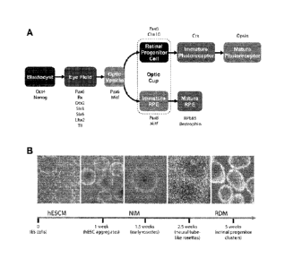
[0033] Fig. 1 shows the stepwise development towards a retinal lineage,
beginning with the establishment of the eye field within the anterior
neuroepithelium.
(A) Each major stage in retinogenesis can be distinguished in part based upon
the
expression of various transcription factors. (B) Schematic of the
differentiation
- 8b -
CA 2771901 2018-10-15
CA 02771901 2012-02-22
WO 2011/028524
PCMJS2010/046488
protocol used to generate cells of a retinal lineage. (C) RT-PCR analysis of
the
changes in gene expression towards an eye field fate through the first 16 days
of
differentiation. (D-F) Immunocytochemistry of typical human ESC aggregates 10
days after differentiation, demonstrating the expression of the eye field
transcription
factors 0tx2 (D) and Lhx2 (E) and the definitive neural transcription factor
Sox1 (F).
Scale bar equals 200 um.
[0034] Fig. 2 shows highly efficient derivation of eye field phenotypes
from
human ESC. (A) RT-PCR analysis showing the onset of Pax6 and Rx gene
expression and concomitant loss of 0ct4. (B-C) qPCR analysis of 0ct4 gene
expression (B) and Pax6 and Rx gene expression (C). Values were expressed as
fold change relative to undifferentiated human ESC. (D) Immunocytochemical
analysis of cells at day 10 showing uniform coexpression of Pax6 and Rx. (E)
FACS
analysis confirming the rapid loss of 0ct4 expression and the onset of both
Pax6 and
Rx protein expression. Negative controls for FACS analyses are indicated by
the
white histograms. (F) Quantification of the FACS analysis. (G-H) qPCR (G) and
Western analysis (H) demonstrating the endogenous expression of the BMP and
Wnt antagonists Noggin and Dkk-1. (I) qPCR showing the near complete loss of
Pax6 and Rx gene expression in cells treated with BMP4 and Wnt3A. Scale bar
equals 40 um.
[0035] Fig. 3 shows repression of neural and eye field fate specification
by
BMP4 and Wnt3A. (A) In the absence of exogenous VVnt and BMP antagonists,
cells in typical human ESC neuroepithelial colonies at day 10 of
differentiation were
tightly packed together and individual cells were nearly indistinguishable.
(B) When
100 ng/ml BMP4 and Wnt3A were added to cultures at the onset of
differentiation,
human ESC adopted altered, non-neuroepithelial morphologies by day 10.
[0036] Fig. 4 shows the dependence of eye field specification upon FGF
signaling. RT-PCR showing complete loss of Rx and Pax6 gene expression at day
of differentiation in the presence of 10 1.11\A SU5402.
[0037] Fig. 5 shows acquisition of optic vesicle and optic cup cell
phenotypes.
(A) Miff protein expression in neurospheres after 30 days of differentiation.
(B)
qPCR analysis of Miff gene expression over the first 80 days of
differentiation. (C-E)
lmmunocytochemical analyses of the time course of Miff and Chx10 protein
expression in neurospheres at 30 (C), 40 (D) or 50 (E) days of
differentiation. (F)
- 9 -
CA 02771901 2012-02-22
WO 2011/028524 PCMJS2010/046488
qPCR analysis of Chx10 gene expression over the first 80 days of
differentiation.
(G) Uniform Chx10 expression throughout a subset of neurospheres by day 40. (H-
I)
Quantification of immunocytochemistry data showing the percentage of Chx10+
spheres (H) and the percentage of Chx10+ cells within those spheres (I) from
day 20
to day 50 of differentiation. (J) FAGS analysis demonstrating the percentage
of all
cells expressing Chx10 at day 40. (K) lmmunocytochemical analysis showed all
Chx10+ cells co-expressed Pax6 at day 40. (L) Rosettes of Chx10-expressing
cells
expressed the tight junction protein ZO-1 within their core. (M) Rare Chx10+
cells
co-expressed pill tubulin at day 40. (N) qPCR demonstrating the increased
expression in Miff and corresponding decrease in Chx10 in cultures treated
with the
FGF inhibitor SU5402. qPCR values were expressed as fold change relative to
cultures at day 16 (B and F) or day 10 (N) of differentiation. Scale bars
equal 500 prn
in panels A & G, 50 m in panels C, D, E, L, & M, and 75 gm in panel K.
[0038] Fig. 6 shows generation of RPE. (A) Photomicrograph of adherent
cultures showing pigmented, hexagonal RPE-like cells. (B) Immunostaining
revealing expression of Miff within RPE-like cells, as well as the tight
junction protein
ZO-1, at day 40. (C) FAGS analysis demonstrating the percentage of all
adherent
cells expressing Miff and Pax6 at day 40 of differentiation. (D) RT-PCR
studies
showing expression of genes associated with an RPE fate. Scale bars equal 100
p.m.
[0039] Fig. 7 shows generation of early photoreceptor phenotypes. (A)
Immunocytochemical detection of cells expressing the photoreceptor-specific
transcription factor Crx at 80 days of differentiation. (B-C) Expression of
the
photoreceptor-specific protein recoverin (B) and the cone photoreceptor-
specific
protein red-green opsin (C) among Crx-expressing cells at day 80. (D) RT-PCR
analysis demonstrated the stepwise acquisition of a cone photoreceptor fate
from an
eye field population. (E) Schematic of the timing of retinal lineage marker
expression
during human ESC differentiation in comparison to normal human retinal
development (Barishak, Y., 2001; Finlay, B.L., 2008). Scale bars equal 50 pi
[0040] Fig. 8 shows quantitative RT-PCR analysis of Pax6(+5a) expression
relative to total Pax6 message in differentiating human ESC-derived
neurosphere
cultures. Values are expressed as fold change relative to cultures at day 4 of
differentiation.
- P10-
CA 02771901 2012-02-22
WO 2011/028524 PCMJS2010/046488
[0041] Fig. 9 shows schematic of the differentiation protocol used to
generate
cells of a retinal lineage from human iPSC.
[0042] Fig. 10 shows stepwise retinal specification from human iPSC. (A)
Various stages of retinal differentiation were observed in IMR90-4 iPSC,
beginning
with Pax6+/Rx+ eye field cells by day 10. (B-D) Mitf+ and Chx10+ cells,
indicative of
the optic vesicle and optic cup stages, are evident by day 40. (E) By day 80,
clusters
were present containing Chx10+ retinal progenitors and Crx+ photoreceptor
precursor cells. (F-H) Many Crx-expressing cells were associated with the
expression of the photoreceptor-specific protein recoverin (F) and the cone-
specific
protein red-green opsin (G-H). (I) RT-PCR analysis demonstrating the stepwise
expression of retina- and photoreceptor-associated genes in differentiating
iPS cell
neurospheres over time. (J-K) RPE cells derived from iPSC acquired a typical
hexagonal morphology and pigmentation (J) and expressed Miff and ZO-1 (K).
Scale bars equal 50
[0043] Fig. 11 shows expression of eye field characteristics in
differentiating
IMR90-4 iPSC. After 10 days of differentiation, iPSC co-expressed Pax6 with
eye
field transcription factors such as Lhx2 (A), and 0tx2 (B). (C) Eye field
colonies
expressed the definitive neural marker Sox1. (D) RT-PCR over the first 16 days
of
differentiation demonstrated the expression of a full complement of eye field
transcription factors, as well as neuroepithelial markers.
[0044] Fig. 12 characterizes FPC neurospheres. Differentiated non-retinal
cells retained an anterior neural phenotype. At 40 days of differentiation,
all
neurospheres expressed the general neural markers Sox1 (A-C) and 8111-tubulin
(D-
F). (G-I) Many 811I-tubulin+ cells possessed a GABAergic phenotype. (J-L) The
forebrain fate of these cells was determined by the widespread expression of
0tx2.
(M) RT-PCR experiments confirmed that these cells expressed both general and
anterior neural markers, but did not express markers of other germ layers,
midbrain
or spinal cord. Insets demonstrate the nuclear specificity of the signal.
[0045] Figure 13 shows the identification of retinal cells in mixed
neurosphere
populations using brighffield microscopy (A-C), immunocytochemistry (D-I), and
RT-
PCR (J). Neurospheres bearing varied morphologies (A) can be separated into
two
distinct populations of phase-bright vesicular shaped neurospheres with a
pseudostratified cellular border (B) and darker, rosette-containing (non-
vesicular)
- 11 -
CA 02771901 2012-02-22
WO 2011/028524 PCMJS2010/046488
neurospheres (C). The definitive retinal progenitor marker Chx10 was
identified in a
subset of mixed neurospheres (D), and was restricted to only the phase-bright
vesicular shaped neurospheres (E) and was absent from the non-vesicular,
rosette-
containing neurospheres (F). Additionally, the expression of Islet-1 was
identified in
a subset of mixed neurospheres (G), was not found in the phase-bright
vesicular
shaped neurospheres (H), but was found to be expressed in the non-vesicular,
rosette-containing neurospheres (I). RT-PCR analysis (J) identified a number
of
transcription factors that were either commonly or differentially expressed
between
these two populations. Figure 13J results for isolated cultures of vesicular
neurospheres are labeled "Retina," and results for isolated cultures of non-
vesicular
neurospheres are labeled "Forebrain."
[0048] Figure 14 shows the results of a comparative microarray expression
analysis of isolated cultures of vesicular and non-vesicular neurospheres.
Numerous
transcription factor genes were identified by microarray analysis to be
differentially
expressed between the two cell populations. Differences are expressed as a
fold
change in expression for vesicular neurospheres relative to non-vesicular
neurospheres.
[0047] Figure 15 shows varied neural/retinal specification from hiPSCs
(human induced pluripotent stem cells) using bright field microscopy (A and
E),
immunocytochemistry (B and F), and fluorescence-activated cell sorting (FAGS)
analysis (G). When subjected to differentiation conditions, hiPSCs commonly
adopted a non-neural, epithelial-like morphology (A) that lacked the
expression of
Pax6 (B). When several lines of hESCs and hiPSCs were compared, IMR90-4 and
OAT hiPSCs were found to express lower amounts of Dkk1 and Noggin as
compared to H9 and H1 hESCs after 2 days of differentiation (C), which was
found
to be correlated with lower levels of Pax6 and Rax after 10 days of
differentiation (D).
When recombinant Dkk1 and Noggin were added to the differentiating cells from
2-4
days of differentiation, a neuroepithelial morphology was identified (E) and
the
expression of Pax6 was restored (F). Treatment of cells with Dkk1 and Noggin
also
increased the expression of Chx10 after 20 days of differentiation, as seen
using
FAGS analysis (G).
[0048] Figure 16 shows the acquisition of mature anterior neural fate from
non-vesicular neurospheres using immunocytochemistry (A-F) and RT-PCR (G).
After a total of 20 days of differentiation, non-vesicular neurospheres began
to
- 12-
CA 02771901 2012-02-22
WO 2011/028524
PCMJS2010/046488
express the neuronal marker pill-tubulin (A-C), the neural transcription
factors Pax6
(A) and Sox1 (B), and the anterior neural marker 0tx2 (C). Following a total
of 70
days of differentiation, nearly all cells possessed neural morphologies and
expressed
features of different neural cell types, including GABAergic neurons (D), TH-
positive
dopaminergic neurons (E), as well as GFAP-positive astrocytes (F). RT-PCR
analysis (G) of these cells at 20 and 70 days of differentiation exhibits
certain
transcription factors whose expression is maintained at both timepoints, as
well as
some transcription factors that are expressed only at either 20 days or 70
days of
differentiation.
[0049] Figure 17 shows the acquisition of mature retinal identities from
vesicular neurospheres using bright field microscopy (A and C),
immunocytochemistry (B, D, F-J), RT-PCR (M), and qPCR (E). Vesicular
neurospheres at 20 days of differentiation (A) nearly uniformly expressed
Chx10,
many of which co-expressed Ki67 (B). After a total of 50 days of
differentiation, this
vesicular morphology tends to be lost (C) but the cells still maintain the
expression of
Chx10 and Ki67 (D). qPCR analysis of these cells during the differentiation
process
from day 20 to 120 of differentiation identifies the onset of expression of
markers for
each of the major retinal cell types in ten day intervals from left to right
(E). Those
cells known to be born early during normal retinal development were identified
as
early-born neurons in this system, as identified by orange to red bars,
whereas late-
born neurons such as PKC-positive bipolar cells and Nrl-positive rod
photoreceptors
were also found to be born later in this system, as indicated by the green to
blue
bars. The identity of the different retinal cell types was confirmed by
immunocytochemistry with antibodies against cell-type specific proteins,
including
retinal ganglion cells (F), amacrine and horizontal cells (G), bipolar cells
(H), cone
precursor cells (I), cone photoreceptor cells (J) including those possessing a
morphology similar to photoreceptors found in vivo (K), as well as rod
photoreceptor
precursors (L). RT-PCR analysis demonstrated the expression of numerous genes
associated with the phototransduction cascade (M).
[0050] Figure 18 shows RPE specification from vesicular neurospheres using
brightfield microscopy (A-D) and qPCR (E-F). At 20 days of differentiation,
retinal
vesicular neurospheres can be enriched (A). After a total of 50 days of
differentiation, pigmentation characteristic of the RPE was rarely observed
(B). After
-13-
CA 02771901 2012-02-22
WO 2011/028524
PCMJS2010/046488
the addition of Activin-A from 20 to 50 days of differentiation, a subset of
neurospheres adopted a pigmented, RPE-like morphology (C). These pigmented
spheres could be plated onto laminin and expanded in the presence of FGF2 and
EGF to form monolayers of RPE (D). When compared to untreated vesicular
spheres (leftmost bar in each bar), Activin-A treated spheres (rightmost bar
in each
pair) expressed higher levels of RPE-associated genes as determined by qPCR
(E),
whereas neural retinal-associated genes were found to be expressed at lower
levels
in Activin-A treated neurospheres (rightmost bar in each pair) (F).
[0051] While the invention is susceptible to various modifications and
alternative forms, specific embodiments thereof have been shown by way of
example in the drawings and are herein described in detail. It should be
understood,
however, that the description herein of specific embodiments is not intended
to limit
the invention to the particular forms disclosed, but on the contrary, the
intention is to
cover all modifications, equivalents, and alternatives falling within the
spirit and
scope of the invention as defined by the appended claims.
DESCRIPTION OF THE INVENTION
[0052] We have developed a novel and simplified protocol to both produce
and isolate retinal progenitor cell (RPC) and forebrain progenitor cell (FPC)
neurospheres from human pluripotent cells. Human ESC (e.g., lines H1 or H9)
and
iPSC (e.g., lines developed from an individual, or in this proof of concept,
reprogrammed iPSC lines derived from somatic cell lines such as IMR90 fetal
fibroblast cells, ATCC CCL-186) following this protocol undergo a targeted,
stepwise
differentiation process that follows a normal developmental timeline and
initially
yields highly enriched populations of eye field cells that eventually separate
into
discrete, morphologically distinct RPC and FPC cell populations that can be
mechanically isolated from one another as highly enriched or substantially
pure
neurosphere cultures. Thereafter, the RPC neurospheres acquire features of
advancing retinal differentiation, including production of RPE neurospheres,
in a
sequence and time course that mimic in vivo human retinal development. The
resulting RPE neurospheres can also be mechanically separated to produce a
culture of enriched or substantially pure RPE.
[0053] Accordingly, the invention encompasses both methods for separating
neuroepithelial lineage cells by progenitor cell type and the substantially
pure
-14-
CA 02771901 2012-02-22
WO 2011/028524
PCMJS2010/046488
cultures of human neuroepithelial lineage cells that can be produced by such
methods.
[0054] It is envisioned that the methods and substantially pure cell
cultures of
the present invention are useful in the following areas:
[0055] 1. Transplantation. Non-limiting examples include the use of FPC in
therapeutic cell rescue therapy, and the use of RPC, RPE, FPC or cells
differentiated
from any of the foregoing in lost cells replacement therapy to help restore
previously
lost vision. The methods and cultures could also be used for developing
tissues for
use in whole tissue replacement therapy.
[0056] 2. Drug screening for agents to protect or enhance the function of
all
cells, including ganglion cells, rods, cones, RPE, forebrain cells, and
midbrain cells.
[0057] 3. Producing retinal disease models from pluripotent cells,
especially
from iPSC, which can also be used to study pathophysio logy and for drug
screening
or customized therapy using stem cells or derivatives thereof.
[0058] 4. As a unique model of human neural development, which would be a
useful resource to study a variety of processes, including without limitation
retinal
development, tissue formation, and synapse formation.
[0059] The protocol used for generating neuroepithelial rosettes from
human
ESC or iPSC is as follows. First, human ESC or iPSC lifted from an irradiated
mouse embryonic fibroblast (MEF) cell layer are grown as aggregates in
suspension
in embryonic stem cell medium (ESCM) without fibroblast growth factor 2 (FGF2)
for
four days. In a non-limiting example, ESCM contains DMEM/F12 (1:1) (Gibco
#11330-065), 20% knockout serum replacement (Gibco #10828-028), 0.1mM 13
mercaptoethanol, 1mM L-glutamine (Gibco #25030-081), 1% MEM nonessential
amino acids (Gibco #11140-050), and 4ng/mL FGF2 (Invitrogen #13256-029). The
pluripotent cells can alternatively be cultured using a defined medium such as
TESR
medium using a matrix such as Matrigel, or under other conditions known to
support
culture of such cells. The aggregates are then cultured in a chemically
defined
neural induction medium (NIM) for two days. The aggregates are then allowed to
attach to the surface of the culture dish, preferably with the addition of
laminin. By
about day 15, columnar cells will have developed which often form
neuroepithelial
structures.
[0060] The invention relates to various methods for producing and
isolating
other retinal lineage cells, including RPC, FPC and RPE, from the
neuroepithelial
-15-
CA 02771901 2012-02-22
WO 2011/028524
PCMJS2010/046488
rosettes. On about day 16 of differentiation, the neuroepithelial rosettes can
be
mechanically detached from adherent cultures with light trituration upon
change of
medium and placed into suspension culture in non-adherent culture dishes. Over
the next 24-48 hours, sphere-like cell aggregates (neurospheres) highly
enriched for
RPC and FPC form. Within three to five days after detachment, using, e.g., a
polished Pasteur pipette, one can mechanically separate and isolate the RPC
neurospheres and FPC neurospheres, based on the observed morphological
differences that appear between the two neurosphere types (See Figures 13 A -
C).
[0061] Specifically, RPC neurospheres appear phase-bright and golden in
color with a ring-like, outer pseudostratified, laminar structure (a
"vesicular" or
"laminar" morphology) under bright field microscopy (Figure 13 B), and FPC
neurospheres appear more uniform in color and density under bright-field
microscopy (a "non-vesicular" or "non-laminar" morphology; Figure 13 C). If
the two
neurosphere populations are not separated in this short culture window
following the
appearance of these morphological differences, they will attach to one another
and
become inseparable.
[0062] The RPC and FPC neurospheres are maintained separately in flasks
containing a Retinal Differentiation Medium (RDM). In a non-limiting example,
RDM
includes DMEM/F12 (3:1) supplemented with 2% B27. By mechanically separating
the neurospheres according to these morphological differences, one can obtain
a
multipotent RPC neurosphere culture having greater than 90% purity as assessed
by
immunocytochemical analysis (e.g., Chx10), meaning that greater than 90% of
the
cells in the neurosphere culture are RPCs. In this disclosure, the term
"substantially
pure" culture refers a culture wherein at least 90% of the cells are of a
given cell
type. Other cells remaining in the culture are more primitive optic vesicle
cells
(expressing Mitf) or eye field cells (expressing Pax6, Rx and Lhx2). The FPC
neurospheres in the other flask express neural and forebrain markers such as
0tx2,
Pax6, Sox1 and Sox2.
[0063] The RPC neurospheres begin to lose their unique morphological
structure after 1 week in RDM and produce non-pigmented, Chxl 0+/Pax6+/Mitf-
neural retinal neurosphere populations. These neurospheres in turn yield
Crx+/recoverin+/opsin+ and Nr1+ photoreceptors and 8111 tubulin+/Brn3/HuC-D+
- 16-
CA 02771901 2017-02-22
64181-371
retinal ganglion cells in a sequence and time frame that mimics normal human
retinogenesis.
[0064] In addition to the non-pigmented neurosphere populations, the
RPC
neurospheres produce pigmented, Mitf+/Chx10- RPE neurospheres, the latter of
which
are identifiable in culture by about day 30-50 of overall differentiation. As
they are
brown-black in color, they can then be mechanically removed from the remaining
neural
RPC neurospheres and transferred to a separate flask and maintained in RPE
Propagation Medium (DMEM/F12 (3:1) with 2% B27 and 20 ng/ml each of FGF2 and
EGF along with 5 pg/ml of heparin.
[0065] To expand the RPE neurosphere populations, the pigmented (RPE)
neurospheres are plated onto laminin-coated culture dishes and allowed to
adhere in RPE
Propagation Medium. Within 24 hours, RPE cells begin to proliferate and expand
outward
from the plated RPE neurosphere. After 1 week, the RPE neurospheres are gently
triturated to remove them from the flask and transferred to another laminin-
coated flask to
repeat this process (up to 3 times). The remaining skirt of RPE is dissociated
enzymatically
and replated at a density of 100,000 cells/cm2 and passaged up to 3 times as
described for
human fetal RPE (Gamm et al, IOVS 49:788 2008). To promote maturation of RPE,
FGF2,
EGF and heparin are removed from the RPE Propagation Medium for 1-3 weeks. RPE
propagated and differentiated in this manner express numerous RPE markers,
including
Bestrophin, RPE65, CRALBP, ezrin, Mitf, and ZO-1 among others.
[0066] The present invention cultures human pluripotent ESC or iPSC
under
differentiating conditions to yield major neural retinal cell types and RPE,
including
populations of RPC and FPC, in convenient sphere forms (neurospheres) that can
be
easily and inexpensively maintained in culture. The spheres allow
recapitulation of more
complex 3-D structure of the retina.
[0067] The following Examples are offered for illustrative purposes
only, and are
not intended to limit the scope of the present invention in any way. Indeed,
various
modifications of the invention in addition to those shown and described herein
will
become apparent to those skilled in the art from the foregoing description and
the
following examples and fall within the scope of the appended claims.
- 17-
CA 02771901 2012-02-22
WO 2011/028524
PCMJS2010/046488
EXAMPLES
Example 1: Modeling early retinal development with human ESC and iPSC
[0068] We demonstrate below that cell fate specification and maturation
from
human ESC of definitive retinal cell populations follows a sequence and time
course
highly reminiscent of normal retinal development. We also demonstrate that
retinal
differentiation can be selectively altered by manipulating endogenous
developmental
signaling pathways. Additionally, we show that an identical cohort of
developing
retinal cell types can be generated from human iPSC, although variation can
occur
between lines. Cell populations expressing morphologic features and/or markers
of
the eye field, RPE, neural retinal progenitors, photoreceptor precursors and
photoreceptors were observed in cultures of cells differentiated from human
iPSC at
time points predicted by results using human ESC.
[0069] The findings presented here demonstrate that human ES cells and
human iPSC can differentiate in vitro into cells having signature features
associated
with early eye and retinal development, while following an expected timeline
for
human retinal development (Barishak, Y., 2001; Finlay, B.L., 2008), thereby
meeting
the criteria (Keller, G., 2005; Pera, M.F., et al., 2004) for a comprehensive
in vitro
model system for investigating mechanisms of human retinogenesis involved in
retinal specification and differentiation of individual retinal cell types.
Furthermore,
the highly enriched neurosphere populations described herein can be
selectively
cultured and isolated from one another and from other cell populations for
further
differentiation, isolation and use.
[0070] Results
[0071] Maintenance of human ESC
[0072] Human ESC (H9 line) were expanded on a feeder layer of irradiated
MEFs in ESCM containing DMEM/F12 (1:1) (Gibco #11330-065), 20% knockout
serum replacement (Gibco #10828-028), 0.1mM B-mercaptoethanol, 1mM L-
glutamine (Gibco #25030-081), 1% MEM nonessential amino acids (Gibco #11140-
050), and 4ng/mL FGF2 (Invitrogen #13256-029). Cells were passaged every 5-6
days, and morphologically identifiable differentiated cells were mechanically
removed at each passage.
[0073] Differentiation of human ESC
[0074] Human ESC were differentiated as follows:
- 18-
CA 02771901 2012-02-22
WO 2011/028524 PCMJS2010/046488
[0075] (1) Making human ESC aggregates: Human ESC were grown on
irradiated MEFs to approximately 80% confluence in a 6-well plate. Upon
reaching
80% confluence, the human ESC medium was aspirated off and 1 ml of dispase (1
mg/ml, Gibco #17105-041) solution was added to each well. After 5 minutes, the
cells were examined for curling of human ESC colony edges, indicative of the
colonies beginning to lift off of the plate. If noticeable curling was
evident, the
dispase solution was aspirated off of the plate and replaced with 1 ml of
DMEM/F12
per well. The human ESC colonies were mechanically lifted from the plate by
pipetting a few times with a 10 ml pipette. When all colonies were detached,
the
cells were transferred to a 15 ml conical tube and allowed to settle by
gravity. After
the aggregates had settled (-5 minutes), the supernatant was aspirated and
replaced with 10 ml of Embryoid Body (EB) medium, which contained DMEM/F12
(1:1) (Gibco #11330-065), 20% knockout serum replacement (Gibco #10828-028),
0.1mM P-mercaptoethanol, 1mM L-glutamine (Gibco #25030-081) and 1% MEM
nonessential amino acids (Gibco #11140-050). The pellet of cells was
resuspended
by repeated pipetting 2-3 times with a 10 ml pipette, just enough to separate
the
individual aggregates while minimizing their dissociation. Aggregates were
approximately 400 mm in diameter. These aggregates were then transferred to a
T25 flask and placed in the incubator. The next day, the aggregates formed
floating,
sphere-like structures. (If significant attachment of human ESC and/or
residual MEF
cells was observed, the unattached aggregates were transferred to a new
flask.)
These cells were fed with fresh EB medium every day for the first 4 days.
[0076] (2) Differentiating to anterior neuroepithelial fate: The aggregates
were
then collected by settling in a 15 ml conical tube and washed once with 10 ml
of
DMEM/F12. Upon resettlement of the aggregates, the supernatant was removed
and the aggregates were re-suspended in 10 ml of Neural Induction Medium (NIM)
and transferred to a new T25 flask. NIM included DMEM/F12 (1:1) (Gibco #11330-
065), 1% N2 supplement (Gibco # 17502-048), 1% MEM nonessential amino acids
(Gibco #11140-050), and 2 pg/ml heparin (Sigma #H-3149). Two days later, a 6-
well
plate was coated with laminin (20 g/ml diluted in DMEM) and the coated plate
was
left in the incubator overnight. By this time, aggregates had become brighter
and
acquired clearly defined edges. The next day, approximately 50 aggregates were
plated in each well of a 6 well plate. An additional 2 ml of fresh NIM was
added to
- 19-
CA 02771901 2012-02-22
WO 2011/028524
PCT/US2010/046488
each well. The cells were then placed in the incubator, making sure the cells
were
distributed evenly in the plate by gently shaking the plate back and forth as
well as
side-to-side a few times. Within the first couple of days (by a total of 9
days), most
aggregates attached to the laminin-coated surface. These aggregates then
flattened
somewhat with cells arranged in a monolayer fashion towards the periphery, yet
retaining a more 3-dimensional appearance in the center of the aggregate.
These
cultures were routinely fed with fresh NIM every 2 days. Within a few days,
columnar cells developed and formed neural tube-like structures. After a total
of 15
days of differentiation (8 days following attachment), it was possible to
identify
columnar rosette structures within many of these aggregates. A small
population of
cells that did not display the columnar rosette structures were found on the
periphery
of these colonies.
[0077] (3) Generating retinal progenitor cell (RPC) neurospheres and
forebrain progenitor cell (FPC) neurospheres: To allow for retinal
differentiation, the
central regions of these colonies that possess columnar rosette structures
were
dislodged from the culture plate using a 1000 I pipette tip on day 16 of the
differentiation, by drawing up some of the medium within the well and
squirting the
medium directly onto the cell colonies. The columnar rosette cells found in
the
center of these colonies were denser than those cells in the periphery, so
they were
easily dislodged by this pipetting technique, leaving the non-rosette cells
attached to
the plate. We took care not to break up the lifted colonies during this step.
Detached colonies were collected in a 15 ml conical tube and spun in a
centrifuge at
600 rpm for 1 minute to effectively pellet the detached colonies while leaving
most
single cells in suspension in the culture medium. The supernatant was then
aspirated from the tube and the clusters were re-suspended in 10 ml of Retinal
Differentiation Medium (RDM) and transferred to a new T25 flask. RDM included
DMEM/F12 (3:1) supplemented with 2% B27. Over the next 24-48 hours, these
detached colonies rolled up to form sphere-like clusters of cells
(neurospheres),
while some of the remaining non-neural cells attached to the flask.
Neurospheres
were fed every 2-3 days with fresh RDM. Within 3-5 days after detachment of
the
cells (19th-21st day of the differentiation), neurospheres readily adopted one
of two
major appearances. Some neurospheres appeared uniform in color and density
under bright-field microscopy, typically without defining structural
characteristics, but
- 20 -
CA 02771901 2012-02-22
WO 2011/028524
PCMJS2010/046488
occasionally with a neuroepithelial rosette within them ("non-vesicular" or
"non-
laminar" morphology). Other neurospheres were distinctly phase-bright,
appeared
golden in color, with a ring-like, laminar structure on the outside of the
neurosphere
("vesicular" or "laminar" morphology). The outer ring of cells appeared to
have cells
oriented radially outwards from the center. The phase-bright, golden, ring-
like
neurospheres are comprised of definitive early retinal progenitor cells (RPC).
The
uniform appearing, non-ring-like neurospheres are comprised of forebrain
progenitor
cells (FPC).
[0078] (4) Isolation of human ESC-derived Retinal Progenitor Cell
Neurospheres (RPC neurospheres) and Forebrain Progenitor Neurospheres (FPC
neurospheres): To isolate RPC neurospheres from FPC neurospheres, neurosphere
populations were separated based on morphological characteristics (see above)
at
approximately day 20 of differentiation (5 days following detachment), a stage
at
which there were a minimal number of these clusters sticking to one another
and
maximal morphological differences. At later time points, the cell aggregates
begin to
lose their distinguishing characteristics and attach firmly to one another. To
collect
the neurospheres, the contents of the flask were transferred to a 15 ml
conical tube
and allowed to settle by gravity (-2-3 minutes). The medium was aspirated and
the
neurospheres were re-suspended in 5 ml of fresh RDM, and the mixture was then
transferred to a 60 mm Petri dish. The Petri dish was placed on a microscope
stage
and swirled several times to collect the neurospheres within the center of the
dish. A
P20 pipette was used to gently collect the phase-bright, golden laminar RPC
neurospheres into a 15 ml conical tube with 10 ml of RDM inside. This process
was
repeated to harvest the RPC neurospheres. After the RPC neurospheres were
removed, FPC neurospheres were collected according to their morphological
characteristics into a separate 15 ml conical tube containing 10m1 RDM. After
the
FPC neurospheres were isolated, the remaining cells, mixed clusters that were
stuck
together or those too small to definitively identify, were discarded.
[0079] (5) Generating retinal pigment epithelium (RPE): RPE were generated
in two ways. In one way, the RPC neurospheres isolated above were cultured in
RDM to allow for further maturation. Within an additional 2 weeks of culture
of the
RPC neurosphere population, a subset of these neurospheres began to develop
pigmentation. Within an additional month of culture (2 months total), all
cells
observed in a subset of neurospheres were pigmented. The pigmentation
identified
-21-
CA 02771901 2012-02-22
WO 2011/028524 PCMJS2010/046488
those RPC neurospheres that had adopted a RPE fate (Le., RPE spheres that
express multiple markers of mature RPE such as bestrophin, RPE65, CRALBP,
ezrin, Mitf, ZO-1). Additionally, neuroepithelial rosettes (described above)
kept
attached to the culture surface in RDM after day 15 differentiated over time
to RPE.
These cells were expanded, as described in Gamm, Ophthalmol Vis Sc! 2008.
Additionally, neuroepithelial rosettes kept attached to the culture surface in
RDM
after day 15 differentiated over time to RPE.
[0080] (6) Further differentiation of FPC: FPC neurospheres differentiated
to
more mature forebrain neuronal populations within an additional week and
expressed such markers as GABA, HuC/D, 811Itubulin and Sox1 as well as 0tx2,
Sox2 and other forebrain and neural markers.
[0081] Eye field specification from human embryonic stem cells
[0082] As described above in section (1) and (2) of "Differentiation of
human
ESC" and as shown in Fig. 1B, human ESC were differentiated as free-floating
human ESC aggregates (EBs) and prompted to adhere to the culture dish to
permit
neuroepithelial rosette formation. After 16 days of differentiation, rosette-
containing
colonies were mechanically removed and allowed to grow as neurospheres (see
section (3), "generating retinal progenitor cell neurospheres and forebrain
progenitor
cell neurospheres").
[0083] RT-PCR experiments were done as follows to check gene expression:
Total RNA was isolated from cell cultures from various stages of
differentiation using
the RNAeasy0 kit (Qiagen) and treated with DNAse I. Reverse transcription was
performed with the Superscript Ill RT-PCR kit (Invitrogen). PCR was performed
with
GoTaq PCR master mix (Promega) and subsequent PCR products were run on 2%
agarose gels. For quantitative RT-PCR (qPCR), reactions were performed with
Sybr
Green Supermix (Applied Biosystems) and the Opticon 2 DNA Engine and Opticon
Monitor 2.02 software (MJ Research). Primer sets used are listed in Table 1.
All
primer sets listed were run for 30 cycles at an annealing temperature of 60 C
unless
otherwise noted. Quantitative RT-PCR primers were run for 40 cycles.
Table I. Primers used for RT-PCR.
Gene amplified Forward Reverse Size (bp)
-22 -
CA 02771901 2012-02-22
WO 2011/028524
PCT/1TS2010/046488
AGA ACC TGT CAC GAC AGC AAG CTG
a-fetoprotein MG CTG TG AGG ATG TC 676
(SEQ ID NO:1) (SEQ ID NO:2)
GCG AGA AGA TGA CCA GTG GTA CGG
fl¨Actin (qPCR) CCC AGA IC CCA GAG G 103
(SEQ ID NO:3) (SEQ ID NO:4)
ATT TAT AGG CTG TGT TCT GCC GGA
Bestrophin GCC CTC ACG GAA GTC ATA AAG
CCT 359
(SEQ ID NO:5) (SEQ ID NO:6)
ACC CAG TTC ATA CM TTG TCA TGG
Brachyury GCG GTG AC GAT TGC AG 392
(SEQ ID NO:7) (SEQ ID NO:8)
ATT CAA CGA AGC ATC CU GGC TGA
Chxl 0 CCA CTA CCC AGA CTT GAG GAT
GGA 229
(SEQ ID NO:9) (SEQ ID NO:10)
GGC GAC ACA GGA TTC CGG CAG CTC
Chx10 (qPCR) CAA TCT TTA CGT TTT C 122
(SEQ ID NO:11) (SEQ ID NO:12)
TAT TCT GTC MC TGC ATT TAG CCC
Crx GCC TTG GCC CTA TCC GGT TCT
TGA 253
(SEQ ID NO:13) (SEQ ID NO:14)
AGC ACC TTG GAT ACA CM TCC TGA
Dkk-1 (qPCR) GGG TAT TCC AGA GGC ACA GTC
TGA 114
(SEQ ID NO:15) (SEQ ID NO:16)
CCC TGG TTT CTC GCA GTC TGT GGG
En-1 TGG GAC TT GTC GTA TT 162
(SEQ ID NO:17) (SEQ ID NO:18)
GAPDH (23 ACC ACA GTC CAT TCC ACC
ACC CTG
GCC AT CAC TTG CTG TA 450
cycles) (SEQ ID NO:19) (SEQ ID NO:20)
GCA AAG AGC CCG CGT GTC AGG TAG
HoxB4 (55 C) TCG TCT AC CGG TTG TA 160
(SEQ ID NO:21) (SEQ ID NO:22)
CM GAT CTC GGA CCG TGG TCA GCA
Lhx2 CCG CTA CT TCT TGT TA 284
(SEQ ID NO:23) (SEQ ID NO:24)
TTC ACG AGC GTC TTG CM AGC AGG
Miff CTG TAT GCA GAT ATC CAT CAA
GCC 106
(SEQ ID NO:25) (SEQ ID NO:26)
CM AGG CM ACA TCT GCT GGA GGC
Nanog ACC CACTI TGA GGT AT 158
(SEQ ID NO:27) (SEQ ID NO:28)
CCA TCA TTT CCG MG CTA GGT CTC
Noggin (qPCR) AGT GCA AGT GCT TGT AGC CCA
GM 189
(SEQ ID NO:29) (SEQ ID NO:30)
CGA GCA ATT TGC TTC GGG CAC TGC
0ct4 CM OCT CCT GM AGG AAC MA TTC
324
(SEQ ID NO:31) (SEQ ID NO:32)
GTG GAG GM GCT ATT CTC CAG GTT
0ct4 (qPCR) GAC MC M GCC TCT CA 120
(SEQ ID NO:33) (SEQ ID NO:34)
TAC CTG GAC CAT TM GTC CAG CCC
Opsin TGG TAT TGG CGT ATG GU ACG
arT 379
(SEQ ID NO:35) (SEQ ID NO:36)
CM CAG CAG MT CTG GGT GGA MG
0tx2 GGA GGT CA AGA GAA GC TG 429
(SEQ ID NO:37) (SEQ ID NO:38)
- 23 -
CA 02771901 2012-02-22
WO 2011/028524 PCMTS2010/046488
CGG AGT GM TCA CCG CU ATA CTG 300 (+5a)
Pax6 GCT CGG TG GGC TAT TTT GC
(SEQ ID NO:39) (SEQ ID NO:40) 258 (-5a)
AGT GM TCA GCT TGC AGA ATT CGG
Pax6 (qPCR) CGG TGG TGT CU GAA ATG TCG
CAC 120
(SEQ ID NO:41) (SEQ ID NO:42)
Pax6"-5& CTC GGT GGT GTC ACT TTT GCA
TCT
TTT GTC AAC GCA TGG GTC 130
(qPCR) (SEQ ID NO:43) (SEQ ID NO:44)
GCC CTC CTG CAC AGT TGG TCT CTG
RPE65 AAG TTT GAC TTT TGC AAG CGT
AGT 259
(SEQ ID NO:45) (SEQ ID NO:46)
GM TCT CGA MT CU CAC TM TTT
Rx CTC AGC CC GCT CAG GAC 279
(SEQ ID NO:47) (SEQ ID NO:48)
AGC GAA ACT GTC TCA TGC AGC TGG
Rx (qPCR) AGA GGA GGA ACA TAC GTG GTG
AAA 81
(SEQ ID NO:49) (SEQ ID NO:50)
CGA GCA GAA GAC CGG CCT TGG CTA
Six3 (55 C) GCA TTG CU CM TCA TAC ATC ACA
394
(SEQ ID NO:51) (SEQ ID NO:52)
AU TGG GAC GGC ATC CTG GAT GGG
Six6 GM CAG AAG ACA CM CTC AGA TOT
385
(SEQ ID NO:53) (SEQ ID NO:54)
CM TGC GGG GAG CTC TGG ACC MA
SOxi GAG AAG TC CTG TGG CG 464
(SEQ ID NO:55) (SEQ ID NO:56)
CCC CCG GCG GCA TCG GCG CCG GGG
Sox2 (55 C) ATA GCA AGA TAC AT 448
(SEQ ID NO:57) (SEQ ID NO:58)
ATG GCA MT TCT GCG CTG AU TCC
TII GTG GCG CTG AAG CAA GTG CAT
TCT 352
(SEQ ID NO:59) (SEQ ID NO:60)
[0084] During this process, human ESC rapidly lost expression of the
pluripotency genes 0ct4 and Nanog and acquired expression of transcription
factors
associated with eye field specification (Rx, Six3, Six6, Lhx2, TII and 0tx2)
as well as
neural induction (Pax6, Sox1 and Sox2) (Fig. 1C). In RT-PCR experiments, Pax6
was present as a doublet band at 6 days of differentiation, reflecting the
expression
of both the Pax6(-5a) and Pax6(+5a) isoforms, also known as Pax6a and Pax6b,
respectively. The appropriate staging and lineage of this early cell
population was
further confirmed by the absence of expression of the retinal progenitor
transcription
factor Chx10 (also known as Vsx2), the photoreceptor precursor-specific
transcription factor Crx and the spinal cord-associated transcription factor
Hox64.
[0085] I
mmunocytochemistry was done as follows to check protein
expression: human ESC aggregates or neurospheres were plated onto poly-
ornithine- and laminin-coated coverslips overnight to allow for attachment,
and then
-24-
CA 02771901 2012-02-22
WO 2011/028524 PCMJS2010/046488
fixed with 4% paraformaldehyde. Cells were then permeabilized in 0.2% Triton X-
100 for 10 minutes. For Mitf immunocytochemistry, cells were incubated in ice-
cold
90% methanol for 10 minutes as an additional permeabilization step.
lmmunostaining was performed in 0.1% Triton X-100 and 5% donkey serum using
the antibodies listed in Table 2. Labeled cells were visualized with either
Alexafluor
488- or Cy3-conjugated secondary antibodies, and nuclei were counterstained
with
either Hoechst or To-Pro-3 nuclear dyes. Images were obtained from a Nikon
TE600 fluorescent microscope equipped with a SPOT camera and software or from
z-stacks of cell clusters obtained on a Nikon Cl Laser Scanning Confocal
microscope.
[0086] lmmunocytochemistry showed that nearly all cells within these
colonies
expressed the anterior neural/eye field transcription factors Otx2 (Fig. 1D)
and Lhx2
(Fig. 1E), as well as the definitive neuroepithelial marker Sox1 (Fig. 1F) by
day 10 of
differentiation.
[0087] We demonstrate below that cell fate specification and maturation
from
human ESC of definitive retinal cell populations follows a sequence and time
course
highly reminiscent of normal retinal development.
Table 2. Primary antibodies used for immunocytochemistry and Western analysis.
Antibody Type Source Dilution
811Itubulin Rabbit polyclonal Covance 1:100
Chx10 Goat polyclonal Santa Cruz 1:200
Mouse
Crx monoclonal Abnova 1:100
Dkk1 Mouse Upstate Biotechnology 1:500
-25-
CA 02771901 2012-02-22
WO 2011/028524 PCMJS2010/046488
monoclonal
Lhx2 Goat polyclonal Santa Cruz 1:200
Mouse
Mitf monoclonal Neomarkers 1:50
Mouse
Noggin Monoclonal Chemicon 1:2000
0ct4 Mouse polyclonal Santa Cruz 1:1000
Opsin, red/green Rabbit polyclonal Chemicon 1:5000
0tx2 Goat polyclonal R & D Systems 1:2000
Mouse Developmental Studies
Pax6 monoclonal Hybridoma Bank 1:50
Recoverin Rabbit polyclonal Chemicon 1:2000
Rx Rabbit polyclonal Abcam 1:1000
Sox1 Goat polyclonal R & D Systems 1:1000
ZO-1 Rabbit polyclonal Zymed 1:100
[0088] The gene and protein expression of Pax6 and Rx was examined in
further detail, because eye field cells are often characterized by the
coexpression of
these two transcription factors (Osakada, F., et al., 2008; Mathers, P.H.,
etal.,
2000). RT-PCR and quantitative PCR (qPCR) analysis were done as described
above and revealed the onset of expression of both Pax6 and Rx within the
first few
days of differentiation (Fig. 2A-C), which was closely correlated with loss of
0ct4
expression. At the protein level, nearly all H9-derived cells co-expressed
both Pax6
and Rx within 10 days of differentiation as determined by immunocytochemistry
(Fig.
2D).
[0089] FACS analysis was done as follows to quantify the percentage of
cells
acquiring expression of Pax6 and Rx: Cells were dissociated with either
trypsin or
Accutase (Chemicon), washed with a fluorescence-activated cell sorting (FAGS)
buffer (PBS, 0.1% sodium azide, and 2% donkey serum), and fixed with 0.1%
paraformaldehyde for 10 minutes. Cells were then permeabilized with ice-cold
90%
- 26 -
CA 02771901 2012-02-22
WO 2011/028524 PCMJS2010/046488
methanol for 20 minutes and incubated overnight in primary antibodies at a
concentration of 1 pg of antibody per 1 million cells in FACS buffer. Antibody
information is found in Table 2. Immunostaining was then completed with either
donkey-anti-mouse or donkey-anti-rabbit Alexa 488 secondary antibodies for 2
hours, after which cells were washed with FAGS buffer, and then sorted with a
Becton Dickinson FACSCaliber. Data retrieved from the sorting was analyzed
with
CellQuest Pro software (Becton Dickinson).
[0090] Cell populations were analyzed by FAGS over the first 16 days of
differentiation (Fig. 2E-F). The onset of Pax6 and Rx expression was detected
by
day 6, when approximately 25% of all cells expressed these factors. Expression
of
Pax6 and Rx surpassed 90% of cells by day 10 of differentiation and increased
to
greater than 95% by day 16. Conversely, protein expression of 0ct4 decreased
to
an undetectable level by 10 days of differentiation.
[0091] The generation of a high percentage of cells with eye field
characteristics in the absence of exogenous Wnt and BMP antagonists prompted
further investigation into the endogenous expression of Dkk-1 and Noggin in
differentiating human ESC cultures. Both genes were upregulated during eye
field
specification (Fig. 2G) as determined by qPCR.
[0092] Furthermore, Western analysis was done as follows to determine
expression of Dkk-1 and Noggin; 20 lig protein samples obtained from cell
lysates
were separated on 4% to 20% gradient Tr's-CI gels (Bio-Rad), electroblotted
onto
PVDF membranes and stained with Ponceau red to confirm transfer. Membranes
were blocked with 5% nonfat dry milk and 2.5% BSA in TBST for 1 hour at room
temperature followed by consecutive 1 hour incubations at room temperature
with
primary antibody in TBST+1.5% BSA and HRP-conjugated secondary antibody in
TBST+1')/0 nonfat dry milk. Primary antibodies used for Western blot analysis
were
directed against Noggin and Dkk-1 (see Table 2). Protein bands were visualized
by
chemiluminescence (ECL Plus Western Blot Analysis Detection Kit; GE
Healthcare,
Chalfont St. Giles, UK).
[0093] Western analysis detected protein expression of Dkk-1 and Noggin at
day 10 of differentiation (Fig. 2H). Addition of Wnt3A and BMP4 to cultures
over the
first 10 days of differentiation abolished both the expression of Pax6 and Rx
(Fig. 21)
and the appearance of neuroepithelial colonies (Fig. 3).
-27 -
CA 02771901 2012-02-22
WO 2011/028524 PCMJS2010/046488
[0094] Endogenous FGF signaling was also involved in the acquisition of
early
eye field features, since the addition of the specific FGF receptor inhibitor
SU5402
led to a complete loss of both Pax6 and Rx expression at 10 days of
differentiation
(Fig. 4, tested by RT-PCR).
[0095] Acquisition of optic vesicle and optic cup cell phenotypes
[0096] When neuroepithelial rosettes corresponding to the eye field stage
of
retinal development were lifted and grown to neurospheres, nearly uniform
expression of Mitf protein was observed within 14.7 2.1% of all spheres by
day 30
of differentiation as shown by immunocytochemistry (Fig. 5A). qPCR analysis
further
demonstrated that gene expression of Miff increased from day 16 to day 37 of
differentiation (Fig. 5B).
[0097] Next, the relationship between Miff and Chx10 protein expression was
examined in differentiating neurosphere cultures over time using
immunocytochemistry. Chx10 expression was only occasionally observed at day 30
(Fig. 5C). Coexpression of Miff and Chx10 was prevalent by day 40 (Fig. 5D),
followed by mutually exclusive expression of Chx10 and Miff by day 50 as Mitf
expression diminished within Chx10+ neurospheres (Fig. 5E). qPCR analysis
confirmed that Chx10 gene expression was delayed relative to Miff (Fig. 5F).
Similar
to Miff, Chx10 protein was eventually detected by immunocytochemistry in
nearly all
cells of the subset of neurospheres in which it was expressed (Fig. 5G).
Quantification of Chx10 protein expression demonstrated that 18.0 3.3% of
all
neurospheres contained Chx10+ cells by day 40 of differentiation (Fig. 5H),
and
within these Chx10-expressing neurospheres, greater than 90% of cells
expressed
Chx10 by day 50 (Fig. 51). By FACS, 26% of the entire cell culture population
expressed Chx10 at day 40 (Fig. 5J). The remaining Chx10-negative neurospheres
that were derived from the early eye field cell population maintained an
anterior
neural identity.
[0098] Chx10 expression was associated with a neural cell type that had not
yet acquired a mature neuronal phenotype. Among the neurospheres that
expressed Chx10, greater than 99% of cells maintained expression of Pax6,
which is
a requirement of early RPC (Belecky-Adams, T., etal., 1997; Toy, J., etal.,
2002)
(Fig. 5K). Furthermore, many of the Chx10+ clusters within neurospheres were
arranged in rosettes with cells oriented radially away from a core that was
positive
for the tight junction protein ZO-1 (Fig. 5L), another feature associated with
- 28 -
CA 02771901 2012-02-22
WO 2011/028524
PCMJS2010/046488
progenitor cell populations (Elkabetz, Y., etal., 2008). When the expression
of 13111
tubulin, a marker for early post-mitotic neurons, was examined, we found that
the
Chx10+ rarely co-expressed 0111tubulin, although some clusters that contained
Chx10+ cells included a small number of 13111tubulin-positive neurons (Fig.
5M).
[0099] Because
FGF signaling is suggested to be involved in the specification
of the neural retina (Muller, F., et aL, 2007), we next examined the effect of
SU5402,
a potent and specific inhibitor of the FGFR1 receptor, on Miff and Chx10 gene
expression. The addition of SU5402 to human ESC cultures during the optic
vesicle
stage and optic cup stage of differentiation (days 16-40) resulted in an 11.8-
fold
increase in Miff gene expression at day 40, as measured by qPCR (Fig 5N). By
contrast, Chx10 expression was reduced 15.9-fold as a result of this
treatment.
[00100] The FPC neurospheres were also characterized, as is shown in
Fig. 12. Differentiated non-retinal cells retained an anterior neural
phenotype. At 40
days of differentiation, all neurospheres expressed the general neural markers
Sox1
(A-C) and 3111-tubulin (D-F). (G-I) Many bIll-tubulin+ cells possessed a
GABAergic
phenotype. (J-L) The forebrain fate of these cells was determined by the
widespread
expression of 0tx2. (M) RT-PCR experiments confirmed that these cells
expressed
both general and anterior neural markers, but did not express markers of other
germ
layers, midbrain or spinal cord. Insets demonstrate the nuclear specificity of
the
signal.
[00101] Retinal pigment epithelium (RPE) specification
[00102] When neuroepithelial rosettes corresponding to the eye field
stage of retinal development were maintained as an adherent culture in RDM as
described above, distinct patches of polygonal, pigmented cells were initially
observed at approximately day 30 of human ESC differentiation (Fig. 6A). These
cells maintained expression of the transcription factor Miff while also
expressing the
RPE-associated tight junction protein ZO-1 as shown by immunocytochemistry
(Fig.
68). At day 40 of differentiation, FACS analysis revealed that 25% of all
adherent
cells expressed Miff, and 77% of all cells expressed Pax6 (Fig. 6C). RT-PCR
analysis demonstrated maintained expression of Pax6 in this cell population
over
time, as well as the acquisition of more mature RPE-associated markers such as
RPE65 and bestrophin (Fig. 6D).
- 29 -
CA 02771901 2012-02-22
WO 2011/028524 PCMJS2010/046488
[00103] Retinal cell types generated from human ESC-derived
retinal progenitors
[00104] After prolonged maintenance of the human ESC-derived retinal
progenitor neurospheres, the neurospheres matured toward a photoreceptor
phenotype. By a total of 3 months of differentiation, the unpigmented, neural
retinal
RPC neurospheres produced multiple retinal cell types, predominantly
photoreceptor
precursors (e.g., Crx+) or photoreceptor-like cells (e.g., opsin+ and
recoverin+) as
well as retinal ganglion-like cells expressing HuC/D and possessing long
pIlltubulin+
processes.
[00105] Early photoreceptor phenotypes generated from human
ESC-derived retinal progenitors
[00106] As shown by immunocytochemistry, by day 80 of differentiation,
19.4 3.1% of all neurospheres contained Crx+ photoreceptor precursors (Fig.
7A).
Within these colonies, 63.0 7.6% of all cells expressed Crx. Furthermore,
the
majority of Crx+ cells expressed the photoreceptor-specific protein recoverin
(Fig.
7B) and the cone photoreceptor-specific protein opsin (Fig. 7C).
[00107] To analyze the time course and sequential acquisition of
neuroretinal- and photoreceptor-associated gene expression, RT-PCR analysis
was
performed as described above (Fig. 7D). Throughout the differentiation process
from day 16 through day 80, Pax6 gene expression was detected. Rx gene
expression was also present early in differentiation, followed by the
consecutive
expression of Chx10, Crx and opsin. Overall, the timing of expression of the
gene
and protein markers used in this study coincided with that of normal human
retinal
development (Barishak, Y., 2001; Finlay, B.L., 2008) (Fig. 7E).
[00108] Further evidence of the progression toward a more mature
retinal phenotype in these cultures was provided by monitoring Pax6 isoform
expression over time by RT-PCR. RT-PCR results from the present study
suggested
that the Pax6(+5a) isoform became more prevalent during human ESC
differentiation
(Figs. 2A and 7D). To verify this observation, qPCR of the Pax6(+5a) isoform
relative
to total Pax6 expression was performed as described above(Fig. 8). This
analysis
confirmed the onset of Pax6(+5a) expression between days 4 and 16 of
differentiation and demonstrated a relative increase in the expression of this
isoform
- 30 -
CA 02771901 2012-02-22
WO 2011/028524 PCMJS2010/046488
between days 60 and 70, which corresponded to the appearance of photoreceptor-
like cells in culture.
[00109] Differentiation of retinal cell types from human iPSC
[00110] To determine the potential for stepwise derivation of retinal cell
types
from human iPSC, we applied the human ESC differentiation protocol described
above to four different human iPS cell lines. Consistent with a previous
report (Yu,
J., et al., 2007), considerable variation was found in the ability of these
lines to
produce Pax6+ neuroectodermal cells at day 10 of differentiation, with
efficiencies
ranging from 5% to 56% of the total cell population.
[00111] Based on these results, we chose to study the IMR90-4 cell line,
one of
the highest Pax6-expressing lines (Yu et al., 2007), in greater detail, since
Pax6 is
necessary for retinal development, although we have obtained, with varying
efficiency, RPC and FPC neurospheres from all iPSC lines examined, including
lines
produced without viral vectors. The cells chosen for extensive study were
derived by
Yu et al. by reprogramming fetal foreskin IMR-90 (ATCC CCL-186) cells. Upon
differentiation, the appearances of the iPS cell colonies, aggregates,
neuroepithelial
rosettes and RPC and FPC neurospheres were indistinguishable from those of
human ESC and the cells differentiated therefrom (Fig. 9). During
differentiation,
immunocytochemistry revealed early eye field cells co-expressing Pax6 and Rx
by
day 10 (Fig. 10A). These cells also expressed a full complement of eye field
and
neuroepithelial transcription factors (Fig. 11). Discrete populations of Mitf+
cells
were observed upon further differentiation of eye field colonies as
neurospheres (Fig.
10B). Like their human ESC counterparts, many of these iPS cell neurospheres
appeared to lose Mitf expression in favor of Chx10 expression (Fig. 10C),
yielding
neurospheres that were highly enriched for Chx10+ cells (Fig 10D). Among the
total
population, 12.9 4.3% of all neurospheres expressed Chx10 at 40 days of
differentiation, within which 90.1 1.2% of all cells expressed Chx10. Over
time,
photoreceptor markers appeared, such as the rod- and cone-specific
transcription
factor Crx, which was present in 14.4 5.1% of all neurospheres by day 80
(Fig.
10E). Similar to the expression of earlier markers of retinal differentiation,
Crx+ cells
were common within individual positive neurospheres, constituting 65.5 9.3%
of
cells. These Crx+ cells frequently expressed the photoreceptor-specific
protein
recoverin (Fig. 10F) and the cone photoreceptor-specific protein opsin (Fig.
10G-H).
RT-PCR analysis confirmed the sequence and timing of gene expression of these
- 31 -
CA 02771901 2012-02-22
WO 2011/028524 PCMJS2010/046488
markers, along with the early loss of 0ct4 expression (Fig. 101). RPE cells
were
found within iPS cell cultures as well, with pigmentation first apparent at
approximately day 35 of differentiation and typical monolayers arising by day
50 (Fig.
10J). Like human ESC-derived RPE, these cells possessed morphological
characteristics of mature RPE and expressed Mitf and ZO-1 (Fig. 10K).
[00112] Discussion
[00113] We have demonstrated that the production of Pax6+/Rx+ cells is
highly
efficient, with 95% of all cells co-expressing these essential transcription
factors.
This efficiency is likely due in part to a relative lack of influence from
endogenous
BMP and Wnt signaling, since both pathways are known to antagonize neural
specification (Glinka, A., etal., 1998; Lamb, T.M., etal., 1993). In support
of this
theory, increasing expression of BMP and Wnt antagonists (Noggin and Dkk-1,
respectively) was observed in human ESC cultures shortly after the onset of
differentiation. Early exposure of differentiating human ESC to recombinant
BMP4
and Wnt3a eliminated the expression of Pax6 and Rx, as well as the subsequent
formation of neuroepithelial rosettes.
[00114] The enriched Pax6+/Rx+ cell population derived in this study most
closely resembled a primitive stage of human eye field development, which
preceded
the appearance of committed retinal progenitors, because the majority of the
early
Pax6+/Rx+ population did not subsequently adopt cellular phenotypes of the
optic
vesicle or optic cup despite retaining an anterior neural identity.
[00115] Our results provide evidence that human ESC proceed through
analogous stages of early retinal differentiation, as indicated by the
spatiotemporal
expression of Miff and Chx10 in neurospheres. Furthermore, mechanisms
governing
cell fate choice in the developing retina may also function in differentiating
human
ESC cultures, as inhibition of endogenous FGF signaling during the optic
vesicle and
optic cup stages of human ESC differentiation resulted in a profound increase
in Miff
gene expression and a corresponding decrease in Chx10 gene expression.
[00116] After adopting a retinal fate, individual neurospheres yielded
photoreceptor precursors in a time frame predicted by normal human
retinogenesis.
As with earlier stages of retinal differentiation, this was achieved without
adding
specific exogenous agents.
[00117] Taking into account the entire human ESC population present at the
start of the differentiation process, we observed a decrease in targeted cell
- 32 -
CA 02771901 2012-02-22
WO 2011/028524 PCMJS2010/046488
production with each subsequent stage of retinal differentiation. This
observation is
consistent with normal retinal development, where early cell types often give
rise to
multiple distinct progeny of the same lineage. However, there now exist
opportunities to introduce exogenous factors for defined time periods to
augment
production of retinal cell types at specific developmental stages. Such
precision is
likely to be important, since a single factor can have diverse effects on
cellular fate
choice depending on the stage of development (Esteve, P., et aL, 2006). For
example, we observed that early inhibition of endogenous FGF signaling in
differentiating human ESC resulted in a loss of eye field specification,
whereas later
inhibition differentially regulated genes important for the induction of RPE
and neural
retina progenitors. Manipulation of the culture environment with signaling
factors may
also alter the time course of retinal cell differentiation from human ESC.
[00118] Given the ability of human ESC to mimic normal human retinogenesis,
we investigated whether another source of human pluripotent stem cells, iPSC,
displayed a similar potential using the same culture method, and confirmed the
previous report (Yu etal., 2007) that human iPSC lines differed in their early
expression of Pax6. While IMR90-4, one of the highest Pax6-expressing lines,
was
efficient at producing retinal cell populations, other iPSC lines displayed
reduced
competency to produce neural and retinal cell types, a phenomenon also
observed
by Hirami et al. (2009). Therefore, present techniques for deriving iPSC from
somatic cells do not always yield uniform lineage competencies between lines.
[00119] A detailed knowledge of the stages and time course of retinal
differentiation from human ESC and iPSC not only provides an opportunity to
study
fundamental questions of human retinal development, but may also aid efforts
to use
pluripotent stem cell derivatives for pharmaceutical testing and retinal
repopulation
studies. The near absence of contamination from non-neural cell types and the
potential to enrich for discrete retinal cell types, including RPC and RPE and
FPC
further add to the possible clinical and scientific utility of these
differentiating
cultures. Convenient isolated neurosphere populations of multipotent RPC, RPE
and
FPC can be produced and supplied for a number of uses. Moreover, the potential
for iPSC to generate multiple retinal cell types will aid in the development
of in vitro
models of human retinal degenerative diseases and stimulate investigation into
customized stem cell therapies for patients afflicted by these disorders
(Ebert, A.D.,
etal., 2009; Park, I.H., etal., 2008).
- 33 -
CA 02771901 2012-02-22
WO 2011/028524 PCT/1JS2010/046488
Example 2: Additional aspects of the methods disclosed in Example 1 to
separate neuroepithelial lineage cells by progenitor cell type
[00120] Introduction
[00121] Previous studies have demonstrated the ability of hPSCs (human
pluripotent stem cells, which include both hESCs and hiPSCs) to differentiate
towards the retinal lineage. However, these retinal cells have often been
found
within a mixed population of cells that have either been unidentified or of a
non-
retinal lineage. This feature has complicated studies of the cell fate
decisions
leading to the development of the retina, particularly because many
characteristics
typically used to identify retinal cell types within the retina are often
shared
throughout the central nervous system. When these cells are derived from a
pluripotent cell source such as hESCs or hiPSCs, the mere expression of these
characteristics does not sufficiently identify them as definitively of a
retinal nature.
Thus, it would be necessary to be able to identify and enrich for multipotent
retinal
progenitor cells in order to properly study the development of the retina and
disorders associated with it.
[00122] In this example, we were able to identify and isolate neurosphere
populations of cells that were highly enriched for Chx10-positive retinal
progenitor
cells. With this population of cells, it was possible to study principles of
human
retinal development, including the sequence and timing of generation of all of
the
major classes of retinal cell types, as well as mechanisms cell fate
determination. In
our application of this approach to a hiPSC line derived from a patient with
gyrate
atrophy, it was possible to study disease progression and provide the proof of
principle that hiPSCs can be used as a tool for pharmacological screening for
disorders of the retina. These findings are among the first to demonstrate the
ability
to highly enrich a cell population for a specific progenitor cell population,
and serves
as the first demonstration of the utility of hiPSCs to serve as a tool for
studies of
retinal disorders.
[00123] Results
[00124] Identification, characterization, and separation of optic vesicle-
like
cells.
[00125] Among the earliest stages of retinal development is the
establishment
of the optic vesicle. Formed as an evagination of the primitive forebrain, the
cells of
-34-
CA 02771901 2012-02-22
WO 2011/028524 PCMJS2010/046488
the optic vesicle share many characteristics with the early anterior
neuroepithelium,
but also have some key differences as well. In Example 1 above, we
demonstrated
the ability to differentiate hPSCs, including ESCs and hiPSCs, towards a
population
of cells including both retinal progenitor cells and forebrain progenitor
cells.
Interestingly, Chx10-positive retinal progenitor cells in these cultures
became
segregated into individual neurospheres, whereas other neurospheres seemed to
be
entirely 0tx2-positive forebrain progenitor cells.
[00126] The ability to identify and isolate the Chx10-positive retinal
progenitor
neurospheres would allow for numerous experimental possibilities. A highly
enriched population of retinal progenitor cells would have implications for
cell-based
therapy for retinal disorders. Furthermore, such a retinal progenitor cell
population
would allow for studies of human retinal specification at stages of
development that
would otherwise be inaccessible. Finally, at their earliest stages of
differentiation,
the ability to identify and isolate enriched populations of both retinal and
forebrain
progenitor cells could lead to a better understanding of the mechanisms
underlying
the decision of a primitive anterior neuroepithelial cell to develop into
either a cell of
the retina or the forebrain.
[00127] To this end, we first looked for differences within our early
neurosphere
population derived from hES cells. Following our previous studies (see Example
1),
neural-rosette containing clusters of cells were mechanically isolated after
16 days of
differentiation and grown in suspension culture as neurospheres. After 20
total days
of differentiation, morphological differences became apparent between
individual
neurospheres (Figure 13A). When observed using bright field microscopy, some
neurospheres appeared phase-bright along the periphery of the cluster and
possessed a vesicular or nearly cup-like structure (a "vesicular" or "laminar"
morphology). Other clusters appeared more uniform without the phase-bright
characteristic along the periphery (a "uniform," "non-vesicular," or "non-
laminar"
morphology). These latter phenotypes occasionally displayed neural rosette-
like
structures within them. Based on these morphological criteria, neurospheres
could
be mechanically isolated into RPC (vesicular) neurospheres (Figure 13B) and
FPC
(non-vesicular) neurospheres (Figure 13C).
[00128] Chx10 is known to be the first definitive marker of retinal
progenitor
cells. Using Chx10 to identify retinal progenitor cells in populations of
mixed,
vesicular, and non-vesicular neurospheres, immunoreactive cells were abundant
- 35 -
CA 02771901 2012-02-22
WO 2011/028524 PCMJS2010/046488
(greater than 90%) within the vesicular clusters (Figure 13E) and completely
absent
within the non-vesicular clusters (Figure 13F). Thus, Chx10-positive retinal
progenitor cells could be readily identified using morphological features of
the cell
clusters after 20 days of differentiation. The progenitor cell state of these
populations was also further confirmed by the expression of the proliferative
marker
Ki-67 in both populations (Data not shown). Additionally, when Islet-1 was
used to
identify forebrain progenitor cells in populations of mixed, vesicular, and
non-
vesicular neurospheres, immunoreactive cells were abundant within the non-
vesicular clusters (Figure 131) and completely absent within the vesicular
clusters
(Figure 13H). Given the differential expression patterns of Chx10 between the
two
cell populations, RT-PCR was performed to identify similarities and
differences in
gene expression (Figure 13J). Interestingly, the vesicular neurospheres
differentially
expressed numerous transcription factors known to play a role in early retinal
development, including Rax, Lhx2 and Six6, further confirming the identity of
these
neurospheres as comprising retinal progenitor cells.
[00129] To further establish the retinal and forebrain identities of the
hES cell-
derived vesicular and non-vesicular neurospheres, respectively, microarray
analysis
was performed on the two populations of cells. Vesicular neurospheres
differentially
expressed many transcription factors associated with retinal development when
compared to non-vesicular neurospheres (see Figure 14). These transcription
factors included the eye field transcription factors Six6 and Rax, as well as
transcription factors that have been shown to be involved in the optic vesicle
stage of
retinal development, including Mitf, Tbx2, Tbx5, and Vax2. Conversely, non-
vesicular neurospheres differentially expressed numerous transcription factors
implicated in the development of anterior neural populations, including DIx1,
DIx2,
Islet-1, and FoxG1.
[00130] Human induced pluripotent stem cells (hiPSCs) represent an
alternative source of pluripotent stem cells. Derived through the
reprogramming of
somatic cells with known key transcription factors, hiPSCs have the potential
to
serve as an autologous source of pluripotent stem cells for transplantation.
Furthermore, given the ability to derive hiPSCs from a patient with a known
genetic
disorder, hiPSCs can serve as an in vitro model for disease progression and
drug
screening. Before this potential can be realized, however, it is instrumental
to
- 36 -
CA 02771901 2012-02-22
WO 2011/028524 PCT/1JS2010/046488
determine whether or not hiPSCs respond to inductive cues similarly to their
hESC
counterparts.
[00131] To this end, several lines of hiPSCs were screened for their
ability to
produce anterior neural phenotypes upon differentiation. Previous studies have
demonstrated the highly efficient derivation of anterior neural phenotypes
from
several established hESC lines through a nearly default mechanism (see, e.g.,
Elkabetz, 2008). Following identical induction protocols applied to hESCs,
significant
variability was found in the ability of hiPSC lines to adopt an anterior
neural
phenotype, as determined by the expression of Pax6 and Rax, with some lines
producing these neural phenotypes poorly and yet others in a rather efficient
manner. When we subjected hiPSCs to the differentiation protocol, the cells
commonly adopted a non-neural morphology (Figure 15A) that lacked expression
of
Pax6 (Figure 15B).
[00132] To establish the underlying mechanisms responsible for this
inability to
produce neural phenotypes, signaling pathways known to inhibit neural
specification
were studied. During the development of the nervous system, the BMP and Wnt
signaling pathways are known to antagonize neural specification, and
endogenous
inhibitors of these pathways, Noggin and Dkk-1, have been demonstrated to be
conducive to a neural cell fate acquisition. In Example 1, we demonstrated the
expression of BMP and Wnt molecules during the neural differentiation of hES
cells,
along with their respective inhibitors Noggin and Dkk-1. With a focus upon the
endogenous expression of Noggin and Dkk-1, several lines of hES and hiPSCs
were
demonstrated to express these inhibitors at varying levels after 2 days of
differentiation (Figure 15C). This variability was correlated with varying
abilities to
generate Pax6-positive, Rax-positive anterior neural populations after 10 days
of
differentiation (Figure 15D), with those populations more greatly expressing
Dkk-1
and Noggin at 2 days of differentiation typically possessing higher levels of
Pax6 and
Rax after 10 days.
[00133] Given the correlation between early expression of Dkk-1 and Noggin
and the later acquisition of a Pax6-positive, Rax-positive anterior neural
fate, it was
likely that the addition of inhibitors of the BMP and Wnt signaling pathways
early
during the differentiation process might improve the neural specification of
those
hiPSC lines that had a reduced capacity for such differentiation. To test this
hypothesis, inhibitors of BMP signaling (Noggin or Dorsomorphin) and Wnt
signaling
- 37 -
CA 02771901 2012-02-22
WO 2011/028524
PCMJS2010/046488
(Dkk-1 or XAV-939) were added to cultures from days 2 to 4 of differentiation,
a
timeframe that was shown in Example 1 to be prior to the onset of neural
specification based on Pax6 and Rax expression. Following exposure to BMP and
Wnt inhibitors, and after a total of 10 days of differentiation, those lines
that had
previously exhibited a reduced capacity to acquire a neural fate expressed
higher
levels of Pax6 (Figure 15F) and Rax. These observations were confirmed through
the morphology of hiPSC-derived colonies after 10 days of differentiation,
where
untreated colonies of cells possessed a flattened appearance and those
colonies
treated with BMP and Wnt inhibitors possessed a more uniform, epithelial
appearance (Figure 15E). This was further confirmed through
immunocytochemistry, where cells treated with BMP and Wnt inhibitors were more
likely to express the neural transcription factors Pax6 and Sox1 as well as
eye field
transcription factors Lhx2 and Six6. After the culture was maintained for a
total of
20 days, cells treated with BMP and Wnt inhibitors demonstrated a subset of
neurospheres with a morphology similar to the RPC vesicular neurospheres, with
these cells largely expressing the retinal progenitor marker Chx10 (Figure
15G).
[00134]
Differentiation and separation of retina and forebrain populations
[00135] The
neural retina is known to develop in a precise sequence of events
and according to a predicted timecourse. Studies of the differentiation of
each of the
retinal cell types from pluripotent stem cells have been hampered, however,
since
many of the markers used to identify several cell types within the retina are
expressed elsewhere in the central nervous system. As such, one cannot
typically
identify many retinal cell types unequivocally.
[00136] In the
current study, non-vesicular (FPC) neurospheres maintained an
anterior neural fate and were capable of further differentiation to generate
varied
neural phenotypes. After 20 total days of differentiation, early non-vesicular
neurospheres expressed numerous transcription factors associated with a neural
fate including Pax6 (Figure 16A) and Sox1 (Figure 16B), as well as those of an
anterior neural fate such as 0tx2 (Figure 16C). Further maturation of these
non-
vesicular neurospheres for a total of 70 days yielded varied neural
phenotypes,
including GABAergic (Figure 16D) and TH-positive dopaminergic neurons (Figure
16E), as well as GFAP-positive astrocytes Figure 16F). RT-PCR analysis (Figure
16G) of differentiating cells at these timepoints highlights the dynamic
nature of this
system, where some genes are expressed at both timepoints, others are
expressed
- 38 -
CA 02771901 2012-02-22
WO 2011/028524 PCMJS2010/046488
early but lost in later cultures, while yet other genes are not expressed in
earlier
cultures but become evident in later cultures.
[00137] Unlike their non-vesicular neurosphere counterparts, hiPSC-derived
vesicular (RPC) neurospheres were demonstrated to possess numerous
characteristics associated with early stages of retinal development. These
hiPSC-
derived vesicular neurospheres (Figure 17A) are comprised of Chx10-positive,
Ki-67-
positive retinal progenitor cells nearly exclusively at day 20 of
differentiation (Figure
17B). Further maturation of these cells in vitro demonstrated that although
the
vesicular morphology tends to be lost (Figure 17C), many of these cells
remained
Chx10-positive and Ki-67 positive until at least 50 days of differentiation
(Figure
17D), after which time Chx10 expression began to be lost in favor of more
differentiated markers.
[00138] Given the high purity of retinal progenitor cells within hiPSC-
derived
vesicular neurospheres, it was possible to identify differentiated retinal
cell types with
high degrees of certainty. To accomplish this task, hiPSC-derived vesicular
neurospheres were sampled every ten days of differentiation from day 20 until
day
120. qPCR analysis (Figure 17E) demonstrated the onset of expression of
individual
retinal cell types. Early-born retinal cell types included Crx-positive cone
photoreceptors and Brn3-positive retinal ganglion cells, consistent with what
is
known about retinal development from traditional model systems. Late-born
retinal
neurons included Nrl-positive rod photoreceptors and PKC-alpha-positive
bipolar
cells. The existence of each of the major types of retinal neurons was further
confirmed with immunocytochemistry against known markers of retinal ganglion
cells
(Figure 17F), amacrine and horizontal cells (Figure 17G), bipolar cells
(Figure 17H),
cone precursor cells (Figure 171), cone photoreceptor cells (Figure 17J),
including
those possessing a morphology similar to photoreceptors found in vivo (Figure
17K),
and rod photoreceptor precursors (Figure 17L). Quantification of
immunocytochemistry results indicated that the predominant cell types
generated in
this system included Crx-positive cone and Nrl-positive rod photoreceptors as
well as
Brn3-positive retinal ganglion cells. Furthermore, photoreceptor-like cells
generated
from hiPSC-derived vesicular neurospheres expressed numerous genes of the
phototransduction cascade as determined by RT-PCR (Figure 17M).
[00139] These hiPSC-derived vesicular neurospheres were demonstrated to
be capable of differentiating into all of the major cell types of the neural
retina. If left
- 39 -
CA 02771901 2012-02-22
WO 2011/028524 PCMJS2010/046488
in a mixed population of vesicular and non-vesicular neurospheres, a subset of
vesicular neurospheres became deeply pigmented and appeared to acquire
characteristics of the retinal pigment epithelium. However, when these
vesicular
neurospheres were isolated within the first 3 week of differentiation (Figure
18A), this
ability to differentiate toward an RPE fate was largely absent (Figure 18B).
To better
understand the signaling required for the acquisition of an RPE fate, we
explored
candidate factors known to influence the development of the RPE. If these
hiPSC-
derived optic vesicle neurospheres truly represent a stage of development
analogous to the optic vesicle, they should be able to generate cells of the
neural
retina and the RPE.
[00140] Activin signaling has previously been implicated in this cell fate
determination and as such, we sought to determine if Activin could influence
these
cells to adopt an RPE fate. Activin was added to the vesicular neurosphere
cultures
upon their isolation from the non-vesicular neurospheres and maintained in the
culture until a total of 40 days of differentiation, a timepoint that was
demonstrated in
Example 1 to be late enough for the acquisition of pigmentation in mixed
cultures. In
the presence of Activin, a subset of the neurospheres was capable of
developing a
pigmented phenotype, characteristic of the RPE (Figure 18C). Based on this
feature, these aggregates could be manually isolated to generate highly
enriched
populations of RPE. When plated onto a laminin-coated substrate and in the
presence of the mitogens FGF2 and EGF, these cells were capable of
proliferation
and outgrowth from the aggregate (Figure 18D). Subsequent removal of FGF2 and
EGF allowed for the maturation of the RPE cells, as demonstrated by the re-
establishment of pigmentation and the adoption of a characteristic hexagonal
shape.
qPCR analysis (Figure 18E-F) of Activin-treated and untreated optic vesicle
populations demonstrated the ability of Activin signaling to influence the
relative
expression of Miff and Chx10, transcription factors associated with the
development
of the RPE and neural retina, respectively. Further maturation of these
treated cells
confirms that Activin signaling plays a role in the acquisition of an RPE fate
at the
expense of the neural retina, as Activin-treated cultures expressed RPE-
specific
genes such as RPE65 at higher levels than the untreated cells, whereas
untreated
cells expressed higher levels of genes associated with the neural retina
including
Brn3A (Figure 18E-F).
- 40 -
CA 02771901 2012-02-22
WO 2011/028524 PCT/US2010/046488
[00141] Using hiPSC to Model an RPE Disorder
[00142] Beyond the ability to study the development of the visual system,
the
studies presented here also allow for the ability to use hiPSCs to model human
disease. Gyrate atrophy is a disorder of the visual system that specifically
affects
the RPE, with a secondary loss of cells in the neural retina. This disorder is
characterized by a defect in the gene encoding for ornithine aminotransferase
(OAT).
With the goal of developing a unique, human-RPE based system to study this
disease and demonstrate its suitability for pharmacological screening, we
established lines of hiPSCs from skin fibroblasts derived from a patient with
gyrate
atrophy. These cells were demonstrated to express all of the pluripotency-
associated genes that were examined, and these cells were capable of forming
teratomas in a mouse model.
[00143] While specific embodiments of the subject matter have been
discussed, the above specification is illustrative and not restrictive. Many
variations
will become apparent to those skilled in the art upon review of this
specification and
the claims below. The full scope of the invention should be determined by
reference
to the claims, along with their full scope of equivalents, and the
specification, along
with such variations.
- 41 -
CA 02771901 2017-02-22
64181-371
CITED DOCUMENTS
Bailey, T. J., et al. (2004) Regulation of vertebrate eye development by Rx
genes.
Int J Dev Biol 48(8-9):761-770.
Banin, E., et al. (2006) Retinal incorporation and differentiation of neural
precursors
derived from human embryonic stem cells. Stem Cells 24(2):246-257.
Barishak, Y. (2001) Embryology of the Eye and its Adnexa (Karger, New York)
2nd Ed.
Belecky-Adams, T., et al. (1997) Pax-6, Prox 1, and Chx10 homeobox gene
expression correlates with phenotypic fate of retinal precursor cells.
.. Invest Ophthalmol Vis Sci 38(7):1293-1303.
Bharti, K., et al. (2008) Alternative promoter use in eye development: the
complex
role and regulation of the transcription factor MITF. Development 135(6):1169-
1178.
Chen, S., etal. (1997) Crx, a novel Otx-like paired-homeodomain protein, binds
to
and transactivates photoreceptor cell-specific genes. Neuron 19(5):1017-1030.
.. Chow, R. L., etal. (2001) Early eye development in vertebrates. Annu Rev
Cell Day Biol 17:255-296.
Cicero, S. A., et al. (2009) Cells previously identified as retinal stem cells
are
pigmented ciliary epithelial cells. PNAS 106(16):6685-6690.
Clegg, D. 0., at al. (2009) Derivation of Functional Retinal Pigmented
Epithelium from
Induced Pluripotent Stem Cells. Stem Cells 27(10):2427-2434.
Ebert, A. D., et al. (2009) Induced pluripotent stem cells from a spinal
muscular
atrophy patient. Nature 457(7227):277-280.
- 42 -
CA 02771901 2012-02-22
WO 2011/028524
PCMJS2010/046488
Elkabetz, Y., et a/. (2008) Human ES cell-derived neural rosettes reveal a
functionally distinct early neural stem cell stage. Genes Dev22(2):152-165.
Esteve, P., of a/. (2006) Secreted inducers in vertebrate eye development:
more
functions for old morphogens. Curr Opin Neurobiol 16(1):13-19.
Finlay, B. L. (2008) The developing and evolving retina: using time to
organize
form. Brain Res 1192:5-16.
Furukawa, T., etal. (1997) Crx, a novel Otx-like homeobox gene, shows
photoreceptor-specific expression and regulates photoreceptor differentiation.
Cell 91(4):531-541.
Furukawa, T., etal. (1997) Rax, a novel paired-type homeobox gene, shows
expression in the anterior neural fold and developing retina. Proc Nat! Acad
Sci U
S A 94(7):3088-3093.
Gamm, D. M., et al. (2008) A novel serum-free method for culturing human
prenatal retinal pigment epithelial cells. Invest Ophthalmol Vis Sc! 49(2):788-
799.
Gamm D.M., Wright L.S, Capowski, E., Kim, H.J., Shearer R.L., Melvan J.N.,
Schroeder, B. and Svendsen C.N. (2008) Regulation of human retinal
neurosphere growth and cell fate potential by retinal pigment epithelia and
Mash1. Stem Cells, Dec;26(12):3182-93.
Glinka, A., et al. (1998) Dickkopf-1 is a member of a new family of secreted
proteins and functions in head induction. Nature 391(6665):357-362.
Gualdoni, S., etal. (2010) Adult ciliary epithelial cells, previously
identified as
retinal stem cells with potential for retinal repair, fail to differentiate
into new rod
photoreceptors. Stem Cells 28 (6):1048-1059.
- 43 -
CA 02771901 2012-02-22
WO 2011/028524
PCMJS2010/046488
Hirami, Y., etal. (2009) Generation of retinal cells from mouse and human
induced pluripotent stem cells. Neurosci Lett.
Horsford, D. J., etal. (2005) Chx10 repression of Miff is required for the
maintenance of mammalian neuroretinal identity. Development 132(1):177-187.
Hu, B., et al. (2010) Neural differentiation of human induced pluripotent stem
cells
follows developmental principles but with variable potency. PNAS 107 (9):4335-
4340.
Keller G (2005) Embryonic stem cell differentiation: emergence of a new era in
biology and medicine. Genes Dev 19(10)1129-1155.
Klassen, H., etal. (2008) Stem cells in a new light. Nat Biotechnol 26(2):187-
188.
Klimanskaya, I., et al. (2004) Derivation and comparative assessment of
retinal
pigment epithelium from human embryonic stem cells using transcriptomics.
Cloning Stem Cells 6(3):217-245.
Lamba, D. A., etal. (2006) Efficient generation of retinal progenitor cells
from
human embryonic stem cells. Proc Natl Acad Sci USA 103(34):12769-12774.
Lamba, D. A., etal. (2009) Transplantation of human embryonic stem cell-
derived
photoreceptors restores some visual function in Crx-deficient mice. Cell Stem
Cell 4:73-79.
Lamb, T. M., et al. (1993) Neural induction by the secreted polypeptide
noggin.
Science 262(5134):713-718.
Li, H., etal. (1997) A single morphogenetic field gives rise to two retina
primordia
under the influence of the prechordal plate. Development 124(3):603-615.
MacLaren, R. E., at al. (2006) Retinal repair by transplantation of
photoreceptor
precursors. Nature 444:203-207.
-44-
CA 02771901 2012-02-22
WO 2011/028524
PCMJS2010/046488
Mathers, P. H., etal. (2000) Regulation of eye formation by the Rx and Pax6
homeobox genes. Cell Mo/ Life Sci 57(2):186-194.
Muller, F., et al. (2007) Bone morphogenetic proteins specify the retinal
pigment
epithelium in the chick embryo. Development 134(19):3483-3493.
Osakada, F., et al. (2008) Toward the generation of rod and cone
photoreceptors
from mouse, monkey and human embryonic stem cells. Nat Biotechnol
26(2):215-224.
Pankratz, M. T., etal. (2007) Directed neural differentiation of human
embryonic
stem cells via an obligated primitive anterior stage. Stem Cells 25(6):1511-
1520.
Park, I. H., etal. (2008) Disease-specific induced pluripotent stem cells.
Cell
134(5):877-886.
Pera, M. F., et al. (2004) Human embryonic stem cells: prospects for
development. Development 131(22):5515-5525.
Pinson, J., etal. (2005) Regulation of the Pax6: Pax6(5a) mRNA ratio in the
developing mammalian brain. BMC Dev Biol 5:13.
Rowan, S., et al. (2004) Transdifferentiation of the retina into pigmented
cells in
ocular retardation mice defines a new function of the homeodomain gene Chx10.
Development 131(20):5139-5152.
Takahashi, K., etal. (2007) Induction of pluripotent stem cells from adult
human
fibroblasts by defined factors. Cell 131(5):861-872.
Toy, J., etal. (2002) Effects of homeobox genes on the differentiation of
photoreceptor and nonphotoreceptor neurons. Invest Ophthalmol Vis Sci
43(11):3522-3529.
- 45 -
CA 02771901 2012-02-22
WO 2011/028524
PCMJS2010/046488
Vugler, A., et al. (2008) Elucidating the phenomenon of HESC-derived RPE:
anatomy of cell genesis, expansion and retinal transplantation. Exp Neurol
214(2):347-361.
Yu, J., et al. (2007) Induced pluripotent stem cell lines derived from human
somatic cells. Science 318(5858):1917-1920.
Yu, J., et al. (2009) Human Induced Pluripotent Stem Cells Free of Vector and
Transgene Sequences. Science 324(5928):797-801
Zuber, M. E., et aL (2003) Specification of the vertebrate eye by a network of
eye
field transcription factors. Development 130(21):5155-5167.
- 46 -
CA 02771901 2012-05-14
SEQUENCE LISTING IN ELECTRONIC FORM
In accordance with Section 111(1) of the Patent Rules, this
description contains a sequence listing in electronic form in ASCII
text format (file: 64181-371 Seq 08-05-12 vl.txt).
A copy of the sequence listing in electronic form is available from
the Canadian Intellectual Property Office.
The sequences in the sequence listing in electronic form are
reproduced in the following table.
SEQUENCE TABLE
<110> Wisconsin Alumni Research Foundation
Gramm, David M.
Meyer, Jason S.
<120> SUBSTANTIALLY PURE HUMAN RETINAL PROGENITOR, FOREBRAIN
PROGENITOR, AND RETINAL PIGMENT EPITHELIUM CELL CULTURES AND
METHODS OF MAKING THE SAME
<130> 960296.01070
<150> US 61/274,962
<151> 2009-08-24
<160> 60
<170> PatentIn version 3.5
<210> 1
<211> 20
<212> DNA
<213> Artificial Sequence
<220>
<223> Synthetic oligonucleotide
<400> 1
agaacctgtc acaagctgtg 20
<210> 2
<211> 20
<212> DNA
<213> Artificial Sequence
<220>
<223> Synthetic oligonucleotide
<400> 2
gacagcaagc tgaggatgtc 20
4 6a
CA 02771901 2012-05-14
<210> 3
<211> 20
<212> DNA
<213> Artificial Sequence
<220>
<223> Synthetic oliqonucleotide
<400> 3
gcgagaagat gacccagatc 20
<210> 4
<211> 19
<212> DNA
<213> Artificial Sequence
<220>
<223> Synthetic oligonucleotide
<400> 4
ccagtggtac ggccagagg 19
<210> 5
<211> 24
<212> DNA
<213> Artificial Sequence
<220>
<223> Synthetic oligonucleotide
<400> 5
atttataggc tggccctcac ggaa 24
<210> 6
<211> 24
<212> DNA
<213> Artificial Sequence
<220>
<223> Synthetic olionucleotide
<400> 6
tgttctgccg gagtcataaa gcct 24
<210> 7
<211> 20
<212> DNA
<213> Artificial Sequence
<220>
<223> Synthetic oligonucleotide
46b
CA 02771901 2012-05-14
<400> 7
acccagttca tagcggtgac 20
<210> 8
<211> 20
<212> DNA
<213> Artificial Sequence
<220>
<223> Synthetic oligonucleotide
<400> 8
caattgtcat gggattgcag 20
<210> 9
<211> 24
<212> DNA
<213> Artificial Sequence
<220>
<223> Synthetic oligonuleotide
<400> 9
attcaacgaa gcccactacc caga 24
<210> 10
<211> 24
<212> DNA
<213> Artificial Sequence
<220>
<223> Synthetic oligonucleotide
<400> 10
atccttggct gacttgagga tgga 24
<210> 11
<211> 21
<212> DNA
<213> Artificial Sequence
<220>
<223> Synthetic oligonucleotide
<400> 11
ggcgacacag gacaatcttt a 21
<210> 12
<211> 19
<212> DNA
<213> Artificial Sequence
46c
CA 02771901 2012-05-14
<220>
<223> Synthetic oligonucleotide
<400> 12
ttccggcagc tccgttttc 19
<210> 13
<211> 24
<212> DNA
<213> Artificial Sequence
<220>
<223> Synthetic oligonucleotide
<400> 13
tattctgtca acgccttggc ccta 24
<210> 14
<211> 24
<212> DNA
<213> Artificial Sequence
<220>
<223> Synthetic oligonucleotide
<400> 14
tgcatttagc cctccggttc ttga 24
<210> 15
<211> 24
<212> DNA
<213> Artificial Sequence
<220>
<223> Synthetic oligonucleotide
<400> 15
agcaccttgg atgggtattc caga 24
<210> 16
<211> 24
<212> DNA
<213> Artificial Sequence
<220>
<223> Synthetic oligonucleotide
<400> 16
acacaatcct gaggcacagt ctga 24
<210> 17
<211> 20
46d
CA 02771901 2012-05-14
<212> DNA
<213> Artificial Sequence
<220>
<223> Synthetic oligonucleotide
<400> 17
ccctggtttc tctgggactt 20
<210> 18
<211> 20
<212> DNA
<213> Artificial Sequence
<220>
<223> Synthetic oligonucleotide
<400> 18
gcagtctgtg gggtcgtatt 20
<210> 19
<211> 20
<212> DNA
<213> Artificial Sequence
<220>
<223> Synthetic oligonucleotide
<400> 19
accacagtcc atgccatcac 20
<210> 20
<211> 20
<212> DNA
<213> Artificial Sequence
<220>
<223> Synthetic oligonucleotide
<400> 20
tccaccaccc tgttgctgta 20
<210> 21
<211> 20
<212> DNA
<213> Artificial Sequence
<220>
<223> Synthetic oligonucleotide
<400> 21
gcaaagagcc cgtcgtctac 20
46e
CA 02771901 2012-05-14
<210> 22
<211> 20
<212> DNA
<213> Artificial Sequence
<220>
<223> Synthetic oligonucleotide
<400> 22
cgtgtcaggt agcggttgta 20
<210> 23
<211> 20
<212> DNA
<213> Artificial Sequence
<220>
<223> Synthetic oligonucleotide
<400> 23
caagatctcg gaccgctact 20
<210> 24
<211> 20
<212> DNA
<213> Artificial Sequence
<220>
<223> Synthetic oligonucleotide
<400> 24
ccgtggtcag catcttgtta 20
<210> 25
<211> 24
<212> DNA
<213> Artificial Sequence
<220>
<223> Synthetic oligonucleotide
<400> 25
ttcacgagcg tcctgtatgc agat 24
<210> 26
<211> 24
<212> DNA
<213> Artificial Sequence
<220>
<223> Synthetic oligonucleotide
46f
CA 02771901 2012-05-14
<400> 26
ttgcaaagca ggatccatca agcc 24
<210> 27
<211> 20
<212> DNA
<213> Artificial Sequence
<220>
<223> Synthetic oligonucleotide
<400> 27
caaaggcaaa caacccactt 20
<210> 28
<211> 20
<212> DNA
<213> Artificial Sequence
<220>
<223> Synthetic oligonucleotide
<400> 28
tctgctggag gctgaggtat 20
<210> 29
<211> 24
<212> DNA
<213> Artificial Sequence
<220>
<223> Synthetic oligonucleotide
<400> 29
ccatcatttc cgagtgcaag tgct 24
<210> 30
<211> 24
<212> DNA
<213> Artificial Sequence
<220>
<223> Synthetic oligonucleotide
<400> 30
aagctaggtc tctgtagccc agaa 24
<210> 31
<211> 24
<212> DNA
<213> Artificial Sequence
46g
CA 02771901 2012-05-14
<220>
<223> Synthetic oligonucleotide
<400> 31
cgagcaattt gccaagctcc tgaa 24
<210> 32
<211> 24
<212> DNA
<213> Artificial Sequence
<220>
<223> Synthetic oligonucleotide
<400> 32
ttcgggcact gcaggaacaa attc 24
<210> 33
<211> 20
<212> DNA
<213> Artificial Sequence
<220>
<223> Synthetic oligonucleotide
<400> 33
gtggaggaag ctgacaacaa 20
<210> 34
<211> 20
<212> DNA
<213> Artificial Sequence
<220>
<223> Synthetic oligonucleotide
<400> 34
attctccagg ttgcctctca 20
<210> 35
<211> 24
<212> DNA
<213> Artificial Sequence
<220>
<223> Synthetic oligonucleotide
<400> 35
tacctggacc attggtattg gcgt 24
<210> 36
<211> 24
46h
CA 02771901 2012-05-14
<212> DNA
<213> Artificial Sequence
<220>
<223> Synthetic oligonucleotide
<400> 36
taagtccagc ccatggttac ggtt 24
<210> 37
<211> 20
<212> DNA
<213> Artificial Sequence
<220>
<223> Synthetic oligonucleotide
<400> 37
caacagcaga atggaggtca 20
<210> 38
<211> 22
<212> DNA
<213> Artificial Sequence
<220>
<223> Synthetic oligonucleotide
<400> 38
ctgggtggaa agagagaagc tg 22
<210> 39
<211> 20
<212> DNA
<213> Artificial Sequence
<220>
<223> Synthetic oligonucleotide
<400> 39
cggagtgaat cagctcggtg 20
<210> 40
<211> 23
<212> DNA
<213> Artificial Sequence
<220>
<223> Synthetic oligonucleotide
<400> 40
ccgcttatac tgggctattt tgc 23
46
CA 02771901 2012-05-14
<210> 41
<211> 24
<212> DNA
<213> Artificial Sequence
<220>
<223> Synthetic oligonucleotide
<400> 41
agtgaatcag ctcggtggtg tctt 24
<210> 42
<211> 24
<212> DNA
<213> Artificial Sequence
<220>
<223> Synthetic oligonucleotide
<400> 42
tgcagaattc gggaaatgtc gcac 24
<210> 43
<211> 21
<212> DNA
<213> Artificial Sequence
<220>
<223> Synthetic oligonucleotide
<400> 43
ctcggtggtg tctttgtcaa c 21
<210> 44
<211> 21
<212> DNA
<213> ArLificial Sequence
<220>
<223> Synthetic oligonucleotide
<400> 44
acttttgcat ctgcatgggt c 21
<210> 45
<211> 24
<212> DNA
<213> Artificial Sequence
<220>
<223> Synthetic oligonucleotide
4 6j
CA 02771901 2012-05-14
<400> 45
gccctcctgc acaagtttga cttt 24
<210> 46
<211> 24
<212> DNA
<213> Artificial Sequence
<220>
<223> Synthetic oligonucleotide
<400> 46
agttggtctc tgtgcaagcg tagt 24
<210> 47
<211> 20
<212> DNA
<213> Artificial Sequence
<220>
<223> Synthetic oligonucleotide
<400> 47
gaatctcgaa atctcagccc 20
<210> 48
<211> 21
<212> DNA
<213> Artificial Sequence
<220>
<223> Synthetic oligonucleotide
<400> 48
cttcactaat ttgctcagga c 21
<210> 49
<211> 24
<212> DNA
<213> Artificial Sequence
<220>
<223> Synthetic oligonucleotide
<400> 49
agcgaaactg tcagaggagg aaca 24
<210> 50
<211> 24
<212> DNA
<213> Artificial Sequence
46k
CA 02771901 2012-05-14
<220>
<223> Synthetic oligonucleotide
<400> 50
tcatgcagct ggtacgtggt gaaa 24
<210> 51
<211> 24
<212> DNA
<213> Artificial Sequence
<220>
<223> Synthetic oligonucleotide
<400> 51
cgagcagaag acgcattgct tcaa 24
<210> 52
<211> 24
<212> DNA
<213> Artificial Sequence
<220>
<223> Synthetic oligonucleotide
<400> 52
cggccttggc tatcatacat caca 24
<210> 53
<211> 24
<212> DNA
<213> Artificial Sequence
<220>
<223> Synthetic oligonucleotide
<400> 53
atttgggacg gcgaacagaa gaca 24
<210> 54
<211> 24
<212> DNA
<213> Artificial Sequence
<220>
<223> Synthetic oligonucleotide
<400> 54
atcctggatg ggcaactcag atgt 24
<210> 55
<211> 20
461
CA 02771901 2012-05-14
<212> DNA
<213> Artificial Sequence
<220>
<223> Synthetic oligonucleotide
<400> 55
caatgcgggg aggagaagtc 20
<210> 56
<211> 20
<212> DNA
<213> Artificial Sequence
<220>
<223> Synthetic oligonucleotide
<400> 56
ctctggacca aactgtggcg 20
<210> 57
<211> 18
<212> DNA
<213> Artificial Sequence
<220>
<223> Synthetic oligonucleotide
<400> 57
cccccggcgg caatagca 18
<210> 58
<211> 20
<212> DNA
<213> Artificial Sequence
<220>
<223> Synthetic oligonucleotide
<400> 58
tcggcgccgg ggagatacat 20
<210> 59
<211> 24
<212> DNA
<213> Artificial Sequence
<220>
<223> Synthetic oligonucleotide
<400> 59
atggcaaatt ctgtggcgct gaag 24
46m
CA 02771901 2012-05-14
<210> 60
<211> 24
<212> DNA
<213> Artificial Sequence
<220>
<223> Synthetic oligonucleotide
<400> 60
gcgctgattt cccaagtgca ttct 24
46n