Note: Descriptions are shown in the official language in which they were submitted.
CA 02772280 2012-02-27
WO 2011/017143 PCT/US2010/043470
COMPOSITIONS AND METHODS OF PREPARING ALLOREACTIVE
CYTOTOXIC T CELLS
FIELD OF THE INVENTION
The invention relates to compositions and methods for preparing alloreactive
cytotoxic T cells. In one aspect of the invention, alloreactive cytotoxic T
cells are generated
by activating donor cells by patient stimulator cells.
BACKGROUND OF THE INVENTION
T cells can be activated by an antigen presenting cell. An activated T cell
can bind to
a cell that presents an antigen to which the T cell was activated via an
interaction between a T
cell receptor and a major histocompatibility complex, and the activated T cell
can kill the cell
to which it is bound. It is possible to activate T cells from a donor against
cells from a patient
and generate cytotoxic T cells that kill patient cells. Such T cells are
referred to as
"alloreactive" T cells as they are activated from donor cells and are active
against patient
cells.
Alloreactive cytotoxic T cells can be prepared by isolating blood from a
patient,
separating white blood cells, and inactivating them. These inactivated patient
cells can be
mixed with white blood cells from a donor in a one-way lymphocyte reaction. In
the
lymphocyte reaction, T cells among the donor cell population are activated
against antigens
presented by cells in the patient population, and activated cytotoxic T cells
are generated
against the patient cells. The activated cytotoxic T cells can be collected
and administered to
the patient. Cells in the patient, such as cancer cells, that display antigens
recognized by the
cytotoxic T cells will be killed.
The methods in the prior art, while effective, have shown to produce results
of
varying reliability.
1
CA 02772280 2012-02-27
WO 2011/017143 PCT/US2010/043470
SUMMARY OF THE INVENTION
The present invention relates to compositions and methods of preparing
alloreactive T
cytotoxic cells. Methods according to the invention exhibit greater
reliability over methods in
the prior art and generate compositions that are highly effective in treating
a subject of
interest, for example, a patient having a cancer such as a glioma.
In an exemplary method according to the invention, alloreactive cytotoxic T
cells are
prepared by isolating monocytes from a subject and by differentiating the
isolated monocytes
into dendritic cells. In successive steps, the dendritic cells are matured and
then inactivated,
and the inactivated dendritic cells are contacted with T cells from a donor to
generate the
alloreactive cytotoxic T cells.
The monocytes may include adherent cells and may be isolated from peripheral
blood
of the subject.
In different embodiments of this exemplary method, the monocytes may be
differentiated into dendritic cells by using GM-CSF, IL-4.
Also in different embodiments of this exemplary method, the dendritic cells
may be
matured by exposing the dendritic cells to TNFalpha, IL-6, IL-Ibeta, and/or
one or more
pathogen-associated molecular patterns (PAMPs).
Still in different embodiments of this exemplary method, the matured dendritic
cells
may be inactivated with irradiation or with mitomycin C.
In yet different embodiments of this exemplary method, the inactivated
dendritic cells
may be contacted with the T cells from the donor in different ratios of donor
cells to subject
cells, preferably in ratios ranging from about 1:1 to about 10:1. The donor
and the subject
should be human leukocyte antigen (HLA) disparate, and, preferably, the
patient and the
donor are partially HLA disparate.
This exemplary method may further include the steps of administering the
alloreactive
cytotoxic T cells to the subject of interest. For example, the alloreactive
cytotoxic T cells may
be employed to treat a patient having cancer such a glioma in the brain. In
one embodiment
of the invention, the alloreactive cytotoxic T cells are administered by
injection or using other
means that cause a direct contact of the alloreactive cytotoxic T cells with
at least some of the
2
CA 02772280 2012-02-27
WO 2011/017143 PCT/US2010/043470
cancerous cells. In another embodiment, the alloreactive cytotoxic T cells are
administered to
an immune semi-privileged site of a patient.
An exemplary composition according to the invention includes alloreactive
cytotoxic
T cells from a donor, which have been activated to recognize a predetermined
cell type in a
subject of interest, for example, a cancerous cell in a patient. Preferably,
the alloreactive
cytotoxic T cells are derived from a donor that is HLA disparate with the
patient, most
preferably partially HLA disparate, and have been activated to recognize a
peptide, or, more
generally, one or more peptides derived from the HLA of the patient.
The cytotoxic T cells in this exemplary composition may have been derived from
monocytes of the patient and may have been contacted with matured dendritic
cells from the
patient. In one embodiment of the invention, the dendritic cells have been
activated during
maturation by exposure to cytokines with or without one or more PAMP
molecules.
This exemplary composition may also include inactivated dendritic cells that
have
been derived from the patient.
BRIEF DESCRIPTION OF THE DRAWINGS
The drawings constitute a part of this specification and include exemplary
embodiments of the invention, which may be embodied in various forms. It is to
be
understood that in some instances various aspects of the invention may be
shown exaggerated
or enlarged to facilitate an understanding of the invention.
FIG. 1 shows a bifurcated protocol for the production of alloreactive
cytotoxic T cells.
DETAILED DESCRIPTION OF EMBODIMENTS OF THE INVENTION
Detailed descriptions of embodiments of the invention are provided herein. It
is to be
understood, however, that the present invention may be embodied in various
forms.
Therefore, the specific details disclosed herein are not to be interpreted as
limiting, but rather
as a representative basis for teaching one skilled in the art how to employ
the present
invention in virtually any detailed system, structure, or manner.
3
CA 02772280 2012-02-27
WO 2011/017143 PCT/US2010/043470
Brain tumor cells, such as glioma cells, express human leukocyte (HLA)
antigens,
which are generally not expressed on normal, mitotically quiescent neuroglia.
Accordingly,
the HLA expressed by the glioma cells can act as therapeutically useful tumor
directed
antigens.
Alloreactive cytotoxic T lymphocytes (alloCTL) are activated T cells that
respond to
peptide(s) derived from the allogeneic HLA. An immune response to major
alloantigen often
is stronger than an immune response engendered to minor tumor associated
antigens (TAA).
Furthermore, CTL precursor frequencies generally are higher to major
alloantigens than to
TAA. Preclinical and clinical data indicate that a11oCTL adoptively
transferred into the brain
can induce selective destruction of glioma cells. The lack of expression of
HLA antigens on
normal brain tissue cells may limit the immune reaction only to tumor cells,
and the relative
immune privilege of the brain may extend the useful life-span of therapeutic
a11oCTL.
Malignant gliomas are a uniformly fatal disease. The length of survival is
generally
inversely related to the pathologic grade of the tumor at diagnosis. The
tumors are usually
resistant to conventional radiotherapy and/or chemotherapy modalities. A
variety of
promising immune-based protocols in Phase I testing have primarily targeted
the WHO grade
IV glioblastoma multiforme (GBM) patient population. Given the grave
prognosis, the FDA
has set precedence to allow certain experimental treatments to be given
upfront rather than at
recurrence.
Few protocols are available specifically for recurrent lower grade gliomas,
such as
anaplastic astrocytomas (AA). After conventional radiotherapy, and in suitable
cases
treatment with chemotherapy, median survival times are 2-3 year for patients
with AA, 3-5 yr
for anaplastic oligodendroglioma (AODG), and 12-15 months for GBM. Reoperation
for
patients with recurrent grade 11I AA, without other adjuvant therapy,
prolonged median
survival an additional 5 -10 months. Reoperation may be indicated in recurrent
GBM
patients with mass effect, but alone will have limited value in prolonging
survival. Some
neuro-oncologists believe that the biology and outcomes of secondary GBMs are
different
than primary GBMs.
Invasive glioma cells are the origin of tumor recurrence after surgery and
radiation in
nearly 100% of patients. Therefore, a successful therapeutic regimen must not
only eradicate
the bulk of the tumor, but it must also eliminate these small pockets of
infiltrating cells that
diffuse away from the main tumor mass. Immune cells are normally circulating
cells that can
4
CA 02772280 2012-02-27
WO 2011/017143 PCT/US2010/043470
move through tissue, can kill tumor cells upon contact, and can produce
cytokines that induce
apoptosis or initiate an endogenous immune response.
Accordingly, alloreactive cytotoxic T cells prepared by the methods and
compositions
herein can be useful for treating glioma and other proliferative disorders
upon administration
to a patient.
Stimulator Cell and Donor Cell Preparation
After identifying the presence of a partial mismatch for a donor/patient pair,
cytotoxic
T cells may be prepared by mixing cells of the donor with inactivated cells of
the patient for
donor/patient pairs exhibiting a partial mismatch in HLA. Stimulator cells and
responder
cells are prepared before such an activation reaction is conducted.
Stimulator cells, which are derived from a patient, and responder cells, which
are
derived from a donor, independently can be from any suitable source. A source
of cells
includes, without limitation, blood, blood fraction (e.g., plasma, serum,
buffy coat, red blood
cell layer), bone marrow, biological fluid (e.g., urine, blood, saliva,
amniotic fluid, exudate
from a region of infection or inflammation, saliva, cerebral spinal fluid,
synovial fluid), or
organ, tissue, cell, cell pellet, cell extract or biopsy (e.g., brain, neck,
spine, throat, heart,
lung, breast, kidney, liver, intestine, colon, pancreas, bladder, cervix,
testes, skin and the
like). The source can be direct removal from the patient or donor, and
sometimes is frozen,
and at times is provided as a cell suspension. A source of cells includes,
without limitation, a
human or an animal (e.g., canine, feline, ungulate (e.g., equine, bovine,
caprine, ovine,
porcine, buffalo, camel and the like), rodent (e.g., murine, mouse, rat),
avian, amphibian,
reptile, fish).
Cells from a patient sometimes are from patient blood, and in certain
embodiments
are white blood cells or lymphocytes from the blood. Cells from a donor
sometimes are from
donor blood, and in certain embodiments are white blood cells or lymphocytes
from the
blood. Donor blood sometimes is from a blood bank. Blood sometimes is
peripheral blood,
sometimes is a blood fraction (e.g., buffy coat), sometimes is zero to seven
days old, and at
times is frozen blood or frozen blood fraction (e.g., blood cells are vitally
cryopreserved).
A patient from whom stimulator cells are derived often is afflicted with a
medical
condition. A medical condition can be a cell proliferation condition, an
autoimmune
condition and/or inflammation condition (non-limiting examples are provided
herein).
5
CA 02772280 2012-02-27
WO 2011/017143 PCT/US2010/043470
Donor cells or patient cells, or stimulator cells or responder cells,
sometimes include
an enriched fraction of a particular type of cell. The term "enriched
fraction" as used in the
foregoing sentence refers to 25% or more than higher of normal physiologic
numbers of cells
in a container (e.g., flask, tube, plate; and may be as high as 95% or more).
Particular cell
types include, without limitation, white blood cell, granulocyte,
agranulocyte, monocyte,
lymphocyte, B cell, T cell, CD4+ T cell, CD8+ T cell, natural killer cell,
stem cell (e.g.,
CD34+ cell), lymphoblast, antigen presenting cell, dendritic cell, macrophage,
neutrophil,
eosinophil, basophil. An antigen presenting cell sometimes is a professional
antigen
presenting cell, which can include, without limitation, a dendritic cell,
macrophage, B cell
and activated epithelial cell.
Donor cells and/or patient cells sometimes are subjected to a treatment
process before
combining for activation of T cells into cytotoxic T cells. A treatment
process can increase
the relative amount of a particular cell type in a composition, or can
generate a new cell type
in a population. For example, a treatment process may be utilized to
differentiate patient
cells into dendritic cells or activate patient cells into lymphoblasts.
Certain treatments of
donor cells into stimulator cells can improve the immunogenic action of
responder cells when
the stimulator cells are combined with the responder cells.
However, donor cells and/or patient cells may not be subjected to a treatment
process
prior to combining them with one another for production of cytotoxic T cells
(e.g., by mixing
white blood cells from the donor with stimulator cells). In the latter
embodiments, the donor
cells and patient cells are responder cells and stimulator cells,
respectively.
In certain treatment methods, white blood cells from a patient or donor are
provided
and certain cell types are separated. White blood cells sometimes are
collected by isolating
peripheral blood mononuclear cells (PBMC) by a suitable method (e.g., density
gradient
centrifugation, such as on Ficoll or Percoll gradients). In some embodiments,
monocytes are
separated (e.g., for differentiation into dendritic cells), and sometimes are
separated by
collecting cells that adhere to a solid support in a particular medium (e.g.,
AIM-V medium).
Lymphocytes are separated (e.g., for activation of lymphoblasts) in some
embodiments, and
sometimes are separated by collecting cells that do not adhere to a solid
support in a
particular medium (e.g., commercially available AIM-V medium).
An exemplary treatment method according to the principles of the invention
involves
the preparation of dendritic cells (DCs). Dendritic cells can be prepared by
any suitable
6
CA 02772280 2012-02-27
WO 2011/017143 PCT/US2010/043470
method known in the art, and non-limiting examples of DC differentiation
methods are
described herein (see, e.g., Examples section). In some embodiments, DCs are
separated
from other cells in a population and then expanded. In such methods, DCs may
be contacted
with one or more antibodies that bind to DC cell markers, and the DCs may be
separated by
flow cytometry.
DCs may also be differentiated from precursor cells. In some DC
differentiation
methods, monocytes from PBMC are differentiated into immature DCs and then to
mature
DCs. Immature DCs sometimes are differentiated from monocytes by contacting
the
monocytes with one or more suitable stimulants. Any suitable medium can be
utilized for
differentiation of dendritic cells, for example, an AIM-V or RPMI 1640 medium.
In certain
embodiments, DCs are differentiated from stem cells. DCs derived from a
patient and
selected for combination with donor cells are of any suitable maturation or
activation state
and can express Toll-like receptors of various types. In certain embodiments,
cultures having
mature DCs are selected for combination with donor cells.
Examples of stimulants include, without limitation, cytokines, which include,
for
example, interleukins (e.g., IL-1 - IL-18 and the like), interferons (e.g.,
IFN-beta, IFN-
gamma and the like), tumor necrosis factors (e.g., TNF-alpha, TNF-beta and the
like),
lymphokines, monokines and chemokines; growth factors (e.g., transforming
growth factors
(e.g., TGF-alpha, TGF-beta and the like); colony-stimulating factors (e.g.,
granulocyte
macrophage colony-simulating factor (GM-CSF), granulocyte colony-simulating
factor (G-
CSF) etc.); and the like.
Other stimulants include pattern recognition receptors (PRRs), which are
proteins
expressed by cells of the innate immune system to identify pathogen-associated
molecular
patterns (PAMPs) that are associated with microbial pathogens or cellular
stress (such as heat
shock proteins). Examples of PRRs include, without limitation, such molecules
as toll-like
receptors (TLRs) which include members TLR-3, TLR-7, TLR-8, and TLR-9.
Examples of
PAMPs include, without limitation, such molecules such as TLR-agonists,
imiquimod,
Monophosphoryl lipid A (MPL), fibroblast-stimulating lipopeptide-I (FSL-1),
Pam3CSK4,
lipolysaccharide (aka LPS or endotoxin), peptidoglycan (cell walls),
lipoproteins (bacterial
capsules), hypomethylated DNA (such as CpG found in bacteria and other
parasites), double-
stranded DNA as found in viruses, and flagellin (bacterial flagella).
7
CA 02772280 2012-02-27
WO 2011/017143 PCT/US2010/043470
In some embodiments, monocytes are contacted with one or more interleukins
(e.g.,
IL-4), and/or one or more colony-stimulating factors (e.g., GM-CSF). In
certain
embodiments, monocytes and/or immature DCs are contacted with one or more
interleukins
(e.g., IL-6, IL-Ibeta) and/or one or more tumor necrosis factors (e.g., TNF
alpha). A suitable
amount of stimulant is selected as known in the art, and the amount of a
stimulant can range
from about 5 units to about 5000 units (e.g., International Units). In some
embodiments,
about 0.2 ng/ml to about 1000 ng/ml of a stimulant is utilized. A stimulant
can be native
polypeptide purified from a cell and often is recombinant polypeptide. A
stimulant often is a
human polypeptide, and often is produced by recombinant methods (e.g.,
recombinant human
IL-2 (rhIL-2)).
A DC can be differentiated from a stem cell in some embodiments. In certain
non-
limiting DC differentiation methods, a hematopoietic stem cell (e.g., a human
CD34+ stem
cell) can be differentiated into a dendritic cell. Stem cells can be isolated
by methods known
in the art. For example, bone marrow aspirations from iliac crests can be
performed e.g.,
under general anesthesia in the operating room. The bone marrow aspiration
sometimes is
approximately 1,000 ml in quantity and often is collected from the posterior
iliac bones and
crests. If the total number of cells collected is less than about 2x 10⁸/kg, a
second aspiration is
optionally performed (e.g., using the sternum and/or anterior iliac crests in
addition to
posterior crests). During the operation, two units of irradiated packed red
cells can be
administered to replace the volume of marrow taken by the aspiration. Human
hematopoietic
progenitor cells and stem cells can be characterized by the presence of a CD34
surface
membrane antigen. This antigen often is used for purification.
After the bone marrow is harvested, the mononuclear cells can be separated
from
other components by density gradient centrifugation. This centrifugation can
be performed
by a semi-automated method using a cell separator (e.g., a Baxter Fenwal
CS3000+ or
Terumo machine). The light density cells, composed mostly of mononuclear
cells, are
collected and the cells are incubated in plastic flasks at 37 C for 1.5 hours.
The adherent
cells (e.g., monocytes, macrophages and B-Cells) often are discarded. The non-
adherent cells
can be collected can be incubated with a monoclonal anti-CD34 antibody (e.g.,
the murine
antibody 9C5) at 4 C for 30 minutes with gentle rotation. The final
concentration for the
anti-CD34 antibody often is 10 micrograms/ml.
After two washes, paramagnetic microspheres (Dyna Beads, supplied by Baxter
Immunotherapy Group, Santa Ana, Calif.) coated with sheep antimouse IgG (Fc)
antibody
8
CA 02772280 2012-02-27
WO 2011/017143 PCT/US2010/043470
can be added to the cell suspension at a ratio of 2 cells/bead. After a
further incubation
period of 30 minutes at 4 C, the rosetted cells with magnetic beads are
collected with a
magnet. Chymopapain (supplied by Baxter Immunotherapy Group, Santa Ana,
Calif.) at a
final concentration of 200 U/ml can be added to release the beads from the
CD34+ cells.
Alternatively, an affinity column isolation procedure can be used which binds
to CD34, or to
antibodies bound to CD34.
Stem cells can be differentiated in vitro using appropriate cytokines (e.g.,
GM-CSF).
The concentration of GM-CSF in culture can be about 0.2 ng/ml or more,
sometimes about 1
ng/ml or more, and at times between about 20 ng/ml and about 200 ng/ml (e.g.,
about 100
ng/ml), in certain embodiments. In some embodiments, TNF-alpha also is added
to facilitate
differentiation, sometimes in about the same concentration range as for GM-
CSF.
Optionally, a proliferation ligand (e.g., stem cell factor (SCF), Flt 3
ligand) is added in
similar concentration ranges to differentiate human DCs, and in some
embodiments, IL-4 is
added in similar ranges to promote DC differentiation. In certain embodiments,
a DC or DC
precursor cell is transduced with a nucleic acid. The nucleic acid may encode
an interleukin
and/or a colony-stimulating factor (e.g., IL-4 and/or GM-CSF; U.S. Patent No.
7,378,277,
Hwu et al.). A transduction-facilitating agent (e.g., lipofectamine) can be
introduced to
facilitate nucleic acid transfer to cultured cells. Optimized concentrations
of stimulants
described in this paragraph can be assessed by titrating stimulant and
observing effects (e.g.,
U.S. 7,378,277, supra).
In certain non-limiting DC differentiation methods, peripheral blood
mononuclear
cells (PBMCs) from healthy donors can be isolated by density centrifugation of
heparinized
blood on Lymphoprep (Nycomed, Oslo, Norway). PBMCs can be washed with PBS,
resuspended in CellGenix DC medium (Freiburg, Germany) and allowed to adhere
in culture
plates for 2 h at 37 C and 5% CO2. Nonadherent cells can be removed by
extensive
washings, and adherent monocytes can be cultured for 5 days in the presence of
500 U/ml
hIL-4 and 800 U/ml hGM-CSF (R&D Systems, Minneapolis, MN). As assessed by
morphology and FACS analysis, resulting immature DCs (imDCs) often are MHC-
class I,
Ilhi, and often express CD401o, CD80Io, CD831o, and/or CD861o. Immature DCs
often are
CD14 neg and contain less than 3% of contaminating CD3+ T, CD 19+ B, and CD16+
NK
cells. DCs can be stimulated with monophosphoryl lipid A (MPL), fibroblast-
stimulating
lipopeptide-1 (FSL-1), Pam3CSK4 (InvivoGen, San Diego, CA), lipopolysaccharide
(LPS)
(Sigma-Aldrich, St. Loucan be, MO), AP20187 (ARIAD Pharmaceuticals, Cambridge,
MA)
9
CA 02772280 2012-02-27
WO 2011/017143 PCT/US2010/043470
or maturation cocktail (MC), containing 10 ng/ml TNF-alpha, 10 ng/ml IL-Ibeta,
150 ng/ml
IL-6 (R&D Systems, Minneapolis, MN) and 1 micrograms/ml of PGE2 (Cayman
Chemicals,
Ann Arbor, MI). Other methods for differentiating DCs from PBMC of a patient
are
described herein (e.g., Examples section).
Lymphoblasts also may be prepared as stimulator cells by activating patient
lymphocytes, in certain embodiments. Any suitable method may be used to treat
lymphocytes and activate lymphoblasts, and an example is provided herein
(e.g., Examples
section). Lymphoblasts can be activated from lymphocytes by contacting the
latter with one
or more suitable stimulants.
In certain embodiments, patient lymphocytes are contacted with one or more
suitable
interleukins (e.g., IL-2). An amount of an interleukin often is selected for
specific expansion
of sensitized cells, as known in the art (e.g., 60 International Units of
recombinant human IL-
2 can be utilized). Lymphocytes also can be contacted with an agent that
interacts with T
cells (e.g., binds to a T cell receptor), such as an antibody for example
(e.g., OKT3 murine
monoclonal IgG2a antibody that binds to CD3 T cell receptor complex). Any
suitable
medium can be utilized for activation of lymphoblasts (e.g., AIM-V medium).
Methods are known in the art for isolating and expanding T cells. In certain
non-
limiting T cell isolation and expansion methods, Ficoll-Hypaque density
gradient
centrifugation can be used to separate PBMC from red blood cells and
neutrophils according
to established procedures. Cells can be washed with modified AIM-V (i.e., AIM-
V
(Invitrogen Corporation) supplemented with 2 mM glutamine, 10 micrograms/ml
gentamicin
sulfate, 50 micrograms/ml streptomycin supplemented with 1% fetal bovine serum
(FBS).
Enrichment for T cells can be performed by negative or positive selection with
appropriate
monoclonal antibodies coupled to columns or magnetic beads according to
standard
techniques. An aliquot of cells can be analyzed for cell surface phenotype
including CD4,
CD8, CD3 and CD 14.
Cells can be washed and resuspended at a concentration of 5x 10⁵ cells per ml
of AIM-
V modified as above and containing 5% FBS and 100 U/ml recombinant IL-2 (rIL-
2) (in
supplemented AIM-V). Where cells are isolated from a HIV+ patient, 25 nM CD4-
PE40 (a
recombinant protein consisting of the HIV-1-binding CD4 domain linked to the
translocation
and ADP-ribosylation domains of Pseudomonas aeruginosa exotoxin A), or other
similar
recombinant cytotoxic molecule which selectively hybridizes to HIV, can be
added to the cell
CA 02772280 2012-02-27
WO 2011/017143 PCT/US2010/043470
cultures for the remainder of the cell expansion to selectively remove HIV
infected cells from
the culture. CD4-PE40 has been shown to inhibit p24 production in HIV-1-
infected cell
cultures and to selectively kill HIV-1-infected cells. To stimulate
proliferation, OKT3
monoclonal antibody (Ortho Diagnostics) can be added at a concentration of
about 10 ng/ml
and the cells can be plated in 24 well plates with 0.5 ml per well. The cells
can be cultured at
37 C in a humidified incubator with 5% CO2 for 48 hours.
In some embodiments, stimulator cells are subjected to a process that yields
inactivated stimulator cells. Inactivated stimulator cells often are not
capable of dividing, and
often are not capable of certain functions (e.g., killing other cells).
Inactivated stimulator
cells are capable of activating T cells present in the responder cell
population against patient
antigens. Inactivated stimulator cells often retain cell surface structure,
and generally are
capable of presenting antigen to responder cells (e.g., presentation of
antigen by way of MHC
to T cell receptor of a responder cell). Methods for inactivating stimulator
cells are known in
the art, which include, without limitation, irradiating stimulator cells or
contacting stimulator
cells with mitomycin C.
Combining Stimulator Cells and Responder Cells
Stimulator cells, from a patient or derived from patient cells, and responder
cells,
from a donor or derived from donor cells, may be combined with one another to
generate
activated cytotoxic T cells. Such activated cytotoxic T cells generally arise
from the
responder cell population, and often are "alloreactive," meaning that they are
active against
the stimulator cells. Without being bound by theory, responder cells include T
cells that are
activated by antigens presented by stimulator cells, and the resulting
activated cytotoxic T
cells are capable of killing the stimulator cells, and cells of the patient.
In certain
embodiments, stimulator cells include (i) inactivated dendritic cells
differentiated from
patient cell monocytes, (ii) inactivated lymphoblasts activated from patient
cell lymphocytes,
and/or (iii) inactivated patient cell white blood cells (e.g., PBMC). In some
embodiments,
responder cells are lymphocytes from a donor. Combining stimulator cells and
responder
cells with the expectation of generating alloreactive cytotoxic T cells
sometimes is referred to
herein as an "activation reaction."
11
CA 02772280 2012-02-27
WO 2011/017143 PCTIUS2010/043470
Certain donors are selected as sources of responder cells for generation of
cytotoxic T
cells in an activation reaction. In some embodiments, a donor is selected who
is unrelated by
family relationship to the patient.
In certain embodiments, a donor is selected based on having a partial antigen
mismatch with a patient. A partial mismatch generally is not a full match and
often is at a
less restrictive degree of matching than for an organ donor-patient pairing. A
partial
mismatch generally is a greater degree of matching than a total mismatch. An
"antigen unit"
as used herein refers to antigen information that can be assessed by a method
known in the art
(e.g., HLA group allele; measure of T cell receptor/MHC peptide interaction.
An example of
a mismatch at the serologic level would be HLA Al vs A2. An example of a
mismatch at the
molecular level would be if responder and donor are A2, the difference might
be at the as
level such as A*0201 or A*0202. Where antigen units are compared, a partial
mismatch
sometimes is 1, 2, 3, 4, 5 or 6 patient/donor antigen units mismatched short
of a full match in
some embodiments, and in certain embodiments, a partial mismatch sometimes is
1, 2, 3, 4, 5
or 6 patient/donor antigen units matched short of a full mismatch. A partial
mismatch may be
identified when there are one or more amino acid mismatches between
counterpart HLA
molecules of a donor and patient.
Patient antigen information and donor antigen information can be any suitable
antigen
information useful for determining antigen discrepancy for the preparation of
cytotoxic T
cells. In certain embodiments, major histocompatibility complex (MHC)
information, which
also is referred to as human leukocyte antigen (HLA) information, is provided.
HLAs are
encoded by the HLA loci on human chromosome 6p. HLA information includes,
without
limitation, HLA class I information, HLA class II information, a combination
of both, and
any other suitable antigen information.
HLA class I molecules often present peptides from about I to 9 amino acids in
length,
and HLA class II molecules often present peptides from about I to 15-24 amino
acids in
length. HLA class I molecules often present peptides from within the cell, and
HLA class II
molecules often present peptides from a source outside the cell that is
brought into the cell for
presentation. An HLA molecule can interact with a CD8+ activated T cell that
recognizes the
peptide presented by the HLA molecule, and the T cell can kill the cell
bearing the HLA
molecule with which the T cell interacts.
12
CA 02772280 2012-02-27
WO 2011/017143 PCT/US2010/043470
There are different groups of HLA class I molecules that include, without
limitation,
HLA-A - HLA-L groups. Each group of HLA class I molecules includes multiple
alleles.
For example, HLA-A*0101, *0102, *0103, ... *0130 are assigned to the serotype
Al. The
"A*01" prefix signifies that the gene products (expressed proteins) of the
alleles are primarily
identified by the Al serotype or most similar to alleles recognized by the
serotype. There are
different groups of HLA class 11 molecules that include, without limitation,
HLA-DM, HLA-
DQ, HLA-DP, HLA-DO and HLA-DR groups. Each group of class 11 molecules encodes
alpha-beta heterodimer proteins, and includes multiple alleles. For example,
the HLA-DR
group of HLA class II molecules includes DRB 1 *0101, DRB 1 *0102, DRB 1 *0103
and other
alleles. For mammalian patients and donors (e.g., humans), each patient and
donor cell bears
two alleles (fraternal and paternally derived) in each group. Thus, patient
and donor cells
each have two HLA-A alleles, two HLA-B alleles and so on.
Patient and donor antigen information sometimes are referred to herein as
"antigen
units," and each antigen unit sometimes is an allele. Antigen information is
one or more
alleles in certain embodiments, and in some embodiments is between about 2 to
about 38
alleles. Antigen information sometimes includes one allele for each HLA group
provided, or
both alleles of each HLA group provided. In some embodiments, antigen
information
includes one or two alleles from HLA groups (e.g., about 1 to about 19 HLA
groups).
Methods for determining an HLA allele are known in the art. For example, an
HLA
allele can be determined by methods that include, but are not limited to,
molecular typing,
haplotyping, gene sequencing, cellular typing and serotyping. In molecular
typing methods,
for example, an amplification reaction (e.g., polymerase chain reaction
(PCR)), can be
utilized with sequence specific primers (SSPs), where the size of an
amplification product,
and/or a sequence in or of an amplification product, can be assessed to
determine an HLA
type (e.g., HLA allele). The latter method sometimes is referred to as SSP-PCR
when PCR is
utilized as the amplification process.
A molecular typing method, in some embodiments, can involve identification of
a
sequence in or of a product of an amplification reaction (e.g., sequence base
typing (SBT)).
In SBT an amplification product sometimes is immobilized and contacted with
sequence
specific primers to determine a sequence of the product. Molecular typing also
can be
accomplished in some embodiments by a restriction fragment length polymorphism
(RFLP)
method in which one or more amplification products are digested with one or
more enzymes,
and the resulting fragments are analyzed. In molecular typing methods that
utilize an
13
CA 02772280 2012-02-27
WO 2011/017143 PCT/US2010/043470
amplification reaction, nested amplification reactions can be utilized in some
embodiments.
Haplotyping often involves determining multiple HLAs on one nucleic acid
strand of a
subject.
Gene sequencing methods generally involve sequencing all or a part of an HLA
from
a patient or donor using known sequencing methodology (e.g., SBT-PCR).
Serotyping often
involves reacting cells from a patient or donor with blood, antiserum and/or
an antibody and
determining which HLA antigens are present in the cell. In serotyping
procedures, a cross-
reacting HLA antigen can be recognized by monospecific antibodies (e.g.,
monoclonal or
polyclonal) in certain embodiments. A cellular typing method, such as a mixed
lymphocyte
culture (MLC) method, can be used to determine presence of an HLA allele by
selective
activation of a particular T cell type. In some embodiments, a molecular
typing method (e.g.,
SSP-PCR, SBT and/or RFLP method) is utilized to generate antigen information
for a donor
and/or patient, and in certain embodiments, antigen information from a donor
and/or a patient
is obtained, or is complemented, with a cellular typing and/or cellular typing
method.
Stimulator cells and responder cells can be combined in any suitable ratio for
generating activated cytotoxic T cells. In certain embodiments, the ratio of
stimulator:responder cells is about 1:10, and but different ratios may be
employed in other
embodiments, for example, 1:2, - 1:20. The stimulator cells and responder
cells are
combined under conditions conducive to generating activated cytotoxic T cells.
Such
conditions can include one or more stimulants (e.g., low dose IL-2 (60 IU/ml
for DC
stimulator cells)). Culture conditions can include a suitable medium (e.g.,
AIM-V medium)
with or without serum (e.g., 5% autologous serum). In embodiments where serum
is utilized
in culture medium, cells may be weaned from serum-containing medium over time.
Stimulator cells and responder cells may be combined for any suitable period
of time,
including, without limitation, 2-25 or more days. Responder cells may be re-
stimulated one
or more times (e.g., 1-10 or more times) with additional stimulator cells,
which can be
combined at a stimulator:responder cell ratio described above. Re-stimulation
can be for any
suitable period of time, such as a period of time described above for the
initial stimulation.
Alloreactive cytotoxic T cells resulting from the combination of stimulator
cells and
responder cells can be identified, separated and/or purified by methods
described herein.
Cytotoxic T cells also may be administered to a patient, with or without
identification,
separation or purification, to treat a condition or disorder, as addressed in
more detail
hereafter.
14
CA 02772280 2012-02-27
WO 2011/017143 PCT/US2010/043470
Characterization of Cells and Activities
Methods for assessing stimulator cells, responder cells and activated
cytotoxic T cells
are known in the art. Such methods can be carried out at a suitable time
point, and some are
performed before patient cells are exposed to activation or differentiation
conditions, before
stimulator cells and responder cells are combined and/or after the latter
cells are combined.
For example, certain methods assess the ability of antigen presenting cells
(e.g., patient cells,
DCs, lymphoblasts) to activate responder cells (e.g., donor cells, T cells),
and some methods
assess the activity of activated responder cells (e.g., donor cells, T cells).
Examples of such
methods are described herein (e.g., Examples section).
Presence, absence or amount of cell surface markers and/or production of
certain
cytokines can be utilized to determine whether certain cells have reached a
particular
maturation or activation state (e.g., mature dendritic cell, activated T
cell). Levels of a
stimulant in the cytoplasm of cells, or secreted by cells, also can be
assessed. For example,
activated T cells produce interferon (IFN) gamma, which can be assayed as
described herein
(e.g., using an antibody that binds IFN-gamma; Examples section). Cytokines
can be
measured in culture supernatants using commercially available enzyme-linked
immunosorbent assay kits (e.g., human IL-6 and IL-I2p70 (BD Biosciences)).
A cell having a certain feature (e.g., one or more cell surface markers) can
be
identified, separated and/or purified from cells not having that feature.
Presence, absence of
amount of a surface marker facilitates identification, separation and/or
purification of
immunologic cells known in the art. For example, cells in a population can be
contacted with
an antibody that binds to a particular cell marker on a subset of the cells.
Cells that display
the marker and bind the antibody can be separated from cells that do not
display the marker
and do not bind the antibody. A fluorescence activated cell sorter (FACS) can
be utilized to
separate certain cell types from others, and the separated cells can be
assessed and/or further
manipulated.
Cell surface markers expressed, or not expressed, on the cell surface at a
particular
state of differentiation or activation are known. For example, markers are
available to
identify activated cytotoxic T cells (e.g., CD8+, CD3+, CD69+); immature T
cells (e.g.,
CD4- and CD8-); helper T cells (e.g., CD3+, CD4+ and CD8+); regulatory T cells
(e.g.,
CD4+/CD25+ or Foxp3+ and production of certain cytokines (e.g., IL-10 and/or
TGF-beta));
CA 02772280 2012-02-27
WO 2011/017143 PCT/US2010/043470
NK cells (CD3-, CD16+), human stem cells (e.g., CD34+, CD15+). DCs express MHC
molecules (e.g., HLA class I molecules, HLA class 11 molecules), co-
stimulatory molecules
(e.g., CD80+ (B7.1), CD86+ (B7.2), and CD40+, which are co-receptors in T-cell
activation
that enhance the DC's ability to activate T-cells) and chemotactic receptor
(e.g., CCR7+).
Other markers that can be detected on DCs include, without limitation, CD 11
c, CD83 and
CD86. DCs lack markers specific for granulocytes, NK cells, B cells, and T
cells. In some
instances, DCs express 33D1 (DC from spleen and Peyer's patch, but not skin or
thymic
medulla), NLDC 145 (DC in skin and T-dependent regions of several lymphoid
organs and
CD I 1 c (CD 11 c also reacts with macrophage)). Agents that bind to markers
are known in the
art and are commercially available (e.g., antibodies bound to a detectable
label) and methods
for identifying, separating and purifying cells using such agents are known
(e.g., described
herein). Cell surface staining can be performed using fluorochrome-conjugated
monoclonal
antibodies (BD Biosciences, San Diego, CA). Cells can also be phenotypically
analyzed
using a flow cytometer (e.g., FACSCalibur or LSR II cytometer (BD Biosciences,
San Jose,
CA)).
Cells can be identified, separated and/or purified before being treated (e.g.,
differentiation into DCs or activation into lymphoblasts), after being
treated, after exposure to
a condition that generates inactivated cells, after being combined with a
stimulator or
responder counterpart, or after administration to a patient. For example,
separated cells may
be exposed to conditions that produce differentiated cells (e.g., DCs),
activated cells (e.g.,
lymphoblasts, activated T cells) and/or inactivated cells (e.g., inactivated
DCs, inactivated
lymphoblasts), in some embodiments. Separated cells also may be administered
to a subject
for cell therapy (e.g., activated T cells may be administered), in certain
embodiments.
Separated cells can be substantially free from other cell types (e.g.,
substantially isolated). A
cell having a particular marker, or a particular cell type, maybe enriched or
represent about
60% or more of the cells in a population of cells, up to 95% or more in a
population of cells).
Depending upon the assay or separation technique utilized, various components,
including an antibody, sometimes are bound to a solid surface. For instance,
in certain
embodiments, unwanted cells are panned out of bone marrow using appropriate
antibodies
bound to a substrate over which cells are passed. Methods for immobilizing
biomolecules to
a variety of solid surfaces are known in the art. For instance, a solid
surface sometimes is a
membrane (e.g., nitrocellulose), a microtiter dish (e.g., PVC, polypropylene,
or polystyrene),
a test tube (glass or plastic), a dipstick (e.g. glass, PVC, polypropylene,
polystyrene, latex,
16
CA 02772280 2012-02-27
WO 2011/017143 PCT/US2010/043470
and the like), a microcentrifuge tube, a flask, or a glass, silica, plastic,
metallic or polymer
bead. The desired component sometimes is covalently bound, or non-covalently
attached
(e.g., through nonspecific bonding) in certain embodiments. Organic and
inorganic
polymers, natural and synthetic, are known and sometimes employed as a solid
surface
material. Illustrative polymers include polyethylene, polypropylene, poly(4-
methylbutene),
polystyrene, polymethacrylate, poly(ethylene terephthalate), rayon, nylon,
poly(vinyl
butyrate), polyvinylidene difluoride (PVDF), silicones, polyformaldehyde,
cellulose,
cellulose acetate, nitrocellulose, and the like. Other materials sometimes
include paper,
glasses, ceramics, metals, metalloids, semiconductive materials, cements and
the like.
Substances that form gels, such as proteins (e.g., gelatins),
lipopolysaccharides, silicates,
agarose and polyacrylamides can be used. Polymers which form several aqueous
phases, such
as dextrans, polyalkylene glycols or surfactants, such as phospholipids, long
chain (12-24
carbon atoms) alkyl ammonium salts also can be selected and utilized.
Certain assays can detect cell proliferation. In certain embodiments, T cells
in a
responder cell population proliferate in response to stimulator cells, and
progress or success
(or lack thereof) of an activation reaction can be assessed. In certain non-
limiting examples
of a cell proliferation assay, cells can be pulsed with a radiolabeled
nucleotide (e.g., tritiated
thymidine), and the amount of radiolabeled nucleotide incorporated into
cellular DNA can be
assessed (e.g., the higher amount of incorporation the high level of
proliferation). An
example of such an assay is described herein (e.g., Examples section).
In some embodiments, certain assays detect one or more ratios of stimulators
(e.g.,
cytokines) produced during activation reactions. Such ratios can be indicative
of the progress
or success (or lack thereof) of an activation reaction. In some assay
embodiments, a T helper
1 (Th1) to T helper 2 (Th2) cytokine ratio is assessed. A ratio of suitable
stimulators can be
assessed, and in some embodiments, a ratio between any two of the following
stimulators can
be determined: IFN-gamma, TNF-alpha, IL-2, IL-4, IL-5 and IL-10. In certain
embodiments, a ratio is determined for (i) IFN-gamma to IL-10, and/or (ii) TNF-
alpha to IL-
4.
Certain assays can assess cytotoxic T cell activity by detecting one or more
cytokines
generated by activated T cells (e.g., granulocyte-macrophage colony-
stimulating factor (GM-
CSF), interferon (IFN) gamma, tumor necrosis factor (TNF) alpha). In a non-
limiting
example of an IFN-gamma assay, DCs from HLA-A2-positive healthy volunteers can
be
pulsed with MAGE-3 A2.1 peptide (residues 271-279; FLWGPRALV) on day 4 of
culture,
17
CA 02772280 2012-02-27
WO 2011/017143 PCT/US2010/043470
followed by transduction with Ad-iCD40 and stimulation with various stimuli on
day 5.
Autologous T cells can be purified from PBMCs by negative selection (Miltenyi
Biotec,
Auburn, CA) and mixed with DCs at DC:T cell ratio 1:3. Cells can be incubated
in complete
RPMI with 20 U/ml hIL-2 (R&D Systems) and 25 micrograms/ml of MAGE 3 A2.1
peptide.
T cells can be restimulated at day 7 and assayed at day 14 of culture. For
quantification, flat-
bottom, 96-well nitrocellulose plates (MultiScreen-HA; Millipore, Bedford, MA)
can be
coated with IFN-gamma mAb (2 gg/ml, 1-D 1 K; Mabtech, Stockholm, Sweden) and
incubated overnight at 4 C. After washings with PBS containing 0.05% TWEEN 20,
plates
can be blocked with complete RPMI for 2 hat 37 C. A total of 1 x 10⁵
presensitized CD8+ T
effector cells can be added to each well and incubated for 20 h with 25
micrograms/ml
peptides. Plates then can be washed thoroughly with PBS containing 0.05% Tween
20, and
anti-IFN-mAb (0.2 gg/ml, 7-B6-1-biotin; Mabtech) can be added to each well.
After
incubation for 2 h at 37 C, plates can be washed and developed with
streptavidin-alkaline
phosphatase (1 gg/ml; Mabtech) for 1h at room temperature. After washing,
substrate (3-
amino-9-ethyl-carbazole; Sigma-Aldrich) can be added and incubated for 5 min.
Plate
membranes displaying dark-pink spots that can be scanned and analyzed by
ZellNet
Consulting Inc. (Fort Lee, NJ).
Certain assays for cytotoxic T cell activity can assess the cell-killing
(e.g., cell lysis)
activity of activated T cells. Certain assays detect a component inside a cell
released when it
is killed by an activated T cell, and one example is a chromium release assay.
In a non-
limiting example of a chromium release assay, antigen recognition can be
assessed using
target cells labeled with 51 Chromium (Amersham) for 1 h at 37 C and washed
three times.
Labeled target cells (5000 cells in 50 l) can be then added to effector cells
(100 l) at certain
effector:target cell ratios in V-bottom microwell plates at certain
concentrations. Supernatants
can be harvested after 6-h incubation at 37 C, and chromium release is
measured using
MicroBeta Trilux counter (Perkin-Elmer Inc, Torrance CA). Assays involving
LNCaP cells
can be run for 18 hours. The percentage of specific lysis is calculated as:
100
[(experimental - spontaneous release)/(maximum - spontaneous release)].
Specificity of activated T cells also can be assessed by methods known in the
art. For
example, a tetramer staining assay which identifies TAA can be utilized to
determine
activated T cell specificity. In a non-limiting example of a tetramer staining
assay, HLA-A2
tetramers assembled with MAGE-3.A2 peptide (FLWGPRALV) can be obtained from
Baylor
College of Medicine Tetramer Core Facility (Houston, TX). Presensitized CD8+ T
cells in
18
CA 02772280 2012-02-27
WO 2011/017143 PCT/US2010/043470
50 l of PBS containing 0.5% FCS can be stained with PE-labeled tetramer for
15 min on ice
before addition of FITC-CD8 mAb (BD Biosciences). After washing, results can
be analyzed
by flow cytometry. The assay described in this paragraph utilizes a particular
peptide (i.e.,
MAGE-3.A2 peptide) that may or may not be applicable to certain therapeutic
methods and
compositions described herein, and another relevant peptide may be
substituted.
A polarization assay can be utilized to determine whether antigen presenting
cells are
capable of activating T cells from a donor by assaying for activated cells
that display CD4
and IFN-gamma markers. In a non-limiting example of a polarization assay,
naive
CD4+CD45RA+ T-cells from HLA-DR11.5-positive donors (genotyped using FASTYPE
HLA-DNA SSP typing kit; BioSynthesis, Lewisville, TX) can be isolated by
negative
selection using naive CD4+ T cell isolation kit (Miltenyi Biotec, Auburn, CA).
T cells can be
stimulated with autologous DCs pulsed with tetanus toxoid (5 FU/ml) and
stimulated with
various stimuli at a stimulator to responder ratio of 1:10. After 7 days, T
cells can be
restimulated with autologous DCs pulsed with the HLA-DR1 1.5-restricted helper
peptide
TTp30. Cells can be stained with PE-anti-CD4 Ab (BD Biosciences), fixed and
permeabilized using BD Cytofix/Cytoperm kit (BD Biosciences), then stained
with hIFN-
gamma mAb (eBioscience, San Diego, CA) and analyzed by flow cytometry.
Supernatants
can be analyzed using human TH1/TH2 BD Cytometric Bead Array Flex Set on BD
FACSArray Bioanalyzer (BD Biosciences). The assay described in this paragraph
utilizes a
particular peptide (i.e., peptide TTp30) that may or may not be applicable to
certain
therapeutic methods and compositions described herein, and another relevant
peptide may be
substituted (e.g., another HLA peptide may be utilized and donors having an
HLA that
presents the peptide can be selected).
Any suitable assay can be utilized to determine the activity of DCs as they
are
differentiated. A migration assay (e.g., chemotaxis assay) can be utilized to
determine
whether viable dendritic cells are present in a culture medium, for example,
and methods for
assessing DC migration are known in the art. In a non-limiting example,
migration of DCs
can be measured by passage through a polycarbonate filter with 8 micrometer
pore size in 96-
Multiwell HTS Fluoroblok plates (BD Biosciences). Assay medium (250 L)
containing 100
ng/ml CCL 19 (R&D Systems) or assay medium alone (as a control for spontaneous
migration) can be loaded into a lower chamber. DCs (50,000) can be labeled
with Green-
CMFDA cell tracker (Invitrogen), unstimulated or stimulated for 48 h with the
indicated
reagents, and can be added to an upper chamber in a total volume of 50 L for
1 hour at 37 C
19
CA 02772280 2012-02-27
WO 2011/017143 PCT/US2010/043470
and 5% C02. Fluorescence of cells, which have migrated through the microporous
membrane, can be measured using the FLUOstar OPTIMA reader (BMG Labtech Inc.,
Durham, NC). The mean fluorescence of spontaneously migrated cells can be
subtracted from
the total number of migrated cells for each condition.
Administration of Cytotoxic T Cells and Treatments
Cytotoxic T cells herein provided may be formulated in a pharmaceutical
composition
in any manner appropriate for administration to a subject. A composition may
be prepared by
washing cells one or more times with a medium compatible with cells of the
subject (e.g.,
phosphate buffered saline). Cells also may be combined with components that
form a time-
release matrix or gel in some embodiments. Non-limiting examples of components
that form
a matrix include, without limitation, fibrin, proteoglycans or
polysaccharides. A matrix
sometimes is a thrombus or plasma clot in some embodiments.
Compositions comprising cytotoxic T cells can be administered to patients for
treatment of a condition. The cytotoxic T cells often are administered to the
same patient
from whom stimulator cells were derived used to generate the T cells. In some
embodiments,
cytotoxic T cells are administered to a subject who is not the patient from
which the
stimulator cells used to prepare the T cells were derived.
A composition can be administered to a subject in need thereof in an amount
effective
to treat a cell proliferative condition (e.g., cancer, tumor), inflammation
condition or
autoimmune condition. The terms "treat" and "treating" as used herein refer to
(i) preventing
a disease or condition from occurring (e.g. prophylaxis); (ii) inhibiting the
disease or
condition or arresting its development; (iii) relieving the disease or
condition; and/or (iv)
ameliorating, alleviating, lessening, and removing symptoms of the disease or
condition. The
terms also can refer to reducing or stopping a cell proliferation rate (e.g.,
slowing or halting
tumor growth) or reducing the number of proliferating cancer cells (e.g.,
removing part or all
of a tumor).
Given that activated T cells often are alloreactive and can kill cells of a
patient that
present patient antigen to which the cytotoxic T cells are sensitized, the T
cells often are
administered in a manner that does not lead to significant killing of non-
afflicted tissue.
Activated T cells also often are administered to a part of the body that does
not rapidly
inactivate the administered T cells. In certain embodiments, activated T cells
can be
CA 02772280 2012-02-27
WO 2011/017143 PCT/US2010/043470
administered to an immuno-privileged region of a subject. An immuno-privileged
region
sometimes is characterized by one or more of the following non-limiting
features: low
expression of MHC molecules; increased expression of surface molecules that
inhibit
complement activation; local production of immunosuppressive cytokines such as
TGF-beta;
presence of neuropeptides; and constitutive expression of Fas ligand that
controls the entry of
Fas-expressing lymphoid cells. An immuno-privileged region can be semi-immuno-
privileged, where a minority subset of cells are subject to the immune system.
In certain
embodiments, a composition is administered to the brain, an immuno-privileged
region, to
treat a cancer, where cancer cells are the predominant antigen presenting
cells and are
preferentially killed by the T cells over non-cancer cells. Other non-limiting
examples of
immuno-privileged regions of the body are portions of the eye (e.g., ocular
anterior chamber,
ocular uveal tract, cornea, central nervous system), testis, liver and
pregnant uterus.
Activated T cells also may be administered to another part of the body that is
not
immuno-privileged, in certain embodiments. In some embodiments, activated T
cells are
administered to a part of the body where T cells are not substantially cleared
or inactivated.
For example, activated T cells may be administered directly to a solid tumor
mass, where the
T cells may not be readily transported to other parts of the body or
inactivated (e.g., injected
into the tumor). Compositions can be administered to the subject at a site of
a tumor, in some
embodiments. Diffuse cancers are treatable where the composition is maintained
in contact
with cells within a limited area (e.g., within the cranial cavity), in certain
embodiments.
Cytotoxic T cells are delivered in any suitable manner. A dose can be
administered
by any suitable method, including, but not limited to, systemic
administration, intratumoral
administration, bolus injection, infusion, convection enhanced delivery, blood-
brain barrier
disruption, intracarotid injection, implant delivery (e.g., cytoimplant), and
combinations
thereof (e.g., blood-brain barrier disruption followed by intracarotid
injection). Blood-brain
barrier disruption can include, without limitation, osmotic disruption; use of
vasoactive
substances (e.g, bradykinin); exposure to high intensity focused ultrasound
(HIFU); use of
endogenous transport systems, including carrier-mediated transporters such as
glucose and
amino acid carriers, for example; receptor-mediated transcytosis for insulin
or transferrin;
blocking of active efflux transporters such as p-glycoprotein, for example;
intracerebral
implantation; convection-enhanced distribution; use of a liposome; and
combinations of the
foregoing. Cytotoxic T cells are delivered by injection in a suitable volume
(e.g., about 5 ml
to about 20 ml volume (e.g., about 10 ml volume)), and in a suitable medium
(e.g., saline;
21
CA 02772280 2012-02-27
WO 2011/017143 PCT/US2010/043470
phosphate buffered saline), and with or without cytokines that help maintain
activation state,
(i.e., IL-2, IL-12). An implant sometimes includes a gel or matrix. In certain
embodiments,
an infusion is via a catheter and/or reservoir (e.g., Rickham, Ommaya
reservoir).
The dose given is an amount "effective" in bringing about a desired
therapeutic
response (e.g., destruction of cancer cells) by the alloreactive cytotoxic T
cells in the
composition. For pharmaceutical compositions described herein, an effective
dose often falls
within the range of about 108 to 1011 cells. The cells can include allogeneic
stimulators and
responders, or may be purified to a certain degree (e.g., substantially pure)
for responder cells
(e.g., activated T cells). About 1 x 10⁹ to about 5 x 10'0 cells sometimes are
delivered, in some
embodiments, and in certain embodiments, about 108 to about 1010 cells, about
109 to about
10" cells, about 108 to about 109 cells, about 109 to about 1010 cells, about
1010 to about 101'
cells, about 2x 10⁹ to about 2x 101 cells, or about 2x 10⁹ to about 2x 10'0
cells, are delivered.
Multiple doses can be delivered over time to achieve a desired effect, and
often, each dose
delivers an effective amount of cells. Cells in the composition delivered can
contain a
mixture of responder cells and inactivated stimulator cells, sometimes in a
ratio between
about 1:1 and about 100:1, and sometimes in a ratio between about 5:1 and
about 25:1, and
sometimes about 10:1. In some embodiments, cytotoxic T cells are enriched or
purified to a
certain degree (e.g., cytotoxic T cells could be about 30% or more of cells in
the composition,
up to more than 95% of cells in the composition) and other accessory cells (NK
or NKT or
CE4+ T cells) may be carried along in the preparation that may have cytotoxic
or helper
function(s). Any number of component cells or other constituents may be used,
as long as the
composition is effective as a whole. The number of cells utilized in a
composition also can
depend on culture conditions and other factors during preparation.
A pharmaceutical composition provided herein may be administered following,
preceding, in lieu of, or in combination with, one or more other therapies
relating to
generating an immune response or treating a condition in the subject (e.g.,
cancer). For
example, the subject may previously or concurrently be treated by
chemotherapy, radiation
therapy, surgery, cell therapy and/or a form of immunotherapy and adoptive
transfer. Where
such modalities are used, they often are employed in a way or at a time that
does not interfere
with the immunogenicity of compositions described herein. The subject also may
have been
administered another vaccine or other composition to stimulate an immune
response. Such
alternative compositions may include tumor antigen vaccines, nucleic acid
vaccines encoding
22
CA 02772280 2012-02-27
WO 2011/017143 PCT/US2010/043470
tumor antigens, anti-idiotype vaccines, and other types of cellular vaccines,
including
cytokine-expressing tumor cell lines.
Non-limiting examples of chemotherapeutic agents include, without limitation,
alkylating agents (e.g., cisplatin); antimetabolites (e.g., purine,
pyrimidine); plant alkaloids
and terpenoids (e.g., taxanes); vinca alkaloids and topoisomerase inhibitors.
Surgeries
sometimes are tumor removal or cytoreduction, the latter of which is removal
of as much
tumor as possible to reduce the number of tumor cells available for
proliferation. Surgeries
include, without limitation, surgery through the nasal cavity (trans-nasal),
surgery through the
skull base (trans-sphenoidal), and craniotomy (opening of the skull).
Radiotherapies include,
without limitation, external beam radiotherapy (EBRT or XBRT) or teletherapy,
brachytherapy or sealed source radiotherapy, systemic radioisotope therapy or
unsealed
source radiotherapy, virtual simulation, 3-dimensional conformal radiotherapy,
intensity-
modulated radiotherapy, particle therapy and radioisotope therapy.
Conventional external
beam radiotherapy (2DXRT) often is delivered via two-dimensional beams using
linear
accelerator machines. Stereotactic radiotherapy is a type of external beam
radiotherapy that
focuses high doses of radiation within the body (e.g., cyberknife, gamma knife
and Novalis
Tx). Cell therapies include, without limitation, administration alone or in
combination of
dendritic cells, alloreactive cytotoxic T-lymphocytes, stem cells, and
monocytes.
A composition may be administered in intervals, and may be replenished one or
more
times. A composition may be administered about 1 to about 20 times. The time
interval
between each administration independently may be of days or even months, for
example 1
month to about 6 months, or about l day to about 60 days, or about I day to
about 7 days.
Subsequent administration of a composition described herein can boost
immunologic activity
and therapeutic activity.
Timing for administering compositions is within the judgment of a managing
physician, and depends on the clinical condition of the patient, the
objectives of treatment,
and concurrent therapies also being administered, for example. Suitable
methods of
immunological monitoring include a one-way mixed lymphocyte reaction (MLR)
using
patient lymphoblasts as effectors and tumor cells as target cells. An
immunologic reaction
also may manifest by a delayed inflammatory response at an injection site or
implantation
site. Suitable methods of monitoring of a tumor are selected depending on the
tumor type
and characteristics, and may include CT scan, magnetic resonance imaging
(MRI),
radioscintigraphy with a suitable imaging agent, monitoring of circulating
tumor marker
23
CA 02772280 2012-02-27
WO 2011/017143 PCT/US2010/043470
antigens, and the subject's clinical response. Additional doses may be given,
such as on a
monthly or weekly basis, until the desired effect is achieved. Thereafter, and
particularly
when an immunological or clinical benefit appears to subside, additional
booster or
maintenance doses may be administered.
When multiple compositions are administered to a patient, it is possible that
an anti-
allotype response could manifest. The use of a mixture of allogeneic cells
from a plurality of
donors, and the use of different allogeneic cell populations in each dose, are
strategies that
can help minimize the occurrence of an anti-allotype response. During the
course of therapy,
a subject sometimes is evaluated on a regular basis for general side effects
such as a febrile
response. Side effects are managed with appropriate supportive clinical care.
In some embodiments, methods and compositions provided herein are utilized to
treat
a cell proliferative condition. Examples of cell proliferation disorders,
include, without
limitation, cancers of the colorectum, breast, lung, liver, pancreas, lymph
node, colon,
prostate, brain, head and neck, skin, liver, kidney, and heart. Examples of
cancers include
hematopoietic neoplastic disorders, which are diseases involving
hyperplastic/neoplastic cells
of hematopoietic origin (e.g., arising from myeloid, lymphoid or erythroid
lineages, or
precursor cells thereof). The diseases can arise from poorly differentiated
acute leukemias,
e.g., erythroblastic leukemia and acute megakaryoblastic leukemia. Additional
myeloid
disorders include, but are not limited to, acute promyeloid leukemia (APML),
acute
myelogenous leukemia (AML) and chronic myelogenous leukemia (CML) (reviewed in
Vaickus, Crit. Rev. in Oncol./Hemotol. 11:267-297 (1991)); lymphoid
malignancies include,
but are not limited to acute lymphoblastic leukemia (ALL), which includes B-
lineage ALL
and T-lineage ALL, chronic lymphocytic leukemia (CLL), prolymphocytic leukemia
(PLL),
hairy cell leukemia (HLL) and Waldenstrom's macroglobulinemia (WM). Additional
forms
of malignant lymphomas include, but are not limited to non-Hodgkin lymphoma
and variants
thereof, peripheral T cell lymphomas, adult T cell leukemia/lymphoma (ATL),
cutaneous T-
cell lymphoma (CTCL), large granular lymphocytic leukemia (LGF), Hodgkin's
disease and
Reed-Sternberg disease. In a particular embodiment, a cell proliferative
disorder is non-
endocrine tumor or endocrine tumors.
Illustrative examples of non-endocrine tumors include but are not limited to
adenocarcinomas, acinar cell carcinomas, adenosquamous carcinomas, giant cell
tumors,
intraductal papillary mucinous neoplasms, mucinous cystadenocarcinomas,
pancreatoblastomas, serous cystadenomas, solid and pseudopapillary tumors. An
endocrine
24
CA 02772280 2012-02-27
WO 2011/017143 PCT/US2010/043470
tumor may be an islet cell tumor. Also included are pancreatic tumors (e.g.,
as pancreatic
ductal adenocarcinomas); lung tumors (e.g., small and large cell
adenocarcinomas, squamous
cell carcinoma, and bronchoalveolar carcinoma); colon tumors (e.g., epithelial
adenocarcinoma, and liver metastases of these tumors); liver tumors (e.g.,
hepatoma,
cholangiocarcinoma); breast tumors (e.g., ductal and lobular adenocarcinoma);
gynecologic
tumors (e.g., squamous and adenocarcinoma of the uterine cervix, anal uterine
and ovarian
epithelial adenocaroinoma); prostate tumors (e.g., prostatic adenocarcinoma);
bladder tumors
(e.g., transitional, squamous cell carcinoma); tumors of the
reticuloendothelial system (RES)
(e.g., B and T cell lymphoma (nodular and diffuse), plasmacytoma and acute and
chronic
leukemia); skin tumors (e.g., malignant melanoma); and soft tissue tumors
(e.g., soft tissue
sarcoma and leiomyosarcoma).
A cell proliferation disorder may be a tumor in an immune semi-privileged
site, such
as the brain, for example. A brain tumor is an abnormal growth of cells within
the brain or
inside the skull, which can be cancerous or non-cancerous (benign). Benign
tumors may be
considered malignant only because of its location (nonresectable) in the
brain. A brain tumor
is any intracranial tumor having (and/or arising from) abnormal and
uncontrolled cell
division, often in the brain itself (neurons, glial cells (astrocytes,
oligodendrocytes,
ependymal cells), lymphatic tissue, blood vessels), in the cranial nerves
(myelin-producing
Schwann cells), in the brain envelopes (meninges), skull, pituitary and pineal
gland, or spread
from cancers primarily located in other organs (metastatic tumors). Primary
brain tumors
sometimes are located infratentorially in the posterior cranial fossa (often
in children) and in
the anterior two-thirds of the cerebral hemispheres or supratentorial location
(often in adults),
although they can affect any part of the brain. Non-limiting types of brain
tumors include
glioma (e.g., mixed glioma), glioblastoma (e.g., glioblastoma multiforme),
astrocytoma (e.g.,
anaplastic astrocytoma), oligodendroglioma, medulloblastoma, ependymoma, brain
stem
tumors, primitive neural ectodermal tumor, pineal region tumors or tumor cells
that are in the
cerebrospinal fluid such as leptomeningeal gliomatosus carcinomatosus.
As certain embodiments are directed to administering a composition containing
cytotoxic T cells can be administered to an immuno-privileged region of a
subject, any
disorder occurring in such a region can be treated. For example, a disorder of
the eye, liver,
testis or pregnant uterus amenable to treatment by alloreactive cytotoxic T
cells can be treated
with a composition of cytotoxic T cells described herein.
CA 02772280 2012-02-27
WO 2011/017143 PCT/US2010/043470
Certain matters are considered when compositions described herein are utilized
to
treat a brain tumor. If a tumor mass is resectable or partly resectable, then
the composition
can be administered at or near the site or in a cavity generated by the
resection. If a brain
tumor is completely removed it still often is beneficial to administer the
composition to
surrounding tissue to kill remaining cancer cells. A convenient time to
administer
alloactivated cells to a resectable site is during the time of surgery, in
some embodiments. To
keep the cells at the site until completion of the surgical procedure, it is
convenient to
administer the cells in a pharmaceutically compatible artificial gel, or in
clotted plasma.
When the solid tumor mass is not resectable, or where less invasive procedures
are
desired, the composition can be injected at or near the tumor site through a
needle. For deeper
sites, the needle can be positioned using ultrasound, radioscintigraphy, or
some other imaging
technique, alone or in combination with the use of an appropriate scope or
cannula. For such
applications, the cell population is conveniently administered when suspended
in isotonic
saline or a neutral buffer in a suitable volume (e.g., about 5 to about 20 ml
(e.g., 10 ml)).
EXAMPLES
[0001] The examples set forth below illustrate certain embodiments and do not
limit the
invention.
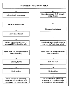
[0002] T lymphocytes that are transformed into cytotoxic lymphocytes (CTL) are
capable
of destroying brain tumor cells. When directed to destroy cells displaying non-
self
transplantation antigen markers known as human leukocyte antigens or HLA, they
are
referred to as "alloCTL." The goal of developing alloCTL is to develop a
population of CTL
that have strong recognition of allogeneic HLA peptides. Historically, alloCTL
have been
generated by one-way mixed lymphocyte reaction (MLR) where peripheral blood
monocytes
(PBMC) from a healthy donor are mixed with irradiated lymphoblasts from a
genetically
disparate individual (or patient). However, Dendritic cells (DC) are potent
antigen presenting
cells (APC) that display strong surface HLA.
Example 1: Sources of responder and stimulator cells.
For PBMC for preclinical studies IRB approvals have been obtained for: 1)
normal
blood donor collections at 100 ml or less, 2) purchase of buffy coats from the
San Diego
Blood Bank, and 3) limited leukapheresis of donors. Donors must test negative
for all
infectious disease agents. The density gradient isolated PBMC is washed then
fractionated,
26
CA 02772280 2012-02-27
WO 2011/017143 PCT/US2010/043470
using standard plastic adherence, into monocytes and lymphocytes. The
nonadherent cells
from the PBMC containing T, B and NK cells are either used fresh or
cryopreserved in vials
containing 10'-108 cells for the MLR generation method.
Experience using PBMC as responders indicates that the MLR can be applied
equally
well to fresh PBMC as well as to vitally-frozen PBMC. For LDCR, the adherent
monocytes
are differentiated to DC.
Example 2: Standardizing alloCTL generated by one-way MLR or LDCR.
Irradiated stimulator (S) lymphocytes and responder (R) lymphocytes from
normal,
healthy HLA-mismatched donors are collected. A small pool of young (18-50
years old)
normal blood donors is employed to help standardize the PBMC reactivity to
alloantigen
given that PBMC from older people do not respond to antigenic stimulation as
well in that
they have quantitative and functional defects in the CD4 T helper cell
compartment and cells
that lack CD40L.
Furthermore, it was demonstrated that resting lymphocytes, activated
lymphocytes
(aka lymphoblasts), as well as lymphocytes or lymphoblasts that have been
cryopreserved
and then thawed, all have high HLA surface expression levels, thus can
adequately serve as
stimulators.
Example 3: Isolation and expansion of stimulator lymphocytes for sensitization
of alloCTL by
MLR
A 100 ml blood draw from a healthy donor would be expected to yield 1 to 2 x
108
PBMC after isolation from Ficoll density gradients. After washing several
times with Hank's
balanced salt solution (HBSS) the PBMC are suspended in 20 ml of AIM V
synthetic
medium containing 5% autologous serum. The cells are injected into the
extracapillary space
(ECS) of the artificial capillary cartridge and perfused with medium
containing Orthoclone
OKT3 antibody (50 ng/5x 107 cells) and 240 IU/ml of rIL 2. The perfusion
volume may be
doubled every 2 to 4 days by adding fresh rIL 2 containing medium. Lactic acid
concentration is measured daily (7 days/week, YSI Stat lactate/glucose
analyzer) to determine
the rate of lactate production (usually 0.2-0.25 gm/109 cells/day). Cells are
fed every 4 to 5
days or when the concentration of lactate is at 0.5-0.7 gm/liter.
Lactic acid production parallels the expansion rate of the cells. Multiple
vials of
stimulator lymphocytes are vitally-frozen to maintain the capability of
performing multiple
27
CA 02772280 2012-02-27
WO 2011/017143 PCT/US2010/043470
alloCTL cultures from any given responder to stimulator (R:S) pairs; for
statistical purposes
multiple cultures are generated from one R:S pair. The number of stimulator
PBMC frozen is
based upon starting cultures at a R:S ratio of 10:1. Cells harvested from one
starter culture
are cryopreserved in 10% DMSO/autologous serum and stored at 80 C. The
stimulator
lymphocytes are thawed prior to inactivation with gamma-irradiation (127Cs-
source, 2000
Rads), then washed before combining with allogeneic responder lymphocytes.
Example 4: Isolation of monocytes and generation of stimulator DC.
The isolation of PBMC from whole blood is by density gradient centrifugation
and is
washed 2x with Hank's balanced salt solution (HBSS). The PBMC are suspended at
a density
of 5 x 10⁶/ml in serum-free, AIM V synthetic medium in plastic tissue culture
flasks. After 30
min incubation at 370 C, the nonadherent cells containing lymphocytes are
recovered and
cryopreserved; the adherent monocytic cells are washed with HBSS to removed
loosely
adherent cells then overlaid with fresh AIM-V medium and cultivated overnight
at standard
conditions.
The next day the adherent cells are washed with HBSS to remove residual
platelets,
then overlaid with AIM-V medium supplemented with 1,000 IU/ml of GM-CSF and
500
IU/ml of rIL-4 and cultivated for 6 days to differentiate monocytes into
immature DC. At day
6, the medium is supplemented with recombinant human TNF-a, IL-6 and IL-113
(10 ng/ml
for each cytokine) and cultured an additional 2 days to mature the DC. An
estimated
approximately 10% of the starting cell number are obtained as mature DC. The
DC are
subjected to gamma-irradiation (127Cs-source, 2000 Rads), and washed lx with
HBSS in
preparation for the LDCR protocol; these represent the stimulator DC.
Example 5: Generation of alloCTL by one-way MLR
The generation of alloCTL by MLR is as depicted on the right side of the Flow
Diagram in FIG. 1. Responder PBMC, from a donor genetically distinct from the
donor
supplying the stimulator cells, are isolated with Ficoll Hypaque and washed 2x
with HBSS.
The responder lymphocytes are combined with 127Cs-irradiated stimulator
lymphocytes, at a
responder to stimulator (R:S) ratio of 10:1 (i.e., one-way MLR). They are
placed into the
artificial capillary cartridges and cultivated at 37oC with 5% C02 with AIM V
medium
containing 5% autologous serum and 60 International Units (IU)/ml of rIL 2 for
14 days; the
cells over a 7 10 day period are weaned from serum containing medium.
28
CA 02772280 2012-02-27
WO 2011/017143 PCT/US2010/043470
A restimulation of the a11oCTL occurs, if growth patterns indicate it is
necessary, at
day 12 post-MLR with relevant lymphoblasts at a R:S of 10:1. Cytotoxicity
assessments,
proliferation, phenotypically-defined cytotoxic subsets and cytokine
production is determined
on day 14 post-MLR cells.
Example 6: Generation ofalloCTL by one-way LDCR
The generation of alloCTL by LDCR is as depicted on the left side of the Flow
Diagram shown in FIG. 1. The allodonors used for responders or pCTL are HLA-
disparate to
the donor supplying stimulator cells. The adherent cells are grown with growth
factors that
encourage DC (immature) growth. Growth factors are then added to the culture
medium to
mature the DC. Briefly, the plastic adherent monocytic cells are cultured in
serum free AIM-
V medium supplemented with 1000 units/ml rhGM-CSF and 500 units/ml rhIL-4 at
37 C in a
humidified, 5% C02 incubator. Six days later, the immature DC are stimulated
with
recombinant human TNF-a, IL-6 and IL-113 (10 ng/ml for each cytokine) to
induce their
maturation for 2 days. DCs are harvested, irradiated and combined with
responder PBMC for
LDCR at a R:S ratio of 10:1.
The DC present peptides from alloantigen (i.e., stimulators) to the T
lymphocytes of
the allodonor in the presence of low dose 1L-2 (601U/ml). Reactive responder
lymphocytes
develop into alloCTL capable of recognizing the HLA on the stimulator cells
over a 12 day
period. They are restimulated with DC at a 10:1 R:S on day 12 post-LDCR and
assessed 2
days later in 4 hour 51 Cr-release cytotoxicity assays, for proliferation, and
for phenotype and
cytokine production.
Example 7: Chromium release cytotoxicity assays
AIIoCTL preparations are generated from the same R:S pairs by either MLR or by
LDCR. The cytotoxicity of the alloCTL to relevant target, i.e., stimulator
lymphoblasts
displaying the HLA to which they are sensitized, is then determined. 51Cr-
release assays are
used determine the lytic activity of alloCTL effector cells when they are co-
incubated with
the target cells. Four hour assays are run in 96-well plates at multiple
effector to target (E:T)
ratios of 3:1, 10:1, 30:1 with triplicate samples as previously described.
Percent specific
release is calculated by the formula: [(cpmexperimentai - cpmspontaneous) /
(Cpmmaximal -
Cpmsp ntaneous)] x 100%. Spontaneous release is measured for targets in assay
medium alone
and maximal release is produced by lysis of the targets with 2% Triton X-100
(Sigma, St.
29
CA 02772280 2012-02-27
WO 2011/017143 PCT/US2010/043470
Louis, MO). Lysis obtained at each given E:T ratio is determined and the
thresholds of low,
moderate and high cytotoxicity can be defined accordingly.
Day 14 alloCTL generated by 1-way MLR and 1-way LDCR are compared.
Statistical assessment of lytic activity and the effects of reaction type (MLR
vs LDCR), the
three E:T ratios evaluated as an ordered factor, the samples, and their
possible interactions are
made by ANOVA with planned post-hoc comparisons. All statistical operations
are
accomplished in R, version 2.9 or higher. Optimization of alloCTL by DC
presentation is
considered possible if the cytotoxic responses, by DC-generated a11oCTL
compared to 1-way
MLR generated alloCTL, against stimulator lymphoblast target cells is >15%
higher when all
data are grouped and normalized from three equivalent E:T ratios tested.
The a11oCTL preparations have the ability to elicit alloantigen-specific
immune
responses against relevant target cells in vitro. Target cells are OKT3 or PHA-
stimulated
lymphoblasts, which display high levels of HLA antigen. "Relevant" targets are
the
lymphoblasts derived from stimulator PBMC. Responding donor lymphoblasts
express HLA
that should be regarded as "self' and therefore should not be targets of the
alloreactive T
cells. They are used as a background, negative control. Additionally, K562
natural killer
(NK)-sensitive cell targets do not express HLA antigen and are used as
"irrelevant" target
cells to assess nonspecific injury caused by NK cells (non-MHC-restricted
killing) that is
unrelated to T-cell alloreactivity (MHC-restricted killing). Lysis of K562 is
subtracted from
stimulator lymphoblast lysis for these comparisons also. The levels of HLA
expression by
lymphoblasts is analyzed by flow cytometry using the pan HLA-ABC antibody
(W6/32) to
assess whether the cytotoxicity directly relates to the relative antigen
density (MFIs) of HLA
on the relevant target cells.
Example 8: Phenotypic characterization of activated, mature dendritic cells
Aliquots of DC are stained with monoclonal antibodies (mAbs) against DC
surface
markers (anti-HLA class I conjugated to fluorescein isothiocyanate (FITC),
anti-HLA class II
DR conjugated to PerCp, anti-CD I lc conjugated to APC, anti-CD80, anti-CD83,
and anti-
CD86 conjugated to phycoerythrin (PE) (BD Biosciences/Pharmingen, San Diego,
CA) on
ice for lhour. The cells are washed three times with cold PBS before analyzing
on an LSR II
flow cytometer.
Example 9: Phenotypic characterization of activated, CD3 cytotoxic T cell
subsets by f low
cytometric analyses.
CA 02772280 2012-02-27
WO 2011/017143 PCT/US2010/043470
The cytotoxic subsets with alloCTL preparations for production of IFN-y are
examined. This cytokine has previously been shown to be most relevant to the
Thl cell-
mediated responses to immunotherapy exhibited by T lymphocytes. Additionally,
IFN-y has
been used as an in vitro monitoring tool to predict GVH in renal transplant
patients where
slight mismatches in donor to patient HLA are expected.
AIIoCTL preparations generated from the same responder/stimulator pairs by MLR
or
by LDCR are used to determine the fold-increase in the phenotypically-defined
CD3/CD8
cytotoxic subset displaying the activated T cell marker (CD69) that produces
IFN-y within
the alloCTL upon exposure to relevant target, i.e., stimulator patient
lymphoblasts displaying
the HLA to which they are sensitized. The cell subset positive for CD3, CD8,
CD69, and
intracellular IFN-y (BD Fast Immune Kit, BD Biosciences) is assessed at 24 hr
after
incubation with or without relevant target cells (stimulator lymphoblasts at a
R: S of 10:1). In
the last 6 hr of the 24 hr incubation, 10 gg/ml of Brefeldin A, a secretion
inhibitor, is added.
Nonstimulated or stimulated alloCTL are each aliquoted into three tubes (106
cells/tube) and
pelleted at 100x g. Flow cytometric analysis is performed, staining for cell
surface markers
(e.g., CD3+, CD8+, CD69+) and cytoplasmic IFN-y cytokine expression. The Fix
and Perm
reagents are used where indicated according to the manufacturer's protocol. In
brief,
alloCTL cell pellets are resuspended and incubated with a fluorochrome-
conjugated
monoclonal antibody (mAb) cocktail on ice for 30 minutes. The cells are
washed, fixed and
permeabilized, then incubated with a fluorochrome-conjugated mAb specific for
IFN-y for 30
min. Following the second antibody incubation, the cells are washed again and
resuspended
in PBS and analyzed by flow cytometry. The analyses is performed with a six-
color capable
BD LSR II flow cytometer. Percentages of the positive activated T cell subset
and the mean
fluorescence intensities (MFI) of IFN-y are obtained.
The fold increases in the percentages of the activated subset in the alloCTL
that are
restimulated versus those not are determined. As well, the fold increases in
the MFIs for
IFN-y in the alloCTL subsets that are or are not restimulatcd are determined.
Each of these
measures may be usefully predictive of the extent of cytolysis. An increase in
the cytotoxic
subset or the degree of IFN-y expression that is >1.5-fold may reach
significance based upon
other observations with patient PBMC in vaccine trials for gliomas. Other
investigators' data
collected by this flow cytometric method compare well to that collected by
limiting dilution
analyses.
31
CA 02772280 2012-02-27
WO 2011/017143 PCT/US2010/043470
Example 10: Determination of the proliferative response of the alloCTL made by
MLR or
DC presentation upon their exposure to relevant stimulator lymphoblasts.
The CTL precursor frequency within a donor mononuclear cell pool to patient
HLA
antigens is variable. It may be as high as 10% to allogeneic MHC antigen or as
low as 0.1-
0.01%. Anticipating that the precursor CTL frequency is identical in any given
responder/stimulator pair, DC presentation is compared to T lymphocyte
presentation in an
MLR for enhancing the proliferative events of alloresponders.
The overall intent is to generate therapeutically significant quantities of
alloCTL. The
ability of T cells to proliferate when exposed to the antigens to which they
are sensitized has
been used as an indicator of the presence of antigen-specific CD4+ helper T
cells.
The proliferative response of the alloCTL preparations upon their exposure to
relevant
patient lymphoblasts displaying the HLA to which they are sensitized is
characterized. The
proliferative response of the alloCTL upon their exposure to relevant
stimulator lymphoblasts
is determined for alloCTL preparations generated from the same R:S pairs made
by MLR or
by LDCR and converted to stimulation indices for comparison.
The capacity to proliferate in response to HLA presentation by relevant
stimulator
cells is measured by tritiated thymidine uptake at a R:S ratio of 10:1. In
response to the
alloCTL seeing relevant antigen, proliferation should ensue. After 48 hr, the
culture is pulse-
labeled with 3H-thymidine. DNA synthesis, as a measure of proliferation, is
quantified by
using a liquid scintillation counter to measure the amount of radiolabeled
thymidine
incorporated into the DNA. A stimulation index (SI) is calculated by dividing
the number of
cpm for the resensitized al1oCTL by the number of cpm for the cells incubated
without
sensitizing cells.
The Sts obtained for each alloCTL preparation can be categorized as having a
high
proliferative population versus a low proliferative population. The in vitro
proliferative
capacity of the alloCTL can thereby be compared to their cytotoxicity,
phenotypic analyses,
and the level of HLA mismatch between the responder and stimulator.
In general, while there is some consensus in the literature that proliferative
events
correlate with responder/stimulator MHC disparities at Class 11, while
cytolytic activity is a
function of disparities at Class I, the separation of proliferative and
cytolytic functions should
be confirmed by analyzing the data with molecular HLA types of the responder
and
stimulator. This is to be addressed using both conventional and robust
regression analyses.
32
CA 02772280 2012-02-27
WO 2011/017143 PCT/US2010/043470
In addition to comparing the proliferative differences in alloCTL generated by
MLR
vs LDCR methods, proliferation of the alloresponder enriched cultures at
restimulation
results from HLA Class II disparities should be discerned, whereas the
functionality of the
cells as determined by cell injury, and cytotoxic cell phenotype/cytokine
production, relates
to HLA Class I disparities between responder and stimulators.
Example 11: Determination of the soluble Thl to Th2 cytokine ratios produced
upon alloCTL
exposure to relevant target.
Other researchers have compared IFN-y/IL-10 ratios as an in vitro monitoring
tool for
assessing tumor host response using PBMC pre- and post-vaccination, and for T
cell induced
GVH development and rejection in transplant patients. Here the soluble Thl to
Th2 (i.e.,
IFN-y to IL- 10 or TNF-a to IL-4) cytokine ratios produced upon alloCTL
exposure to
relevant stimulator target are determined with alloCTL preparations generated
from the same
R:S pairs by MLR or by LDCR. Higher Thl to Th2 ratios may correlate with
induction of a
proinflammatory response in vivo and/or correspond to better cytolysis to
relevant target.
Supernatants from alloCTL coincubated for 24 hr in the presence or absence of
relevant irradiated stimulator lymphoblasts are examined. The cell suspensions
are clarified
by refrigerated centrifugation at 400 x g for 10 min. The clarified medium, or
dilutions of it
if necessary, are analyzed using the BD cytometric bead array system. The
cytokines tested
include Thl and Th2 cytokines: IL-2, IL-4, IL-5, IL-10, gamma interferon (y-
IFN) and tumor
necrosis factor alpha (TNF-a). The array system allows for collection of
multiple cytokine
results from a single small sample at relatively sensitive levels of detection
(2.0 - 4.0 pg/ml).
Therefore, not only IFN-y/IL- 10 ratios but other alternative Th I /Th2
cytokine permutations
(i.e., TNF-a/IL-4) as well are analyzed. For this reason, the array can be
considered a cost
effective alternative to ELISAs specific for the four cytokines.
Statistical Evaluation. Statistical analysis is performed using conventional
and
statistically robust techniques with the R statistical package. Data
descriptions include
standard 5-point summaries as well as the first four moments and MAD (median
absolute
deviations). To elucidate the interrelationships of functional
alloresponsiveness (i.e.,
cytotoxicity of the alloCTL, fold-increases in the phenotypic subset
displaying the activated
T cell marker, proliferation in response to exposure to relevant antigens
and/or
proinflammatory cytokine production) relative to HLA mismatch, correlative
studies include
both pairwise analyses with confidence intervals and additional analyses to
investigate
33
CA 02772280 2012-02-27
WO 2011/017143 PCTIUS2010/043470
systematic nonlinearities. Both conventional ANOVA and its robust analogues
are used to
investigate the relationships. The mean averages of triplicate samples are
compared in three
separate experiments using the same R:S pairs. The higher the number of
alloCTL
preparations made by both methods, the better the statistic will be. The
number of
experiments needed depends upon the pilot data and power analyses. The
implication to
obtaining significantly higher cytotoxic assessments with alloCTL generated by
LDCR vs
MLR is that an alteration of the generation method for alloCTL for clinical
studies could be
made by amending the existing IND to the FDA.
Example 12: Validation using patient tumor/lymphocyte sets as targets and
stimulator of
alloCTL
A tissue bank that contains matched patient lymphocyte and glioma specimens
could
be used for validation purposes. Tissues in the bank are obtained from
patients by IRB-
approved protocols. The patient lymphocytes act as stimulators of alloCTL. The
cultured
glioma specimens act as relevant targets in cytotoxicity assays. It has been
documented that
tumor cells in situ and in culture express MHC class I antigens.
Functional characteristics of alloCTL generated by MLR and by LDCR are
compared,
including selective cytotoxicity toward relevant HLA-bearing targets, and in
response to
incubation with relevant target cells, upregulation of proinflammatory
cytotoxic subsets, Th1
cytokine production, and proliferation.
Final Considerations
While the invention has been described in connection with the above described
embodiments, it is not intended to limit the scope of the invention to the
particular forms set
forth, but on the contrary, it is intended to cover such alternatives,
modifications, and
equivalents as may be included within the scope of the invention. Further, the
scope of the
present invention fully encompasses other embodiments that may become obvious
to those
skilled in the art and the scope of the present invention is limited only by
the appended
claims.
34