Note: Descriptions are shown in the official language in which they were submitted.
CA 02772418 2012-03-26
SYSTEM AND METHOD FOR UV TACKING AN IMPLANT
BACKGROUND
Technical Field
[0002] The present disclosure relates to endoscopic surgical instruments. More
particularly, the present disclosure relates to a system and method for
ultraviolet (UV) tacking an
implant via an endoscopic surgical instrument having a UV light source
mechanism distally
disposed.
Background of Related Art
[0003] Surgical instruments which include a tool assembly mounted on a distal
end of a
body portion of the surgical instrument for articulation are well known.
Typically, such surgical
instruments include articulation control mechanisms, which allow an operator
to remotely
articulate the tool assembly in relation to the body portion of a surgical
instrument to allow the
operator to more easily access, operate on, and/or manipulate tissue.
[00041 Such articulating tool assemblies have become desirable, especially in
the
endoscopic surgical procedures. In an endoscopic surgical procedure, the
distal end of a surgical
instrument is inserted through small incisions in the body to access a
surgical site. Typically, an
appropriately sized cannula, e.g., 5 mm, 10 mm, etc., is inserted through the
body incision to
provide a guide channel for accessing the surgical site. Because it is
desirable to provide small
1
CA 02772418 2012-03-26
body incisions, i.e., less scarring, reduced trauma to the patient, faster
healing time, the
tolerances between the surgical instrument and the inner diameter of the
cannula are small.
[0005] Conventional articulating tool tips have limited functionality mainly
due to
mechanical design limitations of actuating mechanisms. Thus, it is desirable
to provide an
articulating surgical instrument, which includes an articulation mechanism
that would provide a
wider range of functions for the articulation tip.
SUMMARY
[0006] Accordingly, an improved surgical instrument is provided. The surgical
instrument includes a handle portion and a body portion extending distally
from the handle
portion and defining a longitudinal axis. The surgical instrument also
includes a grasper
disposed at a distal end of the body portion, the grasper including an
ultraviolet (UV) light
mechanism for performing UV tacking of an implant.
[0007] In another exemplary embodiment, the grasper is an end effector
assembly having
a first jaw member and a second jaw member. The first and second jaw members
are movable
from a first position in spaced relation relative to one another to a second
position where the first
and second jaw members cooperate to grasp the implant therebetween.
[0008] In another exemplary embodiment, the implant is a mesh having a UV
reactive
polymeric coating. The mesh is positioned between the first and second jaw
members: (i) to be
placed at a surgical site and (ii) to be exposed by a UV light emitted from
the UV light
mechanism such that the UV tacking of the mesh to the surgical site is
performed. The mesh
includes one or more tack regions each having a polymer coating embedded
therein, the polymer
coating being chemically induced by a UV light of the UV light mechanism.
2
CA 02772418 2012-03-26
[0009] A mesh having a UV reactive polymeric coating suitable for some
embodiments
of the present invention is found in U.S. Provisional Application Ser. No.
61/348896 filed on
May 27 2010, the entire contents of which are incorporated by reference
herein. In other
embodiments, polymers as disclosed above are applied directly to tissue and
then used to affix
the mesh to tissue when polymerized with UV light.
[0010] In another exemplary embodiment, tack regions may be a uniform coating
of the
mesh surface or may be distinct regions. In yet another exemplary embodiment,
the tack regions
are visually designated along a length of the mesh. In a further embodiment,
the regions tacked
by the instrument change color when subjected to UV light or pressure,
indicating locations on
the mesh that have been tacked.
[0011] The UV light mechanism may be positioned on a non-grasping portion of
the
grasper. However, the UV light mechanism may be positioned on at least one
grasping portion
of the grasper.
[0012] In yet another exemplary embodiment, the surgical instrument further
includes at
least one sensor adapted to continuously or intermittently monitor UV light
emission from the
UV light mechanism. Additionally, the surgical instrument may include a
trigger mechanism
positioned on the handle portion for selectively activating the UV light
mechanism.
[0013] In another exemplary embodiment, an improved surgical instrument
assembly is
provided. The surgical instrument assembly includes a handle portion and a
body portion
extending distally from the handle portion. The surgical instrument assembly
also includes an
end effector assembly disposed at a distal end of the body portion, the end
effector assembly
including a light source for tacking a mesh in position at a surgical site.
3
CA 02772418 2012-03-26
[0014] In another exemplary embodiment a method of UV tacking a mesh at a
surgical
site is provided. The method includes the steps of providing a surgical
instrument including an
ultraviolet (UV) light mechanism for performing UV tacking of an implant;
providing a mesh
implant having a polymeric coating activated by UV light; endoscopically
positioning the mesh
over the surgical site; and selectively applying UV light emitted from the UV
light source to the
mesh to tack the mesh to the site The mesh may include a polymeric coating
that is activated
upon exposure from the UV light emitted from the UV light source.
BRIEF DESCRIPTION OF THE DRAWINGS
[0015] The accompanying drawings, which are incorporated in and constitute a
part of
this specification, illustrate embodiments of the disclosure and, together
with a general
description of the disclosure given above, and the detailed description of the
embodiment(s)
given below, serve to explain the principles of the disclosure, wherein:
[0016] FIG. IA is a perspective view of a surgical instrument in accordance
with the
present disclosure;
[0017] FIG. 1B is a perspective view of the end effector assembly of the
surgical
instrument of FIG. IA, illustrating one or more ultraviolet (UV) light sources
on a non-grasping
portion of the end effector assembly, in accordance with the present
disclosure;
[0018] FIG. IC is a perspective view of the end effector assembly of the
surgical
instrument of FIG. IA, illustrating one or more UV light sources on grasping
portions of the end
effector assembly, in accordance with the present disclosure;
[0019] FIG. 2A is a perspective view of another surgical stapling instrument
in
accordance with the present disclosure;
4
CA 02772418 2012-03-26
[0020] FIG. 2B is a perspective view of the end effector assembly of the
surgical
instrument of FIG. 2A, illustrating one or more UV light sources on a non-
grasping portion of
the end effector assembly, in accordance with the present disclosure;
[0021] FIG. 2C is a perspective view of the end effector assembly of the
surgical
instrument of FIG. 2A, illustrating one or more UV light sources on grasping
portions of the end
effector assembly, in accordance with the present disclosure;
[0022] FIG. 3A is a perspective view of the mesh, in accordance with the
present
disclosure;
[0023] FIG. 3B is a perspective cross-sectional view of the mesh of FIG. 3A,
in
accordance with the present disclosure;
[0024] FIG. 4A is a perspective view of the surgical instrument of FIG. 1A
grasping the
mesh of FIG. 3A, in order to apply UV light via the one or more UV light
sources to the mesh, in
accordance with the present disclosure; and
[0025] FIG. 4B is a perspective view of the surgical instrument of FIG. 2A
grasping the
mesh of FIG. 3A, in order to apply UV light via the one or more UV light
sources to the mesh, in
accordance with the present disclosure.
DETAILED DESCRIPTION
[0026] Embodiments of the presently disclosed apparatus will now be described
in detail
with reference to the drawings, in which like reference numerals designate
identical or
corresponding elements in each of the several views. As used herein, the term
"distal" refers to
that portion of the tool, or component thereof which is further from the user
while the term
"proximal" refers to that portion of the tool or component thereof which is
closer to the user.
CA 02772418 2012-03-26
[0027] Referring to FIGS. IA-IC, a surgical system for use in a surgical
procedure, e.g.,
a minimally invasive procedure is illustrated.
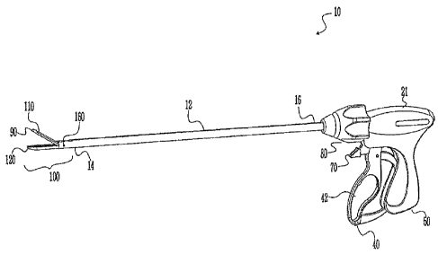
[0028] FIG. IA shows a surgical instrument 10 according to the present
disclosure.
More particularly, surgical instrument 10 generally includes a housing 21, a
handle assembly 40,
a rotating assembly 80, and a trigger assembly 70, which mutually cooperate
with the end
effector assembly 100 to grasp and treat tissue. Such a grasping instrument 10
is further
exemplified by laparoscopic grasping instruments such as Covidien order codes
173030, 174317,
174001 and 174233.
[0029] The surgical instrument 10 also includes a shaft 12, which has a distal
end 14 that
mechanically engages the end effector assembly 100 and a proximal end 16 that
mechanically
engages the housing 21 proximate the rotating assembly 80. Handle assembly 40
includes a
fixed handle 50 and a movable handle 42. Handle 42 moves relative to the fixed
handle 50 to
actuate the end effector assembly 100 and enable a user to grasp and
manipulate tissue.
[0030] The end effector assembly 100 includes opposing jaw members 110, 120.
The
jaw members 110, 120 are activated by using a drive assembly (not shown)
enclosed within the
housing 21. The drive assembly cooperates with the movable handle 42 to impart
movement of
the jaw members 110, 120 from the open position to the clamping or closed
position.
[0031] The surgical instrument 10 also includes a rotating assembly 80
mechanically
associated with the shaft 12 and the drive assembly (not shown). Movement of
the rotating
assembly 80 imparts similar rotational movement to the shaft 12 which, in
turn, rotates the end
effector assembly 100.
[0032] As best seen with respect to FIG. 1A, the end effector assembly 100
attaches to
the distal end 14 of shaft 12. The jaw members 110, 120 are pivotable about a
pivot 160 from
6
CA 02772418 2012-03-26
the open to closed positions upon relative reciprocation, i.e., longitudinal
movement, of the drive
assembly (not shown). It is envisioned that the surgical instrument 10 may be
designed such that
it is fully or partially disposable depending upon a particular purpose or to
achieve a particular
result. For example, end effector assembly 100 may be selectively and
releasably engageable
with the distal end 14 of the shaft 12 and/or the proximal end 16 of the shaft
12 may be
selectively and releasably engageable with the housing 21 and handle assembly
40. In either of
these two instances, the surgical instrument 10 may be either partially
disposable or reposable,
such as where a new or different end effector assembly 100 or end effector
assembly 100 and
shaft 12 are used to selectively replace the old end effector assembly 100 as
needed.
[00331 Additionally, FIG. IA illustrates a UV light source 90 (or UV light
mechanism)
disposed at the distal end of the first jaw 110. The UV light source 90 is
better seen in FIGS. 1 B
and 1 C, which illustrate the end effector assembly 100. FIG. I B illustrates
one or more UV light
sources 90 on the first jaw 110 and the second jaw 120 (i.e., the non-grasping
portions of the
jaws 110, 120). FIG. IC illustrates one or more UV light sources 90 on the
grasping portions of
the second jaw 120. One skilled in the art may contemplate using a number of
different UV light
sources on one jaw or on both jaws and may contemplate positioning such UV
light sources on
or about any desired portion(s) of the end effector assembly 100.
[00341 In operation, the jaw members 110, 120 are positioned in the vicinity
of an
incision of a surgical site for placement of an implant or mesh (see FIGS. 3A
and 3B). The light
sources 90 positioned on the first and second jaws 110, 120 (see FIG. 1B) are
triggered to emit
UV light to activate an adhesive on the implant or mesh to permanently secure
the implant or
mesh to the incision of the surgical site. The adhesive may be a UV activated
adhesive. Thus,
the surgical instrument 10 may perform a full cure to permanently attach or
tack the implant or
7
CA 02772418 2012-03-26
mesh by using the UV light source 90 to activate UV adhesive sprayed on the
implant or mesh.
A less than full cure for temporarily securing the implant may be achieved by
applying a lower
energy of UV light.
[00351 Energy is transmitted to the implant or mesh from one or more energy
transmission devices such as a laser or lasers. In at least one embodiment,
the laser is a UV
laser, however in some alternative embodiments the laser may be an IR laser,
diode laser, C02,
visible light, or any other form of laser device or combinations thereof. One
skilled in the art
may contemplate using a plurality of different forms of energy in order to
tack the implant or
mesh to the incision of the surgical site. For example, one skilled in the art
may use thermal
energy, microwave energy, chemical energy, and/or ultrasonic energy or a
combination thereof.
[00361 Referring to FIG. 2A, a perspective view of another surgical stapling
instrument
500 in accordance with the present disclosure is presented.
[0037] In FIG. 2A, surgical instrument 500 includes a handle portion 510, a
body portion
512, and a disposable loading unit ("DLU") 540. Handle portion 510 includes a
stationary
handle 514 and a movable handle or trigger 516. Movable handle 516 is movable
in relation to
stationary handle 514 to advance a control rod 520 (not shown), which projects
from the distal
end of body portion 512. Alternately, other surgical instruments may be used
with DLU 540 to
perform endoscopic surgical procedures. The surgical instrument 500 also
includes an
articulation mechanism 400 for articulating a tool assembly 17 of the DLU 540.
The tool
assembly 17 may include a first jaw 520 and a second jaw 522.
[0038] DLU 540 includes a tool assembly 17, a proximal body portion 200 and a
mounting assembly 235. Body portion 200 has a proximal end adapted to
releasably engage the
distal end of a surgical instrument 500. Mounting assembly 235 is pivotally
secured to a distal
8
CA 02772418 2012-03-26
end of body portion 200 and is fixedly secured to a proximal end of tool
assembly 17. Pivotal
movement of mounting assembly 235 about an axis perpendicular to a
longitudinal axis of body
portion 200 effects articulation of tool assembly 17 between a non-articulated
position in which
the longitudinal axis of tool assembly 17 is aligned with the longitudinal
axis of body portion
200 and an articulated position in which the longitudinal axis of tool
assembly 17 is disposed at
an angle to the longitudinal axis of body portion 200.
[0039] Additionally, the surgical instrument 500 includes a UV light source
590 (or UV
light mechanism) disposed at the distal end of the tool assembly 17. The UV
light source 590 is
better seen in FIGS. 2B and 2C, which illustrate the tool assembly 17. FIG. 2B
illustrates one or
more UV light sources 590 on the lower jaw (i.e., the non-grasping portion of
the tool assembly
17). FIG. 2C illustrates one or more UV light sources 590 on the grasping
portions of the tool
assembly 17. One skilled in the art may contemplate using a number of
different UV light
sources on one jaw or on both jaws and may contemplate positioning such UV
light sources on
or about any desired portion(s) of the tool assembly 17.
[0040] Referring to FIGS. 2B and 2C, perspective views of the tool assembly 17
of the
surgical instrument 500 of FIG. 2A, illustrating one or more UV light sources
590 on a non-
grasping portion and a grasping portion of the tool assembly 17, respectively,
in accordance with
the present disclosure is presented.
[0041] In operation, the jaw members 520, 522 are positioned in the vicinity
of an
incision of a surgical site for placement of an implant or mesh (see FIGS. 3A
and 3B). The light
sources 590 positioned on the second jaw 520 (see FIG. 2C) are triggered to
emit UV light to
activate an adhesive on the implant or mesh to permanently secure the implant
or mesh to the
incision of the surgical site. The adhesive may be a UV activated adhesive.
Thus, the surgical
9
CA 02772418 2012-03-26
instrument 500 may perform a full cure to pennanently attach or tack the
implant or mesh by
using the UV light source 590 to activate UV adhesive sprayed on the implant
or mesh.
[0042] Referring to FIG. 3A, a perspective view of the mesh 310, in accordance
with the
present disclosure is presented, whereas referring to FIG. 3B a perspective
cross-sectional view
of the mesh 310 of FIG. 3A, in accordance with the present disclosure is
presented.
[0043] The surgical mesh 310 (or implant) is suitable for surgical repair of
hernias and
other surgical procedures requiring reinforcement or repair of soft tissue,
such as muscle or wall
tissue defects, pelvic organ prolapse, and urinary incontinence, for example.
The mesh 310 of
the present disclosure may be in the form of sheets, patches, slings,
suspenders, and other
implants and composite materials such as pledgets, buttresses, wound
dressings, drug delivery
devices, and the like. The present surgical mesh 310 may be implanted using
open surgery or by
a laparoscopic procedure.
[0044] The surgical mesh 310 may be fabricated from monofilament and/or
multifilament yarns 312, which may be made of any suitable biocompatible
material. Suitable
materials from which the mesh 310 may be made should have the following
characteristics:
sufficient tensile strength to support tissue; sufficiently inert to avoid
foreign body reactions
when retained in the body for long periods of time; easily sterilized to
prevent the introduction of
infection when the mesh 310 is implanted in the body; and sufficiently strong
to avoid tearing of
portions thereof.
[004] Referring now to FIGS. 3A and 3B, the mesh 310 is illustrated including
a porous
mesh substrate 311. The substrate 311 may be formed from fibers, filaments,
threads or yarns
312 defining a plurality of pores 314 therebetween. The yarns 312 of the
substrate 311 may be
made up of multiple filaments 338 (see FIG. 3B). The pores 314 may include one
or more intra-
CA 02772418 2012-03-26
pore films 316. The intra-pore films 316 of the present disclosure are non-
contiguous with
respect to one another, with each intra-pore film 316 being located in a
single pore 314 of the
porous substrate 311. In embodiments, multiple intra-pore films 316 may also
be formed within
each of the pores 314 of the substrate 311. The term "non-contiguous" as used
herein, is used to
denote one or more films 316 that are wholly contained within a corresponding
pore 314 and are
not in physical contact with another intra-pore film 316 of any other pore
314, as compared to a
conventional film-coated porous substrate in which the film stretches across
multiple pores. The
intra-pore films 316 are solely contained within the pores of the substrate.
The intra-pore film
does not span across the yarns 312 of the substrate. The intra-pore films 316
are non-contiguous
and are not bridged together by applying a film over the entire substrate, but
rather, the intra-pore
films 316 are created at discrete locations, within the individual pores.
[0046] The intra-pore films 316 may be formed at any plane within the pores
314 relative
to the plane of the substrate 311 such that the intra-pore film 316 does not
contact any adjacent
intra-pore film 316. In embodiments, the intra-pore film 316 may be textured,
smooth and/or
porous.
[0047] In a preferred embodiment, the yams 312 may be sprayed with a UV
polymer
adhesive that is activated when a UV light source 90, 590 (see FIGS. 1A-2C) is
placed in the
proximity of the yams 312 of the mesh 310.
[0048] As illustrated in FIG. 3A, not every pore 314 includes an intra-pore
film. In
certain embodiments, the pores including intra-pore films may be from about
10% to about 95%
of the pores. In further embodiments, about 15% to about 90% of the pores of
the substrate 311
include at least one intra-pore film. In other embodiments, from about 25% to
about 75% of the
11
CA 02772418 2012-03-26
pores of the substrate 311 include at least one intra-pore film. In other
embodiments, all of the
pores of the substrate 311 may include an intra-pore film.
[0049] The substrate 311 may include at least a center and a periphery. In
embodiments
where less than 100% of the pores of the substrate 311 include intra-pore
films, the location of
the intra-pore films may be random or patterned. For example, the pores of the
substrate 311
that include the intra-pore films may be solely disposed in the center of the
substrate 311 or the
pores that include the intra-pore films may be solely disposed on the
periphery of the substrate.
In embodiments, the location of intra-pore films may be varied (e.g., random,
patterned, etc.)
depending upon the intended use of the substrate 311. The intra-pore films may
form a
discontinuous layer covering intermittent portions of the surface of the
substrate 311. In one
example, the intra-pore films may form a discontinuous layer on the surface of
the substrate 311,
wherein the porosity of the substrate 311 is maintained by the discontinuous
layer of the intra-
pore films.
[0050] Each intra-pore film 316 of a substrate 311 may be made from the same
materials
or different materials. In particular, one or more of the intra-pore films 316
may be formed from
one material, while one or more different intra-pore films 316 may be formed
from another
material. The intra-pore film 316 may be permanent (e.g., non-bioabsorbable),
biodegradable, or
may be formed from any suitable combination of natural, synthetic,
biodegradable and non-
biodegradable materials. In the present application, the terms
"biodegradable," "bioresorbable,"
and "bioabsorbable" are used interchangeably and are intended to mean the
characteristic
according to which an implant and/or a material is resorbed by biological
tissues and the
surrounding fluids, and disappears in vivo after a given period of time. The
time period may
12
CA 02772418 2012-03-26
vary, from about one minute to about several months or more, depending on the
chemical nature
of the implant and/or of the material utilized to form the implant.
[0051] In alternate embodiments, the substrate may include intra-pore films
that have a
varying degradation rates, such that some of the intra-pore films degrade at a
rate different from
that of other intra-pore films. The type of material used to form the film,
concentration of the
material, and structure of the film, are some factors which may affect the
degradation time of the
film.
[0052] In some embodiments, the yarns 312 include at least two filaments,
which may be
arranged to create openings therebetween, the yarns 312 also being arranged
relative to each
other to form openings in the mesh 310. Alternatively, the mesh 310 may be
formed from a
continuous yarn 312 that is arranged in loops that give rise to the openings
in the mesh 310. The
use of a mesh 310 having yarns spaced apart in accordance with the present
disclosure has the
advantage of reducing the foreign body mass that is implanted in the body,
while maintaining
sufficient tensile strength to securely support the defect and tissue being
repaired by the mesh
310. Moreover, the openings of the mesh 310 of the present disclosure may be
sized to permit
fibroblast through-growth and ordered collagen laydown, resulting in
integration of the mesh 310
into the body. Thus, the spacing between the yarns 312may vary depending on
the surgical
application and desired implant characteristics as envisioned by those skilled
in the art.
[0053] All the above alternate embodiments of the mesh 310 may include one or
more
yarns 312 and/or pores 314 having UV adhesive sprayed thereon during
manufacturing for being
activated by any type of UV light source 90, 590 of any type of surgical
instrument/system 10,
500. Therefore, the mesh 310 may be any type of biodegradable polymeric
coating having UV
properties for interacting with UV light sources 90, 590.
13
CA 02772418 2012-03-26
[0054] It may desirable to reposition the mesh 310. In that instance, the mesh
adhesive
may be initially tacky to allow repositioning of the mesh. Alternatively, the
mesh adhesive may
be partially polymerized by a relatively briefer application or lower energy
application of UV
light to achieve tackiness or a light bonding to tissue. In any case, when
mesh is repositioned
after application of UV light, it is desirable to know what regions of the
mesh 310 have been
originally subjected to UV light to enable applying the light to an uncured
region of the mesh.
This may be aided by marking certain adjacent zones of the mesh 310 with
numeric or alphabetic
sequences such as A, B, C so that the surgeon may locate the mesh positions of
a first bonding
attempt during repositioning. Further, the mesh 310 may be treated with a heat
or pressure
reactant dye to display a visual indication that UV light has been applied or
that the jaws of
grasping instrument 10 have applied pressure indicative of bonding to the
mesh.
[0055] Referring to FIG. 4A, a perspective view 400A of the surgical
instrument 10 of
FIG. 1A grasping the mesh 310 of FIG. 3A, in order to apply UV light via the
one or more UV
light source 90 to the mesh 310, in accordance with the present disclosure is
presented.
[0056] Referring to FIG. 4B, a perspective view 400B of the surgical
instrument 500 of
FIG. 2A grasping the mesh 310 of FIG. 3A, in order to apply UV light via the
one or more UV
light sources 590 to the mesh 310, in accordance with the present disclosure
is presented.
[0057] In operation, the mesh 310 is positioned between the first and second
jaw
members 110, 120 of surgical instrument 10: (i) to be placed at a surgical
site and (ii) to be
exposed by a UV light 91 emitted from the UV light mechanism 90, such that the
UV tacking of
the mesh 310 to the surgical site is performed (see FIG. 4A). Similarly, the
mesh 310 is
positioned between the first and second jaw members 520, 522 of instrument
500: (i) to be
placed at a surgical site and (ii) to be exposed by a UV light 591 emitted
from the UV light
14
CA 02772418 2012-03-26
mechanism 590 such that the UV tacking of the mesh 310 to the surgical site is
performed (see
FIG. 4B).
[0058] The mesh 310 may include one or more tack regions each having a polymer
coating embedded therein, the polymer coating being chemically induced by a UV
light of the
UV light mechanism 90, 590. Additionally, the one or more tack regions may
have a thickness
that is greater than a thickness of the mesh 310. Alternatively, the one or
more tack regions may
be positioned substantially equidistant from each other along a length of the
mesh 310.
[0059] Therefore, in accordance with the present disclosure, the method of UV
tacking a
mesh includes the step of applying energy to a handle portion of a surgical
instrument having a
body portion extending distally therefrom from a handle portion. The next
steps may be
positioning an end effector assembly at a distal end of the body portion and
incorporating a UV
light source at the end effector assembly. A user may then selectively apply a
UV light emitted
from the UV light source to the mesh and UV-tack the mesh to the surgical
site. The mesh may
include a biodegradable polymeric coating that is activated upon exposure from
the UV light
emitted from the UV light source.
[0060] In another exemplary embodiment, at least one sensor may be adapted to
continuously or intermittently monitor UV light emission from the UV light
mechanism.
[0061] While several embodiments of the disclosure have been shown in the
drawings, it
is not intended that the disclosure be limited thereto, as it is intended that
the disclosure be as
broad in scope as the art will allow and that the specification be read
likewise. Therefore, the
above description should not be construed as limiting, but merely as
exemplifications of
presently disclosed embodiments. Thus the scope of the embodiments should be
determined by
the appended claims and their legal equivalents, rather than by the examples
given.
CA 02772418 2012-03-26
[0062) Persons skilled in the art will understand that the devices and methods
specifically
described herein and illustrated in the accompanying drawings are non-limiting
exemplary
embodiments. The features illustrated or described in connection with one
exemplary
embodiment may be combined with the features of other embodiments. Such
modifications and
variations are intended to be included within the scope of the present
disclosure. As well, one
skilled in the art will appreciate further features and advantages of the
present disclosure based
on the above-described embodiments.
16