Note: Descriptions are shown in the official language in which they were submitted.
CA 02772437 2012-02-28
WO 2011/026215
PCT/CA2010/001251
1
CRYOTREATMENT DEVICE USING A SUPERCRITICAL GAS
FIELD OF THE INVENTION
The present invention relates to a coolant system and method of use for a
cryogenic medical device.
BACKGROUND OF THE INVENTION
A number of cooled catheter systems have been developed for treating tissue
in a cardiac setting, either to cool the tissue sufficiently to stun it and
allow cold
mapping of the heart and/or confirmation of catheter position with respect to
localized
tissue lesions, or to apply a more severe level of cold to ablate tissue at
the site of the
catheter ending. In general, the range of treatments which may be effected by
a
cryocatheter is comparable to the range of applications for radio frequency or
thermal
ablation catheters, and in particular, these instruments may be configured to
achieve
either small localized ball shape lesions at the tip of the catheter, or one
or more
elongated linear lesions extending a length of several centimeters or more
along the
tip. The latter form of lesion is commonly used to achieve conduction block
across a
region of the cardiac wall so as to sever an aberrant pathway over a length,
preventing
conduction across the region, in order change the cardiac signal path
topology, for
example, to eliminate a faulty pathway responsible for atrial fibrillation or
a
tachycardia.
A cryogenic device uses the energy transfer derived from thermodynamic
changes occurring in the flow of a refrigerant through the device. Various
fluids with
low operating temperatures (such as cryogens or cryogenic refrigerants) have
been
used in the medical and surgical field to treat such tissue aberrations. In
general, a
cryogenic device uses the energy transfer derived from thermodynamic changes
occurring in the flow of a cryogen therethrough to create a net transfer of
heat flow
from the target tissue to the device, typically achieved by cooling a portion
of the
device to very low temperature through conductive and convective heat transfer
between the cryogen and target tissue. The quality and magnitude of heat
transfer is
regulated by the device configuration and control of the cryogen flow regime
within
the device.
CA 02772437 2012-02-28
WO 2011/026215
PCT/CA2010/001251
2
Structurally, cooling can be achieved through injection of high pressure
refrigerant through an orifice. Upon injection from the orifice, the
refrigerant
undergoes two primary thermodynamic changes: (i) expanding to low pressure and
temperature through positive Joule-Thomson throttling, and (ii) undergoing a
phase
change from liquid to vapor, thereby absorbing heat of vaporization. The
resultant
flow of low temperature refrigerant through the device acts to absorb heat
from the
target tissue and thereby cool the tissue to the desired temperature.
A number of different fluids have been used for the coolant component of
cryotreatment catheters, such as a concentrated saline solution or other
liquid
providing some degree of thermal conductivity and heat capacity. However,
typical
refrigerants and their respective refrigeration systems may be limited in
their thermal
conductivity and/or capacity to remove heat, either because of their
particular thermal
properties or because of insufficient temperature reduction prior to delivery
of the
refrigerant to a catheter.
To some extent these considerations have been addressed by using a phase
change material as the cryogenic fluid, and arranging the catheter such that
the phase
change, e.g., from a liquid to a gas, occurs in the treatment portion of the
catheter tip.
Another possible approach is to employ a pressurized gas, and configure the
catheter
for cooling by expansion of the gas in the tip structure. However, owing to
the small
size that such a catheter is required to assume for vascular insertion, or the
awkwardness of handling a cryogenic treatment probe generally, the design of a
safe
and effective coolant circulation system which nonetheless dependably provides
sufficient cooling capacity at a remote tip and minimizes treatment times
while
increasing ablative lesion depth and quality remains a difficult goal.
Accordingly, it is desirable to provide a coolant system consistently,
controllably delivering coolant to a treatment device with a cooling capacity
that
minimizes treatment time and improves the depth and quality of treatment.
SUMMARY OF THE INVENTION
The present invention advantageously provides a method and system for
delivering coolant to a medical device and thermally treating a tissue region.
In
particular, a method of delivering coolant to a medical device is provided,
including
transferring a coolant in a supercritical state to a treatment region of the
medical
CA 02772437 2012-02-28
WO 2011/026215
PCT/CA2010/001251
3
device; and changing the coolant from the supercritical state to at least one
of a liquid
phase and a gaseous phase at the treatment region. The method may include
changing
the coolant from the supercritical state to at least one of a liquid phase and
a gaseous
phase involving ejecting the coolant from a Joule-Thompson valve. The coolant
may
be changed from a supercritical state into a mixed liquid-gaseous state, and.
transferring the coolant in a supercritical state to a treatment region of the
medical
device can include subcooling the coolant. The method may include drawing
coolant
from a reservoir in a liquid phase, and transitioning the coolant into a
supercritical
phase for delivery to the medical device, where transitioning the coolant into
a
supercritical phase for delivery to the medical device includes raising the
pressure of
the coolant with a pressure regulator. The method may also include monitoring
a
pressure level within the medical device and evacuating coolant from the
medical
device when the monitored pressure level varies from a predetermined target
pressure.
A method of cryogenically treating a tissue region is also provided, including
positioning a treatment region of a medical device proximate the tissue
region;
transferring coolant in a substantially liquid phase from a coolant reservoir
to a
subcooler; transitioning the coolant from the liquid phase into a
supercritical state;
transferring the supercritical coolant to the treatment region; changing the
coolant
from the supercritical state to at least one of a liquid phase and a gaseous
phase at the
treatment region; ablating the tissue region; and evacuating coolant from the
treatment
region of the medical device.
BRIEF DESCRIPTION OF THE DRAWINGS
A more complete understanding of the present invention, and the attendant
advantages and features thereof, will be more readily understood by reference
to the
following detailed description when considered in conjunction with the
accompanying
drawings wherein:
FIG. I illustrates an embodiment of a medical system constructed in
accordance with the principles of the present invention;
FIG. 2 illustrates an embodiment of a medical device constructed in
accordance with the principles of the present invention;
FIG. 3 is a schematic representation of an embodiment of a cooling system
constructed in accordance with the principles of the present invention;
CA 02772437 2012-02-28
WO 2011/026215
PCT/CA2010/001251
4
FIG. 4 is another schematic representation of an embodiment of a cooling
system constructed in accordance with the principles of the present invention;
FIG. 5 schematically represents an embodiment of a subcooler for the medical
system according to the present invention;
FIG. 6 schematically represents another embodiment of a subcooler for the
medical system according to the present invention;
FIG. 7 schematically represents an additional embodiment of a subcooler for
the medical system according to the present invention;
FIG. 8 schematically represents still another embodiment of a subcooler for
the medical system according to the present invention;
FIG. 9 schematically represents an additional embodiment of a subcooler for
the medical system according to the present invention;
FIG. 10 schematically represents an embodiment of a subcooler for the
medical system according to the present invention; and
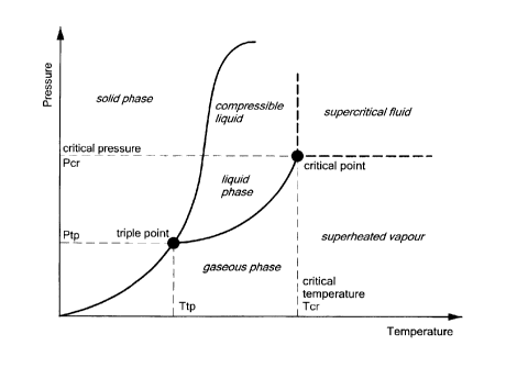
FIG. 11 is a diagram illustrating phase relationship to pressure and
temperature.
DETAILED DESCRIPTION OF THE INVENTION
The present invention includes a cryogenic cooling system and a medical
device for use therewith. Referring now to the drawing figures in which like
reference
designations refer to like elements, an embodiment of a medical system
constructed in
accordance with principles of the present invention is shown in FIG. 1 and
generally
designated as "10." The system generally includes a cooling unit or console 12
coupled to a medical device 14 through an umbilical system 16. The medical
device
14 may be a medical probe, a catheter, a balloon-catheter, as well as other
devices
deliverable or otherwise positionable through the vasculature and/or proximate
to a
tissue region for treatment. In particular, the medical device 14 may include
a device
operable to thermally treat a selected tissue site, including cardiac tissue.
Umbilical system 16 may include three separate umbilicals: a coaxial cable
umbilical 18, an electrical umbilical 20 and a vacuum umbilical 22. An outer
vacuum
umbilical may be suitable for a medical device having multiple layers or
balloons. If
the user wishes to perform a radiofrequency (-RF") ablation procedure,
radiofrequency energy can be provided to electrodes on the medical device 14
via
CA 02772437 2012-02-28
WO 2011/026215
PCT/CA2010/001251
electrical umbilical 20 to perform an RF ablation technique. Electrical
umbilical 20
can include an electrocardiograph ("ECG") box 24 to facilitate a connection
from
electrodes on medical device 14 to an ECG monitor (not shown). Coaxial
umbilical
18 may include both a cooling injection umbilical and a vacuum umbilical that
5 provide respective inlet and return paths for a refrigerant or coolant
used to cool a
tissue-treating section of the device 14. The vacuum umbilical 22 may provide
a
safety conduit allowing excess coolant or gas to escape from the device 14 if
the
pressure within the medical device 14 exceeds a predefined limit. The vacuum
umbilical 22 can also be used to capture air through a leak of the outer
vacuum system
where it is outside the patient and as a lumen to ingress blood when inside
the patient.
Now referring to FIG. 2, the medical device 14 is shown in more detail. The
medical device may include a treatment region 26 for energy interaction
between the
medical device 14 and a treatment site. The treatment region 26 may include,
for
example, a balloon structure that can be a single wall or a double wall
configuration.
In a double-wall or dual-balloon configuration, the space or junction between
balloon
walls may be in communication with a vacuum source. In particular, the medical
device may include a handle 28 having a number of proximal connector ports 30a-
30d. Port 30a may be a first vacuum connector, having a first vacuum lumen
therein,
such as a 10 French lumen. Port 30b may be a coaxial connector having both a
vacuum lumen and injection therein, the vacuum lumen being a second vacuum
lumen, such as an 8 French lumen. Port 30c may be an electrical connector and
port
30d may be a guidewire fuer hub. The medical device 14 may include an
elongate,
flexible catheter body 32 having a guidewire 34 and an inner shaft 36 and
outer shaft
38 having one or more lumens defined therethrough for the circulation and or
deliver
of a fluid or coolant to the treatment region 26 of the medical device 14.
The handle 28 may include blood detection circuitry 40 and a pressure relief
valve 42. The treatment region 26 of the medical device 14 may include a
first, inner
expandable element (such as a balloon) 44 and a second, outer expandable
element 46
surrounding the first expandable element 44. Radiopaque marker bands 48 may be
located proximate the exit point of coolant injected into the treatment region
26 to aid
in the positioning and tracking of the device.
CA 02772437 2012-02-28
WO 2011/026215
PCT/CA2010/001251
6
The medical system 10 may also include one or more sensors to monitor the
operating parameters throughout the system, including for example, pressure,
temperature, flow rates, volume, or the like in the console 12, the umbilical
system
16, or the medical device 14.
Now referring to FIG. 3, a schematic representation of the console 12 for use
with a medical device is shown. The console 12 includes various mechanical
and/or
electrical components to assist in the operation, control, and/or monitoring
of the
medical device 14. Primarily, the console 12 may be coupled to the medical
device 14
through the umbilical system 16 to place a fluid supply lumen 50 and an
exhaust
lumen 52 of the console 12 in fluid communication with the treatment region 26
of
the medical device 14. In general, the console 12 may further include a first
coolant
reservoir 54, a second coolant reservoir 56, and a vacuum source 58 As used
herein,
the term 'reservoir' is intended to include any container or chamber able to
contain a
fluid. As such, either of the first or second reservoirs may include a tank,
container, or
even a length of tubing or the like defining an interior space between two or
more
valves. The second coolant reservoir 56 may have a volumetric capacity smaller
than
the volumetric capacity of the first coolant reservoir 54 (such as 20 cubic
centimeters
for example), which has been shown to reduce the likelihood of cardiac
abnormalities
and/or failure due to coolant egress into the vascular system. The vacuum
source 58
may include any structure and/or apparatus able to provide a negative pressure
gradient for providing fluid flow, including pumps, plunger devices, or the
like.
One or more valves may be disposed about the console 12 in fluid
communication with the supply lumen 50 and/or the exhaust lumen 52 for
manipulating and/or providing fluid flow along a desired path. For example,
the
console 12 may include a pair of valves, 60 and 62, in fluid communication
with the
first coolant reservoir 54 such that the first coolant reservoir 54 may be
selectively
switched from being in fluid communication with the second coolant reservoir
56 to
being in fluid communication with the supply lumen 50. Moreover, a valve 64
may be
disposed on the exhaust lumen 52 such that the exhaust lumen 52 may be
selectively
switched from being in fluid communication with the second coolant reservoir
56 to
being in fluid communication with the vacuum source 58. In addition, the
console 12
may include one or more check valves and/or pressure relief valves CV
configured to
CA 02772437 2012-02-28
WO 2011/026215
PCT/CA2010/001251
7
open to atmosphere or to a recovery tank should a pressure level and/or flow
rate
within a portion of the console 100 exceed a desired or predetermined level.
Such
valves may further be operated to open portions of the system if so desired.
The console 12 may include a valve 66 in fluid communication with both the
supply lumen 50 and the exhaust lumen 52. In particular, the valve 66 may be
in fluid
communication with the supply lumen 50 at a position upstream of the umbilical
connector, while being in fluid communication with the exhaust lumen 52
downstream from the umbilical connector. The valve 66 may further be placed in
fluid communication with the surrounding atmosphere to equalize pressure in
both the
exhaust and supply lumens. During operation, the console 12 may detect a
failure of
the medical device 14, such as an indication of the presence of blood or
bodily fluid
being entrained into the coolant system. Upon such detection, coolant flow may
be
terminated. However, despite the termination of coolant flow, due to the built-
up
pressure levels in the supply and exhaust lumens, bodily fluid may continue to
be
siphoned into the medical device and thus into portions of the console 12. To
reduce
the likelihood that siphoning occurs, the valve 66 may be actuated to place
both the
supply lumen 50 and the exhaust lumen 52 into fluid communication with the
atmosphere. By doing so, the pressure in either lumen will be substantially
equalized
and thus will prevent the further ingress of bodily fluids into the medical
device and
thus the console. Of course, the equalization and/or subjection of both the
supply and
exhaust lumens may be achieved by using one or more valves in various
configuration.
The console 12 may also include a subcooler 68 disposed about a portion of
the supply lumen 50 for achieving a desired temperature and/or coolant phase
of fluid
flowing therethrough. The subcooler 68 may include a compressor, condenser and
the
like placed in thermal communication with the supply lumen 50 as previously
discussed.
FIG. 5 discloses an example of a closed-loop subcooler in schematic form. As
shown, the subcooler includes a heat exchange chamber 70 having a coiled
refrigerant
transfer line 72 passing therethrough. A compressor 76 and condenser 78
provide
liquid refrigerant that is transferred into the chamber 70 as shown by the
arrow
marked "Ref. in." The coolant, if compressed gas expands, or if liquid changes
state to
CA 02772437 2012-02-28
WO 2011/026215
PCT/CA2010/001251
8
gas, thereby chilling the transfer line 72 and its contents. The expanded, gas-
state
coolant is exhausted from the chamber 70 as shown by the arrow marked "Ref.
out"
and returned to the compressor 76; A capillary tube 80 can be interposed
between the
condenser 78 and the chamber 70 in order to reduce the coolant flow into the
heat
exchanging chamber 70.
Another example of a subcooler 68 of the present system is shown in FIG. 6.
The subcooler includes an insulated enclosure 82 (like chamber 70) encloses a
coiled
portion of a coolant supply line 84 leading to a medical implement (not shown)
as
described above. The coolant supply line 84 is in communication with a coolant
reservoir 86 to allow coolant to be directed into the enclosure 82. An outlet
88 in
communication with a vacuum source 90 is provided to exhaust coolant from the
enclosure 82 whereupon it is directed to a scavenging system. Cooling
performance
can be controlled with a coolant flow regulator 92 that can be made responsive
to a
temperature sensor 94 within the enclosure 82 that outputs a signal to a
temperature
controller 96 that controls the flow regulator 92.
Referring now to FIG. 7, an alternate subcooling configuration is shown.
Chamber 98 is depicted having an outlet 100. Provided within the chamber 98 is
a
conduit 102, having a first end 104 and a second end 106, defining a fluid
flow path
for a coolant or a refrigerant. The conduit 102 defines an aperture 108. In
practice, a
refrigerant is supplied to the first end 104 which then passes through the
body of the
conduit 102 to the second end 106. After the refrigerant enters the conduit
102, a
portion of the refrigerant is directed into the chamber 98 via the aperture
108. The
refrigerant then expands to thereby cool the chamber 98 and in turn the
conduit 102.
The expanded refrigerant is then evacuated from the chamber 98 via the outlet
100.
The rate of flow through the aperture 108 can be controlled by the size of the
aperture
as well as by flow control valves as discussed herein (not shown). The
diameter of the
aperture can range from 0.0001 to 0.03 inches, for example. The rate of
subcooling
affected within the chamber 98 can be regulated by adjusting the flow rate of
the
outlet 100. By decreasing the flow rate allowed at the outlet 100, the amount
of
refrigerant entering the chamber 98 via the aperture 108 is thereby decreased
and the
subcooling reduced. Further, it is contemplated that the location of the
aperture along
the conduit 102 can be varied.
CA 02772437 2012-02-28
WO 2011/026215
PCT/CA2010/001251
9
Referring now to FIG. 8 which is a schematic view of another alternate
embodiment of a subcooler illustrated in more detail. In the illustrated
arrangement,
refrigerant is supplied to the system from a coolant source 110. The
refrigerant passes
through a filter or contaminant remover 112 (optional) and then to a junction
114.
One branch of the junction passes through a vent system 116 and the other
branch
passes through subcooler 118. The subcooler 118 chills the refrigerant to a
temperature that causes the refrigerant to be in the liquid state prior to
transfer to the
medical device 14. The illustrated arrangement permits placement of the
subcooler
within accessories external to the console, for example, in an connection box
or
intermediary console (not shown), in a catheter handle assembly or any other
such
device located between the medical device 14 and the console 12.
Referring now to FIG. 9, yet another configuration for a subcooler is
illustrated in conjunction with a control system for the subcooler. As with
configurations described above, this illustration depicts a heat exchange
chamber 120,
having an inlet 122 and an outlet 124, provides a flow path for refrigerant
such as
nitrous oxide or another fluid. A conduit 126 that defines a second fluid flow
path for
the same refrigerant passes through the chamber 120 and is in fluid
communication
with a refrigerant supply upstream of the chamber and a medical device
downstream
from the chamber. As shown, a fluid flow splitter 128 can allow a common
refrigerant
source to be used for supplying the chamber 120 and the conduit 126.
A programmable controller 130 is in communication with and controls one or
more valves, such as a first valve 132, to regulate flow of coolant through
the conduit
126 and into the medical device in response to a programmed cooling profile
and in
response to sensor outputs from the catheter. Additionally, the controller 130
can be
used to control a second valve 134 to regulate flow of coolant through the
chamber
120 in response to sensed temperature within the chamber. For example, the
controller
130 can establish a duty cycle that opens and closes the second valve 134
repeatedly
over time. If the temperature rises in the chamber 120, the second valve 134
can be
opened and closed more frequently. By contrast, if the temperature in the
chamber
falls too far, the second valve 134 can be cycled less frequently. Another
example
includes establishing a duty cycle to specifically regulate the temperature
increases
and decreases at the treatment site. It is advantageous to be able to
precisely control
CA 02772437 2012-02-28
WO 2011/026215
PCT/CA2010/001251
the freezing and thawing rates of the treatment region 26 of the medical
device 14
when performing a medical treatment procedure. Further, by sensing the actual
temperatures and adjusting the opening and closing of the system valves, the
application of specific temperature regimens can be accomplished.
5 Referring now to FIG. 10, yet another configuration for a subcooler is
illustrated in conjunction with a control system for the subcooler. The
subcooler
feature is provided by a thermoelectric cooler 136, such as a peltier cooler,
the
operation of which is known in the art. The thermo-electric cooler has a hot
side 138
and a cold side 140. A conduit 142 is provided adjacent and in thermally-
conductive
10 communication with the cold side 140 of the thermo-electric cooler 136.
A
supplemental cooler 144 is provided adjacent to and in thermally-conductive
communication with the hot side 138 of the thermoelectric cooler. The conduit
142,
the thermoelectric cooler 136 and the supplemental cooler 144 are enclosed by
a
housing 146. The supplemental cooler 144 is connected to an external cooling
source
148 which can be any of the cooling arrangements disclosed herein or other
such
devices.
When the thermoelectric cooler is activated, the temperature of the cold side
140 is reduced and thereby reduces the temperature of the adjacent conduit
412,
which in turn reduces the temperature of refrigerant passing through the
conduit 142.
Further, the hot side 138 increases in temperature. The cooling source 148
supplies
cold energy to the supplemental cooler 144 which thereby cools the adjacent
hot side
138. By cooling the hot side 138, heat is removed from the housing 146 and the
cooling efficiency of the supplemental cooler 144 is increased. It is further
contemplated that the hot side 138 can be cooled by more conventional means
such as
moving air across it. Additionally, a heat sink can be provided in thermal
communication with the hot side 138 to increase cooling efficiency.
Again referring to FIG. 3, one or more sensors may be disposed about the
supply and exhaust lumens of the console 12 for detecting temperature,
pressure,
and/or flow rates through a particular portion of the console plumbing. For
example, a
first pressure sensor 150 may be disposed about the exhaust lumen 52 proximate
to
the umbilical connector. In addition, a second pressure sensor 152 may be
disposed
about the supply lumen 50. Additional sensors SS may be included throughout
the
CA 02772437 2012-02-28
WO 2011/026215
PCT/CA2010/001251
11
console 12 for monitoring and/or controlling particular portions of the
console and
properties thereof.
In addition to the one or more sensors, one or more controllers may be coupled
to the sensors, and in turn, coupled to one or more of the valves situated
throughout
the console 12 such that the valves may be controllably manipulated in
response to
information obtained by the sensors. For example, a first controller 154 may
be
coupled to the first pressure sensor 150, wherein the first controller 154 is
further
coupled to a valve 156 disposed on a portion of the exhaust line, and where
the valve
156 may also be in fluid communication with the vacuum source 58. In addition,
a
second controller 158 may be coupled to the second pressure sensor 152, where
the
second controller 158 is further coupled to a valve 160 disposed about the
supply
lumen 50. Accordingly, fluid flow through portions of the exhaust and/or
supply
lumens may be controllably manipulated in direct response to the information
obtained by sensors contained therein.
Now referring to FIG. 4, an embodiment of the console 12, such as a cooling
system for a cryogenic medical device, is shown. As shown, the console
contains
several of the valves, sensors and components discussed above with respect to
FIG. 3.
The console 12 further includes a bypass coolant supply line 162 extending
from a
junction between valves 62 and 160. The bypass coolant supply line 162
includes a
bypass valve 164, and rejoins the coolant supply line 50 on a distal side of
the
subcooler 68. The bypass coolant supply line 162 provides an avenue, conduit,
or
fluid pathway for delivery of coolant to the medical device without
interacting or
being exposed to the subcooler. The bypass may provide for the delivery of
relatively
warmer (or non-subcooled) coolant to the medical device 14 to inflate it
without
cooling, or to thaw or otherwise increase the temperature of a portion of the
medical
device 14, such as the treatment region 26.
In an exemplary use, the console 12 may be operated to deliver a refrigerant
or
coolant in a supercritical state to the medical device 14 for subsequent
thermal
treatment of selected tissue. A supercritical fluid is a substance at a
temperature and
pressure above its defined critical point. A critical point, also called a
critical state,
specifies the conditions (temperature, pressure and sometimes composition) at
which
a phase boundary ceases to exist. To reach or exceed a materials critical
point,
CA 02772437 2012-02-28
WO 2011/026215
PCT/CA2010/001251
12
predetermined temperatures and pressure must be obtained. Critical properties
vary
from material to material, similar to melting points and boiling points.
As shown in the phase diagram of FIG. 11, the supercritical phase of a
substance lies beyond the liquid and gaseous phases ¨ resulting in a fluid
having
characteristics of both. Supercritical fluids typically have gaseous
characteristics
(such as the ability to diffuse through solids) as well as liquid
characteristics (such as
the ability to dissolve materials). In the pressure-temperature phase diagram
of FIG.
11, boiling a material separates the gas and liquid region and ends in the
critical point,
where the liquid and gas phases disappear to become a single supercritical
phase. As
the critical temperature is approached, the density of the gas at equilibrium
becomes
denser, and that of the liquid lower. At the critical point, there is no
difference in
density, and the liquid and gaseous phases become one fluid phase. Thus, above
the
critical temperature a gas cannot be liquefied by pressure. A small increase
in pressure
causes a large increase in the density of the supercritical phase, allowing
many
properties of a supercritical fluid to be selectively and controllably
manipulated.
Many other physical properties also show large gradients with pressure near
the
critical point, e.g. viscosity, the relative permittivity and the solvent
strength, which
are all closely related to the density.
By delivering a supercritical fluid to the treatment region 26 of the medical
device 14, lower temperatures can be achieved through the expansion and/or
evaporation of the coolant once delivered to the treatment region. The lower
temperatures may be obtained by using a Joule-Thompson valve to obtain the
desired
expansion. The supercritical coolant has increased thermodynamic capacity for
cooling upon expansion compared to a liquid or gaseous phase, resulting in
lower
thermal temperatures ¨ which reduces the time needed for tissue ablation.
In an exemplary method of operation, a coolant or refrigerant having a pre-
defined critical point may be supplied by or otherwise contained in the first
coolant
reservoir 54. Exemplary coolants may include methane, argon, nitrogen, oxygen,
krypton, and neon. The coolant in the first reservoir 54 may be at a pressure
and/or
temperature combination such that the coolant is in a liquid phase, or in a
mixed
liquid-gaseous phase. The first coolant reservoir 54 may include a dip tube or
other
structure to ensure that only the liquid-phase coolant is drawn from the
reservoir 54
CA 02772437 2014-01-08
13
during use. The coolant may then proceed through the valves and conduits
described above,
which may direct the coolant through a subcooler prior to reaching the medical
device 14. The
subcooler may operate to modify the temperature and pressure characteristics
of the coolant to
ensure the supercritical state of the coolant passing through the supply lumen
50. Once passing
through the subcooler, the remaining lengths of conduit leading to the
treatment region 26 of the
medical device may include insulative properties to reduce thermal exchange
with the
surrounding environment. To further increase the stability of the
supercritical state of the
coolant, the dimensions of the fluid supply tube leading to and through the
length of the medical
device 14 may be dimensioned to reduce the volume of coolant passing
therethrough and to
further maintain a desired pressure throughout the delivery path. Upon
reaching the treatment
region of the medical device 14, the supercritical coolant may be dispersed
through a valve or
expansion element, thereby allowing at least a portion of the ejected coolant
to change phase into
a liquid, gas and/or combination thereof. The expansion into a gaseous phase
and subsequent
evaporation of the liquid phase coolant within the treatment region 26
provides increased cooling
capacity and reduced temperatures for thermal ablation of a selected tissue
region.
It will be appreciated by persons skilled in the art that the present
invention is not limited
to what has been particularly shown and described herein above. In addition,
unless mention
was made above to the contrary, it should be noted that all of the
accompanying drawings are not
to scale. A variety of modifications and variations are possible in light of
the above teachings.