Note: Descriptions are shown in the official language in which they were submitted.
CA 02772447 2012-02-28
WO 2011/032284 PCT/CA2010/001460
Title: Steroidogenesis Modified Cells and Methods for Screening for
Endocrine Disrupting Chemicals
[0001] This Patent Cooperation Treaty application claims the benefit of 35
USC 119 based on the priority of co-pending US Provisional Patent Application
61/242,822, filed September 16, 2009 which is incorporated herein in its
entirety
by reference.
Field of the Disclosure
[0002] The disclosure relates to steroidogenesis modified cells such as
modified H295R cells which are modified to reduce the expression of one or
more enzymes (knocked down) involved in steroidogenesis. The disclosure also
relates to methods, uses and compositions comprising these cells for
identifying
endocrine disrupting substances.
Background of the Disclosure
[0003] Over the past two decades, there has been increasing concern
about the possible effects of exposure to chemicals in the environment on
endocrine and reproductive systems in humans and wildlife (Kavlock et al.
1996). To address these concerns, national and international programs have
been initiated to develop new guidelines for the screening and testing of
potential endocrine-disrupting chemicals (EDCs) in vertebrates. The Safe
Drinking Water Act Amendments of 1995 and the Food Quality Protection Act of
1996 mandate screening for endocrine-disrupting properties of chemicals in
drinking water or pesticides used in food production. In response to this
legislation, the federal Endocrine Disrupter Screening and Testing Advisory
Committee (EDSTAC) has identified disrupting the process of steroidogenesis
as one of the important toxicity pathways of endocrine disruption in addition
to
binding to three key endocrine nuclear receptors, i.e. estrogen receptors
(ER),
androgen receptors (AR) and thyroid hormone receptor (ThR) (Hilscherova et
al. 2004; Sanderson et al. 2002; Zhang et al. 2005). The human H295R
adrenocarcinoma cell-based steroidogenesis assay has been approved by the
United States Environmental Protection Agency (USEPA) for use in Tier I of the
1
CA 02772447 2012-02-28
WO 2011/032284 PCT/CA2010/001460
Endocrine Disruptor Screening Program (EDSP) and is currently in the last
phase of validation through OECD as an international standard to test
chemicals for endocrine disrupting effects. While previously used assays have
used production of different mRNAs as endpoints, currently used assays use as
endpoints the production and release to the medium of the steroid hormones
testosterone (T) and 1713-estradiol (E2) (Hecker et al. 2006).
(0004] H295R cells express genes that encode for all the key enzymes
involved in steroidogenesis (Figure. 1) (Gazdar et al. 1990; Staels et al.
1993;
Rainey et al. 1994). This is a unique property, because in vivo expression of
these genes is tissue- and developmental stage-specific with no one tissue or
developmental stage simultaneously expressing all of the genes involved in
steroidogenesis. H295R cells have physiological characteristics of zonally
undifferentiated human fetal adrenal cells. H295R cells represent a unique in
vitro system with the ability to produce the steroid hormones found in the
adult
adrenal cortex and the gonads, which allows testing for effects on both
corticosteroid synthesis and the production of sex steroid hormones such as
androgens and estrogens.
[0005] One of the key hormones of interest, E2, is produced by H295R
cells at relatively small and varying concentrations (-10 - 50 pg E2/ml in
culture
medium) that are difficult to measure by use of automated ELISA or the more
laborious LC\MS-MS method. Concentrations of E2 released by H295R cells
into the medium are near the current limit of quantification (LOQ,
approximately
2 - 10 pg E2/ml), which makes it difficult to measure reductions in E2 release
caused by EDCs. The relatively great variance in E2 production around the
detection limit is also a limiting factor. In addition, due to the small basal
concentrations released by the H295R cells into the medium, it is difficult to
demonstrate a decrease in production, which is also important for use as a
screening tool. This is especially true with regard to the assessment of weak
inhibitors.
[0006] Another endpoint of interest in screening for potential endocrine
disruption is changes in expression of the aromatase (CYP19) gene, protein
2
CA 02772447 2012-02-28
WO 2011/032284 PCT/CA2010/001460
and enzyme activity, the enzyme that transforms (aromatizes) Testosterone to
E2.
Summary of the Disclosure
[0007] In an aspect, the disclosure provides an isolated steroidogenesis
modified cell comprising a steroid biosynthesis knock down nucleic acid
operatively linked to a promoter, wherein the steroid biosynthesis knock down
nucleic acid reduces the expression of a gene selected from the group
CYP21A2, CYP11A1, CYP17A1, CYP19A1, 3-PHSD1, 3-(3HSD2, 17-(3HSD1,
StAR, HMGR, CYP11 B2, CYP11131, 5a-Reductase 2, SULT1 E1, CYP3A4 and
UTG1A1 wherein the cell comprises reduced expression of one or more of said
genes.
[0008] In an embodiment, the knock down nucleic acid comprises a
siRNA nucleic acid, a shRNA nucleic acid or an antisense nucleic acid.
[0009] In an embodiment, the one or more genes comprises CYP21A2. In
an embodiment, the steroid biosynthesis knock down nucleic acid comprises
CCGGCGACAACTTAATGCCTGCCTACTCGAGTAGGCAGGCATTAAGTTGTC
GTTTTTG (SEQ ID NO:1).
[0010] In an embodiment, the one or more genes comprises CYP11A1. In
an embodiment, the steroid biosynthesis knock down nucleic acid comprises
TGCTGTTGACAGTGAGCGACCTGCAGAGATATCTTGTAAATAGTGAAGCCA
CAGATGTATTTACAAGATATCTCTGCAGGGTGCCTACTGCCTCGGA (SEQ
ID NO:2).
[0011] In an embodiment, the one or more genes comprises CYP17A1. In
an embodiment, the steroid biosynthesis knock down nucleic acid comprises
TGCTGTTGACAGTGAGCGCGGGCACAGAAGTTATCATCAATAGTGAAGCCA
CAGATGTATTGATGATAACTTCTGTGCCCTTGCCTACTGCCTCGGA (SEQ ID
NO:3).
[0012] In an embodiment, the one or more genes comprises CYP19A1.
In an embodiment, the steroid biosynthesis knock down nucleic acid comprises
TGCTGTTGACAGTGAGCGAAGAACCAGGCTACAAGAGAAATAGTGAAGCCA
3
CA 02772447 2012-02-28
WO 2011/032284 PCT/CA2010/001460
CAGATGTATTTCTCTTGTAGCCTGGTTCTCTGCCTACTGCCTCGGA (SEQ ID
NO:4).
[0013] In an embodiment, the one or more genes comprises 3-(3HSD1. In
an embodiment, the steroid biosynthesis knock down nucleic acid comprises
CCGGCGCCTGTATCATTGATGTCTTCTCGAGAAGACATCAATGATACAGGC
GTTTTTG (SEQ ID NO:5).
[0014] In an embodiment, the one or more genes comprises 3-PHSD2. In
an embodiment, the steroid biosynthesis knock down nucleic acid comprises
TGCTGTTGACAGTGAGCGACCACACAGTCACATTATCAAATAGTGAAGCCA
CAGATGTATTTGATAATGTGACTGTGTGGCTGCCTACTGCCTCGGA (SEQ ID
NO:6).
[0015] In an embodiment, the one or more genes comprises 17-(3HSD1.
In an embodiment, the steroid biosynthesis knock down nucleic acid comprises
TGCTGTTGACAGTGAGCGCGGGTGGCTAATTAAGATAGATTAGTGAAGCCA
CAGATGTAATCTATCTTAATTAGCCACCCATGCCTACTGCCTCGGA (SEQ ID
NO:7).
[0016] In an embodiment, the one or more genes comprises StAR. In an
embodiment, the steroid biosynthesis knock down nucleic acid comprises
CCGGGCTGCCCAAGAGCATCATCAACTCGAGTTGATGATGCTCTTGGGCA
GCTTTTTG (SEQ ID NO:8).
[0017] In an embodiment, the one or more genes comprises HMGR. In an
embodiment, the steroid biosynthesis knock down nucleic acid comprises
CCGGGCAGTGATAAAGGAGGCATTTCTCGAGAAATGCCTCCTTTATCACTG
CTTTTT (SEQ ID NO:9).
[0018] In an embodiment, the one or more genes comprises CYP11 B2. In
an embodiment, the steroid biosynthesis knock down nucleic acid comprises
CCGGCCTCACTTTCAGAGCGATTAACTCGAGTTAATCGCTCTGAAAGTGAG
GTTTTTG (SEQ ID NO:10).
[0019] In an embodiment, the one or more genes comprises CYP11 B1. In
an embodiment, the steroid biosynthesis knock down nucleic acid comprises
4
CA 02772447 2012-02-28
WO 2011/032284 PCT/CA2010/001460
CCGGCCCTCAACAGTACACCAGCATCTCGAGATGCTGGTGTACTGTTGAGG
GTTTTTG (SEQ ID NO:11).
[0020] In an embodiment, the one or more genes comprises 5a-
Reductase 2. In an embodiment, the steroid biosynthesis knock down nucleic
acid comprises
CCGGCCTCAAGATGTTTGAGGACTACTCGAGTAGTCCTCAAACATCTTGAG
GTTTTTG (SEQ ID NO:12).
[0021] In an embodiment, the one or more genes comprises SULT1E1. In
an embodiment, the steroid biosynthesis knock down nucleic acid comprises
CCGGCCAGAAATTGTCGCCCTTCATCTCGAGATGAAGGGCGACAATTTCTG
GTTTTTG (SEQ ID NO:13).
[0022] In an embodiment, the one or more genes comprises CYP3A4. In
an embodiment, the steroid biosynthesis knock down nucleic acid comprises
CCGGCCTTACATATACACACCCTTTCTCGAGAAAGGGTGTGTATATGTAAG
GTTTTTG (SEQ ID NO:14).
[0023] In an embodiment, the one or more genes comprises UGT1A1. In
an embodiment, the steroid biosynthesis knock down nucleic acid comprises
CCGGCCCACTGTATTCTTCTTGCATCTCGAGATGCAAGAAGAATACAGTGG
GTTTTTG (SEQ ID NO:15).
[0024] In an embodiment, the isolated steroidogenesis modified cell is an
isolated steroidogenesis H295R modified cell.
[0025] In an embodiment, the isolated steroidogenesis modified cell is an
isolated steroidogenesis H295, JEG-3 or R2C modified cell.
[0026] In an embodiment, the isolated steroidogenesis modified H295R
cell comprises a CYP21A2 knock down nucleic acid operatively linked to a
promoter, wherein the CYP21A2 knock down nucleic acid reduces the
expression of CYP21A2.
[0027] In an embodiment, the isolated isolated steroidogenesis modified
H295R cell comprises a CYP17A1] knock down nucleic acid operatively linked
5
CA 02772447 2012-02-28
WO 2011/032284 PCT/CA2010/001460
to a promoter, wherein the CYP17A1 knock down nucleic acid reduces the
expression of CYP17A1.
[0028] In an embodiment, the isolated isolated steroidogenesis modified
H295R cell comprises a CYP19A1 knock down nucleic acid operatively linked
to a promoter, wherein the CYP19A1 knock down nucleic acid reduces the
expression of CYP19A1.
[0029] In another aspect, the disclosure further provides a screening
assay for identifying an endocrine disruptor comprising: a) contacting a
steroidogenesis cell such as a steroidogenesis modified H295R cell of the
present disclosure with a test substance; b) determining a level of at least
one
steroid or steroidogenic gene expression product, e.g. mRNA or protein, or
enzyme activity; wherein a modulation in the level of the at least one steroid
or
steroidogenic gene expression product or enzyme activity compared to a
control is indicative that the test substance is an endocrine disruptor.
[0030] A further aspect includes a kit for screening for an endocrine
disruptor comprising a steroidogenesis cell described herein and a component
for determining the level of at least one steroid.
[0031] In yet a further aspect, the disclosure provides a system for
predicting the mechanism of action of an endocrine disruptor with unknown
mechanism comprising: (i) a control module for receiving a steroid production
profile for the endocrine disruptor wherein the steroid production profile is
obtained by contacting the endocrine disruptor with a steroidogenesis cell
such
as a steroidogenesis modified H295R cell disclosed herein and determining a
level of at least one steroid or steroidogenic gene expression product or
activity
produced by the cell line; (ii) a database comprising steroid production
profiles
for a plurality of reference endocrine disruptors; (iii) analysis module for
comparing the steroid production profile of the endocrine disruptor with the
steroid production profiles of the plurality of reference endocrine
disruptors; and
for identifying a best match for the steroid production profile of the
endocrine
disruptor with the steroid production profiles of the plurality of reference
endocrine disruptors, wherein the mechanism of action of the best match
6
CA 02772447 2012-02-28
WO 2011/032284 PCT/CA2010/001460
reference endocrine disruptor is predicted to be the mechanism of action of
the
endocrine disruptor.
[0032] Other features and advantages of the present disclosure will
become apparent from the following detailed description. It should be
understood, however, that the detailed description and the specific examples
while indicating preferred embodiments of the disclosure are given by way of
illustration only, since various changes and modifications within the spirit
and
scope of the disclosure will become apparent to those skilled in the art from
this
detailed description.
Brief description of the drawings
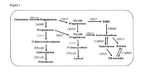
[0033] Figure 1 Steroidogenesis pathways in H295R cells. CYP11A,
desmolase (20,22 Desmolase); CYP17, steroid 17a-hydroxylase; CYP21,
steroid 21-hydroxylase; CYP19, aromatase; 313HSD, 313-hydroxysteroid
dehydrogenase; CYP11 B1, steroid 1113 hydroxylase, CYP11 B2, aldosterone
synthetase; 1713HSD, 1713 hydroxysteroid dehydrogenase. The enzyme CYP21
encoded by human CYP21A2 gene is highlighted.
[0034] Figure 2 Western blot analysis of human CYP21A2 protein
expression in the stable H295R/CYP21A2-KD and unaltered H295R cells.
Unrelated CYP1 1A protein detected on the same plot membrane was used as a
reference. Values presented are means +/- standard deviation.
[0035] Figure 3 Production of progesterone (P), T, El and E2 in
H295R and H295R/CYP21A2-KD cell line after exposure to forskolin (FOR) and
prochloraz (PRO). SC: Solvent Control.
[0036] Figure 4 Comparison of the hormone production (pg/mL)
between the H295R/CYP21A2-KD and unaltered H295R cells under the same
chemical exposure conditions.
[0037] Figure 5 Comparison of the hormone basal production (pg/mL)
between H295R/CYP17-KD, H295R/CYP19-KD and unaltered H295R
cells.
7
CA 02772447 2012-02-28
WO 2011/032284 PCT/CA2010/001460
Detailed description of the Disclosure
1. Definitions
[0038] As used herein "a", "an" and/or "the" includes one and/or more than
one.
[0039] The term "endocrine disrupting chemical" or "endocrine disrupting
compound" also referred to as "endocrine disruptor" or "hormonally active
agent" as used herein refers to an exogenous substance that interferes with
the
synthesis, secretion, transport, binding, action, or elimination of endogenous
hormones in vertebrates including human and/or invertebrates (mollusks,
crustacean, etc.). Endocrine disrupting compounds include a number of
chemical classes, including for example, pesticides, compounds used in the
plastics industry and in consumer products, compounds used as food additives
and in cosmetics and other industrial by-products, pharmaceuticals, naturally
occurring hormones (e.g., phytoestrogens), degradation products and
metabolites of any of these classes of compounds and by-products of
manufacture, and pollutants.
[0040] The term "steroidogenesis cell" as used herein refers to any cell,
modified or unmodified that produces one or more steroid hormones including
sex steroids, mineral- and corticosteroids. Specifically, these include
estrogens
(17beta-estradiol, estrone) and androgens (testosterone, androstenedione,
dihydrotestosterone), as well as the mineral- and corticosterdoids and/or
their
precursors, andincludes for example a H295R cell, a steroidogenesis modified
H295 cell, a steroidogenesis modified H295R cell, a JEG-3, a R2C cell, a
steroidogenesis modified JEG-3 cell and a steroidogenesis modified R2C cell .
[0041] The term "steroidogenesis modified H295R cell" as used herein
refers to a H295R cell that has been modified by, for example, recombinant
technology to knock down the gene expression of one or more genes involved
in a steroidogenesis pathway e.g. a steroid biosynthesis gene, including but
not
limited to the following genes: CYP21A2, CYP11A1, CYP17A1, CYP19A1, 3-
RHSD1, 3-3HSD2, 17-RHSD1, StAR, HMGR, CYP11B2, CYP11B1, 5a-
Reductase 2, SULT1E1, UGT1Al and CYP3A4. For example, the expression of
8
CA 02772447 2012-02-28
WO 2011/032284 PCT/CA2010/001460
two or more of the genes listed above can be knocked down simultaneously or
contemporaneously e.g. using a construct that produces two or more siRNA
nucleic acids, each targeting a gene involved in a steroidogenesis pathway, or
two or more of the genes can be knocked down sequentially. For example, a
H295R cell modified to knock down gene expression of CYP21A2 can be
further modified to knock down expression of another gene, for example
CYP11A1. Further, for example, the expression of related steroidogenesis
gene family members could also be knocked down to produce steroidogenesis
modified cells. For example, if a new steroidogenesis gene for example a new
CYP21 subfamily gene were identified, also involved in steroidogenesis, and/or
if a steroidogeneis cell to be modified expressed additional steroidogenesis
genes other than the specific ones mentioned herein e.g. additional CYP21
family members, the expression of one or more of these genes could also be
knocked down to produce a cell useful for the methods described herein.
[0042] The term "steroid ogenesis modified H295 cell" as used herein
refers to a H295 cell that has been modified by for example, recombinant
technology to knock down the gene expression of one or more genes involved
in a steroidogenesis pathway e.g. a steroid biosynthesis gene, including but
not
limited to the following genes: CYP21A2, CYP11A1, CYP17A1, CYP19A1, 3-
(3HSD1, 3-(3HSD2, 17-(3HSD1, StAR, HMGR, CYP11 B2, CYP11 B1, 5a-
Reductase 2, SULT1 El, UGT1A1 and CYP3A4. For example, the expression of
two or more of the genes listed above can be knocked down simultaneously or
contemporaneously e.g. using a construct that produces two or more siRNA
nucleic acids, each targeting a gene involved in a steroidogenesis pathway, or
two or more of the genes can be knocked down sequentially. For example, a
H295 cell modified to knock down gene expression of CYP21A2 can be further
modified to knock down expression of another gene, for example CYP11A1.
[0043] Similarly, the term "steroidogenesis modified JEG-3 cell" as used
herein refers to a JEG-3 cell that has been modified by for example,
recombinant technology to knock down the gene expression of one or more
genes involved in a steroidogenesis pathway e.g. a steroid biosynthesis gene,
9
CA 02772447 2012-02-28
WO 2011/032284 PCT/CA2010/001460
including but not limited to the following genes: CYP21A2, CYP11A1,
CYP17A1, CYP19A1, 3-(3HSD1, 3-(3HSD2, 17-(3HSD1, StAR, HMGR,
CYP11B2, CYP11B1, 5a-Reductase 2, SULT1E1, UGT1A1 and CYP3A4 and
the term "steroidogenesis modified R2C cell" as used herein refers to a R2C
cell that has been modified by for example, recombinant technology to knock
down the gene expression of one or more genes involved in a steroidogenesis
pathway e.g. a steroid biosynthesis gene, including but not limited to the
following genes: CYP21A2, CYP11A1, CYP17A1, CYP19A1, 3-(3HSD1, 3-
3HSD2, 17-I3HSD1, StAR, HMGR, CYP11 B2, CYP11 B1, 5a-Reductase 2,
SULT1E1, UGT1A1 and CYP3A4.
[0044] The term "modified" as used herein in terms of a steroidogenesis
modified cell means genetically altering the cell, for example by recombinant
technology, to knock down and/or reduce with the expression of one or more
genes involved in a steroidogenesis pathway. This can for example be
accomplished by antisense technology and/or replacing the gene by
homologous recombination with a variant that exhibits decreased expression
for example because of a weaker promoter etc.
[0045] The term "a steroid biosynthesis gene" as used herein refers to a
gene involved in a steroidogenesis pathway and includes but is not limited to
a
gene selected from CYP21A2, CYP11A1, CYP17A1, CYP19A1, 3-j3HSD1, 3-
RHSD2, 17-(3HSD1, StAR, HMGR, CYP11B2, CYP11B1, 5a-Reductase 2,
SULT1E1, UGT1A1 and CYP3A4 (for example see Figure 1, Table 1).
[0046] The term "CYP21A2" also known as "steroid 21-hydroxylase" or
"21-hdoxylase" and optionally "CYP21" or "CYP21 B" refers to cytochrome
P450, family 21, subfamily A, polypeptide 2,preferably human to cytochrome
P450, family 21, subfamily A polypeptide 2, for example as disclosed in Entrez
GenelD1589.
[0047] The term "CYP11A1" also referred to as "desmolase" or "20,22
Desmolase" refers to a cytochrome P450 family 11, submfamily A polypeptide
1, preferably human cytochrome P450 family 11, submfamily A polypeptide 1,
for example as disclosed in Entrez Gene ID 1583.
CA 02772447 2012-02-28
WO 2011/032284 PCT/CA2010/001460
[0048] The term "CYP17A1" is for example also referred to as steroid
17a-hydroxylase, CYP17A and CYP17.
[0049] The term "CYP19A1" is for example also referred to as aromatase,
"CYP19", "CYP19A", and "-450AROM", for example as disclosed in Entrez
GeneID1588.
[0050] The terms "313HSD1" also referred to for example as "Type 1
3(3HSD", "313HSD", 33-hydroxysteroid dehydrogenase Type 1 or 313-
hydroxysteroid dehydrogenase, for example as disclosed in Entrez
GenelD3283.
[0051] The term "313HSD2" is also for example referred to as The term
"313HSD2" also referred to as "Type 2 313HSD", "3(3HSD", "HSD3B2", 313-
hydroxysteroid dehydrogenase Type 2 or 33-hydroxysteroid dehydrogenase, for
example as disclosed in Entrez 3284.
[0052] The term "CYP11B1" is also for example referred to "P450C11",
steroid 1113 hydroxylase, for example as disclosed in Entrez GeneID1584.
[0053] The term "CYP11 B2" is also referred to for example as
"P450C18", aldosterone synthetase, for example as disclosed in Entrez
GenelD1585.
[0054] The term "1713HSD1" is also for example referred to as "1713HSD",
1713 hydroxysteroid dehydrogenase Type 1 or 1713 hydroxysteroid
dehydrogenase, for example as disclosed in Entrez GenelD3292.
[0055] The term "StAR" is also for example referred to as steroidogenic
acute regulatory protein, for example as disclosed in Entrez Gene ID: 6770.
[0056] The term "HMGR" is also for example referred to as "HMGCR", 3-
hydroxy-3-methylglutaryl-coenzyme A reductase, for example as disclosed in
Entrez Gene ID: 3156.
[0057] The term "SULTIEI" is also for example referred to as "EST-l",
"EST", 5a-Reductase 2, estrogen-preferring sulfotransferas, for example as
disclosed in Entrez GeneID6783.
11
CA 02772447 2012-02-28
WO 2011/032284 PCT/CA2010/001460
[0058] The term "CYP3A4" is also for example referred to as "CYP3A",
"P450C3", cytochrome P450 family 3 subfamily A polypeptide 4, for example
as disclosed in Entrez GeneID1576.
[0059] The term "UGT1A1" is also for example referred to as "UGT1A1"
also referred to as "UGT1", "UDPGT", UDP-glucuronosyltransferase, for
example as disclosed in Entrez GeneID54658.
[0060] The term "steroidogenesis pathway" as used herein refers to the
genes, enzymes, substrates, intermediates and final products involved in
steroid biosynthesis including for example corticosteroid synthesis, including
mineralo- and gluco- corticosteroids such as aldosterone and cortisol
respectively, and the production of sex steroid hormones such as androgens
and estrogens.
[0061] The term "steroidogenesis gene" or "steroid bysynthesis gene"
refers to a gene selected from CYP21A2, CYP11A1, CYP17A1, CYP19A1, 3-
I3HSD1, 3-(3HSD2, 17-I3HSD1, StAR, HMGR, CYP11 B2, CYP11 B1, 5a-
Reductase 2, SULT1E1, UGT1A1 and CYP3A4.
[0062] The term "steroid biosynthesis knockdown nucleic acid" refers to a
nucleic acid molecule that is specific for reducing or "knocking down" the
expression of a steroid biosynthesis gene, such as a gene selected from
CYP21A2, CYP11A1, CYP17A1, CYP19A1, 3-(3HSD1, 3-(3HSD2, 17-I3HSD1,
StAR, HMGR, CYP11 B2, CYP11131, 5a-Reductase 2, SULT1 E1, UGT1A1 and
CYP3A4. For example, expression of a steroid biosynthesiss gene can be
reduced by introducing a small interfering RNA (siRNA), small hairpin RNA or
short hairpin RNA (shRNA), or antisense nucleic acid that is specific for the
steroid biosynthesis gene. Reference to a specific knockdown nucleic acid that
targets a gene is made by referring to the gene being knocked down such that
for example, a CYP21A2 knockdown nucleic acid refers to a steroid
biosynthesis knockdown nucleic acid that reduces expression of CYP21A2, and
a StAR knockdown nucleic acid refers to a steroid biosynthesis knockdown
nucleic acid that reduces expression of StAR. Several genes can for example
be targeted simultaneously, and/or several regions of a single gene can be
12
CA 02772447 2012-02-28
WO 2011/032284 PCT/CA2010/001460
targeted, for example multiple RNAi can be accomplished by introducing
multiple steroid biosynthesis knockdown nucleic acids (e.g. multiple siRNA or
shRNA species), for example by transfection and/or viral transfer.
Alternatively,
a construct with several consecutive steroid biosynthesis knockdown nucleic
acids (e.g. each optionally operatively linked to a promoter) or expression of
a
composite steroid biosynthesis knockdown nucleic acid (e.g. RNA) comprising
several consecutive steroid biosynthesis knockdown nucleic acids that is
cleaved into multiple shRNAs (e.g. individual steroid biosynthesis knockdown
nucleic acids) can be used.
[0063] The term "a cell" as used herein includes a plurality of cells and
includes a cell line.
[0064] The term an "isolated cell" as used herein refers to a cell or
population of cells including a cell line that has been removed from the
environment in which the cell occurs naturally and/or that have been modified
from the state in which the cell occurs in its natural environment.
[0065] The term "H295R" or "NCI-H295R" as used herein refers to a strain
of H295 cells selected for attachment to culture dishes and which are a
pluripotent human adrenocortical carcinoma cell line that expresses genes that
encode all the key enzymes involved in steroidogenesis. H295R is publicly
available from, for example the American Type Culture Collection (ATCC). The
term H295R also includes sub-strains and sub-clones of H295R cells. H295R
cells are derived from H295 cells which also express all of the key enzymes
involved in steroidogenesis (Gazdar et al. 1990; Staels et al. 1993; Rainey et
al. 1994). As H295R cells are a strain of H295 cells, a person skilled in the
art
would recognize that parental H295 cells and other strains thereof can also be
used to make the modified cells of the disclosure for use for example in
screening assays described herein. Accordingly, the disclosure is intended to
encompass steroidogenesis modified H295 cells, the making of such cells and
the use of steroidogenesis modified H295 cells in the methods disclosed
herein.
[0066] The term "cell line" as used herein refers to a group of genetically
uniform immortal cells that be propagated in vitro for an indefinite term. The
cell
13
CA 02772447 2012-02-28
WO 2011/032284 PCT/CA2010/001460
line can derive from a single clone (e.g., monoclonal cell line) or from more
than
one clone (e.g., polyclonal cell line).
[0067] The term "stable cell line" as used herein refers to a cell line that
shows consistent growth and/or maintenance of one or more parameters or
introduced properties, for example, maintenance of puromycin resistance (e.g.,
which is a proxy for maintenance of a construct comprising the puromycin-
resistance gene) after multiple freeze-thaw cycles.
[0068] The term "selection marker nucleic acid" as used herein refers to a
nucleic acid that encodes a marker, such as an antibiotic resistance marker
such as a puromycin resistance gene, which are well known in the art.
[0069] The term "selection marker" as used herein refers to a gene
introduced into a cell that confers a trait suitable for artificial selection.
Selection
markers are often antibiotic resistance genes, such as puromycin resistance
gene or neomycin resistance gene. Selection markers function as a type of
reporter to indicate the success of a transfection or other procedure meant to
introduce foreign DNA into a cell.
[0070] The term "nucleic acid" and/or "oligonucleotide" as used herein
refers to a sequence of nucleotide or nucleoside monomers consisting of
naturally occurring bases, sugars, and intersugar (backbone) linkages, and
includes single-stranded and double-stranded molecules, RNA and DNA. The
term also includes modified or substituted oligomers comprising non-naturally
occurring monomers or portions thereof, which function similarly, which are
referred to herein as "chemical analogues" and/or "oligonucleotide analogues"
such as "peptide nucleic acids". Such modified or substituted nucleic acids
may
be preferred over naturally occurring forms because of properties such as
enhanced cellular uptake or increased stability in the presence of nucleases.
The term also includes chimeric nucleic acids that contain two or more
chemically distinct regions. For example, chimeric nucleic acids may contain
at
least one region of modified nucleotides that confer beneficial properties
(e.g.,
increased nuclease resistance, increased uptake into cells), or two or more
nucleic acids of the disclosure may be joined to form a chimeric nucleic acid.
14
CA 02772447 2012-02-28
WO 2011/032284 PCT/CA2010/001460
The term "nucleic acid" includes, for example, "antisense nucleic acids or
oligonucleotides", "siRNA nucleic acids or oligonucleotides", "shRNA
oligonucleotides" and "miRNA" as well as oligonucleotide analogues such as
"morpholino oligonucleotides", "phosphorothioate oligonucleotides", and
"peptide nucleic acids". The term "nucleic acid" also includes aptamers.
[0071] The term "isolated nucleic acid" or "isolated nucleic acid molecule"
as used herein refers to a nucleic acid substantially free of cellular
material or
culture medium when produced by recombinant DNA techniques, or chemical
precursors, or other chemicals when chemically synthesized. An isolated
nucleic acid is also substantially free of sequences which naturally flank the
nucleic acid (i.e. sequences located at the 5' and 3' ends of the nucleic
acid)
from which the nucleic acid is derived.
[0072] An "antisense nucleic acid" or "antisense oligonucleotide"
comprises a nucleotide sequence, which is complementary to a "sense" nucleic
acid encoding a protein, e.g., complementary to the coding strand of a double-
stranded cDNA molecule or complementary to a messenger RNA (mRNA)
sequence. Accordingly, an antisense nucleic acid can hydrogen bond to a
sense nucleic acid. For example, the nucleic acid can comprise DNA, RNA or a
chemical analog that binds to the mRNA produced by the target gene. Binding
of the antisense nucleic acid prevents translation and thereby inhibits or
reduces target protein expression. Antisense nucleic acid molecules may be
chemically synthesized using naturally occurring nucleotides or variously
modified nucleotides designed to increase the biological stability of the
molecules or to increase the physical stability of the duplex formed with mRNA
or the native gene e.g., phosphorothioate derivatives and acridine substituted
nucleotides. The antisense nucleic acid can be complementary to an entire
target gene coding strand, or only to a portion thereof. The antisense
sequences may be produced biologically using an expression vector introduced
into cells in the form of a recombinant plasmid, phagemid or attenuated virus
in
which antisense sequences are produced under the control of a high-efficiency
CA 02772447 2012-02-28
WO 2011/032284 PCT/CA2010/001460
regulatory region, the activity of which may be determined by the cell type
into
which the vector is introduced.
[0073] The term "antisense technologies or methods" as used herein
refers to technologies and methodologies that use for example, antisense
nucleic acids or oligonucleotides; ribozymes or deoxyribozymes, which are
catalytically active oligonucleotides that cause RNA cleavage; siRNA nucleic
acids and/or shRNA nucleic acids, which employ the RNA interference
pathway; to inhibit expression of a target gene. The term "coding region"
refers
to the region of the nucleotide sequence comprising codons which are
translated into amino acid residues.
[0074] The term "noncoding region" refers to 5' and 3' sequences which
flank the coding region that are not translated into amino acids (i.e., also
referred to as 5' and 3' untranslated regions).
[0075] The term "siRNA", "siRNA nucleic acid" and/or "siRNA
oligonucleotide" refers to a short inhibitory RNA that can be used to reduce
or
inhibit gene expression of a specific gene by RNA interference (i.e., RNAi).
For
example, siRNAs can be double-stranded RNA nucleic acids consisting of for
example, 21-23 nucleotides that correspond to a target region in a gene of
interest (e.g., comprise a sense strand homologous to the target mRNA).
[0076] The term "small hairpin RNA", "short hairpin RNA" and/or "shRNA"
refers to a short nucleic acid that gives rise to a RNA hairpin that can be
used to
silence the expression of a target gene via RNA interference. For example, the
shRNA comprises a short nucleotide sequence ranging for example from 19-29
nucleotides derived from the target gene, a short spacer, for example, of 4-15
nucleotides (which forms the loop) and a nucleotide sequence that is the
reverse complement of the initial target sequence. The shRNA is optionally
comprised in a vector that is introduced into cells and utilizes, for example
the
human H1 RNA or U6 pol III promoters, or other promoter to ensure that the
shRNA is always expressed. The vector is usually passed on to daughter cells,
allowing the gene silencing to be inherited. For example, in a stable cell,
the
vector comprising the shRNA is maintained in progeny cells.
16
CA 02772447 2012-02-28
WO 2011/032284 PCT/CA2010/001460
[0077] The term "RNA interference" as used herein refers to a pathway
that can be used to reduce or silence gene expression of a target gene. RNAi
activates a cellular degradation pathway directed at mRNAs corresponding
(e.g., homologous) to the siRNA or shRNA. Methods of designing specific
siRNA and shRNA nucleic acids and administering them are known to a person
skilled in the art. It is known in the art that efficient silencing is
obtained with
siRNA duplex complexes paired to have a two nucleotide 3' overhang. The
siRNA or shRNA can also be modified to increase stability. For example, adding
two thymidine nucleotides and/or 2'O methylation is thought to add nuclease
resistance. A person skilled in the art will recognize that other nucleotides
can
also be added and other modifications can be made. As another example
deoxynucleotide residues (e.g., dT) can be employed to increase stability.
[0078] The term "miRNA" refers to microRNAs which are single stranded
RNAs, for example, consisting of 22 nucleotides, that are processed from
hairpin RNA precursors, for example, about 70 nucleotides long. miRNAs can
inhibit gene expression through targeting homologous mRNAs.
[0079] The term "morpholino oligonucleotides" refers to an antisense
technology used to block access of other molecules to the target mRNA
sequence. Morpholino oligonucleotides are short chains of about 25 morpholino
subunits. Each subunit is comprised of a nucleic acid base, a 6 membered
morpholine ring and a non-ionic phosphorodiamidate intersubunit linkage.
Morpholinos block small (-25 base) regions of the base-pairing surfaces of
ribonucleic acid (RNA).
[0080] As used herein, the terms "peptide nucleic acids" or "PNAs" refer
to nucleic acid mimics, e.g., DNA mimics, in which the deoxyribose phosphate
backbone is replaced by a pseudopeptide backbone and only the four natural
nucleobases are retained. The neutral backbone of PNAs has been shown to
allow for specific hybridization to DNA and RNA under conditions of low ionic
strength. The synthesis of PNA oligomers can be performed using standard
solid phase peptide synthesis protocols as described in Hyrup B. et al. (1996)
supra; Perry-O'Keefe et al. Proc. Natl. Acad. Sci. 93: 14670 675.
17
CA 02772447 2012-02-28
WO 2011/032284 PCT/CA2010/001460
[0081] By "at least moderately stringent hybridization conditions" it is
meant that conditions are selected which promote selective hybridization
between two complementary nucleic acid molecules in solution. Hybridization
may occur to all or a portion of a nucleic acid sequence molecule. The
hybridizing portion is typically at least 15 (e.g., 20, 25, 30, 40 or 50)
nucleotides
in length. Those skilled in the art will recognize that the stability of a
nucleic acid
duplex, or hybrids, is determined by the Tm, which in sodium containing
buffers
is a function of the sodium ion concentration and temperature (Tm = 81.5 C -
16.6 (Log10 [Na+]) + 0.41(%(G+C) - 600/1), or similar equation). Accordingly,
the parameters in the wash conditions that determine hybrid stability are
sodium
ion concentration and temperature. In order to identify molecules that are
similar, but not identical, to a known nucleic acid molecule a 1 % mismatch
may
be assumed to result in about a 10C decrease in Tm, for example, if nucleic
acid molecules are sought that have a >95% identity, the final wash
temperature will be reduced by about 5 C. Based on these considerations those
skilled in the art will be able to readily select appropriate hybridization
conditions. In preferred embodiments, stringent hybridization conditions are
selected. By way of example the following conditions may be employed to
achieve stringent hybridization: hybridization at 5x sodium chloride/sodium
citrate (SSC)/5x Denhardt's solution/1.0% SDS at Tm - 5 C based on the above
equation, followed by a wash of 0.2x SSC/0.1% SDS at 60 C. Moderately
stringent hybridization conditions include a washing step in 3x SSC at 42 C.
It
is understood, however, that equivalent stringencies may be achieved using
alternative buffers, salts and temperatures. Additional guidance regarding
hybridization conditions may be found in: Current Protocols in Molecular
Biology, John Wiley & Sons, N.Y., (1989, 2002), and in: Sambrook et al.,
Molecular Cloning: a Laboratory Manual, Cold Spring Harbor Laboratory Press,
(2001).
[0082] The term "antibody" as used herein is intended to include
monoclonal antibodies, polyclonal antibodies, and chimeric antibodies. The
antibody may be from recombinant sources and/or produced in transgenic
animals. The term "antibody fragment" as used herein is intended to include
18
CA 02772447 2012-02-28
WO 2011/032284 PCT/CA2010/001460
Fab, Fab', F(ab')2, scFv, dsFv, ds-scFv, dimers, minibodies, diabodies, and
multimers thereof and bispecific antibody fragments. Antibodies can be
fragmented using conventional techniques. For example, F(ab')2 fragments can
be generated by treating the antibody with pepsin. The resulting F(ab')2
fragment can be treated to reduce disulfide bridges to produce Fab' fragments.
Papain digestion can lead to the formation of Fab fragments. Fab, Fab' and
F(ab')2, scFv, dsFv, ds-scFv, dimers, minibodies, diabodies, bispecific
antibody
fragments and other fragments can also be synthesized by recombinant
techniques.
[0083] The term "peptide mimetics" as used herein refers are structures
which serve as substitutes for peptides in interactions between molecules (See
Morgan et al. (1989), Ann. Reports Med. Chem. 24:243-252 for a review).
Peptide mimetics include synthetic structures which may or may not contain
amino acids and/or peptide bonds but retain structural and functional features
of
a peptide, such as its ability to bind and inhibit the expression or activity
of a
steroidogenic enzyme of interest. Peptide mimetics also include peptoids,
oligopeptoids (Simon et al. (1972) Proc. Natl. Acad, Sci USA 89:9367); and
peptide libraries.
[0084] The term "aptamer" as used herein refers to short strands of
nucleic acids that can adopt highly specific 3-dimensional conformations.
Aptamers can exhibit high binding affinity and specificity to a target
molecule.
These properties allow such molecules to specifically inhibit the functional
activity of proteins and are included as agents that inhibit, for example,
steroidogenesis enzyme such as CYP21A2.
[0085] The term "a steroid production profile for an endocrine disruptor"
as used herein refers to a plurality of data points each corresponding to a
level
of steroid produced by a particular modified or unmodified cell in response to
a
particular endocrine disruptor or a mixture of chemicals under a set of
conditions. Which steroids are increased or decreased and to what extent they
are increased or decreased, can provide information on which endocrine
19
CA 02772447 2012-02-28
WO 2011/032284 PCT/CA2010/001460
pathway is affected by a particular endocrine disruptor or putative endocrine
disruptor.
[0086] The definitions and embodiments described in particular sections
are intended to be applicable to other embodiments herein described for which
they are suitable as would be understood by a person skilled in the art or
unless
otherwise stated.
II. Steroidogenesis Modified Cells
[0087] Disclosed herein are steroidogenesis modified cells such as
steroidogenesis modified H295R cells, which are useful for identifying
endocrine disrupting chemicals or endocrine disruptors, for example, using a
steroidogenesis modified H295R steroidogenesis assay also disclosed herein.
As required by USEPA and OECD, over the next decade thousands of
chemicals will have to be screened for their endocrine disrupting properties
using EPA's Tier 1 screening battery. The steroidogenesis modified H295R
cells and assays disclosed herein will provide a unique and significantly
improved screening assay, for example, as a replacement of one of the current
Tier 1 tests of EPA's EDSP, and that overcomes some of current uncertainties
and limitations of the present H295R Steroidogeneis assay. For example, it has
been found that several of the steroidogenesis modified H295R cell lines not
only exhibit increased basal estradiol production but also exhibit better
stability
in terms of hormone production compared to parental H295R cells. It is also
demonstrated herein that JEG-3 and R2C exhibit increased basal steroid,
production for one or more steroids (e.g. estradiol, and also
17ahydorxyprogesterone and estone in the case of R2C cells) compared to
H295R cells making these cells also useful in the methods disclosed herein. It
is predicted that knock down of one or more genes in the steroidogenesis
pathway in JEG-3 or R2C cells would similarly produce cells lines with further
increases in basal steroid levels.
[0088] Accordingly, an aspect of the disclosure provides an isolated
steroidogenesis modified cell comprising a steroid biosynthesis knockdown
nucleic acid operatively linked to a promoter, wherein the steroid
biosynthesis
CA 02772447 2012-02-28
WO 2011/032284 PCT/CA2010/001460
knock down nucleic acid reduces the expression of a steroidogenesis pathway
gene, for example a gene selected from desmolase (20,22 Desmolase)
(CYP11A1), steroid 17a-hydroxylase (CYP17A1); steroid 21-hydroxylase
(CYP21A2); aromatase (CYP19A1); 3(3-hydroxysteroid dehydrogenase
(33HSD1 & 3RHSD2); steroid 11(3 hydroxylase (CYP11 B1); aldosterone
synthetase (CYP11B2); 17R hydroxysteroid dehydrogenase (17(3HSD1);
steroidogenic acute regulatory protein (StAR); 3-hydroxy-3-methylglutaryl-
coenzyme A reductase (HMGR); 5a-Reductase 2, estrogen-preferring
sulfotransferase (SULT1 E1); cytochrome P450 family 3 subfamily A polypeptide
4 (CYP3A4); and UDP-glucuronosyltransferase (UGT1A1). Examples of NCBI
Entrez Gene ID for each of these genes is provided in Table 1, and the
corresponding genomic, mRNA and protein sequences are herein incorporated
by reference.
[0089] In an embodiment, the isolated modified steroidogenesis cell is an
isolated modified H295R cell comprising a steroid biosynthesis knockdown
nucleic acid operatively linked to a promoter, wherein the steroid
biosynthesis
knock down nucleic acid reduces the expression of a gene selected from
CYP11A1, CYP17A1,CYP21A2, CYP19A1, 3(3HSD1 & 3(3HSD2, CYP11 B1,
CYP11B2, 17I3HSD1, StAR, HMGR, 5a-Reductase 2, SULT1E1, CYP3A4 and
UGT1A1.
[0090] As H295R cells are a strain of H295 cells, person skilled in the art
would recognize that parental H295 cells and other strains thereof can also be
used to make steroidogenesis modified H295 cells. Accordingly, in an
embodiment, the disclosure provides an isolated steroidogenesis modified
H295 cell comprising a steroid biosynthesis knockdown nucleic acid operatively
linked to a promoter, wherein the steroid biosynthesis knock down nucleic acid
reduces the expression of a gene selected from CYP11A1,
CYP17A1,CYP21A2, CYP19A1, 3(3HSD1 & 3(3HSD2, CYP11B1, CYP11B2,
17(3HSD1, StAR, HMGR, 5a-Reductase 2, SULT1E1, CYP3A4 and UGT1A1.
[0091] Similarly, other cells such as other undifferentiated fetal adrenal
cell lines with similar properties to H295R cells that produce steroid
hormones,
21
CA 02772447 2012-02-28
WO 2011/032284 PCT/CA2010/001460
for example which produce sex hormones namely androgens and estrogens,
can similarly be manipulated to knock down expression of a steroidogenesis
gene to increase expression of for example estradiol (E2), and are useful in
methods described herein.. Examples include JEG-3 cells and R2C cells.
[0092] Accordingly, in another embodiment, the isolated modified
steroidogenesis cell is an isolated modified JEG-3 cell. In a further
embodiment,
the isolated modified steroidogenesis cell is an isolated modified R2C cell.
[0093] In an embodiment, the isolated cell is a stable cell line. In a further
embodiment, the steroid biosynthesis knock down nucleic acid comprises a
siRNA nucleic acid, a shRNA nucleic acid or an antisense nucleic acid.
Antisense technologies such as siRNA, shRNA and antisense nucleic acids are
well known in the art and are further described below.
[0094] The steroidogenesis modified cell, for example the steroidogenesis
modified H295R cell, can also comprise combinations of knocked down
steroidogenesis genes. Accordingly, in an embodiment, the isolated modified
steroidogenesis cell comprises knock down of one or more genes selected from
CYP11A1, CYP17A1,CYP21A2, CYP19A1, 3I3HSD1 & 3PHSD2, CYP11B1,
CYP11 B2, 17(3HSD1, StAR, HMGR, 5a-Reductase 2, SULT1E1, CYP3A4 and
UGT1A1. In an embodiment, the steroid biosynthesis nucleic acid comprises In
an embodiment, the cell comprises, at least two steroid biosynthesis
knockdown nucleic acids operatively linked to a promoter, wherein each steroid
biosynthesis knock down nucleic acid reduces the expression of a gene
selected from the group CYP21A2, CYP11A1, CYP17A1, CYP19A1, 3-RHSD1,
3-(3HSD2, 17-(3HSD1, StAR, HMGR, CYP11B2, CYP11B1, 5a-Reductase 2,
SULT1E1, CYP3A4 and UGT1A1. In a further embodiment, the cell comprises
at least three or at least four steroid biosynthesis knockdown nucleic acids,
operatively linked to a promoter. For example, a bicistronic vector can be
used
to knock down the expression of two genes, or two vectors can be employed.
Further, a single promoter is useful to drive expression of multiple
constructs.
Alternatively, each or a subset of constructs, e.g. each shRNA nucleic acid,
can
be driven by a dedicated promoter. For example, each steroid biosynthesis
22
CA 02772447 2012-02-28
WO 2011/032284 PCT/CA2010/001460
knock down nucleic acid can be operatively linked to a separate promoter,
and/or a single promoter can be operatively linked to two or more steroid
biosynthesis knock down nucleic acids.
a)CYP21A2 Modified Cells
[0095] The steroidogenic properties of the H295R cell line have been
intensively investigated and the CYP21A gene was identified to be one of the
key factors that could alter the production of steroid sex hormones by these
cells. A commercially available RNAi technique was applied to genetically
knockdown the CYP21A gene in the parent H295R cell, and thus, successfully
generate a novel stable cell line. This new CYP21A knockdown H295R cell line
carries a favourable characteristic in that the basal 17[3-estradiol
production was
increased from approximately 10-50 to 400 pg/ml. As a consequence, basal
17[3-estradiol levels in the stable CYP21A knockdown H295R cell line is 200-
times greater than the detection limit of current technologies, which
significantly
increases the sensitivity over the current H295R steroidogenesis assay. Basal
levels of 17[3-estradiol are increased almost 10 fold in JEG-3 cells and about
61
fold in R2C cells compared to H295R cells and it is expected that modification
of steroidogenesis pathway genes would further increase the levels of some
steroids, for example 17[3-estradiol.[
[0096] Accordingly, in an embodiment of the disclosure, the one or more
genes for which expression is reduced comprises CYP21A2. In another
embodiment, the one or more genes is CYP21A2. In an embodiment, the
steroidogenesis modified cell comprises a CYP21A2 knockdown H295R cell
(e.g. H295R/CYP21A2) wherein the steroid biosynthesis knockdown nucleic
acid reduces the expression of CYP21A2. For modified cells wherein the
expression of CYP21A2 is reduced, the steroid biosynthesis knock down
nucleic acid comprises a nucleic acid that targets the CYP21A2 to reduce its
expression (i.e. a CYP21A2 knock down nucleic acid). For example, a siRNA,
shRNA or antisense nucleic acid that is specific for CYP21A2 can be used as a
CYP21A2 knock down nucleic acid. In an embodiment, the CYP21A2 knock
23
CA 02772447 2012-02-28
WO 2011/032284 PCT/CA2010/001460
down nucleic acid comprises
CCGGCGACAACTTAATGCCTGCCTACTCGAGTAGGCAGGCATTAAGTTGT
CGTTTTTG (SEQ ID NO:1).
[0097] In an embodiment, the disclosure provides an isolated
steroidogenesis modified H295R cell comprising a CYP21A2 knockdown
nucleic acid operatively linked to a promoter, wherein the CYP21A2-specific
knock down nucleic acid reduces the expression of CYP21A2, but not other
genes.
b) Knockdown of Other Steroidogenesis Pathway Enzymes
[0098] Steroidogenesis modified cells comprising reduced or knockdown
expression of other enzymes involved in steroidogenesis are also herein
provided. Such modified cells are in certain embodiments alternatives, and/or
are employed in addition, to CYP21A2 in the steroidogenesis assays, and
include the alteration of CYP11 B1 and 2 to achieve a partial or complete
suppression of the corticoid synthesis pathways. In addition to the alteration
of
sex steroid synthesis, effects on corticoid synthesis such as cortisol or
aldosterone are increasingly of concern in context with the phenomenon of
endocrine disruption. Therefore, steroidogeneisis modified knockdown cells of
steroid biosynthesis genes that are expected to affect mineralo- and/or gluco-
corticoid synthesis pathways, for example due to - but not limited to -
alteration
of the expression of CYP17, CYP21, and CYP11 B1 and 2 can be usefully
exploited in steroidogenesis assays to assess the effect of a test substance
on
corticoid synthesis.
[0099] Accordingly, in an embodiment of the disclosure, the one or more
genes which expression of is reduced comprises CYP11A1. In another
embodiment, the one or more genes is CYP11A1. In an embodiment, the
steroidogenesis modified cell comprises a CYP11A1 knockdown cell (e.g..,
H295R/CYP11A1), wherein the steroid biosynthesis knockdown nucleic acid
reduces the expression of CYP11A1. In an embodiment, the steroidogenesis
modified H295R cell comprises a CYP11A1 knockdown cell (i.e.
H295R/CYP11A1), wherein the steroid biosynthesis knockdown nucleic acid
24
CA 02772447 2012-02-28
WO 2011/032284 PCT/CA2010/001460
reduces the expression of CYP11A1. For modified cells wherein the expression
of CYP11A1 is reduced, the steroid biosynthesis knock down nucleic acid
comprises a nucleic acid that targets the CYP11A1 to reduce its expression
(i.e., a CYP11A1 knock down nucleic acid). For example, a siRNA, shRNA or
antisense nucleic acid that is specific for CYP11A1 can be used as a CYP11A1
knock down nucleic acid. In an embodiment, the knock down nucleic acid
comprises
TGCTGTTGACAGTGAGCGACCTGCAGAGATATCTTGTAAATAGTGAAGCCA
CAGATGTATTTACAAGATATCTCTGCAGGGTGCCTACTGCCTCGGA (SEQ
ID NO:2).
[00100] In another embodiment, the one or more genes comprises
CYP17A1. In another embodiment, the one or more genes is CYP17A1. In a
further embodiment, the steroidogenesis modified H295R cell comprises a
CYP17A1 knockdown cell (i.e., H295R/CYP17A1), wherein the steroid
biosynthesis knock down nucleic acid comprises
TGCTGTTGACAGTGAGCGCGGGCACAGAAGTTATCATCAATAGTGAAGCC
ACAGATGTATTGATGATAACTTCTGTGCCCTTGCCTACTGCCTCGGA (SEQ
ID NO:3).
[00101] In another embodiment, the one or more genes comprises
CYP19A1. In another embodiment, the one or more genes is CYP19A1. In an
embodiment, the steroidogenesis modified cell comprises a CYP19A1
knockdown cell (e.g.., H295R/CYP19A1), wherein the steroid biosynthesis
knockdown nucleic acid reduces the expression of CYP19A1. In a further
embodiment, the steroidogenesis modified H295R cell comprises a CYP19A1
knockdown cell (i.e., H295R/CYP19A1), wherein the steroid biosynthesis knock
down nucleic acid comprises
TGCTGTTGACAGTGAGCGAAGAACCAGGCTACAAGAGAAATAGTGAAGCC
ACAGATGTATTTCTCTTGTAGCCTGGTTCTCTGCCTACTGCCTCGGA (SEQ
ID NO:4).
[00102] In another embodiment, the one or more genes comprises 3-
(3HSD1. In a further embodiment, the one or more genes is 3-(3HSD1. In an
CA 02772447 2012-02-28
WO 2011/032284 PCT/CA2010/001460
embodiment, the steroidogenesis modified cell comprises a 3-I3HSD1
knockdown cell (e.g.., H295R/3-(3HSD1), wherein the steroid biosynthesis
knockdown nucleic acid reduces the expression of 3-(3HSD1. In yet a further
embodiment, the steroidogenesis modified H295R cell comprises a 3-(3HSD1
knockdown cell (i.e., H295R/3-I3HSD1), wherein the steroid biosynthesis knock
down nucleic acid comprises
CCGGCGCCTGTATCATTGATGTCTTCTCGAGAAGACATCAATGATACAGGC
GTTTTTG (SEQ ID NO:5).
[00103] In a further embodiment, the one or more genes comprises 3-
(3HSD2. In another embodiment, the one or more genes is 3-RHSD2. In an
embodiment, the steroidogenesis modified cell comprises a 3-Bhsd2
knockdown cell (e.g.., H295R/3-Bhsd2), wherein the steroid biosynthesis
knockdown nucleic acid reduces the expression of 3-Bhsd2. In yet another
embodiment, the steroidogenesis modified H295R cell comprises a 3-PHSD2
knockdown cell (i.e., H295R/3-(3HSD2), wherein the steroid biosynthesis knock
down nucleic acid comprises
TGCTGTTGACAGTGAGCGACCACACAGTCACATTATCAAATAGTGAAGCCA
CAGATGTATTTGATAATGTGACTGTGTGGCTGCCTACTGCCTCGGA (SEQ
ID NO:6).
[00104] In another embodiment, the one or more genes comprises 17-
(3HSD1. In another embodiment, the one or more genes is 17-(3HSD1. In an
embodiment, the steroidogenesis modified cell comprises a 17-
PHSD1knockdown cell (e.g.., H295R/17-RHSD1), wherein the steroid
biosynthesis knockdown nucleic acid reduces the expression of 17-(3HSD1. In
a further embodiment, the steroidogenesis modified H295R cell comprises a 17-
(3HSD1 knockdown cell (i.e., H295R/17-(3HSD1), wherein the steroid
biosynthesis knock down nucleic acid comprises
TGCTGTTGACAGTGAGCGCGGGTGGCTAATTAAGATAGATTAGTGAAGCC
ACAGATGTAATCTATCTTAATTAGCCACCCATGCCTACTGCCTCGGA (SEQ
ID NO:7).
26
CA 02772447 2012-02-28
WO 2011/032284 PCT/CA2010/001460
[00105] In yet another embodiment, the one or more genes comprises
StAR. In another embodiment, the one or more genes is StAR. In an
embodiment, the steroidogenesis modified cell comprises a StAR knockdown
cell (e.g.., H295R/StAR), wherein the steroid biosynthesis knockdown nucleic
acid reduces the expression of StAR. In yet a further embodiment, the
steroidogenesis modified H295R cell comprises a StAR knockdown cell (i.e.,
H295R/StAR), wherein the steroid biosynthesis knock down nucleic acid
comprises
CCGGGCTGCCCAAGAGCATCATCAACTCGAGTTGATGATGCTCTTGGGCA
GCTTTTTG (SEQ ID NO:8).
[00106] In another embodiment, the one or more genes comprises HMGR.
In another embodiment, the one or more genes is HMGR. In an embodiment,
the steroidogenesis modified cell comprises a HMGR knockdown cell (e.g..,
H295R/HMGR), wherein the steroid biosynthesis knockdown nucleic acid
reduces the expression of HMGR. In yet a further embodiment, the
steroidogenesis modified H295R cell comprises a HMGR knockdown cell (i.e.,
H295R/HMGR) wherein the steroid biosynthesis knock down nucleic acid
comprises
CCGGGCAGTGATAAAGGAGGCATTTCTCGAGAAATGCCTCCTTTATCACTG
CTTTTTG (SEQ ID NO:9).
[00107] In another embodiment, the one or more genes comprises
CYP1 1 B2. In another embodiment wherein the one or more genes is CYP1 1 B2.
In an embodiment, the steroidogenesis modified cell comprises a CYP11 B2
knockdown cell (e.g.., H295R/ CYP11 B2), wherein the steroid biosynthesis
knockdown nucleic acid reduces the expression of CYP1 1 B2. In yet a further
embodiment, the steroidogenesis modified H295R cell comprises a CYP11 B2
knockdown cell (i.e., H295R/ CYP11132) wherein the steroid biosynthesis knock
down nucleic acid comprises
CCGGCCTCACTTTCAGAGCGATTAACTCGAGTTAATCGCTCTGAAAGTGAG
GTTTTTG (SEQ ID NO:10).
27
CA 02772447 2012-02-28
WO 2011/032284 PCT/CA2010/001460
[00108] In another embodiment, the one or more genes comprises
CYP11 B1. In a further embodiment, the one or more genes is CYP1 1131. In an
embodiment, the steroidogenesis modified cell comprises a CYP11 B1
knockdown cell (e.g.., H295R/ CYP11 B1), wherein the steroid biosynthesis
knockdown nucleic acid reduces the expression of CYP11131. In yet a further
embodiment, the steroidogenesis modified H295R cell comprises a CYP11B1
knockdown cell (i.e., H295R/ CYP11 B1), wherein the steroid biosynthesis knock
down nucleic acid comprises
CCGGCCCTCAACAGTACACCAGCATCTCGAGATGCTGGTGTACTGTTGAG
GGTTTTTG (SEQ ID NO: 11).
[00109] In yet another embodiment, the one or more genes comprises 5a-
Reductase 2. In another embodiment, the one or more genes is 5a-Reductase
2. In an embodiment, the steroidogenesis modified cell comprises a 5a-
Reductase 2 knockdown cell (e.g.., H295R/ 5a-Reductase 2), wherein the
steroid biosynthesis knockdown nucleic acid reduces the expression of 5a-
Reductase 2. In a further embodiment, the steroidogenesis modified H295R
cell comprises a 5a-Reductase 2 knock down cell (i.e., H295R/5a-Reductase
2), wherein the steroid biosynthesis knock down nucleic acid comprises
CCGGCCTCAAGATGTTTGAGGACTACTCGAGTAGTCCTCAAACATCTTGAG
GTTTTTG (SEQ ID NO:12).
[00110] In another embodiment, one or more genes comprises SUMP.
In another embodiment, the one or more genes is SULT1 El. In an embodiment,
the steroidogenesis modified cell comprises a SULTIE1 knockdown cell (e.g..,
H295R/SULTIEI), wherein the steroid biosynthesis knockdown nucleic acid
reduces the expression of SULTIE1. In a further embodiment, the
steroidogenesis modified H295R cell comprises a SUMP. knockdown cell
(i.e., H295R/ SULT1 E1.), wherein the steroid biosynthesis knock down nucleic
acid comprises
CCGGCCAGAAATTGTCGCCCTTCATCTCGAGATGAAGGGCGACAATTTCT
GGTTTTTG (SEQ ID NO:13).
28
CA 02772447 2012-02-28
WO 2011/032284 PCT/CA2010/001460
[00111] In another embodiment, one or more genes comprises CYP3A4. In
another embodiment, the one or more genes is CYP3A4. In an embodiment,
the steroidogenesis modified cell comprises a CYP3A4 knockdown cell (e.g..,
H295R/ CYP3A4), wherein the steroid biosynthesis knockdown nucleic acid
reduces the expression of CYP3A4. In a further embodiment, the
steroidogenesis modified H295R cell comprises a CYP3A4 knockdown cell (i.e.,
H295R/ CYP3A4), wherein the steroid biosynthesis knock down nucleic acid
comprises
CCGGCCTTACATATACACACCCTTTCTCGAGAAAGGGTGTGTATATGTAAG
GTTTTTG (SEQ ID NO:14).
[00112] In another embodiment, one or more genes comprises UGT1A1. In
another embodiment, the one or more genes is UGT1A1. In an embodiment,
the steroidogenesis modified cell comprises a UGTlAlknockdown cell (e.g..,
H295R/ UGT1A1), wherein the steroid biosynthesis knockdown nucleic acid
reduces the expression of UGT1A1. In a further embodiment, the
steroidogenesis modified H295R cell comprises a UGT1A1 knockdown cell (i.e.,
H295R/ UGT1A1.), wherein the steroid biosynthesis knock down nucleic acid
comprises
CCGGCCCACTGTATTCTTCTTGCATCTCGAGATGCAAGAAGAATACAGTGG
GTTTTTG (SEQ ID NO: 15).
Gene Knock Down Levels and Hormone Levels
[00113] The expression level of one or more steroidogenesis genes e.g.,
genes for one or more enzymes involved in steroidogenesis, is decreased in the
modified cells described herein.
[00114] In an embodiment, the modified cell expresses at least 10%, at
least 15%, at least 20%, at least 25%, at least 30%, at least 35%, at least
40%,
at least 45%, at least 50% at least 55%, at least 60%, at least 65%, at least
70%, at least 80%, at least 85%, or at least 90% less of the one or more genes
(e.g. mRNA or protein) compared to a control, assessed for example by
determining the level of expressed mRNA or protein and/or enzyme activity. In
an embodiment, the control is unmodified H295R cells.
29
CA 02772447 2012-02-28
WO 2011/032284 PCT/CA2010/001460
[00115] In another embodiment, the cell produces an increased level of at
least one steroid or steroid precursor. The increased level is, in an
embodiment,
an increased concentration of the steroid or steroid precursor.
[00116] In an embodiment, the at least one steroid is a sex steroid. In
another embodiment, the modified cell produces an increased level of
androstenedione (AD), testosterone (T), dihydrotestosterone (DHT), estrone
(El) and/or 179 estradiol (E2).
[00117] In another embodiment, the steroid is a corticosteroid. In a further
embodiment the corticosteroid is a mineralocorticosteroid or a
glucocorticosteroid. In another embodiment, the steroid is cortisol and/or
aldosterone.
[00118] In another embodiment, the cell produces at least 10%, at least
15%, at least 20%, at least 25%, at least 30%, at least 35%, at least 40%, at
least 45%, at least 50% at least 55%, at least 60%, at least 65%, at least
70%,
at least 80%, at least 85%, at least 90%, or more of the at least one steroid.
In a
further embodiment, the cell produces at least 1x, at lest 2x, at least 3x, at
least
4x, at least 5x, at least 6x, at least 7x, at least 8x, at least 9x, at least
10x, at
least 11x, at least 12x , at least 15x, at least 20x, at least 25X, at least
30x, at
least 40x, at least 50x, at least 60x, at least 70x, at least 80x, at least
90x, at
least 100x, at least 125x, at least 150x, at least 175x, at least 200x or more
of
the at least one steroid.
[00119] Hormone production is typically a function of time which differs
greatly among hormones. For example, E2 production can be less than 100
pg/mL/48h (e.g. per 200,000 - 300,000 cells) while concentrations of
androstenedione can be around 100ng/mL/48h (e.g. per 200,000 - 300,000
cells), and some of the corticosteroids can even be produced at greater
concentrations. In an embodiment, the steroidogenesis modified cells produce
at least 10 pg/ml, least 20 pg/ml, at least 30 pg/ml, at least 40 pg/ml, at
least 50
pg/ml at least 60 pg/ml, at least 70 pg/ml, at least 80 pg/ml, at least 90
pg/ml, at
least 100 pg/ml, at least 125 pg/ml, at least 150 pg/ml, at least 175 pg/ml,
at
least 200 pg/ml, at least 250 pg/ml, at least 300 pg/ml, at least 350 pg/ml,
at
CA 02772447 2012-02-28
WO 2011/032284 PCT/CA2010/001460
least 400 pg/ml, at least 500 pg/ml, at least 600 pg/ml, at least 800 pg/ml,
or at
least 1,000 pg/ml of 17beta-estradiol.
[00120] In a further embodiment, the cell produces at least 1 attog/cell per
48hrs, at least 3 attog/cell per 48hrs, at least 10 attog/cell per 48hrs, at
least 20
attog/cell per 48hrs, at least 30 attog/cell per 48hrs, at least 40 attog/cell
per
48hrs, at least 50 attog/cell per 48hrs, at least 60 attog/cell per 48hrs, at
least
70 attog/cell per 48hrs, at least 80 attog/cell per 48hrs, at least 90
attog/cell per
48hrs, at least 100 attog/cell per 48hrs, at least 125 attog/cell per 48hrs,
at least
150 attog/cell per 48hrs, at least 175 attog/cell per 48hrs, , at least 200
attog/cell per 48hrs, at least 250 attog/cell per 48hrs, at least 300
attog/cell per
48hrs, or at least 400 attog/cell per 48hrs or at least 800 attog/cell per
48hrs of
the at least one steroid. In another embodiment, the cell produces at least 1
femtog/cell/48h, at least 3 femtog/cell/48h, at least 5 femtog/cell/48h, at
least 10
femtog/cell/48h, at least 20 femtog/cell/48h, at least 30 femtog/cell/48h, at
least
40 femtog/cell/48h, at least 50 femtog/cell/48h, at least 60 femtog/cell/48h,
at
least 70 femtog/cell/48h, at least 80 femtog/cell/48h, at least 90
femtog/cell/48h,
at least 100 femtog/cell/48h, at least 125 femtog/cell/48h, at least 150
femtog/cell/48h, at least 175 femtog/cell/48h, at least 200 femtog/cell/48h,
at
least 250 femtog/cell/48h, at least 300 femtog/cell/48h, at least 400
femtog/cell/48h, at least 800 femtog/cell/48h, or at least 1,000
femtog/cell/48h
of at least one steroid.
[00121] In an embodiment, the cells produces at least 1 attog/cell per
48hrs, at least 3 attog/cell per 48hrs, at least 10 attog/cell per 48hrs, at
least 20
attog/cell per 48hrs, at least 30 attog/cell per 48hrs, or at least 100
attog/cell per
48hrs of E2. In another embodiment, the cell produces at least 1
femtog/cell/48h, at least 3 femtog/cell/48h, at least 5 femtog/cell/48h, at
least 10
femtog/cell/48h, at least 20 femtog/cell/48h, at least 30 femtog/cell/48h, at
least
40 femtog/cell/48h or at least 200 femtog/cell/48h of testosterone. In another
embodiment, the cell produces at between about10 femtog/cell/48h and about
500 femtog/cell/48h of androstenedione. In another embodiment, the cell
31
CA 02772447 2012-02-28
WO 2011/032284 PCT/CA2010/001460
produces at between about 1 femtog/cell/48h and about 100 femtog/cell/48h of
estrone.
[00122] In another embodiment, the modified cell further comprises an
antibiotic resistance gene nucleic acid operatively linked to a promoter. In
an
embodiment, the cell is resistant to the antibiotic puromycin. In addition to
puromycin acetyltransferase, other antibiotic selection markers include
without
limititation genes that encode the protein neomycin phosphotransferase and
hygromycin B phosphotransferase.
Delivery Vectors
[00123] It will be appreciated by one skilled in the art that a variety of
delivery vectors and expression vehicles are usefully employed to introduce
the
nucleic acids described herein into a cell. Vectors that are useful comprise
lentiviruses, oncoretroviruses, expression plasmids, adenovirus, and adeno-
associated virus. The commonly used shRNA delivery vectors are plasmids,
retroviral and lentiviral vectors.
[00124] The shRNA nucleic acid introduced into the H295R cell and
described in Example 2 comprised in a pLKO.1 plasmid (Thermo Scientific
Open Biosystems). As a person skilled in the art would understand, other
vectors such as other stably integrating vectors, can also be used. For
example,
lentiviral vectors suitable for shRNA technologies include vectors available
for
example, from Thermo Scientific Open Biosystems, Santa Cruz Biotechnology,
Inc (www.scbt.com), Ambion (www.ambion.com), Invitrogen
(www.invitrogen.com) and Signosis BioSignal Capture (www.signosisinc.com).
[00125] The antisense and/or shRNA nucleic acid is in an embodiment,
operatively linked to a promoter. Any promoter that provides sufficient
expression of the antisense or shRNA molecule to knock down expression of
the target gene can be used. Suitable promoters include for example, human
H1 RNA promoter, human U6 promoter, human phosphorglycerate kinase
promoter (hPGK) SV40, and CMV early enhancer/chicken R actin (CAG)
promoter.
32
CA 02772447 2012-02-28
WO 2011/032284 PCT/CA2010/001460
[00126] As other methods can be used to knock down expression of a
target gene, in an embodiment, the disclosure provides an isolated
steroidogenesis modified cell, for example an isolated steroidogenesis
modified
H295R cell, comprising a steroid biosynthesis knock down agent, wherein the
steroid biosynthesis knock down agent reduces the expression of a gene
selected from CYP21A2, CYP11A1, CYP17A1, CYP19A1, 3-(3HSD1, 3-(3HSD2,
17-I3HSD1, StAR, HMGR, CYP11 B2, CYP11 B1, 5a-Reductase 2, SULT1E1,
CYP3A4 and UTG1A1.
Ill. Methods
i) Method of producing cell lines
[00127] The disclosure provides a method of making an isolated
steroidogenesis modified cell such an isolated steroidogenesis modified H295R
or H295 cell comprising a steroid biosynthesis knock down nucleic acid
operatively linked to a promoter, wherein the steroid biosynthesis knockdown
nucleic acid reduces the expression of a gene selected from the group
CYP21A2, CYP11A1, CYP17A1, CYP19A1, 3-I3HSD1, 3-I3HSD2, 17-(3HSD1,
StAR, HMGR, CYP11 B2, CYP11131, 5a-Reductase 2, SULT1 E1, CYP3A4 and
UGT1A1.
[00128] Accordingly, another aspect provides a method of making a
steroid-biosynthesis-modified- cell, in an embodiment, a steroid-biosynthesis-
modified H295R or a a steroid-biosynthesis-modified H295 cell comprising
introducing a steroid biosynthesis knock down nucleic acid operatively linked
to
a promoter into a steroidogenesiscell (e.g. a H295R, H295, JEG-3 or RC2 cell),
and selecting cells wherein the steroid biosynthesis knock down nucleic acid
reduces the expression of a gene selected from the group CYP21A2,
CYP11A1, CYP17A1, CYP19A1, 3-(3HSD1, 3-(3HSD2, 17-I3HSD1, StAR,
HMGR, CYP11 B2, CYP11 B1, 5a-Reductase 2, SULT1E1, CYP3A4 and
UGT1A1.
[00129] A number of technologies can be used to make the isolated cells
described herein. Recombinant and antisense technologies can be used to
33
CA 02772447 2012-02-28
WO 2011/032284 PCT/CA2010/001460
make the steroidogenesis modified cells (such as the steroidogenesis modified
H295R and/or H295 cells) by introducing a nucleic acid specific for the gene
to
be knocked down. In an embodiment, the steroid biosynthesis knock down
nucleic acid comprises a siRNA nucleic acid, a shRNA nucleic acid or an
antisense nucleic acid. In another embodiment, zinc finger proteins are used
to
knock down expression the desired gene. Chemical inhibition and chemical
mutagenesis methods can also be used in other embodiments. Other methods
that knockdown or knockout expression of the desired gene are also suitable.
[00130] Accordingly in an embodiment, the disclosure provides a method of
making an isolated steroidogenesis modified cell comprising introducing a
steroid biosynthesis knock down agent, and selecting cells wherein the steroid
biosynthesis knock down agent reduces the expression of a gene selected from
CYP21A2, CYP11A1, CYP17A1, CYP19A1, 3-(3HSD1, 3-I3HSD2, 17-I3HSD1,
StAR, HMGR, CYP11 B2, CYP11131, 5a-Reductase 2, SULT1 E1, CYP3A4 and
UTG1A1. In an embodiment the isolated steroidogenesis modified cell is a
isolated steroidogenesis modified H295R cell.
[00131] A number of siRNA, shRNA and antisense nucleic acids are
suitable for making the cells disclosed herein. In an embodiment, the siRNA
nucleic acid (each strand) is between 20 and 30, 31 and 40, 41 and 50 residues
long. In an embodiment the siRNA (each strand) is 20, 21, 22, 23, 24 or 25
residues long. In another embodiment, the shRNA comprises a hairpin loop and
is between 40 and 80 residues long. In an embodiment, the shRNA comprises a
hairpin loop and is 40, 41, 42, 43, 44, 45, or 46 residues long.
[00132] In an embodiment, the shRNA nucleic acid comprises a sequence
listed in Table 2.
[00133] The steroid biosynthesis knock down nucleic acid for example, a
shRNA, siRNA or antisense nucleic acid, can be comprised in a vector that is
maintained in the modified cell, for example, by stable integration. In an
embodiment, the siRNA, shRNA and/or antisense nucleic acid is comprised in a
vector. A retroviral vector is suitable for stably integrating a shRNA, siRNA
or
antisense nucleic acid comprised therein. Accordingly in an embodiment, the
34
CA 02772447 2012-02-28
WO 2011/032284 PCT/CA2010/001460
steroid biosynthesis knock down nucleic acid operatively linked to a promoter
is
comprised in a lentiviral plasmid construct. Other suitable vectors are
described
herein.
[00134] In an embodiment, a selection marker nucleic acid operatively
linked to a promoter is also introduced, optionally wherein the selection
marker
nucleic acid encodes an antibiotic resistance gene and the cell is selected by
antibiotic selection. In an embodiment, the cell is selected with puromycin
using
known selection techniques.
[00135] The nucleic acid molecules described herein, can be constructed
using chemical synthesis and enzymatic ligation reactions using procedures
known in the art. For example, an antisense nucleic acid (e.g., an antisense
oligonucleotide) or a siRNA oligonucleotide can be chemically synthesized
using naturally occurring nucleotides or variously modified nucleotides
designed
to increase the biological stability of the molecules or to increase the
physical
stability of the duplex formed between the hybridizing strands, e.g.,
phosphorothioate derivatives and acridine substituted nucleotides can be used.
Examples of modified nucleotides which can be used to generate the antisense
nucleic acid include 5-fluorouracil, 5-bromouracil, 5-chlorouracil, 5-
iodouracil,
hypoxanthine, xantine, 4-acetylcytosine, 5-(carboxyhydroxyImethyl) uracil, 5-
carboxymethylaminomethyl-2-thiouridine, 5-carboxymethylaminomethyluracil,
dihydrouracil, beta-D-g alactosylqueosine, inosine, N6-isopentenyladenine, 1-
methylguanine, 1-methylinosine, 2,2-dimethylguanine, 2-methyladenine, 2-
methylguanine, 3-methylcytosine, 5-methylcytosine, N6-adenine, 7-
methylguanine, 5-methylaminomethyluracil, 5-methoxyaminomethyl-2-thiouracil,
beta-D-mannosylqueosine, 5'-methoxycarboxymethyluracil, 5-methoxyuracil, 2-
methylthio-N6-isopentenyladenine, uracil-5-oxyacetic acid (v), wybutoxosine,
pseudouracil, queosine, 2-thiocytosine, 5-methyl-2-thiouracil, 2-thiouracil, 4-
thiouracil, 5-methyluracil, uracil-5-oxyacetic acid methylester, uracil-5-
oxyacetic
acid (v), 5-methyl-2-thiouracil, 3-(3-amino-3-N-2-carboxypropyl) uracil,
(acp3)w,
and 2,6-diaminopurine. Alternatively, the nucleic acid molecules can be
produced biologically using an expression vector into which a nucleic acid has
CA 02772447 2012-02-28
WO 2011/032284 PCT/CA2010/001460
been subcloned in an antisense orientation (e.g., RNA transcribed from the
inserted nucleic acid will be of an antisense orientation to a target nucleic
acid
of interest, or in the case of shRNA, the RNA transcribed will be in a short
hairpin orientation corresponding to a target nucleic acid of interest).
[00136] Other oligonucleotides may contain nucleotides containing polymer
backbones, cyclic backbones, or acyclic backbones. For example, the
nucleotides may have morpholino backbone structures (U.S. Patent No.
5,034,506). Oligonucleotides may also contain groups such as reporter groups,
a group for improving the pharmacokinetic properties of an oligonucleotide,
and/or a group for improving the pharmacodynamic properties of an
oligonucleotide. Oligonucleotides may also have sugar mimetics.
[00137] The siRNA, shRNA or antisense nucleic acids can target a coding
region of the target gene or a non-coding region of the target gene. For
example, gene expression can be inhibited by targeting nucleotide sequences
complementary to the regulatory region of the target gene (e.g., promoters
and/or enhancers) to form triple helical structures that prevent transcription
of
the target gene. See generally, Helene, C. (1991) Anticancer Drug Des.
6(6):569 84; Helene, C. et al. (1992) Ann. N.Y. Acad. Sci. 660:27 36; and
Maher, L. J. (1992) Bioassays 14(12):807 15.
[00138] The nucleic acids disclosed herein can be modified at the base
moiety, sugar moiety or phosphate backbone to improve, e.g., the stability,
hybridization, or solubility of the molecule. For example, the deoxyribose
phosphate backbone of the nucleic acid molecules can be modified to generate
peptide nucleic acids (see Hyrup B. et at. (1996) Bioorganic & Medicinal
Chemistry 4 (1): 5 23).
[00139] In an embodiment, the nucleic acid molecule is a PNA. The neutral
backbone of PNAs has been shown to allow for specific hybridization to DNA
and RNA under conditions of low ionic strength. The synthesis of PNA
oligomers can be performed using standard solid phase peptide synthesis
protocols as described in Hyrup B. et at. (1996) supra; Perry-O'Keefe et al.
Proc. NatI. Acad. Sci. 93: 14670 675.
36
CA 02772447 2012-02-28
WO 2011/032284 PCT/CA2010/001460
ii) Assays for Identifying Endocrine Disrupting Chemicals (EDC) Using
Steroidogenesis modified Cells such as Steroidogenesis modified H295R
and/or Steroidogenesis modified H295 Cells - Modified Steroidogenesis
Assay
[00140] In response to emerging concerns that substances may alter the
function of endocrine systems and result in adverse effects to human health,
the U.S. Congress included a provision in the Food Quality Protection Act of
1996 adding section 408 to the Federal Food Drug and Cosmetic Act (FFDCA).
This section of the FFDCA requires EPA to-
... develop a screening program, using appropriate validated test
systems and other scientifically relevant information, to determine
whether certain substances may have an effect in humans that is
similar to an effect produced by a naturally occurring estrogen, or
other such endocrine effect as the Administrator may designate [21
U.S.C. 346 (p)]
[00141] In fulfillment of these requirements EPA has been developing and
validating in vitro and in vivo assays to determine the potential of chemicals
to
interact with the endocrine systems and related functions in humans and
wildlife. The EPA is recommending a two-tiered approach in the evaluation
process. The Tier 1 Screening battery of assays is based on EPA's Endocrine
Disruptor Screening and Testing Advisory Committee (EDSTAC)
recommendations and aims to identify chemicals affecting the estrogen,
androgen & thyroid hormone systems. Tier 2 testing is intended to confirm,
characterize and quantify those effects for estrogen, androgen and thyroid
active chemicals.
[00142] Included in the Tier 1 Screening battery is the H295R
Steroidogenesis Assay using the H295R human adrenocortical carcinoma cell
line (http-//www.epa.gov/endo/pubs/assayvalidation/status.htm). EPA's
Endocrine Receptor Screening Program (EDSP) has included this cell-based
37
CA 02772447 2012-02-28
WO 2011/032284 PCT/CA2010/001460
assay in their Tier 1 Screening battery programs. Furthermore, the H295R
Steroidogenesis Assay is currently in the last validation phase of the Test
Method Validation Program for the Organization for Economic Cooperation &
Development (OECD), which will ultimately result in the development of an
OECD Test Guideline for assessing the potential of chemicals to affect steroid
hormone synthesis (OECD 2002).
[00143] EPA published in the Federal Register a list of pesticide active
ingredients and HPV/pesticide inert chemicals selected for initial Tier 1
screening. This initial list for testing was prioritized from a universe of
87,000
chemicals included on the TSCA Inventory, active pesticide ingredients, and
ingredients in cosmetics and food additives.
[00144] In present disclosure, several stable modified steroidogenesis cell
lines were developed including a CYP21A knockdown H295R cell line, a
CYP17A1 knockdown H295R cell line and a CYP19A1 knockdown H295R cell
line. It is demonstrated herein for example that the CYP21A knockdown H295R
cell line overcomes some of the issues associated with the original H295R
Steroidogenesis Assay, namely low basal hormone production of estradiol and
significantly improved sensitivity now permitting to also identify weak
inhibitors
of this hormone's production while the properties of the cells to detect
inducers
of 17(3-estradiol and testosterone remain unchanged. Therefore, an increase at
the basal production level of 17(3-estradiol will significantly improve the
sensitivity of this assay, and will help overcoming its current limitations
with
regard to the identification of inhibitors of different strength.
[00145] The stable CYP21A knockdown H295R cell line has herein been
developed as a significantly improved screening assay for endocrine disrupting
chemicals. A genetic alteration was introduced into the genome of the parent
H295R cell line. This modification significantly increased the basal
production
of testosterone and 17(3-estradiol, but without altering the structure of the
endogenous steroidogenesis pathway. These novel steroid-producing
properties, render this new H295R/CYP21 knock down cell a superior screening
tool that outperforms the original assay, making it the preferable assay for
its
38
CA 02772447 2012-02-28
WO 2011/032284 PCT/CA2010/001460
application as part of the EDSP and other national and/or international
screening programs.
[00146] Accordingly, in another aspect, the disclosure provides a screening
assay for identifying an endocrine disruptor comprising:
a) contacting a steroidogenesis cell for example a steroidogenesis
modified H295R or H295 cell disclosed herein with a test substance, e.g. a
putative EDC;
b) determining a level of at least one steroid or steroidogenic gene
expression product (e.g. mRNA or protein concentration) or enzyme activity;
wherein a modulation in the level of the at least one steroid or steroidogenic
gene expression product or enzyme activity compared to a control is indicative
that the test substance is an endocrine disruptor.
[00147] In an embodiment, the steroidogenesis cell is a JEG-3 cell or a
steroidogenesis modified JEG-3 cell. In an embodiment, the steroidogenesis
cells is a RC2 cell or a steroidogenesis modified RC-2 cell.
[00148] In an embodiment, the level of the at least one steroid detected is
steroid released by the cell into the culture medium. In another embodiment,
the
level of the at least one steroid detected is a steroid that is intracellular.
In an
embodiment, the level of the at least one steroidogenic gene expression
product (e.g. mRNA or protein concentration) or enzyme activity is an
intracellular level.
[00149] The test substance can be any chemical or substance that
putatively affects an endocrine system of a vertebrate or invertebrate (e.g. a
putative EDC). In an embodiment, the test substance is selected from
industrial
chemicals, pharmaceuticals, herbicides, fungicides, polycarbonate plastic
monomers, agricultural chemicals, antineopolastic agents, contraceptives
postcoitals, synthetics, drugs, therapeutic agents, polyvinyl additives,
organophosphorus insecticides, peptide hormones, excipients, pharmaceutical
aids, spermaticides, preservatives in food, cosmetics, toiletries and
pharmaceuticals, and pesticide synergists. Examples of each category are
39
CA 02772447 2012-02-28
WO 2011/032284 PCT/CA2010/001460
listed in Table 3. The "effect type" as assessed in a steroidogenesis assay
using for example H295R cells (e.g. parental or unmodified cells) is provided
in
Table 3.
[00150] A significant proportion of the substances listed in Table 3 are
categorized as weak effectors or negatives using H295R cells. Accordingly, in
an aspect of the disclosure, the herein described steroidogenesis modified
H295R cells provide a significantly improved screening tool that eliminates
uncertainty associated with the below listed assessment of chemicals.
[00151] In an embodiment, the at least one steroid is a sex steroid. In an
embodiment, at least one steroid is selected from androstenedione,
testosterone, dihydrotestosterone, estrone and E2.
[00152] In another embodiment, the steroid is a corticosteroid. In a further
embodiment, the corticosteroid is a mineralocorticosteroid or a
glucocorticosteroid. In another embodiment, the steroid is cortisol or
aldosterone.
[00153] In another embodiment, the at least one steroidogenic gene
expression product or enzyme activity is selected from CYP21A2, CYP11A1,
CYP17A1, CYP19A1, 3-RHSD1, 3-(3HSD2, 17-I3HSD1, StAR, HMGR,
CYP11 B2, CYP11131, 5a-Reductase 2, and SULT1 E1 gene expression product
or enzyme activity.
[00154] In another embodiment, the modulation in the level of the at least
one steroid or steroidogenic gene expression product or enzyme activity is at
least 10%, at least 15%, at least 20%, at least 25%, at least 30%, at least
35%,
at least 40%, at least 45%, at least 50% at least 55%, at least 60%, at least
65%, at least 70%, at least 80%, at least 85%, or at least 90% more or less
than control. In an embodiment, the modulation is an increase compared to
control. In another embodiment, the modulation is a decrease compared to
control.
[00155] The control is, for example, the same assay performed in the
absence of test substance (e.g. no test substance is added to the cell been
CA 02772447 2012-02-28
WO 2011/032284 PCT/CA2010/001460
added), or solvent or carrier (e.g. the solvent or carrier the test substance
is
dissolved in is added to the steroid ogenesiscel1).
[00156] A person skilled in the art will be familiar with assays that can be
employed to determine the level of a steroid or steroidogenic gene product
(e.g.
mRNA, protein concentration) or enzyme activity. In an embodiment, an
antibody-based method is used to directly or competitively detect the level of
steroid. In an embodiment the antibody or other agent to detect the steroid
level
is labeled.
[00157] A label bound to the antibody or other agent to detect the steroid
level is suitably capable of producing, either directly or indirectly, a
detectable
signal. For example, the label may be radio-opaque or a radioisotope, such as
3H 14C 32p, 35S, 1231, 1251 or 1311; a fluorescent (fluorophore) or
chemiluminescent (chromophore) compound, such as fluorescein
isothiocyanate, rhodamine or luciferin; an enzyme, such as alkaline
phosphatase, beta-galactosidase or horseradish peroxidase; acetylcholine
esterase, an imaging agent; or a metal ion.
[00158] In another embodiment, the detectable signal is detectable
indirectly. For example, a labeled secondary antibody can be used to detect
the
steroid of interest.
[00159] In an embodiment, the level of at least one steroid is detected by
ELISA, optionally automated ELISA, immunoblotting or liquid chromatography
mass spectrometry (LC-MS). Steroidogenic gene mRNA levels can be for
example, determined by quantitative RT-PCR methods, and/or northern
blotting. In addition, arrays (including microarrays for example, for
detecting
mRNA) can be used. Spectrometry methods can also be used. Protein
expression levels of steroidogeneic genes can be determined for example by
ELISA or immunoblotting. Furthermore, enzyme enzyme activity of
steroidogenic enzymes can be determined using substrate conversion assay
such as the tritium release assay or cellular incorporation and metabolism of
E2, for example as described in the Examples below.
41
CA 02772447 2012-02-28
WO 2011/032284 PCT/CA2010/001460
[00160] In an embodiment, the screening assay is a steroidogenesis
modified H295R steroidogenesis assay, wherein the steroidogenesis modified
H295R cell is used in a steroidogenesis assay described herein or known in the
art.
[00161] In an embodiment, the object of the Steroidogenesis modified
H295R Steroidogenesis Assay is to detect substances that affect for example
androgen or estrogen steroid hormone productions of steroidogenesis modified
H295R cells. The assay can detect substances that inhibit the enzymes of the
steroidogenesis pathway and substances that induce enzymes responsible for
hormone synthesis by measuring concentrations of e.g. estradiol, estrone,
androstenedione, testosterone and dihydrotestosterone in medium after at least
1, 3, 6, 12, 24, 48, 60, and at least 72 h addition of the test substance.
[00162] In an embodiment, the assay is performed under standard cell
culture conditions in 24 or 48 culture well plates. In another embodiment, the
assay can also be performed under standard cell culture conditions in 6, 12,
96
or 384 culture well plates. In another embodiment, cells are acclimatized for
about 24 hours and exposed for about 48 hour to, for example 1 to 10 different
concentrations. In an embodiment six or seven concentrations of the test
substance are tested in triplicate. In another embodiment, a solvent and a
known inhibitor and inducer of hormone production are run at one, two or three
fixed concentrations as negative and positive controls. In an embodiment, the
inhibitor of hormone production is prochloraz. In an embodiment, the inducer
of
hormone production is forskolin. At the end of the exposure period, the medium
is in an embodiment removed from each well and cell viability in each well is
analyzed after removal of medium. In an embodiment photomicrographs of cells
in the wells are taken prior to removal of medium for the purpose of cell
viability
assessment to enable retrospective analysis of cell conditions. Concentrations
of hormones in the medium can be measured using a variety of methods
including for example antibody based immunoassays such as enzyme linked
immuno assays (ELISA) and radio immuno assays (RIA) and other bioanalytical
hormone detection assays known to persons skilled in the art, and/or
42
CA 02772447 2012-02-28
WO 2011/032284 PCT/CA2010/001460
instrumental techniques such as liquid chromatography-mass spectrometry
(LC-MS) or gas chromatography-mass spectrometry (GC-MS).
[00163] In an embodiment, data are expressed as fold change relative to
the solvent control and the lowest observed effect concentration (LOEC), no
observable effect concentration (NOEC), relative potencies compared to model
compounds, and effective concentrations (EC) are reported. If the assay is
negative, the highest concentration tested is reported as the no observable
effect concentration (NOEC). Conclusions regarding the ability of a chemical
to
affect steroidogenesis in an embodiment, based on three independent test runs.
[00164] In an embodiment, steroidogenesis cells, for example the
steroidogenesis modified H295R cells are plated in a 24 well plate. In another
embodiment, steroidogenesis cells, for example the steroidogeneis modified
H295R cells are incubated at 37 C to allow cells to attach to the wells. In an
embodiment, after about 8, about 16 or about 24 hours, the medium is replaced
with fresh medium. Cells are then exposed to the test substance by adding for
example 0.1 % v/v (e.g. 1 microlitre for 24 well plate) of a stock solution in
DMSO/mL of medium (e.g in the well). In an embodiment, solvent controls
receive 0.1 % v/v (e.g. 1 microlitre for 24 well plate) DMSO/mL of medium. The
plate is optionally incubated for about 6, about 12, about 24, about 36, about
48, about 60 or about 72 hours at 37 C.
[00165] In another embodiment, the medium is removed and optionally
aliquoted and optionally frozen at < -20 C, at < -80 C and at < -196 C.
[00166] In another embodiment, a standard curve is run.
[00167] Final hormone concentrations are calculated for example as
described in Example 7.
[00168] In an embodiment, to evaluate the relative increase/decrease in
hormone production, the results are normalized to the mean solvent (SC) value
of each test plate and results expressed as changes relative to the SC in each
test plate.
43
CA 02772447 2012-02-28
WO 2011/032284 PCT/CA2010/001460
[00169] If the data are normally distributed or approximate a normal
distribution, differences between test substance treatments and solvent
controls
are analysed using a parametric test (such as Dunnett's Test). If the data are
not normally distributed, an appropriate non-parametric test is used (e.g.
Kruskal Wallis, Steel's many-one rank test). Relative changes are for example
calculated as follows: Relative change = (concentration in well/ average
concentration of SCs in same plate)
[00170] In an embodiment, a test substance is positive if the fold induction
is statistically different from the solvent control at doses that fall within
the
increasing or decreasing portion of the concentration response curve. For
example, statistically significant increases in fold induction indicates the
test
substance is an inducer of one or more enzymes in the steroid synthesis
pathway and statistically significant decreases in fold induction indicate the
test
substance is an inhibitor of one or more enzymes in the steroidogenesis
pathway.
[00171] The steroidogenesis cell for example the steroidogenesis modified
H295R cells can be used in a steroidogenesis assay that assesses
steroidogenic enzyme activity, for example by assessing steroid metabolism
and steroid cellular incorporation. In another aspect, the disclosure provides
a
method of screening for the effect of a test substance on the rate of
metabolism
of a steroid comprising:
a) contacting a steroidogenesis cell, for example a steroidogenesis modified
H295R or H295 cell of the present disclosure with the test substance in
culture medium;
b) removing the test substance, optionally by washing the cells;
c) contacting the cell with a labeled steroid, optionally a radiolabeled
steroid;
d) separating water soluble steroid metabolites from the intact steroid by
solvent extraction, optionally using dichloromethane (DCM);
e) detecting a level of a water soluble labeled steroid metabolite; and
44
CA 02772447 2012-02-28
WO 2011/032284 PCT/CA2010/001460
f) determining the rate of steroid metabolism, wherein the rate of steroid
metabolism is determined by the rate of formation of water soluble steroid
metabolites.
[00172] In another embodiment, the test substance and the radiolabeled
steroid are added simultaneously. Accordingly, the disclosure provides in an
embodiment, a method of screening for the effect of a test substance on the
rate of metabolism of a steroid comprising:
a) contacting a steroidogenesis cell such as a steroidogenesis modified
H295R or H295 cell of the present disclosure with a labeled steroid,
optionally a radiolabeled steroid together with the test substance;
b) separating water soluble steroid metabolites from the intact steroid by
solvent extraction, optionally using dichloromethane (DCM);
c) detecting a level of a water soluble labeled steroid metabolite; and
d) determining the rate of steroid metabolism, wherein the rate of steroid
metabolism is determined by the rate of formation of water soluble steroid
metabolites.
[00173] In another aspect, the disclosure provides a method of screening
for the effect of a test substance on cellular incorporation of a steroid
comprising:
a) contacting a steroidogenesis cell, for example a steroidogenesis modified
H295R or H295 cell of the present disclosure with the test substance in
culture medium;
b) removing the test substance, optionally by washing the cells;
c) contacting the cell with a labeled steroid, optionally a radiolabeled
steroid;
d) removing extracellular labeled steroid, optionally by washing the cells;
e) homogenizing the cells; and
f) detecting intracellular labeled steroid;
CA 02772447 2012-02-28
WO 2011/032284 PCT/CA2010/001460
wherein the cellular incorporation of labeled steroid is evaluated by the
radioactivity of the cell homogenates.
[00174] In an embodiment, the test substance and labeled steroid are
added simultaneously. Accordingly, in an embodiment, the disclosure provides
a method of screening for the effect of a test substance on cellular
incorporation
of a steroid comprising:
a) contacting a steroidogenesis cell, optionally a steroidogenesis modified
H295R or H295 cell of the present disclosure with a labeled steroid,
optionally a radiolabeled steroid together with the test substance;
b) removing extracellular labeled steroid, optionally by washing the cells;
c) homogenizing the cells; and
d) detecting intracellular labeled steroid;
wherein the cellular incorporation of labeled steroid is evaluated by the
radioactivity of the cell homogenates.
[00175] In an embodiment, test substance induced effects on cellular
incorporation and metabolism of E2 are evaluated. In another embodiment, the
effects are evaluated using radiolabeled estradiol (Modified from Lancon et
al.
2004). For example, after at least 1, at least 3, at least 6, at least 12, at
least 24,
at least 48, at least 60 or at least 72 h exposure, the steroidogenesis cells
(e.g.
the steroidogenesis modified H295R cells) are washed twice and then are
incubated with supplemented medium comprising labeled steroid, for example
comprising 1 nM 6,7 [3H]-E2 (Perkin Elmer, Boston, MA) and a test substance if
for direct exposure. In an embodiment, DMSO is used as the carrier solvent and
does not exceed 0.1% v/v. In another embodiment, after incubation, for example
incubation for 30 min at 37 C and 5% C02, cells are placed on ice to stop the
reaction. Cell culture media are collected for extraction by adding for
example
200 L of medium into 500 L dichloromethane (DCM). Supernatant (medium)
is collected for hormone measurement for example by scintillation as described
elsewhere. In an embodiment, E2 metabolism activity is determined. For
example, the E2 metabolism activity is determined by the rate of formation of
46
CA 02772447 2012-02-28
WO 2011/032284 PCT/CA2010/001460
water soluble E2 metabolites. In an embodiment, after removal of medium the
cells remaining in the plate are washed two times with ice-cold PBS buffer. In
an embodiment, lysis buffer for example 250 pL of lysis buffer containing 0.1
M
NaOH, 0.1 % SDS and 0.1 % Na2CO3 is added to each well to lyse the cells for
example for about 15 min at room temperature. In another embodiment, the
cellular incorporation of estradiol is evaluated by the radioactivity of the
cell
homogenates.
IV. Kits and Systems
i) Detection Kits for Endocrine Disruptors
[00176] In another aspect, the disclosure provides a kit for screening for an
endocrine disruptor comprising a cell disclosed herein and an analyte specific
detection agent for determining the level of at least one steroid.
[00177] The analyte specific detection agent for determining the level of at
least one steroid, for example, comprises an antibody that binds the steroid,
or
an ELISA for the steroid.
[00178] In an embodiment, the kit comprises an analyte specific detection
agent for determining the level of at least one steroid or a kit control e.g.
a
standard such as a quantity of a steroid such as estradiol for example that
can
be used for calibration and/or comparison.
[00179] In another embodiment, the kit comprises one or more of
phosphate-buffered saline (PBS), trypsin, dimethylsulfoxide (DMSO), and/or
model compounds such as forskolin and/or prochloraz which can be used as
controls. In another embodiment, the kit comprises a known quantity of one or
more steroids for use as a standard, for example when detecting steroid levels
by liquid chromatography (LC) and/or mass spectrometry (MS).
[00180] Other items that can be included in a kit include for example
reagents to assess cell viability, gene expression and/or enzyme
concentrations
and activities.
[00181] Gene expression can be for example, determined by quantitative
real time-polymerase chain reaction (RT-PCR) methods (e.g. using SYBR
47
CA 02772447 2012-02-28
WO 2011/032284 PCT/CA2010/001460
Green, EvaGreen, Molecular becon or Taqman probes), and/or northern
blotting.
[00182] Cell viability assays use different biomarker detection to quantify
the number of both live and dead cells, such as the MTT assay. The MTT assay
are laboratory tests and standard colorimetric assays (an assay which
measures changes in color) for measuring the activity of enzymes that reduce
MTT (3-(4,5-Dimethylthiazol-2-yl)-2,5-diphenyltetrazolium bromide, a
tetrazole)
to formazan, giving a purple color.
[00183] Another aspect provides a composition comprising a
steroidogenesis modified cell for example a steroidogenesis modified H295R
cell of the present disclosure and a suitable buffer or carrier. In an
embodiment,
the modified cell is lyophilized. In another embodiment, the modified cell is
frozen and optionally resuspended in a suitable buffer. In another embodiment,
the modified cell is in culture. In an embodiment, a suitable buffer comprises
cell culture medium.
ii) System for predicting mechanism of action of EDC
[00184] In an embodiment of the present disclosure, the profiles of steroid
production in the modified cell lines of the present disclosure in the
presence of
known substances will be used as "fingerprints". A database will be built to
house such fingerprints. After the chemical-caused profile of steroid
productions are collected, strategies will be applied to search in the
database
for a profile that is a "best match" for a test compound, which could indicate
the
mechanisms of action of that test compound.
[00185] Accordingly, another aspect of the disclosure provides a system for
predicting the mechanism of action of an endocrine disruptor with unknown
mechanism comprising-
(i) a control module for receiving a steroid production profile for the
endocrine disruptor wherein the steroid production profile is obtained
by contacting the endocrine disruptor with a steroidogenesis cell ,
optionally a steroidogenesis modified cell, preferably a
48
CA 02772447 2012-02-28
WO 2011/032284 PCT/CA2010/001460
steroidogenesis modified H295R cell and determining a level of at
least one steroid or steroidogenic gene mRNA, protein or enzyme
activity produced by the cell line;
(ii) a database comprising steroid production profiles for a plurality of
reference endocrine disruptors;
(iii) an analysis moducle for comparing the steroid production profile of the
endocrine disruptor with the steroid production profiles of the plurality
of reference endocrine disruptors; and
for identifying a best match for the steroid production profile of the
endocrine
disruptor with the steroid production profiles of the plurality of reference
endocrine disruptors,
wherein the mechanism of action of the best match reference endocrine
disruptor is predicted to be the mechanism of action of the endocrine
disruptor.
[00186] The following non-limiting examples are illustrative of the present
disclosure:
Examples
Example 1
[00187] A method to increase the accuracy and precision of the
measurement of basal production of E2 as well as allow for greater sensitivity
to
detect the inhibitory effects of chemicals and to better discriminate among
chemicals is needed. To achieve this goal two approaches were considered
and applied. The first was to reduce the LOQ for ELISA and LC\MS-MS. While
improvements in both methods have been achieved, the improvement was not
deemed to be sufficient and the procedures were more time consuming. The
alternative approach considered was to enhance the production of E2 released
into the medium.
49
CA 02772447 2012-02-28
WO 2011/032284 PCT/CA2010/001460
[00188] Progesterone and 17a-hydroxyprogesterone are the common
precursors of mineralocorticoids, glucocorticoids, T and E2 (Figure 1).
Steroid
21-hydroxylase (CYP21) is an essential enzyme for the biosynthesis of
mineralo- and glucocorticoids by converting progesterone and 17a-
hydroxyprogesterone to 11-deoxycorticosterone (mineralocorticoid pathway)
and 11- deoxycortisol (glucocorticoid pathway), respectively (Sasano et al.,
1988). These precursors, in turn, are converted to the biologically active
hormones aldosterone and cortisol by aldosterone synthetase (CYP11 B2) and
steroid 11 b-hydroxylase (CYP11 B1), respectively. It was hypothesized that
genetic inhibition of the CYP21 gene will reduce the biosynthesis of mineralo-
and glucocorticoids and accumulate more substrates for Zona reticularis
pathways, which should lead to greater production of the steroid hormones of
interest, testosterone, androstenedione, dihydrotestosterone, estrone and E2
by
H295R cells. Such an inhibition would improve the accuracy and precision of
measurements of these hormones and make H295R cells more sensitive to
determine the inhibitory effects of chemicals on steroid hormone production
and
metabolism. To test this hypothesis, the following experiments were conducted:
1) genetically inhibit the CYP21A2 gene in H295R cells by use of siRNA and
develop a stable H295R in which expression of CYP21A2 was knocked down;
2) to characterize the new H295R/CYP21A2 knockdown (H295R/CYP21A2-KD)
cells, 3) compare production of steroid hormones, especially E2, to that of
the
parent H295R cells by measuring both the amount of CYP21A2 protein, and
steroid hormone concentrations in the medium. The results are described in
several of the following examples.
Methods of producing cell lines
Construct
[00189] The shRNA constructs were designed to include a hairpin of 21
base pair sense and antisense stem and a 6 base pair loop. Each hairpin
sequence was cloned into a lentiviral vector and sequence verified. Multiple
constructs were created per gene to ensure adequate coverage of the target
CA 02772447 2012-02-28
WO 2011/032284 PCT/CA2010/001460
gene. Rules-based shRNA deisgn allows for efficient gene knockdown. The
lentiviral vector allows for consistent high expression of the shRNA
constructs in
the cells and contains antibiotic resistance marker to allow stable selection.
The human CYP21A2 pLKO.1 lentiviral shRNA vector was ordered from
Thermo Scientific Open Biosystems (Huntsville, AL USA). The sequences of
the shRNA molecules are available online (http://www.openbiosystems.com).
The siRNA (small interfering RNA) or shRNA(short hairpin) is a recently well-
developed technology, and many other companies also provide similar products
such as:
= Santa Cruz Biotechnology, Inc (www.scbt.com)
= Ambion (www.ambion.com)
= Invitrogen (www.invitrogen.com)
= Signosis BioSignal Capture (www.signosisinc.com)
Cell culture and transfection
[00190] The culture and maintenance of H295R cells (ATCC, Beltsville,
MD) was following the protocol previously described (Hecker et al. 2006;
Hilscherova et al. 2004; Zhang et al. 2005). Briefly, H295R cells were
cultured
in Dulbecco's modified Eagle's medium supplemented with Ham's nutrient
mixture F-12 (Sigma, St. Louis, MO) with 1 ml/100 ml ITS+ Premix (BD
Bioscience, San Jose, CA) and 2.5% BD Nu-Serum (BD Bioscience, San Jose,
CA) at 37 C in a 5% CO2 atmosphere. For transfection, cells were seeded onto
6-well plates and were transfected with human CYP21A2 pLKO.1 lentiviral short
hairpin RNA (shRNA) (Thermo Scientific Open Biosystems, Huntsville, AL USA)
by Arrest-In transfection reagent (Thermo Scientific Open Biosystems,
Huntsville, AL USA) for 5 h. After two d of culture, transfected cells were
selected using culture medium containing 0.4 ug/mL puromycin (Sigma, St.
Louis, MO) for an extended selection period (>3 weeks). Surviving cells were
expanded and tested for production of various steroids.
1. Hormone measurement
51
CA 02772447 2012-02-28
WO 2011/032284 PCT/CA2010/001460
For instrumental detection of steroids, culture medium samples were extracted
by ethyl acetate/hexane (v/v, 50/50) followed by LC-MS/MS analysis.
Surrogate deuterated standards, Estrone-d4, Estradiol-d4, Testosterone-d5,
Androstenedione-d7, Progesterone-d9, 17aOH Progesterone-d8, 21aOH
Progesterone-d8 and Cortisol-d4 were spiked into samples before the
extraction. C-d4 was provided by Cambridge (Andover, MA, USA), other
labelled standards were provided by C/D/N Isotope (Pointe-Claire, Quebec,
Canada). Test chemicals (e.g. test substances) were obtained from Sigma.
For El and E2, the mobile phase was consisting of acetonitrile and 0.1 %
formic
acid in water. For all other hormones, chromatography was performed using a
nano-pure water and methanol. Sample extract was separated by a 100 mm x
2.1 mm Thermo Scientific Betasil C18 column (5 pm pore size) and then
analyzed by a triple quadrapole tandem mass spectrometer (Waters, USA). All
data were acquired and processed with Analyst Software, Ver. 1.4.1 (ABI-
Sciex).
[00191] ELISA measurement of testosterone and estrogens was conducted
as previously described (Hecker et al. 2006). Briefly, the hormones were
extracted twice with diethyl ether (5 ml) and solvent was evaporated under a
stream of nitrogen. The residue was dissolved in ELISA assay buffer and was
measured by competitive ELISA using the manufacturer's recommendations
(Cayman Chemical Company, Ann Arbor, MI; Testosterone [Cat # 582701],
1713-Estradiol [Cat # 582251]).
2. Statistical Analysis
Prior to conducting statistical comparisons, data were tested for normality
using
the Shapiro-Wilkes test and probability plots. If necessary, values were log-
transformed to approximate normality. Two-sample comparisons were made by
use of student t test. A linear regression model was used to compare the
hormone production in the two cell lines in response to chemical exposure.
Statistical analysis was conducted using the R project language (http://www.r-
52
CA 02772447 2012-02-28
WO 2011/032284 PCT/CA2010/001460
project.org/). Differences with p < 0.05 were considered to be statistically
significant.
Western Blotting
[00192] After determination of the total protein concentration, aliquots of
cell lysate were electrophoresed on 8% SDS-PAGE gel, and then electro-
blotted onto a 0.45 pm pore size nitrocellulose membrane as previously
described (Zhang et a/. 2009). The nitrocellulose membrane was blocked in
skimmed milk, and then was incubated with the goat anti-human CYP21A2 (C-
17) antibody (Santa Cruz Biotechnology, Inc. USA). Immunoblots were
detected by anti-rabbit IgG conjugated with horseradish peroxidise (HRP) -
using an ECL Plus kit (Amersham) and visualized using the VersaDoc Imaging
System 4000 (BioRad, CA, USA).
Results
Establishment of a stable H295R/CYP21A2-KD cell line.
[00193] A steroidogenesis modified H295R cell line, H295R/CYP21A2-KD,
was developed by using a human CYP21A2 pLKO.1 lentiviral shRNA plasmid
construct carrying a selection marker. Stable transfected colonies were
selected with puromycin and followed by further expansion. The puromycin-
resistance of the stable H295R/CYP21A2-KD was preserved after multiple
freezing-thaw cycles, which confirmed the stable transfection with the plasmid
carrying the siRNA. The knockdown of CYP21A2 was confirmed by western
blotting (Figure 2). CYP21A2 protein level was 48% in the stable knockdown
cell line relative to the parental H295R cells. Instrumental measurement of
concentration of different steroids in the culture medium displayed that both
cell
line produce similar level of progesterone within 48-h incubation. The stable
H295R/CYP21A2-KD cell line produced - 10 fold more T and greater than 15-
fold more El and E2, relative to that of the unaltered H295R cells (Table 4).
[00194] Although the H295R cell has been identified as one of the best in
vitro assays to assess chemical induced effects on steroidogenesis, and was
very effective in detecting chemicals that increased production of E2, it had
53
CA 02772447 2012-02-28
WO 2011/032284 PCT/CA2010/001460
been limited in its ability to detect decreases in production of E2. This was
due
to the fact that the most potent estrogen E2 is detected at relatively small
concentrations (-10 - 50pg/ml in cell culture medium) by H295R cells under
standard culture condition. Since the LOQs for E2 by current ELISA and
LC\MS-MS is approximately 2 - 10 pg E2/ml, it was difficult to accurately
measure decreases in production of E2. Also, the observed great variance
around the LOQ further reduced the sensitivity of the H295R assay to detect
inhibition of E2 production by chemicals. Therefore, an increase at the basal
production of E2 was deemed to be useful in improving sensitivity as well as
accuracy and precision of the H295R assay.
[00195] CYP21A is a protein localizes to the endoplasmic reticulum and
hydroxylates steroids at the 21 position. Its activity is required for the
synthesis
of steroid hormones including cortisol and aldosterone. There are two CYP21A
genes in human genome, CYP21A1P and CYP21A2, respectively. Human
CYP21A1 is a pseudo gene and has lost its protein-coding ability. Human
CYP21A2 encode the cytochrome P450 family 21 enzyme and mutations in this
gene cause congenital adrenal hyperplasia, which is an autosomal recessive
disorder caused by defective adrenal steroid biosynthesis, resulting in
reduced
glucocorticoid and increased androgen production (Merino et al., 2007). The
stable H295R/CYP21A2-KD cells are significant improvement in the utility of
the
H295R cells for detecting decreases in E2 production without altering the
structure of the endogenous steroidogenesis pathway or affecting the pattern
of
responses to model EDCs. Thus, the stable H295R/CYP21A2-KD cells that
overcome the limitations in basal E2 production by H295R cells improved
sensitivity such that it is now possible to detect weak inhibitors of E2
production.
The newly developed H295R/CYP21A2-KD cells are a superior screening tool
that outperforms the original H295R cells and makes the knockdown cells
preferable for application as part of the EDSP and other national and/or
international screening programs.
[00196] Figure2 Western blot analysis of human CYP21A2 protein
expression in the stable H295R/CYP21A2-KD and unaltered H295R cells.
54
CA 02772447 2012-02-28
WO 2011/032284 PCT/CA2010/001460
Unrelated CYP1 1A protein detected on the same plot membrane was used as a
reference. Values presented are means +/- standard deviation.
Example 2
[00197] Gene Knock Downs of other enzymes important for
Steroidogenesis
[00198] The knock down cell lines listed in Table 1 were developed using
the methods described in Example 1 to examine the gene knock-down on the
production of different hormones, which can serve to assess EDCs and serve
as models to simulate the enzyme inhibition by endocrine disrupting chemicals.
The shRNA nucleic acids used to make these cell lines are listed in Table 2.
The expected cell properties are listed in Table 5. Cell properties were
confirmed for the CYP19A1 KD and the CYP17A1 KD. The CYP19A KD was
confirmed with less production of Estrone (El) which is a metabolite of
estradiol
suggesting El and E2 levels are reduced as predicted the CYP17A KD was
confirmed with increased production of 21a hydroxyprogesterone and less
production of 17 hydroxyprogesterone, androstenedione, testosterone and
estrone, confirming increased production progesterone and decreased
production of sex steroids as predicted.
[00199] These cell lines will be incorporated into mathematical and
statistical models to evaluate the endocrine disrupting potency of
environmental
chemicals (Zhang, X., Newsted, J. L., Hecker, M., Higley, E. B., Jones, P. D.,
and Giesy,
J. P. (2009). Classification of chemicals based on concentration-dependent
toxicological
data using ToxClust. Environ. Sci. Technol. 43(10), 3926-3932.).
Example 3
Multiple Gene Knockdowns
[00200] Similar methods to those described in Example 1 will be used to
make steroidogenesis modified H295R cells with multiple gene knockdowns.
The genes to be knocked down include for example, any two or more genes
listed in Table 1. For transfection, cells are seeded onto 6-well plates and
are
CA 02772447 2012-02-28
WO 2011/032284 PCT/CA2010/001460
transfected with single vector carrying multiple shRNA constructs for 5 h.
After
two d of culture, transfected cells are selected using culture medium
containing
0.4 ug/mL puromycin (Sigma, St. Louis, MO) for an extended selection period
(>3 weeks). Surviving cells are expanded and tested for production of various
steroids.
[00201] As an example, H295R/CYP21A2&SULT1E1 knockdown cell aims
to knockdown the protein expression of CYP21A2 and SULT1 El. SULT1 El is a
sulfotransferase enzyme which catalyzes the sulfate conjugation of estrogen.
Simultaneous knockdown of both CYP21A2 and SULT1E1 can further raise the
basal sexual steroid production and thereby increases the sensitivity of the
steroidogenesis assay in the detection of endocrine disruptors.
Example 4
Steroidogenesis Assay Using Steroidogenesis modified H295R cells:
[00202] Because it simultaneously expresses all of the genes involved in
synthesis and conversion of steroid hormones, the H295R human
adrenocortical carcinoma cell line has been suggested as a useful in vitro
assay
for examining effects of chemicals on the adrenal and the general process of
steroidogenesis. The H295R steroidogenesis assay has been approved for use
in the tiered screening approach developed by the USEPA. It has been
approved to test chemicals for endocrine disrupting effects of chemicals in
the
current Tier I tests of EPA's Endocrine Disruptor Screening Program (EDSP)
and is currently in its last phase of validation through OECD as an
international
standard assay. The endpoints of the current assay are production of two
sexual hormones, testosterone and 173-estradiol, but effects on mRNA
expression of steroidogenic genes has also been used as an endpoint.
However, concentrations of 1713-estradiol, (E2) is produced at relatively
small
(-10 - 50 pg/ml in cell culture medium) and difficult to measure by automated
ELISA methods. Therefore, a knockdown version of the H295R cells in which
endogenous production of testosterone, estrone and E2 were enhanced by
genetically reducing the expression of CYP21A2 which can use the same
precursors of sexual hormone, pregnenolone and progesterone to synthesize
56
CA 02772447 2012-02-28
WO 2011/032284 PCT/CA2010/001460
mineralo- and glucocorticoids. The resulted stable H295R CYP21A2
knockdown (H295R/CYP21A2-KD) cell line produces more than 10-fold greater
concentrations of the steroid hormones testosterone, estrone and 17(3
estradiol
in the culture medium but the same level of progesterone as the parent H295R
cells. Furthermore, stable H295R/CYP21A2-KD cells displayed the same
response to the inducer (forskolin) and inhibitor (prochloraz) as did
unaltered
H295R cells. In addition, other stable steroidogenesis modified H295R cell
lines comprising reduced expression of other steroidogenesis pathway
enzymes have also been constructed. Therefore, stable steroidogenesis
modified H295R cells will provide a unique and significantly improved
screening
assay by increasing both the accuracy and precision compared to unaltered
H295R cells.
Model chemicals, e.g. forskolin and prochloraz.
Chemical exposure
[00203] Cells were exposed to model chemicals in 24-well cell culture
plates (COSTAR, Bucks, UK). One ml of cell suspension was added to each
well and cells cultured for 24 to 48 h. Cells were exposed to chemicals
dissolved in dimethyl sulfoxide (DMSO; Sigma-Aldrich, St. Louis, MO).
Forskolin (FOR) was obtained from Sigma Chemical Co. (St. Louis, MO, USA)
and Prochloraz (PRO) was purchased from Aldrich (St. Louis, MO, USA). The
culture medium was collected from each well after 48 h and stored at -80 C
until
further measurement.
Results
Chemical exposure
[00204] Unaltered H295R and stable H295R/CYP21A2-KD cells were
exposed to several concentrations of forskolin and prochloraz for 48 h and
concentrations of different steroids were measured in the culture medium.
H295R/CYP21A2-KD cells exhibited similar fold modulation responses to the
chemical exposure as unaltered H295R cells (Figure 3). Exposure to 0.1 pM to
10 pM prochloraz increased concentrations of progesterone production in both
57
CA 02772447 2012-02-28
WO 2011/032284 PCT/CA2010/001460
H295R and H295R/CYP21A2-KD cells in a dose-dependent manner.
Production of T, El and E2 was inhibited by prochloraz in the both cell types.
Comparable increases in concentrations of T, El and E2 by forskolin was also
observed in the two cell types. Further analysis using linear regression model
suggested that concentration of all four measured steroids in stable
H295R/CYP21A2-KD cell line were significantly correlated to that in the
unaltered H295R cells (Figure 4).
Discussion
[00205] In response to emerging concerns that substances may alter the
function of endocrine systems and result in adverse effects to human health,
many countries and international organizations have developed strategies to
test and evaluate the potential endocrine disrupting chemicals (ECDs). The
USEPA has been developing and validating in vitro and in vivo assays to
determine the potential of chemicals to interact with the endocrine systems
and
related functions in humans and wildlife. The EPA is recommending a two-
tiered approach in the evaluation process. The Tier I Screening battery of
assays is based on EPA's Endocrine Disruptor Screening and Testing Advisory
Committee (EDSTAC) recommendations and aims to identify chemicals
affecting the estrogen, androgen & thyroid hormone systems. Tier 2 testing is
intended to confirm, characterize and quantify those effects for estrogen,
androgen and thyroid active chemicals. Included in the Tier 1 Screening
battery
is the H295R Steroidogenesis Assay which uses the H295R human
adrenocortical carcinoma cell line. Furthermore, the H295R Steroidogenesis
Assay is currently in the last validation phase of the Test Method Validation
Program for the Organization for Economic Cooperation & Development
(OECD), which will ultimately result in the development of an OECD Test
Guideline for assessing the potential of chemicals to affect steroid hormone
synthesis.
Example 5
58
CA 02772447 2012-02-28
WO 2011/032284 PCT/CA2010/001460
[00206] Two other knockdown cell lines, CYP17A-KD and CYP19A-KD,
were tested for their level of steroids production (see figure 5). CYP17A is
the
enzyme converting progesterone into 17a hydroxyprogesterone, and it also
catalyzes the conversion from 17a hydroxyprogesterone to androstenedione.
As indicated in figures, CYP17A knockdown in H295R resulted in favorable
results, in which both 17a Hydroxyprogesterone and androstenedione
production were significantly reduced comparing to control. On the other hand,
the production of 21a hydroxyprogesterone was significantly enhanced. In the
CYP19A-KD h295R cells, the level of estrone was significantly lower comparing
to the control cell line.
[00207] In both these KD cells lines, favorable properties have been
achieved. The steroid production profiles have great value to test chemicals
and to predict chemicals induced effect on the steroidogenesis pathway.
Example 6
[00208] The basal production of steroids in other two different
steroidogenic cell lines, JEG-3 and R2C have recently been determined. JEG-3
] and R2C cells secrete increased levels of Estradiol (E2) compared to H295R
cells (see Table 6). R2C cells also secrete increased levels of 17a
hydroxyprogesterone. These cell lines, can also be further modified according
to the methods described herein to knock down expression of one or more
steroidogenesis genes. Modified or unmodified R2C and/or JEG-3 cells can be
used for example in place of modified H295R cells in the methods described
herein.
Example 7
Method for Detecting Steroid cellular intake and metabolism using H295,
H295R or steroidogenesis modified H295R cells
[00209] Chemical induced effects on cellular incorporation and metabolism
of E2 are evaluated using radiolabeled estradiol (Modified from Lancon et al.
2004). After 48h exposure steroidogenesis modified H295R cells are washed
59
CA 02772447 2012-02-28
WO 2011/032284 PCT/CA2010/001460
twice and then are incubated with 0.25 ml of supplemented medium containing
1 nM 6,7 [3H]-E2 (Perkin Elmer, Boston, MA) and chemicals if for direct
exposure. DMSO is used as the carrier solvent and does not exceed 0.1 % v/v.
After 30 min incubation at 37 C and 5% C02, cells are placed on ice to stop
the
reaction. Cell culture media are collected for extraction by adding 200 L of
medium into 500 L DCM. After vortexing, 100 L of supernatant (medium) is
collected for the scintillation measurement as described above. The E2
metabolism activity is determined by the rate of formation of water soluble E2
metabolites. Immediately after removal of medium the cells remaining the plate
are washed two times with ice-cold PBS buffer. 250 pL of lysis buffer
containing 0.1 M NaOH, 0.1 % SDS and 0.1 % Na2CO3 is added to each well to
lyse the cells for 15 min at room temperature. The cellular incorporation of
estradiol is evaluated by the radioactivity of the cell homogenates.
Example 8
Steroidogenesis modified H295R Steroidogenesis Assay
[00210] The object of the Steroidogenesis modified H295R
Steroidogenesis Assay is to detect substances that affect for example androgen
or estrogen steroid hormone productions. The assay can detect substances that
inhibit the enzymes of the steroidogenesis pathway and substances that induce
enzymes responsible for hormone synthesis.
[00211] The assay is performed under standard cell culture conditions in 24
culture well plates. Cells are acclimatized for 24 hours and exposed for 48
hour
to six concentration of the test substance in triplicate. A solvent and a
known
inhibitor and inducer of hormone production are run at a fixed concentration
as
negative and positive controls. At the end of the exposure period, the medium
is
removed from each well. Cell viability or cytotoxicity in each well is
analyzed
after removal of medium. Concentrations of hormones in the medium can be
measured using a variety of methods including bioanalytical immunoassays
such as ELISA or RIA and/or instrumental techniques such as liquid
chromatography- mass spectrometry (LC-MS). Data are expressed as fold
CA 02772447 2012-02-28
WO 2011/032284 PCT/CA2010/001460
change relative to the solvent control and the lowest observed effect
concentration (LOEC). If the assay is negative, the highest concentration
tested
is reported as the no observable effect concentration (NOEC). Conclusions
regarding the ability of a chemical to affect steroidogenesis should be based
on
three independent test runs. the first test run may function as a ragne
finding
run with subsequent adjustment of concentrations for runs 2 and 3 if
solubility or
cytotoxicity problems are encountered or the activity of the chemical seems to
be at the extreme end of the range of the concentrations tested.
[00212] Steroidogenesis modified H295R cells are plated in a 24 well plate,
incubated at 37 C to allow cells to attach to the wells. After 24 hours, the
medium is replaced with fresh medium. Cells are exposed to the test substance
by adding 1 microlitre of a stock solution in DMSO/mL of medium. Sovent
controls receive 1 microlitre DMSO/mL of medium. The plate is incubated for 48
hours at 37 C.
[00213] The medium is removed and optionally aliquoted and optionally
frozen.
[00214] Cell viability is determined for each exposure plate.
[00215] Hormone is analyes using one of a variety of hormone detection
systems such as ELISA, RIA or LC-MS.
[00216] A standard curve is run.
[00217] Final hormone concentrations are calculated for example as
follows:
Example:
Extracted: 450 microlitres,
Reconstituted in: 250 mL assay buffer;
Dilution in Assay: 1:10 (to bring sample within the line range of
the standard curve);
Hormone concentration in Assay: 150 pg (already adjusted for final
concentration per ml);
61
CA 02772447 2012-02-28
WO 2011/032284 PCT/CA2010/001460
Recovery: 89%
Final hormone concentration = (hormone concentration (per
mL)/recovery)(dilution factor)
Final hormone concentration = (150 pg x (250 microlitres/450
microlitres) x 10 / 0.89 = 936.33 pg/mL
[00218] To evaluate the relative increase/decrease in hormone production,
the results should be normalized to the mean solvent (SC) value of each test
plate and results expressed as changes relative to the SC in each test plate.
[00219] The average response in each well should be divided by the
relative cell viability measured in the same well to normalize for possible
differences due to variations in the number of live cells. If the data are
normally
distributed or approximate a normal distribution, differences between test
substance treatments and solvent controls should be analysed using a
parametric test (such as Dunnett's Test). If the data are not normally
distributed,
an appropriate non-parametric test should be used (e.g. Kruskal Wallis,
Steel's
many-one rank test). Relative changes should be calculated as: Relative
change = (concentration in well/average concentration of SCs in same plate).
[00220] A test substance is positive if the fold induction is statistically
different from the solvent control at doses that fall within the increasing or
decreasing portion of the concentration response curve. Statistically
significant
increases in fold induction indicates the test substance is an inducer of one
or
more enzymes in the steroid synthesis pathway and statistically significant
decreases in fold induction indicate the test substance is an inhibitor of one
or
more enzymes in the steroidogenesis pathway.
[00221] While the present disclosure has been described with reference to
what are presently considered to be the preferred examples, it is to be
understood that the disclosure is not limited to the disclosed examples. To
the
contrary, the disclosure is intended to cover various modifications and
62
CA 02772447 2012-02-28
WO 2011/032284 PCT/CA2010/001460
equivalent arrangements included within the spirit and scope of the appended
claims.
[00222] All publications, patents and patent applications are herein
incorporated by reference in their entirety to the same extent as if each
individual publication, patent or patent application was specifically and
individually indicated to be incorporated by reference in its entirety.
63
CA 02772447 2012-02-28
WO 2011/032284 PCT/CA2010/001460
Table 1, List of genes, which have been knocked down individually. Separate
stable H295R knockdown cell lines have been developed for each individual
gene.
NO Knockdown Entrez Gene Function
Gene gene ID
1 CYP11A1 1583 Cholesterol side-chain cleavage
2 CYP17A1 1586 17-hydroxylation, scission of the C-17,20 carbon
bond
3 CYP19A1 1588 Conversion of androgens to estrogens
4 CYP21A2 1589 21-hydroxylation
3-PHSD1 3283 3(3-hydroxysteroid dehydrogenation and
isomerization
6 3-(3HSD2 3284 33-hydroxysteroid dehydrogenation and
isomerization
7 17-(3HSD1 3292 NAD(H)- and/or NADP(H)-dependent enzymes
that catalyze the oxidation and reduction of 17,-
hydroxy- and 17,-ketosteroids respectively.
8 StAR 6770 Mediates cholesterol transport to the inner
mitochondrial membrane (to CYP1 1A)
9 HMGR 3156 4-electron reduction of HMG CoA into CoA and
mevalonate -step leading to the cholesterol
synthesis
CYP11 B1 1584 Making cortisol from 11 -deoxycortisol
11 CYP11 B2 1585 11- and 18-hydroxylation, 18-oxidation
12 5a-Reductase 6716 Catalyzing the conversion of testosterone to
2 dihydrotestosterone
13 SULT1E1 6783 Catalyzing the sulfate conjugation of many
hormones, estrogen preferring
14 CYP3A4 1576 CYP3A4 encodes a monooxygenase which
catalyzes many reactions involved in drug
metabolism and synthesis of cholesterol, steroids
and other lipids.
UGT1A1 54658 Encoding a UDP-glucuronosyltransferase that
transforms small lipophilic molecules, such as
steroids, and drugs, into water-soluble, excretable
metabolites
Table 2. shRNA sequences used in this study*
Gene Gene
Name ID shRNA Sequence
TGCTGTTGACAGTGAGCGCGGGCACAGAAGTT SEQ ID NO:3
ATCATCAATAGTGAAGCCACAGATGTATTGATG
CYP17A1 1586 ATAACTTCTGTGCCCTTGCCTACTGCCTCGGA
CA 02772447 2012-02-28
WO 2011/032284 PCT/CA2010/001460
TGCTGTTGACAGTGAGCGACCACACAGTCACAT SEQ ID N0:6
TATCAAATAGTGAAGCCACAGATGTATTTGATA
3-(3HSD2 3284 ATGTGACTGTGTGGCTGCCTACTGCCTCGGA
TGCTGTTGACAGTGAGCGCGGGTGGCTAATTA SEQ ID N0:7
AGATAGATTAGTGAAGCCACAGATGTAATCTAT
17-(3HSD1 3292 CTTAATTAGCCACCCATGCCTACTGCCTCGGA
TGCTGTTGACAGTGAGCGAAGAACCAGGCTAC SEQ ID N0:4
AAGAGAAATAGTGAAGCCACAGATGTATTTCTC
CYP19A1 1588 TTGTAGCCTGGTTCTCTGCCTACTGCCTCGGA
TGCTGTTGACAGTGAGCGACCTGCAGAGATATC SEQ ID N0:2
TTGTAAATAGTGAAGCCACAGATGTATTTACAA
CYP11A1 1583 GATATCTCTGCAGGGTGCCTACTGCCTCGGA
CCGGCGACAACTTAATGCCTGCCTACTCGAGTA SEQ ID N0:1
CYP21A2 1589 GGCAGGCATTAAGTTGTCGTTTTTG
CCGGCGCCTGTATCATTGATGTCTTCTCGAGAA SEQ ID N0:5
3-(3HSD1 3283 GACATCAATGATACAGGCGTTTTTG
CCGGGCTGCCCAAGAGCATCATCAACTCGAGTT SEQ ID N0:8
StAR 6770 GATGATGCTCTTGGGCAGCTTTTTG
CCGGGCAGTGATAAAGGAGGCATTTCTCGAGA SEQ ID N0:9
HMGR 3156 AATGCCTCCTTTATCACTGCTTTTTG
CCGGCCCTCAACAGTACACCAGCATCTCGAGAT SEQ ID N0:11
CYP11B1 1584 GCTGGTGTACTGTTGAGGGTTTTTG
CCGGCCTCACTTTCAGAGCGATTAACTCGAGTT SEQ ID NO:10
CYP11B2 1585 AATCGCTCTGAAAGTGAGGTTTTTG
5a- SEQ ID NO:12
Reductase CCGGCCTCAAGATGTTTGAGGACTACTCGAGTA
2 6716 GTCCTCAAACATCTTGAGGTTTTTG
6783 CCGGCCAGAAATTGTCGCCCTTCATCTCGAGAT SEQ ID NO:13
SULT1E1 GAAGGGCGACAATTTCTGGTTTTTG
1576 CCGGCCTTACATATACACACCCTTTCTCGAGAA SEQ ID NO:14
CYP3A4 AGGGTGTGTATATGTAAGGTTTTTG
54658 CCGGCCCACTGTATTCTTCTTGCATCTCGAGATG SEQ ID NO:15
UGT1A1 CAAGAAGAATACAGTGGGTTTTTG
*The sequences of the shRNA molecules are available online
(http://www.openbiosystems.com) (Thermo Scientific Open Biosystems
,Huntsville, AL USA).
Table 3 Examples of Test Substances
Name CAS# Mode of action Product class Effect type
Cell toxicant: No known endocrine function other
2,4-Dinotrophenol 51-28-5 phosphorylation uncoupler Industrial chemical than
cell toxicity and altered
bioenergetics.
CA 02772447 2012-02-28
WO 2011/032284 PCT/CA2010/001460
Inhibits CYP19 aromatase Pharmaceutical Medium to weak inhibitor of T and E2
Aminoglutethimide 125-84-8 and other cytochrome (phased out) production.
P450 enzymes
Atrazine 1912-24-9 Aromatase inducer in vitro Herbicide Weak inducer of E2
production.
Weak inhibitor of T production; Weak
inducer or negative for E2 production.
Benomyl 17804-35-2 Aromatase inducer in vitro Fungicide Has been shown to
induce aromatase
activity in human ovarian tumor cells
(KGN).
Some evidence that alters
Progesterone in vitro, but mechanism
Cyclic-AMP second Monomer in may or may not be c-AMP second
80-05-7 messenger system; polycarbonate messenger system. Medium to weak
Bisphenol A proported ER binder plastics inhibitor of testosterone and inducer
of
estradiol. Tested positive for ER
binding in vitro and in uterrotrophic
assay.
Agricultural
Chemical,
3HSD; P450c17 (17 Antineoplastic
Danazol 17230-88-5 hydrolaselC17-20lyase); agents, Negative for effects on
testosterone;
17KSR Contraceptives, medium to strong inhibitor of estradiol.
postcoital,
synthetic, Drug i
Therapeutic Agent
Inhibits FSH-stimulated Metabolite monoethylhexyl phthalate
cAMP accumulation. (MEHP) has been shown to suppress
Effects have been aromatase and estradiol production in
demonstrated at the level
Di (2-ethylhexyl) 117-81-7 of P450scc and aromatase. Polyvinyl additive female
rat primary granulosa cells.
considered
phthalate (DEHP) Note: Compound that has Parent compound is not ot considered
been hypothesized to be active.; Negative for effects on
active is the metabolite testosterone but medium to strong
MEHP, not DEHP. inducer of estradiol in H295R cells.
Has been shown to decrease
Inhibits steroidogenesis by progesterone synthesis in vitro; does
Dimethoate 60-51-5 disrupting transcription of insecticide Org nop us not
affect aromatase activity in vitro;
StAR ticide increases testosterone and estradiol
in the H295R cells.
Ethane dimethane 4672-49-5 Cytotoxic No effect expected at non-cytotoxic
sulfonate (EDS) concentrations.
Shown to inhibit aromatase (CYP19)
Fenarimol 60168-88-9 Aromatase inhibition Fungicide in vitro, evidence from in
vivo studies
not as unequivocal; weak inhibitor of
estradiol.
Pharmaceutical,
therapeutic agent Weak inhibitor of testosterone;
Finasteride 98319-26-7 5-a reductase inhibitor for prostrate negative for
effects on estradiol.
cancer, hirsutism,
and alopecia
Flutamide 13311-84-7 P450c17 (17 Pharmaceutical Negative for effects on
testosterone;
hydrolaselC17-201yase) weak inducer of estradiol.
CA 02772447 2012-02-28
WO 2011/032284 PCT/CA2010/001460
Forskolin 66575-29-9 Cyclic-AMP second Pharmaceutical Strong inducer of T and
E2
messenger system production.
Anti-oxidant, Medium inducer of E2 and weak
Genistein 446-72-0 topoisomerase inhibitor/ Pharmaceutical inhibitor of T
production. Weak
tyrosine kinase inhibitor estrogen receptor agonist
Glyphosate Unknown. Has not shown to
(Roundup) 1071-83-6 Herbicide conclusively affect reproduction in
laboratory in vivo studies.
Human chorionic 9002-61-3 Binds to GtH receptor Peptide hormone No effect on T
and E2 production in
gonadotropin (hcG) H295R cells.
Inhibiting the microsomal
cytochrome P450 mixed Strong inhibitor of T production'
-
function oxidases. This Medium to weak inhibitor of E2
Ketoconazole 65277-42-1 drug inhibits 17 alpha- Fungicide production; Induces
progesterone
hydroxylase, C17-20 lyase, production.
and the cholesterol-side-
chain cleavage enzyme
Specifically inhibits Strong inhibitor of E2 production.
Letrozole 112809-51-5 catalytic aromatase Pharmaceutical Weak inhibitor of T
production.
activity.
Anti-cholinesterase/
neurotoxicant. Note: In
vitro, molinate is a poor
inhibitor of esterase
activity, whereas molinate
sulfoxide, a major Weak inducer of E2 and
Molinate 2212-67-1 metabolite of molinate in Pesticide negative/weak inhibitor
of T
rats, and molinate sulfone production.
were shown to be potent
inhibitors of esterase
activity, suggesting that
metabolic activation of
molinate is required in vivo.
Excipients,
Pharmaceutical aid
[surfactant] , Negative for effects on testosterone
Nonoxynol-9 26027-38-3 Unknown Pharmaceutical aid and estradiol.
[wetting and or
solubilizing agent],
Spermaticide
Preservative in
Paraben (Butyl food, cosmetics, Weak inducer of E2, and weak
araben 94-26-8 ER binder
p ) toiletries, inhibitor of T production.
pharmaceutical.
This compound is used to inhibit
several P450s involved in metabolism
Piperonyl butoxide 51-03-6 Cytochrome P450 inhibitor Pesticide synergist but
not necessarily steroidogenesis;
weak inhibitor of testosterone and
weak inducer of estradiol.
General inhibitor of
Prochloraz 67747-09-5 microsomal cytochrome Fungicide Strong inhibitor of T
and E2
P450 mixed function production.
oxidases.
CA 02772447 2012-02-28
WO 2011/032284 PCT/CA2010/001460
Prometon 1610-18-0 Photosynthetic inhibitor Wide-spectrum Weak inducer of E2
production;
herbicide Negative for T.
Negative for ER very
weakly positive for AR at
RU Negative for effects on testosterone;
486/mifepristone 84371-65-3 high conc., blocking the Pharmaceutical medium to
strong inducer of estradiol.
progesterone receptor,
incr. levels of EST.
Antiandrogen action
Spironolactone 52-01-7 through inhibition of 17 Pharmaceutical Unknown
hydroxlase;Glucocorticoid
& PXR-ligand
Pharmaceutical,
Trilostane 13647-35-3 3B-HSD competitive used in treatment Strong inducer of T
and E2
inhibitor of Cushings production.
disease
Metabolized to Mi and M2, Weak inducer of and moderate
Vinclozolin 50471-44-8 which are strong AR Fungicide inhibitor of T
production.
antagonists
Table 4. Fold change of basal steroid hormones production in culture
medium of stable H295R/CYP21A KD cells comparing to unaltered H295R
cells. Cell culture medium from each well of 24-well plate was replaced with
new culture medium 24 h after cell seeding. By the end of 48 h further
incubation, the culture medium from each well was collected for LC\MS-MS
measurement.
Steroid Fold Change
Progesterone 1.19 0.08
Testosterone (T) 9.41 3.05
Estrone (E 1) 14.02 + 1.36
Estradiol (E2) 17.78 + 2.0*
*, significance level < 0.001
CA 02772447 2012-02-28
WO 2011/032284 PCT/CA2010/001460
Table 5. Expected cell properties of the newly established knockdown cell
lines
NO Knockdown Entrez Expected cell properties
Gene gene ID
1 CYP11A1 1583 Decreased production of most steroids including
prognenolone, progesterone, and T, El, E2, etc.
2 CYP17A1 1586 Increased production of Pregnenolone,
progesterone and aldosterone, while decreased
cortisol and sex steroids (AD, T, El and E2)
production
3 CYP19A1 1588 Decreased production of El and E2.
4 CYP21A2 1589 Increased production of Pregnenolone,
progesterone and sex steroids (AD, T, El and E2),
but decreased aldosterone, and cortisol
production
3-(3HSD1 3283 Increased pregnenolone production, but
decreased production of progesterone
aldosterone, cortisol and sex steroids (AD, T, El
and E2)
6 3-(3HSD2 3284 Increased pregnenolone production, but
decreased production of progesterone
aldosterone, cortisol and sex steroids (AD, T, El
and E2)
7 17-(3HSD1 3292 Increased production of AD and El, but
decreased T and E2
8 StAR 6770 Decreased production of most steroids including
prognenolone, progesterone, and T, El, E2, etc.
9 HMGR 3156 Decreased production of most steroids including
prognenolone, progesterone, and T, El, E2, etc.
CYP1 1 B1 1584 Decreased production of cortisol
11 CYP11 B2 1585 Decreased production of corticosterone and
aldosterone
12 5a-Reductase 6716 Increased production of T, but decreased
2 dihydrotestosterone
13 SULT1E1 6783 Increased production of most steroids including
prognenolone, progesterone, and T, El, E2, etc.
14 CYP3A4 1576 Increased production of most steroids including
prognenolone, progesterone, and T, El, E2, etc.
UGT1A1 54658 Increased production of most steroids including
prognenolone, progesterone, and T, El, E2, etc.
CA 02772447 2012-02-28
WO 2011/032284 PCT/CA2010/001460
Table 6. Basal production of steroids in three different steroidogenic cell
lines
Cell line H295R JEG-3 R2C
21a Hydroxyprogesterone 29.90(4.85) ND 20.13(6.38)
17a Hydroxyprogesterone 6.68(0.50) ND 47.07(10.96)
Corticosterone 27.73(2.81) ND 6.13(1.49)
Androstenedione 121.67(20.23) ND 0.55(0.21)
Testosterone 1.80(0.16) ND 0.31(0.11)
Estone 0.76(0.03) ND 20.10(4.53)
Estradiol 0.15(0.03) 1.13(0.12) 9.20(1.08)
CA 02772447 2012-02-28
WO 2011/032284 PCT/CA2010/001460
CITATIONS FOR REFERENCES REFERRED TO IN THE SPECIFICATION
1. Gazdar, A. F., Oie, H. K., Shackleton, C. H., Chen, T. R., Triche, T. J.,
Myers,
C. E., Chrousos, G. P., Brennan, M. F., Stein, C. A., and La Rocca, R. V.
(1990).
Establishment and characterization of a human adrenocortical carcinoma cell
line
that expresses multiple pathways of steroid biosynthesis. Cancer Res 50(17),
5488-
5496.
2. Gracia, T., Hilscherova, K., Jones, P. D., Newsted, J. L., Zhang, X.,
Hecker,
M., Higley, E. B., Sanderson, J. T., Yu, R. M. K., Wu, R. S. S., and Giesy, J.
P.
(2006). The H295R system for evaluation of endocrine-disrupting effects.
Ecotoxicology and Environmental Safety 65(3), 293-305.
3. Hecker, M., Newsted, J. L., Murphy, M. B., Higley, E. B., Jones, P. D., Wu,
R., and Giesy, J. P. (2006). Human adrenocarcinoma (H295R) cells for rapid in
vitro
determination of effects on steroidogenesis: hormone production. Toxicol.
Appl.
Pharmacol 217(1), 114-124.
4. Hilscherova, K., Jones, P. D., Gracia, T., Newsted, J. L., Zhang, X. W.,
Sanderson, J. T., Yu, R. M. K., Wu, R. S. S., and Giesy, J. P. (2004).
Assessment of
the effects of chemicals on the expression of ten steroidogenic genes in the
H295R
cell line using real-time PCR. Toxicol. Sci. 81(1), 78-89.
5. Kavlock, R. J., Daston, G. P., DeRosa, C., Fenner-Crisp, P., Gray, L. E.,
Kaattari, S., Lucier, G., Luster, M., Mac, M. J., Maczka, C., Miller, R.,
Moore, J.,
Rolland, R., Scott, G., Sheehan, D. M., Sinks, T., and Tilson, H. A. (1996).
Research
needs for the risk assessment of health and environmental effects of endocrine
disruptors: a report of the U.S. EPA-sponsored workshop. Environ. Health
Perspect.
104 Suppl 4, 715-740.
6. Sanderson, J. T. (2006). The steroid hormone biosynthesis pathway as a
target for endocrine-disrupting chemicals. Toxicol. Sci. 94(1), 3-21.
7. Sanderson, J. T., Boerma, J., Lansbergen, G. W. A., and van den Berg, M.
(2002). Induction and inhibition of aromatase (CYP19) activity by various
classes of
pesticides in H295R human adrenocortical carcinoma cells. Toxicology and
Applied
Pharmacology 182(1), 44-54.
8. Staels, B., Hum, D. W., and Miller, W. L. (1993). Regulation of
steroidogenesis in NCI-H295 cells: a cellular model of the human fetal
adrenal. Mol
Endocrinol 7(3), 423-433.
9. Zhang, X., Yu, R. M., Jones, P. D., Lam, G. K., Newsted, J. L., Gracia, T.,
Hecker, M., Hilscherova, K., Sanderson, T., Wu, R. S., and Giesy, J. P.
(2005).
Quantitative RT-PCR methods for evaluating toxicant-induced effects on
steroidogenesis using the H295R cell line. Environ. Sci. Technol. 39(8), 2777-
2785.
10. Zhang, X., Moore, J. N., Newsted, J. L., Hecker, M., Zwiernik, M. J.,
Jones, P.
D., Bursian, S. J., and Giesy, J. P. (2009). Sequencing and characterization
of mixed
function monooxygenase genes CYP1A1 and CYP1A2 of Mink (Mustela vison) to
facilitate study of dioxin-like compounds. Toxicology and Applied Pharmacology
234(3), 306-313.
CA 02772447 2012-02-28
WO 2011/032284 PCT/CA2010/001460
11. Merino, P., Bachega, T., Cespedes, P., Trejo, L., Billerbeck, A. E., and
Codner, E. (2007). [Molecular study of CYP21A2 gene for prenatal diagnosis of
congenital adrenal hyperplasia. Report of a family]. Rev. Med. Chil. 135(11),
1450-
1455.
12. Hecker M, Newsted JL, Murphy MB, Higley EB, Jones PD, Wu R, Giesy JP
(2006a) Toxicol Appl Pharmacol 217:114-124.
13. Hilscherova K, Jones PD, Gracia T, Newsted JL, Zhang X, Sanderson JT, Yu
RMK, Wu RSS, Giesy JP (2004) Toxicol Sci 81:78-89.
14. Sanderson, J. T., Boerma, J., Lansbergen, G., and Van den Berg, M. (2002)
Toxicol. Appl. Pharmacol. 182, 44-54.
15. Gazdar AF, Oie HK, Shackleton CH, Chen, TR, Triche, TJ, Myers CE,
Chrousos GP, Brennan MF, Stein CA., La Rocca RV (1990) Cancer Res 50:5488-
5496.
16. Rainey, W. E., Bird, I. M., Sawetawan, C., Hanley, N. A., Mccarthy, J. L.,
Mcgee, E. A., Wester, R., and Mason, J. I. (1993) J. Clin. Endocrinol. Metab.
77,
731-737.
17. Puddefoot, J.R., Barker, S., Glover, H.R., Malouitre, S.D.M., Vinson, G.P.
(2002) Noncompetitive steroid inhibition of oestrogen receptor functions. Int
J Cancer
101:17-22.
18. Mosman, T., 1983. Rapid colorimetric assay for growth and survival:
application to proliferation and cytotoxicity. J. Immunol. Methods. 100, 45-
50.
19. Battelle. (March 2005) Detailed Review Paper on Steroidogenesis. Available
at http://www.epa.gov/endo/pubs/edmvs/steroidogenesis_drp_final_3_29_05.pdf
20. OECD Conceptual Framework for the Testing and Assessment of Endocrine
Disrupting Chemicals http://www.oeed.
org/document/58/0,3343,en 2649 34377 2348794 1 1 1 1 00 html
21. OECD 2002. Detailed Review Paper: Appraisal of Test Methods for Sex
Hormone Disrupting Chemicals. OECD Series on Testing and Assement No. 21.
ENV/JM/MONO(2002)8.
22 Rainey, W. E.; Bird, I. M.; Mason, J. I. The NCI-H295 cell line: a
pluripotent
model for human adrenocortical studies. Mol. Cell Endocrionol. 1994, 100, 45-
50.
23 Zhang, X., Newsted, J. L., Hecker, M., Higley, E. B., Jones, P. D., and
Giesy,
J. P. (2009). Classification of chemicals based on concentration-dependent
toxicological data using ToxClust. Environ. Sci. Technol. 43(10), 3926-3932.
24 A. Lancon, D. Delma, H. Osman, J.P. Thenot, B. Jannin and N. Latruffe,
Human hepatic cell uptake of resveratrol: involvement of both passive
diffusion and
carrier-mediated process, Biochem Biophys Res Commun 316 (2004), pp. 1132-
1137.