Note: Descriptions are shown in the official language in which they were submitted.
CA 02773046 2013-10-30
Y UBL01 \4473 CA\CIPO \Rplcmt Desc 131030 wpd
METHOD AND APPARATUS FOR UNIVERSAL IMPROVEMENT OF VISION
BACKGROUND OF THE INVENTION
The present invention relates generally to a method and apparatus for
improving the vision of an eye, and, more particularly concerns a method and
apparatus that improve vision at all distances, hereafter referred to as
"universal
improvement."
Most common defects in human vision are caused by the inability of the eye
to focus incoming light to a common focal point on the retina. For example,
nearsightedness can be attributed to an eye which focuses light anterior to
the
retina, farsightedness can be attributed to an eye which focuses incoming
light
.. posterior to the retina, and astigmatism can be attributed to an eye which
does not
have a common focal point. Human optical scientists frequently model the
cornea
as a portion of an ellipsoid defined by orthogonal major and minor axes.
Today, vision is commonly improved in one of two ways: either a lens is
.. placed in front of the eye (e.g. a contact lens or a spectacle lens) or
within the eye
(e.g. an intraocular lens) to refocus incident light into the eye
appropriately.
Alternatively, the effective external surface shape of the cornea is changed,
as by
laser ablation surgery or other surgical means to alter the anterior surface
shape of
the cornea. Such surgical procedures for correcting visual acuity are
typically
directed at increasing or decreasing the surface curvature of the cornea. Some
procedures are intended to make the corneal shape more spherical, and others
are
intended to change the corneal shape to an "average" ellipse, or more recently
to
making corrections based on wavefront analysis, a methodology that is intended
to
correct for the "higher order aberrations" of the eye.
Contact lenses or spectacles are used to provide vision correction for objects
of regard at different distances from the eye, for example objects relatively
close to
the eye or for objects remotely displaced from the eye. In this regard,
different
1
CA 02773046 2013-10-30
zones of a lens have been provided with different lens powers so as to permit
the
wearer to see objects at different distances. The traditional "multifocal"
contact lens
is one wherein there are power differences located in different areas or zones
on the
surface of the lens. Such zones have been designed as spherical segments and
.. spherical lunes of different power formed on the lens. Although such lenses
have
provided vision correction at certain distances, they have not provided
sufficient
universal vision improvement to restore natural visual acuity for an eye that
requires
multiple levels of depth correction in addition to the distance refractive
error. In
addition, variable focus spectacle lenses have been provided in which a
central
optical region is formed with a curvature that varies continuously with
vertical
position, to provide vision correction at all distances. However, the wearer
must
raise or lower his head to make an adjustment for distance. Some contact lens
designs provide two or more zones of refractive power in distinct bands on the
anterior surface. This lens translates in position depending on lid position.
In order
to provide clear vision with the translating design, the wearer must,
similarly, raise
or lower his head in order to adjust for the distance of the object being
viewed. It
is less than optimal to require the wearer to make such adjustments.
It would be desirable to provide universal vision improvement without the
need for any extraneous physical movements by the wearer.
SUMMARY OF THE INVENTION
Making use of the analysis of clinical measurements in accordance with the
surface modeling techniques disclosed in U.S. Patent No. 5,807,381 the
applicants
.. have discovered that the cornea of an eye which has an ideal "turtleback"
shape will
exhibit universal improvement in vision if its surface curvature is modified
to correct
only for defective distance vision. As used herein, a "turtleback" shape will
be
understood to exhibit the flattest surface curvature at a point lying at the
edge
closest to the nose, where surface curvature is determined along a half-
meridian
from that point to a central point on the cornea. Moving upwardly and about
the
perimeter of the cornea, the surface curvature will increase continuously
until it
reaches a maximum at the vertical extreme of the cornea. The surface curvature
2
CA 02773046 2013-10-30
will then decrease continuously until it reaches an intermediate value at the
edge of
the cornea most distant from the nose, will increase continuously to a maximum
at
the vertically lowermost edge of the cornea, and will decrease continuously
until it
returns to its minimum at the edge of the cornea closest to the nose.
Universal improvement of vision may be achieved by effectively changing the
shape of the cornea to an ideal turtleback shape, on which is imposed the
necessary curvature adjustment to achieve vision correction for distant
objects of
regard. In accordance with one embodiment, the cornea is actually formed to
the
desired shape through corneal surgery, preferably laser ablation surgery. In
accordance with a second embodiment, a contact lens with the desired distance
vision and adjusted ideal turtleback shape on its anterior surface is
positioned over
the cornea.
In accordance with another aspect of the present invention there is provided
a method for use in improving or planning the improvement of the vision of an
eye
having a cornea and a retina, the method including the steps of, on a surface
model
of the cornea of the eye, determining points of focus for different locations
on the
surface model and modifying the shape of the model so as to shift points of
focus
to predetermined locations relative to a predefined reference axis, without
forcing
them to a common axis, the modified model representing a desired restructuring
of
the cornea.
The modifying step may be representative of effectively re-shaping the
cornea by one of physically changing its shape and applying to the eye an
optical
lens intended to correct refractive error. The physical changing may include
an
intended corneal ablation on the cornea of the eye.
The reference axis may pass through the HIGH point. The reference axis
may be the LOCALZ-AXIS.
The method may be performed with the aid of computer program which
3
CA 02773046 2013-10-30
produces the surface model of the cornea, which closely represents at least a
portion of the surface of a cornea in three dimensions as a smooth, free-form
surface, the modifying step comprising changing the shape of at least a
portion of
the model to produce a modified surface model.
The shape may be modified so that a plurality of points of focus are shifted
so as to form a predefined pattern on the retina of the eye. The predetermined
pattern may be one of a circle, a spiral, a rose pattern and a dual rose
pattern.
In another aspect, the present invention provides an optical lens for
improving
the vision of an eye, the lens including areas of focus on a surface thereof
corresponding to different locations on the corneal surface of the eye, each
area of
focus being shaped to shift the focus of the corresponding location of the
cornea to
a predetermined location relative to a predefined reference axis in the eye,
without
forcing the focus of each location to a common axis.
The lens may be one of a cataract lens, a phakic lens, an intraoccular lens,
an intracorneal lens and a spectacle lens.
The reference axis may pass through the HIGH point. The reference axis
may be the LOCAL Z-AXIS.
A plurality of areas of focus may be shaped so that corresponding points of
focus are shifted so as to form a predefined pattern on the retina of the eye.
The
predetermined pattern may be one of a circle, a spiral, a rose pattern and a
dual
rose pattern.
BRIEF DESCRIPTION OF THE DRAWINGS
The foregoing brief description and further objects, features and advantages
of the present invention will be understood more completely from the following
detailed description of presently preferred embodiments, with reference being
had
4
CA 02773046 2013-10-30
to the accompanying drawings in which:
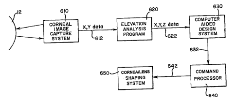
Fig. 1 is a block diagram illustrating a method for achieving vision
correction in accordance with the present invention through either laser
ablation of
the cornea or an appropriately shaped corrective lens;
Fig. 2 is a schematic diagram illustrating a plan view of a point cloud
as obtained with a corneal image capture system;
Fig. 3 is a schematic plan view similar to Fig. 2 illustrating a plurality
of splines and how they are connected through the data points of the point
cloud;
Fig. 4 is a perspective view of a cornea matching surface illustrating
how characterizing curves are constructed;
Fig. 5 is a diagram exemplifying the axial focus scatter of a cornea at
a 3 millimeter diameter;
Fig. 6 illustrates the radial focus scatter corresponding to Fig. 5;
Fig. 7 is a diagram exemplifying the axial focus scatter of a cornea at
a 5 millimeter diameter;
Fig. 8 illustrates the radial focus scatter corresponding to Fig. 7;
Fig. 9 is a diagram exemplifying the axial focus scatter of a cornea at
a 7 millimeter diameter;
Fig. 10 illustrates the radial focus scatter corresponding to Fig. 9;
Fig. 11 illustrates a method for modifying the corneal model by
orthogonalizing to the central axis;
Fig. 12 illustrates the concept of decentered orthogonalization;
Figs. 13-15 are plan views of the macula showing the 72 focus points
P distributed in spiral, rose and dual rose patterns, respectively, on the
anterior
surface of the macula; and
Fig. 16 illustrates three waveforms useful in describing the ideal
turtleback shape adjustment to the cornea that provides universal vision
improvement
DETAILED DESCRIPTION OF THE PREFERRED EMBODIMENTS
In conjunction with modern corneal procedures, such as corneal ablation
surgery, for clinical applications, and for contact lens design and
manufacture, high
5
CA 02773046 2013-10-30
resolution cameras are used to obtain a digitized array of discrete data
points on the
corneal surface. One system and camera which have been available for mapping
the cornea is the PAR Corneal Topography System (PAR CTS) of PAR Vision
Systems. The PAR CTS maps the corneal surface topology in three-dimensional
Cartesian space, i.e., along x- and y- coordinates as well as depth (Z)
coordinate.
The "line-of-sight" is a straight line segment from a fixation point to the
center
of the entrance pupil. As described more fully in Mandell, "Locating the
Corneal
Sighting Center From Videokeratography," J. Refractive Surgery, V. 11, pp. 253-
259
(July/August 1995), a light ray which is directed toward a point on the
entrance pupil
from a point of fixation will be refracted by the cornea and aqueous humor and
pass
through a corresponding point on the real pupil to eventually reach the
retina.
The point on the cornea at which the line-of-sight intersects the corneal
surface is the "optical center" or "sighting center" of the cornea. It is the
primary
reference point for refractive surgery in that it usually represents the
center of the
area to be ablated in photorefractive keratectomy. The line-of-sight has
conventionally been programmed into a laser control system to govern corneal
ablation surgery. However, some surgeons prefer to use the pupillary axis as a
reference line. Other surgeons center the ablation profile about the corneal
apex
usually defined as the area on the cornea of greatest curvature change.
Experienced practitioners have employed various techniques for locating the
sighting center. In one technique, the angle lambda is used to calculate the
position
of the sighting center relative to the pupillary ("optic") axis. See Mandell,
supra,
which includes a detailed discussion of the angles kappa and lambda.
During the LASIK corneal ablation procedure a portion of the corneal surface
is reflected and the ablation performed on the exposed surface. The gathered
elevational data is used to direct an ablation device, such as a laser, so
that the
corneal surface can be selectively ablated to more closely approximate a
spherical
surface of appropriate radius about the line-of-sight, or an "average"
ellipse, or a
wavefront fingerprint within the ablation zone. The use of the line-of-sight
as a
6
CA 02773046 2013-10-30
reference line for the procedures may reduce myopia or otherwise correct a
pre-surgical dysfunction or a visual abnormality. However, a more irregularly
shaped cornea may result, which may exacerbate existing astigmatism or
introduce
astigmatism or spherical aberration in the treated eye. This will complicate
any
subsequent vision correction measures that need be taken. Also, any
substantial
surface irregularities which are produced can cause development of scar tissue
or
the local accumulation of tear deposits, either of which can adversely affect
vision.
Implicit in the use of the-line-of sight or the pupillary axis as a reference
axis
for surgical procedures is the assumption that the cornea is symmetric about
an axis
extending along a radius of the eye. The cornea, however, is an
"asymmetrically
aspheric" surface. "Aspheric" means that the radius of curvature along any
corneal
"meridian" is not a constant (a "meridian" could be thought of as the curve
formed
by the intersection of the corneal surface and a plane containing the
pupillary axis).
Indeed, the corneal curvature tends to flatten progressively from the
geometric
center to the periphery. "Asymmetric" means that the corneal meridians do not
exhibit symmetry about their centers. The degree to which the cornea is
aspheric
and/or asymmetrical varies from patient to patient and from eye to eye within
the
same person.
Analysis of clinical measurements in accordance with the surface modeling
techniques of U.S. Patent No. 5,807,381 reveals that the point on the surface
of the
cornea which is most distant from the reference plane of the PAR CTS
(hereafter
referred to as the HIGH point) is a far more effective reference point for
corneal
ablation and lens design than the center of the cornea or the pupillary
center.
Specifically, as demonstrated in Patent No. 5,807,381 laser ablation about an
axis
passing through the HIGH point produces a much more regularly shaped cornea
and removes less corneal material than the same operation performed about an
axis close to the center of the eye, such as the pupillary axis.
Analysis of clinical measurements in accordance with the methods of U.S.
Patent No. 5,807,381, and International Application No. PCT/US03/1763
(published
7
CA 02773046 2013-10-30
as W003/101341) raises questions about assumptions that have been made about
the structure of the human cornea which are inherent in such well-known
corneal
analysis technologies as wave-front analysis and Placido disc technology. In
particular, it was found that, unlike other optical systems, the central
portion of the
cornea (for example, out to a 3mm diameter) is not necessarily optically
superior to
substantially greater portions of the cornea (for example, out to a 7mm
diameter) in
its ability to focus. The central portion of the cornea exhibits a great deal
of focus
scattering. That is, different regions on the cornea do not focus to the same
point
on a focal axis. Indeed, they do not even focus on the same axis. This focus
difference is usually most pronounced in the central portion of the cornea and
decreases substantially at increasing diameters from the center.
As disclosed in PCT/US03/1763, vision can be improved by adjusting the
focus of the cornea, referred to herein as "orthogonalizing," so that
different regions
focus substantially to the same axis. This can be accomplished by shaping the
cornea (e.g., through ablation) or by applying an appropriate corrective lens,
effectively reducing radial and axial focus scatter. An additional benefit of
orthogonalization for many patients was that presbyopia (defective near
vision) was
substantially reduced. That is, many presbyopic patients fitted with
orthogonalized
contact lenses that did not have components that focused at different
distances
could achieve simultaneous improvement in near and distance vision. However,
sufficient improvement could not be achieved in both distance and near vision
to
provide universal improvement for most near sighted individuals with
substantial age
related defects in near vision, as is very common.
A process for achieving laser ablation of the cornea and contact lens shaping
in accordance with the present invention is illustrated in block diagram form
in Fig.
1. The process makes use of a Corneal Image Capture System 610, an Elevation
Analysis Program 620, a Computer Aided Design System 630, a Command
Processor 640 and a Cornea Shaping System 650. The Corneal Image Capture
System 610, in conjunction with the Elevation Analysis Program 620, generates
a
three dimensional topographic map of the cornea of the patient. The Computer
8
CA 02773046 2013-10-30
Aided Design System 630 is used as an aid in editing or modifying the corneal
topographic data, to create a surface model, and data relating to the model is
sent
to a Cornea Shaping System 650 via the Command Processor 640. The Command
Processor 640 uses the topographic data describing the surface of the cornea
to be
shaped from the Computer Aided Design System 630 to generate a sequence of
commands/control signals required by the Cornea/Lens Shaping System 650. The
Cornea/Lens Shaping System 650 accepts, from the Command Processor 640, a
sequence of commands that describe the three dimensional movements of the
Cornea/Lens Shaping System (any coordinate system may be used; e.g.,
Cartesian,
radial or spherical coordinates) to shape the cornea or machine (e.g., a
lathe)
manufacturing a contact lens.
The Corneal Image Capturing System 610 and the Elevation Analysis
Program 620 are preferably components of the PAR Corneal Topography System
("the PAR System"), which is available from PAR Vision Systems. The Elevation
Analysis Program 620 is a software program executed by a processor, for
example
an IBMTm compatible PC. Program 620 generates a third dimension element (a Z
coordinate representing distance away from a reference plane inside the eye)
for
each of a plurality of sample points on the surface of the cornea measured by
system 610. Each point is defined by its X-Y coordinates as mapped into the
reference plane, and its Z coordinate is determined from brightness of the
point.
One method of calculating the elevation of each point, i.e., the Z coordinate,
is by
comparing the X-Y and brightness values measured from the patient's cornea 14
with the coordinates and brightness of some reference surface with known
elevation, e.g., a sphere of a known radius. The reference values can be
pre-stored.
The final output of the Elevation Analysis Program 620 is the X-Y-Z
coordinates for a multiplicity of sample points, commonly known as a point
cloud,
on the surface of the cornea 14. It will be apparent to those skilled in the
art that
any method can be used that can generate X, Y, Z corneal data providing both
location and elevation information for points on the corneal surface with the
required
9
CA 02773046 2013-10-30
accuracy. In the preferred embodiment about 1200 points are spaced in a grid
pattern, as viewed in the X-Y plane, so the projections of the points into the
X-Y
plane are about 200 microns apart.
The X-Y-Z data output from the Elevation Analysis Program 620 can be
formatted in any number of well-known machine-specific formats. Preferably,
the
data are formatted in Data Exchange File (DXF) format, an industry standard
format
which is typically used for the inter-application transfer of data. A DXF file
is an
ASCII data file, which can be read by most computer aided design systems.
Referring now to Figs. 2 and 3, a point cloud 100 is depicted as it would
appear
when viewing the reference plane along the Z-axis (i.e., as projected into the
X-Y
plane). Each point corresponds to a particular location on the patient's
cornea. The
data are usually generated from an approximately lOmm x lOmm bounded area of
the cornea, the working area. Thus, there may be as many as 50 rows of data
points. A surface 108 (see Fig. 4) that models or matches the topography of
the
surface of the patient's cornea is generated by the computer aided design
system
630 from the data points generated by the Elevation Analysis Program. In a
preferred embodiment, Computer Aided Design System 630 is the Anvil 5000TM
program which is available from Manufacturing Consulting Services of
Scottsdale,
Arizona.
Cornea matching surface 108 is preferably produced by first generating a
plurality of splines 102, each defined by a plurality of the data points of
the point
cloud 100. The generation of a spline that intersects a plurality of data
points (i.e.,
knot points) is, per se, known to those skilled in the art and can be
accomplished by
the Anvil 5000" program once the input data have been entered. For more
information regarding the generation of a surface model, see U.S. Patent No.
5,807,381. In a preferred embodiment, the known non-uniform rational B-spline
formula is used to generate the splines, but they could be generated by other
well-known mathematical formulas for splines, such as the cubic spline formula
or
the rational uniform B-spline formula. As illustrated in Fig. 3, in a
preferred
embodiment, each of the splines 102 lies in a plane that is parallel to the X
and Z
CA 02773046 2013-10-30
axes and includes a row of points from the cloud 100 in Fig. 3.
Surface 108, which matches the corneal surface of the scanned eye, is then
generated from splines 102. There are a number of well-known mathematical
formulas that may be used to generate a surface from a plurality of splines
102. In
the preferred embodiment, the well known nurb surface equation is used to
generate a corneal surface from splines 102. In the embodiment, because the
scanned area of the eye is approximately lOmm x lOmm, approximately 50 splines
102 are created. As illustrated in Fig. 3, a skinned surface segment 104 is
created
for a small number (e.g., five) of the adjacent splines. Adjacent skinned
surface
segments 104 share a common border spline. Thus, about ten skinned surface
segments are generated from the point cloud and are then merged together by
the
Anvil 5000' program in a manner known to those skilled in the art, to produce
one
composite surface 108.
Neither the original data points, nor the knot points of splines 102
necessarily
lie on-surface 108, owing to the mathematical generation of the surface when
using
the nurb surface equation formula. However, the surface 108 estimates those
points within a predefined tolerance.
The HIGH point on the generated corneal matching surface 108 (i.e., the
point having the greatest Z value) is determined. A cylinder 106 of a
predetermined
diameter is then projected onto the corneal matching surface 108 along an axis
which is parallel to the Z-axis and passes through the HIGH point. Cylinder
106
preferably has a diameter of about 3mm to about 8mm, typically about 7mm, and
the closed contour formed by the intersection of cylinder 106 with surface 108
projects as a circle 106' in the X-Y plane. On the matching surface 108, this
contour
defines the outer margin 26 of the working area of the cornea. The cornea is
the
most symmetric and spherical about the HIGH point and, therefore, provides the
best optics at this point.
The outer margin 26 must fit within the point cloud, so that the surfaces of
the
11
CA 02773046 2013-10-30
cornea can be formed based on the measured corneal data. The computer aided
design system 630 can then illustrate a default circle 106' (in the X-Y plane)
with
respect to the point cloud, for example on a monitor screen, so that the
operator can
be assured that circle 106' falls within the point cloud. Additionally, system
630 can
be set up to determine if circle 106' falls within point cloud 100 and, if it
does not fall
completely within point cloud 100, to alert the user to manipulate the circle
(i.e.,
move the center point and/or change the radius of the circle) so that circle
106' lies
within the corneal data point cloud 100. In a worst case scenario, the eye
should
be rescanned if insufficient data is available from the scanned eye to ensure
that the
working area of the cornea will fit properly within the point cloud.
Alternatively, the
area of the point cloud can be made larger.
It is to be understood that circle 106' is only a circle when viewed in the X-
Y
plane (i.e., looking along the Z-axis). Actually, the periphery 26 is
approximately
elliptical and lies in a plane which is tilted relative to the reference
plane. A line
Perpendicular to this tilted plane which passes through the HIGH point will be
referred to as the "LOCAL Z-AXIS" or "tilted axis," and the tilt of the tilted
plane
relative to the reference plane will be considered the tilt angle of the
working area
of the cornea.
The cornea is about 600pm thick. In most corneal ablation procedures, less
than 100pm depth of cornea is ablated because there is virtually no risk of
scarring
with the type of lasers that are typically used. Beyond the 100pm depth, the
risk of
scar-like imperfections. For example, 120pm depth ablation is known to cause
scarring. However, there exists the possibility that the risk of scarring for
surface
ablations may be reduced by drug therapy prior to or contemporaneous with the
laser treatment. However, most of today's laser surgery does not cause
scarring,
as most procedures are under the LASIK flap. The fear in LASIK is ablating too
deep wherein the residual bed is less than -250pm. If the bed is less than
this
amount, structural failure can occur. The magnitude of the corneal undulations
is
typically about fifteen to twenty microns from the crest of a hill to the
trough of a
valley and may be as great as about thirty microns.
12
CA 02773046 2013-10-30
The surgical procedures performed in accordance with the present invention
and optical lenses manufactured in accordance with the invention, will seek to
correct the patient's vision in accordance with the required corrections
established
in a "refraction test." When this test is performed, the patient sits in chair
which is
fitted with a special device called a "phoropter," through which the patient
looks at
an eye chart approximately 20 feet away. As the patient looks into the
phoropter,
the doctor manipulates lenses of different strengths into view and, each time,
asks
the patient whether the chart appears more or less clear with the particular
lenses
in place. In practice, the doctor is able to vary the power or diopter
correction about
two orthogonal axes, as well as the degree of rotation of those axes about a Z-
axis
along the line-of-sight. The doctor continues to modify these three parameters
until
he achieves the optimum vision. The results of the refraction test are usually
given
in the form "a, b, c," where " a" is the diopter correction at the first axis,
"b" is the
additional diopter correction required at the second, orthogonal axis, and "c"
is the
angle of rotation of the first axis relative to the horizontal. This form of
information
is given for each eye and is immediately useful in grinding a pair of lenses
for
eyeglasses.
There will now be described a technique for generating characterizing curves
on surface 108, which will be useful below. A plane 110 is constructed which
contains the LOCAL Z-AXIS (See Fig. 4). The intersection between plane 110 and
surface 108 defines a first characterizing curve 112. Plane 110 is then
rotated
about the LOCAL Z-AXIS, for example by a 5 increment counterclockwise, as
represented by line 114, where its intersection with surface 108 defines a
second
characterizing curve 116, which is illustrated as a dashed line in Fig. 4.
This
process continues at fixed rotational increments about the LOCAL Z-AXIS, for
example every 5 , until plane 110 has swept 360 , to produce a complete set of
characterizing curves (meridians), in this case seventy-two (360 % 5 ).
Each of these characterizing curves is then estimated by a best-fit spherical
(circular) arc. One manner of doing this is simply to select a circular arc
which
passes through three known points for each curve (e.g., the point at which it
touches
13
CA 02773046 2013-10-30
the contour 106', the HIGH point, and that point which is halfway between
those two
points when viewed in projection along the local Z axis). Once the spherical
arcs
are generated, the focal point of a portion of the cornea represented by a
circular
arc can be estimated by the center of that arc. Techniques for locating the
center
of a spherical arc are well-known. The resulting set of arc centers then
provides a
representation of focus scattering.
For purposes of illustration, the preceding procedure was performed on the
corneal model of a patient having 20/15 uncorrected visual acuity. Fig. 5 is a
focus
scatter diagram along the LOCAL Z-AXIS for that portion of the cornea
extending
out to a 3.0mm diameter. In this case, the focal points start at 7.06mm along
the
LOCAL Z-AXIS and extend out an additional 6.91mm. Fig. 6 illustrates that the
radial scatter within a 3mm diameter is 1.2mm. Similarly, Fig. 7 illustrates
that the
axial focus scatter of a 5mm diameter portion of the cornea begins at 8.99mm
and
extends for an additional 1.69mm. As shown in Fig. 8, the radial scatter of
the same
portion of the cornea is .49mm. Fig. 9 illustrates that the axial focus
scatter at 7mm
begins at 8.68mm and extends axially for an additional .47mm, whereas Fig. 10
illustrates that the corresponding radial scatter is .33mm. Clearly, focus
scatter is
most severe in the central portion of the cornea, and decreases significantly
as
larger portions of the cornea are considered. Therefore, it would clearly
be
desirable to reduce or eliminate the focus scatter at least in central
portions of the
cornea. This can be accomplished by "orthogonalizing" at least a portion of
the
cornea. The term "orthogonalizing" refers to a re-shaping of the surface model
so
as to piecewise re-focus the cornea towards the LOCAL Z-AXIS. The re-shaped
surface model can then be applied to the cornea (e.g., through ablation) or to
shape
the posterior surface of a contact lens (or another type of optical lens) so
as to
achieve the required focus scatter correction. It has been found that
orthogonalizing
the cornea not only reduces radial focus scatter, but simultaneously reduces
axial
focus scatter substantially and produces more uniformity in the radius of
curvature
of the orthogonalized portion of the cornea.
Fig. 11 illustrates the process of orthogonalization. The process is carried
out
14
CA 02773046 2013-10-30
on each of the arcs which represent characteristic curves, in the manner
explained
below. After this piecewise refocusing, the modified arcs are reassembled into
a
modified surface model having the re-focused characteristics.
In Fig. 11, 130 represents one of the half-meridian arcs corresponding to a
characterizing curve. Arc 130 has a center point C, the location of which has
been
exaggerated to demonstrate focus which is radially spaced from the LOCAL Z-
AXIS.
Orthogonalization of arc 130 begins with creating a chord 132 between the two
ends
of the arc. A perpendicular bisector 134 of chord 132 may be constructed, and
it will
pass through point C and intersect the LOCAL Z-AXIS at a point X. Using the
distance of point X from point H (the HIGH point) as a radius, a new arc 130'
can
now be drawn between the two end points of arc 130. Arc 130' will be focused
on
the LOCAL Z-AXIS and will have a larger radius of curvature than arc 130.
At this point, arc 130' could be accepted as an arc defining the modified
surface model 108'. However, it would be desirable to avoid too great a change
in
the thickness of the cornea. Accordingly, a certain threshold is defined (for
example, .0075mm), and if any portion of arc 130' is more than a distance
inside or
outside the surface 108, arch 130' is not accepted for use in the modified
surface
model. Instead, point x can be moved up or down on the LOCAL Z-AXIS
(depending upon which direction arch 130' needs to be moved) by half the
excess
over. Arc 130' can then be re-drawn and re-tested against the threshold. This
readjustment and testing continues until an acceptable arc 130' has been
found.
Then, the next arc is orthogonalized. After all of the arcs are
orthogonalized, a new
surface model 108' is created based upon all of the arcs.
As has been explained above, the orthogonalization process is applicable to
corneal ablation procedures. Prior to the procedure, a corrected corneal
surface
model is generated, which is shaped to provide relief from macular
degeneration
and correction of refraction established by an eye test (as described in the
patents
cited above), and all the arcs are orthogonalized. The corrected corneal
surface
model is then registered with the unmodified corneal surface model, and it is
moved
CA 02773046 2013-10-30
towards the unmodified surface until the corrected surface just contacts the
unmodified surface. If the point of initial contact is at the center of the
corrected
surface, it is moved toward the uncorrected surface until the periphery of the
corrected surface just contacts the uncorrected surface at the diameter of the
proposed ablation procedure. If the point of initial contact is at the
periphery of the
corrected surface, it is moved toward the uncorrected surface until the center
of the
corrected surface just contacts the uncorrected surface. The corrected surface
will
then be displaced so that it is, at least partially, inside the cornea, and
the cornea
is ablated until the displaced corrected surface becomes its new surface.
The central region of the retina is called the macula, and the very center of
the macula, called the foveola, is the most sensitive. Although the macula
typically
has a diameter in the range of 6 to 7 millimeters, the central foveola
typically has a
diameter of about 0.35mm. With perfect orthogonalization, all sub-portions of
the
cornea are refocused to the center of the macula, the foveola. When
orthogonalization is performed by refocusing all of the sub regions onto the
LOCAL
Z-AXIS, orthogonalization is not perfect.
In accordance with one aspect of the present invention, sub-portions of the
.. cornea may be refocused so as to place their focal points outside the
foveola yet still
within the macula at a controlled lateral distance from the LOCAL Z-AXIS. The
macula has approximately the shape of a cap-shaped segment of a sphere, is
usually between 6 millimeter and 7 millimeters in diameter and is
approximately 0.88
millimeters deep.
The difference should be kept in mind between introducing de-focus and the
decentered focus of the invention. Ophthalmologists have long known that, in
prescribing corrective lenses, distance focus can be reduced through de-focus,
and
a benefit in near vision can result. In accordance with the present invention,
there
.. is no de-focus. All sub-portions of the cornea remain fully focused, but
the focus
point is moved away from the LOCAL Z.
16
CA 02773046 2013-10-30
Fig. 12 illustrates the concept of decentered orthogonalization. The arc 130
is a sub-portion of the cornea which has a scattered focal point X. Ordinary
orthogonalization as shown in Fig. 11 would move the focal point X to the
LOCAL
Z-AXIS, LZ. Perfect orthogonalization would move it to the foveola F on the
macula
M. Decentered orthogonalization creates a new arc 130" which focuses at a
point
X', which is at a predefined radius r from the foveola. The axis Z' is
parallel to the
LOCAL Z-AXIS and passes through the point X. For purposes of estimation, the
macula can be considered flat in the region between the axes LZ and Z'.
The preferred manner of performing decentered orthogonalization utilizes the
technique discussed with respect to Fig. 4. Specifically, the anterior surface
of the
cornea is broken down into 72 arcs spaced 50 apart rotationally, and each arc
is
subjected to decentered orthogonalization. The 72 resulting focus points
should be
well distributed in a working region W' of the foveola which preferably has a
diameter less than .07 millimeters. Fig. 13 is a top plan view of the foveola
showing
the 72 points P distributed in a spiral pattern on the surface of the foveola.
A more preferred configuration for the points is illustrated in Fig. 14. This
pattern is described by the polar equation R=ajicos2y, where R is the
two-dimensional radius of the point from the foveola, a is a constant selected
to
spread the points well over the entire working area M', and Si is the
rotational angle
of the particular arc on the cornea. This pattern is preferred to the spiral,
because
every quadrant of the working area M' has focus points at a full range of
distances
from the foveola.
Another preferred pattern for the focus point is illustrated in Fig. 14. In
this
case, the pattern is formed from two overlaid rose patterns, a large one 150
and a
small one 150', which is offset by 45 from the pattern 150. Only one petal of
each
rose pattern is shown to have points, but it will be understood that each of
the other
petals is similarly provided with points. The points are shared evenly between
the
patterns 150 and 150'. However, the pattern 150 provides the outermost points
and
has points distributed at over its outermost two-thirds. Pattern 150' provides
the
17
CA 02773046 2013-10-30
' .
innermost points and has them evenly distributed. As a result, the pattern in
Fig. 14
provides a good distribution of points near to and distant from the foveola.
It should be appreciated that, in all the focus point patterns that have been
shown, in most instances the points are equally spaced along a curve. However,
those skilled in the art will appreciate that unequal spacing could be
provided for the
points so as to concentrate them more in a specific region (e.g., the center
or the
outermost area of the working region.
A further method, defining a further embodiment of the invention, has been
developed for decentered orthogonalization which is preferred over all those
described previously to enhance universal improvement, in some instances. This
method will be referred to as "offset" decentralized orthogonalization. The
method
proceeds exactly as in the Fig. 11, except that once arc 130' has been
reshaped,
it is tilted clockwise so as to move the point X, the endpoint of the arc's
axis, to the
left in Fig 11, across the local z-axis, so that it lies at a preselected
distance, or
offset, from the local z-axis. Biases at values below about .01mm are
contemplated
at present, with a bias of approximately .0025mm being preferred. However,
distances in the range of approximately .0025mm to approximately 0.01mm still
being effective.
Figure 16 illustrates three waveforms which are useful in describing the
idealized turtleback shape. Each of the waveforms is a polar graph of
curvature
(given in diopters) as a function of rotational position. For example,
waveform A
represents the cornea of an actual patient that is nearsighted, astigmatic,
and
exhibits age-related presbyopia. The polar angle is the rotational angle of a
plane
containing the local z axis (about the tilted local z axis) relative to a
reference
position at which the plane intersects the base of the cornea at a position
closest to
the nose. The curvature is the diopter equivalent of the radius of a circular
arc
which most closely approximates the half-meridian arc created by the
intersection
between the surface of the cornea and the plane when it has the particular
rotational
orientation. The following well-known formula relates the diopter value to the
radius
18
CA 02773046 2013-10-30
of the arc:
337.5/Arc Radius = Diopter Value
Ideally (for the best universal improvement of vision), waveform A should be
shaped substantially like a letter "M" and it is therefore referred to herein
as the
"M-wave" of the cornea. It is, in the present instance, a somewhat distorted
M.
As an initial step in redesigning the shape of a cornea to exhibit universal
vision improvement, an idealized M wave is generated for the cornea. Starting
with
a polar representation of the patient's cornea showing the surface curvature
along
the natural half-meridian arcs of the particular corneal surface, such as
waveform
A, an idealized waveform is generated. This waveform is not related to
waveform
A, except the lowest diopter values are preferably approximately the same in
the two
waveforms, but waveform B preferably meet certain criteria. However, in some
instances improved vision performance may be obtained by making the baseline
of
waveform B 1.5 diopters higher than waveform A. First of all, the peak to peak
diopter variation of the waveform is adjusted to be approximately 3 diopters,
preferably about 2.875 diopters. It has been found that there is substantial
deterioration in near vision correction if this diopter range drops below
about 2
diopters or exceeds about 4 diopters. In addition, the dip D in the M wave is
adjusted so as to lie between approximately 40% and 60% of the peak to peak
amplitude of the M wave. Preferably, it is approximately 50%. Then, the entire
waveform is adjusted so as to transition smoothly between values. Preferably,
the
peaks occur at about 900 and 270 and the dip at approximately 180 , while
producing a smooth curve. This results in the ideal M wave to represent the
patient's cornea. This wave is represented by waveform B in Fig. 16.
As a practical matter, every lens will have the same M-wave shape, except
for the adjustment to match the flattest curvature (K value) of the cornea and
the
necessary distance vision correction, as determined, for example, by a
refraction
test. K value and refraction are measurements normally taken by an eye care
professional when fitting lenses and would typically be available. To
customize the
19
CA 02773046 2013-10-30
M-wave for a patient, it is only necessary to pick a baseline for it that
corresponds
to his K value and to shift the waveform vertically to provide the diopters
necessary
for correction of distance vision. This defines the lens shape of a custom
lens for
that patient.
It will be appreciated that waveform B exhibits the flattest surface curvature
at 0 (a point corresponding to the edge of the cornea that would be closest
to the
nose in waveform B). Increasing the polar angle, the surface curvature
increases
continuously until it reaches a maximum at about 90 (corresponding to the
vertically
uppermost edge of the cornea). The surface curvature then decreases
continuously
until it reaches an intermediate value at about 180 (corresponding to the
edge of
cornea most distant from the nose), and it increases continuously to a maximum
at
about 270 (corresponding to the vertically lowermost edge of the cornea), and
it
decreases continuously until it reaches 0 , where it returns to its minimum.
Thus,
the surface described by this M-wave has the idealized turtleback shape
discussed
previously.
In the preceding paragraph it was assumed that the M wave for the patient's
right eye was being considered. The reference or 0 angle was selected as the
point closest to the nose and the polar angle increased in a clockwise
direction.
The M-wave for the left eye could be identical (i.e. with 0 at the point
furthest from
the nose and polar angle increasing clockwise), or it could be a mirror image
of the
right eye (i.e. with 0 at the nose but polar angle increasing
counterclockwise). The
former approach would simplify manufacture and reduce cost, since the same
lens
would be used for both eyes.
In some instances, better universal improvement of vision will be attained if
the surface model represented by waveform B is provided with one additional
adjustment. That is, if offset, decentered orthogonalization is performed on
the
surface model with an offset of less than approximately .005 mm from the local
z
axis. Most preferably, the offset is about .0025 mm. The upper offset limit of
.005
mm was selected because experimentation has shown that a significant
CA 02773046 2013-10-30
deterioration in distance or near vision is reached at that value. Distance
vision
continues to deteriorate significantly as offset is increased further.
In one embodiment, the surface model represented by waveform B,
.. represents the shape of the posterior surface of a contact lens for use by
the
patient. In accordance with the present invention, the shape of the anterior
surface
of the lens is derived by providing a diopter adjustment along waveform B
which is
determined to be necessary to correct the patient's distance vision.
Typically, such
diopter correction would be determined from a conventional refraction test. At
each
angle, the anterior surface diopter value Da and radius Ra are determined by
the
Zeiss lens formulas:
Da = (-P Dp)/(1-(((T/1000)/Na)*Dp))
Ra = (NTNIA)*1000/Da
where Da is the diopter value of the anterior arc
Dp is the diopter value of the posterior arc
NL is the index of refraction of the material of
which the
lens is made
NA is the index of refraction of air
is the power adjustment factor, and
T is the lens thickness.
Following this diopter adjustment, waveform C results.
Those skilled in the art will appreciate that the posterior surface of the
contact
lens need not be shaped as defined by waveform B. In fact, it could be any
shape
calculated to conform generally to the patient's cornea, such as a spherical
surface
or an ellipsoidal surface. The idealized M wave is a turtleback shape, not
spherical
or ellipsoidal and, except for preferably having the same minimum curvature as
the
cornea, is universal and has no relationship to the patient's native cornea.
.. Moreover, matching the flattest curvature of the cornea is not related to
vision
correction, but is done to assure that the lens will have a more comfortable
fit.
21
CA 02773046 2013-10-30
. .
When the lens is placed on the eye, the lens, the cornea and the tear film
therebetween will have substantially the same index of refraction. Thus, only
the
interface between the air and anterior surface of the lens will have a
significant
effect on vision improvement. Using a surface shape defined by waveform B for
the
posterior surface of the lens minimizes unnecessary thickness variation in the
lens,
which can introduce certain distortions.
Those skilled in the art will appreciate that the surface model represented by
waveform C could also be used to define the desired shape of the cornea
following
a surgical procedure. The surgical procedure constitutes an actual reshaping
of the
cornea, while the use of the contact lens constitutes an effective reshaping.
It should be appreciated that the contact lens described immediately above
is a custom designed contact. However, it is contemplated that M wave lenses
could be provided in ready made prescription form as in current mass produced
lenses. For example, in the case where a lens has an M wave posterior surface,
lenses could be provided in different base curvature variations or "sizes"
(e.g. a
large base curve for a relatively flat cornea, a medium for a cornea of medium
or
average curvature, and a small for a relatively steeply shaped cornea). In all
cases,
the M-wave has the idealized shape described earlier, so the only difference
between the sizes is the actual values of the initial curvature. Each
posterior curve
set would include a subset of lenses with different anterior curves such that
each
size would include a subset of lenses with the necessary diopter adjustment to
correct for different distance refractive errors. The patient would only
require two
optometric tests in order to obtain the correct prescription. First, the
optometrist
would perform a conventional refraction test to determine the diopter
correction
required for distance vision. Second, during the initial visit the optometrist
or lens
fitter also could perform a conventional keratometer test, which yields the
diopter
readings for the flattest and steepest portions of the cornea. The flattest
curvature
of the keratometer test determines whether the patient needs a lens with a
small,
medium, or large posterior surface base curve (in order to obtain best fit),
and the
refraction test establishes the required distance correction. Given this
prescription,
22
CA 02773046 2013-10-30
an eye care professional could easily fit the patient with the most
comfortable M
wave lens that will provide universal vision improvement.
Although preferred embodiments of the invention have been disclosed for
illustrative purposes, those skilled in the art will appreciate that many
additions,
modifications, and substitutions are possible without departing from the scope
of the
invention. For example, the present invention is applicable not only to
corneal
ablation and contact lenses, but to any other kind of lens, including
cataract, phakic,
intraocular, intracorneal and spectacle lenses.
23