Note: Descriptions are shown in the official language in which they were submitted.
CA 02773186 2012-03-05
WO 2011/037692 PCT/US2010/044586
COMPOSITIONS, METHODS, AND KITS FOR ISOLATING AND ANALYZING
NUCLEIC ACIDS USING AN ANION EXCHANGE MATERIAL
CROSS REFERENCE TO RELATED APPLICATIONS
This application claims priority from US Provisional Patent Application Serial
No.
61/231,371 (filed September 24, 2009), US Provisional Patent Application
Serial No. 61/295,269
(filed January 15, 2010), and European Patent Application Serial No. EP 10 006
062 (filed June
11, 2010), the contents of which are incorporated herein by reference in their
entireties. A US
non-provisional application entitled "Compositions, Methods, and Kits for
Isolating and
Analyzing Nucleic Acids Using an Anion Exchange Material," filed concurrently
herewith on
August 5, 2010, is also incorporated herein by reference in its entirety.
FIELD OF INVENTION
The present invention relates to methods, compositions, and kits for
isolating, eluting,
and analyzing nucleic acids using anion exchange materials.
BACKGROUND
Ion-exchange is a process that allows the separation of ions and polar
molecules based on
the charge properties of the molecules. In principle, ion exchange functions
by retaining a target
analyte molecules in a stationary phase based on reversible ionic
interactions. Typically, the ion
exchange composition comprises a stationary phase bearing a capture molecule.
Under
appropriate buffer and ionic conditions, the capture molecule bears a net
charge, the target
molecule bears an opposite net charge, while contaminants bear a net neutral
charge or a similar
net charge to the capture molecule. As a result, the target molecule is
retained by the ion
exchange composition, while contaminants are removed. The target molecule can
then be eluted
by neutralizing the charge of the capture or target molecule (e.g. by altering
the pH) or treating
the composition with another molecule that displaces the target molecule (e.g.
by treating with a
high salt solution). Such processes are broadly categorized as either cation
exchange or anion
exchange, depending upon the net charge of the molecule that is being
isolated.
Anion exchange is commonly used to purify nucleic acids from complex
biological solutions.
The nucleic acids can be selectively adsorbed because of their negative charge
onto anion
exchange resins and then eluted. For low molecular weight nucleic acids,
elution buffers
typically include salts in high concentrations. Simple organic and inorganic
anions have lower
SUBSTITUTE SHEET (RULE 26)
CA 02773186 2012-03-05
WO 2011/037692 PCT/US2010/044586
selectivity coefficients than nucleic acid, but at high concentrations they
can displace nucleic
acid from the resins. High molecular weight nucleic acid cannot be eluted by
salts because of
their extremely high selectivity for the resin. In this case, elution is most
often performed by
raising the pH, since at high pH weak anion exchange groups of the resin lose
their charge.
Thus, elution of nucleic acids from ion exchange resin usually is based on an
interrelationship of pH and salt concentration. However, these procedures
present some
difficulties for downstream processing and analysis of the target nucleic
acid. Alkali buffers
cannot be used for RNA isolation since RNA is degraded in alkaline solutions.
DNA is also
alkali-sensitive to some extent, most notably, to the deamination of cytosine
(conversion of
deoxycytosine into deoxyuracil), especially when heated. Storage of alkali
buffers is also
problematic, as such solutions are often corrosive, toxic, and have poor shelf
stability due to
absorbance of CO2 from the air. High salt concentrations that are often
employed for elution
often interfere with downstream enzymatic reactions such as polymerase chain
reaction (PCR),
ligation reactions, restriction analysis, cDNA generation or isothermal
amplification. Therefore,
the resulting nucleic acid eluates need to be desalted, which adds additional
handling steps to the
nucleic acid isolation procedure and may lead to loss of nucleic acids due to
the increased
number of handling steps or possible contamination. DNA cannot be directly
processed
enzymatically because most enzymes are completely denatured under alkaline
conditions. Thus,
high pH elution also requires additional handling steps because it requires
neutralization of the
eluate, which further dilutes the eluate, increases labor steps, and may not
be appropriate for all
enzymatic downstream assays.
In addition, the presence of commonly-used anion exchange materials can
inhibit
downstream analysis of nucleic acids. As such, current protocols require that
the nucleic acid be
separated from the anion exchange material before the purified nucleic acid
can be analyzed.
Unfortunately, complete separation of the eluate and the anion exchange
material often cannot be
achieved, thus reducing both the efficiency and the accuracy of subsequent
analyses and
amplifications. Moreover, the additional step of eluting and separating the
eluate from the anion
exchange material hampers the utility of anion exchange in fully automated
nucleic acid
purification and analysis procedures.
2
CA 02773186 2012-03-05
WO 2011/037692 PCT/US2010/044586
Thus, materials and methods are disclosed for isolating nucleic acids which do
not
require additional handling steps prior to a further use of the isolated
nucleic acids. In particular,
the isolated nucleic acids should be directly suitable for any amplification
or analysis procedures.
SUMMARY OF INVENTION
The present disclosure relates to materials and methods for isolating and
analyzing
nucleic acids comprising the use of anion exchange chromatography in
conjunction with an
anionic compound. Using anionic compounds, in particular anionic compounds
having at least
two anionic groups, nucleic acids can be eluted from the anion exchange
material without the use
of unfavourably high pH values and/or to use high salt concentrations.
Moreover, the presence
of an anionic compound having at least two anionic groups has been found to
permit
amplification of nucleic acids in the presence of an anion exchange material.
In one aspect, a method of isolating nucleic acids is disclosed, said method
comprising
providing a nucleic acid complexed with an anion exchange material and eluting
the bound
nucleic acid by adding a solution comprising at least one anionic compound
comprising at least
two anionic groups. In one embodiment, the anionic compound comprising at
least two anionic
groups is a polyanionic compound, preferably a polyanionic organic compound.
In another
embodiment, the compound comprising at least two anionic groups is a non-
polymeric
compound, preferably a non-polymeric organic compound. In yet another
embodiment, the
elution step is performed without high salt conditions or severe changes in
pH. In another
embodiment, the nucleic acid complexed with the anion exchange material is
provided by a
method comprising the steps of contacting an anion-exchange composition with a
sample
comprising the nucleic acid, wherein the anion-exchange composition is capable
of reversibly
binding nucleic acid; and optionally washing the anion-exchange composition to
remove
unbound sample components. In the present methods, the compound comprising at
least two
anionic groups displaces the nucleic acids from the anion-exchange material.
Using an anionic
compound comprising at least two anionic groups for elution of the bound
nucleic acid provides
the advantages that these compounds do not have to be removed for performing
subsequent
analyses steps, including amplification steps, as they do not or only slightly
interfere with such
steps. Furthermore, only low concentrations of the compound comprising at
least two anionic
groups is necessary for effectively eluting the nucleic acid.
3
CA 02773186 2012-03-05
WO 2011/037692 PCT/US2010/044586
In another aspect, a method of analyzing a nucleic acid in the presence of an
anion
exchange material is provided, said method comprising providing a nucleic acid
complexed with
the anion exchange material to form a nucleic acid-anion exchange complex and
analyzing the
nucleic acid in the presence of at least one compound comprising at least two
anionic groups. In
one embodiment, the anionic compound comprising at least two anionic groups is
a polyanionic
compound. In another embodiment, the anionic compound comprising at least two
anionic
groups is a non-polymeric compound. In another embodiment, the nucleic acid
complexed with
the anion exchange material is provided by a method comprising the steps of
contacting an
anion-exchange composition with a sample comprising the nucleic acid, wherein
the anion-
exchange composition is capable of reversibly binding nucleic acid; optionally
washing the
anion-exchange composition to remove unbound sample components; and optionally
separating
the nucleic acid-anion exchange complex from other material.
Yet another aspect is an elution composition comprising an anionic compound
comprising at least two anionic groups and further comprising at least one
buffer. One
advantage of the provided methods and compositions is that they provide
flexibility with respect
to the used elution conditions and elution buffers and furthermore, may make
the elution more
effective. Therefore, the present elution composition may comprise any buffer
or composition
commonly used for elution. By way of example and not limitation, the elution
buffer may be
formed by combining the anionic compound may be combined with water, low salt
buffers, or
biological buffers such as CHAPS, MES, HEPES, MOPS, TRIS, TRICINE and PIPES.
In one
embodiment, the compound comprising at least two anionic groups is a
polyanionic compound.
In another embodiment, the compound comprising at least two anionic groups is
a non-polymeric
compound.
Yet another aspect relates to an eluate obtained by the elution methods
disclosed herein.
Yet another aspect is an amplification composition comprising a polymerase and
an
anionic compound comprising at least two anionic groups. In another
embodiment, the
amplification composition optionally comprises: an enzyme having reverse
transcriptase activity;
and enzyme having helicase activity; a nick-inducing agent; Mgt or a source
thereof, such as
MgCl2; a ribonucleotide triphosphate (NTP); a deoxyribonucleotide triphosphate
(dNTP); K_'_ or a
source thereof, such as KC1; NH4-'- or a source thereof, such as (NH4)2SO4; a
buffer, such as Tris;
and/or a reducing agent, such as 2-mercapthoethanol or dithiothreitol (DTT).
In one
4
CA 02773186 2012-03-05
WO 2011/037692 PCT/US2010/044586
embodiment, the compound comprising at least two anionic groups is a
polyanionic compound.
In another embodiment, the compound comprising at least two anionic groups is
a non-polymeric
compound.
Yet another aspect is a kit for isolating nucleic acids from a sample, the kit
comprising an
anion exchange material and an anionic compound comprising at least two
anionic groups.
In another aspect, a kit for amplifying a nucleic acid is provided comprising
a
polymerase, an anion exchange material, and an anionic compound comprising at
least two
anionic groups. In another embodiment, the amplification composition
optionally comprises:
Mg 2+ or a source thereof, such as MgC12; a ribonucleotide triphosphate (NTP);
a
deoxyribonucleotide triphosphate (dNTP); K+ or a source thereof, such as KC1;
NH4+ or a source
thereof, such as (NH4)2SO4; a buffer, such as Tris; and/or a reducing agent,
such as 2-
mercapthoethanol or dithiothreitol (DTT). In one embodiment, the compound
comprising at
least two anionic groups is a polyanionic compound. In another embodiment, the
compound
comprising at least two anionic groups is a non-polymeric compound.
In a further aspect, the use of an anionic compound comprising at least two
anionic
groups for displacing a nucleic acid reversibly bound to an anion-exchange
material from said
anion-exchange material is provided. In one embodiment, the compound
comprising at least two
anionic groups is a polyanionic compound. In another embodiment, the compound
comprising at
least two anionic groups is a non-polymeric compound.
BRIEF DESCRIPTION OF THE FIGURES
Fig. 1 shows a gel-electrophoretic analysis of the bound and eluted nucleic
acids. A 300
bp DNA fragment was bound onto diethylaminopropyl magnetic beads and eluted
using a 500 bp
DNA fragment. The gels are loaded as follows:
Lane Sample
1 lkb+ Ladder
2 1 300bp control
3 lug, 2 , 3 500b control
4 Flow-through, binding, wash 1, wash 2
Eluate 1, pH 7.0, 1 500b
6 Eluate 1, pH 7.0, 2 500bp
7 Eluate 1, pH 7.0, 3 500b
8 Eluate 2, pH 8.5, 50mM MES, 5OmM NaCl
9 Flow-through, binding, wash 1, wash 2
5
CA 02773186 2012-03-05
WO 2011/037692 PCT/US2010/044586
Eluate 1, pH 7.0, 1 500b
11 Eluate 1, pH 7.0, 2 500bp
12 Eluate 1, pH 7.0, 3 500b
13 Eluate 2, pH 8.5, 50mM MES, 50mM NaCl
14 lkb+ Ladder
Fig. 2 shows a Gel-electrophoretic analysis of the bound and eluted nucleic
acids.
Plasmid DNA was bound onto polyethylenimine magnetic beads and eluted using
dextransulfate,
polyacrylic acid, oxalic acid, mellitic acid or genomic DNA. A: Binding and
elution at pH 8Ø
B: Binding and elution at pH 8.5. The gels are loaded as follows:
A
Lane Sample
1 lkb+ ladder
2 2 pUC21 control
3 2 DNA control
4 low-through, binding, pH 8.0
5 Wash 1, MilliQ 1
6 Wash 2, MilliQ1
7 Eluate 1, 500n dextran sulfate
8 Eluate 1, 500n of ac lic acid
9 Eluate 1, 500n oxalic acid
10 Eluate 1, 500n mellitic acid
11 Eluate 1, 2 DNA
12 Eluate 2, pH 8.5, 50mM MES, 50mM NaCl, (dextran sulfate)
13 Eluate 2, pH 8.5, 50mM MES, 50mM NaCl, (polyacrylic
acid)
14 Eluate 2, pH 8.5, 50mM MES, 50mM NaCl, (oxalic acid)
Eluate 2, pH 8.5, 50mM MES, 50mM NaCl, (mellitic acid)
16 Eluate 2, pH 8.5, 50mM MES, 50mM NaCl, 2 gDNA)
17 lkb+ ladder
B
Lane Sample
1 lkb+ ladder
2 2 pUC21 control
3 2 DNA control
4 low-through, binding, pH 8.5
5 Wash 1, MilliQ 1
6 Wash 2, MilliQ1
7 Eluate 1, 500n dextran sulfate
8 Eluate 1, 500n of ac lic acid
9 Eluate 1, 500n oxalic acid
10 Eluate 1, 500ng mellitic acid
6
CA 02773186 2012-03-05
WO 2011/037692 PCT/US2010/044586
11 Eluate 1, 2 DNA
12 Eluate 2, pH 8.5, 50mM MES, 50mM NaCl, (dextran sulfate)
13 Eluate 2, pH 8.5, 50mM MES, 50mM NaCl, (polyacrylic
acid)
14 Eluate 2, pH 8.5, 50mM MES, 50mM NaCl, (oxalic acid)
15 Eluate 2, pH 8.5, 50mM MES, 50mM NaCl, (mellitic acid)
16 Eluate 2, pH 8.5, 50mM MES, 50mM NaCl, 2 gDNA)
17 lkb+ ladder
Fig. 3 shows a Gel-electrophoretic analysis of the bound and eluted nucleic
acids.
Plasmid DNA was bound onto spermine magnetic beads and eluted using
carboxymethyl
dextran, dextransulfate, polyacrylic acid, poly(4-styrenesulfonic maleic
acid), acetic acid, oxalic
acid, citric acid, pyromellitic acid, or mellitic acid. The gels are loaded as
follows:
CM-Dextran sodium salt
Lane Sample
1 200bp ladder
2 2 pUC21 control
3 Flow-through binding
4 Wash 1, MilliQ
Wash 2, MilliQ
6 Eluate 1, 2000n buffer 5
7 Eluate 1, 5000n buffer 5
8 Eluate 1, 10000n buffer 5
9 Eluate 2 (buffer 5), 50mM NaCl, 50mM Tris pH 8.5
Dextran sulfate sodium salt, Mw 6500-10000
Lane Sample
1 Eluate 1, 2000n buffer 2
2 Eluate 1, 5000n buffer 2
3 Eluate 1, 00OOng buffer 2
4 Eluate 2, (buffer 2) 50mM NaCl, 50mM TRIS pH 8.5
5 200bp ladder
Dextran sulfate sodium salt, Mw 9000-20000
Lane Sample
1 200bp ladder
2 2 pUC21 control
3 Flow-through binding
4 Wash 1, MilliQ
5 Wash 2, MilliQ
6 Eluate 1, 2000n buffer 3
7 Eluate 1, 5000ng buffer 3
7
CA 02773186 2012-03-05
WO 2011/037692 PCT/US2010/044586
8 Eluate 1, 10000n buffer 3
9 Eluate 2, (buffer 3) 50mM NaCl, 50mM TRIS pH 8.5
Polyacrylic acid sodium salt
Lane Sample
1 Eluate 1, 2000n buffer 4
2 Eluate 1, 5000n buffer 4
3 Eluate 1, 10000n buffer 4
4 Eluate 2, (buffer 4) 50mM NaCl, 50mM TRIS pH 8.5
200bp ladder
Poly (4-styrenesulfonate maleic acid)
Lane Sample
1 lkb+ ladder
2 2 pUC21 control
3 Flow-through binding
4 Wash 1, MilliQ
5 Wash 2, MilliQ
6 Eluate 1, 2000n buffer 5
7 Eluate 1, 5000n buffer 5
8 Eluate 1, 10000n buffer 5
9 Eluate 2 (buffer 5) 50mM NaCl, 50mM TRIS pH 8.5
Acetic acid
Lane Sample
1 Eluate 1, 2000n buffer 6
2 Eluate 1, 5000n buffer 6
3 Eluate 1, 10000n buffer 6
4 Eluate 2, (buffer 6) 50mM NaCl, 50mM TRIS pH 8.5
5 lkb+ ladder
Oxalic acid
Lane Sample
1 lkb+ ladder
2 2 pUC21 control
3 Flow-through binding
4 Wash 1, MilliQ
5 Wash 2, MilliQ
6 Eluate 1, 2000n buffer 7
7 Eluate 1, 5000n buffer 7
8 Eluate 1, 00OOng buffer 7
9 Eluate 2 (buffer 7) 50mM NaCl, 50mM TRIS pH 8.5
Citric acid
Lane I Sample
8
CA 02773186 2012-03-05
WO 2011/037692 PCT/US2010/044586
1 Eluate 1, 2000n buffer 8
2 Eluate 1, 5000n buffer 8
3 Eluate 1, 10000n buffer 8
4 Eluate 2, (buffer 8) 50mM NaCl, 50mM TRIS pH 8.5
lkb+ ladder
Pyromellitic acid
Lane Sample
1 lkb+ ladder
2 2 pUC21 control
3 Flow-through binding
4 Wash 1, MilliQ
5 Wash 2, MilliQ
6 Eluate 1, 2000n buffer 9
7 Eluate 1, 5000n buffer 9
8 Eluate 1, 10000n buffer 9
9 Eluate 2 (buffer 9) 50mM NaCl, 50mM TRIS pH 8.5
Mellitic acid
Lane Sample
1 lkb+ ladder
2 Flow-through binding
3 Wash 1, MilliQ
4 Wash 2, MilliQ
5 Eluate 1, 2000n buffer 11
6 Eluate 1, 5000n buffer 11
7 Eluate 1, 10000n buffer 11
8 Eluate 2 (buffer 11) 50mM NaCl, 50mM TRIS pH 8.5
9 2 pUC21 control
lkb+ ladder
Fig. 4 shows a Gel-electrophoretic analysis of the bound and eluted nucleic
acids.
Plasmid DNA was bound onto spermine or polyethylenimine magnetic beads and
eluted using
"base-free" DNA. The gels are loaded as follows:
0.25mg Ethyleneimine beads
Lane Sample
1 lkb+ ladder
2 5 pUC21 control
3 Flow-through binding pH 8.0
4 Flow-through binding pH 8.5
5 Wash 1, MilliQ, pH 8.0
6 Wash 1, MilliQ, pH 8.5
9
CA 02773186 2012-03-05
WO 2011/037692 PCT/US2010/044586
7 Eluate 1, pH 8.0, 2 `base-free'
8 Eluate 1, pH 8.0, 5 `base-free'
9 Eluate 1, pH 8.0, l0 `base-free'
Eluate 1, pH 8.5, 2 `base-free'
11 Eluate 1, pH 8.5, 5 `base-free'
12 Eluate 1, pH 8.5, l0 `base-free'
13 Binding pH 8.0, Elutate 2, pH 8.5, 50mM MES/NaC1, 2/5/10 ('base-free')
14 Binding pH 8.5, Elutate 2, pH 8.5, 50mM MES/NaC1, 2/5/10 ('base-free')
lkb+ ladder
Fig. 5 shows a PCR inhibition assay. The influence of different carboxylic
acids on the
ct-value of a (3-actin PCR was tested.
Fig. 6 shows a PCR inhibition assay. The influence of different citric acid
concentrations
on the ct-value of a (3-actin PCR was tested.
Fig. 7 shows a Gel-electrophoretic analysis of the bound and eluted nucleic
acids. A 100
bp DNA fragment was bound onto spermine magnetic beads and eluted using a 500
bp or 1000
bp DNA fragment. The gels are loaded as follows:
Lane Sample
1 lkb+ ladder
2 Flow-through, binding, 1 100b
3 Wash 1, 100 1 MilliQ
4 Wash 2, 100 1 MilliQ
5 1 100b control
6 Control 500b p, 1
7 Control 500b p, 2
8 Control 500b p, 3
9 Control 1000b p, 1
10 Control 1000b p, 2
11 Control 1000b p, 3
12 500bp elution, 25mM MES, pH7.0, 1
13 500b elution, 25mM MES, H7.0, 2
14 500b elution, 25mM MES, H7.0, 3
15 1000b elution, 25mM MES, H7.0, 1
16 1000b elution, 25mM MES, H7.0, 2
17 000bp elution, 25mM MES, pH7.0, 3
18 lkb+
Fig. 8 shows a Gel-electrophoretic analysis of the bound and eluted nucleic
acids. A 500
bp or 1000 bp DNA fragment was bound onto spermine magnetic beads and eluted
using a 200
bp DNA fragment. The gels are loaded as follows:
CA 02773186 2012-03-05
WO 2011/037692 PCT/US2010/044586
Lane Sample
1 lkb+ ladder
2 1 500b control
3 1 1000bp control
4 Control 200b, 1
Control 200b, 2
6 Control 200b, 3
7 Flow-through, binding, pH 7.0
8 Wash 1, MilliQ
9 Wash 2, MilliQ
binding 500b p, 200bp elution, 1
11 binding 500b p, 200bp elution, 2
12 binding 500b p, 200bp elution, 3
13 binding 1000b , 200bp elution, 1
14 binding 1000b p, 200bp elution, 2
binding 1000b p, 200bp elution, 3
16 2" elution (50mM NaCl, 50mM TRIS, pH 8.5)
Fig. 9 shows a Gel-electrophoretic analysis of the bound and eluted nucleic
acids. A 500
bp DNA fragment was bound onto spermine magnetic beads and eluted using RNA.
The gels
are loaded as follows:
DNA, 1 % agarose gel
Lane Sample
1 lkb+ ladder
2 1 500b control
3 1 , 2 , 3 RNA control
4 Flow-through binding
5 Wash 1
6 Wash 2
7 Eluate 1, pH 7.0, 1 RNA
8 Eluate 1, pH 7.0, 2 RNA
9 Eluate 1, pH 7.0, 3 RNA
10 Eluate 2, pH 8.5, 50mM NaCl, 50mM TRIS
11 lkb+ ladder
RNA, 1 % formaldehyde gel
Lane Sample
1 1 500b control
2 1 , 2 , 3 RNA control
3 Flow-through binding
4 Wash 1
11
CA 02773186 2012-03-05
WO 2011/037692 PCT/US2010/044586
Wash 2
6 Eluate 1, pH 7.0, 1 RNA
7 Eluate 1, pH 7.0, 2 RNA
8 Eluate 1, pH 7.0, 3 RNA
9 Eluate 2, pH 8.5, 50mM NaCl, 50mM TRIS
Fig. 10 shows a Gel-electrophoretic analysis of the bound and eluted nucleic
acids. RNA
was bound onto spermine magnetic beads and eluted using a 500 bp DNA fragment.
The gels
are loaded as follows:
Lane Sample
1 lkb+ ladder
2 2 RNA control
3 1 , 2 , 3 500bp control
4 Flow-through binding
5 Wash 1
6 Wash 2
7 Eluate 1, 1 500b
8 Eluate 1, 2 500bp
9 Eluate 1, 3 500b
Eluate 2, 50mM NaCl, 50mM TRIS, pH 8.5
11 lkb+ ladder
Fig. 11 shows a Gel-electrophoretic analysis of the bound and eluted nucleic
acids.
Genomic DNA was bound onto spermine magnetic beads and eluted using plasmid
DNA. The
gels are loaded as follows:
Lane Sample
1 lkb+ ladder
2 1 DNA control
3 1 UC21 control
4 2 pUC21 control
5 3 pUC21 control
6 0.25mg NK 04, binding
7 0.25mg NK 04, wash 1
8 0.25mg NK 04, wash 2
9 0.125m NK 04, binding
10 0.125m NK 04, wash 1
11 0.125m NK 04, wash 2
12 Elution 1, 0.25mg NK 04, 12.5mg MES, pH 7.0, 1
13 Elution 1, 0.25mg NK 04, 12.5mg MES, pH 7.0, 2
14 Elution 1, 0.25mg NK 04, 12.5mg MES, pH 7.0, 3
Elution 1, 0.125mg NK04, 12.5mg MES, pH 7.0, 1 g
12
CA 02773186 2012-03-05
WO 2011/037692 PCT/US2010/044586
16 Elution 1, 0.125m NK 04, 12.5mg MES, pH 7.0, 2
17 Elution 1, 0.125m NK 04, 12.5mg MES, pH 7.0, 3
18 Elution 2, 0.25mg NK 04, 50mM TRIS, 50mM NaCl H8.5
19 Elution 2, 0.125mg NK04, 50mM TRIS, 50mM NaCl pH8.5
Fig. 12 shows a Gel-electrophoretic analysis of the bound and eluted nucleic
acids.
Plasmid DNA was bound onto spermine magnetic beads and eluted using genomic
DNA. The
gels are loaded as follows:
Lane Sample
1 lkb+ ladder
2 1 UC21 control
3 1 DNA control
4 2 DNA control
3 DNA control
6 0.25mg NK 04, binding
7 0.25mg NK 04, wash 1
8 0.25mg NK 04, wash 2
9 125mg NK 04, binding
0.125m NK 04, wash 1
11 0.125m NK 04, wash 2
12 Elution 1, 0.25mg NK 04, 12.5mg MES, pH 7.0, 1
13 Elution 1, 0.25mg NK 04, 12.5mg MES, pH 7.0, 2
14 Elution 1, 0.25mg NK 04, 12.5mg MES, pH 7.0, 3
Elution 1, 0.125m NK 04, 12.5mg MES, pH 7.0, 1
16 Elution 1, 0.125m NK 04, 12.5mg MES, pH 7.0, 2
17 Elution 1, 125mg NK 04, 12.5mg MES, pH 7.0, 3
18 Elution 2, 0.25mg NK 04, 50mM TRIS, 50mM NaCl H8.5
19 Elution 2, 0.125mg NK04, 50mM TRIS, 50mM NaCl pH8.5
Fig. 13 shows a Gel-electrophoretic analysis of the bound and eluted nucleic
acids.
siRNA was bound onto polyethylenimine magnetic beads and eluted using a 300 bp
DNA
fragment. The gels are loaded as follows:
Lane Sample
1 lkb+ ladder
2 1,5 siRNA control
3 1 , 2 , 300bp control
4 Flow-through binding
5 Wash 1
6 Wash 2
7 Eluate 1, 2 300bp
8 Eluate 1, 3 g 300bp
13
CA 02773186 2012-03-05
WO 2011/037692 PCT/US2010/044586
9 Eluate 2, H 8.5, 50mM TRIS, 100mM NaCl
l kb+ ladder
Fig. 14 shows that amplification curves in PCR are normal and PAA does not
promote
artifact formation in PCR. Figures 14A-14B show the results of an experiment
where threshold
cycles (Ct) were detected using a TagMan -MGB probe. Fig. 14C provides the
results of an
experiment where melting curves were recorded to confirm product identity in
PCR. See
Example 15.
Fig. 15 provides the results of real-time PCR of various concentrations of
Neisseria
Gonorrhoeae (NG) DNA comparing the different elutions: AXpHTM beads, alkali or
PAA.
Fig. 16 demonstrates the inhibitory effect of anion exchange materials on PCR.
Real
time PCR amplifications were performed in triplicate in the presence of no
beads (Fig. 16A) or
1:10 (Fig. 16A), 1:100 (Fig. 16B), and 1:1000 (Fig. 16B) dilutions of AXpHTM
beads.
Fluorescent signals were generated using reporter probes labeled with 6-FAMTM
("FAM") dye,
which is an isomer of carboxyfluorescein as described in Brandis, Dye
structure affects Taq
DNA polymerase terminator selectivity, Nucl. Acids Res. 1999 27(8): 1912-1918.
Inhibition is
shown by a shift of the linear phase of the amplification curve to the right.
Fig. 17 demonstrates that bovine serum albumin does not rescue PCR in the
presence of
anion exchange materials. Real time PCR amplifications were performed in
triplicate in the
presence of 1:50 dilutions of AXpHTM beads and 0, 1:1, and 1:10 dilutions of 1
mg/ml stock
bovine serum albumin to a final concentration of 0.lmg/ml and 0.01 mg/ml of
BSA. Fluorescent
signals were generated using reporter probes labeled with either FAM (top
curve) or 5'-
Tetrachloro-Fluorescein ("TET") dye (lower curve).
Fig. 18 demonstrates that addition of carrier DNA reverses inhibition of PCR
reaction by
anion exchange bead carryover. Real time PCR amplifications were performed in
triplicate in
the presence of 1:50 dilutions of AXpHTM beads and 0 or 100 ng/ml of carrier
DNA. Fluorescent
signals were generated using reporter probes labeled with either FAM (top
curve) or TET (lower
curve). As can be seen, carrier DNA appeared to quench any signal generated by
FAM.
However, use of TET dye indicated that carrier DNA reversed the inhibitory
effect of the
presence of the AXpHTM beads.
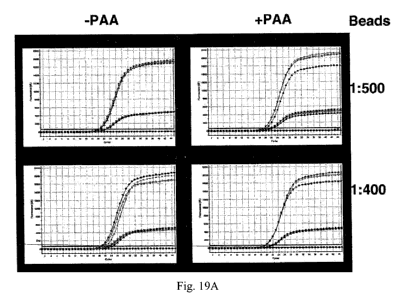
Fig. 19 demonstrates that polyacrylic acid ("PAA") also rescues PCR in the
presence of
anion exchange materials. Real time PCR amplifications were performed in
triplicate in the
14
CA 02773186 2012-03-05
WO 2011/037692 PCT/US2010/044586
presence of 1:200 (Fig. 19A), 1:250 (Fig. 19A); 1:400 (Fig. 19B); and 1:500
(Fig. 19B) dilutions
of AXpHTM beads in the presence ("PAA+") or absence ("PAA-") of 25 ng/ml PAA.
Fluorescent signals were generated using reporter probes labeled with either
FAM (top curve) or
TET (lower curve).
Fig. 20 demonstrates that the rescue effect of PAA an effect that holds for
low target
concentration in the presence of high anion exchange concentrations. Real time
PCR
amplifications were performed in triplicate on 10, 103, and 105 copies of a
target nucleic acid in
the presence of a 1:25 dilution of AXpHTM beads and 0 or 25 ng/mL PAA.
Fluorescent signals
were generated using reporter probes labeled with either FAM or 5'-Tetrachloro-
Fluorescein dye
("TET") as indicated. As can be seen, three distinct curves are present in the
control (Fig. 20A,
top curves), which is extinguished by the addition of AXpHTM beads for all but
the highest
concentration of target nucleic acid (Fig. 20A, bottom curves). Addition of
PAA reverses this
effect (Fig. 20B).
Fig. 21 demonstrates the inhibitory effects of anion exchange materials on
tHDA and
shows that amplification can be rescued by PAA. Real time amplifications of NG
DNA were
performed in triplicate on 10, 103, and 105 copies of a target nucleic acid in
the presence of a
1:25 dilution of AXpHTM beads and 0 or 25 ng/mL PAA. Fluorescent signals were
generated
using labeled reporter probes. Fig. 21A demonstrates control tHDA activity (no
PAA or beads),
Fig. 21 B demonstrates tHDA activity in the presence of beads, and Fig. 21 C
demonstrates tHDA
activity in the presence of beads and PAA.
Fig. 22 demonstrates that the presence of high molecular weight PAA (PAA-H),
low
molecular weight PAA (PAA-L), or polymethacrylic acid ("+PMA") permits
amplification of a
target NG DNA in the presence of AXpHTM beads. A standard three step real time
PCR reaction
was performed in triplicate for all examples using TagMan probes labeled with
either TET (top
curves) or FAM (bottom curves). All amplifications were performed in the
presence of 2.5mM
MgC12. Fig. 22A: 1 is a positive amplification control utilizing a target
nucleic acid in wash
buffer (10mM Tris, pH 8; 0.1 % NP-40 alternative); 2 is a bead control
utilizing a target nucleic
acid in wash buffer in the presence of AXpHTM beads. Fig. 22B: 3 is a target
amplified in wash
buffer plus PAA-H (0.25%) and AXpHTM beads; 4 is a target amplified in wash
buffer plus
PAA-L (0.25%) and AXpHTM beads. Fig. 22C: 5 is a target amplified in wash
buffer plus PMA
(0.25%) and AXpHTM beads.
CA 02773186 2012-03-05
WO 2011/037692 PCT/US2010/044586
Fig. 23 demonstrates that the presence of polyglutamic acid ("PGA") permits
PCR
amplification in the presence of anion exchange materials. A standard three
step real time PCR
reaction was performed on a NG DNA in triplicate for all examples using TagMan
probes.
B7-9 is an absolute negative control using wash buffer (10mM Tris, pH 8; 0.1 %
NP-40
alternative) in the absence of both target nucleic acid and AXpHTM beads. The
curves at A are
positive amplification controls using target nucleic acid dissolved in either
wash buffer alone or
wash buffer plus 1.25% PGA, 1.25% poly-adenylate ("poly-A"), or 1.25%
carboxymethyldextran ("CMD"). As can be seen, none of the polyanionic
compounds
significantly affected amplification of the target. B4-6 is a bead control to
demonstrate the
effect of anion exchange materials on amplification of the target. E4-6
indicates amplification
of the target in the presence of 1.25% CMD and AXpHTM beads. D4-6 indicates
amplification
of target in the presence of 1.25% PGA and AXpHTM beads. The left plot shows
the data
expressed as derivative curves. The right plot shows raw data.
Fig. 24 demonstrates that PAA inhibits PCR, which can be corrected by
increasing the
concentration of Mgt+. Target NG DNAs were amplified in the presence of either
0.05 or 0.1 %
PAA in the presence of two types of anion exchange materials (BCD and EFG,
respectively) and
3, 7, or 11 mM Mg2+. As can be seen in the figure, increasing the
concentration of Mg2-,- reversed
the inhibitory effect of PAA.
DETAILED DESCRIPTION OF THE INVENTION
It has long been known that nucleic acids can be releasably adsorbed onto
certain
positively ionized materials, owing to the negatively ionized phosphates of
the nucleic acid
backbone. This property is frequently manipulated to purify nucleic acids from
complex
biological materials through a procedure termed anion exchange. In the typical
scenario, a solid
phase is coated with "capture moieties" to form an anion exchange material.
Under appropriate
ionic and pH conditions, the phosphate backbone of nucleic acids will bear a
net negative charge,
while the capture moiety will bear a net positive charge. Thus, the nucleic
acid will bind to the
capture moiety, while neutrally or positively charged molecules, such as
proteins, will not. The
solid phase with the bound nucleic acid can then be separated from the
remaining by various
means, such as by centrifugation or application of a magnetic field. The anion
exchange material
can then be washed and, in the typical scheme, the nucleic acid eluted back
into solution by
16
CA 02773186 2012-03-05
WO 2011/037692 PCT/US2010/044586
altering the salt and/or pH conditions to form an eluate. The eluate can then
be separated from
the anion exchange material and used in subsequent analyses. Numerous such
methods have
been previously described. These methods are clean and easy to perform, result
in relatively
high nucleic acid yields, and do not require the dangerous and expensive
chemicals necessary
with tradition chemical extraction methods of purification. However, they do
present some
problems.
For example, elution of nucleic acids from anion exchange materials typically
requires
some combination of pH and/or ionic strength manipulation to elute bound
nucleic acids from
the anion exchange material. Nucleic acids eluted in this manner generally
cannot be used
directly in analytical methods, such as PCR, as the resulting eluate has a non-
optimal pH and/or
elevated ionic strength. Therefore, it would be desirable to develop materials
and methods that
permit elution of nucleic acids anion exchange materials at analytically
appropriate pH and ion
concentrations.
Accordingly, the present disclosure relates to materials, methods, and kits
for isolating a
nucleic acid with an anion exchange material, wherein an anionic compound
comprising at least
two anionic groups is added during an elution step.
In one aspect, a method of eluting a nucleic acid from an anion exchange
material is
disclosed, said method comprising: a) providing a nucleic acid-anion exchange
complex; and b)
adding a composition comprising an anionic compound comprising at least two
anionic groups to
the nucleic acid-anion exchange complex, wherein the anionic compound
displaces the nucleic
acid from the anion exchange material.
As used herein, the verb "to complex" shall refer to the process of a
positively ionized
capture moiety on the anion exchange material associating directly with a
negatively ionized
moiety on either the phosphate backbone of a target nucleic acid or an anionic
compound. The
noun "complex" shall refer to the chemical structure formed by such an
association. The term
"nucleic acid-anion exchange complex" shall refer to the chemical structure
formed when a
nucleic acid complexes with a positively ionizable capture moiety on an anion
exchange
material.
In one embodiment, the nucleic acid-anion exchange material is provided by a
method
comprising: (1) contacting an anion exchange material with a sample comprising
a nucleic acid
under conditions in which a complex forms between the anion exchange material
and the nucleic
17
CA 02773186 2012-03-05
WO 2011/037692 PCT/US2010/044586
acid, and (2) isolating the complex from the sample. In a further embodiment,
the step of
forming a complex between the anion exchange material and the nucleic acid is
performed under
pH, ionic strength, and/or detergent conditions such that the nucleic acid of
interest complexes
with the anion exchange material, but contaminants such as proteins,
endotoxins, and liposomes
do not. The conditions may be further refined such that only specific nucleic
acids form a
complex with the anion exchange material.
In another embodiment, the nucleic acid-anion exchange material is provided by
a
method comprising: (1) contacting a sample comprising the nucleic acid with
the anion exchange
material, (2) forming a complex between the anion exchange material and the
nucleic acid, (3)
isolating the complex from the sample, and (4) washing the complex to remove
impurities. The
wash conditions may be selected such that substantially all non-nucleic acid
material is removed.
Suitable wash buffers are known in the art and include but are not limited to
solutions
comprising water, alcohols in particular branched or unbranched alcohols
having 1 to 5 carbon
atoms, such as ethanol or isopropanol, polyethylenglycols,
polypropylenglycols, acetone,
carbohydrates, aqueous solutions comprising salts and mixtures of the
foregoing. By way of
example and not limitation, the wash buffer may comprise 0.1 % NP-40 in 0.1 mM
Tris, pH 8Ø
The wash conditions further may be refined such that specific nucleic acids
are removed as
impurities as well.
Another problem with isolating nucleic acids using anion exchange materials is
that the
eluate often does not completely separate from the anion exchange material,
resulting in
analytical samples contaminated with the anion exchange material, commonly
referred to as
"bead carryover". This is problematic because the presence of anion exchange
materials often
interferes with subsequent analysis of the nucleic acid. Moreover, molecular
biological analyses
are increasingly automated. Ideally, one would like to combine both the
isolation and the
analytical methods into a single automated process with as few steps and
reagents as possible.
The potential for anion exchange materials interfering with analysis of the
nucleic acid makes
predictable and reliable automation of such processes difficult. Therefore, it
would be desirable
to develop materials and methods that permit analysis of nucleic acids in the
presence of anion
exchange materials.
Accordingly, the present disclosure relates to materials, methods, and kits
for analyzing a
nucleic acid in the presence of an anion exchange material and an anionic
compound comprising
18
CA 02773186 2012-03-05
WO 2011/037692 PCT/US2010/044586
at least two anionic groups, wherein the anionic compound reverses inhibition
of the analytical
process caused by the anion exchange material.
It should be understood that the analysis step of the methods disclosed herein
is intended
to be performed in the presence of the anion exchange material. The presence
of the anion
exchange material can be either unintentional, as in the case of carry-over
during nucleic acid
purification, or it can be intentional, as when nucleic acids are analyzed
directly from the isolated
nucleic acid-anion exchange complex.
Nucleic Acid
The nucleic acids according to the present disclosure are not limited and
include any
nucleic acid. By way of example and not limitation, the nucleic acid may be:
DNA, including
but not limited to genomic DNA, mitochondrial DNA, bacterial DNA, viral DNA,
plasmids,
cosmids, linear oligodeoxynucleotides and polydeoxynucleotides, cDNA, PCR
fragments, PCR
amplicons, tHDA amplicons, LCR amplicons, long-range PCR amplicons,
oligonucleotides,
primers, probes, artificial or synthetic DNA; RNA, including but not limited
to mRNA, tRNA,
rRNA, viral RNA, siRNA, miRNA, RNAi, linear oligonucleotides, linear
polynucleotides,
probes, artificial or synthetic RNA; artificial nucleic acids such as PNA and
LNA as well as
combinations thereof such as nucleic acids comprising both DNA and RNA, and
hybrids thereof
such as RNA:DNA hybrids; complexes of nucleic acids with other biological
components. The
nucleic acid may be single-stranded or double-stranded. It may contain
modifications such as
natural modifications and artificial modifications, and may contain artificial
nucleotides
comprising, e.g., artificial bases, artificial sugar moieties and/or
artificial connections between
the nucleotides.
Nucleic acids can include, without limitation, nucleic acids found in
specimens or
cultures (e.g. cellular, microbiological and viral cultures) including
biological and environmental
samples. The ribonucleic acids may be found in any biological samples from
cell culture,
bacteria, viruses, an animal, including a human, fluid, solid (e.g., stool) or
tissue samples. Target
nucleic acids may further be found in biological samples including, but not
limited to cervical
samples (e.g., a sample obtained from a cervical swab), adenoid cells, anal
epithelial cells, blood,
blood products such as serum, plasma or buffy coat, saliva, cerebral spinal
fluid, pleural fluid,
milk, lymph, sputum, urine and semen.
19
CA 02773186 2012-03-05
WO 2011/037692 PCT/US2010/044586
In other embodiments, the nucleic acids are from other viral, bacteria,
mycobacteria or
plasmodia, for example cytomegalovirus (CMV), herpes, HIV, Chlamydia,
Gonorrhea,
Staphylococcus aureus, tubercolis, Sars Coronavirus and/or influenza.
In one embodiment, the nucleic acids are human papillomavirus (HPV) and
include
genetic variants of HPV. A variant includes polymorphisms, mutants,
derivatives, modified,
altered, or other forms of the nucleic acid. In one embodiment, the nucleic
acid is an HPV
nucleic acid. In another embodiment, the HPV nucleic acid is HPV DNA and/or
RNA of a high
risk HPV type. In another embodiment, the nucleic acids are high risk HPV
types such as, 16, 18,
31, 33, 35, 39, 45, 51, 52, 56, 58, 59, 68, 26, 66, and/or 82.
Sample comprising a nucleic acid
Samples that contain nucleic acid include, but are not limited to, a specimen
or culture
(e.g., cellular, microbiological and viral cultures) including biological and
environmental
samples. Biological samples may be from any source such as cell culture,
bacteria, viruses, an
animal, including a human, fluid, solid (e.g., stool) or tissue samples, as
well as liquid and solid
food and feed products and ingredients such as dairy items, vegetables, meat
and meat
byproducts, and waste. Environmental samples include, for example,
environmental material
such as surface matter, soil, water and industrial samples, as well as samples
obtained from food
and dairy processing instruments, apparatus, equipment, utensils, disposable
and non-disposable
items. Exemplary biological samples including, but not limited to, cell
samples, such as cervical
epithelial cells (e.g., a sample obtained from a cervical swab), adenoid
cells, anal epithelial cells,
blood, blood products such as serum, plasma or buffy coat, saliva, cerebral
spinal fluid, pleural
fluid, milk, lymph, sputum and semen, and may be collected, for example, in
Preservcyt,
Surepath and/or Digene Collection Medium ("DCM"). The sample may comprise a
deoxyribonucleic acid (DNA) and/or ribonucleic acid (RNA).
The sample may be processed prior to contacting it with the anion-exchange
material if
desired for any reason. For example, a biological sample comprising cells
comprising nucleic
acids may be treated to lyse the cells in order to release the nucleic acid.
The lysate comprising
the nucleic acids may then be added to the anion-exchange material. One
skilled in the art would
appreciate desirable methods for treating a sample containing nucleic acids
before contacting the
solution with an anion-exchange material. For example, the cells may be lysed
with a suitable
lysis buffer comprising, for example, 2% Triton X-100, 0.2 M EDTA, 40 mM
sodium citrate, 40
CA 02773186 2012-03-05
WO 2011/037692 PCT/US2010/044586
mM boric acid in 100 mM Tris HC1, pH 7Ø If an alkali lysis buffer is used to
prepare the
sample, the pH of the sample may need to be neutralized to a pH that allows
the nucleic acids to
bind to the anion-exchange material prior to contacting the sample with the
anion-exchange
material.
In other embodiments, the sample may comprise nucleic acids from other viral,
bacteria,
mycobacteria or plasmodia, for example cytomegalovirus (CMV), herpes, HIV,
Chlamydia,
Gonorrhea, Staphylococcus aureus, tubercolis, Sars Coronavirus or influenza.
In a further embodiment, the sample is treated such that the nucleic acid is
"free." As
used herein, the phrase "free nucleic acid" shall indicate that the nucleic
acid is not associated
with large macromolecular structures, such as vesicles, liposomes, micelles,
ribosomes, nuclei,
mitochondria, viral caspids and/or envelopes, endosomes, or exosomes.
Anion-Exchange Material
The present disclosure advantageously utilizes anion exchange material. The
terms
"anion exchange material", "anion exchange material", "anion exchange matrix"
and "anion
exchange resin" are used synonymously herein and in particular are not
restricted to materials
which are resins in the chemical meaning. As used herein, the term "anion
exchange material"
shall refer to any material that can be used to selectively remove nucleic
acids from a solution
via the formation of a complex between the phosphate backbone of the nucleic
acid and a
positively ionizable capture moiety of the material. Numerous anion exchange
materials have
previously been described and would be immediately recognized by a person
having ordinary
skill in the art, including for example those described in U.S. Pat. No.
6,914,137, US Pat. No.
5,990,301, US 20100009351A1, and EP 0 268 946 B1, the disclosures of which are
hereby
incorporated by reference. Any anion exchange material suitable for purifying
nucleic acids may
be used.
In one aspect, the anion exchange material material comprises a solid phase
and a
positively ionizable capture moiety.
Any solid phase suitable for anion exchange chromatography may be used,
including but
not limited to silica, borosilicates, silicates, anorganic glasses, organic
polymers such as
poly(meth)acrylates, polyurethanes, polystyrene, agarose, polysaccharides such
as cellulose,
metal oxides such as aluminum oxide, magnesium oxide, titanium oxide and
zirconium oxide,
metals such as gold or platinum, agarose, sephadex, sepharose, polyacrylamide,
divinylbenzene
21
CA 02773186 2012-03-05
WO 2011/037692 PCT/US2010/044586
polymers, styrene divinylbenzene polymers, dextrans, and derivatives thereof,
and/or silica gels,
beads, membranes, and resins; glass or silica surfaces, such as beads, plates,
and capillary tubes;
magnetizable or magnetic (e.g. paramagnetic, superparamagnetic, ferromagnetic
or
ferrimagnetic) particles, including but not limited to polystyrene, agarose,
polyacrylamide,
dextran, and/or silica materials having a magnetic material incorporated
therein or associated
therewith. In some embodiments, the capture moieties can be linked to the
surfaces of the
processing vessels such as micro-tubes, wells of micro-plates, or capillaries,
and using these
surfaces nucleic acids can be isolated on a micro scale. In one embodiment,
the solid phase will
be treated, manufactured, or otherwise processed such that the nucleic acid of
interest will not
bind directly to the solid phase during the purification step.
Anion exchange materials include, but are not limited to, materials modified
with
positively ionizable capture moieties. Examples of such ionizable groups are
monoamines,
diamines, polyamines, and nitrogen-containing aromatic or aliphatic
heterocyclic groups. In one
embodiment, the positively ionizable capture moiety comprises at least one
primary, secondary
or tertiary amino group. In another embodiment, the positively ionizable
capture moiety is
selected from the group consisting of a primary amine of the formula R3N, a
secondary amine of
the formula R2NH, and a tertiary amine X-(CH2)ri Y, wherein:
X is R2N, RNH or NH2,
Y is R2N, RNH or NH2,
R is independently of each other a linear, branched or cyclic alkyl, alkenyl,
alkynyl or aryl substituent which may comprise one or more heteroatoms,
preferably
selected from 0, N, S and P, and
n is an integer in the range of from 0 to 20, preferably 0 to 18.
Examplary capture moieties include, but are not limited to, aminomethyl (AM),
aminoethyl (AE), aminoalkyl, alkylaminoalkyl, dialkylaminoalkyl such as
diethylaminoethyl
(DEAE), ethylendiamine, diethylentriamine, triethylentetraamine,
tetraethylenpentaamine,
pentaethylenhexaamine, trimethylamino (TMA), triethylaminoethyl (TEAE), linear
or branched
polyethylenimine (PEI), carboxylated or hydroxyalkylated polyethylenimine, j
effamine,
spermine, spermidine, 3-(propylamino)propylamine, polyamidoamine (PAMAM)
dendrimers,
polyallylamine, polyvinylamine, N-morpholinoethyl, polylysine, and
tetraazacycloalkanes.
22
CA 02773186 2012-03-05
WO 2011/037692 PCT/US2010/044586
Biological buffer compounds also may be used as functional groups of the anion
exchange material, such as those described in the patents or applications US
6,914,137 and EP 1
473 299. Further examples of the positively ionizable groups are
polyhydroxylated amines,
detergents, surfactants, heterocycles, dyes, negatively charged groups in
combination with metal
ions or metal oxides, histidine and polyhistidine. Such groups are also
described, for example, in
the patent application WO 2003/101494 and paten application EP 09 007 338.8.
Also
zwitterionic groups such as amino acids or betaines may be used. In one
embodiment, the anion
exchange material comprises spermine-modified magnetic silica beads.
The solid phase may be functionalized for attachment of the capture moieties,
for
example with functionalities such as Si-O-Si, Si-OH, alcohol, diol or polyol,
carboxylate, amine,
phosphate or phosphonate. The positively ionizable capture moieties may be
attached to the solid
phase, for example, by using epoxides, (activated) carboxylic acids, acid
anhydrides, acid
chlorides, formyl groups, tresyl groups or pentafluorophenyl groups. The
functional groups may
be attached directly to the solid phase or via (linear or branched) spacer
groups, e.g.
hydrocarbons such as -(CH2)õ- groups, carbohydrates, polyethylenglycols and
polypropylenglycols. Alternatively, a polymer composed of monomers comprising
a capture
moiety such as an amino functional group can be used as anion exchange
material.
In some embodiments, ionizable groups can be linked to the surfaces of
processing
vessels, such as micro-tubes, micro-plates, or capillaries, and using these
surfaces nucleic acids
can be isolated on a micro scale.
In a further embodiment, the solid phase is treated, manufactured, or
otherwise processed
such that the nucleic acid of interest will not bind directly to the solid
phase during the
purification step.
In one embodiment, a PEI-modified paramagnetic silica bead is used as the
anion
exchange material. Exemplary PEI-modified magnetic silca beads include AXpHTM
beads
(commercially available from Qiagen GmbH, Hilden Germany). AXpHTM beads can be
separated magnetically from wash buffer and eluate, which is easier than the
filtration needed
when working with cellulose and other non-magnetic resins.
23
CA 02773186 2012-03-05
WO 2011/037692 PCT/US2010/044586
Nucleic Acid Binding
The ion exchange process known in the art usually involves two primary steps:
(1)
binding nucleic acids to the anion-exchange material at a pH that causes the
material to be
positively charged; and (2) elution and/or displacement of nucleic acids from
said material, by
either increasing salt concentration and/or increasing the pH above the pKa of
the capture
moieties ("pH shift method"). In the present disclosure, a solution comprising
at least one
compound comprising at least two anionic groups is applied to the anion-
exchange material to
elute the nucleic acids.
It will be understood by the person having ordinary skill in the art that the
precise ionic
and pH conditions necessary to cause complex formation and elution necessarily
depends on
both the identity and the concentration of both the capture moiety and the
nucleic acid of interest.
Methods of charging the anion-exchange material to prepare it for loading the
nucleic acid are
known to those skilled in the art.
In one aspect, the positively ionizable capture moiety bears a first net
positive charge at a
first pH and ionic strength and either a neutral charge or a second net
positive charge that is
lower than the first net positive charge at a second pH and ionic strength,
such that the nucleic
acid of interest binds to the positively ionizable capture moiety at the first
pH and ionic strength
and is released at the second pH and ionic strength.
In a further aspect, the positively ionizable capture moiety bears a first net
positive
charge at a first pH and ionic strength and a second net positive charge at a
second pH and ionic
strength, wherein a first nucleic acid and a second nucleic acid bind to the
positively ionizable
capture moiety at the first pH and ionic strength and the first nucleic acid,
but not the second
nucleic acid, is released from the positively ionizable capture moiety at the
second pH and ionic
strength.
The binding of the nucleic acid to the anion exchange material is usually done
in an
aqueous solution, preferably comprising buffer substances. Suitable biological
buffers are
CHAPS, MES, HEPES, MOPS, TRIS, TRICINE and PIPES. Furthermore, the solution
may
contain chaotropic salts such as sodium perchlorate, guanidium hydrochloride,
and guanidium
thiosulfate, as described, e.g., in US 5,234,809, and/or cosmotropic salts
such as ammonium
sulfate, zinc sulfate, potassium sulfate and cobalt sulfate, as described,
e.g., in WO 2004/055207.
24
CA 02773186 2012-03-05
WO 2011/037692 PCT/US2010/044586
The binding buffer may also contain further salts such as chlorides, sulfates,
phosphates,
acetates, formiates, citrates, azides, and nitrates, as well as further
organic compounds such as
alcohols, diols, triols, polyols, polyethylenglycols, polypropylenglycols,
acetone, acetonitrile,
urea, guanidine, carbohydrates and surface-active substances such as
surfactants or detergents,
for example tween, triton, brij, nonidet or pluronic. Salts preferably are
present in a concentration
of about 1 M or less, preferably 0,5 M or less, more preferably 250 mM or
less, most preferably
100 mM or less. Binding of the nucleic acids is preferably done at a pH in the
range of about 3 to
about 10, preferably about 5 to about 9.
Furthermore, binding of the nucleic acid to the anion exchange material may be
combined with a treatment of the sample such as lysis of cells or tissue in
the sample or digestion
of specific compounds such as proteins, DNA and/or RNA in the sample. To this
end, the
binding buffer may further comprise lysis agents such as enzymes, surfactants,
chaotropic salts
or chelators (e.g. EDTA or NTA), and/or digestion agents such as proteases,
DNases and
RNases. These sample treatment, however, may also be done in a preceding step
prior to the
binding of the nucleic acid to the anion exchange material (see also above).
Binding, as well as pre-treatment steps such as lysis and/or enzymatic
pretreatment
processes can be performed at elevated temperatures in order to speed up the
respective
processes.
It may be possible to wash the anion exchange material after the sample has
been added
to the anion exchange material to remove unbound material in the sample, such
as proteins,
polysaccharides, et cetera. Suitable washing buffers which can be used to
remove non-target
materials are known in the prior art and include but are not limited to
solutions comprising water,
alcohols in particular branched or unbranched alcohols having 1 to 5 carbon
atoms, such as
ethanol or isopropanol, polyethylenglycols, polypropylenglycols, acetone,
carbohydrates,
aqueous solutions comprising salts and mixtures of the foregoing. An exemplary
wash buffer
comprises, for example, 0.1 % NP-40 in 0.1 mM Tris, pH 8Ø
Elution
After the nucleic acid is bound to the anion exchange material, the bound
nucleic acid can
be eluted by adding a solution comprising an anionic compound comprising at
least two anionic
groups or a mixture of such anionic compounds.
CA 02773186 2012-03-05
WO 2011/037692 PCT/US2010/044586
1. Anionic compound
The anionic compound(s) displaces the nucleic acids from the anion exchange
material.
The present method does not need to involve a change in pH in particular above
the pKa of the
capture moiety to cause elution, but rather relies on the anionic compound(s)
to elute nucleic acid
from anion-exchange material bound with nucleic acid. The anionic compound(s)
can displace
nucleic acid from anion-exchange materials at relatively low concentrations
due to their own
high selectivity to the material.
The optimal concentration of anionic compounds to be used in the eluate should
be
determined for any particular application; this concentration should be high
enough to provide
high recovery of nucleic acids, but not too high to compromise any subsequent
operations and
processes. The anionic compounds may be used at varying concentrations
necessary to elute the
nucleic acid. For example, the concentration may be 0.1 % to about 2.0 % and
in certain
embodiments, the concentration is from about 0.5 % to about 1.0%. It is
understood that any
numerical value recited herein includes all values from the lower value to the
upper value (and
including the lower value and the upper value). For example, all possible
combinations of
numerical values between the lowest value and the highest value enumerated are
to be
considered expressly stated in this application. For example, for a
concentration range stated as
0.025 % to about 2.5 %, it is intended that values such as 0.05, 0.2, 0.3,
0.4, 1.8, 1.9, etc. or any
ranges within this range such as 0.3 to 1.0 or 0.4 to 2.4, etc., are expressly
enumerated in this
specification. These low concentrations have little or no effect on subsequent
detection/analysis
steps such as amplification or enzymatic reactions. Thus, the eluate can be
used directly without
a neutralization step typically required when using pH based or a desalting
step when using a salt
buffer based elution.
As used herein, "anionic compound" shall refer to any compound having a net
negative
charge at the pH and salt conditions used to elute or analyze the nucleic
acid.
As used herein, the phrase "anionic group" shall refer to any functional
group, covalently
bound to the anionic compound, that bears a net negative charge at the pH and
salt conditions
used to elute or analyze the nucleic acid. In all cases, each anionic group of
each anionic
compound may be the same or different.
Suitable anionic compounds include polyanionic compounds, as well as non-
polymeric
anionic compounds. Preferably, the anionic compound is organic.
26
CA 02773186 2012-03-05
WO 2011/037692 PCT/US2010/044586
Suitable non-polymeric anionic compounds useful in the methods and
compositions
disclosed herein include, for example, any non-polymeric organic compounds
comprising at least
two anionic groups. By way of example and not limitation, the non-polymeric
anionic
compound may comprise at least 2, at least 3, at least 4, at least 5, or at
least six anionic groups.
In another embodiment, the non-polymeric anionic compound comprises from 2 to
6 or 3 to 6
anionic groups. In another embodiment, the non-polymeric anionic compound may
comprise not
more than 15 anionic groups, not more than 10 anionic groups, not more than 8
anionic groups,
or not more than 6 anionic groups. The non-polymeric organic anionic compound
preferably has
2 to 40 carbon atoms, more preferably 2 to 20 carbon atoms, even more
preferably 2 to 12 carbon
atoms. It may be linear, branched or cyclic and may be aliphatic (being
saturated or unsaturated)
or aromatic (having one or more conjugated or fused rings).
In one embodiment, the at least two anionic groups are selected from the group
consisting
of a carboxylic acid group, a sulfonic acid group, a phosphonic acid group, a
phosphate group, a
carbonate group, and combinations thereof.
As used herein, the term "acid group" shall encompass both the referenced acid
and,
alternatively, its conjugated base.
In one embodiment, the at least two anionic groups are selected from the group
consisting
of. a carboxylate group, a sulfonate group, a phosphonate group, and an
ionized phosphate
group. In another embodiment, the anionic group is a carboxylic
acid/carboxylate group. In
another embodiments, the non-polymeric anionic compound is selected from the
group
consisting of a dicarboxylic acid, tricarboxylic acid, tetracarboxylic acid,
pentacarboxylic acid,
hexacarboxylic acid, heptacarboxylic acid, octacarboxylic acid, nonacarboxylic
acid, and a
decacarboxylic acid.
In another embodiment, the non-polymeric compound comprising at least two
anionic
groups is a carboxylic acid, such as oxalic acid, fumaric acid, glutaric acid,
maleic acid, malic
acid, malonic acid, succinic acid, glutaric acid, adipic acid, pimelic acid,
suberic acid, azelaic
acid, tartronic acid, tartaric acid, citric acid, isocitric acid, citraconic
acid, mesaconic acid,
itaconic acid, aconitic acid, propane-1,2,3-tricarboxylic acid, aconitic acid,
butane-1,2,3,4-
tetracarboxylic acid, triethyl-1,1,2-ethanetricarboxylic acid, cyclopropane
dicarboxylic acid,
cyclobutane dicarboxylic acid, cyclobutane tricarboxylic acid, cyclopentane
dicarboxylic acid,
cyclohexane dicarboxylic acid, cyclohexane tricarboxylic acid, cyclohexane
tetracarboxylic acid,
27
CA 02773186 2012-03-05
WO 2011/037692 PCT/US2010/044586
cyclohexane hexacarboxylic acid,cyclooctane dicarboxylic acid, phthalic acid,
isophthalic acid,
terephthalic acid, hemimellitic acid, trimellitic acid, trimesic acid,
mellophanic acid, prehnitic
acid, pyromellitic acid, benzene pentacarboxylic acid, and mellitic acid,
hexacarboxylic acid of
dierythritol, octaacetic acid of trierythritol, iminodiacetic acid,
nitrilotriacetic acid,
ethylenediamine tetraacetic acid, diethylenetriamine pentaacetic acid, a-
ketoglutaric acid,
glutamic acid, aspartic acid, dicarboxymalonic acid, (18-crown-6)-2,3,11,12-
tetracarboxylic acid,
oligomers of 2 to 10, preferably 2 to 6, polymerizable or condensable acidic
monomers such as
acrylic acid, methacrylic acid, or vinylacetic acid.
In another embodiment, the non-polymeric compound comprising at least two
anionic
groups is an organosulfonic acid, such as methyl disulfonic acid, ethyl
disulfonic acid, benzene
disulfonic acid, phenole disulfonic acid, or naphthalene disulfonic acid.
In another embodiment, the non-polymeric compound comprising at least two
anionic
groups is an organophosphonic acids, such as hydroxyethane diphosphonic acid.
In another embodiment, the non-polymeric compound comprising at least two
anionic
groups is an organophosphate.
In another embodiment, the non-polymeric compound comprising at least two
anionic
groups is a carbonate.
In another embodiment, the non-polymeric compound comprising at least two
anionic
groups is a diacetylacetone.
In another embodiment, the non-polymeric anionic compound is selected from the
group
consisting of oxalic acid, mellitic acid, pyromellitic acid and citric acid.
The non-polymeric anionic compound may optionally be substituted, as long as
the
substituents do not interfere with or inhibit the ability of the compound to
displace the nucleic
acid from the anion exchange material. Exemplary substituents include, for
example, halogen
atoms such as fluorine, chloride, bromide or iodine, and hydroxyl, oxy,
aldehyde, keto, alkoxy,
ether, ester, amino, thiol, thioether, thioester, linear or branched alkyl,
alkenyl, alkynyl, saturated
or unsaturated cycloalkyl and aryl groups. In one embodiment, the anionic
compound comprises
1 to 6 substituents or 1 to 3 substituents. In another embodiment, these
substituents comprise - if
present - not more than 20, 10, 8, 6, 4 or 2 carbon atoms.
In one embodiment, the non-polymeric anionic compound is a low molecular
weight
compounds. In another embodiment, the non-polymeric anionic compound has a
molecular
28
CA 02773186 2012-03-05
WO 2011/037692 PCT/US2010/044586
weight of 5,000 Da or less. In another embodiment, the non-polymeric anionic
compound has a
molecular weight of 3,000 Da or less. In another embodiment, the non-polymeric
anionic
compound has a molecular weight of 2,000 Da or less.
Non-polymeric anionic compounds having at least two anionic groups have the
advantage that only relatively low concentrations of the anionic compound are
necessary to
effectively elute the bound nucleic acid. Furthermore, the presence of non-
polymeric anionic
compounds do not interfere with or inhibit subsequent amplification reactions,
even at rather
high concentrations.
In another aspect, the anionic compound comprising at least two anionic groups
is a
polyanionic compounds. As used herein, the phrase "polyanionic compound" shall
refer to a
polymeric anionic compound.
In one embodiment, the polyanionic compound is selected from the group
consisting of:
polymerized unsaturated carboxylic acids (e.g. acrylic, methacrylic, maleic,
etc.) or copolymers
of these acids with other monomers, such as acrylamide or acrylonitrile;
acidic polypeptides such
as polyglutamic or polyaspartic acid, or copolymers of acidic polypeptides
with other amino
acids; modified dextran and other modified or polyanionic polycarbohydrates
bearing covalently
attached ionized groups, such as carboxymethyl dextran, dextran sulfate and
dextran phosphate
or mixtures thereof; polystyrene with anionic groups, such as
polystyrenesulfonates; and even
other nucleic acids such as double-stranded or single-stranded DNA or RNA, or
analogs thereof
such as "base-free" nucleic acid (nucleic acid which only comprises the
backbone structure and
does not comprise any bases attached to the sugar moieties). In another
embodiment, polyacrylic
acid (PAA), polymethacrylic acid (PMA), polyglutamic acid (PGA), and/or
dextran sulfate (DS)
are selected. In another embodiment, a "low molecular weight" polyacrylic acid
(a weight
average Fw-5,100; approximately a 70-mer) is selected. In another embodiment,
"high
molecular weight" polyacrylic acid (a weight average Fw>200,000; i.e. longer
than a 2,800-mer),
is selected.
In one embodiment, the polyanionic compound is a second nucleic acid. When
using a
second nucleic acid for elution of the bound, first nucleic acid, the second
nucleic acid should not
interfere with the subsequent process (e.g. RNA can be used to elute DNA if
the next step is PCR
analysis). The second nucleic acid preferably comprises at least 100 base
pairs (bp), more
preferably at least 200 bp, at least 500 bp or at least 1000 bp. It may be,
for example, plasmid
29
CA 02773186 2012-03-05
WO 2011/037692 PCT/US2010/044586
DNA or genomic DNA or a carrier nucleic acid, such as polydeoxyadenosine
("poly-dA"),
polydeoxythymidine ("poly-dT") or a co-polymer of polydeoxyadenosine and
polydeoxythymidine ("poly-dA:dT")
The anionic compound may be added in the presently disclosed methods either as
free
acid or as a salt. Suitable cations for use in such salts are, for example,
any alkaline cation, such
as sodium and lithium.
2. Elution
Elution of the bound nucleic acid from the anion exchange material is achieved
using the
anionic compound comprising at least two anionic groups as defined herein.
Preferably, elution
is performed using an elution buffer containing the anionic compound. The
elution buffer
preferably is an aqueous solution which may further contain, for example, a
buffering agent, e.g.
as described above with respect to the binding solution, organic components
and/or salts. In one
embodiment, the pH used for elution preferably is in the range of from about 5
to about 13, from
about 5 to about 9.5, or from about 5 to about 8.5. In another embodiment, the
pH lies in a range
from about 8.2 to about 9.0, in particular when the eluate is supposed to be
used directly in a
amplification reaction such as PCR, RT-PCR, or a isothermal amplification
reaction.
Elution of nucleic acids from the anion exchange material can be performed
without the
necessity of severe changes in the pH. In one embodiment, the pH during the
elution step does
not render the anion exchange material neutral or negatively charged. In
another embodiment,
the pH during elution does not significantly reduce the positive charge of the
anion exchange
material. In yet another embodiment, the pH during the elution step is not
above the pKa of the
anion exchange material and/or the capture moieties thereof.
In another embodiment, the solution used for eluting the nucleic acid from the
anion
exchange material does not comprise a high salt concentration. In another
embodiment, the total
salt concentration in the elution solution does not exceed 1 M, is at or below
0.5 M, 300 MM,
200 mM, 150 mM, 100 mM, 50 mM or 30 mM. In another embodiment, the elution
solution
does not contain any salts except for the anionic compound comprising at least
two anionic
groups.
Nucleic acids in biological samples are preferably first bound to anion-
exchange material,
for example, anion-exchange magnetic beads. The material is then washed to get
rid of proteins
and other undesirable impurities. Nucleic acids are then eluted by a solution
comprising at least
CA 02773186 2012-03-05
WO 2011/037692 PCT/US2010/044586
one anionic compound comprising at least two anionic groups as defined herein.
This procedure
requires no change or at least no severe change in pH for the elution step. In
fact, the pH of the
wash and elution buffer can be the same.
During elution, the anionic compound is present in a concentration high enough
to effect
elution of at least a part of the bound nucleic acids. In one embodiment, the
anionic compound is
present in a concentration high enough to effect elution of a majority of the
bound nucleic acids.
In one embodiment, the anionic compound is present in a concentration high
enough to effect
elution of substantially all of the bound nucleic acids. As just one example,
the concentration of
a non-polymeric anionic compound during elution is selected in the range of
from about 1 mg/l
to about 1 g/1, more preferably from about 10 mg/l to about 500 mg/l, even
more preferably from
about 20 mg/l to about 100 mg/l. As another example, the amount of the
polyanionic compound
is in the range of from about 0.01% to about 5%, more preferably from about
0.025% to about
2.5%, even more preferably from about 0.1% to about 2.0% or from about 0.5% to
about 1.0%.
The optimal concentration of anionic compounds to be used in the eluate should
be
determined for any particular application. Namely, the concentration should
preferably be high
enough to provide high recovery of nucleic acids, but not too high to
compromise any
subsequent operations and processes. On the other hand, if the anionic
compounds used in
nucleic acid isolation step decrease efficiency of the subsequent procedures
such as
amplifications, such negative effects can be overcome by specific adjustments
of conditions.
For example, when DNA elution from AXpH beads, which are specific magnetic
silica
beads bearing polyethyleneimine groups available from QIAGEN, Germany, was
performed
with I% polyacrylic acid as an example of a polyanionic compound, it
completely inhibited PCR
when the eluates comprised 1/10th of the PCR volume. However, PCR efficiency
was completely
restored when MgC12 concentration was increased from 5 mM to 11 MM.
Accordingly, in certain
embodiments, after isolation, the isolated nucleic acid can be amplified in
the presence of Mg2+
at a concentration of about 10 to about 40mM. The polyanionic compounds may be
used at
varying concentrations necessary to elute the nucleic acid. For example, the
concentration may
advantageously be 0.1 % to about 2.0 % and in certain embodiments, the
concentration is from
about 0.5 % to about 1.0%. It is understood that any numerical value recited
herein includes all
values from the lower value to the upper value (and including the lower value
and the upper
value). For example, all possible combinations of numerical values between the
lowest value and
31
CA 02773186 2012-03-05
WO 2011/037692 PCT/US2010/044586
the highest value enumerated are to be considered expressly stated in this
application. For
example, for a concentration range stated as 0.025 % to about 2.5 %, it is
intended that values
such as 0.05, 0.2, 0.3, 0.4, 1.8, 1.9, etc. or any ranges within this range
such as 0.3 to 1.0 or 0.4 to
2.4, etc., are expressly enumerated in this specification. These low
concentrations have little or
no effect on subsequent detection/analysis steps such as amplification or
enzymatic reactions.
Thus, the eluate can be used directly in such downstream reactions without
neutralization (which
is typically required when using pH based elution) and/or without using a
desalting step (when
using a salt buffer based elution).
By way of example, PAA elutes DNA from anion-exchange magnetic beads at
concentrations that do not significantly affect PCR or isothermal
amplification such as tHDA
(thermophilic Helicase Dependent Amplification). The concentration of PAA is
typically, but
not limited to, from 0.025-2.5%. In one embodiment, PAA is present at a
concentration of from
0.1% to 2.0 %. In another embodiment, and more preferably 0.5-1.0 %), and the
exact
concentration depends also on the volume of the sample that is included into
the PCR. For
example, higher PAA concentrations provide better nucleic acid recovery from
AXpH beads but
tend to somewhat slow down amplification. Thus, if higher PAA concentrations
are used for
elution, then it may be preferable to dilute the sample in downstream
reactions or use the sample
in a larger overall total volume in downstream reactions. As an example, if
DNA was eluted by
I% PAA, this may lead to unsatisfactory results when 8 L of such eluates are
loaded per 25 L
PCR reaction. However, the same eluates will yield the expected results if
either the volumes of
eluates are smaller (e.g., 2 L of such eluate per 25 L PCR) or total PCR
volumes are higher
(e.g., 8 L of the eluate is loaded into 100 L PCR reaction). Thus, PAA
concentration in the
elution buffer, sample volume per PCR and the volumes of individual PCR should
be found
experimentally as a reasonable compromise between required assay sensitivity
and limitations of
PCR volume. Alternatively, concentrations of polyanionic compounds that could
be inhibitory to
PCR or other nucleic acid amplification techniques can be tolerated if the
eluates are used in
applications that do not require amplification; for example, in nucleic acid
hybridization
techniques. Thus, eluates can be used directly for nucleic acid detection.
According to one embodiment, the elution is performed at elevated temperatures
such as
e.g. at temperatures > 50 C, > 60 C or even > 70 C.
32
CA 02773186 2012-03-05
WO 2011/037692 PCT/US2010/044586
The present disclosure also provides methods for nucleic acid isolation,
analysis and in
particular amplification that allow automation utilizing an anion exchange
material that is
suitable for the gentle isolation of DNA as well as RNA. Further, the eluted
DNA or RNA does
not generally need additional handling steps such as a neutralization step or
desalting step in
order to be fully compatible with downstream reactions.
Eluates, Analysis, and Amplification Compositions
One advantage of the isolation and elution methods disclosed herein is that an
eluate
comprising the nucleic acid of interest can be generated without resorting to
pH switch or high
salt methods. As such, the eluates generated according to the methods
described herein may be
used directly in various analytical methods, including, for example,
amplification procedures
such as PCR, RT-PCR, helicase-dependent amplification, hybrid capture assays,
sequencing or
transfection. In case the eluted nucleic acid is used in amplification
procedures, any anionic
compound comprising at least two anionic groups as defined herein can be used.
However, in
certain embodiments including a subsequent amplification reaction, in
particular a PCR, the
anionic compound is not dextran sulfate.
Thus, in one aspect, an eluate obtained according to any of the above-
described methods
is provided. In one embodiment, the eluate is directly compatible with a
subsequent enzymatic
reaction. By "directly compatible," it is meant that the resulting eluate does
not require any
further processing steps (such as desalting, neutralization, et cetera) before
it maybe included in
an enzymatic reaction mixture. In another embodiment, the eluate is obtained
by a method
comprising: a) providing a nucleic acid-anion exchange complex; and b) adding
a composition
comprising an anionic compound comprising at least two anionic groups to the
nucleic acid-
anion exchange complex, wherein the anionic compound displaces the nucleic
acid from the
anion exchange material, and further wherein the composition comprising the
anionic compound
further comprises amplification components. By way of example and not
limitation, said
amplification components may be: an enzyme having a polymerase activity; an
enzyme having a
reverse transcriptase activity; an enzyme having a helicase activity;
nucleotides; primer nucleic
acids; amplification buffers; a nick-inducing agent; a source of Mg2+ and/or
Mn2+, for example,
MgC12 and/or MnC12; a ribonucleotide triphosphate (NTP); a deoxyribonucleotide
triphosphate
(dNTP); a source of K+, for example, KC1; a source of NH4-,-, for example,
(NH4)2SO4; or a
reducing agent, for example, 2-mercapthoethanol and/or dithiothreitol (DTT).
33
CA 02773186 2012-03-05
WO 2011/037692 PCT/US2010/044586
In another aspect, a method of analyzing a nucleic acid is provided, said
method
comprising, consisting essentially of, or consisting of. a) providing a
nucleic acid-anion
exchange complex; b) adding a composition comprising an anionic compound
comprising at
least two anionic groups to the nucleic acid-anion exchange complex; and c)
analyzing the
nucleic acid in the presence of the anionic compound.
In one embodiment, the analysis method comprises a nucleic acid amplification.
Nucleic
acid amplifications can be broadly separated into two categories: temperature
cycled
amplifications and isothermic amplifications. Either class of amplification
may be used. The
amplification in the disclosed methods can be either a temperature cycled
amplification or an
isothermic amplification. Exemplary methods of amplification include, but are
not limited to,
polymerase chain reaction ("PCR"), reverse transcriptase ("RT") reaction,
thermophilic helicase-
dependent amplification ("tHDA"), whole genome amplification, and ligase chain
reaction
("LCR"). Exemplary analytical methods comprising an amplification step
include, but are not
limited to, PCR, RT-PCR, real time PCR, real time RT-PCR, quantitative real
time PCR,
quantitative real time RT-PCR, multiplex analysis, melting curve analysis,
high resolution
melting curve analysis, tHDA, RT-tHDA, real time tHDA, quantitative real time
tHDA,
quantitative real time RT-tHDA, NIA, RT-NIA, real time NIA, quantitative real
time NIA, and
quantitative real time RT-NIA.
In temperature cycled amplifications, such as PCR, the temperature typically
is raised
above the melting point of the target nucleic acid to "melt" any double
stranded portions, and
then lowered to a point at which oligonucleotide primers anneal with single
stranded portion of
the target nucleic acid, then raised again to a temperature at which the
primers remain annealed
and the polymerase is active.
In isothermic amplifications, such as tHDA and whole genome amplification, an
agent is
added to the reaction mixture to permit amplification without temperature
cycling. For example,
in tHDA, an enzyme having helicase activity is added to the amplification
mixture. As used
herein, "helicase" or "an enzyme with, or having, helicase activity" refers to
any enzyme capable
of unwinding a double stranded nucleic acid. The helicase functions to unwind
double stranded
nucleic acids, thus obviating the need for repeated melting cycles. Exemplary
helicases include
E. coli helicase I, II, III, & IV, Rep, DnaB, PriA, PcrA, T4 Gp41 helicase, T4
Dda helicase, T7
Gp4 helicases, SV40 Large T antigen, yeast RAD. Additional helicases that may
be useful
34
CA 02773186 2012-03-05
WO 2011/037692 PCT/US2010/044586
include RecQ helicase, thermostable UvrD helicases from T. tengcongensis and
T. thermophilus,
thermostable DnaB helicase from T. aquaticus, and MCM helicase from archaeal
and eukaryotic
organisms. As another example, in nick-initiated amplification ("NIA"), a nick-
inducing agent is
used to induce breaks in the phosphodiester bonds of the nucleic acid
backbone. A polymerase
having strand displacement activity can then initiate amplification at the
site of the nick, using
one strand of the nucleic acid as a primer and the other strand as a template.
As used herein,
"nick-inducing agent" refers to any enzymatic or chemical reagent or physical
treatment that
introduces breaks in the phosphodiester bond between two adjacent nucleotides
in one strand of a
double-stranded nucleic acid. Examples of nick-inducing enzymes include Bpul O
I, BstNB I,
Alw I, BbvC I, BbvC I, Bsm I, BsrD, and E. coli endonuclease I.
Analysis in the presence of an anion exchange material
Anion exchange materials are know to interfere with interfere with the
analysis of nucleic
acids. Separate and apart from their utility as elution agents, the anionic
compounds as described
throughout also have the unexpected property of permitting analysis of nucleic
acids in the
presence of anion exchange materials. Accordingly, the present disclosure
relates to materials
and methods of analyzing nucleic acids in the presence of anion exchange
materials, wherein a
anionic compound is added during the analysis step.
In one embodiment, a method of analyzing a nucleic acid of interest in the
presence of an
anion exchange material is disclosed, said method comprising: a) purifying the
nucleic acid of
interest from a sample using the anion exchange material; and b) analyzing the
nucleic acid in a
first solution comprising at least one anionic compound, wherein the anionic
compound
comprises at least two anionic groups. In one embodiment, the anionic compound
is non-
polymeric. In another embodiment, the anionic compound is a polyanionic
compound.
In another embodiment, the purifying step comprises the steps of. (1)
contacting a sample
comprising the nucleic acid with the anion exchange material, (2) forming a
nucleic acid-anion
exchange complex between the anion exchange material and the nucleic acid, and
(3) isolating
the nucleic acid-anion exchange complex from the sample.
In a further aspect, the purifying step comprises the steps of. (1) contacting
a sample
comprising the nucleic acid with the anion exchange material, (2) forming a
complex between
the anion exchange material and the nucleic acid, (3) isolating the complex
from the sample, and
(4) washing the complex to remove impurities. The wash conditions may be
selected such that
CA 02773186 2012-03-05
WO 2011/037692 PCT/US2010/044586
substantially all non-nucleic acid material is removed. The wash conditions
further may be
refined such that specific nucleic acids are removed as impurities as well.
In another embodiment, the nucleic acid is eluted from the nucleic acid-anion
exchange
complex before the anionic compound is added. For example, the nucleic acid
may be eluted
using pH switch or high salt solutions. In such a case, the resulting eluant
will likely need to be
further processed before it can be used in an analytical process. As another
example, the nucleic
acid may be eluted using a solution comprising an anionic compound, as
described above. The
eluate may then be separated from a substantial portion of the anion exchange
material before an
analysis (as in bead carryover), or the eluate may be analyzed in the presence
of the entire
portion of the
In another embodiment, an analytical solution comprising the anionic compound
is added
directly to the nucleic acid-anion exchange complex. By way of example and not
limitation,
where the analysis method is an amplification, a nucleic acid amplification
composition
comprising an anionic compound comprising at least two anionic groups may be
added directly
to the nucleic acid-anion exchange complex.
As used herein, the term "nucleic acid amplification composition" shall refer
to any
composition comprising an anionic compound as described above and further
having the
appropriate components and physical properties such that a nucleic acid
amplification may be
conducted therein. Such components may include in the appropriate
circumstance, but are not
limited to: buffers; salt solutions; primers; nucleotide triphosphates;
macromolecules such as
RNase, RNase inhibitors, coenzymes, and/or catalysts; polyethylene glycol;
sorbitol; and DMSO.
Physical properties include, but are not limited to, pH, ionic strength, and
Mg2+ concentration.
The precise identity of the additional components and physical properties will
depend on
numerous factors, including but not limited to: the particular amplification
method being used;
the identity of the nucleic acid being amplified; the length and G/C content
of the desired
amplicon; and the particular polymerase and/or helicase being used. These
additional
components and physical properties will be immediately apparent to the person
having skill in
the art. By way of example and not limitation, said amplification components
may be any
combination of. an enzyme having a polymerase activity; an enzyme having a
reverse
transcriptase activity; an enzyme having a helicase activity; nucleotides;
primer nucleic acids;
amplification buffers; a nick-inducing agent; a source of Mg2+ and/or Mn2+,
for example, MgC12
36
CA 02773186 2012-03-05
WO 2011/037692 PCT/US2010/044586
and/or MnC12; a ribonucleotide triphosphate (NTP); a deoxyribonucleotide
triphosphate (dNTP);
a source of K+, for example, KC1; a source of NH4, for example, (NH4)2SO4; or
a reducing
agent, for example, 2-mercapthoethanol and/or dithiothreitol (DTT).
The nucleic acid amplification composition may be provided at a working (1X)
concentration or in a concentrated format. By way of example and not
limitation, concentrated
formats may be provided as aqueous solutions at 5X, I OX, 20X, or 50X
concentrations, or as
lyophilized concentrates.
In one embodiment, the nucleic acid amplification composition comprises a) at
least one
target nucleic acid; b) an anion exchange material; c) at least one anionic
compound comprising
at least two anionic groups; and d) at least one protein having a polymerase
activity.
Another embodiment relates to a nucleic acid amplification composition
comprising a) at
least one target nucleic acid; b) an anion exchange material; c) at least one
anionic compound
comprising at least two anionic groups; d) at least one protein having a
polymerase activity; and
e) at least one protein having a helicase activity.
Such materials and methods are particularly suitable for use in automated
methods.
Kits
The present disclosure also provides a kit for isolating nucleic acid from a
sample such as
a biological sample, the kit comprising an anion exchange material, preferably
an anion
exchange material as described above; and an elution buffer comprising at
least one anionic
compound comprising at least two anionic groups as described above. The kit
may further
comprise one or more components selected from the group consisting of washing
buffers,
loading buffers, equilibration buffers, lysis buffers (e.g. for lysing the
sample such as a cell or
tissue sample), nucleases such as RNases and/or DNases, RNase or DNase
inhibitors, and
instructions for its use. The kit for isolating nucleic acid may also comprise
further components
suitable for analyzing and/or amplifying the isolated nucleic acid.
Furthermore, the present disclosure also provides a kit for amplifying nucleic
acid, the kit
comprising an enzyme having a polymerase activity and at least one anionic
compound
comprising at least two anionic groups, preferably a non-polymeric anionic
compound as
described above. The kit may further comprise one or more additional
components such as an
enzyme having a reverse transcriptase activity; an enzyme having a helicase
activity;
nucleotides; primer nucleic acids; amplification buffers; a nick-inducing
agent; a source of Mg2+
37
CA 02773186 2012-03-05
WO 2011/037692 PCT/US2010/044586
and/or Mn2, for example, MgClz and/or MnC12; a ribonucleotide triphosphate
(NTP); a
deoxyribonucleotide triphosphate (dNTP); a source of K+, for example, KC1; a
source of NH4
for example, (NH4)2SO4; a reducing agent, for example, 2-mercapthoethanol
and/or dithiothreitol
(DTT) and instructions for its use. The kit for amplifying nucleic acid may
further contain any of
the components of the kit for isolating nucleic acid.
The various components of the kits disclosed herein may be provided at a
working (1X)
concentration or in a concentrated format. By way of example and not
limitation, concentrated
formats may be provided as aqueous solutions at 5X, I OX, 20X, or 50X
concentrations, or as
lyophilized concentrates.
Use
The present disclosure also provides the use of an anionic compound comprising
at least
two anionic groups, preferably a non-polymeric compound comprising at least
two anionic
groups, for displacing a nucleic acid reversibly bound to an anion-exchange
material from said
anion-exchange material. The anionic compound, the non-polymeric compound, the
nucleic acid
and/or the anion-exchange material are preferably as described above.
The present disclosure also provides a method of isolating a nucleic acid from
a sample
containing a nucleic acid. The method comprises the step of. a) contacting the
sample
comprising the nucleic acid with an anion-exchange material to allow binding
of the nucleic
acid. The anion-exchange material is capable of reversibly binding the nucleic
acid. The method
further comprises the steps of. b) optionally washing the anion-exchange
material to remove
unbound sample components; and c) eluting the bound nucleic acid by adding at
least one
compound comprising at least two anionic groups, wherein the anionic
compound(s) is capable
of displacing the nucleic acids from the anion-exchange material.
Details with respect to the compound comprising at least two anionic groups
and the
anion exchange material and the method are discussed in detail above. It is
referred to the above
disclosure.
As discussed above, the present disclosure also provides a method of
analyzing,
preferably amplifying a nucleic acid in the presence of an anion exchange
material, said method
comprising the steps of complexing the nucleic acid with the anion exchange
material to form a
nucleic acid-anion exchange complex; optionally separating the nucleic acid-
anion exchange
complex from other material; and analyzing, preferably amplifying the nucleic
acid in a solution
38
CA 02773186 2012-03-05
WO 2011/037692 PCT/US2010/044586
comprising at least one compound comprising at least two anionic groups. The
anionic
compound preferably does not inhibit or interfere with the amplifying
reaction, respectively
analysis step. Furthermore, the anionic compound preferably reduces or removes
any inhibitory
effect the anion exchange material has on the amplifying reaction or analysis
step. That is, the
anion exchange material, when present, may reduce the efficacy of the
analysis, in particular the
amplification step. This reduction in efficacy is at least partially
neutralized by the presence of
the anionic compound used according to the invention.
As discussed above, also provided is a method of isolating a nucleic acid from
a sample
comprising a nucleic acid, said method comprising:
a) contacting the sample with an anion-exchange material, wherein the anion-
exchange material reversibly binds nucleic acid;
b) optionally washing the anion-exchange material to remove unbound sample
components; and
c) eluting bound nucleic acid by adding at least one non-polymeric compound
comprising at least two anionic groups, wherein the compound displaces the
bound nucleic acid from the anion-exchange material to obtain an isolated
nucleic
acid.
According to one embodiment, the anion-exchange material comprises a solid
phase that
is modified with a positively ionizable capture moiety. The positively
ionizable capture moiety
may have one or more of the following characteristics:
a) it is selected from the group consisting of primary, secondary or tertiary
amins having
the formula
R3N, R2NH, RNH2 and/or X-(CH2)ri Y
wherein
X is R2N, RNH or NH2,
Y is R2N, RNH or NH2,
R is independently of each other a linear, branched or cyclic alkyl, alkenyl,
alkynyl or
aryl substituent which may comprise one or more heteroatoms, preferably
selected
from 0, N, S and P, and
n is an integer in the range of from 0 to 20, preferably 0 to 18, and/or
39
CA 02773186 2012-03-05
WO 2011/037692 PCT/US2010/044586
b) it is selected from the group consisting of aminomethyl, aminoethyl,
diethylaminoethyl, trimethylamino, triethylaminoethyl, spermine, spermidine, 3-
(propylamino)propylamine, polyamidoamine dendrimers, polyethylenimine, N-
morpholinoethyl, and polylysine, and/or
c) it is selected from the group consisting of spermine, spermidine and
polyethylenimine.
The non-polymeric compound may have one or more of the following
characteristics:
a) it comprises at least 2 anionic groups, and preferably between 3 and 6
anionic groups;
b) it is an organic compound, preferably having from 2 to 20 carbon atoms,
more
preferably having from 2 to 12 carbon atoms;
c) it is a carboxylic acids such as oxalic acid, fumaric acid, glutaric acid,
maleic acid,
malic acid, malonic acid, succinic acid, glutaric acid, adipic acid, pimelic
acid, suberic
acid, azelaic acid, tartronic acid, tartaric acid, citric acid, isocitric
acid, citraconic acid,
mesaconic acid, itaconic acid, aconitic acid, propane-1,2,3-tricarboxylic
acid, aconitic
acid, butane- 1,2,3,4-tetracarboxylic acid, triethyl-1,1,2-ethanetricarboxylic
acid,
cyclopropane dicarboxylic acid, cyclobutane dicarboxylic acid, cyclobutane
tricarboxylic acid, cyclopentane dicarboxylic acid, cyclohexane dicarboxylic
acid,
cyclohexane tricarboxylic acid, cyclohexane tetracarboxylic acid, cyclohexane
hexacarboxylic acid,cyclooctane dicarboxylic acid, phthalic acid, isophthalic
acid,
terephthalic acid, hemimellitic acid, trimellitic acid, trimesic acid,
mellophanic acid,
prehnitic acid, pyromellitic acid, benzene pentacarboxylic acid, and mellitic
acid,
hexacarboxylic acid of dierythritol, octaacetic acid of trierythritol,
iminodiacetic acid,
nitrilotriacetic acid, ethylenediamine tetraacetic acid, diethylenetriamine
pentaacetic
acid, a-ketoglutaric acid, glutamic acid, aspartic acid, dicarboxymalonic
acid, (18-
crown-6)-2,3,11,12-tetracarboxylic acid, oligomers of 2 to 10, preferably 2 to
6,
polymerizable or condensable acidic monomers such as acrylic acid, methacrylic
acid
or vinylacetic acid, which are optionally substituted by one or more
substituents;
d) it is a organosulfonic acid such as methyl disulfonic acid, ethyl
disulfonic acid,
benzene disulfonic acid, phenole disulfonic acid, and naphthalene disulfonic
acid,
which are optionally substituted by one or more substituents;
CA 02773186 2012-03-05
WO 2011/037692 PCT/US2010/044586
e) it is a organophosphonic acid, such as hydroxyethane diphosphonic acid;
f) it is an organophosphate; and/or
g) it is carbonate or diacetylacetone.
As described above, anion-exchange material magnetic beads, preferably
magnetic silica
beads or magnetic polymer beads, can be used that are functionalized with
positively ionizable
capture moieties and wherein as said non-polymeric compound a carboxylic acid
selected from
oxalic acid, mellitic acid, pyromellitic acid and citric acid can be used.
The elution step of said method may have one or more of the following
characteristics:
a) the pH during elution does not lie above the pKa of the positively
ionizable groups of
the capture moiety;
b) the pH during elution is in a range from 5 to 13, preferably 5 to 8.5;
c) the total salt concentration during solution does not exceed 1M, preferably
0,5M.
The method may further comprise the step of amplifying the isolated nucleic
acid, preferably
without performing intermediate purification, buffering or desalting steps.
As discussed above, also provided is a method of analyzing, preferably
amplifying a
nucleic acid in the presence of an anion exchange material, said method
comprising:
a) complexing the nucleic acid with the anion exchange material to form a
nucleic
acid-anion exchange complex;
b) separating the nucleic acid-anion exchange complex from other material; and
c) analyzing, preferably amplifying the nucleic acid in a solution comprising
at least
one non-polymeric compound comprising at least two anionic groups.
The analysis step may comprise PCR, RT-PCR, quantitative real time PCR,
quantitative
real time RT-PCR, multiplex analysis, melting curve analysis, high resolution
melting curve
analysis, tHDA, RT-tHDA, quantitative real time tHDA or quantitative real time
RT-tHDA, or a
combination thereof.
According to one embodiment of said method, the nucleic acid is not eluted
from the
anion exchange material prior to the analysis step.
According to one embodiment, the anion exchange material as defined above
and/or a
non-polymeric compound as described above is used.
Also provided is a kit for isolating a nucleic acid from a sample, the kit
comprising:
41
CA 02773186 2012-03-05
WO 2011/037692 PCT/US2010/044586
a) an anion exchange material; and
b) an elution buffer comprising at least one non-polymeric compound comprising
at
least two anionic groups.
According to one embodiment, the anion exchange material as defined above
and/or a
non-polymeric compound as described above is used.
EXAMPLES
1. Materials
1. Anionic exchange modification of magnetic silica gel
In a 100 ml one-necked flask with two-necked cap are provided 23 ml deionised
water, 3
ml 250 mM NaPO4, pH 6.0, 100 gl 3-glycidoxypropyl-trimethoxysilane and 91.7 gl
diethylaminopropyl-triethoxysilane. The solution is adjusted to pH 5.50 with 2
M NaOH. Before
add-on, 2 g MagAttract Beads "G" are put in a nalgene-flask with 15 ml
deionised water, shaken
for 5 minutes on a shaker at level 5, magnetically separated (3 minutes) and
the supernatant
water is drained. The suspension is refilled up to total 7 g and added into
the reaction flask. The
nalgene-flask is washed once with 1 ml deionised water. Then, the flask is
added to a KPG-
stirrer and a reflux condenser. Stirring rate is 500/min. The temperature of
the suspension is
raised up to 90 C using an oil bath (heater without magnet). 90 C is hold
for 4 h. Afterwards,
the oil bath is removed and the suspension is stirred 1 h to cool down. The
flask volume is
converted into a 60 ml nalgene-flask and the stirrer is washed with a little
deionised water. To
convert the rest of the beads, the suspension in the nalgene-flask is
magnetically separated for 3
minutes and the flask is washed once with the liquid. The beads are separated
with a magnet at
the bottle neck and the fluid is drained. For washing the following amount of
liquid is added to
the beads; the suspension is shaken for 5 minutes at the shaker at level 5,
then, the beads are
separated with a magnet at the bottle neck and the fluid is drained. Washing
was done with 2 x
25 ml Tris/NaC1-buffer, 1 x 25 ml deionised water and 3x 25 ethanol.
Subsequently, to convert
into water, washing with 3 x 25 ml deionised-water was performed.
2. Synthesis of "base-free" nucleic acid
Dissolve under cover gas 530 mg (2.2 mmol) 2-cyanoethyl-N,N-
diisopropylchlorophosphoramidite in 15 ml dichloromethane and parallel in a
100 ml three-
necked-flask 146 gl (154 mg, 2 mmol) 1,3-propanediol in water-free
dichloromethane. Put the
42
CA 02773186 2012-03-05
WO 2011/037692 PCT/US2010/044586
solutions together in the three-necked-flask and let them react for 3 hours.
Then, 16 ml
"activator-solution" (0.25 M 4,5-Dicyanoimidazole in CH3CN) is added, shaken
for 1 h by room
temperature, then, add 4 ml "Oxidizer Solution" (1 M iodine in
H20/Pyridin/THF, 2:21:77 v/v/v)
and react overnight. After bleaching, add several iodine balls till the yellow
stain does not
disappear anymore. Afterwards, put the flask into a fridge. If no precipitate
is shown, add once
more 50 ml hexane and then condense because no precipitate has shown. Then,
the residue is
washed twice with 40 ml hexane, decant and resuspend in 50 ml RNase free
water. Dialyse
against 5 150 mM NaH2PO4, pH 7.0 and twice against 5 1 VE-water with a dialyse
tube (MWCO
5,000). Afterwards the solvent is removed with freeze-drying. The yield of
this experiment was
40 mg.
3. Synthesis of amino functional magnetic carboxylate beads
Resuspend 500 mg of magnetic particles (Carboxyl-Adembeads, Ademtech, order
no.
02111) in 10 ml 50 mM MES buffer, pH 6.1 and add 11.5 ml of a 50 mg/ml
solution of N-
hydroxysulfosuccinimide. Mix with a mini shaker. Add 10 ml of a 52 gmol/l
solution of 1-ethyl-
3-(3-dimethylaminopropyl)carbodiimide and mix again. Afterwards, it is reacted
for 30 minutes
on a rotating shaker. Then, the supernatant is removed. Resuspend in 50 ml 50
MM MES buffer,
pH 6.1 and make 10 ml aliquots thereof. After magnetic separation the
supernatants are removed.
Then, resuspend in 1 ml 50 mM MES buffer, pH 6.1 and add 2 ml of the amine in
a
concentration of 500 mg/ml in 50 mM MES and a pH of 8.5, mix well. After 10
minutes
ultrasound treatment it is reacted for 1 h on a rotating shaker. Afterwards
wash twice with 10 ml
50 mM MES buffer, pH 6.1, separate magnetically and discard the supernatant.
Resuspend the
particles in 2 ml MES-buffer with a pH from 4.5 to 7Ø
4. Synthesis of with polyethylenimine modified magnetic carboxylate beads
Resuspend 500 mg of magnetic particles (Estapor) in 10 ml 50 mM MES buffer, pH
6.1
and add 11.5 ml of a 50 mg/ml solution of 1-ethyl-3-(3-
dimethylaminopropyl)carbodiimide and
mix again. Afterwards, it is reacted for 30 minutes on a rotating shaker.
Then, the supernatant is
removed. Resuspend in 50 ml 50 mM MES buffer, pH 6.1 and make 10 ml aliquots
thereof.
After magnetic separation the supernatants are removed. Then, resuspend in 1
ml 50 MM MES
buffer, pH 6.1 add 2 ml of polyethylenimine in a concentration of 500 mg/ml in
50 MM MES
and a pH of 8.5, mix well. After 10 minutes ultrasound treatment it is reacted
for 1 h on the
43
CA 02773186 2012-03-05
WO 2011/037692 PCT/US2010/044586
rotating shaker. Afterwards wash twice with 10 ml 200 mM NaCl, 50 mM MES
buffer, pH 7.0
as well as four times with 10 ml 50 mM MES buffer, pH 6.1. After magnetic
separation, the
supernatants are removed. The particles are resuspended in 2 ml MES buffer
with a pH from 4.5
to 7Ø
II. Working examples
Example 1: Nucleic acid elution using a secondary nucleic acid and magnetic
anion exchange
silica particles
1 gg of a DNA fragment of 300 bp are dissolved in 100 gl buffer (25 mM MES, pH
7.0).
Then, 1 mg of a magnetic silica gel prepared according to Example 1.1 are
added and incubated
at RT and 1,000 rpm. After magnetic separation, the supernatant is discarded.
Then the beads are
washed twice by incubation in 100 gl deionised water at 1,000 rpm on the
thermal shaker for 5
min, magnetic separation and discarding of the supernatant. 50 gl elution
buffer containing 1-3
gg of a 500 bp DNA fragment are added to the beads, and after 5 min incubation
on the thermal
shaker and magnetic separation the supernatant is obtained. Then the elution
is repeated using 50
gl elution buffer containing 50 mM Tris, 50 mM NaCl, pH 8.5.
Fig. 1 shows that there is no DNA in the remaining sample and that the entire
DNA was
bound by the magnetic particles. In the eluates with 1, 2 or 3 gg secondary
DNA and pH 7 DNA
fragments of 300 bp and 500 bp are visible, showing that the secondary DNA
(500 bp) has
displaced at least part of the bound first DNA (300 bp). In the second eluate
at pH 8.5 the
remaining DNA fragments were eluted.
Example 2: Nucleic acid elution with different anionic compounds and slightly
alkaline buffers
2 gg plasmid DNA (pUC21) are solved in 25 mM MES, pH 8.0 or 8.5 and 0.25 mg
magnetic particles coated with polyethylenimine (50 mg/ml in deionised water)
are added. The
dispersion is mixed for 5 min at RT and 1,000 rpm. After magnetic separation,
the supernatant is
removed. Then, the beads are washed twice by incubation in 100 gl deionised
water at RT and
1,000 rpm on the thermal shaker for 5 min, magnetic separation and discarding
of the
supernatant. Elution is performed using 100 gl elution buffer (25 mM MES, pH
8.0 or 8.5)
additionally containing 2,000 ng dextransulfate (Mw 9,000 - 25,000),
polyacrylic acid, oxalic
acid or mellitic acid. Then, a second elution is performed using 50 gl 50 mM
MES, 50 mM
NaCl, pH 8.5.
44
CA 02773186 2012-03-05
WO 2011/037692 PCT/US2010/044586
Fig. 2 shows that using 2,000 ng dextransulfate, polyacrylic acid, oxalic acid
or mellitic acid
most of the pDNA is eluted from the magnetic particles. The second elution at
pH 8.5 only elutes
remainders of the plasmid.
Example 3: Elution using carboxylic acids
2 gg plasmid DNA (pUC21) are solved in 25 mM MES, pH 7.0 and 0.125 mg magnetic
particles coated with spermine (50 mg/ml in deionised water) are added. Then,
the dispersion is
incubated for 5 min at RT and 1,000 rpm. After magnetic separation the
supernatant is removed.
Then, the beads are washed twice by incubation in 100 gl deionised water at RT
and 1,000 rpm
on the thermal shaker for 5 min, magnetic separation and discarding of the
supernatant. Elution
is performed using 100 gl elution buffer (25 mM MES, pH 7.0) additionally
containing 2,000,
5,000 or 10,000 ng of carboxymethyldextrane, dextransulfate (Mw 6,500 -
10,000),
dextransulate (Mw 9,000 - 25,000), polyacrylic acid, poly(4-styrenesulfonate
maleic acid),
acetic acid, oxalic acid, citric acid, pyromellitic acid or mellitic acid.
Then, a second elution is
performed using 50 tl 50mM MES, 50 mM NaCL, pH 8.5.
Fig. 3 shows that using 2,000 - 10,000 ng dextransulfate, polyacrylic acid,
poly(4-
styrenesulfonate maleic acid), oxalic acid, citric acid, pyromellitic acid, or
mellitic acid, elution
of the bound nucleic acid is achieved. Acetic acid eluted only low amounts of
plasmid. Here, the
majority of the plasmid is eluted in the second elution step. The other
compounds, however, are
capable of eluting the bound nucleic acid.
Example 4: Elution using "base-free" nucleic acid
2 gg plasmid DNA (pUC21) are solved in 25 mM MES, pH 7.0 and 0.25 mg magnetic
particles coated with spermine (pH 7.0, 7.5) or polyethylenimine (pH 8.0,
8.5), respectively,
(each 50 mg/ml in deionised water) are added. Then, the dispersion is mixed
for 5 min at RT and
1,000 rpm. After magnetic separation, the supernatants are removed. Then, the
beads are washed
twice by incubation in 100 gl deionised water at RT and 1,000 rpm on the
thermal shaker for 5
min, magnetic separation and removal of the supernatant. Elution is performed
with 100 gl
elution buffer (25 mM MES, pH 7.0, 7.5, 8.0, or 8.5) additionally comprising
2,000, 5,000 or
10,000 ng "base-free" DNA. Then, a second elution is performed using 50 gl 50
mM MES, 50
mM NaCl, pH 8.5.
CA 02773186 2012-03-05
WO 2011/037692 PCT/US2010/044586
Fig. 4 shows binding of the plasmid DNA to the solid phase since no DNA could
be
detected in the remaining sample. At pH 7.0 and 7.5 it is not possible to
remove the DNA from
the spermine beads. However, at pH 8.0 and 8.5 the plasmid is eluted from the
polyethylenimine
beads.
Example 5: PCR inhibition by different anions
To a B-actin PCR reaction mixture 5, 10 or 20 ng genomic DNA and 20 ng each of
acetic
acid, oxalic acid, citric acid, polyacrylic acid or mellitic acid are added.
In parallel the PCR
results without addition of a carboxylic acid as control and a blank value are
determined.
The results of the inhibition tests (Fig. 5) show that PCR performs well in
the presence of
any of the tested carboxylic acids. In particular, the addition of acetic
acid, oxalic acid or citric
acid does not influence the PCR at all. Addition of polyacrylic acid and
mellitic acid likewise do
not inhibit PCR in these examples, although a mild increase of the ct-value is
detectable. As is
shown in the subsequent examples, however, polyacrylic acid can under some
conditions inhibit
PCR. The inhibitory effects of e.g. polyacrylic acid can be reversed/rescued
by increasing the
Mg 2+ concentration as demonstrated at Fig. 24.
Example 6: PCR inhibition assay using citric acid
To a B-actin PCR reaction mixture 5 or 10 ng genomic DNA and 30, 60, 120 or
240 ng
citric acid are added. In parallel, the PCR results without addition of the
carboxylic acid as
control and a blank value are determined. Then, the B-actin PCR is performed
and the Ct-values
are determined. The Ct-value represents the number of reaction cycles of the
PCR until the
amount of the produced nucleic acid exceeds a certain threshold level (which
is used to
distinguish between the signal of the nucleic acid product and background
signals).
As shown in Fig. 6 the Ct-value even decreases with increasing amount of
citric acid.
One possible explanation for this is that the citric acid complexes cationic
components of the
PCR buffer and thereby increases specificity of the PCR reaction. No
inhibitory influence of the
citric acid could be detected when adding up to 360 ng to 5 ng gDNA.
Example 7: DNA binding and release using a secondary DNA
1 gg DNA fragment (100 bp) is solved in 100 gl 25 mM MES buffer, pH 7Ø 15 gl
of a
suspension of spermine-modified magnetic particles (26.4 mg/ml) are added and
the suspension
is mixed for 5 min at RT and 1,000 rpm. After magnetic separation, the
supernatant is removed.
46
CA 02773186 2012-03-05
WO 2011/037692 PCT/US2010/044586
Then, the particles are washed twice by incubation in 100 gl deionised water
at RT and 1,000
rpm for 5 min, magnetic separation and removal of the supernatant. Then, the
primary DNA is
eluted using 50 gl elution buffer (25 mM MES, pH 7.0) containing 1, 2 or 3 gg
DNA of 500 bp
or 1000 bp. Elution is performed by incubation in the elution buffer for 5 min
at RT and 1,000
rpm, magnetic separation and obtaining of the supernatant. Samples of the
supernatants after
binding, washing and elution are then electrophoretically separated on an
agarose gel.
Fig. 7 shows that the shorter primary DNA is bound to the magnetic particles
and
effectively eluted by the longer secondary DNA.
Example 8: DNA binding and release using a secondary DNA having a shorter
length
1 gg DNA fragment of 500 bp or 1000 bp is solved in 100 gl 25 mM MES buffer,
pH
7Ø 15 gl of a suspension of spermine-modified magnetic particles (17 mg/ml)
are added and the
suspension is mixed for 5 min at RT and 1,000 rpm bound to spermine-modified
magnetic
particles. After magnetic separation, the supernatant is removed. Then, the
particles are washed
twice by incubation in 100 gl deionised water at RT and 1,000 rpm for 5 min,
magnetic
separation and removal of the supernatant. Then, elution is performed using 50
gl elution buffer
(25 mM MES, pH 7.0) containing 1, 2 or 3 gg DNA of 200 bp. Elution is
performed by
incubation in the elution buffer for 5 min at RT and 1,000 rpm, magnetic
separation and
obtaining of the supernatant. For control, a second elution using 50 gl 50 mM
Tris, 50 mM NaCl,
pH 8.5 is performed. Samples of the supernatants after binding, washing, first
and second elution
are then electrophoretically separated on an agarose gel.
Fig. 8 shows that also a longer primary DNA can effectively be eluted using a
shorter
secondary DNA. The subsequent control elution only shows low amounts of
residual primary
DNA.
Example 9: DNA binding and release using RNA for elution
0.125 mg spermine-modified beads are suspended in 100 gl 25 mM MES buffer, pH
7.0
and 1 gg of a DNA fragment of 500 bp is added. The suspension is incubated for
5 min at RT
and 1,000 rpm, magnetically separated and the supernatant is removed. Then,
the beads are
washed twice by incubation in 100 gl deionised water for 5 min, magnetic
separation and
removal of the supernatant. Then, elution is performed using 50 gl elution
buffer (25 mM MES,
pH 7.0) containing 1, 2 or 3 gg RNA. Elution is performed by incubation in the
elution buffer for
47
CA 02773186 2012-03-05
WO 2011/037692 PCT/US2010/044586
min, magnetic separation and obtaining of the supernatant. For control, a
second elution using
50 l50 mM Tris, 50 mM NaCl, pH 8.5 is performed. Samples of the supernatants
after binding,
washing, first and second elution are then electrophoretically separated on an
agarose gel.
Fig. 9 shows that significant amounts of the bound DNA are eluted using 3 gg
RNA.
Example 10: RNA binding and release using DNA for elution
0.125 mg spermine-modified beads are suspended in 100 gl 25 mM MES buffer, pH
7.0
and 2 gg RNA are added. The suspension is incubated for 5 min at RT and 1,000
rpm,
magnetically separated and the supernatant is removed. Then, the beads are
washed twice by
incubation in 100 gl deionised water for 5 min, magnetic separation and
removal of the
supernatant. Then, elution is performed using 50 gl elution buffer (25 mM MES,
pH 7.0)
containing 1, 2 or 3 gg DNA of 500 bp. Elution is performed by incubation in
the elution buffer
for 5 min, magnetic separation and obtaining of the supernatant. For control,
a second elution
using 50 gl 50 mM Tris, 50 mM NaCl, pH 8.5 is performed. Samples of the
supernatants after
binding, washing, first and second elution are then electrophoretically
separated on an agarose
gel.
Fig. 10 shows that the bound RNA is eluted from the beads using DNA. Using 3
gg
DNA, nearly the entire bound RNA is eluted.
Example 11: Genomic DNA binding and release using plasmid DNA for elution
0.125 mg or 0.25 mg spermine-modified beads are suspended in 100 gl 25 mM MES
buffer, pH 7.0 and 1 gg genomic DNA is added. The suspension is incubated for
5 min at RT
and 1,000 rpm, magnetically separated and the supernatant is removed. Then,
the beads are
washed twice by incubation in 100 gl deionised water for 5 min, magnetic
separation and
removal of the supernatant. Then, elution is performed using 50 gl elution
buffer (12.5 mM
MES, pH 7.0) containing 1, 2 or 3 gg plasmid DNA. Elution is performed by
incubation in the
elution buffer for 5 min, magnetic separation and obtaining of the
supernatant. For control, a
second elution using 50 gl 50 mM Tris, 50 mM NaCl, pH 8.5 is performed.
Samples of the
supernatants after binding, washing, first and second elution are then
electrophoretically
separated on an agarose gel.
Fig. 11 shows elution of the genomic DNA when 3 gg plasmid DNA are used.
Example 12: Plasmid DNA binding and release using genomic DNA for elution
48
CA 02773186 2012-03-05
WO 2011/037692 PCT/US2010/044586
0.25 mg spermine-modified beads are suspended in 100 gl 25 mM MES buffer, pH
7.0
and 1 gg plasmid DNA (pUC21) is added. The suspension is incubated for 5 min
at RT and
1,000 rpm, magnetically separated and the supernatant is removed. Then, the
beads are washed
twice by incubation in 100 gl deionised water for 5 min, magnetic separation
and removal of the
supernatant. Then, elution is performed using 50 gl elution buffer (12.5 mM
MES, pH 7.0)
containing 1, 2 or 3 gg genomic DNA. Elution is performed by incubation in the
elution buffer
for 5 min, magnetic separation and obtaining of the supernatant. For control,
a second elution
using 50 gl 50 mM Tris, 50 mM NaCl, pH 8.5 is performed. Samples of the
supernatants after
binding, washing, first and second elution are then electrophoretically
separated on an agarose
gel.
Fig. 12 shows that plasmid DNA can effectively be eluted using genomic DNA.
The
subsequent control elution only shows low amounts of residual plasmid DNA.
Example 13: siRNA binding and release using a short DNA fragment for elution
0.125 mg polyethylenimine-modified beads prepared according to example 1.4 are
suspended in 100 gl 25 mM MES buffer, pH 8.0 and 1 gg siRNA (Qiagen, ordering
no.
S100300650) is added. The suspension is incubated for 5 min at RT and 1,000
rpm, magnetically
separated and the supernatant is removed. Then, the beads are washed twice by
incubation in 100
gl deionised water for 5 min, magnetic separation and removal of the
supernatant. Then, elution
is performed using 50 gl elution buffer (25 mM MES, pH 8.0) containing 2 or 3
gg DNA of 300
bp. Elution is performed by incubation in the elution buffer for 5 min,
magnetic separation and
obtaining of the supernatant. For control, a second elution using 50 gl 50 mM
Tris, 50 mM
NaCl, pH 8.5 is performed. Samples of the supernatants after binding, washing,
first and second
elution are then electrophoretically separated on an agarose gel.
Fig. 13 shows that the bound siRNA is effectively eluted using the short DNA
fragment.
The subsequent control elution only shows low amounts of residual RNA.
Example 14: Analysis of inhibitory effects of plasmid DNA on a PCR using
genomic DNA
For analyzing the influence of the plasmid DNA used for elution on a PCR using
the
primary DNA, a serial assay of (3-actin PCRs with different amounts of genomic
DNA and added
plasmid DNA is performed. For the PCR, an Applied Biosystems 7500 Real-Time
PCR system
(Applied Biosystems, Foster City, CA, USA), TaqMan (3-Actin Control Reagents
(PE Applied
49
CA 02773186 2012-03-05
WO 2011/037692 PCT/US2010/044586
Biosystems, ordering no. 401846), a QuantiTect Probe PCRMastermix
(nucleotides, polymerases
and salts; Qiagen, ordering no. 127 137 815) and the fluorescence dye FAM
(from the (3-Actin
Control Reagents) are used. The genomic DNA is obtained from a nucleic acid
preparation from
human blood using the kit QlAamp Blood (Qiagen, ordering no. 51106), and the
plasmid DNA
(pUC21) is prepared by plasmid isolation from E. coli using the Qiagen Plasmid
Mega Kit
(Qiagen, ordering no. 12183). The amounts of genomic DNA and plasmid DNA used
as well as
the recorded Ct-values are shown in Table 1.
Table 1
genomic DNA (ng) plasmid DNA (ng) Ct value
2 0 29
2 10 30
2 25 30
2 50 30
2 100 28
2 250 30
2 500 33
2 1000 35
2 2000 40
0 26
10 10 27
10 25 28
10 50 27
10 100 27
10 250 29
10 500 30
10 1000 33
10 2000 37
0 26
20 10 25
20 25 27
20 50 26
20 100 26
20 250 27
20 500 28
20 1000 36
20 2000 38
CA 02773186 2012-03-05
WO 2011/037692 PCT/US2010/044586
An inhibitory effect - indicated by a higher Ct-value - is only seen when
using a very high
excess of plasmid DNA. As shown in the other examples, only an excess of up to
5-fold of the
secondary nucleic acid is necessary for efficient elution of the bound nucleic
acid, while an
inhibitory effect of the secondary nucleic acid could only be detected at a 25-
fold excess or
more. Therefore, under the conditions used herein the use of a secondary
nucleic acid to elute a
first nucleic acid off from an anion exchange material does not interfere with
a subsequent PCR
reaction.
Example 15: Elution using polyacrylic acid (PAA)
DNA samples containing 5x10⁵ copies of Neisseria gonorrhoeae (NG) DNA were
added
to 1 mL PreserCyt media. 0.5 mL of Lysis buffer (2% Triton X-100, 0.2 M EDTA,
40 mM
Sodium Citrate, 40 mM Boric acid in 100 mM Tris HC1, pH 7.0) was then added,
bringing the
pH of the sample to -7.8. The sample was then mixed with 30 gL of an AXpHTM
bead
suspension and incubated at 60 C for 10 minutes. The AXpHTM beads were
magnetically
separated and washed once with 500 gl of wash buffer (0.1 % NP-40 in 0.1 mM
Tris, pH8.0).
Elution was then conducted by adding either: (a) 25 gL of wash buffer or (b)
solutions of PAA,
Na salt (Fw>200,000; pH adjusted to 8.0 with Tris base) in wash buffer. The
eluates (2.5 gL
samples) were then compared in real-time PCR performed in triplicate 25 gL
reactions for each
eluate to an equivalent amount of control DNA in the same elution buffer that
had not been
processed according to the procedure described above. Thus, control dilutions
of DNA would
mimic a 100% recovery of DNA from the beads and also reveal if PAA has any
negative effect
on PCR. The threshold cycles (Ct) were detected using a TaqMan-MGB probe (TET
channel, see
Figures 14A-14C). To confirm product identity, EvaGreen fluorescent dye was
also added to the
PCR to record post-PCR melting curves (Fig. 14C).
Results are summarized in Table 2. The "Eluate" column shows the results of
using the
indicated buffer to elute the nucleic acid from AXpHTM beads. The "Control"
column shows the
results of diluting the nucleic acid directly in the indicated buffer
composition. "ACt rec." is
calculated by subtracting the average of the "Control" column replicates from
the average of the
"Eluate" column replicates for each buffer composition. "ACt ef" is
calculated by subtracting
the average of the "Control" column replicates for each buffer from the
average of the "Control"
column replicates for the Wash Buffer. Thus, "ACt rec." reflects the
efficiency of DNA elution
51
CA 02773186 2012-03-05
WO 2011/037692 PCT/US2010/044586
from AXpHTM beads, and "ACt eff." shows any potential loss in PCR efficiency
caused by the
presence of PAA in the amplification reaction. Ideally, both parameters should
be close to zero,
reflecting high efficiency recovery and no inhibition of the PCR reaction.
Data in Table 2 demonstrate that recovery of DNA from AXpHTM beads in PAA
elution
is close to 100%. The pH of both wash buffer and elution buffers with PAA was
identical at pH
8.0, which rules out any pH effects in DNA elution. Figures 14A-14C show that
amplification
curves in PCR are normal and PAA does not promote artifact formation in PCR.
Table 2
Channel Eluate Ct Control (Ct) ACt ACt
1 2 3 Avg. 1 2 3 Avg. (rec.) eff.
................ ..............
.................
Wash tet No Ct 39.4 38.9 22.7 22.4 23.0 16.4 0.0
buffer FAM No Ct 40.4 37.6 23.8 23.7 24.1 15.2 0.0
0.025% tet 28.4 29.1 28.2 22.8 22.7 22.8 22v 5.9 0.0
................. ................
.............. .............
PAA FAM 29.1 29.5 28.9 23.6 23.5 23.5 v 5.7 -0.3
.................
..............
0.05% tet 25.8 27.7 26.1 23.3 34.0 23.0 2`6 0.2 4.0
................. ..........
PAA FAM 26.5 27.9 26.7 24.0 33.0 23.6 v 0.5 3.0
...............
.........
01% tet 25.7 25.2 25.1 25 25.1 24.1 25.0 0.4 2.0
................. ..............
PAA FAM 25.9 25.6 25.5 25.7 24.5 25.2 0.4 1.3
0.125% tet 25.1 25.4 25.0 23.6 24.0 24.5 24( 1.2 1.3
PAA FAM 25.7 25.8 25.6 24.2 24.3 24.7 1:1111" 4 1.3 0.5
Example 16: Comparison of elution of DNA with PAA to "pH-shift" elution
The experiment was performed essentially as Example 15 with the following
differences:
(a) each set of 3 samples contained 105, 103 and 10 copies of NG DNA; (b) one
set of samples
was eluted with 25 gL of 0.5% PAA; (c) one set of samples was eluted with an
alkaline solution
(15 gL of 0.1 M NaOH, with subsequent neutralization with 10 gL of 170 mM Tris
HCI, pH
8.0); and (d) a control set of NG DNA was prepared to simulate 100% recovery
of samples from
AXpHTM beads by diluting the appropriate amount of NG DNA in wash buffer. As
shown in
Fig. 15, results for the elution by alkali and PAA are comparable.
Example 17: Elution with different PAA size fractions, polymethacrylic acid,
and polyglutamic
acid
This experiment demonstrated that DNA is eluted by two different samples of
polyacrylic
acid (PAA - heavy "PAAH" and PAA - light "PAAL") and polymethacrylic acid
(PMA) but not
by the wash buffer from AXpHTM beads. Each DNA sample contained 5x10⁵ copies
of Neisseria
52
CA 02773186 2012-03-05
WO 2011/037692 PCT/US2010/044586
gonorrhoeae (NG) DNA in 1 mL PreserCyt media. After adding 0.5 mL of Lysis
buffer (2%
Triton X-100, 0.2 M EDTA, 40 mM Sodium Citrate, 40 mM Boric acid in 100 mM
Tris HC1, pH
7.0); the final pH of the sample was -7.8), the sample was mixed with 30 gL of
AXpHTM bead
suspension and incubated at 60 C for 10 minutes. The AXpHTM beads were
magnetically
separated and washed once with 500 gl of wash buffer (0.1 % NP-40 in 0.1 mM
Tris, pH8.0).
Elution was then conducted by adding either: (a) 25 gL of wash buffer; (b)
0.25% PAAH
(polyacrylic acid, Na salt, Fw>200,000) in wash buffer; (c) 0.1 % PAAL
(polyacrylic acid, Na
salt, Fw -5,100) in wash buffer; (d) 0.1% PMA (polymethacrylic acid, Na salt,
Fw-483,000) in
wash buffer. The eluates (2 gL samples) were then compared in real-time PCR
performed in
triplicate 25 gL reactions for each eluate to an equivalent amount of control
DNA that had not
been processed according to the procedure described above. The threshold
cycles (Ct) were
detected using EvaGreen fluorescent dye. The average Ct's were 27.2 (PAAH),
24.4 (PAAL),
26.1 (PMA) and 23.5 (control DNA), respectively. Wash buffer eluate gave no
amplification
products. These results show that the elution of Neisseria gonorrhoeae (NG)
DNA was caused
not by "pH-shift" but by the presence of PAA and PMA.
Additionally, this experiment demonstrated that PAA of lower formula weight,
PMA, and
polyglutamic acid ("PGA") also eluted DNA from AXpHTM beads. Each DNA sample
contained
i of 105 copies of NG DNA in PreserCyt media. Lysis buffer (as described in
Example 15)
was added, the sample vortexed and incubated at 60 C for 10 minutes. Each
sample was shaken
once after 5 minutes. The AXpHTM beads were magnetically separated, the
supernatant aspirated
and washed with S00 1 of wash buffer (as described in Example 15) followed by
separation of
the beads and aspiration of the wash buffer. Elution was then conducted by
adding either: a)
wash buffer; b) alkaline solution; c) wash buffer containing PAAH; d) wash
buffer containing
PAAL; e) wash buffer containing PMA; or f) wash buffer containing PGA. As seen
in Table 3,
wash buffer control demonstrated good washing off of the DNA in a single step.
Alkali wash
buffer was ineffective. PAAH gave very good recovery. PAAL worked even better
than PAAH.
PMA provided recovery results as good as PAAL but efficiency was somewhat
lower.
53
CA 02773186 2012-03-05
WO 2011/037692 PCT/US2010/044586
Table 3
Dye Eluates Ct ACt ACt
1 2 3 Av 1 2 3 Avg rec. eff.
Wash Tet 43.67 N/A N/A 23.6 23.5 23.5 N/A 0.0
... ... ..............
... ... .................
.................
Buffer FAM 43.72 N/A N/A 23.6 23.5 23.4 N/A 0.0
Alkali Tet N/A N/A N/A 23.5 23.6 23.7 N/A 0.1
................. .................
................. ...........
FAM N/A N/A N/A 23.8 23.8 24 N/A 0.3
................. ..............
.................
................. .................
.................
0.25% Tet 28.1 28.8 28.2 285 26.6 27.2 27.3 1.5 3.5
...... ........
.................
PAA-H FAM 27.0 27.4 27.0 25.8 26.3 26.3 1.1 2.6
.................
.................
.................
0.1% Tet 24.9 24.9 25.0 24.0 23.9 24.0 0.9 0.5
.................
................
.
PAA-L FAM 24.5 24.4 24.5 24.1 23.9 23.9 0.5 0.4
.................
.................
0.1% Tet 26.3 26.6 26.5 24.0 24.0 23.8 2.6 .4
.................
PMA FAM 25.9 26.1 26.0 24.3 24.1 23.9 1.9 0.6
0.1% Tet 35.0 34.4 34.5 23.4 23.2 23.5 11.1 -0.1
PGA FAM 34.9 34.1 34.2 23.5 23.2 23.4 10.8 -0.1
Example 17: Elution of RNA with PAA
This example demonstrates that RNA also can be eluted from AXpHTM beads by
polyacrylic acid. The RNA used in this example was a 7,983 nt transcript of
cloned HPV16
sequence. 25 gL of the RNA solution (7.5x10⁴ copies/gL) in 1 mL PresevCyt
media were added
to the suspension of 60 gL Qiagen AXpHTM beads in 1 mL Lysis Buffer (as
described in
Example 15), and incubated at 60 C and washed as described in Example 15.
After the wash
step, the beads were re-suspended in 500 gL Wash Buffer and divided in 2x250
L. After
magnetic separation of the beads, one portion was eluted with 25 gL of 0.5%
PAA (>200,000)
containing 0.1 % NP-40 for 10 min at 60 C; the other portion of beads was
eluted with 2.5% PAA
containing 0.1 % NP-40 under the same conditions. After separating the beads,
the eluate from
the 1st portion was loaded directly into reverse transcription/PCR while the
eluate from the 2'
portion was diluted 5-fold with water. In both cases 2.5 gL eluate volumes
were added into 25
gL one-step reverse transcription-PCR reactions using Qiagen 1-Step QuantiTech
Virus Kit,
containing the primers and TagMan probe for the HPV16 6467-6590 region. RNA
controls
were prepared by diluting the RNA solution with the corresponding elution
buffers
appropriately, to mimic 100% elution of RNA from the AXpHTM beads (i.e., 10-
and 50-fold
dilution of the 7.5x10⁴ copies/ L RNA stock solution for the 0.5% and 2.5% PAA
eluates,
respectively). The one-step RT/PCR was performed in triplicates for each
eluate/control
according to the manufacturer's protocol. Results are shown in Table 4. The
resulting
54
CA 02773186 2012-03-05
WO 2011/037692 PCT/US2010/044586
difference in Ct's between control dilutions and eluates is within the
experimental error,
suggesting that RNA elution was close to 100%.
Table 4
Ct (dR
Eluate Set Average ACt
1 2 3 Ct
0.5% PAA Control 24.66 24.45 25.04 24.7 0.0
Eluate 24.1 23.63 23.85 23.9 -0.8
2.5% PAA Control 27.44 27.4 27.67 27.5 0.0
Eluate 25.78 25.76 25.65 25.7 -1.8
Example 18: Effect of anion exchange materials on PCR and reversal by
polyanionic
compounds
It has been anecdotally observed that anion exchange materials can interfere
with
effective amplification of purified nucleic acids. To demonstrate this effect,
real time PCR was
performed in the absence of AXpH beads or in the presence of a 1:10,000,
1:1,000, or 1:100
dilution of AXpH beads. Results are shown at Fig. 16. Inhibition is shown by a
shift of the
linear phase of the amplification curve to the right.
Carrier DNA was tested to determine if it could reverse this inhibitory
effect. Bovine
serum albumin ("BSA") was tested as a control. Real time PCR amplifications
were performed
in triplicate in the presence of 1:50 dilutions of AXpHTM beads and either 1
mg/ml BSA (at
dilutions of 1:1 and 1:10 for final concentrations of 0.1 and 0.01 mg/ml BSA)
or 100 ng/ml
carrier DNA. Fluorescent signals were generated using reporter probes labeled
with either FAM
(top curve) or 5'-Tetrachloro-Fluorescein ("TET") dye (lower curve). Results
are shown at Fig.
17 and Fig. 18.
Polyacrylic acid was then tested to determine whether this effect was specific
for nucleic
acids or if it could be replicated with other polyanionic compounds. Real time
PCR
amplifications were performed in triplicate in the presence of 1:200, 1:250;
1:400; and 1:500
dilutions of AXpH beads in the presence ("PAA+") or absence ("PAA-") of 25
ng/ml PAA.
Fluorescent signals were generated using reporter probes labeled with either
FAM (top curve) or
5'-Tetrachloro-Fluorescein dye ("TET") (lower curve). As can be seen at Fig.
18, polyacrylic
acid was similarly able to reverse PCR inhibition by anion exchange materials.
Results are
shown at Fig. 19.
CA 02773186 2012-03-05
WO 2011/037692 PCT/US2010/044586
In addition, PAA was tested over a range of target concentrations to determine
if this
effect holds for low target concentration in the presence of high anion
exchange concentrations.
Real time PCR amplifications were performed in triplicate on 10, 103, and 105
copies of a target
nucleic acid in the presence of a 1:25 dilution of AXpH beads and 0 or 25
ng/mL PAA.
Fluorescent signals were generated using reporter probes labeled with either
FAM (top curve) or
5'-Tetrachloro-Fluorescein dye ("TET") (lower curve). Results are shown at
Fig. 20.
Example 19: Effect of anion exchange materials on tHDA and reversal by
polyanionic
compounds
Although PCR is the most commonly used method of amplifying nucleic acids,
other
methods are known. In order to determine if the effect is specific for PCR,
tHDA amplification
was performed in the presence of anion exchange material either with or
without polacrylic acid.
As can be seen at Fig. 21, the presence of polyacrylic acid rescues
amplification using
tHDA at both low and high target nucleic acid concentrations. Thus,
polyanionic compounds are
effective at rescuing amplifications using either PCR or tHDA.
Example 20: Amplification of nucleic acids in the presence of anion exchange
materials
Nucleic acids commonly are eluted and separated from anion exchange materials
before
analysis, owing to the inhibitory effect of the anion exchange materials. It
would be
advantageous, however, to be able to analyze the nucleic acid without having
to separate it from
the beads. Therefore, anionic compounds as disclosed herein were tested to
determine if they
permit real time PCR amplification of nucleic acids in the presence of the
anion exchange
material.
In one example, three different polyanionic compounds were tested in a real
time PCR:
polyacrylic acid, polymethacrylic acid, and polyglutamic acid. Target NG DNA
was added to
AXpH beads, vortexed, and incubated for 10 minutes. The beads were then
magnetically
separated from the supernatant and washed with wash buffer (l OmM Tris, pH 8;
0.1 % NP-40
alternative) by vortexing. The bead were magnetically separated from the wash
buffer and
resuspended by vortexing the beads in either wash buffer or wash buffer plus
0.25% of the
respective polyanionic compound. A portion of these suspensions were then
added to plate wells
for analysis. Results are shown at Fig. 22.
56
CA 02773186 2012-03-05
WO 2011/037692 PCT/US2010/044586
As another example, polyglutamic acid ("PGA") was compared to polyadenylate
("poly-
A") and carboxymethyl dextran ("CMD"). This analysis was performed
substantially as above,
except 1.25% of PGA, poly-A, or CMD was used where appropriate. Results are
shown at Fig.
23.
Example 22: Effect ofpolyanionic compound on PCR and reversal by Mgt
PAA inhibits PCR, which can be corrected by increasing the concentration of
Mgt_'_.
Target NG DNAs were amplified in the presence of either 0.05 or 0.1 % PAA in
the presence of
two types of anion exchange materials and 3, 7, or 11 MM Mg2 . As can be seen
in the figure,
increasing the concentration of Mgt-'-reversed the inhibitory effect of PAA.
Results are shown at
Fig. 24.
57