Note: Descriptions are shown in the official language in which they were submitted.
CA 02773582 2012-03-07
WO 2011/059716 PCT/US2010/054380
STRUCTURED ILLUMINATION PROBE AND METHOD
TECHNICAL FIELD OF THE INVENTION
The present invention relates generally to surgical instrumentation and
methods. In particular, the present invention relates to surgical systems and
methods for illuminating an area during eye surgery. Even more particularly,
the present invention relates to an illumination probe for providing
structured
illumination of a surgical field.
BACKGROUND OF THE INVENTION
In ophthalmic surgery, and in particular in vitreo-retinal surgery,
surgeons rely on a variety of techniques to provide contrast in order to
1s visualize features of interest on the retina. Techniques that are commonly
used are brightfield imaging, darkfield imaging, and gradient field imaging.
Gradient field imaging is created by illuminating a feature by partially
overlapping an illumination spot so that parts of the feature are well lit by
direct illumination and parts of the feature are dim or back-lit by scattered
light, or through dynamic contrast by moving an illumination beam over the
retinal feature. Because endoscopic illumination is provided by inserting a
probe tip through a small incision, the fact that the probe may have to be
articulated through the incision point, and that the illumination probe is at
a
finite angel of incidence relative to a viewing microscope, providing
desirable
contrast is difficult to realize in a practical surgical setting.
Patterned (structured) illumination can be used to provide contrast by
which a surgeon can visualize ocular structures, such as retinal structures.
To obtain desirable contrast illumination, it is preferable to create a
regular
pattern of illumination (irregular illumination patterns, such as spiral ring
patterns or donut patterns, do not provide favorable contrast). However,
illuminator probes that can efficiently provide structured illumination safe
for
use in ophthalmic procedures are unknown.
FIGUREs 1 and 2 illustrate a prior art method of illuminating an off-axis
portion of the fundus with a pattern of linear stripes. Because the stripe
pattern illuminates the fundus at an off-axis incident angle, any bumps or
depressions in the retinal surface show as curved stripes to an observer
CA 02773582 2012-03-07
WO 2011/059716 PCT/US2010/054380
(FIGURE 3). When viewed through a fundus camera and analyzed, the
stripes can be used to derive information about the retinal topography.
However, this type of prior art illumination device requires a bulky, off-axis
illumination system attached to the fundus camera by a bracket and is not
useful for micro-surgery inside the eye.
Therefore, a need exists for an ophthalmic illuminator that can provide
structured, safe illumination of a surgical field while overcoming the
problems
and disadvantages associated with prior art ophthalmic illuminators.
2
CA 02773582 2012-03-07
WO 2011/059716 PCT/US2010/054380
BRIEF SUMMARY OF THE INVENTION
The embodiments of the structured illumination probe and system and
method for structured illumination of a surgical field of the present
invention
s substantially meet these needs and others. One embodiment of the present
invention is a surgical illumination system comprising: a light source for
providing a light beam; an optical cable, comprising an optical fiber,
optically
coupled to the light source for receiving and transmitting the light beam; a
handpiece, operably coupled to the optical cable; an optical element,
comprising a distal optical fiber and a distal light guide, wherein a proximal
end of each of the distal optical fiber and the distal light guide can be
selectably optically coupled to a distal end of the optical fiber, for
receiving the
light beam and scattering the light beam to illuminate an area (e.g, a
surgical
site), and wherein the surface area of the proximal end of the distal light
guide
is is greater than the surface area of the distal end of the optical fiber; an
actuator, operably coupled to the handpiece, for selectably coupling the
optical fiber to the distal optical fiber and the distal light guide; and a
cannula,
operably coupled to the handpiece, for housing and directing the optical fiber
and the optical element.
The optical element can be a small-gauge light guide, which can be a
square light guide, or a taper. For example, the optical element can be an
acrylic taper or a .9 mm by .9 mm square light guide, and the optical fiber a
20
gauge optical fiber. The combination of an optical fiber with a distal end
surface area smaller than the surface area of the optical element proximal end
combines to spatially under-fill the entrance aperture of the optical element.
The result is structured illumination output from the distal end of the
optical
element.
The cannula, optical element and the handpiece can be fabricated from
biocompatible materials. The optical cable can comprise an optical connector
for coupling the optical cable to the light source. These connectors can be
standard illumination probe optical fiber connectors, as known to those having
skill in the art, such as ACMI connectors. In some embodiments, a separate
optical fiber in the handpiece can be coupled to the optical element proximal
end and to the distal end of a fiber or fibers in the optical cable. The
optical
element, optical fiber and optical cable (i.e., the optical fiber(s) within
the
optical cable) should be of a compatible gauge so as to transmit the light
3
CA 02773582 2012-03-07
WO 2011/059716 PCT/US2010/054380
beam from the light source to the surgical field. For example, all three
elements could be of equal gauge.
Another embodiment of the present invention is a structured
illumination probe, which can be a small-gauge illumination probe, comprising:
an optical fiber, operable to receive a light beam and transmit the light beam
to illuminate an area; a handpiece, operably coupled to the optical fiber; an
optical element, a proximal end of the optical element optically coupled to a
distal end of the optical fiber, for receiving the light beam and scattering
the
light beam to illuminate an area, wherein the surface area of the proximal end
of the optical element is greater than the surface area of the distal end of
the
optical fiber; and a cannula, operably coupled to the handpiece, for housing
and directing the optical fiber and the optical element The area can be a
surgical site, such as the retina.
Other embodiments of the present invention can include a method for
structured illumination of a surgical field using a structured illuminator in
accordance with the teachings of this invention, and a surgical handpiece
embodiment of the structured illuminator of the present invention for use in
ophthalmic surgery. Another embodiment can comprise an illuminator for
providing both structured and conventional illumination by selectively
coupling
either an optical element, as described above, or a distal optical fiber, to
the
optical fiber coupled to the light source. The distal optical fiber and the
optical
element can both be housed in the cannula and selected by means of, for
example, a mechanical actuator. The distal optical fiber can be of the same
diameter as the optical fiber. Further, embodiments of this invention can be
incorporated within a surgical machine or system for use in ophthalmic or
other surgery. Other uses for a structured illuminator designed in accordance
with the teachings of this invention will be known to those having skill in
the
art.
4
CA 02773582 2012-03-07
WO 2011/059716 PCT/US2010/054380
BRIEF DESCRIPTION OF THE SEVERAL VIEWS OF THE DRAWINGS
A more complete understanding of the present invention and the
advantages thereof may be acquired by referring to the following description,
taken in conjunction with the accompanying drawings, in which like reference
numbers indicate like features and wherein:
FIGUREs 1-3 are diagrammatic representations of a prior art system
and method for off-axis illumination of a fundus for retinal topography
determination;
FIGURE 4 is a diagrammatic representation of an embodiment of a
system for structured illumination in accordance with the teachings of this
invention;
FIGURE 5 is a diagrammatic representation of a close-up view of the
distal end of an embodiment of a structured illuminator probe of this
invention;
FIGURE 6 is a diagrammatic representation of an embodiment of a
structured illuminator in accordance with the teachings of this invention;
FIGUREs 7A and 7B illustrate predicted structured illumination patterns
that can be provided by embodiments of a structured illuminator probe in
accordance with the teachings of this invention;
FIGUREs 8A and 8B illustrate predicted structured illumination patterns
that can be provided by other embodiments of a structured illuminator probe
in accordance with the teachings of this invention;
FIGURE 9 is an image of an actual rectilinear grid pattern of circular
spots created by an embodiment of a structured illuminator in accordance with
the teachings of the present invention;
FIGURE 10 is a diagrammatic representation of a cross-sectional view
of an embodiment of a structured illuminator probe of the present invention
comprising a circular 20 gauge optical fiber under-filling a square light
guide;
FIGURE 11 shows a circular bulls-eye structured illumination pattern
5
CA 02773582 2012-03-07
WO 2011/059716 PCT/US2010/054380
that can be provided by an embodiment of a structured illuminator probe in
accordance with the teachings of this invention;
FIGURE 12 is a diagrammatic representation of an embodiment of a
structured illuminator in accordance with the teachings of this invention
comprising a taper optical element;
FIGUREs 13A -13C illustrate the patterned and unpatterned output of
an embodiment of a structured illuminator probe having an optical element
coupled to a high NA source in accordance with the teachings of this
invention;
FIGURE 14 is a diagrammatic representation of an embodiment of a
structured illuminator probe of the present invention comprising a square
light
is pipe optical element;
FIGUREs 15A and 15B are diagrammatic representations of a dual-
function embodiment of a structured illuminator in accordance with the
teachings of the present invention; and
FIGURE 16 is a diagram illustrating the use of an embodiment of a
wide-angle illuminator of the present invention in ophthalmic surgery.
6
CA 02773582 2012-03-07
WO 2011/059716 PCT/US2010/054380
DETAILED DESCRIPTION OF THE INVENTION
Preferred embodiments of the present invention are illustrated in the
FIGURES, like numerals being used to refer to like and corresponding parts of
the various drawings.
The various embodiments of the present invention provide for a small
gauge (e.g., 19, 20, 25 or smaller gauge) optical fiber based endo-illuminator
device for use in surgical procedures, such as in vitreo-retinal/posterior
segment surgery. Embodiments of this invention can comprise a handpiece,
such as the Alcon-Grieshaber Revolution-DSPTM handpiece sold by Alcon
Laboratories, Inc., Fort Worth, Texas, connected to a small gauge cannula
(e.g., 19, 20, 25 or smaller gauge). The inner dimension of the cannula can
house an optical fiber, which can terminate in an optical coupling to an
optical
element, such as a light guide, or in a selectable light guide and distal
optical
fiber combination, in accordance with the teachings of this invention.
Embodiments of the structured illuminator can be configured for use in the
general field of ophthalmic surgery. However, it is contemplated and it will
be
realized by those skilled in the art that the scope of the present invention
is
not limited to ophthalmology, but may be applied generally to other areas of
surgery where structured illumination may be desired.
Embodiments of the present invention provide for an illumination probe,
system and method for producing structured illumination at a surgical site.
The structured illumination can consist of, for example, a rectilinear grid
patter
on squares or circles, a series of parallel lines oriented at any desired
angular
orientation, a single or multiple ring pattern, or a combination of these
patterns. The patterned (structured) illuminator output is created with
minimal
loss to the overall probe transmission by optically coupling a light guide to
the
distal end of a delivery optical fiber so that the entrance aperture (proximal
end surface) of the light guide is spatially under-filled. The degree of under-
fill, the asymmetry of the under-fill, the shape of the light guide and
manicuring the distal end of the delivery fiber into different shapes (e.g.,
circle,
ellipse, or near-square) can provide for design control of the illuminator
output
pattern. Embodiments of the structured illuminator of this invention can be
directional or chandelier-type illuminators, or other ophthalmic illuminator
types as are known to those having skill in the art. Embodiments of the
structured illuminator of this invention can provide for improved
visualization
7
CA 02773582 2012-03-07
WO 2011/059716 PCT/US2010/054380
of retinal structures and/or retinal surface topology.
Embodiments of the illuminator of the present invention can produce
structured illumination in the form of a regular or irregular grid patter of
shapes
(e.g., circles, squares, ellipses, rectangles or more complex shapes) from the
distal end of the illuminator cannula. Other embodiments can produce
structured illumination in the form of a linear pattern, a symmetric or
asymmetric ring (e.g., a bulls-eye) pattern, and, in at least one embodiment,
both structured and conventional illumination, or multiple structured
illumination patterns, simultaneously or sequentially.
An embodiment of the structured illuminator of this invention can
comprise an optical fiber, an optical element, a stem (cannula) and a
handpiece fabricated from biocompatible polymeric materials, such that the
invasive portion of the structured illuminator can be made as a low-cost
disposable surgical item. Unlike the prior art, the embodiments of the
structured illuminator of this invention can provide enhanced contrast of
retinal
structures during vitreo-retinal surgery using only structured illumination,
or in
a dual-function embodiment, a combination of structured and conventional
illumination, either simultaneously or sequentially. Embodiments of this
invention fabricated from biocompatible polymeric materials can be integrated
into a low cost, articulated handpiece mechanism, such that these
embodiments can comprise an inexpensive disposable illuminator instrument.
FIGURE 4 is a diagrammatic representation of a surgical system 2
comprising a handpiece 10 for delivering a beam of light from a light source
12 through cable 14 to a stem 16. Cable 14 can be any gauge fiber optic
cable as known in the art, such as a cable having 19, 20, or 25 gauge fiber.
Further, cable 14 can comprise a single optical fiber or a plurality of
optical
fibers optically coupled to light source 12 to receive and transmit the light
beam to an optical fiber 22 within stem 16 through handpiece 10. Stem 16 is
configured to house an optical fiber 22 and an optical element 20 at the
distal
end of stem 16, as is more clearly illustrated in FIGUREs 5-6. Coupling
system 32 can comprise an optical fiber connector at each end of cable 14 to
optically couple light source 12 to optical fiber 22 within handpiece 10, as
discussed more fully below.
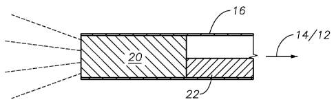
FIGURE 5 is a more detailed diagram illustrating one embodiment of
8
CA 02773582 2012-03-07
WO 2011/059716 PCT/US2010/054380
optical element 20. FIGURE 5 provides a magnified view of the distal end of
stem 16. Stem 16 is shown housing fiber 22 and optical element 20. Optical
element 20 is optically coupled to fiber 22, which can itself be optically
coupled to fiber optic cable 14. In some embodiments, fiber optic cable 14
can comprise an optical fiber that extends from the light source coupling
through the handpiece 10 and is optically coupled directly to optical element
20. For these embodiments, a separate fiber 22 is not used. The distal end
surface of optical fiber 22/14 can be optically bonded to the proximal end
surface of optical element 20, as described more fully below. Optical element
20 can comprise an optical grade machined or injection-molded plastic or other
polymer light guide or taper, for example.
When implemented within handpiece 10, fiber 22 is of a gauge
compatible with the gauge of fiber optic cable 14, such that it can receive
and
transmit light from fiber optic cable 14. Handpiece 10 can be any surgical
handpiece as known in the art, such as the Revolution-DSPTM handpiece sold
by Alcon Laboratories, Inc. of Fort Worth, Texas. Light source 12 can be a
xenon light source, a halogen light source, an LED light source, or any other
light source capable of providing light for delivery through an optical fiber.
Stem 16 can be a small gauge cannula, such as within the range of 18 to 30
gauge, as known to those having skill in the art. Stem 16 can be stainless
steel or a suitable biocompatible polymer (e.g., PEEK, polyimide, etc.) as
known to those having skill in the art.
Light source 12 can be optically coupled to handpiece 10 (i.e., to fiber
22) using, for example, standard ACMI optical fiber connectors at the ends of
fiber optic cable 14. This allows for the efficient coupling of light from the
light
source 12 through fiber optic cable 14 to the handpiece 10 and finally
emanating from optical element 20 at the distal end of the stem 16. Light
source 12 may comprise filters, as known to those skilled in the art, to
reduce
the damaging thermal effects of absorbed infrared radiation originating at the
light source. The light source 12 filter(s) can be used to selectively
illuminate
a surgical field with different colors of light, such as to excite a surgical
dye.
Spatial patterning in light emitted from multi-mode fibers with at least
some degree of radial symmetry can occur whenever the following occurs:
the luminance of the light at the entrance aperture to the fiber is
asymmetrically spatially non-uniform; if the fiber is not straight throughout
its
9
CA 02773582 2012-03-07
WO 2011/059716 PCT/US2010/054380
length (i.e., at least a portion is tapered) then the spatial non-uniformity
of the
luminance at the entrance aperture to the fiber need not be asymmetric; or if
some feature or obstruction prevents all rays of light from passing through
the
fiber with 100% efficiency (such as an absorptive patch in a region of
cladding
on the side of the light pipe). These phenomenons can occur in prior art
illumination probes, but are usually considered objectionable for general
illumination. However, it has been found that certain structured illumination,
such as that provided by the embodiments of the present invention, can be
advantageous for enhancing the ability of an observer (e.g., a surgeon) to
to discern retinal features, such as those retinal features with raised or
depressed areas relative to the surrounding retinal tissue.
FIGURE 6 shows one embodiment of a structured illuminator probe in
accordance with the present invention. Structured illuminator probe 300
comprises handpiece 310, and optical cable 314, comprising a protective
outer covering and an optical fiber 322, operably coupled to handpiece 310.
Optical fiber 322 can be optically coupled to a light source, not shown, for
receiving a light beam and transmitting the light beam to optical element 320.
Optical element 320 and the distal end of optical fiber 322 are housed within
cannula 316. Optical element 320 receives the light beam from optical fiber
322 and provides as an output at its distal end patterned illumination 350. As
can be seen in FIGURE 6, optical fiber 320 has a smaller cross-sectional area
than optical element 320 (i.e., the surface area of the distal end surface of
optical fiber 322 is less than the surface area of the proximal end surface of
optical element 320). Optical element 320 can have a circular, square,
elliptical or other cross-section shape and can be a light guide or taper.
The cannula (stem) 316 can be made of steel and can be smoothly
curved so as to contact the optical element 320 only at the extreme distal
end,
as shown in FIGURE 5, or can contact the optical element at any point along
its length. At the point(s) of contact, the optical element 320 can be bonded
to
the stem 316 by means of adhesive to provide mechanical strength and a seal
to prevent liquid from the eye from entering into any air gap between the
optical element 320 and the stem 316. Optical element 320 can also be
coupled to stem 316 such that its distal end extends out slightly beyond the
distal end of stem 316. Optical fiber 322 and optical element 320 can be
optically coupled by simple contact, using optical adhesive or by other
bonding techniques as are known to those having skill in the art, such that
CA 02773582 2012-03-07
WO 2011/059716 PCT/US2010/054380
optical fiber 322 is asymmetrically coupled to the optical element 320 to
create structured output pattern 350.
FIGUREs 7A and 7B illustrate predicted structured illumination patterns
comprising a rectilinear grid of squares that can be provided by an
embodiment of the illuminator probe of the present invention. The patterns
created in FIGUREs 7A and 7B are created by an embodiment having a distal
end of optical fiber 322 manicured into a square and coupled to an optical
element 320 comprising a larger square light guide. FIGURE 7A represents a
high-density array of square images and FIGURE 7B represents a low density
array of square images.
FIGURES 8A and 8B illustrate predicted structured illumination
patterns comprising a rectilinear grid of squares that can be provided by
is another embodiment of the illuminator probe of the present invention. The
patterns shown in FIGUREs 8A and 8B are created by an embodiment having
an unmanicured optical fiber 322 having a conventional circular end coupled
to an optical element 320 comprising a larger surface area square light guide.
FIGURE 8A represents a high-density array of square images and FIGURE
8B represents a low density array of square images.
FIGURE 9 is an image of an actual rectilinear grid pattern of circular
spots created by an embodiment of the structured illuminator of the present
invention comprising a circular 20 gauge optical fiber 322 under-filling a
.9mm
by .9mm square light guide 320 as shown in cross-sectional detail in FIGURE
10. Light streaks at the perimeter of the output pattern (light beam) can be
removed by aperturing the output of the light guide 320.
Embodiments of the structured illuminator of the present invention can
also provide circular bulls-eye illumination patterns, such as shown in
FIGURE 11. Such a pattern can be created by an optical element 420
comprising a taper optically coupled to the optical fiber 422, as shown in
FIGURE 12. Optical fiber 422 and optical element 420 of FIGURE 12 are
analogous to and provide functions as described for elements 20, 22 and 320,
322 in previous FIGUREs. As shown in FIGURE 12, optical fiber 422
transmits a light beam (as previously described in regards to other
embodiments) to optical element 420, underfilling the entrance aperture of
optical element (taper) 420. Optical element 420 can be an acrylic taper.
11
CA 02773582 2012-03-07
WO 2011/059716 PCT/US2010/054380
The embodiments discussed above provide structured illumination
using standard optical fibers typically having an NA (numerical aperture)
limited to .5 or .63. Other embodiments can instead comprise an optical
element optically coupled to a high NA source, such as an LED or high NA
photonic crystal or photonic bandgap optical fiber. In such embodiments, the
pattern will extend throughout the entire NA of the source coupled to the
optical element. FIGUREs 13A -13C illustrate the patterned and unpatterned
output of such an embodiment. FIGURE 13A shows the bare output from an
LED source without a distal light guide in place. FIGUREs 13B and 13C show
the patterned output created by the same LED source with a light guide in
place, in this example underfilling a square cross-sectioned lightpipe
(optical
element) using a Lambertian emitting LED source. FIGURE 14 is a
diagrammatical representation of such an embodiment, showing an optical
is element 520 comprising a square light pipe. The light pipe 520 is shown
being underfilled by a Lambertian LED light source.
FIGUREs 15A and 15B are diagrammatical representations of a dual-
function embodiment of the structured illuminator of the present invention. In
this embodiment, handpiece 510 includes an actuator 512, which can be a
mechanical, electrical, magnetic or other actuation mechanism as known to
those having skill in the art, for selecting between two distal output paths
of
optical element 520. In this embodiment, optical element 520 comprises a
distal optical fiber 525 to provide for standard illumination and a distal
light
guide 530 to provide structured illumination in accordance with the teachings
described herein. In operation, a user can select between the two output
paths by means of actuator 512 which can, as, for example, is shown in
FIGURE 15A, mechanically align delivery optical fiber 522 with either distal
optical fiber 525 or distal light guide 530. Precision stops 540 ensure proper
alignment between delivery optical fiber 522 and either distal optical fiber
525
or distal light guide 530. A user of such an embodiment of the present
invention thus can have at his disposal a dual-function probe which can
provide either type of illumination on demand without requiring an instrument
change.
FIGURE 16 illustrates the use of one embodiment of the structured
illuminator of this invention in an ophthalmic surgery. In operation,
handpiece
10 delivers a beam of light through stem 16 (via optical fiber 22/14) and
12
CA 02773582 2012-03-07
WO 2011/059716 PCT/US2010/054380
through optical element 20 to illuminate a retina 28 of an eye 30. The light
delivered through handpiece 10 to the optical element 20 is generated by light
source 12 and delivered to illuminate the retina 28 by means of fiber optic
cable 14 and coupling system 32. Optical element 20 is operable to provide
structured illumination in a desired pattern on the retina to provide the
contrast
needed for useful visualization of retinal features.
Although the present invention has been described in detail herein with
reference to the illustrated embodiments, it should be understood that the
description is by way of example only and is not to be construed in a limiting
sense. It is to be further understood, therefore, that numerous changes in the
details of the embodiments of this invention and additional embodiments of
this invention will be apparent to, and may be made by, persons of ordinary
skill in the art having reference to this description. It is contemplated that
all
such changes and additional embodiments are within the spirit and true scope
of this invention as claimed below. Thus, while the present invention has
been described in particular reference to the general area of ophthalmic
surgery, the teachings contained herein apply equally wherever it is desirous
to provide structured illumination of a surgical site.
13