Note: Descriptions are shown in the official language in which they were submitted.
CA 02773882 2012-03-09
WO 2011/039150 PCT/EP2010/064291
-1-
A-FUCOSYLATION DETECTION IN ANTIBODIES
This invention relates to a method for detecting the presence or absence of
fucose residues
within a glycosylated antibody or a fragment thereof.
Background
While the variable regions within the Fab (fragment antigen binding) domains
of
antibodies are responsible for the recognition of the antigen, the Fc
(fragment crystallizable)
region represents an invariant part of the antibody that is responsible for
the mediation of
effector functions. In the case of immunoglobulin G (IgG) these encompass the
fixation of
complement and the binding to Fcy receptors (FcyRs). The presence of an N-
linked
oligosaccharide at a single conserved site (Asn297) within the CH2 domain of
the homodimeric
Fc fragment is mandatory for the mediation of both of these effector
functions. It was only
recently discovered that modification of the attached carbohydrates can also
have an affinity
improving effect for the interaction between FcyRIIIa and IgG. The
carbohydrate modification
responsible for this effect is the absence of a fucose residue which is
usually attached to the first
N-acetylglucosamine (G1cNAc) residue in the biantennary complex-type IgG
glycan (Figure 1).
It could be demonstrated by in vivo and in vitro experiments that such
increased affinity
results in enhanced antibody-dependent cellular cytotoxicity (ADCC) mainly
mediated by
natural killer (NK) cells. Consequently, it is also believed that such a-
fucosylated antibodies
have an improved efficacy in treatments that aim to eradicate opsonized cells.
The generation of a-fucosylated antibodies represents an important
biotechnological
challenge which can be achieved by several methods. While cell lines with a
complete depletion
of enzymes involved in the biosynthesis of fucosylation (e.g. by gene
knockout) may yield
quantitatively a-fucosylated antibodies, most other methods do not. For
example, siRNA
treatment or co-cultivation of antibody-expressing cells with kifunensine
(Zhou et al., Biotechnol
Bioeng (2008) 99, 652-665), as well as carbohydrate modification by N-
acetylglucosaminyltransferase III (GnT-III), which promotes the formation of
bisected
oligosaccharides consequently inhibiting the fucosylation reaction (Umana et
al., Nat Biotech
(1999) 17, 176-180), lead to only partially a-fucosylated antibodies.
CA 02773882 2012-03-09
WO 2011/039150 PCT/EP2010/064291
-2-
These partially a-fucosylated antibodies can principally exhibit a
heterogeneous a-
fucosylation distribution within a pool of antibodies. For example,
fucosylation rates can be
different during fermentation. Also, the event of fucosylation could be
cooperative, i.e. the
second fucosylation on the homodimeric antibody may occur with an increased
(positive
cooperativity) or decreased (negative cooperativity) rate compared to the
first one.
The FcyRIIIa/IgG complex has a 1:1 stoichiometry but IgG has two binding sites
for
FcyRIIIa. Consequently, in a single a-fucosylated antibody the receptor can
bind with high
affinity to the binding site formed by the IgG's a-fucosylated glycan and
protein core or with low
affinity to the binding site consisting of the fucosylated carbohydrate and
the protein core. It can
therefore be concluded that a pool of antibodies with 50% a-fucosylation may
consist of a
homogeneous population of antibodies in which only one of the two N-glycans is
fucosylated, or
50% of antibodies in which both N-glycans are fucosylated while in the other
50% none of the
N-glycans are fucosylated. It is obvious that such a differential partition of
a-fucosylation
influences the overall affinity to FcyRIIIa and results in a different
biological activity. It is
therefore mandatory to analyze the biological activity of such an antibody
preparation either
directly by employing a biological test system (bioassay) or indirectly by
biochemically
measuring the rate and distribution of the a-fucosylation, which yields a more
exact result.
The current state-of-the-art glycoanalytics uses N-glycosidase F (PNGase F)
from
Flavobacterium meningosepticum to cleave off the N-linked carbohydrates with a
subsequent
MALDI-MS (matrix-assisted laser desorption ionization mass spectrometry)
analysis (according
to Papac et aL, Glycobiology (1998) 8, 445-454). By employing such a process,
however, the
linkage information is lost and the determination of fucosylation distribution
within an antibody
preparation is not possible.
On the other hand, analysis of the complete antibody using ESI-MS
(electrospray
ionization mass spectrometry) yields complex mass patterns that do not allow a
quantitative
interpretation due to the various modifications other than fucosylation - like
galactosylation, C-
terminal lysine heterogeneity, deamidation etc. - that may or may not occur in
both subunits of
the homodimeric IgG.
Therefore, there is a need for a new analytical method that eliminates the
mentioned
heterogeneity but maintains the linkage information.
Description of the Invention
CA 02773882 2012-03-09
WO 2011/039150 PCT/EP2010/064291
-3-
The above described drawbacks are overcome by this invention, which provides
for
methods for detecting the presence or absence of fucose residues within a
glycosylated antibody.
Preferably, the quantity of fucose residues and their distribution pattern
within an antibody or a
fragment thereof are determined. The analysis of the distribution of fucose
residues per Fc
molecule in an antibody preparation is also part of this invention. In
addition, the present
invention can be used for the determination of cooperative fucosylation in an
antibody
preparation during fermentation. Hence, this invention provides for a method
that closes a gap in
antibody analytics. With the knowledge of fucosylation patterns within an
antibody or fragment
thereof gained by means of this new method, a more accurate prediction of Fc-
mediated potency
is now possible.
Surprisingly, the inventors of the present invention found that Endo S (an
enzyme with
endoglycosidase activity, originally identified in Streptococcus pyogenes
(Collin and Olsen,
EMBO J (2001) 20, 3046-3055)) cleaves the complex-type glycan moieties from
the Fc region of
human IgG, leaving behind just the first G1cNAc residue to which a fucose
residue might be
attached. The hybrid-type carbohydrates that are discriminated (spared) by
Endo S can be
quantitatively cleaved at the same site by Endoglycosidase H (Endo H). The
combination of both
enzymes thus allows the preparation of a uniformly glycosylated protein that
only varies by the
fucose content. Analysis of such treated Fc fragments not only allows the
determination of the
fucose content of, but also determination of the distribution of fucose
residues within the
analyzed antibody pool. These new findings close an analytical gap and may
allow a potency
estimation of the analyzed antibody in terms of its efficacy in ADCC
induction.
Accordingly, the present invention relates to a method for detecting the
presence or
absence of fucose residues within a glycosylated antibody or a fragment
thereof.
In one embodiment the inventive method comprises the following steps:
a) removal of all heterogeneous saccharide residues from the protein,
b) removal of all other heterogeneous residues from the protein,
c) subsequent analysis of the protein.
In another embodiment, step c) of said method additionally comprises a
purification step
prior to analysis. In a specific embodiment purification is achieved by
affinity chromatography
or size exclusion chromatography. Affinity chromatography can be performed
using for example
Protein A or Protein G.
In one embodiment the protein to be treated and analyzed by the method of the
invention is
an antibody or an antibody fragment. Preferably said antibody is an IgG type
antibody. Said
antibody fragment is preferably an Fc fragment, in particular an Fc fragment
of an IgG type
antibody.
CA 02773882 2012-03-09
WO 2011/039150 PCT/EP2010/064291
-4-
In a specific embodiment the removal of step a) is performed by one or more
enzymes that
specifically cleave complex-type or hybrid-type N-linked carbohydrates.
Preferably, these
enzymes comprise Endo S and Endo H.
In another specific embodiment the removal of step b) is performed by one or
more
enzymes. Preferably these enzymes comprise plasmin and/or carboxypeptidase B.
In a further specific embodiment the analysis of step c) comprises CE-SDS MW
(capillary
electrophoresis-sodium dodecyl sulfate molecular weight) analysis, ESI-MS
analysis or liquid
chromatography-mass spectrometry (LC-MS), or a combination thereof.
In a preferred embodiment, step a) of the above described method comprises
cleavage of
the heterogeneous saccharides from the carbohydrate structures of the protein
after the first
G1cNAc residue of said structures, thereby leaving the fucose residue attached
to the antibody
core. This step can be performed with two enzymes that specifically cleave
complex-type or
hybrid-type N-linked carbohydrates that frequently occur in biotechnologically
produced
antibodies, for example Endo S and Endo H.
In a preferred embodiment, step b) of the above described method comprises
quantitative
removal of C-terminal lysine residues of the antibody heavy chain, preferably
using an enzyme,
said enzyme preferably comprising carboxypeptidase B.
In another preferred embodiment, step b) of the above described method
comprises
cleavage between the Fab and the Fc fragment of an antibody. Preferably the
covalent interchain
disulphide bridges within the hinge peptide of the heavy chains are maintained
within the Fc-
fragment after cleavage between the Fab and the Fc fragment. Preferably the
cleavage is
achieved by an enzyme. Preferably such enzyme comprises plasmin.
In a preferred embodiment, step c) of the above described method comprises
analysis of
the treated antibody molecule or Fc fragment by LC-MS without any prior
purification steps.
Such an analysis normally yields only three masses that correspond to proteins
with two
fucosylated glycans, proteins with one fucosylated and one a-fucosylated
glycan, and proteins in
which both glycans are a-fucosylated.
In another embodiment, step c) of the above described method comprises
purifying the
treated antibody molecule or Fc fragment using standard methods and analyzing
it by ESI-MS
analysis. Such an analysis normally yields only three masses that correspond
to proteins with two
fucosylated glycans, proteins with one fucosylated and one a-fucosylated
glycan, and proteins in
which both glycans are a-fucosylated..
CA 02773882 2012-03-09
WO 2011/039150 PCT/EP2010/064291
-5-
In one embodiment the method of the invention comprises the following steps:
providing
an antibody preparation, optionally isolating the Fc fragment portion of such
antibody
preparation, removing all heterogeneous saccharide residues from said antibody
or Fc fragment
with Endo H and Endo S, removing C-terminal lysine residues from said antibody
or Fc
fragment with carboxypeptidase B, and analysis of the treated antibody or Fc
fragment by ESI-
MS, LC-MS or CE-SDS MW analysis.
In another embodiment said method comprises the following steps: providing an
antibody
preparation, optionally isolating the Fc fragment portion of such antibody
preparation using
plasmin, removing all heterogeneous saccharide residues from said antibody or
Fc fragment with
Endo H and Endo S, removing C-terminal lysine residues from said antibody or
Fc fragment
with carboxypeptidase B, and purification and analysis of the treated antibody
or Fc fragment by
ESI-MS, LC-MS or CE-SDS MW analysis.
In yet another embodiment, this invention is directed to kits suitable for
performing an
assay which detects the presence or absence of fucose residues within a
glycoprotein. The kits of
this invention comprise all components referred to in the methods described
above (e.g. Endo H,
Endo S, carboxypeptidase B, plasmin, suitable buffers), instructions setting
forth a procedure
according to any one of the methods described above and a container for the
contents of the kit.
Use of Endo S for cleavage of complex-type N-linked oligosaccharides of a
glycoprotein,
preferably a glycosylated antibody or a fragment thereof, is also part of this
invention.
De initions
Terms are used herein as generally used in the art, unless otherwise defined
in the
following:
The term "heterogeneous saccharide" as used herein, includes any
monosaccharide moiety
of a glycosylated antibody or antibody fragment that is not connected to a
fucose residue. Non-
limiting examples for heterogeneous saccharides of a glycosylated antibody or
antibody
fragment are mannose, sialate, galactose, acetylglucosamine. Generally,
heterogeneous
saccharides which are removed in step a) of the method according to the
invention will be all
saccharides other than the first G1cNAc residue, i.e. the G1cNAc residue
attached to an
asparagine residue of the protein, and the fucose residue linked to that first
G1cNAc residue.
The term "heterogeneous residues" as used herein, means any other moiety of a
glycosylated antibody or antibody fragment (other than heterogenous
saccharides) that could
interfere with the detection of fucose residues within said antibody or
antibody fragment. Non-
limiting examples of heterogenous residues are various modifications of the
glycosylated
CA 02773882 2012-03-09
WO 2011/039150 PCT/EP2010/064291
-6-
antibody or antibody fragment other than fucosylation, such as
galactosylation, C-terminal lysine
heterogeneity and deamidation. The term "heterogeneous residues" may further
include antibody
fragments that are not glycosylated, for example the Fab fragment, the scFv
fragment and other
fragments.
As used herein, the term "antibody" is intended to include whole anti body mo
lecules,
antibody fragments, or fusion proteins that include a region equivalent to the
Fe region of an
immrrnoglobufin,
The terms "complex-type oligosacc.haride" and "hybrid-type oligosac.cbaride"
refer to the
glycosylation pattern of an antibody or antibody fragment. is on--limiting
examples of "complex-
type oligosaaecharide" and "hybrid-type oligosaccharide" are sho i in Figure
7. As randerstood
by those skilled in the art, glycoproteins enriched in bisected hybrid-type
ol_igosaceharides
typically result from overexpression of GnT-11I in production cell lines.
Exemplary structures of
bisected hybrid-type ohgosaccharides are detailed in Figure 7-ill.
Glycoproteins enriched in
bisected complex type oligosacchaa;rides typ.icaally result from a co--e;
pressiori ofM4aanll and GnT-
111 in production cell lines. Exemplary structures of bisected, complex-type
oligosaecharides are
detailed in figure 7-W (Ferrara et al.., Biotechnol Bioeng (2006) 93, 851-
61).
(:`leavage "after" a sugar residue, as used herein, means cleavage distal to
this residue, i.e.
cleavage of the sugar bond linking this residue with the adjacent one towards
the outer end of the
carbohydrate structure. Cleavage, "after the first GlcN Ac residue" of an N-
lirrked glycan means
cleavage of the chitobiose core of the oligosacch_aride, between the first
(i,e. attached, to the
asparagine residue) and the second (i.e. attached to the first) GlcN Ac
residue.
"Distibuti_ori" of ftucose residues within an antibody preparation refers to
the presence
within that preparation of antibody or Fc molecules differing in the number of
fucose residues
associated with the N.-linked glycans in the Fc region. For example, an IgG
molecule has two >-
linked glycans in its Fe region, each of which can have a frcose residue
linked to the first
Glc (Ac residue of the carbohydrate structure. Thus, iii an lgG preparation
there rraight be three
different molecralar species: lg(:i with two, one or no flicose residues
associated with the N -
linked glycans in the Fc region. The ratio of these different species (i.e.
the distribution of fficose
residues per Fe molecule) can be determined by the method of the invention, in
addition to
determination of the total fucose content, i.c, the fraction of fiacosyla.ted
or a-.tiacosyia.ted N-
glycans.
The exarrrples below explain the iliveriti_on in more detail. The examples are
given to
enable those skilled in the art to more clearly understand and practice the
invention, The present.
invention, however, is not limited in scope by the exemplified enrbodinrents,
which are intended
CA 02773882 2012-03-09
WO 2011/039150 PCT/EP2010/064291
-7-
as illustrations of .single aspects of the invention only, and methods o,,hich
are functionally
equivalent are ,v thin the scope of the invention_.
Short description of the a ures
FIGURE 1. Schematic representation of a carbohydrate moiety attached to Asn-
297 of
human IgGl-Fc. The sugars in bold define the pentasaccharide core of N-linked
glycan
structures; the addition of the other sugar residues is variable. In grey is
represented a bisecting
G1cNAc residue.
FIGURE 2. Deglycosylation of intact Fc fragment of antibodies A (wildtype) and
C
(glycoengineered) monitored by CE-SDS. Electropherograms of non-reduced Fc
fragments are
shown before and after enzymatic treatment. (A) Fc fragment of antibody C
without enzymatic
treatment (dashed line) and deglycosylated with PNGase F (dotted line) or Endo
S (solid line),
(B) Fc fragment of antibody A without enzymatic treatment (dashed line),
deglycosylated with
PNGase F (dotted line) or deglycosylated with Endo S (solid line).
FIGURE 3. Positive-ion MALDI-TOF mass spectra of the N-linked oligosaccharides
released from Fc fragment of antibody C by consecutive treatment with Endo S
and PNGase F or
with Endo S and Endo H. (A) Spectrum of glycans released by treatment with
Endo S. (B)
Spectrum of Endo S-resistant carbohydrates released by subsequent treatment
with PNGase F,
resulting in an isolated signal at m/z = 1663 (possibly corresponding to
hybrid- or complex-type
structures as schematically depicted). (C) Spectrum of glycans released by
subsequent treatment
with the hybrid-type structure specific enzyme Endo H (hybrid-type structures
corresponding to
m/z = 1460 released by Endo H treatment are schematically depicted).
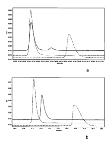
FIGURE 4. Deglycosylation of the Fc fragment of antibody C monitored by CE-SDS
MW
analysis (A) and positive-ion MALDI-TOF mass spectrometry (B). (A) Overlay of
electropherogram of the non-reduced Fc fragment without glycosidase treatment
(dashed line)
and treated with a combination of Endo S and Endo H (solid line). (B) Mass
spectra of the N-
linked oligosaccharides released from the Fc fragment treated with Endo S and
Endo H. Hybrid-
type structures corresponding to m/z = 1460 released by Endo H are
schematically depicted.
FIGURE 5. ESI-MS spectra of Fc fragments after treatment with Endo S and Endo
H. (A)
Fc fragments of antibody A, (B) Fc fragments of antibody B, (C) Fc fragments
of antibody C.
Peak 1: Fc-G1cNAc/G1cNAc, Peak 2: Fc-G1cNAc/G1cNAc + Fuc, Peak 3: Fc-G1cNAc +
Fuc/G1cNAc + Fuc.
FIGURE 6. Deglycosylation of antibody C monitored by CE-SDS (A) and positive-
ion
MALDI-TOF mass spectrometry (B). (A) Electropherograms of non-reduced IgG are
shown
CA 02773882 2012-03-09
WO 2011/039150 PCT/EP2010/064291
-8-
before and after enzymatic treatment: Antibody C without enzymatic treatment
(dashed line) and
deglycosylated with PNGase F (dotted line) or combined treatment with Endo S
and Endo H
(solid line). (B) Mass spectra of the N-linked oligosaccharides released from
entire IgG treated
with Endo S and Endo H.
FIGURE 7. N-linked oligosaccharide biosynthetic pathway leading to complex- or
hybrid-
type structures. MI: mannosidase I, G1: 01,2-N-acetylglucosaminyltransferase
I, G3: 01,4-N-
acetylglucosaminyltransferase III, Gt: 01,4-galactosyltransferase.
FIGURE 8. ESI-MS spectra of entire IgGs after treatment with Endo S and Endo
H. (A)
antibody A, (B) antibody D. Peak 1: Fc-G1cNAc/G1cNAc, Peak 2: Fc-G1cNAc/G1cNAc
+ Fuc,
Peak 3: Fc-G1cNAc + Fuc/G1cNAc + Fuc.
FIGURE 9. LC-MS spectra of entire IgGs after treatment with Endo S and Endo H.
(A)
antibody A, (B) antibody D. Peak 1: Fc-G1cNAc/G1cNAc, Peak 2: Fc-G1cNAc/G1cNAc
+ Fuc,
Peak 3: Fc-G1cNAc + Fuc/G1cNAc + Fuc.
Examples
Example 1: Methods
Generation of Fc from human IgG
Four different human IgGs with a different content of a-fucosylated glycans,
determined
according to Papac et at., 1998 (content in brackets), were used for analysis
of the a-fucosylation
distribution: wildtype antibody A (2.12%), glycoengineered antibody B (47.0%),
glycoengineered antibody C (69.6%), and glycoengineered antibody D (85%).
The proteins were incubated for 72 hours at 25 C in 50 mM Tris pH 8.0, 150 mM
NaCl
with 0.42 U plasmin (Roche) per milligram. Cleaved Fc was separated from Fab-
fragments using
a Protein A affinity column (5 ml HiTrapTM Protein A HP column, GE Healthcare)
equilibrated
and washed (5 column volumes (CV)) with buffer A (50 mM Tris pH 8.0, 100 MM
glycine, 150
mM NaCl). Fc was eluted by a pH-step using buffer B (50 mM Tris pH 3.0, 100 mM
glycine,
150 mM NaCl). Fractions containing Fc were pooled and neutralized by adding
1:40 (v/v) 2 M
Tris pH 8Ø Samples were concentrated to a volume of 2.5 ml using ultra
concentrators
(Vivaspin 15R 10'000 MWCO HY, Sartorius) and subsequently applied to a PD-10
desalting
column (GE Healthcare) equilibrated with 2 mM MOPS pH 7.4, 150 MM NaCl, 0.02 %
(w/v)
NaN3. Purified protein was frozen in liquid nitrogen and stored at -80 C.
Release off-linked oligosaccharides from human Fc
CA 02773882 2012-03-09
WO 2011/039150 PCT/EP2010/064291
-9-
Different enzymes were used for hydrolyzing the N-linked glycans of human IgG.
The N-
linked oligosaccharides were cleaved from 1 mg of Fc by incubation with 0.005
U recombinant
PNGase F (QAbio, Vista Monte, USA). For release of carbohydrates from Fc using
non-tagged
Endo S (Genovis), samples were incubated with either a molar ratio of 1:20 of
Endo S alone or
in combination with 0.1 U/mg Endo H (QAbio). All reactions were incubated in
20 MM Tris pH
8.0 at 37 C for 16 h.
For analyzing carbohydrates spared by Endo S, Fc was purified after Endo S
treatment by
affinity chromatography using Protein A and subsequently digested with either
PNGase F or
Endo H, as described above.
Release off-linked oligosaccharides from entire human IgG
The N-linked glycans of human IgG were released using different enzymes. The N-
linked
oligosaccharides were cleaved from 1 mg of IgG by incubation with 0.005 U of
recombinant
PNGase F (QAbio) in 20 mM Tris pH 8.0 at 37 C for 16 h. For release of
carbohydrates from
IgG using non-tagged Endo S (Genovis), samples were applied to a NAP-5
desalting column
(GE Healthcare) equilibrated with 20 mM Tris pH 8Ø Eluted sample was
concentrated to a final
concentration of 4 mg/ml using ultra concentrators (Amicon 5'000 MWCO,
Millipore) and
incubated with a molar ratio of 1:7 of Endo S combined with 0.1 U/mg Endo H
(QAbio) at 37 C
for 16 h.
Carboxypeptidase B treatment
To remove heterogenicity caused by C-terminal lysine residues, after
deglycosylation
samples were further incubated with carboxypeptidase B (Roche; 1 mg/ml).
Therefore 1 l
carboxypeptidase B per 50 g human Fc or entire antibody was added to the
Endoglycosidase
reaction and incubated again for 1 h at 37 C.
MALDI TOF mass spectrometi >> analysis of released oligosaccharides
Neutral oligosaccharide profiles of the human Fc or entire antibody were
analyzed by mass
spectrometry (Autoflex, Bruker Daltonics GmbH) in positive ion mode (Papac et
al., 1998).
Purification of deglycosylated human Fc or entire antibody
Fc or entire IgG was separated from enzymes and cleaved carbohydrates by
Protein A
affinity chromatography using Agilent HPLC 1100 series (Agilent Technologies).
Samples were
applied to Protein A matrix (Poros 20 A; Applied Biosystems) packed in a guard
column 2x20
mm C-130B (Upchurch Scientific) equilibrated with buffer A (50 MM Tris, 100 MM
glycine, 150
M NaCl, pH 8.0). After washing with 5.5 CV of buffer A, human Fc or entire IgG
was eluted by
CA 02773882 2012-03-09
WO 2011/039150 PCT/EP2010/064291
-10-
a pH-step using buffer B (50 mM Tris, 100 mM glycine, 150 M NaCl, pH 3.0) over
8.3 CV. The
fraction containing the protein was neutralized by adding 1:40 (v/v) 2 M Tris
pH 8Ø
The purified protein was subsequently further used for either treatment with
enzymes to
analyze non-cleaved carbohydrates, CE-SDS analysis or ESI-MS.
CE-SDS MW analysis
Deglycosylation was monitored by CE-SDS-MW analysis, using Beckman PA800 with
UV detection. The buffer of 100 g of each Protein A purified sample was
exchanged to 20 mM
Tris pH 8.0 using spin concentrators (5000 MWCO, Millipore). Non-reduced
samples were
prepared as described in SDS-MW Analyses Guide using the ProteomeLab SDS-MW
Analysis
Kit (Beckman Coulter). The final protein concentration was 1 mg/ml. Samples
were applied to a
preconditioned bare fused silica capillary (50 m ID x 30.2 cm). Pre-
conditioning and separation
were performed according to the instruction manual.
Sample preparation for ESI-MS
The buffer of Protein A purified samples was exchanged to 2 mm MOPS pH 7.4,
150 MM
NaCl, 0.02% (w/v) NaN3 using spin concentrators (5000 MWCO, Millipore).
Proteins were
frozen in liquid nitrogen and stored at -80 C.
ESI-MS analysis of glycan structures of human Fc and entire IgG by direct
infusion Off line
detection)
Desalting by Size Exclusion Chromatography:
20-50 gg (up to 90 l) of Fc after treatment of antibody with the proteases
plasmin and
carboxypeptidase B and with endo-glycosidases Endo S and Endo H, or entire IgG
after
treatment with Endo S, Endo H and carboxypeptidase B, were injected onto a
Sephadex G25
self-packed ECO SR column (5 x 250 mm; KronLab) equilibrated with 2% formic
acid, 40%
acetonitrile at a flow rate of 0.5 ml/min for 30 minutes. The injected protein
sample was desalted
applying an 8 minute isocratic elution with 2% formic acid, 40% acetonitrile
at a flow rate of 1
ml/min. The elution of the desalted protein was recorded by UV at 280 nm and
the eluting
sample (volume about 200-300 l) was collected in a 1.5 ml reaction vial. An
aliquot of the
desalted sample was manually filled into a metal coated glass needle (Proxeon
Biosystems Nano
ESI-needles, cat# ES387), inserted into the nanospray source of the mass
instrument and sprayed
into a ESI-Q-TOF II mass spectrometer from Waters or into a Q-Star Elite mass
spectrometer
from Applied Biosystems.
MS parameters for direct infusion:
CA 02773882 2012-03-09
WO 2011/039150 PCT/EP2010/064291
-11-
A) ofplasmin-treated samples (human Fc) on a Q-TOF II instrument (Waters)
MS spectra were acquired using a capillary voltage of 1000 V, a cone voltage
of 30 V in a
mass range from 1000 - 2000 m/z in positive ion mode using a source
temperature of 80 C.
Desolvation temperature was off. MS data were acquired for approx 2-3 minutes
by the
respective instrument software.
B) of entire antibody on a MaXis-ESI-MS instrument (Bruker)
MS spectra were acquired using a NanoMate device as spray interface. The
values for data
acquisition at the MS instrument were set to 400 Vpp (funnel RF), 120 eV
(ISCID energy) and
400 Vpp (Multipol RF) regarding the transfer parameters, 5.0 eV (ion energy)
and 300 m/z (low
mass) for the quadrupol parameters, 15 eV (collision energy) and 3000 Vpp
(collision RF)
adjusting the collision cell and 800 Vpp, 160 s for transfer time and 20 s
prepulse storage at
the ion cooler. Data were recorded at a mass range from 1000 - 4000 m/z in
positive ion mode.
Molar masses of dimeric Fc-fragments and entire antibody, containing different
combinations of glycan structures truncated by the endoglycosidases applied,
i.e
G1cNAc/G1cNAc, G1cNAc + Fuc/G1cNAc and G1cNAc+Fuc/G1cNAc +Fuc, were determined
from the respective m/z pattern of the Fc fragment or entire antibody species
using an in-house
developed software. The relative ratios of the various residually glycosylated
dimeric Fc
fragments or entire antibodies were calculated with the same in-house software
using the sum of
peak areas of the m/z spectrum of a distinct glycosylation variant of the
dimeric Fc-fragment or
entire antibody.
ESI-MS analysis of glycan structures of entire IgG by LC-MS (On line
detection)
LC-MS was performed on a Dionex HPLC system (Dionex Ultimate 3000) coupled to
a Q-
TOF II mass spectrometer (Waters). The chromatographic separation was
performed on a ACE
C4 column (5 m particle size, 300 A pore size, 1 x 30 mm; Advanced
Chromatography
Technologies). Eluent A was 0.1% formic acid, eluent B was 99.9% acetonitrile
and 0.1% formic
acid. The flow rate was 100 l/min, the separation was performed at 75 C and 2
gg (10 l) of an
intact antibody sample treated with Endo S and Endo H, but without plasmin
treatment, were
used.
TABLE 1. Parameters for LC-MS.
Time (min.) %B remark
0 25 waste
3 25
3.1 25
3.5 25 switch to MS
CA 02773882 2012-03-09
WO 2011/039150 PCT/EP2010/064291
-12-
4.0 25
9.0 50
9.5 100
12.5 100
12.6 25
14.9 25 switch to waste
15.0 255 stop MS-detection
MS spectra were acquired using a capillary voltage of 2700 V, a cone voltage
of 80 V in a
mass range from 1000 - 4000 m/z in positive ion mode using a source
temperature of 100 C.
Desolvation temperature was set to 200 C. MS data were acquired for
approximately 11.4
minutes (gradient time 3.5 to 14.9 min) by the respective instrument software.
Molar masses of intact antibody (consisting of two heavy chains and two light
chains) containing
different combinations of glycan structures truncated by the endoglycosidases
applied, i.e
G1cNAc/G1cNAc, G1cNAc + Fuc/G1cNAc and G1cNAc + Fuc/G1cNAc + Fuc, were
determined
from the respective m/z pattern of the antibody species using an in-house
developed software.
The relative ratios of the various residually glycosylated intact antibodies
were calculated with
the same in-house software using the sum of peak areas of the m/z spectrum of
a distinct
glycosylation variant of the intact antibody.
The ratio of non-fucosylated heavy chains was determined by reducing the EndoS
and EndoH-
treated antibody with TCEP (Tris(2-carboxyethyl)phosphine hydrochloride) and
performing an
LC-MS analysis as described before, using the same column type and gradient
setting but some
modified parameters for MS data acquisition. MS parameters were the same as
described before,
but cone voltage was set to 25 V and mass range was from 600 - 2000 m/z.
Example 2: Results
Deglycosylation of Fc
N-Glycosidase F, also known as PNGase F, is a highly specific deglycosidase
that cleaves
between the innermost N-acetylglucosamine of high mannose-, hybrid-, and
complex-type N-
linked oligosaccharides and the asparagine residue of the glycoprotein to
which the glycan is
attached (Tarentino et at., 1985). Treatment of the Fc fragments of antibody A
and C with
PNGase F according to the instructions of the manufacturer was monitored by CE-
SDS. Under
these conditions PNGase F quantitatively removes the glycan moiety of both
analyzed samples,
resulting in a mobility shift of the main peak from 3.79 x 10⁻⁵ to 3.9 x 10⁻⁵
(Figure 2).
CA 02773882 2012-03-09
WO 2011/039150 PCT/EP2010/064291
-13-
Endo S cleaves the chitobiose core of N-linked oligosaccharides, leaving the
first N-
acetylglucosamine residue - and an a-fucose residue in case of fucosylated
carbohydrates -
attached to the protein. The CE-analysis of a such digested glycoengineered
sample revealed that
approximately 10% of the protein were still undigested (Figure 2a, Table 2),
as demonstrated by
a peak with a mobility of 3.84 x 10⁻⁵. Subsequent analysis by PNGase F
treatment indicated that
the Endo S resistant carbohydrates were entirely of hybrid structure
suggesting specificity of this
enzyme for complex carbohydrates. This result could be corroborated by the
quantitative Endo S
digestion of wildtype antibody A which resulted in homogenously deglycosylated
protein
(Figure 2b).
TABLE 2. Peak area of enzyme-treated Fc fragments evaluated by CE-SDS.
Peak area [%]
Antibody, enzyme Non-cleaved Cleaved
A, no enzyme 99.3 0.7
A, PNGase F 1.3 98.7
A, Endo S 1.8 98.2
C, no enzyme 100.0 0.0
C, PNGase F 0.3 99.7
C, Endo S 10.6 89.4
To confirm this hypothesis, Endo S-treated Fc of antibody C was purified by
affinity
chromatography to remove the enzyme and cleaved carbohydrates, and
subsequently incubated
with PNGase F to remove the entire glycan moiety. The hydrolyzed carbohydrates
were further
analyzed by MALDI TOF MS. The obtained spectra showed that Endo S is
discriminating (i.e.
sparing) either complex- or hybrid-type bisected structures that are
corresponding to m/z= 1663
(Figure 3b).
Further experiments were performed to determine whether the discriminated
carbohydrates
are complex- or hybrid-type bisected structures. After purification by
affinity chromatography,
the Endo S-treated Fc fragment of antibody C was incubated with PNGase F or
Endoglycosidase
H (Endo H). Endo H is a recombinant glycosidase that cleaves within the
chitobiose core of high
mannose- and hybrid-type N-linked oligosaccharides of glycoproteins. It is not
able to cleave
within complex structures. MALDI TOF MS spectra showed that the carbohydrates
discriminated by Endo S are cleaved by Endo H, resulting in a main peak of
m/z=1460 (Figure
3c). These data clearly show that Endo S is not able to release hybrid-type
bisected
carbohydrates from the asparagine-linked N-acetylglucosamine.
To obtain homogenously deglycosylated material that only varies in its a-
linked fucose
content, a combined treatment of the Fc fragment of antibody C with Endo S and
Endo H was
CA 02773882 2012-03-09
WO 2011/039150 PCT/EP2010/064291
-14-
performed resulting in a protein that was quantitatively deglycosylated after
the first G1cNAc
residue as observed by CE-SDS (Figure 4a). MALDI-TOF MS analysis showed that
the hybrid
bisected structures (m/z=1460) are released by combination of these two
enzymes (Figure 4b).
To confirm that there is no other carbohydrate attached to the N-
acetylglucosamine with or
without an a-linked fucose residue, Endo S- and Endo H-treated Fc fragment of
antibody C was
incubated with PNGase F. No MALDI TOF spectra could be obtained after this
treatment
suggesting that no other carbohydrates were remaining that cannot be cleaved
by Endo S or Endo
H (data not shown).
Determination of the fucose distribution in a Fc -preparation
To quantify the distribution of the fucose linked to the N-acetylglucosamine
residue
attached to the Fc, ESI-MS analyses were performed. After incubation with Endo
S and Endo H
before separation by affinity chromatography, the Fc domains of antibodies A,
B and C
(generated by plasmin digestion) were treated with carboxypeptidase B to
remove heterogeneity
introduced by C-terminal lysine.
ESI-MS spectra revealed Fc fragments with either two, one or no fucose linked
to the
residual G1cNAc still attached to the protein after EndoS/EndoH treatment
(Figure 5).
Distribution of these three fucose species is summarized for the investigated
three different IgGs
A, B and C (calculated as relative ratio of the sum of peak areas in the m/z-
spectra). The results
correlate well with the fucose content determined by MALDI-TOF MS (Table 3).
TABLE 3. Comparison of the a-fucosylation degree
determined by mass spectrometry for Fc fragment of antibody A, B and C.
MALDI-TOF ESI-MS
Non-fuc [%] 2 fucose [%] 1 fucose [%] 0 fucose [%] I Non-fuc [%]
A 2.12 94 3 3 4.5
B 47.0 29 41 30 50.5
C 69.6 20 40 40 60.0
Deglycosylation of entire IgG
For deglycosylation of an entire IgG by combined treatment with Endo S and
Endo H,
cleavage conditions had to be optimized. Deglycosylation with a molar ratio of
Endo S to IgG of
1:20, as was used for deglycosylation of the Fc fragment, was insufficient to
deglycosylate entire
IgG. Increasing the concentration of Endo S to a molar ratio of 1:7 was
sufficient to get
homogenously deglycosylated material that only varies in its a-linked fucose
content observed
by CE-SDS (Figure 6a). MALDI-TOF analysis showed that the carbohydrates are
released by
CA 02773882 2012-03-09
WO 2011/039150 PCT/EP2010/064291
-15-
combined treatment with Endo S and Endo H (Figure 6b). Using this approach it
is possible to
analyze the allocation of fucose per IgG without separate generation of the Fc-
fragment.
Determination of the fucose distribution of entire IgG
Quantification of the distribution of fucose linked to the innermost N-
acetylglucosamine
residue of N-linked glycans of entire IgGs was performed using wildtype
antibody A (2.12% a-
fucosylation) and glycoengineered antibody D (85.0% a-fucosylation). After
combined treatment
with Endo S and Endo H, both IgGs were incubated with carboxypeptidase B to
remove
heterogeneity introduced by C-terminal lysine. The antibodies were
subsequently purified by
affinity chromatography.
Allocation of the core fucose per IgG was determined using two different
methods. The
pattern of the m/z-spectra obtained by ESI-MS off line detection revealed IgG-
species with
either two, one or no fucose attached to the residual G1cNAc after EndoS/EndoH
treatment
(Figure 8). Distribution of these three fucose species is summarized for the
investigated two
different IgGs A and D (calculated as relative ratio of the sum of peak areas
in the m/z-spectra)
(Table 4).
LC-MS analyses were also performed to determine the allocation of fucose per
IgG (Figure
9). For both IgGs, m/z-spectra showed a similar ratio of species with either
two, one or no fucose
attached as observed in ESI-MS offline detection, (Table 4). Peak areas below
5% are in the
detection sensitivity of the methods for entire IgG. Ratio for non-fucosylated
heavy chain is
presented in Table 4, column Non-fuc [%].
TABLE 4. Comparison of the a-fucosylation degree and fucose allocation
determined by ESI-MS and LC-MS analyses for antibody A and D.
ESI-MS LC-MS
2 fucose 1 fucose 0 fucose Non-fuc 2 fucose 1 fucose 0 fucose Non-fuc
[%] [%] [%] [%] [%] [%] [%] [%]
A 92 5 <5 8 94 6 <5 12
D 9 24 67 81 10 24 66 80