Note: Descriptions are shown in the official language in which they were submitted.
CA 02774422 2013-07-03
MULTI-MODALITY CONTRAST AND BRIGHTFIELD CONTEXT
RENDERING FOR ENHANCED PATHOLOGY DE'TERMLNATION
AND MULTI-ANALYTE DETECTION IN TISSUE
10 FIELD
The disclosure pertains to methods of providing contrast in tissue sections
for
pathology determination.
BACKGROUND
Microscopic clinical examination of conventional histological stained tissue
sections
can be used to evaluate tissue structures and morphological patterns of
diagnostic
significance. Skilled physicians can view such histological stained tissue
sections for
diagnosis, and to design and evaluate treatments. The contrast of structures
provided by such
images using classical stains is familiar, and permits the physician to devote
her efforts to
interpreting anatomical and morphological tissue section features and
anomalies, and not on
trying to translate how the staining procedure reveals features relevant to
her medical training
and experience.
Additional tissue imaging techniques are being developed that promise to
enhance the
correlative diagnostic information obtainable by the physician on valuable
biopsy material
and archived tissue specimens. For example, fluorescence microscopy can be
used for
detection of specialized molecular markers, but fluorescence based images
typically lack the
familiar structural and anatomical context information found in tissue stained
with
hematoxylin and eosin (H&E) and viewed using brightEeld microscopy.
While fluorescence based images provide useful molecular information for
- 1 -
CA 02774422 2012-03-16
WO 2011/046807
PCT/US2010/051857
confirming and characterizing disease states, conventional histological
stained sections
remain necessary for pathology determination on tissue. Typically, serial
tissue sections
through a specimen must be prepared and evaluated. Commonly, the serial
sections include a
conventional H&E stained section and specially stained section(s) for
diagnostic molecular
markers. Comparing serial sections not only increases the cost and time
necessary for an
evaluation, it may be difficult or impossible to correlate features found in
one section with
features found in the other. Serial sections can be lost or destroyed in the
staining process
pipeline as well.
SUMMARY
The technology described herein provides methods and apparatus that use multi-
modal contrast to produce complimentary contrast components segmented and
displayed in a
manner relevant to physician training and experience for pathology analysis.
Such
complimentary contrast modes may be streamed to display to permit navigation
of tissue
structure, focusing, and changes of magnification. Tissue sections can contain
one or more
probes targeting particular molecules or chemistries of interest. Color
contrast of tissue
structure is provided that can be comparable to the contrast produced with
conventional
color absorbing histological stains such as hematoxylin and eosin stain
(hereinafter "H&E").
The images produced by one or more of the disclosed methods can also include
features
revealed using additional markers and optical or chemical contrast modes.
Typically,
correlation of differentially labeled features between different tissue
sections becomes
unnecessary. The images are presented in digitally rendered color brightfield
context to
provide an image appearance that is comparable to that produced in
conventional histological
slides that have been stained to reveal the same structural features.
In some disclosed examples of multi-modal contrast, contrast is derived from
the
refractive index properties and fluorescent labeling of tissue specially
prepared for markers of
specific molecules. These examples demonstrate the complimentary combination
of
transmitted-light darkfield refraction contrast imaging, with simultaneous
incident light
fluorescence imaging of nuclear counterstain, and the interrogation of
multiplexed molecular
probes. Corresponding correlative images are obtained either simultaneously
(in parallel) or
- 2 -
CA 02774422 2012-03-16
WO 2011/046807
PCT/US2010/051857
sequentially (in serial). In some examples, illumination wavelengths and
detection
wavelengths used to create contrast on unstained or stained tissue may be
tightly controlled to
promote unambiguous segmentation and to prevent interference with multiplexed
probes.
Molecule-specific probe localizations for protein antigens, mRNA expression,
or genetic
rearrangements in DNA can be overlaid on the specimen structure. This contrast
is
associated with changes in refractive index due to tissue structure as
preserved and resolved
through the use of specific histological processing. In typical examples,
disclosed methods
provide image contrast based on refractive index variations in tissue moieties
in combination
with fluorescent counterstains to provide color pathological context for
molecule-specific
multiplexed probes. Examples include formalin-fixed, paraffin embedded tissues
and frozen
tissue. Refractive index contrast can be derived directly from the refractive
or scattering
properties of tissue and probe moieties, or from amplitude of a phase-shift,
or a rate of
change of a phase-shift gradient.
Some disclosed methods comprise exposing a fluorescently stained specimen to a
stimulus beam selected to produce fluorescence by the fluorescent stain, and
producing a
corresponding fluorescence image. The same specimen is also exposed to a high
NA
circumferential dark field illumination, and a corresponding dark field image
representing
changes in refractive index and light scattering moieties is produced. In some
examples, the
fluorescence stimulus beam exposure and the dark field refraction illumination
field exposure
are applied simultaneously, and the complementary images are obtained in
parallel. In other
examples, the fluorescence image and the dark field refraction contrast image
are recorded
serially. Imaging apparatus according to examples comprise a multi-modal
optical system
configured to produce a transmitted dark-field illumination field and an
incident illumination
fluorescence excitation optical system. These sub-systems are configured to
produce
multiple complimentary images that can be combined for correlative analysis: a
refractive
contrast image based on properties of the prepared tissue, a fluorescence
image of a nuclear
counterstain, and a plurality of fluorescent images representing various
molecular markers
that can be segmented by emission wavelength. At least one image capture
device is coupled
to receive the dark field and fluorescence images and an image processor is
configured to
record and process the dark field image and the fluorescence images and
produce a combined
- 3 -
CA 02774422 2016-11-21
image.
Computer readable storage media comprise computer-executable instructions for
receiving images associated with multiple modes of contrast associated with
common
portions of a specimen section prepared for pathological examination, and
overlaying the
multiple modes of contrast to produce a combined image.
In some examples, the image processor is configured to produce a pseudo-color
bright
field rendering of the combined image based on the recorded refraction
contrast darkfield
image and fluorescent images. The fluorescence image and the dark field
refraction image
are individually colored, combined and inverted to produce a combined color
image in an
apparent brightfield context with contrast relevant to conventional staining.
Specific color
mappings to facilitate straightforward physician interpretation are applied to
the refraction
contrast image, fluorescent nuclear counterstain, and specific fluorescent
probes. These
images are subsequently combined to produce a combined-color recorded image in
brightfield rendering. In some examples, the color mapping is based on
quantitative
measures of human perception of preferred color for pathology determination
associated with
at least one color-absorbing histological stain such as an eosin stain. In
still further examples,
a color lookup table is applied to a fluorescence image, wherein the color
mapping is
associated with at least one contrasting color-absorbing histological stain
such as a
hematoxylin stain. In some examples, color lookup tables are applied to the
dark-field
refraction image and the fluorescence counterstain image so as to produce an
image having
inverted contrast associated with complimentary color hue, inverted value and
inverted
saturation compared to that encountered in ideal hematoxylin and eosin
staining. In other
examples, the specimen imaged optically is prepared for further imaging using
mass
spectrometry to provide molecular mapping.
These and other features and aspects of the disclosed technology are described
below with reference to the accompanying drawings.
In accordance with an aspect of the present invention there is provided an
image
generation method, comprising: receiving a fluorescent image of a specimen,
wherein the
specimen is fluorescently stained and a first beam has been selected to
produce fluorescence
by the fluorescent stain so that the first image is a fluorescence image of
the specimen;
receiving a refractive dark field image of the specimen, wherein a second
stimulus beam has
- 4 -
CA 02774422 2016-11-21
been applied to the specimen so that the second image is a refractive dark
field image;
applying a color mapping to the refractive dark field image to produce a
pseudo-color dark
field image; applying a color lookup table to the fluorescence image, and
generating a
converted fluorescent image wherein the color lookup table is associated with
at least one
absorptive stain; combining the pseudo-color dark field image and the
converted fluorescence
image and generating a refractive dark field and fluorescence combined image;
and inverting
the refractive dark field and fluorescence combined image to produce a
brightfield rendered
image.
In accordance with another aspect of the present invention there is provided
an
imaging apparatus, comprising: at least one image capture device that receives
first and
second images, wherein the first image is a refractive dark field image and
the second image
is a fluorescence image; and an image processor coupled to the image capture
device that
applies a color lookup table to at least one of the first and second recorded
images and
generates a pseudo-colored rendering of at least one of the first and second
images, wherein
the image processor combines the pseudo-colored rendering of at least one of
the first and
second images with the other of the at least one of the first and second
images and generates a
refractive dark field and fluorescence combined image, based on the pseudo-
colored
rendering of at least one of the first and second images, wherein when image
processer
generates a pseudo-colored rendering of the first image, the image processor
processes the
first image based on a color lookup table associated with an eosin stain, and
when the image
processor generates a pseudo-colored rendering of the second image, the image
processor
processes the second image based on color lookup table associated with a
hematoxylin stain,
and wherein the image processor inverts the refractive dark field and
fluorescence combined
image to produce a brightfield rendered image.
In accordance with a further aspect of the present invention there is provided
at least
one non-transitory computer readable storage media comprising computer-
executable
instructions for execution by a processor, the instructions for: receiving a
first image and a
second image associated with a common portion of a specimen section, wherein
the first
image is a refractive dark field image and the second image is a fluorescence
image;
combining the refractive dark field image and the fluorescence image and
generating a
- 4a -
CA 02774422 2016-11-21
refractive dark field and fluorescence combined image, wherein before the
refractive dark
field image is combined with the fluorescence image, the refractive dark field
image is
processed based on a color lookup table associated with an eosin stain and the
fluorescence
image is processed based on color lookup table associated with a hematoxylin
stain, and
wherein the refractive dark field and fluorescence combined image is based on
the processed
first and second images; inverting the refractive dark field and fluorescence
combined image,
and generating a pseudo-color brightfield hematoxylin and eosin image based on
the
processed first image and the processed second image.
In accordance with another aspect of the present invention there is provided
an image
processor, comprising: image inputs configured that receives a first image and
a second
image, wherein the first image is a refractive dark field image and the second
image is a
fluorescent image; a color lookup table input that receives, a first color
lookup table
associated with an eosin stain and a second color lookup table associated with
a hematoxylin
stain; an image combiner that processes the refractive dark field image based
on the first
color lookup table associated with an eosin stain and that processes the
fluorescent image
based on the second color lookup table associated with the eosin stain and
produces a pseudo-
color refractive dark field image and a pseudo-color fluorescent image, and
combines the
pseudo-color refractive dark field image with the pseudo-color fluorescent
image and
generates a combined refractive dark field and fluorescent image, and produces
a brightfield
image rendering based on the combined refractive dark field and fluorescent
image after
inverting the combined refractive dark field and fluorescent image.
BRIEF DESCRIPTION OF THE DRAWINGS
FIG. 1 is schematic diagram of a representative imaging system that provides
both
refraction-contrast dark field and fluorescence-based images.
- 4b -
CA 02774422 2012-03-16
WO 2011/046807
PCT/US2010/051857
FIG. 2 is a schematic block diagram of a method of processing and combining
recorded dark field and fluorescent stain based images.
FIG. 3 is a schematic block diagram of a representative method for producing a
specimen image from multiple modes of contrast with contrast corresponding to
that used in
pathology determination with hematoxylin and eosin (H&E) staining.
FIG. 4A is a representative conventional H&E stained image of a human prostate
section.
FIG. 4B is a multiple mode contrast image of a human prostate section based on
a
combination of a dark field refraction image and a fluorescence counterstain
image rendered
in brightfield context.
FIGS. 5A-5B are dual-illumination multiple mode contrast (refractive contrast
and
fluorescence) images recorded with a monochrome CCD with sequential exposures
taken
using interference filters to select either the blue DAPI fluorescence
wavelengths (FIG. 5A)
or the longer wavelength transmitted dark field wavelengths (FIG. 5B).
FIG. 5C is a pseudo-color image obtained by application of inverted color
lookup-
tables for pseudo-color to the images of FIGS. 5A-5B and adding the inverted
color
images.
FIG. 5D is a pseudo-colored, bright field rendering of the image that
corresponds to
the image of FIG. 5C after inversion of the mapped color space.
FIG. 6 is a brightfield context rendering image by overlaying localizations of
quantum
dot fluorescent probes with peak emission wavelengths of 565 nm and 655 nm
from a DAPI
counter-stained formalin-fixed, paraffin embedded sample also imaged for
refractive
contrast.
FIG. 7 contains other example images of multimode imaging for brightfield
context
display. Dark field refractive contrast images and DAPI fluorescence images of
DAPI
counterstained tonsil sections were obtained, overlaid, and rendered as color
bright field
images as shown in FIG. 7 (1a-3a). Protein-specific immuno-probes (localized
in
fluorescence using quantum dots having peak emissions at 565 nm for CD20
antigen and
655 nm for Ki67 antigen) were applied to the DAPI counterstained tonsil
sections to
produce corresponding immuno-probe fluorescence based images. The probe images
were
- 5 -
CA 02774422 2012-03-16
WO 2011/046807
PCT/US2010/051857
overlaid in contrasting pseudo-colors (red and green) as shown in FIG. 7 (1b-
3b). FIG. 7
(1c-3c) shows the images of FIG. 7 (1a-3a) and FIG. 7(1b-3b) after being
combined.
FIGS. 8A-8B are additional representative images in which simulated
brightfield
histological images are obtained, and fluorescent probe images combined using
alternative
methods. FIG. 8A is an additive overlay to a multi-mode pseudo-bright field
image using
QDot probes with 565 nm and 655 nm emission wavelengths from a DAPI
counterstained
formalin fixed paraffin embedded specimen. FIG. 8B is a subtractive overlay in
which
pseudo-color probe images are subtracted from the facsimile H&E rendered
image.
FIG. 9 illustrates an example of the CIEL*a*b* color space used to map
preferential
color characteristics for H&E to refraction contrast and DAPI fluorescence to
render in
brightfield for pathology determination.
FIGS. 10A-10B are grayscale refractive index contrast and DAPI fluorescence
contrast images, respectively. FIGS. 10C-10D are CIEL*a*b* pseudo-color eosin-
converted and hematoxylin-converted images based on the images of FIGS. 10A-
10B,
respectively. FIG. 10E is a merged imaged obtained by combining the converted
images of
FIGS. 10C-10D.
FIG. 11 is a schematic diagram of an optical system that simultaneously
produces
side-by-side refraction dark field images and fluorescence-based images using
a single
CCD camera.
FIG. 12 contains a side-by-side refractive index (dark field) image (A) and a
DAPI
image (B) of the same tissue section acquired and displayed simultaneously.
FIG. 13 contains a two color brightfield rendering overlay image (with pseudo-
color
and image inversion) based on the side-by-side images of FIG. 12. Note this
image is rotated
with respect to the FIG. 12 image.
FIGS. 14A-14B are images of cryosectioned mouse kidney tissue specimens
prepared for deposition of mass spectroscopic imaging tags. Contrast is
produced by
refraction at tissue edges and tissue autofluorescence (blue).
FIGS. 15A-15B are images of mouse kidney tissue specimens prepared for mass
spectroscopic imaging by deposition of an ionizing matrix. Autofluorescence
appears blue
and refractive index contrast associated with ionizing matrix crystals is
apparent.
- 6 -
CA 02774422 2012-03-16
WO 2011/046807
PCT/US2010/051857
FIG. 16 is a schematic diagram illustrating a computing environment for the
apparatus and methods described herein.
FIG. 17 is a multiple mode image providing cellular and nuclear context in
brightfield rendering for a Calu-3 xenograft probed for mRNA in situ
hybridization of two
probes, one for ribosomal RNA (cyan color, dashed black arrow), the other for
HER2
mRNA expression (black color, solid black arrows).
FIG. 18 is a representative image of a prostate cancer imaged using dual-mode
contrast and presented in brightfield context at 20x magnification for
pathology
determination. Prominent nucleoli and anomalous growth patterns characteristic
of
prostate cancer are evident.
FIG. 19 is a portion of the same region imaged at 40x magnification using the
same
simultaneous dual-mode method of combining refraction contrast with
fluorescent nuclear
counterstain and rendering in brightfield context.
DETAILED DESCRIPTION
As used in this application and in the claims, the singular forms "a," "an,"
and "the"
include the plural forms unless the context clearly dictates otherwise.
Additionally, the term
"includes" means "comprises."
The systems, apparatus, and methods described herein should not be construed
as
limiting in any way. Instead, the present disclosure is directed toward all
novel and non-
obvious features and aspects of the various disclosed embodiments, alone and
in various
combinations and sub-combinations with one another. The disclosed systems,
methods, and
apparatus are not limited to any specific aspect or feature or combinations
thereof, nor do the
disclosed systems, methods, and apparatus require that any one or more
specific advantages
be present or problems be solved.
Although the operations of some of the disclosed methods are described in a
particular, sequential order for convenient presentation, it should be
understood that this
manner of description encompasses rearrangement, unless a particular ordering
is required
by specific language set forth below. For example, operations described
sequentially may in
some cases be rearranged or performed concurrently. Moreover, for the sake of
simplicity,
- 7 -
CA 02774422 2013-07-03
the attached figures may not show the various ways in which the disclosed
systems, methods,
and apparatus can be used in conjunction with other systems, methods, and
apparatus.
Additionally, the description sometimes uses terms like "produce" and
"provide" to describe
the disclosed methods. These terms are high-level abstractions of the actual
operations that
are performed. The actual operations that correspond to these terms will vary
depending on
the particular implementation and are readily discernible by one of ordinary
skill in the art..
Theories of operation, scientific principles, or other theoretical
descriptions presented
herein in reference to the apparatus or methods of this disclosure have been
provided for the
purposes of better understanding and are not intended to be limiting in scope.
Introduction
Multiple modes of complimentary contrast generation in tissue can permit
visuali7ation of anatomical and morphological tissue context, presented in a
brightfield
context familiar to the trained physician, along with locali7ations of probes
for specific
molecules or variations in tissue chemistry. Multimodal contrast may leverage
a plurality of
light-tissue and probe detection interactions so long as the information
provided is
complementary. Tissue prepared for pathological examination has constitutive
optical
activity and the optical qualities produced by a particular protocol can be
optimized to
produce useful contrast qualities when combined with appropriate imaging
instrumentation.
Image contrast for non-fluorescent structures can be provided through
components of
optical activity engineered into, or preserved in, a particular tissue
preparation scheme such
as used in automated staining protocols on formalin fixed paraffin embedded as
well as
frozen tissue. This enhanced optical activity may be digitally recorded and
rendered in
artificial bright field contrast to visualize and highlight structures such as
the extracellular
matrix, nucleoli and cell membranes. Such visualization capabilities are used
to diagnose
anomalous growth patterns and morphology characteristic of pathological
conditions in
tissue. Multiple modes of optical or activity or chemical properties in
prepared tissue can be
recorded, in serial or in parallel, and digitally converted into bright field
image contrast for
- 8 -
CA 02774422 2012-03-16
WO 2011/046807
PCT/US2010/051857
visualization, and is referred to herein as "pseudo bright field."
Representative imaged structures are of pathological significance and can be
used by
physicians in tissue screening and in the diagnosis of pre-cancer and cancer
disease states, as
well as for other diagnostic purposes, and in the evaluation of treatment
effectiveness. In an
unstained or specially stained tissue section, such morphological structures
and anatomical
context can be practically invisible under single-mode contrast methods such
as conventional
transmitted light brightfield or fluorescence detection. Complimentary
multiple modality
imaging methods can produce medically relevant structural information and
present this
information in a readily interpretable format without the use of conventional
light absorbing
stain. Quantitative values can be measured and recorded based using one or
more sets of
computer-executable instructions provided by one or more computer readable
storage media.
Morphological metrics can be leveraged to correlate such morphological
characteristics to the
molecular information contained in the same tissue; this approach may help in
ongoing
efforts to stratify disease condition and prognosis as well as monitor
treatment efficacy.
Digital multi-modality images of tissue sections can be captured
simultaneously and rendered
using distinctive colors for complimentary feature components and streamed or
otherwise
stored or delivered for examination by a pathologist or other clinician in
near real-time. Such
methods facilitate high complexity tissue-based diagnostics development and
permit
leveraging physician medical training and experience with conventional
histological stains.
Molecular data, including that from immunohistochemistry, DNA hybridization,
mRNA
hybridization probes, lectins, and mass spectrometry and other analyses can be
integrated for
individual tissue sections, and reported rapidly in a format that is familiar
and pertinent to the
practicing pathologist.
The examples provided herein leverage a multi-modality imaging strategy
utilizing
dual-illumination paths for providing images with complementary contrast of
protein
structure, and DNA counterstain, as well as molecule-specific markers for
medical diagnosis
and evaluation. The example approach includes a combination of dark-field
refraction and
fluorescence contrast; these complimentary contrast modes are digitally
rendered using
specialized color tables derived from physician preferences of classical
histological stain
qualities. With such a combination, multi-color contrast in tissue samples
similar to that
- 9 -
CA 02774422 2012-03-16
WO 2011/046807
PCT/US2010/051857
obtained in samples stained using classical histological methods such as the
hematoxylin and
eosin (H&E) stain can be provided. Such images can be used to develop regions
of interest
for further molecular analysis using luminescent, fluorescent, scattering, or
absorbing probes
for protein, lipid or carbohydrate antigens, mRNA or DNA, probes for charge
properties or
for imaging mass spectrometry (IMS). The multimodal contrast illumination
contrast scheme
exemplified herein can provide contextual information of tissue sections in a
manner
consistent with common stain/counterstain combinations used in conventional
histological
methods. For convenience, optical radiation beams that are directed to a
specimen to obtain
images are referred to herein as stimulus beams. In some examples, stimulus
beams are
selected to produce fluorescence in one or more portions of the specimen, and
may or may
not be at visible wavelengths. Other stimulus beams include illumination beams
that are at
visible wavelengths for direct viewing. Stimulus beams can also be based on
other types of
radiation as well, including in other wavelength ranges and charged particle
beams or
acoustic beams.
In some examples, such methods and apparatus have been applied to fluorescence
in
situ hybridization (FISH), immunohistochemistry (IHC), and mRNA in situ
hybridization
(mRNA-ISH) applications in formalin-fixed paraffin-embedded tissues. Quantum
dot
(QDot)-labeled FISH probes, QDot labeled IHC probes and QDot labeled mRNA-ISH
probes
were specifically detected on tissue using multi-modal contrast and digital
pseudo-brightfield
rendering for visualization of probe localizations within the tissue
anatomical structure
context. In typical examples, a dark field refraction contrast image, a
counter-stained image
obtained with a fluorescent nuclear stain, and one or more probes imaged using
fluorescent
QDot detection are combined. These and other examples are described below.
Representative Imaging Systems
A representative example of a suitable imaging system 100 is illustrated in
FIG. 1. A
fluorescence stimulus light source 102 is situated to deliver a stimulus beam
103 along an
axis 105 to a wavelength dependent beamsplitter (dichroic) 104. The light
source 102 is
typically a light emitting diode (LED), metal halide or other arc lamp, but
other incoherent or
coherent light sources such as lasers can be used. As shown in FIG. 1, the
dichroic 104
- 10-
CA 02774422 2012-03-16
WO 2011/046807
PCT/US2010/051857
reflects the stimulus beam 103 to an objective lens 106 which in turn directs
the stimulus
beam 103 to a specimen 108. In typical examples, the specimen 108 is
selectively labeled
with one or more fluorophores that produce fluorescence in response to the
stimulus beam
103. A portion of the fluorescence is collected by the objective lens 106 and
directed along
an axis 113 through the dichroic 104 to an optional beam splitter 110. The
beam splitter 110
directs a portion of the fluorescence to a camera 112 so that a specimen image
can be
recorded, viewed, or analyzed at a computer system 130. Another portion of the
fluorescence
is directed to an eyepiece 114 for direct viewing of the specimen 108 based on
the
fluorescence light. In addition, a shutter 132 or other beam modulator can be
provided to
substantially prevent the stimulus beam 103 from reaching the specimen 108, or
the
fluorescence source can be controlled (via the computer system 130 or
manually) so that no
stimulus beam is produced. Wavelengths of light used for the stimulus beam can
be selected
as convenient. Typically the stimulus beam includes primarily optical
radiation at
wavelengths or in a wavelength range that is suitable for generating
fluorescence light in
fluorescent dyes or fluorophores associated with any selective markers applied
to the
specimen. Typical wavelength ranges for the stimulus beam is between about 300-
550 nm,
but shorter or longer wavelengths can be used.
In addition to the fluorescence imaging system, a refractive contrast imaging
system
using circumferential oblique dark field illumination is provided. In the
example of FIG. 1,
the circumferential oblique field illumination 117 is selected so that in the
absence of
refractive index differences or scattering moieties in the specimen, light
flux does not reach
the CCD camera 112 or the eyepiece 114, and only refractive index transitions
appear. By
using different magnification objective lenses with the same numerical
aperture (acceptance
angle) or by using a secondary magnification lens, the same illumination
optimizations for
refraction imaging can be used at multiple optical magnifications. There are
multiple
strategies to create contrast based on refraction or scattering of the
illumination field. Such
refractive index contrast images are referred to herein as "dark field"
images. A substage
condenser system 116 is situated so as to deliver an oblique field
illumination 117 to the
specimen 108 at substantially the same location as that illuminated by the
stimulus beam
103. The substage condenser system 116 can direct a suitable light source such
as an LED,
-11-
CA 02774422 2012-03-16
WO 2011/046807
PCT/US2010/051857
tungsten halogen lamp, an arc lamp, or other light source and one or more
lenses, mirrors,
filters, polarizing elements, phase plates, prisms, annuli or apertures that
can produce a
suitable beam. In the example of FIG. 1, the oblique field illumination 117 is
produced by a
carefully sized annulus, but in other examples, different approaches to field
illumination or
point scanning, line scanning, edge illumination, or other strategies designed
to produce
refraction contrast can be provided. In the example of FIG. 1, so-called "dark
field"
illumination is provided in which only portions of the field illumination that
are scattered or
redirected by the specimen 108 are collected by the numerical aperture of the
objective lens
106 and reach the camera 112 or the eyepiece 114. The camera 112 and the
eyepiece 114 are
situated so as to form an image of the specimen 108 based on the redirected
portions of the
transmitted light. Typically, the transmitted illumination system 117 can be
shuttered or its
light source deactivated as desired so that fluorescence-based images can be
acquired or
viewed independently of the transmitted illumination. The example microscope
system 130
of FIG. 1 thus permits recordation of specimen images and viewing of a
specimen based on
fluorescence, dark field refraction contrast, or both, either simultaneously
or sequentially.
Using transmitted circumferential oblique illumination such as illustrated in
FIG. 1,
contrast can be produced based on interfaces and transitions between specimen
portions
having different refractive indices. Typically, the condenser system 116
includes an annulus
118 of an appropriate size (and can be added to a conventional condenser) in
the transmitted
light path of a compound microscope equipped with a transmitted light source.
In this way,
structures that refract and scatter light have appreciable contrast in images
without the use of
a light absorbing color stain. The transmitted illumination wavelength can be
spectrally
filtered with, for example, a near IR filter or other filters or combinations
of filters so that
spectral images of fluorescent probes can be obtained with transmitted
contrast collected at a
longer wavelength in the same data acquisition with both illumination sources
active
simultaneously. The refraction field illumination is generally selected to
provide a suitable
visual image for recording, and is in a wavelength range of between about 400
nm and
900 nm, but different spectral regions within this range can be used if
desired. In other
examples, reflected dark field illumination is used in which the oblique
illumination and the
objective lens are situated on the same side of the specimen.
- 12 -
CA 02774422 2012-03-16
WO 2011/046807
PCT/US2010/051857
The specimen dark field image can be obtained by itself through segmenting
fluorescence with one or more filters, shuttering or temporally modulating or
otherwise
blocking the stimulus beam. In some examples, the fluorescence-based image can
be
obtained with a suitable filter tuned to the fluorescence wavelength and the
refraction contrast
filtered to a different wavelength range; these different wavelength ranges
can be separated to
different sensors, directed to different portions of the same sensor or
recorded sequentially.
The unwanted contribution of dark field illumination to the fluorescence image
or images, or
vice versa, can be reduced by spectrally filtering, but shuttering either the
dark field
illumination field or the fluorescence light path is possible. In addition,
the dark field and
fluorescence images can be viewed separately or simultaneously.
The camera 112 is typically a monochrome charge coupled device (CCD) or
complementary metal oxide semiconductor (CMOS) camera though other image
sensors such
as electron multiplying CCD (EMCCD) and intensified CCD (ICCD) sensors may be
used.
Wavelength filters, dispersive elements, phase plates, prisms, polarizing
elements, tunable
optical crystals and other optical and electro-optical methods can be used to
modify the
optical radiation reaching the CCD and/or the eyepiece so as to produce one or
more
corresponding monochromatic images in the selected wavelength ranges. In some
cases,
fluorescence reaching the camera 112 can be spectrally resolved in a plurality
of wavelength
bins, and a corresponding plurality of fluorescence images obtained for
analysis. Spectral
analysis can be performed with a plurality of absorptive or reflective
filters, a prism, or a
diffraction grating that are inserted into the path of the fluorescence.
Generally, spectral
resolution can be achieved using interferometric, dispersive, or absorptive
optical systems
under the control of the computer system 130 or inserted manually. While
images can be
recorded as one, two, or three dimensional arrays of picture elements with
values associated
with a received light flux intensity (either from fluorescence or other modes
of contrasting
illumination), images can be recorded in other formats and complex data
structures if desired.
For convenience in this description, an image refers to a 2-D mapping of data
in a structured
array as viewed by a clinician through a microscope or other viewing apparatus
and a
recorded image refers to data values stored, processed and/or displayed based
on an image
received by a CCD or other image sensor.
- 13 -
CA 02774422 2012-03-16
WO 2011/046807
PCT/US2010/051857
As noted above, a plurality of spectral images can be obtained based on
fluorescence
and transmitted illumination or both. Spectroscopic information at each pixel
of an image
can be gathered and the resulting data analyzed with spectral image-processing
software. A
series of images can be derived that represent intensities at different
wavelengths that are
__ electronically and continuously selectable and then evaluated with an
analysis program
designed for handling such data. In some examples, quantitative information
from multiple
fluorescent signals and/or optical contrast modalities can be evaluated
simultaneously.
The image sensor 112 is coupled to the computer system 130 that includes a
keyboard, 152, a processing unit 154, and a display 156. In some examples, one
or more
__ additional user input devices such as joysticks, mice, or digitizing
tablets, and one or more
additional output devices such as printers or displays are provided. The
processing unit 154
typically includes a microprocessor and one or more computer readable storage
media such
as read only memory (ROM), random access memory (RAM), a hard disk, a floppy
disk, a
compact disk or digital video disc for storage of image data and computer
executable
__ instructions for recordation, transmission, analysis, and processing of
images or image data.
In typical examples, the computer system 100 is coupled to one or more other
computer
systems via a wired or wireless network connection, and in some examples, is
coupled to the
Internet. Although image processing operations can be conducted at a single
computer
system, in some examples, image data or images are processed at a plurality of
computing
__ systems that can be situated in a common location or distributed on a
network. While laptop
computers can be convenient, other computing devices such as desk top
computers,
workstations, handheld computers, netbook computers, or other devices can be
used for
image capture and processing. In some examples, image data can be processed
and specimen
evaluations can be provided without a display, and evaluations communicated
via the
__ network connection (by email for example), sent to a printer, or delivered
as a text or
multimedia message using a cell phone network.
The imaging system 100 is one example of a suitable imaging system. In other
examples, a reflective or catadioptric objective can be used instead of the
objective lens 106,
a short pass filter can be used instead of the long pass filter 104 by
rearrangement of the
__ fluorescence stimulus source 102 and the camera 112 and eyepiece 114. In
some examples,
- 14 -
CA 02774422 2012-03-16
WO 2011/046807
PCT/US2010/051857
only a camera or an eyepiece is provided for either image recordation or image
viewing.
Additional mirrors or prisms can be used to fold the optical axes as may be
convenient.
Different strategies for multimodal contrast using phase masks, phase
contrast, Rotterman
contrast, oblique illumination contrast, Rheinberg contrast, interference
contrast schemes,
adaptive optics, laser scanning, time or frequency domain lifetime imaging,
structured
illumination, photoswitchable probes, polarization and anisotropy, 21d
harmonic imaging,
two-photon excitation and other strategies may be employed. Specimen
positioning hardware
is not shown for convenient illustration, and in many examples, binocular
viewing with dual
eyepieces can be provided, and suitable filters and beamsplitters can be
provided so that
different image outputs receive an image light flux associated with only one
of multiple
contrast modalities, polarization states or wavelength bandwidths. Additional
filters
(reflective or absorptive) can be provided, typically to reduce the magnitude
of any stimulus
light that reaches the camera 112 or the eyepiece 114, or to control relative
light intensity or
spectral content for viewing or recording.
Another representative imaging system is illustrated in FIG. 11. As shown in
FIG.
11, a combined image light flux 1102 that includes a refraction modulated flux
1102A and
a DAPI fluorescence modulated flux 1102B corresponding to dark field and DAPI
images
is directed along an axis 1103 through an aperture 1104 to a collimating lens
1105. The
collimated, combined light flux is incident to a dichroic mirror 1106 that
reflects a portion
of the modulated light flux (the DAPI modulated flux 1102A in the example of
FIG. 11) to
mirrors 1108A and to a filter 1107A that preferentially transmits DAPI
fluorescence. A
lens 1110 receives the DAPI modulated flux and forms a specimen image on a
first portion
1112A of a CCD or other image sensor1112. The dichroic mirror 1106 transmits
the
longer wavelength refraction modulated beam 1102B to a filter 1107B selected
to reject
DAPI fluorescence and the associated DAPI stimulus beam. A mirror 1108B
directs the
modulated flux 1102B to the lens 1110 which forms a dark field image on a
portion 1112B
of the CCD 1112. The CCD 1112 is coupled to a computer or other processing
device that
can store image data from the CCD 1112 in memory, and provide image data to a
monitor
1118 or other display. With such an imaging system, dark field and
fluorescence images
can be obtained simultaneously and displayed side-by-side as a raw image or
rapidly split
- 15 -
CA 02774422 2012-03-16
WO 2011/046807
PCT/US2010/051857
into two images, color mapped and overlaid in near real-time on the monitor
1118. The
configuration of FIG. 11 is illustrative only, and specimen modulated
refraction and
fluorescence light fluxes can be separated and used in image formation in
other
arrangements and using more, fewer, and different components. The images can
be side by
side on the CCD 1112 or processed by the computer 1114 so that a combined 2-
color
overlay in brightfield-rendered context can be displayed on the monitor 1118.
As shown in
FIG. 11, multiple fluxes (dark field and DAPI) are diverted from an initial
optical axis, but
in other examples, one flux can be transmitted along the initial axis and the
CCD 1112
situated accordingly. The dark field and DAPI images can be produced with
different
lenses that can be arranged to produce a common magnification or different
magnifications.
Additional filters, light sources, and other components can be provided so
that molecular
detection label and other tissue contrast image light fluxes are provided to
and imaged in
one or more CCDs or portions of a single CCD 1112A, 1112B.
Color Lookup Tables and Image Inversion
The system of FIG. 1 permits multi-modality viewing and acquisition of images
based on either fluorescence or dark field illumination, or both
simultaneously. The
acquired images can be manipulated to present specimen features in a common
context
using a representative method illustrated in FIG. 2. In a step 202, a dark
field refraction
image is recorded, typically as a monochromatic image, and in a step 204, one
or more
fluorescence-based images are recorded. The number of such fluorescence-based
images
can depend on numbers and types of fluorescent markers or dyes that are
applied to the
specimen. These images can use different wavelength bands corresponding to
emission
wavelengths of the fluorescent markers. In some examples, the different
wavelength bands
can be overlapping, non-overlapping, or a combination thereof.
In the step 204, the one or more fluorescence based images can be obtained
corresponding to fluorescence from corresponding fluorophores. Appropriate
spectral
segmentation of the fluorescence light can be used to obtain multiple
fluorescence based
images that can reveal different specimen features, typically dependent on the
specific
probe associated with the fluorescent detection marker.
- 16-
CA 02774422 2012-03-16
WO 2011/046807
PCT/US2010/051857
Upon acquisition of the images (either as each is acquired or after all or
some have
been acquired), one or more color map lookup tables (LUTs) can be applied to
the intensity
values of monochrome images in a step 206 to produce pseudo-color rendered
images and
these rendered images can be overlaid in a step 208. One or more or all of the
hue,
intensity or saturation of the acquired overlaid image is inverted in a step
210 to produce an
image having the appearance of colored structure on a bright field. In a
typical application
of a pseudo-color LUT, pixels of monochrome images are assigned RGB color
intensity
values based on grey-scale pixel intensity values and vice versa. Such
inversions may also
invert color coordinates to produce complimentary color mappings. Image
inversion
generally maps large pixel intensity values to smaller pixel intensity values.
For example,
in an image in which pixel intensities are represented with 8 intensity values
(3-bit depth),
intensity values can be re-mapped as shown in Table 1.
Original Re-Mapped
0 7
1 6
2 5
3 4
4 3
5 2
6 1
7 0
Table 1. Image Inversion with 3 Bit Values
Such a mapping scheme can be extended to other bit depths (e.g. 8-bit, 10-bit,
12-bit, 16-bit
and others) and can be applied to different components (e.g. hue, saturation,
value) of a
given color space.
In the step 210, image values that would appear dark are inverted so as to
appear
light, and image values that would appear light are inverted so as to appear
dark. The step
210 can be referred to as producing a pseudo bright field image.
The order of image inversions and pseudo-color LUTs can be varied as needed.
Specific color LUTs can be selected so that, for example, a dark field image
appears in
color contrast similar to histological stains. In this strategy, the image
modes are carefully
chosen to reveal the same structures to an image produced with a conventional
stain
- 17 -
CA 02774422 2012-03-16
WO 2011/046807
PCT/US2010/051857
procedure such as conventional H&E staining. Images can be overlaid in a step
208 with or
without color mapping for contrast components or inversion to bright field
appearance.
Additional color mapped images contrasting different structures can be applied
to the
combined image (typically overlaid with the combined image) in a step 212. The
combined
and processed image can be stored and/or displayed in a step 214. One or more
of these
steps can be omitted, duplicated, or performed in another order if more
convenient.
In many practical examples, it can be advantageous to simulate the coloring of
specific tissue structures produced with conventional histological stains in
multimode
contrast images. Such simulation provides a familiar analytical and diagnostic
setting for a
physician while still permitting correlation with additional specific markers
to reveal
additional information. This simulation also permits the elimination of light
absorbing
stains, so that staining does not interfere with application of other markers
or the evaluation
of image features revealed by these markers. For example, refraction contrast
can be used
to reveal extracellular and membrane proteins while a nucleus specific
fluorescent dye such
as DAPI can be used to reveal details of nuclear chromatin distribution. Thus,
the
refraction/DAPI combination can be used, with appropriate image processing, to
reveal
specimen features in a manner analogous to that achieved with eosin
(eosinophilic, or
protein-specific) and hematoxylin (nucleic acid or DNA-specific). Because
these images
are obtained on the same specimen, the features of each can be registered
spatially and
included in a displayed image for convenient analysis. Optimized color
mappings can be
utilized that permit images displayed as preferred by clinicians to best
reveal features of
interest in the context of medical training and experience. Such color
mappings can be
conveniently described with reference to a CIE 1976 L*a*b* color space, other
color
spaces such as a Hunter 1948 L,a,b color space, an CIE 1931 XYZ color space,
CIE 1976
L*u*v*, HSV, HSI, HSV, HSB color, or RGB color or CMYK color values, or
PANTONE
or MUNSELL color scales can also be used.
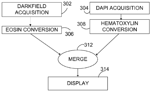
FIG. 3 illustrates a representative method 300 of specimen imaging that
permits
interrogation of specimen features based on refractive index contrast and DAPI
fluorescence. In a step 302, a gray scale intensity map image of refraction
contrast in
specimen is recorded, typically using a monochrome CCD camera. In a step 304,
a DAPI-
- 18 -
CA 02774422 2012-03-16
WO 2011/046807
PCT/US2010/051857
fluorescence-based gray scale intensity map image of the specimen is recorded.
While
color filters are used in acquiring both of these images, the images are
recorded as intensity
values for an array of pixels as gray scale images on a monochrome CCD. In a
step 306,
the refractive index contrast image is processed and mapped to color to have
an appearance
similar to that produced with eosin color absorption under white light
transmitted
illumination. Eosin staining typically produces image contrast in protein
moieties in the
extracellular matrix and in membranes. In some examples, the step 306 can be
configured
so that the processed image has an appearance that is based on clinician
subjective
preferences for eosin staining as quantified and translated to CIE L*a*b*
color space.
These preferred color maps can be based either on a group of clinicians or an
individual
clinician. For convenience, the image resulting from the step 306 is referred
to as a
converted image. For processing based on eosin stains, such images can be
referred to as
eosin-converted images. Such converted images can be either displayed images,
recorded
images, or both.
In a step 308, the DAPI recorded image is processed to produce an image
associated
with an appearance resembling hematoxylin absorption under white light
transmitted
illumination. As noted above, this image can be produced based on individual
or group
subjective preferences, or matched using quantitative spectral color
measurement and
mapping to digital color space. The resulting image of the step 308 can be
referred to as a
converted image as well, or a hematoxylin-converted image.
The converted images are typically produced using one or more color maps or
specialized lookup tables (LUTs). The images are generally pseudo-colored and
inverted
so that the converted image is a complimentary color, image with inverted
saturation, hue
and/or value. A combined image is produced by merging the complimentary images
in a
step 312, for example by addition, and displayed or otherwise analyzed in a
step 314.
The method of FIG. 3 is illustrated with human prostate specimen images shown
in
FIGS. 10A-10E. FIGS. 10A-10B are grayscale refractive contrast and DAPI
fluorescence
contrast images, respectively. FIGS. 10C-10D are eosin-converted and
hematoxylin-
converted images based on the images of FIGS. 10A-10B, respectively. FIG. 10E
is a
merged image obtained by combining the converted images of FIGS. 10C-10D. The
eosin-
- 19 -
CA 02774422 2012-03-16
WO 2011/046807
PCT/US2010/051857
converted image of FIG. 10C and the hematoxylin converted image of FIG. 10D
are
produced by application of a color LUT and image inversion.
Physician-preferential color spaces for hematoxylin and eosin stained tissues
have
been obtained to more closely match the pseudo-color mapping of the refractive
image and
DAPI counterstain to produce a preferred image appearance. Such a color
mapping is
illustrated in FIG. 9. Referring to FIG. 9, a CIE L*a*b* color space 900
includes an a*
axis 902, a b*-axis 904, and an L*-axis 906. CIE L*a*b* coordinates are
represented as
locations on a color sphere 910. Typically, color arcs 912, 914 are assigned
to refractive
index contrast (eosin-analogue) and DAPI fluorescence contrast (hematoxylin-
analogue),
respectively. It is convenient to select the color arcs 912, 914 to produce
contrast similar to
absorption of white light by hematoxylin and eosin in tissue, respectively.
Color mapping
can be provided by assigning a*,b* coordinates based on measured intensities
(L*-values).
The color arc 912 corresponds to a longitudinal arc on the color sphere 910
that is at an
angle of about 30 degrees from the -b-axis. The color arc 914 corresponds to a
longitudinal arc on the color sphere 910 that is at an angle of about 60
degrees from the ¨b-
axis. Other arcs can be used as well. Representative coordinate ranges that
produce H&E
stain-like contrast for selected tissue types are summarized in Table 2 below.
CIE Lab* Cytoplasmic Features Nucleus Features
Ranges
Tissue a* range b* range a* range b* range
Colon +12 to +24 -5 to +5 +15 to +30 -4 to -16
Liver +30 to +50 -4 to -16 +38 to +52 -15 to -27
Table 2. CIE L*a*b* Preferred Coordinate Ranges for Selected Tissues
Computing Environment
FIG. 16 and the following discussion provide a brief, general description of a
suitable computing environment for the software (e.g., computer programs)
configured
to perform the methods described herein. These methods can be implemented in
computer-executable instructions organized in program modules. The program
modules
include the routines, programs, objects, components, and data structures that
perform the
tasks and implement the data types for implementing the techniques described
above.
While Fig. 16 shows a typical configuration of a desktop computer, the
invention
- 20 -
CA 02774422 2012-03-16
WO 2011/046807
PCT/US2010/051857
may be implemented in other computer system configurations, including
multiprocessor
systems, microprocessor-based or programmable consumer electronics,
minicomputers,
mainframe computers, and the like. The invention may also be used in
distributed
computing environments where tasks are performed in parallel by processing
devices to
enhance performance. For example, tasks related to measuring characteristics
of
candidate anomalies can be performed simultaneously on multiple computers,
multiple
processors in a single computer, or both. In a distributed computing
environment,
program modules may be located in both local and remote memory storage
devices.
The computer system shown in Fig. 16 is suitable for implementing the
technologies described herein and includes a computer 1620, with a processing
unit
1621, a system memory 1622, and a system bus 1623 that interconnects various
system
components, including the system memory 1622 to the processing unit 1621. The
system bus may comprise any of several types of bus structures including a
memory bus
or memory controller, a peripheral bus, and a local bus using a bus
architecture. The
system memory includes read only memory (ROM) 1624 and random access memory
(RAM) 1625. A nonvolatile system 1626 (e.g., BIOS) can be stored in ROM 1624
and
contains the basic routines for transferring information between elements
within the
personal computer 1620, such as during start-up. The personal computer 1620
can
further include one or more other computer readable storage devices 1630 such
as a hard
disk drive, a removable memory (thumb-drive), a magnetic disk drive, e.g., to
read from
or write to a removable disk, and an optical disk drive, e.g., for reading a
CD-ROM disk
or to read from or write to other optical media. The hard disk drive, magnetic
disk drive,
and optical disk drive can be connected to the system bus 1623 by a hard disk
drive
interface, a magnetic disk drive interface, and an optical drive interface,
respectively, or
connected in some other fashion. . The drives and their associated computer-
readable
media provide nonvolatile storage of data, data structures, computer-
executable
instructions (including program code such as dynamic link libraries and
executable
files), and the like for the personal computer 1620. Although the description
of
computer-readable media above refers to a hard disk, a removable magnetic
disk, and a
CD, it can also include other types of media that are readable by a computer,
such as
- 21 -
CA 02774422 2012-03-16
WO 2011/046807
PCT/US2010/051857
magnetic cassettes, flash memory cards, digital video disks, and the like.
A number of program modules may be stored in the drives and RAM 1625,
including an operating system, one or more application programs, other program
modules and program data. A user may enter commands and information into the
personal computer 1620 through one or more input devices 1640 such as a
keyboard or a
pointing device, such as a mouse.. Other input devices may include a
microphone,
joystick, game pad, satellite dish, scanner, or the like. These and other
input devices are
often connected to the processing unit 1621 through a serial port interface
that is coupled
to the system bus, but may be connected by other interfaces, such as a
parallel port,
game port, Ethernet, IEEE 1394, Gigabit Ethernet, Camera Link or a universal
serial bus
(USB). One or more output devices 1645 such as a monitor or other type of
display
device is also connected to the system bus 1623 via an interface, such as a
display
controller or video adapter. In addition to the monitor, personal computers
typically
include other peripheral output devices (not shown), such as speakers and
printers.
One or more communication connections 1650 are typically provided such as
wireless connections, wired connections (for example, Ethernet connections) so
that that
the personal computer 1620 can communicate via a communications network. In
addition, although the personal computer 1620 includes a variety of input
devices, output
devices, memory and storage, in some examples some of these components are
located
remotely for access via a network. For example, processed image data obtained
as
discussed above can be forwarded via such a network to a remote terminal or
processing
system for display, evaluation, and further processing by a clinician. Data
storage can
be remote as well. The personal computer 1620 can be configured to record data
in
memory, process data according to the methods disclosed herein and display the
processed data on a local monitor. However, these functions can be performed
by
different processing units at different locations as may be convenient.
The above computer system is provided merely as an example. The technologies
can be implemented in a wide variety of other configurations. Further, a wide
variety of
approaches for collecting and analyzing data related to processing image data
is
possible. For example, the data can be collected, characteristics measured,
colored, and
- 22 -
CA 02774422 2012-03-16
WO 2011/046807
PCT/US2010/051857
processed to provide brightfield-context images for storage and display on
different
computer systems as appropriate. In addition, various software aspects can be
implemented in hardware, and vice versa.
Tissue Analysis and Tissue Processing Optimization
Histological protocol is intended to preserve tissue structure and enhance
contrast
between structures of interest for microscopic examination. In order to
accomplish this,
many approaches are in use and have been used historically. Tissue fixation
can involve a
variety of chemistries, examples include but are not limited to such as
formalin, Bouin's
fixative, ethanol, glutaraldehyde, cryopreservation, microwaves, heat,
acetone, the use of
acids, alkaline solutions, detergents, heavy metals and many other cross-
linking agents or
preservatives. These different chemistries have been used to bring out
details, preserve cell
and tissue structures, assist in labeling and antigen retrieval and other such
efforts to
enhance contrast in single mode imaging for pathology. The material used to
infiltrate and
embed tissue and provide support to structures for microtomy and
ultramicrotomy also
contributes to optical characteristics. The subsequent processing, staining
and mounting
strategies all contribute to optical and chemical characteristics for
multimodal imaging.
With this in mind, studies are underway to optimize multi-modality imaging
parameters
and select appropriate imaging modalities specific to particular fixation,
embedding,
labeling and mounting conditions commonly used for histopathology. This can be
done
using archived tissue prepared through different conventional means and
adjusting imaging
parameters to enhance image quality.
The inverse approach of optimizing tissue preparation protocol to imaging
modalities is also being pursued. Image quality is a synergy between tissue
preparation,
labeling agents, and imaging instrumentation; multi-modal imaging strategy
takes this into
account. Thus the tissue as well as methods of preservation and preparation
are considered
to be parts of the optical or chemical imaging system. Many critical physical
and chemical
steps are involved in tissue processing for histopathology. The principle
phases of
automated tissue processing represent many parameters in the processing
pipeline that
impact image quality. In order to best leverage particular imaging modalities
that produce
-23 -
CA 02774422 2012-03-16
WO 2011/046807
PCT/US2010/051857
complimentary information, the optical and chemical qualities of tissue
processing, labeling
and mounting must be carefully controlled. The use of automated equipment and
optimized protocols for specialized staining and consistency of reagents and
chemistries are
used to permit significant advances in the quality of contrast and
structural/chemical
resolution between complimentary imaging modalities. In the context of the
examples
outlined herein, the methods of tissue preparation such as protein cross-
linking by formalin
fixation, embedding in paraffin, deparaffin steps, preservation of nuclear
chromatin,
counterstaining, specific molecular probes, mounting agent and glass used for
tissue
preparation are all taken to contribute to the multiple modes of imaging. The
multiple
modes of imaging used in examples involve refractive contrast qualities and
fluorescent
signal and/or molecular mass resolution.
Representative Probes
Pseudo-color brightfield-rendered images based on multi-modality contrast can
be
combined with additional detection schemes that use various signal generation
methods.
Some representative probes have been described, but the disclosed technology
is not limited
to these examples. Some probes that are configured to specifically bind to one
or more
targets of interest can be coupled to a label that can be interrogated based
on numerous
optical and chemical-physical properties such as light absorption, emission,
fluorescence
lifetime, chemiluminescence, electronic characteristics, chemical
characteristics,
photoswitchability, intermittent blinking, radioactivity, birefringence or
label mass.
Conjugates comprising signal generating moieties, such as conjugates of
specific-
binding moieties and signal-generating moieties, can be used for detecting
specific target
molecules in biological samples. The signal-generating portion is utilized to
provide a
detectable signal that indicates the presence/and or location of the target.
Examples of signal-
generating moieties include, by way of example and without limitation:
enzymes, such as
horseradish peroxidase, alkaline phosphatase, acid phosphatase, glucose
oxidase, p-
galactosidase, P-glucuronidase or P-lactamase.
When the signal-generating moiety includes an enzyme, a chromagenic compound,
fluorogenic compound, or luminogenic compound can be used to generate a
detectable signal.
- 24 -
CA 02774422 2012-03-16
WO 2011/046807
PCT/US2010/051857
Particular examples of chromogenic compounds include di-aminobenzidine (DAB),
4-
nitrophenylphospate (pNPP), fast red, bromochloroindolyl phosphate (BCIP),
nitro blue
tetrazolium (NBT), BCIP/NBT, fast red, AP Orange, AP blue,
tetramethylbenzidine (TMB),
2,2'-azino-di43-ethylbenzothiazoline sulphonatel (ABTS), o ¨dianisidine, 4-
chloronaphthol
(4-CN), nitrophenyl-P-D-galactopyranoside (ONPG), o-phenylenediamine (OPD), 5-
bromo-
4-chloro-3-indolyl-3¨galactopyranoside (X-Gal), methylumbelliferyl-P-D-
galactopyranoside
(MU-Gal), p-nitorphenyl-a-D-galactopyranoside (PNP), 5-bromo-4-chloro-3-
indolyl- p ¨D-
glucuronide (X-Gluc), 3-amino-9-ethyl carbazol (AEC), fuchsin,
iodonitrotetrazolium (INT),
tetrazolium blue and tetrazolium violet.
One type of detectable conjugate is a covalent conjugate of an antibody and a
fluorophore. Directing photons toward the conjugate that are of a wavelength
absorbed by
the fluorophore stimulates fluorescence that can be detected and used to
qualitate, quantitate
and/or locate the antibody. Some examples described herein are based on
semiconductor
nanocrystals (also referred to as quantum dots or QDots). Quantum dot
bioconjugates are
characterized by quantum yields comparable to the brightest traditional dyes
available.
Additionally, these quantum dot-based fluorophores absorb 10-1000 times more
light than
traditional dyes. Quantum dots typically are stable fluorophores, often are
resistant to photo
bleaching, and have a wide range of excitation, wave-length and narrow
emission spectrum.
Quantum dots having particular emission characteristics, such as emissions at
particular
wave-lengths, can be selected such that plural different quantum dots having
plural different
emission characteristics can be used to identify plural different targets.
Emission from the
quantum dots is narrow and symmetric, which means overlap with other colors is
minimized,
resulting in minimal bleed through into adjacent detection channels and
attenuated crosstalk,
in spite of the fact that many more colors can be used simultaneously.
Symmetrical and
tunable emission spectra can be varied according to the size and material
composition of the
particles, which allows flexible and close spacing of different quantum dots
without
substantial spectral overlap. In addition, their absorption spectra are broad,
which makes it
possible to excite all quantum dot color variants simultaneously using a
single excitation
wavelength, thereby minimizing sample autofluorescence. A quantum dot is a
nanoscale
particle that exhibits size-dependent electronic and optical properties due to
quantum
- 25 -
CA 02774422 2013-07-03
confinement Quantum dots have, for example, been constructed of semiconductor
materials
(e.g., cadmium selenide and lead sulfide) and from crystallites (grown via
molecular beam
epitaxy), etc.
A variety of quantum dots having various surface chemistries and fluorescence
characteristics are commercially available from Invitrogen Corporation,
Eugene, OR (see, for
example, U.S. Patent Nos. 6,815,064, 6,682,596 and 6,649,138. A quantum dot
can be
coupled to a binding moiety selected for a target of interest. After binding
to the target,
the quantum dot can be detected based on, for example, its fluorescence
characteristics,
absorption characteristics, excitation characteristics or fluorescence
lifetime.
While many examples of contrast agents conducive to multi-modal contrast
imaging
with multiplexed probes can be used, including tags based on quantum dots such
as described
above, tags configured for imaging mass spectrometry are also highly useful.
These so-called
"mass tags" can be configured for specific binding to one or more chemistries
or molecules
of interest; and subsequently detected using matrix assisted laser desorption
ionization
(MALDI) mass spectrometry or other mass spectrometry techniques. One or more
mass tags
can be applied to a specimen such as a tissue section that is to be evaluated
or has been
evaluated using refractive index contrast and/or fluorescence as described
above. In one
example, ligands or antibodies are selected for binding to a target molecule
and are secured to
gold nanoparticles or other nanoparticles. Ligands or antibodies that are
present on the
nanoparticle bind to the target protein. After binding to the target, the
small molecules on a
nanoparticle can be subsequently analyzed by laser desorption ionization time-
of-flight mass
spectrometry (LDI-TOF MS). U.S. Patent 7,202,472 discloses representative
nanoparticles
having antibodies coupled thereto for specific binding to a target. Multiple
analytes can be
detected in this way by providing corresponding specific antibodies or ligands
that are bound
to respective nanoparticles, wherein typically each nanoparticle provides a
different mass
signature. In some examples, photocleavable mass tag-labeled antibodies such
as described
in US Patent Appl. Pub. 2009/0088332 can be used. In other examples, such
disclosed in US
2002/0150927, a probe is coupled to a mass modifier, the mass modifier is
cleaved using an
enzyme, and the released mass modifier is detected. In other examples such as
disclosed in
- 26 -
CA 02774422 2012-03-16
WO 2011/046807
PCT/US2010/051857
WO 00/68434 which is incorporated herein by reference, liposome encapsulating
specific
binding oligos are provided, each having specific distinguishing masses
separable by
MALDI.
Representative Examples
In some additional examples, images of formalin-fixed, paraffin embedded
histological tissue sections prepared according to Ventana Medical Systems
(Tucson, AZ)
protocols were obtained. In examples in FIGURES 4,5,6,10,12,13, 20, and 21,
tissue
sections were rendered from prostatectomy and processed for fluorescence in-
situ
hybridization (FISH) with semiconductor nanocrystal quantum dot (QDot) and
counterstained with the fluorescent stain 4',6-diamidino-2-phenylindole
(DAPI). QDot
detection and DAPI fluorescence can be produced with an ultraviolet stimulus
beam in a
wavelength range of 370 +/- 20 nm. Such a stimulus beam is well suited for
simultaneous
multiplex excitation of UV-absorbing nuclear counterstains such as DAPI as
well as
multiplexed QDot probes. Refractive index contrast in the example contrast
scheme is
bright against a dark field and does not depend on light absorbing stains and
thus permits
simultaneous viewing and recording of fluorescence contrast.
Direct viewing of such specimens using a microscope system such as that of
FIG. 1
was found to be useful for direct visualization using long pass (410 nm)
filters installed
directly in microscope eyepieces. The resulting viewed images contained nuclei
that
appeared blue among gold/silver histological structure. Additional color (for
example,
yellow or red) for direct viewing can be induced using one or more wavelength
filters in the
transmitted light path. The contrast provided by either illumination
(fluorescence or
transmitted dark field) can be conveniently shuttered to enable imaging with a
single
contrast method independent of the other. Light source intensities (stimulus
beam, dark
field illumination field) can be controlled so as to balance the contrast for
direct two-color
visualization or recording on a single sensor with a standardized integration
time. A
representative combined dark field (i.e., refractive)/fluorescence contrast
image is shown in
FIG. 4B in 2-color overlay and brightfield rendering along with an image of a
serial section
of the same specimen produced with a conventional H&E stain (FIG. 4A). The
image of
- 27 -
CA 02774422 2012-03-16
WO 2011/046807
PCT/US2010/051857
FIG. 4B is based on both a color LUT and image inversion. The type of data
visible in the
unstained tissue section (FIG. 4B) can be used in the diagnosis of prostatic
intraepithelial
neoplasia (PIN) and anomalous growth patterns present in prostate cancer (FIG
20, FIG 21)
in a manner similar to that of the conventional H&E stained image (FIG. 4A),
but also
interrogated for molecular probe localizations as well. Moreover, the
additional features,
such as prominent nucleoli, are not apparent with DAPI fluorescence alone.
Thus, such
combined images and processing thereof can be useful in diagnosis and
treatment, and can
not only supply the same information as conventional stain based images, but
also yield
additional information.
In these examples, dual-contrast (refraction-dark field and fluorescence,
respectively)
images were recorded with a monochrome CCD with sequential exposures taken
using
interference filters to select either blue DAPI fluorescence wavelengths or
longer wavelength
refraction contrast light flux. FIG. 5A is a monochrome image using DAPI
fluorescence, and
FIG. 5B is a monochrome image obtained of refraction contrast w/ darkfield
illumination.
Additional images based on combinations of the images of FIGS. 5A-5B are shown
in FIGS.
5C-5D. FIG. 5C is a pseudo-color image obtained by overlaying the monochrome
images of
FIGS. 5A-5B and applying a pseudo-color mapping based on contrasting color
lookup tables
(LUTs). FIG. 5D is an image that corresponds to the image of FIG. 5C after
inversion of and
coloring of the image of FIG. 5C. The image of FIG. 5D can be referred to as a
"bright field
rendering" of the image of FIG. 5C.
As discussed above with reference to FIG. 2, multiple fluorescence images can
be
obtained and combined. The ability to localize and render DNA sequence
specific probes
using a pseudo bright field method was tested using the 3'5 ERG break-apart
probe in the
context of an ERG gene break-apart FISH assay on DAPI counterstained prostate
tissue
prepared according to the steps outlined in FIG. 18. The ability to apply
pseudo-color
lookup tables to the probe intensity levels and overlay in an additive color
scheme prior to
inversion was found to generate sufficient contrast to identify at least two
fluorescent probes
simultaneously with the refractive contrast and DAPI counterstained image
rendered in
pseudo-brightfield. Sequential acquisitions of QDot probe localizations at 565
nm and 655
nm were obtained from a DAPI stained sample and processed to produce an image
shown in
- 28 -
CA 02774422 2012-03-16
WO 2011/046807
PCT/US2010/051857
FIG. 6 along with refractive contrast and fluorescence of the DAPI
counterstain. As shown
in the insert to FIG. 6, such acquisitions permit brightfield rendering and
display of dual-
probe FISH localizations (see arrows directed to green and red areas
corresponding to probe
localizations at 565 nm and 655 nm, respectively). In this case, the probe
intensities are
overlaid onto the pseudo-color, pseudo-bright field refractive index/DAPI
contrast image.
Thus, image features similar to those obtained with conventional H&E stains
can be viewed,
along with additional molecular chromosome rearrangements revealed by QDot
probes.
The overall appearance is familiar to those accustomed to H&E stained images,
and little or
no retraining is needed to permit clinicians to comfortably evaluate specimens
based on
these images.
In another example, protein-specific immuno probes (QD565 for CD20 antigen and
QD655 for Ki67 antigen) were applied to DAPI counterstained tonsil tissue
sections to
produce images as shown in FIG. 7. The generalized processing steps used for
tissue
processing and contrast optimization are outlined in FIG. 18. Dark field
refraction images
and DAPI fluorescence based images were obtained, overlaid, and rendered as
pseudo-
color pseudo-bright field images as shown in FIG. 7 (1a-3a). Immuno-probe
fluorescence
based images were obtained for each probe detection and overlaid together as a
fluorescence image in contrasting colors as shown in FIG. 7 (1b-3b). As shown
in FIG.
7(1b-3b), the probe localization images for the QD565 and QD655 probes were
pseudo-
colored in green and red, respectively. The images of FIG. 7(1a-3a) and FIG.
7(1b-3b)
were combined to produce the images final images thereby revealing the probe
localizations on tissue structure context FIG. 7(1c-3c).
Further examples illustrate two methods for overlaying probe localization on
brightfield context. FIG. 8A is an additive overlay of a pseudo-bright field
image with
fluorescent probe images using QD565 and QD655 probes on the DAPI
counterstained
specimen used in obtaining the images of FIG. 7. FIG. 8B is a subtractive
overlay in
which probe image color maps are subtracted from the pseudo H&E image.
Subtractive
overlay may more closely approximate images obtained with light absorbing
stains, and be
advantageous in contrast generation and multiplexed image overlay.
In another example, mRNA-specific ISH probes (QD605 for 18s ribosomal RNA
- 29 -
CA 02774422 2012-03-16
WO 2011/046807
PCT/US2010/051857
and QD625 for HER2 mRNA) were applied to DAPI counterstained Calu-3 xenograft
tissue sections to produce images as shown in FIG. 19. The generalized
processing steps
used for tissue processing and contrast optimization are outlined in FIG. 18.
Dark field
refraction images and DAPI fluorescence based images were obtained, overlaid,
and
rendered as pseudo-color pseudo-bright field images as shown and fluorescence
based
images were obtained for each probe detection and combined together with the
brightfield
context rendering. As shown in FIG. 19, the probe localization images for the
QD605 and
QD625 probes were pseudo-colored in cyan (black arrow) and black (green
arrows),
respectively. The final image of FIG. 19 thereby reveals the probe
localizations on tissue
structure context.
To demonstrate video rate imaging, a 2-color imaging method was tested using
an
imaging beamsplitter similar to that outlined in FIG. 11 to separate DAPI
emission
wavelengths from longer wavelength refraction contrast and project the two
wavelength
components of the exact same field of view side-by-side on a single monochrome
CCD
sensor. Using a secondary beamsplitter permits simultaneous image acquisition
of two color
channels and streaming to a computer display as well as streaming recording of
rapid time
lapse sequences limited only by the required integration time and readout time
of the camera.
FIG. 12 contains a side-by-side refractive index (dark field) image (A) and a
DAPI image (B)
of the same tissue section acquired simultaneously.
The use of monochrome intensity capture of distinct wavelength bands used to
produce complementary multiple mode images permits convenient application of
specialized
lookup tables to the individual grey-scale intensity images for a DAPI
counterstain, the
transmitted dark field image, and one or more probe localizations. A method
which maps the
lowest pixel intensities to white in RGB space and the brightest pixels to
full saturation of a
given hue was tested in the context of acquisition of streaming images. This
alternative
rendering of the transmitted dark field image can be navigated in real time at
various
magnifications and snapshot images may be recorded at will. FIG. 13 contains a
two color
overlay (with pseudo-color and image inversion) based on the side-by-side
images of FIG.
12. (Note this image is rotated with respect to the FIG. 12 image.) Such
images can be
produced and overlaid rapidly, permitting a perception of 'live' color
brightfield viewing of
- 30 -
CA 02774422 2012-03-16
WO 2011/046807
PCT/US2010/051857
tissue structure and counterstain. This approach can be extended to live probe
overlay using
multiple sensors or by dividing light into multiple wavelength bands for
projection on
different areas of a single sensor or combinations of multiple sensors with
multiple
wavelengths projected on one or more sensors. Dark field refraction and
fluorescence
images may alternatively be recorded using sequential detection filters,
sequential
illumination, by using a spectral imaging device as described in Malik et. al.
1996, Hoyt et.
al. 2002, both of which are incorporated herein by reference, or by using a
single shot Bayer-
mask color camera.
While optical based contrast using refractive index, fluorescence, or other
methods
(FIG. 22) can be used in the microscope systems of FIG. 1 and FIG. 11,
specimens evaluated
in this way can also be prepared for further analysis using mass tags in mass
spectrometry. In
a representative example, uncoated and matrix-coated mouse kidney tissues were
prepared
using a standard mass spectrometry imaging protocol. Nuclear counterstain was
not used but
it was possible to image gross section morphology and tissue presence based on
refractive
index differences at tissue edges and by detecting autofluorescence using a
fluorescence
detection optical subsystem. Representative sections are illustrated in FIG.
14. Refraction at
bare tissue edges causes the edges to appear bright, and blue autofluorescence
is associated
with the tissue itself. FIG. 15 contains images illustrating combined dark
field/autofluorescence images of the mouse kidney tissues after deposition of
an ionizing
matrix for mass spectrometry. Matrix crystals appear yellow, and
autofluorescence appears
blue. These images show that dark field and fluorescence images can be
obtained, even after
application of the ionizing matrix.
Additional Discussion
Dark field refractive index contrast and fluorescence have been used
simultaneously
in some disclosed examples so as to produce images with multi-modality
contrast in tissue
samples stained with a fluorescent nuclear counterstain. This approach is
useful in the use
of multiplexed molecule specific probes for IHC, FISH, and mRNA-ISH, with QDot
detection, on the same tissue section, for purpose of determination of
pathological condition,
and may also be used to image tissues prepared for imaging mass spectrometry.
This multi-
-31 -
CA 02774422 2012-03-16
WO 2011/046807
PCT/US2010/051857
modality contrast scheme has been demonstrated to provide complimentary
structural
context information in a manner analogous to routine histological brightfield
stain/counterstain combinations such as H&E. The structures visible through
refractive
index contrast include protein moieties, and such images permit visualization
of structural
anomalies and growth patterns of known pathological significance; including
structures such
as nucleoli, extracellular matrix, and cell and nuclear membranes. Under
fluorescence
illumination alone such structures are not apparent. Particular structures
visualized using
refractive index/fluorescence contrast provide a context for observation of
molecular probe
signals on the same tissue section and will aid physicians in the screening of
tissues and
diagnosis of pre-cancer and cancer disease states. Dark field refractive index
contrast is
particularly useful in that the approach provides bright features against a
dark-field and does
not use light absorbing stains. Thus retraction contrast is compatible for
direct combination
with multiplexed fluorescent emitting probes used for localization of cancer
markers on
transparent tissue sections prepared using specialized tissue fixation,
embedding and staining
protocols. This method does not interfere with probe chemistry or quantitation
when
combined with quantitative spectral imaging of QDot probes. By restricting the
illumination
wavelength for refraction-contrast to a wavelength that is red-shifted from
probe emission,
the illumination methods can be used simultaneously in the context of spectral
image data
acquisition of multiplexed probes. Refraction-contrast combined with
fluorescence also
permits imaging tissue context and pathological state on transparent tissues
intended for
imaging mass spectrometry.
The combined contrast methods (refraction contrast and fluorescence) may be
visualized directly through the eyepieces simultaneously in contrasting color.
Furthermore,
the 2-color image data can be recorded and/or displayed directly in a
streaming fashion for
real-time output and convenient snapshot recording of fields of interest. The
use of
simultaneous multi-wavelength acquisition on a monochrome camera provides a
convenient
means to apply specialized color lookup tables to the streaming grey-scale
intensity images
for the dark field refraction image and a fluorescent nuclear counterstain
image. The
application of CIEL*a*b* lookup tables corresponding to known color values
preferred by
physicians in the context of particular tissue types further refines the
presentation of tissue
- 32-
CA 02774422 2012-03-16
WO 2011/046807
PCT/US2010/051857
structure to the practicing physician. Taken together, careful tissue
processing, multi-modal
contrast acquisition and image data processing can provide information similar
to that which
can be derived from conventional hematoxylin and eosin (H&E) stained tissue
sections.
Such images can also be combined with probe based image data associated with
intranuclear,
cytoplasmic and extracellular genetic, mRNA expression and protein antigen
markers and
other specific probes on otherwise unstained human tissue. By use of suitable
color
mappings and image inversions, image data may be presented and displayed to a
trained
pathologist in a familiar manner, and optically active or chemically
resolvable data from the
same field of view, such as mass spectral data, may be overlaid onto this
familiar context.
Conclusion
As described above, multiple modality contrast can be preserved, enhanced and
revealed in cells and tissue. These contrast elements can be combined and
rendered to
produce images similar to those produced with wavelength absorbing stains
viewed under
transmitted white light illumination. Multimodal contrast images make use of
various
optical and chemical characteristics incorporated into tissue through
specialized processing.
The contrasted components can be effectively segmented and presented digitally
using
engineered color schemes based on classical contrast methods historically used
to reveal the
same anatomical structures and histochemistry, thereby providing relevance to
medical
training and experience. The resulting structural context can be used for
pathology
determination and also to provide context for multiplexed molecular and
chemical markers.
This approach provides important correlative information that may otherwise be
difficult or
impossible to obtain. In some examples, dark-field contrast derived from
refractive index
and fluorescent DAPI counterstain images are combined to produce images
similar to those
obtained with conventional H&E staining. These multi-modal data images have
been shown
to be useful in pathology interpretation of the tissue sections. In addition,
such multi-modal
image data can be streamed to monitor to permit live navigation of
histological samples. In
other examples this structural context is subsequently combined with molecular
localizations
of genetic DNA probes (FISH), sites of mRNA expression (mRNA-ISH), and
immunohistochemical (IHC) probes localized on the same tissue sections. Multi-
modal
- 33 -
CA 02774422 2012-03-16
WO 2011/046807
PCT/US2010/051857
contrast may also used to evaluate and map tissue sections prepared for mass
spectrometry.
Although refractive contrast is a convenient example, other methods are
suitable as
well. Table 3 below lists contrast modalities that may be used to produce
complimentary
information that can be combined to provide useful tissue structural context
combined with
molecular information for pathology determination. Table 4 lists principle
phases of
automated tissue preparation used for molecular labeling of
immunocytochemical, DNA and
mRNA probes on tissue. The details of these standardized phases impact optical
and
chemical qualities that permit multiple mode imaging for pathology
determination.
Table 3. Imaging Modalities Pertinent to Generating Complimentary Contrast in
Tissue for Pathology Determination
Modality Tissue Contrast Factor
Brightfield microscopy Use of absorbance and scattering properties of
tissue or
chemical/molecular markers
Darkfield (refraction) Use of Refractive Index and scattering properties
of tissue
microscopy or chemical/molecular markers
Continuous Wave Fluorescence Use of wavelength resolved fluorescence emission
to image
Intensity (fluorescence map fluorescent molecules
microscopy)
Multiphoton non-linear Use of wavelength resolved fluorescence emission
to image
fluorescence intensity map fluorescent molecules with 2-photon
absorption cross
microscopy section
Total Internal Reflectance Use of excitation light at numerical aperture
exceeding the
Fluorescence Microscopy critical angle to create evanescent wave that
excites
(TIRFM) fluorescent molecules only in near proximity to
the
coverslip interface
Chemiluminescence Imaging Chemical luminescence of chemical marker or
tissue
chemistry
Resonant Energy Transfer Non-emitting (dark) transfer of excited state
from one
Imaging fluorescent molecule (donor) to another
(acceptor) in close
proximity
Excitation Ratio Imaging Imaging the Ratio of fluorescence emission
intensities at
different excitation wavelengths
Emission Ratio Imaging Imaging the Ratio of different fluorescence
emission
intensities at single excitation wavelength
Polarization Microscopy Imaging Contrast produced by polarizing optical
activity of
tissue or probe
Birefringence Polarization Imaging Contrast produced by birefringent
activities of
Microscopy anisotropic crystals in the specimen
- 34 -
CA 02774422 2012-03-16
WO 2011/046807
PCT/US2010/051857
Table 3. Imaging Modalities Pertinent to Generating Complimentary Contrast in
Tissue for Pathology Determination
Modality Tissue Contrast Factor
Fluorescence Lifetime Imaging Temporally resolved imaging excited state
lifetimes of
fluorescent molecules
Interference Contrast Rate of change of phase shift due to changes in
refractive
index in prepared tissue
Phase Contrast Amplitude of phase shift due to changes in refractive
index
in prepared tissue
Harmonic Generation Frequency doubling or tripling of excitation source
by
molecular organization in tissue or the use of specialized
probes
Imaging Raman Spectroscopy Chemical spectral map imaging in which inelastic
scattering depends on vibrational and rotational molecular
states of constitutive molecules or markers
Imaging FTIR Spectroscopy Chemical spectral map imaging in which absorbance
of
organic chemical bonds in markers or constitutive
molecules provides information about sample composition
Imaging Mass Spectroscopy Chemical spectral map imaging in which chemical
composition is determined using mass and charge properties
of constitutive molecules or markers
Polarization Anisotropy Imaging degree of preservation of polarization
state or
Imaging degree of depolarization from emitted, transmitted or
reflected light
Stochastic Photoactivation Use of photoswitchable markers or stochastic
blinking to
Optical Reconstruction (PALM, determine structure, generally used in a
fluorescence
STORM) context
Structured Illumination Use of patterned illumination to resolve details,
generally
Reconstruction Imaging used in a fluorescence context
Stimulated Emission Depletion Use of optical masking to permit de-excitation
of
Microscopy fluorescent molecules to enhance resolution
4Pi Microscopy Use of interference between multiple excitation beams
to
enhance resolution in generation of fluorescence signal
Optical Coherence Use of broad-band frequency light in an
interferometric
Tomography tomography method that identifies scattering and
reflective
interfaces through a volume on a microscopic scale
Near Field Scanning Optical Use of a physical nano-scale optical probe to
limit
Microscopy excitation by means of evanescent waves from a sub-
resolution aperture scanned in close proximity to the
sample surface
Atomic Force Microscopy Use of a nano-scale physical probe to scan
topographic,
mechanical and electromagnetic properties at the sample
surface
- 35 -
CA 02774422 2012-03-16
WO 2011/046807
PCT/US2010/051857
Table 3. Imaging Modalities Pertinent to Generating Complimentary Contrast in
Tissue for Pathology Determination
Modality Tissue Contrast Factor
Scanning Electron Microscopy Use of a scanned electron beam to image surface
topography and molecular markers below the diffraction
limit of light.
Transmission Electron Use of transmitted electron beam to image tissue
Microscopy ultrastructure and molecular markers below the
diffraction
limit of light
Table 4. Principle Phases of Automated Tissue Preparation
Immunohistochemistry DNA Fluorescence In-Situ mRNA Fluorescence In-
(IHC) Hybridization (FISH) Situ Hybridization
(mRNA-ISH)
1. Pre-Analytical Phase: 1. Pre-Analytical Phase: 1.
Pre-Analytical Phase:
Tissue Conservation Tissue Conservation Tissue Conservation
2. Antigen Retrieval 2. Pre-Hybridization 2. Pre-
Hybridization
Phase: Assurance of Phase: Assurance of Target Phase: Assurance of
Target Antigen DNA accessibility Target DNA accessibility
accessibility
3. Antibody Binding 3. Hybridization Phase: 3.
Hybridization Phase:
Phase: Target Antigen Target DNA identification Target DNA
identification
identification with with DNA probes with DNA probes
antibody probes
4. Post Binding Phase: 4. Post Hybridization 4. Post
Hybridization
Removal of non-specific Phase: Removal of non- Phase:
Removal of non-
background labeling specific background specific background
labeling labeling
5. Detection Phase: 5. Detection Phase: 5.
Detection Phase:
Addition of contract- Addition of contrast- Addition of contrast-
generating visualization generating visualization
generating visualization
markers markers markers
6. Post Detection Phase: 6. Post Detection Phase: 6.
Post Detection Phase:
Final tuning of optical Final tuning of optical Final
tuning of optical
quality and preservation quality and preservation
quality and preservation
- 36 -
CA 02774422 2012-03-16
WO 2011/046807
PCT/US2010/051857
Using such contrast modalities, diagnostic methods include providing two or
more
modalities of contrast to features of medical diagnostic relevance in tissue,
wherein the two
or more modalities of contrast provide complimentary correlative information,
and the two
or more modalities provide contextual information pertaining to tissue-level
structure,
anatomy or morphology. Typically, images associated with the two or more
modalities of
contextual context are rendered in a manner consistent with medical training
and familiar to
medical professionals (e.g. pseudo-H&E). Such images (prior to, during, or
after rendering)
can be recorded simultaneously or serially, and streamed to render on display
to permit live
visualization of the tissue for navigation. In some examples, two or more
independent
illumination paths are used. In other examples, transmitted darkfield
refraction contrast
images are acquired or processed simultaneously with incident light
fluorescence contrast.
In some applications, darkfield refraction contrast is segmented by
restricting wavelength of
light used. In other examples, incident light fluorescence contrast is used
simultaneously
with transmitted darkfield contrast.
In some examples, complementary contrast images are provided for direct
viewing in
two or more colorized components through eyepieces or are directed to a
display. In some
cases, it is convenient to acquire two or more complimentary contrast
components in single
acquisition and to simultaneously record complimentary components of multiple
illumination paths in single spectral acquisition. In some examples,
complimentary
components are recorded by simultaneously wavelength segmenting and splitting
the optical
path.
In other examples, complimentary contrast components are rendered to provide a
histological-stain brightfield context, typically based on color maps
generated from
physician preference of light-absorbing stain slides. In some examples, eosin-
like color
maps are used for refractive imaging of eosinophilic protein moieties.
Typically, eosin color
maps are applied, followed by image inversion. Additionally, hematoxylin-like
color map
for fluorescence DAPI imaging of nucleic acid moieties can be used, followed
by image
inversion. These and other complementary contrast components can be colorized
and
streamed. Inverted eosin color maps and inverted hematoxylin color maps can be
provided,
- 37 -
CA 02774422 2013-07-03
and combined images displayed in a brightfleld context.
Spatially registered probe localizations and chemical maps can be overlaid on
structural bright-field context, and probe localizations can be assigned
colors for viewing
probe locali7ations and chemical maps on structural brightfield context_
Imaging modalities,
color lookup tables, inversions, and specimen preparation can be configured to
provide a
selected image appearance based on pathologist preferences. Physical, optical
and chemical
tissue section preparation protocols can be configured in accordance with
multiple mode
imaging strategy. Multiple optical magnifications can be used with the same
darkfield
refraction illumination settings, and multimodal image contrast can be used to
provide
structural context for subsequent MALDI-TOF mass spectrometric imaging
The above disclosure and the examples contained therein are for convenient
explanation, and are not to be taken as limiting the scope of the technology.
- 38 -