Note: Descriptions are shown in the official language in which they were submitted.
CA 02774494 2012-03-16
WO 2011/035001 PCT/US2010/049078
NOVEL HEPARIN ENTITIES AND METHODS OF USE
FIELD OF THE INVENTION
[001] The present invention relates to medical substrates having immobilized
biologically active entities that maintain their biological activity after
sterilization.
Specifically the present invention relates to new heparin entities and their
method of
use.
BACKGROUND OF THE INVENTION
[002] Medical devices which serve as substitute blood vessels, synthetic and
intraocular lenses, electrodes, catheters and the like in and on the body or
as
extracorporeal devices intended to be connected to the body to assist in
surgery or
dialysis are well known. However, the use of biomaterials in medical devices
can
stimulate adverse body responses, including rapid thrombogenic action. Various
plasma proteins play a role in initiating platelet and fibrin deposition on
biomaterial
surfaces. These actions lead to vascular constriction that hinder blood flow,
and the
inflammatory reaction that follows can lead to the loss of function of the
medical
device. Biologically active entities that reduce or inhibit thrombus formation
on the
surface of a biomaterial and/or covering material are of particular interest
for blood
contacting devices. Glycosaminoglycans are generally preferred anti-thrombotic
agents; with heparin, heparin analogs, and derivatives being particularly
preferred.
[003] Immobilization of glycosaminoglycans, such as heparin, to biomaterials
has been researched extensively to improve bio-and hemocompatibility. The
mechanism responsible for reducing thrombogenicity of a heparinzed material is
believed to reside in the ability of heparin to speed up the inactivation of
serine
proteases (blood coagulation enzymes) by anti-thrombin III (ATIII). In the
process,
ATIII forms a complex with a well defined pentasaccharide sequence in heparin,
undergoing a conformational change and thus enhancing the ability of ATIII to
form a
covalent bond with the active sites of serine proteases, such as thrombin. The
formed serine protease-ATIII complex is then released from the heparin,
leaving said
heparin behind for subsequent rounds of inactivation via a catalytic process.
CA 02774494 2012-03-16
WO 2011/035001 PCT/US2010/049078
[004] Immobilization of biologically active entities, such as heparin, on
biomaterials in a biologically active form involves an appreciation of the
respective
chemistries of the entity and the biomaterial. In the field of medical
devices, ceramic,
polymeric, and/or metallic materials are common biomaterials. These materials
can
be used for implantable devices, diagnostic devices or extracorporeal devices.
Modification of the chemical composition of a biomaterial is often required to
immobilize a biologically active entity thereon. This modification is usually
accomplished by treating surfaces of the biomaterial to generate a population
of
chemically reactive moieties or groups, followed by immobilization of the
biologically
active entity with an appropriate protocol. With other biomaterials, surfaces
of a
biomaterial are covered, or coated, with a material having reactive chemical
groups
incorporated therein. Biologically active entities are then immobilized on the
biomaterial through the reactive chemical groups of the covering material. A
variety
of schemes for covering, or coating, biomaterials have been described.
Representative examples of biologically active entities immobilized to a
biomaterial
with a covering, or coating, are described in U.S. Pat. Nos. 4,810,784;
5,213,898;
5,897,955; 5,914,182; 5,916,585; and 6,461,665.
[005] When biologically active compounds, compositions, or entities are
immobilized, the biological activity of these "biologics" can be negatively
impacted by
the process of immobilization. The biological activity of many biologics is
dependent
on the conformation and structure (i.e., primary, secondary, tertiary, etc.)
of the
biologic in its immobilized state. In addition to a carefully selected
immobilization
process, chemical alterations to the biologic may be required for the biologic
to be
incorporated into the covering material with a conformation and structure that
renders the biologic sufficiently active to perform its intended function.
[006] Despite an optimized covering and immobilization scheme, additional
processing, such as sterilization, can degrade the biological activity of the
immobilized biologic. For implantable medical devices, sterilization is
required prior
to use. Sterilization may also be required for in vitro diagnostic devices
having
sensitivity to contaminants. Sterilization of such devices often requires
exposure of
the devices to elevated temperature, pressure, and humidity, often for several
cycles. In some instances, antimicrobial sterilants, such as ethylene oxide
gas (EtO)
or vapor hydrogen peroxide, are included in the sterilization process. In
addition to
2
CA 02774494 2012-03-16
WO 2011/035001 PCT/US2010/049078
sterilization, mechanical compaction and expansion, or long-term storage of an
immobilized biologic can degrade the activity of the biologic.
[007] There exists a need for medical devices having biologically active
entities
immobilized thereon that can be subjected to sterilization, mechanical
compaction
and expansion, and/or storage without significant loss of biological activity.
Such a
medical device would have biologically compatible compositions or compounds
included with the immobilized biological entities that serve to minimize
degradation of
the biological activity of the entities during sterilization, mechanical
compaction and
expansion, and/or storage. In some instances, the additional biologically
compatible
compositions or compounds would increase the biological activity of some
biologically active entities following a sterilization procedure.
SUMMARY OF THE INVENTION
[008] Thus, the present invention comprises medical substrates comprising
heparin entities immobilized onto a substrate. The heparin entities of the
invention
retain significant biological activity following immobilization,
sterilization, mechanical
compaction and expansion, and/or storage, as compared to other coated medical
substrates.
[009] One embodiment of the invention comprises a medical substrate
comprising a heparin entity bound onto a substrate via at least one heparin
molecule, wherein said bound heparin entity is heparinase sensitive. In
another
embodiment, said substrate is selected from the group consisting of
polyethylene,
polyurethane, silicone, polyamide-containing polymers, polypropylene,
polytetrafluoroethylene, expandedmpolytetrafluoroethylene, fluoropolymers,
polyolefins, ceramics, and biocompatible metals. In another embodiment, said
substrate is expanded -polytetrafIuoroethylene. In another embodiment, said
biocompatible metal is a nickel-titanium alloy, such as Nitinol. In another
embodiment, said substrate is a component of a medical device. In another
embodiment, said medical device is selected from the group consisting of
grafts,
vascular grafts, stents, stent-grafts, bifurcated grafts, bifurcated stents,
bifurcated
stent-grafts, patches, plugs, drug delivery devices, catheters, cardiac
pacemaker
leads, balloons, and indwelling vascular filters. In another embodiment, after
3
CA 02774494 2012-03-16
WO 2011/035001 PCT/US2010/049078
heparinase treatment, heparin, or fragments thereof, will not be detected on
said
substrate.
[0010] Another embodiment of the invention comprises a heparin entity
comprising at least one heparin molecule and at least one core molecule such
that
when said heparin entity is bound onto a substrate via a least one heparin
molecule,
said heparin entity is heparinase sensitive. In one embodiment, said core
molecule
is selected from the group consisting of proteins (including polypeptides),
hydrocarbons, aminoglycosides, polysaccharides and polymers. In another
embodiment, said heparin entity is bound onto a substrate via at least one
heparin
molecule and wherein said bound heparin molecule is attached to said substrate
via
end point attachment. In another embodiment, said heparin entity is bound onto
a
substrate via at least one heparin molecule and wherein said bound heparin
molecule is attached to said substrate via loop attachment.
[0011] Another embodiment of the invention comprises an ATIll binding entity
comprising a core molecule, at least one polysaccharide chain attached to the
core
molecule, and at least one free terminal aldehyde moiety on the polysaccharide
chain. In one embodiment, said polysaccharide chain is heparin or a heparin
fragment. In another embodiment, said core molecule is selected from the group
consisting of a protein (including polypeptides), a hydrocarbon, an
aminoglycoside, a
polysaccharide and a polymer. In another embodiment, said substrate is
selected
from the group consisting of polyethylene, polyurethane, silicone, polyamide-
containing polymers, polypropylene, polytetrafluoroethylene, expanded-
polytetrafluoroethylene, fluoropolymers, polyolefins, ceramics, and
biocompatible
metals. In another embodiment, said ATlll binding entity is bound onto a
substrate
via end-point attachment or loop attachment. In another embodiment, said
substrate
is a component of a medical device.
[0012] Another embodiment of the invention comprises a method of determining
the structure of a heparin entity bonded to a substrate, comprising the steps
of
providing a substrate comprising a heparin entity, depolymerizing the heparin
entity
to generate a mixture of soluble heparin fragments, detecting each soluble
heparin
fragment in said mixture using column chromatography, determining the identity
of
each detected heparin fragment from the above step, and deriving the structure
of
the heparin entity from the identities of the detected heparin fragments. In
one
embodiment, said depolymerizing is by heparinase depolymerization. In another
4
CA 02774494 2012-03-16
WO 2011/035001 PCT/US2010/049078
embodiment, said column chromatography is strong anion exchange high
performance liquid chromatography (SAX+IFLC).
[0013] Another embodiment of the invention comprises a system for
determining the structure of a heparin entity bonded to a substrate,
comprising a
depolymerization solution, a labeling reagent solution, and a detector. In
another
embodiment, said depolymerization solution comprises heparinase. In another
embodiment, said labeling reagent solution comprises toluidine blue and
terbium
tris(4-methylthio)benzoate. In another embodiment, said detector comprises SAX-
HPLC, an epifluoroscent microscope, and an absorption spectroscope.
BRIEF DESCRIPTION OF THE DRAWINGS
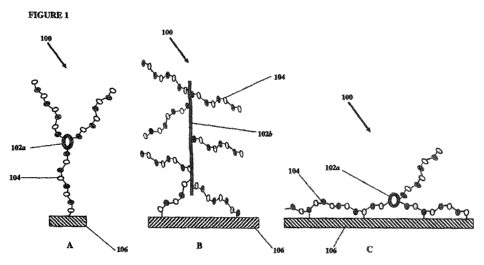
[0014] Figure 1 depicts several heparin entities of the invention and types of
attachment of said heparin entities to a substrate.
[0015] Figure 2 depicts ATIII binding capacity of various aldehyde containing
heparin entities conjugated onto expanded polytetrafluoroethylene (ePTFE) and
having undergone multiple EtO sterilizations. Aldehyde containing heparin
entities
are classified according to the core molecule used in the synthesis of the
heparin
entity. Hence, colistin sulfate as the core refers to Examples 1, neomycin to
Example 2, poly-L-lysine to Example 4, capreomycin to Example 3,
polyethyleneimine (PEI) to Example 5, and ethylene diamine (EDA) to Example 6.
All bars represent mean values of samples numbers with error bars for the
standard
deviation.
[0016] Figures 3 A and B depict light micrographs of heparin entities
comprising
free terminal aldehydes immobilized onto an ePTFE substrate by a single end-
point
attachment method before (3A) and after (3B) treatment with heparinase-1 and
stained with toluidine blue. The absence of coloration in Figure B as compared
to A,
demonstrates that heparin entities comprising free terminal aldehydes
immobilized
onto an ePTFE substrate by a single end-point attachment method is essentially
depolymerized from the surface after heparinase-1 treatment.
[0017] Figure 3 C depicts the normalized change in luminosity before and after
treatment with heparinase-1 for heparin immobilized through end-point aldehyde
and
multi-point attachment, heparin entities comprising a neomycin core
immobilized
through end-point and multi-point attachment through at least one heparin
molecule,
CA 02774494 2012-03-16
WO 2011/035001 PCT/US2010/049078
and USP heparin immobilized through multi-point attachment. The low normalized
change in luminosity values for the heparin end-point aldehyde, heparin entity
comprising heparin and neomycin core with end-point aldehyde, and USP heparin,
all multi-point attached to the substrate, indicated that the surfaces are not
heparinase-1 sensitive and still have substantial heparin on the surface.
[0018] Figures 4 A -C depicts light micrographs of heparin entities comprising
heparin and an EDA, core immobilized onto an ePTFE substrate by a single end-
point attachment method before (4A and 4B) and after (4C) treatment with
heparinase.-1 and stained with toluidine blue. The stained samples demonstrate
the
presence of the heparin entity. Samples 4B and 4C were subjected to a round of
sterilization and rinsed only with DI water post sterilization. The coloration
of Figure
4C after sterilization and heparinase-1 treatment indicates that heparinase-1
did not
recognize heparin entities on the surface.
[0019] Figures 4 D and E depict light micrographs of heparin entities
comprising
heparin and an EDA core immobilized onto an ePTFE substrate by a single end-
point attachment method before (4D) and after (4E) treatment with heparinase-1
and
stained with toluidine blue. These samples where subjected to a round of
sterilization and rinsed with DI water and boric acid post sterilization. The
lack
coloration of Figure 4E after sterilization indicates that heparinase-1 did
recognize
heparin entities on the surface and depolymerized them.
[0020] Figures 5 A-C depicts SAX-HPLC chromatograms from heparinase-1
depolymerization of (A) USP heparin, (B) heparin entities constructed from
heparin
and colistin sulfate, and (C) heparin entities constructed from heparin and
neomycin
sulfate.
[0021] Figures 6 A and B depicts SAX-HPLC chromatograms from heparinase-1
depolymerization of ePTFE surface immobilized (a) USP heparin bound by free
terminal aldehyde and (b) heparin entities constructed from heparin and
colistin
sulfate bound by free terminal aldehyde.
DETAILED DESCRIPTION
[0022] The present invention comprises medical substrates comprising heparin
entities immobilized onto a substrate. The heparin entities of the invention
retain
6
CA 02774494 2012-03-16
WO 2011/035001 PCT/US2010/049078
significant biological activity following immobilization and sterilization as
compared to
other coated medical substrates.
[0023] In the context of this disclosure, a number of terms are used. The
following definitions are provided. As used herein and in the appended claims,
the
singular forms "a", "an", and "the" include plural reference unless the
context clearly
dictates otherwise.
[0024] As used herein the term "heparin entity" means heparin molecules
covalently attached to a core molecule. Said heparin molecules can be attached
to
the core molecule by end point attachment (as described below and as
essentially
described in U.S. Patent 4,613,665, incorporated by reference herein for all
purposes) or other methods known in the art (see e.g. GT Hermanson,
Bioconjugate
Techniques, Academic Press, 1996; HG Garg et af., Chemistry and Biology of
Heparin and Heparan Sulfate, Elsevier, 2005.)
[0025] As used herein the term "core molecule" means a polyfunctional molecule
to which heparin is attached. For the purposes of this invention, said core
molecule
and a substrate are not the same, although a core molecule and a substrate can
be
made from the same material.
[0026] As used herein, the term "substantially pure" means, an object species
is
the predominant species present (i.e., on a molar basis it is more abundant
than any
other individual species in the composition), and preferably a substantially
purified
fraction is a composition wherein the object species comprises at least about
50
percent (on a molar basis) of all macromolecular species present. Generally, a
substantially pure composition will comprise more than about 80 to about 90
percent
of all macromolecular species present in the composition. Most preferably, the
object species is purified to essential homogeneity (contaminant species
cannot be
detected in the composition by conventional detection methods) wherein the
composition consists essentially of a single macromolecular species.
[0027] As used herein, the term "heparinase" means any enzymatic reaction that
depolymerizes (e.g. digests) heparin. Examples of heparinase include, but are
not
limited to, heparinase-1, heparinase-2, heparinase-3, heparanase,
exosulphatases,
bacterial exoenzymes, and glycosidases that can depolymerize heparin.
[0028] As used herein the term "heparinase sensitive" means that after
treatment
of a substrate comprising heparin entities with heparinase and staining said
substrate with toluidine blue, the substrate will not be visibly stained
(essentially as
7
CA 02774494 2012-03-16
WO 2011/035001 PCT/US2010/049078
depicted in Figures 3 B and Figures 4 Q. The term also means that an
insignificant
amount of toluidine blue will bind to residual heparin, or fragments thereof,
and a
reading from a detector that can measure the amount of toluidine blue (or
other
abels) on a substrate, such as a spectrophotometer, luminometer, densitometer,
liquid scintillation counter, gamma counter, or the like, will be about
background
evels, or be insignificantly different from background levels when compared to
a
substrate without heparin entities and stained with toluidine blue, or be
below the
sensitivity of said detectors when compared to a substrate comprising heparin
entities and stained with toluidine blue without heparinase treatment. The
term also
means that a label that binds to heparin, or fragments thereof, will not
detect a
substantial amount of heparin, or fragments thereof, after treatment of a
substrate
comprising heparin entities with heparinase.
[0029] As used herein the terms "bound," "attached," and "conjugate," and
their
derivatives, when referring to heparin entities and/or heparin means
covalently
bound, unless specified otherwise.
[0030] Referring to Figures 1 A-C, one embodiment of the invention comprises a
medical substrate comprising a heparin entity 100 bound onto a substrate 106
via at
east one heparin molecule 104, wherein said bound heparin entity is heparinase
sensitive. Suitable substrate materials for immobilizing or binding said
heparin
entities comprise polymers such as, but not limited to, polyamides,
polycarbonates,
polyesters, polyolefins, polystyrene, polyurethane, poly(ether urethane),
polyvinyl
chlorides, silicones, polyethylenes, polypropylenes, polyisoprenes,
polytetrafluoroethylenes, and expanded-polytetrafluoroethylenes (ePTFE, as
described in I.J.S. Patent 4,187,390). In one embodiment, expanded, or porous,
polytetrafluoroethylene (ePTFE) is the substrate.
[0031] Additional substrates include, but are not limited to, hydrophobic
substrates such as polytetrafluoroethylene, expanded polytetrafluoroethylene,
porous polytetrafluoroethylene, fluorinated ethylene propylene,
hexafluoropropylene,
polyethylene, polypropylene, nylon, polyethyleneterephthalate, polyurethane,
rubber,
silicone rubber, polystyrene, polysulfone, polyester, polyhydroxyacids,
polycarbonate, polyimide, polyamide, polyamino acids, regenerated cellulose,
and
proteins, such as silk, wool, and leather. Methods of making porous
polytetrafluoroethylene materials are described in U.S. Pat. Nos. 3,953,566
and
4,187,390, each of which is incorporated herein by reference. In another
8
CA 02774494 2012-03-16
WO 2011/035001 PCT/US2010/049078
embodiment, said ePTFE may be impregnated, filled, imbibed or coated with at
least
one chemical compound known to cause a bioactive response. Compounds that
cause a bioactlve response comprise anti-microbials (e.g. anti-bacterials and
anti-
virals), anti-inflammatories (e.g. dexamethasone and prednisone), anti-
proliferatives
(e.g. taxol, paclitaxel and docetaxel) and anti-coagulating agents (e.g.
abciximab,
eptifibatlde and tirofibran). In one embodiment, said anti-inflammatory is a
steroid.
In another embodiment, said steroid is dexamethasone. Methods of coating
substrates are well known in the art. In another embodiment, said substrate
comprises the heparin entities of the invention and a coating that comprises a
compound that causes a bioactive response. Said substrate comprises the
materials
referred to above and below. In one embodiment, said substrate is ePTFE.
[0032] Other suitable substrates include, but are not limited to, cellulosics,
agarose, alginate, polyhydroxyethylmethacrylate, polyvinyl pyrrolidone,
polyvinyl
alcohol, polyallylamine, polyaIlylalcohol, polyacrylamide, and polyacrylic
acid.
[0033] Additionally, certain metals and ceramics may be used as substrates for
the present invention. Suitable metals include, but are not limited to,
titanium,
stainless steel, gold, silver, rhodium, zinc, platinum, rubidium, and copper,
for
example. Suitable alloys include cobalt-chromium alloys such as L-605, MF35N,
Flgiloy, nickel-chromium alloys (such as Nitinol), and niobium alloys, such as
Nb-1 %
Zr, and others.
[0034] Suitable materials for ceramic substrates include, but are not limited
to,
silicone oxides, aluminum oxides, alumina, silica, hydroxyapapitites, glasses,
calcium oxides, polysilanols, and phosphorous oxide. In another embodiment,
protein-based substrates, such as collagen can be used. In another embodiment,
polysaccharide-based substrates, such as cellulose can be used.
[0035] Some substrates may have multiplicities of reactive chemical groups
populating at least a portion of its surface to which heparin entities of the
invention
can be bound. Said heparin entities of the invention are covalently bound to
the
substrate material through said reactive chemical groups. Surfaces of said
substrates can be smooth, rough, porous, curved, planar, angular, irregular,
or
combinations thereof. In some embodiments, substrates with surface pores have
internal void spaces extending from the porous surface of the material into
the body
of the material. These porous substrates have internal substrate material
bounding
the pores that often provides surfaces amenable to immobilizing biologically
active
9
CA 02774494 2012-03-16
WO 2011/035001 PCT/US2010/049078
entities. Whether porous or non-porous, substrates can be in the form of
filaments,
films, sheets, tubes, meshworks, wovens, non-wovens, and combinations thereof.
[0036] Substrates lacking reactive chemical groups on their surfaces (or
lacking
appropriately reactive chemical groups) can be covered, at least in part, with
a
polymeric covering material having a multiplicity of reactive chemical groups
thereon
to which said heparin entities can be bound. Polymeric substrates can also be
modified along their surface, or along their polymer backbone using a variety
of
methods, including hydrolysis, aminolysis, photolysis, etching, plasma
modification,
plasma polymerization, carbene insertion, nitrene insertion, etc. Said heparin
entities are covalently attached, or bound, to the polymeric covering material
through
the reactive chemical groups of the covering material or directly to a
substrate that
has been modified. The polymeric covering material may form at least one layer
on
at least a portion of a substrate.
[0037] There are many other surface modifications, such those described U.S.
Patent 4,600,652 and U.S. Patent 6,642,242, which are based on substrates
having
a layer of a polyurethane urea to which heparin modified to contain aldehyde
groups
through oxidation with nitrous acid or periodate, may be bound by covalent
links. A
similar technology is described in U.S. Patent 5,032,666, where the substrate
surface is coated with an amine rich fluorinated polyurethane urea before
immobilization of an antithrombogenic agent, such as an aldehyde-activated
heparin.
Another antithrombogenic surface modification which may be mentioned is
described
in publication W087/07156. The surface of the device is modified through the
coating with a layer of lysozyme or a derivative thereof to which heparin is
adhered.
Yet another surface modification for producing antithrombogenic articles is
described
in J.S. Pat. No. 4,326,532. In this case, the layered antithrombogenic surface
comprises a polymeric substrate, a chitosan bonded to the polymeric substrate
and
an antithrombogenic agent bonded to the chitosan coating. Others have reported
an
antithrombogenic hemofilter also using a chitosan layer for binding heparin.
Another
process for preparing antithrombogenic surfaces is described in W097/07834,
wherein the heparin is admixed with sufficient periodate so as not to react
with more
than two sugar units per heparin molecule. This mixture is added to a surface
modified substrate of a medical device, wherein said surface modification
contains
amino groups. The above listing of processes for adding reactive groups to
substrates are only a small example of how this can be accomplished. The above
CA 02774494 2012-03-16
WO 2011/035001 PCT/US2010/049078
listing is by no means complete. Furthermore, it is clear that the type of
process
used to add reactive chemical groups to a substrate will depend on the
properties of
the substrate of which a person of skill in the art will recognize.
[0038] In another embodiment of the invention, said medical substrate
comprising
said bound heparin entity via at least one heparin molecule is a component of
a
medical device. Medical devices comprise, but are not limited to, grafts,
vascular
grafts, stents, stent-grafts, bifurcated grafts, bifurcated scents, bifurcated
stent-grafts,
hernia patches, hernia plugs, periodontal grafts, surgical fabrics, drug
delivery
devices, catheters, cardiac leads balloons and indwelling filters. In one
embodiment,
said stents can be used in cardiac, peripheral or neurological applications.
In
another embodiment, said stent-grafts can be used in cardiac, peripheral or
neurological applications.
[0039] Another embodiment of the invention comprises a heparin entity
comprising at least one heparin molecule and at least one core molecule. As
shown
in Figure 1, the core molecule 102 is the "backbone" of the heparin entity 100
to
which heparin molecules 104 are bound. Said core molecule 102 can be either
cyclic (102a, Figure 1A and 1 C), linear (102b, Figure 1 B), branched,
dendritic, "Y"
shaped, "T99 shaped, or "star" shaped as described by Freudenberg, U.,
Biomaterials,
30, 5049-5060, 2009 and Yamaguchi, N., Biomacromolecules, 6, 1921-1930, 2005.
In one embodiment, said core molecule is selected from the group consisting of
proteins (including polypeptides), hydrocarbons, lipids, aminoglycosides,
polysaccharides and polymers. Proteins include, but are not limited to,
antibodies,
enzymes, receptors, growth factors, hormones, serpins and any globular
protein.
Specific proteins and polypeptides include, but are not limited to, albumin,
colistin,
collagen, polylysine, antithrombin III, fibrin, fibrinogen, thrombin, laminin,
keratin, and
the like. In another embodiment, said core molecule can be a polypeptide. Said
polypeptide need not be very long and can comprise one or more repetitions of
amino acids, for example repetitions of serine, glycine (e.g. Ser-Gly-Gly-Ser-
Gly),
lysine or ornithine residues. Alternatively, other amino acid sequences can be
used,
for example colistin, polylysine, and polymyxin.
[0040] Examples of polysaccharides include, but are not limited to neutral
polysaccharides such as cellulose, starch, agarose, carboxymethylcellulose,
nitrocellulose, and dextran, anionic polysaccharides such as alginate,
heparin,
heparin sulfate, dextran sulfate, xanthan, hyaluronic acid, carrageenan, gum
arabic,
11
CA 02774494 2012-03-16
WO 2011/035001 PCT/US2010/049078
tragacanth, arabinogalactan, and pectin; macrocyclic polysaccharides such as
cyclodextrin and hydroxypropyl cyclodextrin; and polycationic polysaccharides
such
as chitin and chitosan.
[0041] Examples of synthetic polymers include, but are not limited to,
polyethylene glycol (PEG) 200, 300, 400, 600, 1000, 1450, 3350, 4000, 6000,
8000
and 20000, polytetrafluoroethylene, polypropylene glycol, polyethylene glycol-
co-
propylene glycol), copolymers of polyethylene glycol, copolymers of
polypropylene
glycol, copolymers of tetrafluoroethylene with vinyl acetate and vinyl
alcohol,
copolymers of ethylene with vinyl acetate & vinyl alcohol, polyvinyl alcohol,
polyethyleneimine, polyacrylic acid; polyols such as polyvinyl alcohol and
polyallyl
alcohol; polyanions such as acrylic acid and poly(methacrylic acid).
Polycation
polymers include poly(allylamine), poly(ethyleneimine), poly(guanidine),
polyvinyl
amine), polyethylene glycol diamine, ethylene diamine, and poly(quaternary
amines);
polyacrylonitriles such as hydrolyzed polyacrylonitrile, poly(acrylamide-co-
acrylonitrile), and their copolymers. Other polymers include fluorinated
copolymers
including copolymers of tetrafluoroethylene and vinyl alcohol, vinyl acetate,
vinyl
formamide, acrylamide, and vinyl amine. In another embodiment, said core
molecule can be an aminoglycoside, including, but not limited to, amikacin,
arbekacin, gentamicin, kanamycin, neomycin, netilmicin, paromomycin,
rhodostreptomycin, streptomycin, tobramycin, and apramycin.
[0042] Heparin is a mucopolysaccharide, isolated from pig intestine or bovine
lung and is heterogeneous with respect to molecular size and chemical
structure.
Heparin is built up from alternating glycuronic acid and glucosamine units.
The
glycuronic acid units consist of D-glycuronic acid and L-iduronic acid. These
are
respectively D- and L-(1,4)-bound to the D-glucosamine units. A large
proportion of
the L-iduronic acid residues are sulfated in the 2-position. The D-glucosamine
units
are N-sulfated, sulfated in the 6-position and are a-(1,4)-bound to the uronic
acid
residues. Certain D-glucosamine units are also sulfated in the 3-position.
Heparin
contains material with a molecular weight ranging from about 6,000 Daltons to
about
30,000 Daltons. The hydroxyl and amine groups are derivatized to varying
degrees
by sulfation and acetylation. The active sequence in heparin responsible for
its
anticoagulation properties is a unique pentasaccharide sequence that binds to
the
ligand anti-thrombin III (ATIII). The sequence consists of three D-glucosamine
and
two uronic acid residues. Heparin molecules can also be classified on the
basis of
12
CA 02774494 2012-03-16
WO 2011/035001 PCT/US2010/049078
their pentasaccharide content. About one third of heparin contains chains with
one
copy of the unique pentasaccharide sequence (see, Choay, Seminars in
Thrombosis
and Hemostasis 11:81-85 (1985) which is incorporated herein by reference) with
high affinity for ATI II, whereas a much smaller proportion (estimated at
about 1 % of
total heparin) consists of chains which contain more than one copy of the high
affinity
pentasaccharide (see, Rosenberg et ai., Biochem. Biophys. Res. Comm. 86:1319-
1324 (1979) which is incorporated herein by reference). The remainder (approx.
66%) of the heparin does not contain the pentasaccharide sequence. Thus, so
called "standard heparin" constitutes a mixture of the three species: "high
affinity"
heparin is enriched for species containing at least one copy of the
pentasaccharide
and "very high affinity" heparin refers to the approximately 1 % of molecules
that
contain more than one copy of the pentasaccharide sequence. These three
species
can be separated from each other using routine chromatographic methods, such
as
chromatography over an anti-thrombin affinity column (e.g., Sepharose-AT; see,
e.g.,
Lam et ai., Biochem. Biophys. Res. Comm. 69:570-577 (1976) and Horner Biochem.
J. 262:953-958 (1989) which are incorporated herein by reference).
[0043] In one embodiment, said heparin is derived from an animal. In another
embodiment, said heparin is bovine or porcine derived. In another embodiment,
said
heparin is a synthetic heparin, i.e. not derived from animal sources (e.g.
fondaparinux or enoxaparin). In another embodiment, heparin entities of the
invention comprise heparin that has been enriched and comprises substantially
pure
"high affinity" heparin. In another embodiment, heparin entities of the
invention
comprise heparin that has been enriched and comprises substantially pure "very
high affinity" heparin. In another embodiment, heparin entities of the
invention
comprises heparin has been enriched and comprises a combination of
substantially
pure "high affinity" and "very high affinity" heparin.
[0044] Another embodiment of the invention comprises the binding of said
heparin entity to a medical substrate via at least one heparin molecule. As
shown in
Figure 1, the heparin entities of the invention are bound to said substrate
via at least
one heparin molecule. Thus, in one embodiment, said bound heparin molecule is
attached to said substrate via end point attachment (as depicted in Figures 1A
and
1 B). In another embodiment, said bound heparin molecule is attached to said
substrate via an end point aldehyde. This can be accomplished essentially as
13
CA 02774494 2012-03-16
WO 2011/035001 PCT/US2010/049078
described in U.S. Patent 4,613,665, which is incorporated herein by reference
in its
entirety, and as described below.
[0045] In another embodiment, said heparin entity is bound onto a substrate
via
at least one heparin molecule, wherein said bound heparin molecule is attached
to
said substrate via a "loop attachment." Loop attachment, as depicted in Figure
1 C,
is an attachment of said heparin entity via at least one heparin, wherein the
heparin
is attached loosely to the substrate in a small number of locations, therefore
allowing
substantial portions of the bound heparin to be exposed to heparinase (as
opposed
to more common methods that attach heparin tightly in a large number of
locations).
The more common methods of coupling heparin to a substrate comprise reacting a
majority of functional groups randomly localized along a heparin molecule's
length
(e.g. using coupling agents such as carbodiimides, epoxides, and
polyaldehydes).
These methods result in a high probability that the active sequence (said
unique
pentasaccharide sequence describe above) will be bound to the substrate
resulting
in reduced and/or lost activity. In loop attachment of heparin, only a few
functional
groups on the heparin react and are bound to the substrate. Thus, there is a
high
probability that the active sequence of the attached heparin will not be bound
to the
substrate, therefore allowing said active sequence to bind to its ligand. In
another
embodiment, the invention comprises a heparin entity with multiple attachments
to a
substrate, wherein the active sequence is not bound to the substrate. In
another
embodiment, said bound heparin entity molecule is attached to said substrate
via
loop attachment.
[0046] As discussed above, endpoint and loop attachments allow a substantial
portion of at least one heparin molecule (in a heparin entity) not to be bound
to a
substrate. As used herein the term "substantial portion" means that about 50%,
about 60%, about 70%, about 80%, about 90%, about 95 about 96% about 97%,
about 98% and about 99% of the heparin molecule is not bound to the substrate.
In
another embodiment, the term also refers to the at least one heparin molecule
(in a
heparin entity) wherein said the at least one heparin molecule bound to the
substrate
is not bound to a substrate via its active sequence. Thus, since the active
sequence
is not bound to the surface of the substrate, the active sequence has a
greater
probability of interacting with its ligand. In other words, if the active
sequence is
bound to the surface of the substrate then there is a small chance of heparin
binding
to its ligand.
14
CA 02774494 2012-03-16
WO 2011/035001 PCT/US2010/049078
[0047] However, because said heparin entities are attached via heparin by
endpoint and/or loop attachment, the heparin is sensitive to heparinase. Thus,
after
heparinase treatment, there will be very little, if any, heparin, or fragments
thereof,
on the surface of said substrate. In contrast, some of the more common methods
of
attaching heparin to the surface of a substrate (which comprises multiple
bonds
along the length of the heparin molecule, as described above), after
heparinase
treatment, will have a significant amount of heparin, or fragments thereof,
still
attached to the surface of the substrate. Thus, after heparinase treatment,
heparin,
or fragments thereof, can be detected on the surface of said substrate.
Without
being bound to any particular theory, the inventors have that discovered that
the
more sensitive the bound heparin or heparin entity is to heparinase, the more
biological activity said bound heparin or heparin entity exhibits. This may be
because the active sequence of the bound heparin or heparin entity is not
attached
to the surface of the substrate, thus said bound heparin or heparin entity has
a
greater chance of binding to its ligand.
[0048] Heparin must have intact conformation and structure to be recognized by
ATIII, and if said conformation and structure is lost, heparin will exhibit
poor activity.
In addition, loss of said conformation and structure results in poor
recognition by
other proteins, such as heparinase-1, resulting in said heparin being
resistant to
depolymerization. For example, modification of soluble heparin with
carbodiimide
changes the soluble heparin structure in such a way that it is no longer
recognized
by heparinase-1, and the modified soluble heparin has reduced whole blood
anticoagulant activity (see Olivera, G .B., Biomaterials, 24, 4777-4783,
2003). The
inventors have discovered that heparinase sensitivity of attached heparin or
heparin
entity is predictive of ATIII binding activity of said attached heparin or
heparin entity.
Without wanting to be constrained by any particular theory, if the attached
heparin or
heparin entity retains specificity for specific enzymes such as heparinase-1,
then the
attached heparin or heparin entity retains substantially enough
primary/secondary/tertiary structure for it also to have specificity for
ATIII. Thus, the
inventors have discovered that when an attached heparin or heparin entity is
recognized by heparinase-1, said attached heparin or heparin entity is also
recognized by ATII I, as exemplified by high binding activities.
[0049] The inventors have also shown that a boric acid rinse will restore
heparinase sensitivity to inactivated attached heparin or heparin entities
(inactivated
CA 02774494 2012-03-16
WO 2011/035001 PCT/US2010/049078
by sterilization, mechanical compaction and expansion, or long-term storage,
for
example). Thus, another embodiment of the invention comprises a method of
restoring heparinase sensitivity to heparin or heparin entities bound onto a
substrate
comprising rinsing said substrate in a solution of boric acid. In one
embodiment,
said substrate was exposed to a sterilization cycle. In another embodiment,
said
substrate was exposed to mechanical treatments that reduced heparinase-1
activity.
[0050] In another embodiment of the invention, after treating a medical
substrate
with bound heparin entities of the invention with heparinase, heparin, or
fragments
thereof, will not be detected on said substrate. In another embodiment, after
treating
a medical substrate with bound heparin entities of the invention with
heparinase,
heparin, or fragments thereof, will be detected at a substantially lower level
than
before heparinase treatment. Significantly lower level of detection comprises
very
little detection after staining and/or labeling for heparin.
[00511 In another embodiment, said heparin, or fragments thereof, will not be
detected visually (macroscopically) after staining or labeling. Heparin, or
fragments
thereof, can be detected by a label that binds directly or indirectly to
heparin, or
fragments thereof. In one embodiment, said label that binds to heparin, or
fragments
thereof, is selected from the group consisting of dyes, antibodies, and
proteins.
Examples of labels include, but are not limited to proteins including anti-
heparin
antibodies (polyclonal or monoclonal) and ATIII; metachromatic dyes including
toluidine blue, azure A, alcian blue, victoria blue 4R, night blue, methylene
blue;
radioiodinated labels including radioiodinated toluidine blue, radioiodinated
methylene blue, radioiodinated heparin antibodies, radioiodinated ATIII;
tritiated
labels including tritiated toluidine blue, tritiated azure A, tritiated alcian
blue, tritiated
victoria blue 4R, tritiated night blue, tritiated methylene blue; carbon-14
labels
including 14C-toluidine blue, 14C-azure A, 14C-alcian blue, 14C-victoria blue
4R,
14C-night blue, 14C-methylene blue; fluorescent labels including rhodamine-
labelled
heparin antibodies, fluorescein-labelled heparin antibodies,
rhodaminemlabelled ATIII,
fluorescein-labelled ATIII. In another embodiment, said dye is toluidine blue.
In
another embodiment, after heparinase treatment, an insignificant amount of
toluidine
blue will bind to heparin, or fragments thereof, but will not be visually
detected on
said substrate (essentially as depicted in Figures 3B and 4E). In another
embodiment, after heparinase treatment, a insignificant amount of toluidine
blue will
bind to residual heparin, or fragments thereof, and a reading from a detector
that can
16
CA 02774494 2012-03-16
WO 2011/035001 PCT/US2010/049078
measure the amount of toluidine blue (or other labels described above) on a
substrate (e.g. a spectrophotometer, luminometer, densitometer, liquid
scintillation
counter, gamma counter, or the like) will be about background levels, or be
insignificantly different from background levels when compared to a substrate
without heparin entities. In another embodiment, after heparinase treatment, a
reading from a detector that can measure the amount of toluidine blue (or
other
labels described above) on a substrate will be significantly different when
compared
to a substrate comprising heparin entities and stained with toluidine blue (or
other
labels described above) without heparinase treatment.
[0052] Another embodiment of the invention comprises a heparin entity
comprising at least one heparin molecule attached to a core molecule, wherein
the
entity is bound to a substrate via a heparin molecule, and wherein after
exposure to
heparinase and toluidine blue, the substrate macroscopically evidences
substantially
no toluidine blue on its surface (as depicted in Figures 3 B and Figure 4 E).
[0053] Another embodiment of the invention comprises a heparin entity which
comprises at least one heparin molecule and at least one core molecule such
that
when said heparin entity is bound onto a substrate via a least one heparin
molecule,
said heparin entity is heparinase sensitive. In one embodiment, said substrate
is
selected from the group consisting of polyethylene, polyurethane, silicone,
polyamide-containing polymers, polypropylene, polytetrafluoroethylene,
expanded-
polytetrafluoroethylene, biocompatible metals, ceramics, proteins,
polysaccharides,
and any substrate described above. In another embodiment, said substrate is
expand ed-polytetrafIuoroethylene. In another embodiment, said substrate is a
component of a medical device. In another embodiment, said medical device is
selected from the group consisting of grafts, vascular grafts, stents, stent-
grafts,
bifurcated grafts, bifurcated stents, bifurcated stent-grafts, patches, plugs,
drug
delivery devices, catheters and cardiac leads. In another embodiment, said
stents
can be used in cardiac, peripheral or neurological applications. In another
embodiment, said stent-grafts can be used in cardiac, peripheral or
neurological
applications. In another embodiment, said medical device can be used in
orthopedic, dermal, or gynecologic applications. In another embodiment, said
core
molecule comprises a cyclic, linear, branched, dendritic, "Y", "T", or star
molecular
structure. In another embodiment, said core molecule is selected from the
group
17
CA 02774494 2012-03-16
WO 2011/035001 PCT/US2010/049078
consisting of proteins, polypeptides, hydrocarbons, polysaccharides,
aminoglycosides, polymers, and fluoropolymers.
[0054] In another embodiment, heparin, or fragments thereof, is detected by
labels that bind to heparin, or fragments thereof. In another embodiment, said
label
that binds to heparin, or fragments thereof, is selected from the group
consisting of
dyes, polyclonal antibodies, and proteins. In another embodiment, said dye is
toluidine blue. In another embodiment, after heparinase treatment, an
insignificant
amount of toluidine blue will bind to residual heparin, or fragments thereof,
and will
not be visually detected on said substrate. In another embodiment, after
heparinase
treatment, a insignificant amount of toluidine blue will bind to residual
heparin, or
fragments thereof, and a reading from a detector that can measure the amount
of
toluidine blue (or other labels described above) on a substrate (e.g. a
spectrophotometer, luminometer, densitometer, liquid scintillation counter,
gamma
counter, or the like) will be about background levels, or be insignificantly
different
from background levels when compared to a substrate without heparin entities.
In
another embodiment, after heparinase treatment , a reading from a detector
that can
measure the amount of toluidine blue (or other labels described above) on a
substrate will be significantly different when compared to a substrate
comprising
heparin entities and stained with toluidine blue (or other labels described
above)
without heparinase treatment. In another embodiment, said heparin entity is
bound
onto a substrate via at least one heparin molecule and wherein said bound
heparin
molecule is attached to said substrate via end-point attachment, In another
embodiment, said heparin entity is bound onto a substrate via at least one
heparin
molecule, wherein said bound heparin molecule is attached to said substrate
via
end-point aldehyde. In another embodiment, said heparin entity is bound onto a
substrate via at least one heparin molecule, wherein said bound heparin
molecule is
attached to said substrate via loop attachment. In another embodiment, said
heparin
entity is bound onto a substrate via at least one heparin molecule, wherein
said
bound heparin molecule is attached to said substrate via aldehydes along the
length
said heparin.
[0055] Another embodiment of the invention comprises an ATlll binding entity
comprising: a core molecule, a polysaccharide chain attached to the core
molecule,
and a free terminal aldehyde moiety on the polysaccharide chain. This ATlll
binding
entity can then be end-point attached to a substrate via a terminal aldehyde.
18
CA 02774494 2012-03-16
WO 2011/035001 PCT/US2010/049078
Another embodiment of the invention comprises an ATIII binding entity
comprising: a
core molecule, a polysaccharide chain attached to the core molecule, and free
terminal aldehyde moieties along the length of the polysaccharide chain. This
ATIII
binding entity can then be looped attached to a substrate via the aldehydes
along the
length of the polysaccharide chain. In another embodiment, said polysaccharide
chain is heparin. In another embodiment, said core molecule is selected from
the
group consisting of a protein, a polypeptide, a hydrocarbon, an
aminoglycoside, a
polysaccharide, a polymer, a fluoropolymer, or any core molecule described
herein.
In another embodiment, heparin is bound onto the core molecule via end-point
attachment. In another embodiment, the substrate is selected from the group
consisting of polyethylene, polyurethane, silicone, polyamide-containing
polymers,
and polypropylene, polytetrafluoroethylene, expand ed-polytetrafIuoroethylene
and
biocompatible metals, or any of the substrates described herein. In another
embodiment said biocompatible metal is Nitinol. In another embodiment, said
substrate is expanded -polytetrafIuoroethylene. In another embodiment, said
substrate is a component of a medical device. In another embodiment, said
medical
device is selected from the group consisting of grafts, vascular grafts,
stents, stent-
grafts, bifurcated grafts, bifurcated stents, bifurcated stent-grafts,
patches, plugs,
drug delivery devices, catheters and cardiac leads. In another embodiment,
said
medical device can be used in cardiac, peripheral, neurologic, orthopedic,
gynecologic, or dermal applications.
[0056] Another embodiment of the invention comprises an implantable medical
device comprising a medical substrate, wherein said medical substrate
comprises a
heparin entity bound onto a substrate via at least one heparin molecule,
wherein said
bound heparin entities are heparinase sensitive. In one embodiment, said
medical
device is selected from the group consisting of grafts, vascular grafts,
stents, stent-
grafts, bifurcated grafts, bifurcated stents, bifurcated stent-grafts,
patches, plugs,
drug delivery devices, catheters and cardiac leads. In another embodiment,
said
stent can be used in cardiac, peripheral or neurological applications. In
another
embodiment, said stent can be a balloon expandable and/or a self expanded
stent.
Said stents can be made from any biocompatible material including any polymer
or
metal as described above. In another embodiment, said stent is made from
Nitinol
and/or stainless steel. In another embodiment, said stent comprises a graft.
In
19
CA 02774494 2012-03-16
WO 2011/035001 PCT/US2010/049078
another embodiment, said graft and/or stent comprise heparin entities of the
invention.
[0057] The heparin entities of the invention retain significant biological
activity
following immobilization and sterilization as compared to other coated medical
substrates. Thus, in one embodiment said medical substrate comprises, a
heparin
entity bound onto a substrate via at least one heparin molecule, wherein said
bound
heparin entity is heparinase sensitive has an ATIII activity of about 300
pmol/cmz. In
another embodiment, the ATIII activity is about 250 pmol/cmz, about 200
pmol/cmz,
about 150 pmol/cmz, about 100 pmol/cmz, about 50 pmol/cmz, about 40 pmol/cmz,
about 30 pmol/cm2, about 20 pmol/cmz, about 10 pmol/cmz or about 5 pmol/cmz.
In
another embodiment, after a first round of sterilization the ATIII activity of
said
medical substrate is about 250 pmol/cmz, about 200 pmol/cmz, about 150
pmol/cmz,
about 100 pmol/cmz, about 50 pmol/cmz, about 40 pmol/cmz, about 30 pmol/cmz,
about 20 pmol/cmz, about 10 pmol/cmz or about 5 pmol/cmz. In another
embodiment, after a second round of sterilization, the ATIII activity of said
medical
substrate is about 100 pmol/cmz, about 90 pmol/cmz, about 80 pmol/cmz, about
70
pmol/cmz, about 60 pmol/cmz, about 50 pmol/cmz, about 40 pmol/cmz, about 30
pmol/cmz, about 20 pmol/cmz, about 10 pmoVcm2 or about 5 pmol/cmz. In another
embodiment, after a third round of sterilization, the ATIII activity of said
medical
substrate is above about 50 pmol/cmz, or about 70 pmol/cmz, about 60 pmol/cmz,
about 50 pmol/cmz, about 40 pmol/cmz, about 30 pmol/cmz, about 20 pmol/cmz,
about 10 pmol/cmz or about 5 pmol/cmz. ATIII activity assays are well known in
the
art and at least one is described below. In another embodiment, said heparin
entities of the invention retain significant biological activity following
compression and
expansion of a medical device. In another embodiment, said heparin entities of
the
invention retain significant biological activity following storage conditions
for medical
devices either in a compacted and/or expanded state.
[0058] Another embodiment of the invention comprises methods of determining
the structure of a heparin entity bonded to a substrate. One method of
determining
the structure of a heparin entity bonded to a substrate comprises the steps
of:
providing a substrate comprising a heparin entity, depolymerizing the heparin
entity
to generate a mixture of soluble heparin fragments, detecting each soluble
heparin
fragment in said mixture using column chromatography, determining the identity
of
each detected soluble heparin fragment from above, and deriving the structure
of the
CA 02774494 2012-03-16
WO 2011/035001 PCT/US2010/049078
heparin entity from the identities of the detected soluble heparin fragments.
In one
embodiment, said depolymerization is by heparinase-1. In another embodiment,
column chromatography is strong anion exchange-high performance liquid
chromatography or SAX-HPLC.
[0059] Another embodiment of the invention comprises an implantable medical
device comprising a medical substrate, wherein said medical substrate
comprises a
heparin entity bound onto a substrate via at least one heparin molecule,
wherein said
bound heparin entities are heparinase sensitive. In one embodiment, said
medical
device is selected from the group consisting of grafts, vascular grafts,
stents, stent-
grafts, bifurcated grafts, bifurcated stents, bifurcated stent-grafts,
patches, plugs,
drug delivery devices, catheters, cardiac leads, balloons and indwelling
filters. In
another embodiment, said stent can be used in cardiac, peripheral or
neurological
applications. In another embodiment, said stent can be a balloon expandable
and/or
a self expanded stent. Said stents can be made from any biocompatible material
including any polymer or metal as described above. In another embodiment, said
stent is made from Nitinol and/or stainless steel. In another embodiment, said
stent
comprises a graft. In another embodiment, said graft and/or stent comprise
heparin
entities of the invention.
[0060] Another embodiment of the invention comprises methods of determining
the spatial distribution of a heparin entity bonded to a substrate. One method
of
determining the spatial distribution of a heparin entity bonded to a substrate
comprises the steps of: providing a substrate comprising a heparin entity,
depolymerizing the heparin entity to generate a surface comprising surface-
bonded
unsaturated heparin fragments, reacting the surface with a labeling reagent
which
introduces a detectable component to said surface-bonded unsaturated heparin
fragments, detecting said surface-bonded unsaturated heparin fragment via said
detectable component, and deriving the spatial distribution of the heparin
entity from
the presence of the surface-bonded unsaturated heparin fragments. In one
embodiment, depolymerization is by heparinase-1. In another embodiment, said
labeling reagent is a lanthanoid Michael-like addition organo-complex. In
another
embodiment, said labeling reagent is terbium tris(4-methylthio)benzoate. In
another
embodiment, said organo-complex comprises chemisorbed gold nanoparticles. In
another embodiment, said detecting is by epifluoroscent microscopy or
transmission
electron microscopy.
21
CA 02774494 2012-03-16
WO 2011/035001 PCT/US2010/049078
[0061] Another embodiment of the invention comprises a system for determining
the structure of a heparin entity bonded to a substrate, comprising a
depolymerization solution, a labeling reagent solution, and a detector. A
system is
an assembly of reagents and instruments used to detect the structure and type
of
binding of heparin entities to a substrate. In one embodiment, said
depolymerization
solution comprises heparinase-1. In another embodiment, said labeling reagent
solution comprises toluidine blue, and terbium tris(4-methylthio)benzoate. In
another
embodiment, said detector comprises SAX-HPLC, an epifluoroscent microscope,
and an absorption spectroscope. In another embodiment, said assembly of
reagents
can be a kit.
[0062] After enzymatic heparinase-1 depolymerization of heparin and/or heparin
entities that are end-point attached, heparin fragments are left are on the
surface
that are unsaturated, i.e. they comprise a carbon-carbon double bond ("nubs").
Enzymatic heparinase depolymerization involves cleavage of the non-reducing
terminal uronic acid residue to a 4,5-unsaturated derivative. This produces
residual
surface-bonded unsaturated heparin fragments bonded to the substrate that
comprises a carbon-carbon double bond. Thus, the structure of the residual
surface-
bonded heparin fragment is unsaturated, and can react with various detection
molecules, including those that comprise Michael-like addition complexes, such
as
thiol-containing compounds and thiol-containing fluorescent compounds, such as
terbium tris(4-methylthio)benzoate. Thus, in another embodiment of the
invention,
after enzymatic heparinase-1 depolymerization of an end-point attached heparin
entity, said residual surface-bonded unsaturated heparin fragments bonded to
the
substrate comprising a carbon-carbon double bond are detected. This method can
determine if heparin and/or heparin entities were end-point attached to a
substrate.
In another embodiment, nub detection is combined with any of the detection
and/or
characterization methods described above.
[0063] This invention is further illustrated by the following Examples which
should
not be construed as limiting. The contents of all Figures and references are
incorporated herein by reference.
EXAMPLES
Example 1
22
CA 02774494 2012-03-16
WO 2011/035001 PCT/US2010/049078
[0064] This example describes the construction of heparin entities comprising
heparin and colistin sulfate as the core. This heparin entity contains free
terminal
aldehydes that can be used for attachment to a surface of a substrate.
[0065] Colistin sulfate (0.10 g, Alpharma, Inc.) was dissolved in 300 ml of
deionized (DI) water containing MES buffer (pH 4.7, SupHTM Thermo Scientific).
To
this was added 10 g USP heparin, 4 g N-hydroxysulfosuccinimide (sulfo-NHS,
Thermo Scientific), and 4 g of 1 -ethyl -3-(3-di methylaminopropyl)carbodiim
ide (EDC
hydrochloride, Sigma-Aldrich, St. Louis, MO). The reaction was allowed to
proceed
at room temperature for 4 hours, followed by dialysis overnight with a
50,000MWCC
membrane (Spectra/Por ). The retentate (about 350 ml out of 500 ml) was
transferred to a beaker, and cooled to 0 C. Sodium nitrite (10 mg) and acetic
acid (2
ml) were added and the reaction was allowed to proceed for 1 hour at 0 C.
Dialysis
was performed overnight with a 50,000 MWCO membrane with the addition of 1 g
NaCl to the dialysis liquid. Freezing and Iyophilization of the retentate
produced a
fine powder.
Example 2
[0066] This example describes the construction of heparin entities comprising
heparin and neomycin sulfate as the core. This heparin entity contains free
terminal
aldehydes that can be used for attachment to a surface of a substrate.
[0067] Neomycin sulfate (0.0646 g, Spectrum Chemical) was dissolved in 300 ml
of Dl water containing MES buffer (pH 4.7, DupHTM Thermo Scientific). To this
was
added 10 g USP heparin, 4 g N-hydroxysulfosuccinimide (sulfo-NHS), and 4 g of
EDC hydrochloride. The reaction was allowed to proceed at room temperature for
4
hours, followed by dialysis overnight with a 50,000 MWCO membrane
(Spectra/Por ). The retentate (about 400 ml out of 505 ml) was transferred to
a
beaker and cooled to 0 C. Sodium nitrite (10 mg) and acetic acid (2 ml) were
added
and the reaction was allowed to proceed for 1 hour at 0 C. Dialysis was
performed
overnight with a 50,000 MWCO membrane with the addition of 1 g NaCl to the
dialysis liquid. The dialyzed retentate was filtered twice using a 20
micrometer,
0.00079 inches U.S.A. standard testing sieve, A.S.T.M.E.-11 specification
NO.635 to
remove small particles. Freezing of the filtrate and lyophilization produced a
fine
powder.
23
CA 02774494 2012-03-16
WO 2011/035001 PCT/US2010/049078
Example 3
[0068] This example describes the construction of heparin entities comprising
heparin and capreomycin sulfate as the core. This heparin entity contains free
terminal aldehydes that can be used for attachment to a surface of a
substrate.
[0069] Capreomycin sulfate (0.0501 g, Sigma-Aldrich, St. Louis, MO) was
dissolved in 300 ml of DI water containing MES buffer (pH 4.7, SupHTMThermo
Scientific). To this was added 10 g USP heparin, 4 g N-hydroxysulfosuccinimide
(sulfo-NHS), and 4 g of EDC hydrochloride. The reaction was allowed to proceed
at
room temperature for 4 hours. The reaction mixture was filtered once using a
20
micrometer, 0.00079 inches U.S.A. standard testing sieve, A.S.T.M.E.-11
specification NO.635 to remove small particles and the filtrate was dialyzed
overnight
with a 50,000 MWCO membrane (Spectra/Por ). The retentate (about 400 ml out of
515 ml) was transferred to a beaker and cooled to 0 C. Sodium nitrite (10 mg)
and
acetic acid (2 ml) were added and the reaction was allowed to proceed for 1
hour at
0 C. Dialysis was performed overnight with a 50,000 MWCO membrane with the
addition of 1 g NaCl to the dialysis liquid. The retentate was filtered twice
using a 20
micrometer, 0.00079 inches U.S.A. standard testing sieve, A.S.T.M.E.-11
specification NO.635 to remove small particles. Freezing of the filtrate and
lyophilization produced a fine powder.
Example 4
[0070] This example describes the construction of heparin entities comprising
heparin and poly-L-lysine as the core. This heparin entity contains free
terminal
aldehydes that can be used for attachment to a surface of a substrate.
[0071] Poly-L-lysine (0.1776 g, Sigma-Aldrich, molecular weight 1,000 to 5,000
g/mole) was dissolved in 300 ml of DI water containing MES buffer (pH 4.7,
DupHTM
Thermo Scientific). To this was added 10 g USP heparin, 4 g N-
hydroxysulfosuccinimide (sulfa-NHS), and 4 g of EDC hydrochloride. The
reaction
was allowed to proceed at room temperature for 4 hours followed by dialysis
overnight with a 50,000 MWCO membrane (Spectra/Por ). The retentate (about
400 ml out of 505 ml) was transferred to a beaker and cooled to 0 C. Sodium
nitrite
(10 mg) and acetic acid (2 ml) were added and the reaction was allowed to
proceed
for 1 hour at 0 C. Dialysis was performed overnight with a 50,000 MWCO
24
CA 02774494 2012-03-16
WO 2011/035001 PCT/US2010/049078
membrane with the addition of 1 g NIaCI to the dialysis liquid. Freezing of
the
retentate and Iyophilizatlon produced a fine powder.
Example 5
[0072] This example describes the construction of heparin entities comprising
heparin and polyethyleneimine (PEI) as the core. This heparin entity contains
free
terminal aldehydes that can be used for attachment to a surface of a
substrate.
[0073] PEI (Lupasol, BASF, 1.7756 g) was dissolved in 300 ml of DI water
containing MES buffer (pH 4.7, DupHTM Thermo Scientific). To this was added 10
g
LISP heparin, 4 g N. hydroxysulfosuccinimide (sulfa-NHS), and 4 g of EDC
hydrochloride. The reaction was allowed to proceed at room temperature for 4
hours
followed by dialysis overnight with a 50,000 MWCO membrane (Spectra/Por ). The
retentate (about 400 ml out of 505 ml) was transferred to a beaker and cooled
to
0 G. Sodium nitrite (10 mg) and acetic acid (2 ml) were added and the reaction
was
allowed to proceed for 1 hour at 0 O. Dialysis was performed overnight with a
50,000 MWCO membrane with the addition of 1 g NaCl to the dialysis liquid.
Freezing of the retentate and Iyophilization produced a fine powder.
Example 6
[0074] This example describes the construction of heparin entities comprising
heparin and ethylene diamine (EDA) as the core. This heparin entity contains
free
terminal aldehydes that can be used for attachment to a surface of a
substrate.
[0075] EDA (0.0043 g, Sigma-Aldrich, St. Louis, MO ) was neutralized to a pH
of
4.7 with equal volume dilution of HC and DI water, with the use of an ice
bath, then
dissolved in 300 ml of DI water containing MES buffer (pH 4.7, SupHTM Thermo
Scientific). To this was added 10 g USP heparin, 4 g N-hydroxysulfosuccinimide
(sulfo-NHS), and 4 g of EDC hydrochloride. The reaction was allowed to proceed
at
room temperature for 4 hours followed by dialysis overnight with a 50,000 MWCO
membrane (Spectra/Por ). The retentate (about 400 ml out of 505 ml) was
transferred to a beaker and cooled to 0 O. Sodium nitrite (10 mg) and acetic
acid (2
ml) were added and the reaction was allowed to proceed for 1 hour at 0 C.
Dialysis
was performed overnight with a 50,000 MWCO membrane with the addition of 1 g
NaCl to the dialysis liquid. Freezing of the retentate and lyophilization
produced a
fine powder.
CA 02774494 2012-03-16
WO 2011/035001 PCT/US2010/049078
Example 7
[0076] The heparin entities containing free terminal aldehydes of Examples 1
through 6 were immobilized onto the surface of an ePTFE substrate and tested
for
ATIII activity.
[0077] An ePTFE substrate material in sheet form was obtained from W.L. Gore
& Associates, Inc., Flagstaff, AZ under the trade name GORETM Microfiltration
Media
(GMM-406). A covering material in the form of a base coating was applied to
the
ePTFE material by mounting the material on a ten centimeter (10 cm) diameter
plastic embroidery hoop and immersing the supported ePTFE material first in
100%
isopropyl alcohol (IPA) for about five minutes (5 min) and then in a solution
of
polyethylene imine (PEI, Lupasol, BASF) and IPA in a one to one ratio (1:1).
LUPASOL water-free PEI was obtained from BASF and diluted to a concentration
of about four percent (4%) and adjusted to pH 9.6. Following immersion of the
ePTFE material in the solution for about fifteen minutes (15 min), the
material was
removed from the solution and rinsed in DI water at pH 9.6 for 15 min. PEI
remaining on the ePTFE material was cross-linked with a 0.05% aqueous solution
of
glutaraldehyde (Amresco) at pH 9.6 for 15 min. Additional PEI was added to the
construction by placing the construction in a 0.5% aqueous solution of PEI at
pH 9.6
for 15 min and rinsing again in DI water at pH 9.6 for 15 min. The imine
formed as a
result of the reaction between glutaraldehyde and the PEI layer is reduced
with a
sodium cyanborohydride (NaCNBH3) solution (5 g dissolved in 1 L DI water, pH
9.6)
for 15 min and rinsed in D1 water for thirty minutes (30 min).
[0078] An additional layer of PEI was added to the construction by immersing
the
construction in 0.05% aqueous glutaraldehyde solution at pH 9.6 for 15 min,
followed
by immersion in a 0.5% aqueous solution of PEI at pH 9.6 for 15 min. The
construction was then rinsed in DI water at pH 9.6 for 15 min. The resultant
imines
were reduced by immersing the construction in a solution of NaCNBH3 (5 g
dissolved
in 1 L DI water, pH 9.6) for 15 min followed by a rinse in DI water for 30
min. A third
layer was applied to the construction by repeating these steps. The result was
a
porous hydrophobic fluoropolymeric base material, or disk having a hydrophilic
cross-linked polymer base coat on substantially all of the exposed and
interstitial
surfaces of the base material.
26
CA 02774494 2012-03-16
WO 2011/035001 PCT/US2010/049078
[0079] An intermediate chemical layer was attached to the polymer base coat in
preparation for placement of another layer of PEI on the construction. The
intermediate ionic charge layer was made by incubating the construction in a
solution
of dextran sulfate (Amersham Pharmacia Biotech) and sodium chloride (0.15 g
dextran sulfate and 100 g NaCl dissolved in 1 L DI water, pH 3) at 60 C for
ninety
minutes (90 min) followed by rinsing in CSI water for 15 min.
[0080] A layer of PEI, referred to herein as a "capping layer" was attached to
the
intermediate layer by placing the construction in a 0.3% aqueous solution of
PEI (pH
9) for about forty-five minutes (45 min) followed by a rinse in a sodium
chloride
solution (50 g NaCl dissolved in 1 L DI water) for twenty minutes (20 min). A
final DI
water rinse was conducted for 20 min.
[0081] The heparin entities containing free terminal aldehydes of Examples 1
through 6 were attached, or conjugated, to the PEI layer(s) by placing the
construction in a heparin entity-containing sodium chloride salt solution
(approximately 0.9 g of heparin entity containing free terminal aldehydes,
5.88 g
NaCl dissolved in 200 ml DI water, pH 3.9) and kept for ten minutes (10 min)
at
60 C. A 572 pL volume of a 2.5% (w/v) aqueous NaCNBH3 solution was added to
the (200 ml) heparin entity solution. Samples were kept for additional one
hundred
ten minutes (110 min) at the above temperature.
[0082] The samples were then rinsed in DI water for 15 min, borate buffer
solution (10.6 g boric acid, 2.7 g NaOH and 0.7 g NaCl dissolved in 1 L DI
water, pH
9.0) for 20 min, and finally in DI water for 15 min followed by lyophilization
of the
entire construction to produce a dry construct comprising a heparin entity
bound to
the surface of the ePTFE substrate material The presence and uniformity of the
macromolecular construct of heparin was determined by staining samples of the
construction on both sides with toluidine blue. The staining produced an
evenly
stained surface indicating heparin was present and uniformly bound to the
ePTFE
material
[0083] Samples approximately one square centimeter (1 cm2) in nominal size
were cut from the construction and assayed for heparin activity by measuring
the
ATlll binding capacity of the heparin entities containing free terminal
aldehydes that
were end-point attached onto the surface of the ePTFE substrate. The assay is
described by Larsen M.L., et a1., in "Assay of plasma heparin using thrombin
and the
chromogenic substrate H-D-Phe-Pip-Arg-pNA (S-2238)." Thromb Res 13:285-288
27
CA 02774494 2012-03-16
WO 2011/035001 PCT/US2010/049078
(1978) and Pasche B., et aL., in "A binding of antithrombin to immobilized
heparin
under varying flow conditions." Artif. Organs 15.261-491 (1991), both of which
are
incorporated by reference herein for all purposes. The results were expressed
as
amount of ATIII bound per unit surface area substrate material in picomoles
per
square centimeter (pmol/cm2). All samples were maintained in a wet condition
throughout the assay. It is important to note that while the approximately one
square
centimeter (1 cm2) samples each have a total surface area of two square
centimeters
(2 Cm) if both sides of the material are considered, only one surface on the
sample
(Le., 1 cm2) was used for calculating ATIII heparin entity-binding activity in
pmol/cm2.
[0084] Lyophilized samples representing each conjugated constructs produced in
Examples 1 through 6 were placed in an individual Tower DUALFEEL Self Sealing
Pouch (Allegiance Healthcare Corp., McGraw Park, IL) and sealed for EtO
sterilization. Ethylene oxide sterilization was carried out under conditions
of
conditioning for one hour (1 hr), an EtO gas dwell time of 1 hr, a set point
temperature of 55 C, and an aeration time of twelve hours (12 hrs).
Sterilization with
EtO was repeated up to 3 times with samples taken after each EtO
sterilization.
[0085] Figure 2 is a bar graph illustrating the ATIII binding capacity of
heparin
entities containing free terminal aldehydes from Examples 1 through 6
immobilized
onto an ePTFE surface and having undergone up to three EtO sterilization
cycles.
Anti-thrombin III binding activity is expressed as picomoles of bound anti-
thrombin III
per square centimeter of substrate material. As seen from the results, all
conjugated
heparin entities containing free terminal aldehydes resulted in high anti-
thrombin III
binding activity before sterilization and following up to three EtO
sterilizations. All
bars represent mean values of sample numbers with error bars for the standard
deviation.
Example 8
[0086] The heparin entities containing free terminal aldehydes produced in
Examples 2, 3, 4, and 6 were analyzed in order to determine their absolute
molecular weights.
[0087] A Waters 2414 RI detector in conjunction with Wyatt ASTRA 5.3.4.10
software was used to determine the do/dc for USP heparin in 100 mM NaNO3 with
0.02% NaN3 at a laser wavelength of 660 nm. The do/dc (change in refractive
index
divided by change in concentration) for USP heparin was determined by plotting
28
CA 02774494 2012-03-16
WO 2011/035001 PCT/US2010/049078
known concentrations of heparin versus the RI detector response and
calculating the
slope.
[0088] The heparin entities were analyzed with a Wyatt-Dawn Helleosall 18-
angle
light scattering detector (Wyatt Technology Corp.) for measurement of absolute
molecular weight, with detectors 1, 2, 3. 4, 17, and 18 not utilized. A stock
solution
of the heparin entity was prepared in 100 mM NaNO3 with 0.02% NaN3 mobile
phase. From this stock solution the following concentrations were made: 0.5
mg/mL,
1.0 mg/mL, 1.5 mg/mL, 2.0 mg/mL, and 2.5 mg/mL for the heparin entities of
Examples 3 and 4. For the heparin entities of Examples 2 and 6, concentrations
of
0.25 mg/mL, 0.5 mg/mL, 1.0 mg/mL, 1.5 mg/mL, and 2.0 mg/mL were made. Each
sample was filtered with a 0.02 micron syringe filter using a 5 ml syringe
prior to
injection into the light scattering detector. Batch data analysis was
performed on all
samples using a Zimm plot and the do/dc for USP heparin (0.126 L/g). Table 1
depicts the absolute molecular weights.
Table 1. Absolute Molecular Weight Values for Heparin Entities
Example # Core Mw (g/mol)
Molecule
2 Neomycin 18,570
3 Capreomycin 17,710
4 Poly-L-Lysine 20,300
6 EDA 21,850
USP Heparin 14,810
[0089] All heparin entities analyzed for absolute molecular weight showed
values
larger than USP heparin (14,810 g/mol), with the values ranging from 17,710
g/mol
for the heparin entity comprising heparin and capreomycin as the core, to
21,850
g/mol for the heparin entity comprising heparin and EDA as the core.
29
CA 02774494 2012-03-16
WO 2011/035001 PCT/US2010/049078
Example 9
[0090] This example demonstrates a detection method for discerning the method
of attachment of heparin or a heparin entity onto a surface of a substrate.
Specifically, this example looks at attachment of heparin and heparin entities
via
immobilization onto an ePTFE substrate, using a single point attachment
comprising
free-terminal aldehydes, and using a multi-point attachment comprising
carbodiimide
conjugation.
[0091] Heparin end-point aldehyde was made according to U.S. Patent 4,613,665
and immobilized onto PEI-ePTFE substrates as described in Example 7. This
produced a surface in which the heparin was immobilized by end-point
attachment.
Heparin attachment was demonstrated by staining a sample with toluidine blue
and
noting the coloration, as shown in Figure 3 A.
[0092] A surface was also produced in which the heparin end-point aldehyde was
attached not by the free terminal aldehyde, but by multiple carboxylic acid
residues
along the heparin chain length using 1-ethyl-3-(3-
dimethylaminopropyl)carbodiimide
(EDC). EDC conjugation of heparin onto a surface is known to bind the heparin
through a multiplicity of sites. A PEI containing disk of Example 7 was
immersed into
300 ml of 0.1 IVIES buffer (pH 4.7). To this solution, 1 gram of heparin with
end-point
aldehydes and 4 grams EDC hydrochloride was added. The reaction was allowed to
proceed at room temperature for 4 hours. The immobilized heparin disk was
rinsed
with CSI water, borate buffer, and a final DI water rinse.
[0093] A surface was also produced in which USP heparin containing no free
terminal aldehydes was attached by EDC through multiple bond sites on the
surface.
A PEI containing disks of Example 7 was immersed into 300 ml of 0.1 MES buffer
(pH 4.7). To the solution, 1 gram USP heparin and 4 grams EDC hydrochloride
was
added. The reaction was allowed to proceed at room temperature for 4 hours.
The
immobilized heparin disk was rinsed with DI-water, borate buffer, and a final
DI water
rinse.
[0094] The heparin entity of Example 2 immobilized onto ePTFE/PEI as
described in Example 7 was also produced. Alternatively, the heparin entity of
Example 2 was immobilized onto ePTFE/PEI using carbodiimide conjugation. A PEI
containing disk of Example 7 was immersed into 300 ml of 0.1 ME S buffer (pH
4.7).
To the solution, 1 gram of the heparin entity of Example 2 and 4 grams EDC
hydrochloride was added. The reaction was allowed to proceed at room
temperature
CA 02774494 2012-03-16
WO 2011/035001 PCT/US2010/049078
for 4 hours. The immobilized heparin entity disk was rinsed with DI water,
borate
buffer, and a final DI water rinse.
[0095] To demonstrate that heparin and heparin entities were immobilized by
each technique described above, samples approximately 1 x 1 cm were stained
with
toluidine blue. It was noted that all samples stained with toluidine blue
similar to
Figure 3 A (which depicts USP heparin end-point aldehyde immobilized by end-
point
attachment).
[0096] For each of the various ePTFE-PEI disks conjugated with heparin and
heparin entity, a 2 x 2 cm square was cut and placed in a 1.5 ml vial. To this
was
added 1 ml of heparinase-1 (from Flavobacterium heparinum, E.C. 4.2.2.7, Sigma-
Aldrich, St. Louis, MO) diluted to 1 mg/mL in the following buffer: 20 mM Tris-
HCI,
pH 7.5, 50 mM NaCl, 4 mil CaC12, 0.01% BSA. The sample was incubated for 30
minute at room temperature, rinsed with DI-water, and stained with toluidine
blue.
Samples that where end-point attached, and not multi-point attached, appeared
substantially less stained, as shown in Figure 3 B for LISP heparin end-point
aldehyde immobilized by end-point attachment. Multi-point immobilized heparin
entities retained stain.
[0097] Quantitation of the staining was performed utilizing luminosity
measurements for each of the samples. Samples were mounted onto glass slides
and secured with a single strip of adhesive tape. Digital images were taken
with an
Olympus SZX12 microscope (Olympus America Inc.) equipped with an Olympus
DP71 digital camera controlled with DP Controller 3.1.1.267 software. Images
were
captured using a 1 X lens at 7X magnification with exposure set to 1/350 sec
and
lighted with an overhead ring-light. Before capture of final images, images
were
examined to ensure saturation was not exceeded. It is important to note that
stained
samples, l.e., those that stained substantially with toluidine blue, produced
low
luminosity values, while those that did not stain substantially produced high
luminosity values (the luminosity scale for this example ranged from 0 to
255).
[0098] The luminosity of each captured digital image was assessed using Adobe
Photoshop Elements 2.0 (Adobe Systems Inc., San Jose, CA). Within Adobe
Photoshop Elements 2.0 the image was loaded (resolution of 144 pixels/inch)
and a
representative rectangular region of the sample was outlined using the
rectangular
marquee tool. From the top tool bar, image was selected followed by selection
of
31
CA 02774494 2012-03-16
WO 2011/035001 PCT/US2010/049078
histogram. The histogram window opened, with the channel set to luminosity.
The
mean is recorded as the mean luminosity.
[0099] All samples conjugated with heparin and heparin entity (tabulated with
luminosity values in Table 2) stained substantially with toluidine blue,
indicating
dense coverage of attached heparin and heparin entity on the substrate.
Luminosity
values after immobilization and staining ranged from 27.3 for heparin end-
point
aldehyde immobilized by end-point attachment to 139.1 for USP heparin
immobilized
by multi-point attachment with EDC. After heparinase-1 treatment and staining,
luminosity values increased for all samples, indicating a loss of heparin and
a
consequential decrease in staining and in coloration. For samples more
sensitive to
heparinase-1, the change is more significant. This change is demonstrated in a
graph of normalized change in luminosity. The term "normalized change in
luminosity" is defined as the luminosity value after immobilization subtracted
from the
luminosity value after heparinase-1 treatment divided by the luminosity after
immobilization value, with the resultant multiplied by 100, i.e.,
{[(luminosity(post
heparinase) - luminosity(pre heparinase)] = luminosity(pre heparinase)} * 100.
Normalized change in luminosity for each of the samples in Table 2 is shown in
Figure 3 C, displayed as a function of heparin entity type and immobilization
attachment method. The normalized change in luminosity of heparin end-point
aldehyde was dependent upon the immobilization attachment method, with end-
point
attachment giving a value of 603 and multi-point attachment giving a value of
66.
This dependency was observed for the heparin entity of heparin and neomycin
with
an end-point attachment value of 231 and multi-point attachment of 16. USP
heparin
with multi-point attachment also exhibited a low normalized change in
luminosity with
a value of 14. Low values in normalized change in luminosity indicated a
surface
resistant to heparinase-1 and hence small quantities of heparin removed.
[00100] The heparinase-1 was effective at removing the heparin or heparin
entity
from the surface, as indicated by a lack of substantial staining by toluidine
blue and a
consequential lack of coloration, and a consequential high value for
normalized
change in luminosity. Heparinase-1 was utilized to discern whether heparin or
a
heparin entity was attached via free terminal aldehydes or via multi-point
attachment
using carbodiimide conjugation.
32
CA 02774494 2012-03-16
WO 2011/035001 PCT/US2010/049078
Table 2. Luminosity Values
Immobilization Luminosity after Luminosity after
Heparin Entity Method Immobilization & Heparinase-1 & Staining
Staining
Heparin end- End-point 27.3 192.3
hint aldehyde
Heparin end- EDC multi- 73.6 122.8
point aldehyde point
USP heparin ECG multi- 139.1 158.9
pint
Heparin and EDC multi-
neomycin core point 82.8 96.2
Example 2
Heparin and
neomycin core End-point 57.0 189.3
Example 2
Example 10
[00101] This example demonstrates a detection method for discerning the method
of attachment of a heparin entity onto a surface of a substrate after
sterilization.
Specifically, this example looks at attachment of heparin entities via
immobilization
onto an ePTFE substrate, using a single point attachment comprising free-
terminal
aldehydes followed by sterilization and a boric acid rinse.
[00102] The heparin entity of Example 6 was immobilized onto ePTFE as
described in Example 7. Samples where shown to have good heparin coverage as
indicated by toluidine blue staining (as shown in Figure 4A). Other end-point
attached samples were sterilized, as described in Example 7, via 3 cycles of
EtO, as
described in Example 7. A portion of these samples was rinsed in DI water for
15
min, borate buffer solution (10.6 g boric acid, 2.7 g NaOH and 0.7 g NaCl
dissolved
in 1 L DI water, pH 9.0) for 20 min, and finally in DI water for 15 min after
sterilization
and before toluidine blue staining and measuring luminosity as described in
Example
9.
[00103] Samples having undergone sterilization but no boric acid rinse stained
with
toluidine blue before heparinase-1 treatment (Figure 4B) and after (Figure
4C). Both
samples indicate the presence of heparin entity by coloration and low
luminosity
values of 31. 7 and 85.6, respectively. Sterilization has appeared to diminish
the
ability of heparinase-1 to depolymerize the heparin entity bound by the free
terminal
aldehyde, as compared to heparin entity that was not sterilized.
33
CA 02774494 2012-03-16
WO 2011/035001 PCT/US2010/049078
[00104] Samples having undergone sterilization and boric acid rinse were
stained
with toluidine blue before heparinase-1 treatment (Figure 4D) and after
(Figure 4E).
Dense heparin entity coverage was indicated before heparinase-1 treatment by
toluidine blue stain and a luminosity value of 54.4, while the sample
receiving the
boric acid rinse and heparinase-1 had essentially no toluidine blue stain and
a
luminosity value of 186.3, indicating substantial heparinase sensitivity of
attached
heparin entity.
[00105] This example shows that boric acid restored heparin conformation of
the
attached heparin entity, exemplified by high ATlll specificity and heparinase
sensitivity. Without wishing to be bound by theory, it is hypothesized
sterilization
altered the conformation of the immobilized heparin entity layer,
substantially
reducing specificity for ATI II (as evidenced by low activity) and reducing
heparinase
sensitivity (as evidenced by substantial staining with toluidine blue). It is
further
hypothesized the boric acid rinse restored conformation to the attached
heparin
entity layer that was altered by sterilization. Restoration of conformation
resulted in
sensitivity of the attached heparin entity to heparinase-1 depolymerization,
as shown
by lack of staining in Figure 4E. It is further hypothesized that if an
attached heparin
entity has a conformation that heparinase-1 recognizes, then ATIII will
recognize the
attached heparin entity, and visa versa.
Example 11
[00106] This example demonstrates a detection method for determining the
composition of the heparin entities using oligosaccharide mapping of
heparinase-1
depolymerized heparin entities with strong anion exchange-high performance
liquid
chromatography (SAX-HPLC).
[00107] LISP Heparin and the heparin entities of Examples 1 and 2 were
dissolved
at 0.1 mg in 100 pl of 50 mM acetate buffer, pH 7.3, containing 2.5 mmol of
calcium
acetate. The LISP heparin and the heparin entities of Examples 1 and 2 were
depolymerized to their constituent oligosaccharides by the addition of 6
milliunits of
heparinase-1 for 15 hrs at 30 C, and flash frozen at -85 C.
[00108] Analysis of the oligosaccharides from each sample were performed by
SAX-HPLC and quantified at 232 nm using a 5 micron SAX column (150 x 4.6 mm;
Spherisorb, Waters). Isocratic separation was performed from 0 to 5 min with
50
mM NaCl, pH 4.0, and linear gradient separation was performed from 5 to 90 min
34
CA 02774494 2012-03-16
WO 2011/035001 PCT/US2010/049078
with 100% 50 mM NaCl, pH 4.0, to 100% 1.2 M NaCI, pH 4.0, at a flow of 1.2
mL/min.
[00109] Figure 5 shows qualitative maps of (A) depolymerized USP heparin, (B)
the depolymerized heparin entity of Example 1 comprising heparin and a core
comprising colistin sulfate, and (C) the depolymerized heparin entity of
Example 2
comprising heparin and a core comprising neomycin sulfate. The chromatogram
for
USP heparin was the base line case and served as a standard of reference for
the
heparin entities of Examples 1 and 2. Each peak in Figure 5A represents a
unique
depolymerized oligosaccharide fragment characteristic of USP heparin. New
peaks,
as indicated by the vertical arrows in Figure 5 B and C, represent novel
oligosaccharides units distinct from USP heparin, and hence, allowed the
identification of heparin entities through these distinct signature peaks.
[00110] For the heparin entity of Example 1 comprising heparin and a core
comprising colistin sulfate, the chromatogram of Figure 5B exhibits at least 3
distinct
peaks relative to the USP heparin chromatogram at 15.581, 24.699, and 35.023
minutes (shown by vertical arrows). Structurally, these new peaks are related
to the
core molecule colistin sulfate utilized in the construction of the heparin
entity. When
the heparin entity was depolymerized with heparinase-1, new structurally
distinct
polysaccharide units that contained the core molecule colistin sulfate were
produced.
[00111] For the heparin entity of Example 2 comprising heparin and a core
comprising neomycin sulfate, the chromatogram of Figure 5C exhibits at least 3
distinct peaks relative to the USP heparin chromatogram at 8.276, 25.386, and
34.867 minutes (shown by vertical arrows). Structurally, these new peaks are
related to the core molecule neomycin sulfate utilized in the construction of
the
heparin entity. When the heparin entity was depolymerized with heparinase-1,
new
structurally distinct polysaccharide units that contained the core molecule
neomycin
sulfate were produced.
Example 12
[00112] This example demonstrates a detection method for determining the
composition of heparin entities immobilized on a surface using oligosaccharide
mapping of heparinase-1 depolymerized heparin entities with strong anion
exchange-high performance liquid chromatography (SAX-HPLC).
CA 02774494 2012-03-16
WO 2011/035001 PCT/US2010/049078
[00113] Heparin comprising free-terminal aldehydes was immobilized onto disks
of
ePTFE/PEI according to Example 9. The heparin entity of Example 1 was
immobilized onto disks of ePTFE/PEI according to Example 7. Samples of
approximately 4 cm2 of each disk were placed in individual tubes. These
samples
were depolymerized to their constituent oligosaccharides by the addition of 1
ml of
acetate buffer (consisting of 50mM sodium acetate, 2.5mM calcium acetate, pH
7.3)
to each tube along with 60 pl of heparinase-1 solution. The heparinase-1
solution
comprised acetate buffer (50mM sodium acetate, 2.5mM calcium acetate, pH 7.3)
with heparinasem1 (EC 4.2.2.7, Sigma-Aldrich) at a concentration of 1.67
lU/ml.
Tubes were incubated at 30 C for 18 hours, and liquid samples of approximately
0.5
ml were taken and flash frozen at -85 C for SAX-HPLC analysis as described in
Example 11.
[00114] Figure 6 shows the qualitative SAX-HPLC maps of surface depolymerized
(A) heparin comprising free-terminal aldehydes immobilized on ePTFE and (B) a
heparin entity constructed of heparin and a core of colistin sulfate
immobilized on
ePTFE. The chromatogram for heparin comprising free-terminal aldehydes
immobilized on ePTFE was the base line case and served as a standard of
reference for the heparin entity of Example 1. Each peak in Figure 6A
represents a
unique oligosaccharide that is characteristic for heparin comprising free-
terminal
aldehydes immobilized on ePTFE. New peaks, as indicated by arrows in Figure
613,
represent additional oligosaccharides units distinct from heparin comprising
free-
terminal aldehydes immobilized on ePTFE, and hence, identify the heparin
entity of
heparin and a core of colistin sulfate immobilized on ePTFE.
Example 13
[00115] This example describes the construction of a heparin entity comprising
heparin and a core comprising poly-L-lysine. This heparin entity does not
contain
free terminal aldehydes that can be used for attachment to a surface of a
substrate.
This heparin entity can be used for attachment to a surface of a substrate
through
ionic bonding.
[00116] Poly-L-lysine hydrobromide with molecular weight of 1,000 to 5,000
(0.1776g, Sigma-Aldrich, St. Louis, MO) was dissolved in 300 ml of DI water
containing MES buffer (pH 4.7, SupHTM Thermo Scientific) and pH adjusted to
4.7.
To this was added, 10 g USP heparin, 4 g N-hy4roxysulfosuccinimide (sulfa-
NHS),
36
CA 02774494 2012-03-16
WO 2011/035001 PCT/US2010/049078
and 4 g of EDC hydrochloride. The reaction was allowed to proceed at room
temperature for 4 hours followed by dialysis overnight with 50,000 MWCO
membrane (Spectra/Por ). The retentate was transferred to 50 ml tubes, flash
frozen, and lyophilized to produce a fine powder. This powdered product was
further
used to immobilize the construct of heparin and a core of poly-L-lysine on an
ePTFE
sheet material through ionic bonding.
Example 14
[00117] The heparin entity of Example 13 was immobilized on the surface of the
substrate ePTFE through ionic bonding.
[00118] Disks of ePTFE/PEI were prepared according to Example 7. The heparin
entity of Example 13 containing a core of poly-L-lysine and no free terminal
aldehydes, was attached, via ionic bonding, to the PEI layer(s) by placing 5 1
x 1 cm
square ePTFE samples of the construction in a heparin entity-containing sodium
chloride salt solution (approximately 0.247 g of heparin containing a core of
polymL-
lysine containing no aldehydes, 0.16 g sodium citrate tri basic dehydrate, and
1.607
g NaCl dissolved in 55 ml DI water, pH 3.9) and kept for one hundred and
twenty
minutes (120 min) at 60 C.
[00119] The samples were then rinsed in Dl water for 15 min, borate buffer
solution (10.6 g boric acid, 2.7 g NaOH and 0.7 g NaCl dissolved in 1 L DI
water, pH
9.0) for 20 min, and finally in DI water for 15 min followed by Iyophilization
of the
entire construction to produce dry heparin bound to the ePTFE material. The
presence and uniformity of the heparin containing a core of poly-L-lysine was
determined by staining samples of the construction on both sides with
toluidine blue.
The staining produced an evenly stained surface indicating heparin was present
and
uniformly bound to the ePTFE material.
Example 15
[00120] This example demonstrates a detection method for determining the
conjugation method for immobilizing heparin and heparin entities on a surface.
Specifically, this example looks at the detection of surface-bonded
unsaturated
heparin fragments, or "nubs," on the surface of immobilized heparin or heparin
entities after heparinase-1 depolymerization. Heparinasem1 depolymerization of
heparin involves an enzymatic cleavage of heparin's non-reducing terminal
uronic
37
CA 02774494 2012-03-16
WO 2011/035001 PCT/US2010/049078
acid to a 4,5-unsaturated derivative that can react with various detection
molecules,
such as a thiol-terbium fluorescent molecule. A negative control of ionic
bound
heparin (Example 14) is included.
[00121] A thiol-terbium based florescent molecule was utilized. 5 grams
hydroxypropyl [3-cyclodextrin was dissolved into 50 ml DI-water, and 0.03894
grams
terbium tris(4-methylthio) benzoate [Tb(4MTB3)] was dissolved into 10 ml N,N-
dimethylacetamide (DMAc). The Tb(4MTB3) solution was then added drop wise into
the hydroxypropyl J3-cyclodextrin solution, yielding a clear colorless
solution. The
solution was then filtered through a 0.22 pm Sterix filter cartridge before
use.
[00122] Samples of 1 cm x 1 cm ePTFE coated substrates of Example 14, and 1 cm
x 1 cm samples of the heparin entity of Example 1, comprising heparin and a
core
comprising colistin sulfate, immobilized according to Example 7, were
depolymerized
with heparinase-1 before reaction with thiol-terbium. For comparison, heparin
end-
point aldehyde (made according to U.S. Patent 4,613,665) immobilized onto PEI-
ePTFE substrates in accordance with Example 7, was also utilized; this
produced a
surface in which the heparin was immobilized by end-point attachment.
[00123] Samples were depolymerized with 100 units heparinase-1 (EC 4.2.2.7,
Sigma-Aldrich) diluted in 1 ml of buffer (20 mM tris, 50 mM NaCl, 10 mM CaC12,
0.01 % BSA, and pH 7.5) for 35 min on a shaker at room temperature. This
resulted
in small fragment "nubs" of surface-bonded unsaturated heparin fragments bound
to
the surface of the ePTFE substrate. The samples were then rinsed in DI water
for
15 min, borate buffer solution (10.6 g boric acid, 2.7 g NaOH and 0.7 g NaCl
dissolved in 1 L DI water, pH 9.0) for 20 min, and stored in DI-water until
used for
final analysis. Fluorescence labeling of samples, through a Michael-like
addition of
the thiol-terbium compound to the unsaturated heparin fragment bound to the
surface of the ePTFE substrate, was performed by placing each sample into
vials
containing Tb(4MTB3)/hydroxypropyl 3-cyclodextrin solution, purged with
nitrogen for
1 minute, capped, and incubated overnight at 70 C. Samples were removed from
vials, rinsed with 10 wt% hydroxypropyl 3-cyclodextrin in DI-water, and placed
on a
glass microscope slide for imaging.
[00124] Imaging of samples was performed with a Nikon E-6000 microscope using
an Ocean Optics Deuterium short-wavelength excitation source at an oblique
angle.
Both white light and UV excitation fluorescence images were taken using a FITC
38
CA 02774494 2012-03-16
WO 2011/035001 PCT/US2010/049078
filter cube. All samples were maintained in a wet state during imaging to
minimize
background scattering, and imaged with a black and white camera. Samples
excited
with UV light were imaged, and green tinting was artificially added to the
image for
visualization purposes.
[00125] Distinct UV fluorescence, and hence the detection of surface-bonded
unsaturated heparin fragments bound to the surface of the ePTPE substrate
("nubs"), was noted for the end-point aldehyde heparin and heparin entity
comprising
heparin and a core comprising colistin sulfate samples. An absence of UV
fluorescence was noted for the macromolecular construct of ionically bound
heparin
and poly-L-lysine containing no aldehydes.
Example 16
[00126] This example describes the construction and utilization of an
embodiment
of the present invention in which high heparin anti-thrombin III (ATIII)
binding is
present for a heparin entity comprising heparin and a core comprising an amine-
containing fluoropolymer. This heparin entity contains free terminal aldehydes
that
can be used for attachment to a surface of a substrate.
[00127] The amine-containing fluoropolymer was prepared using the following
conditions. A copolymer comprising a mole ratio of 20:80 tetrafluoroethylene
and
vinyl acetate was prepared. To a nitrogen purged 1 L pressure reactor under
vacuum
were added 500 g DI water, 2 g of 20% aqueous ammonium perfluorooctanoate, 30
ml of distilled vinyl acetate, 10 g of n-butanol, and 0.2g of ammonium
persulfate.
Tetrafluoroethylene monomer was then fed into the reactor until the reactor
pressure
reached 1500 KPa. The mixture was stirred and heated to 50 C. When a pressure
drop was observed, 25ml of vinyl acetate was slowly fed into the reactor. The
reaction was stopped when the pressure dropped another 150 KPa after vinyl
acetate addition. The copolymer was obtained from freeze-thaw coagulation of
the
latex emulsion and cleaned with methanol/water extraction. The copolymer then
was
hydrolyzed. To a 50 ml round bottle flask were add 0.5 g of the copolymer, 10
ml
methanol and 0.46g NaOH in 2 ml DI water. The mixture was stirred and heated
to
60 C for 5 hrs. The reaction mixture was then acidified to pH 4 and
precipitated in LEI
water. The hydrolyzed copolymer was then acetalized. The hydrolyzed copolymer
was dissolved in methanol at 2.5% w/v. To 50 g of this solution was added 33
ml of
D water with vortexing to produce a homogeneous solution. To this solution was
39
CA 02774494 2012-03-16
WO 2011/035001 PCT/US2010/049078
added 0.153g of aminobutyraldehyde dimethyl acetal, and 0.120ml of a 37% HCI
solution. The solution was reacted with stirring under nitrogen, 60 G, for 48
hrs.
Sodium hydroxide from a 1 M solution was added drop wise to a pH of about 9Ø
The resulting copolymer of poly(tetrafluoroethylene-co-vinyl alcohol-co-
vinyl[aminobutyraldehyde acetal]) (TFE-VOH-AcAm) was recovered by
precipitation
into copious Dl water. The precipitate was filtered, redissolved into
methanol, and
reprecipitated into copious DI water for two more cycles. The final product
was dried
under vacuum at 60 C for 3 hrs. FTIR and carbon NMR confirmed a polymer
structure of poly(tetrafluoroethylene-co-vinyl alcohol-co-vinyl [am
inobutyraldehyde
acetal]).
[00128] 48 mg of aldehyde-modified-heparin (made according to J.S. Patent
4,613,665) was dissolved in 30 ml of Dl water. To this solution was added 56pl
of
2.5% sodium cyanoborohydride solution (Aldrich) and the pH was adjusted to 3.8
with HCL Separately, the TFE-VOH-AcAm copolymer was dissolved in superheated
methanol at 2.5% w/v and then cooled to room temperature. To 20 ml of the TFE-
VOH-AcAm solution was added 13 ml of the heparin solution drop wise, to
produce a
slightly milky emulsion. The emulsion was maintained at 60 C for 2.5 hrs and
then
at room temperature for an additional 2 hrs. The emulsion was dialyzed against
DI
water using a 50KDa membrane (SpectraPor) for 18 hrs, flash frozen at -60 O
and
then lyophilized to a powder. 10 mg of the powder was suspended in 2.5 ml of
ice
cold DI water supplemented with 0.1 mg sodium nitrite (Sigma) and 20 pl of
acetic
acid (Baker). After reacting for 2 hrs at 0 O, the suspension was dialyzed
against DI
water using a 10KDa membrane (SpectaPor) for 18 hrs, flash frozen at -60 C and
then lyophilized.
Example 17
[00129] This example describes the construction and utilization of an
embodiment
of the present invention in which high heparin ATIll binding is present for
heparin
entity comprising heparin and a core comprising an amine-containing
fluoropolymer.
This heparin entity contains aldehydes along the length of the heparin
component
that can be used for attachment to a surface of a substrate.
[00130] 48 mg of aldehyde-modified-heparin (made according to U.S. Patent
4,613,665) was dissolved in 30 ml of DI water. To this solution was added 86
p1 of
2.5% sodium cyanoborohydride solution (Aldrich), and the pH adjusted to 3.8
with
CA 02774494 2012-03-16
WO 2011/035001 PCT/US2010/049078
HCL Separately, the TFE-VOH-AcAm copolymer of Example 16 was dissolved in
superheated methanol at 2.5% w/v and then cooled to room temperature. To 20 ml
of the TFE-VOH-AcAm solution was added 13 ml of the heparin solution drop wise
to
produce a slightly milky emulsion. The emulsion was maintained at 60 C for 2.5
hrs
and then at room temperature for an additional 2 hrs. The emulsion was
dialyzed
against D water using a 5OKDa membrane (SpectraPor) for 18 hrs, flash frozen
at -
80 C and then lyophilized to a powder.
[00131] A solution was prepared containing 100 ml DI water, 0.82 g sodium
acetate, and 0.128 g sodium periodate (ICN). To 12 ml of this solution was
suspended 12 mg of the powder. After reacting for 30 min in the dark, 1.2 ml
of
glycerol was added to quench the reaction, the suspension was dialyzed against
LEI
water using a 1 OKIOa membrane (SpectaPor) for 18 hrs, flash frozen at -60 C
and
then lyophilized.
Example 18
[00132] The heparin entities of Examples 16 and 17, comprising heparin and a
core comprising amine-containing fluoropolymer, were immobilized onto the
ePTFE/PEI substrates, following the method described in Example 7, except that
the
samples were not exposed to Eta). ATIII binding activity was measured
following the
method described in Example 7.
Table 3. ATIII binding activity
Example Attachment type pmolVcm2
16 Free terminal aldehyde 106
17 Aldehyde along chain length 66
loo attachment)
Example 19
[00133] This example describes the construction and utilization of an
embodiment
of the present invention in which high heparin ATIII binding is present for
heparin
entity comprising heparin and a core comprising an amine containing
fluoropolymer.
This heparin entity contains free terminal aldehydes that can be used for
attachment
to a surface of a substrate.
41
CA 02774494 2012-03-16
WO 2011/035001 PCT/US2010/049078
[00134] The amine containing fluoropolymer was prepared using the following
conditions. A 4 L reactor was charged with 2 L of t-butanol. 50 g of
tetrafluoroethylene (TFE), 200 g of perfIuoromethylvinylether (PMVE) and 100 g
of
N-vinyl formamide (NFA) were added, along with 0.4 g of diisopropyl
peroxydicarbonate as initiator. The solution was stirred at a speed of 800 rpm
at
70 O for 3 hrs. The precipitate was removed from the reactor, air-dried for 2
hrs, and
dried at 40 O under vacuum for 24 hrs. Proton and fluorine NMR analysis
confirmed
a TFE-PMVE-NFA polymer composition of 46 weight % NFA, 27 weight % PTFE and
27 weight % PMVE. This polymer was soluble in methanol and swelled in water.
[00135] 25 g of the TFE-PMVE-NFA polymer was dispersed in 100 mL of DI water.
The mixture was heated to 70 O, and 30 mL of 37% HCl was slowly added. The
solution was kept at 90 C for 4 hrs. Hydrolyzed polymer was recovered from
acetone precipitation, air-dried for 2 hrs, and dried at 40 O under vacuum for
24 hrs.
FTIR analysis confirmed hydrolysis of the vinyl formamide groups to vinyl
amine (VA)
groups. The TFE-PMVE-VA polymer was water soluble.
[00136] Ina vial, 2 g of USP Heparin was dissolved in 50 mL of OA M MES
buffer,
containing 0.8 g of EDC and 0.8 g sulfo-NHS. In a second vial, a second
solution
was prepared consisting of l g of TFE-PMVE-VA polymer and 30 mL of OA M MES
buffer. The heparin solution was added drop wise into the polymer solution
over 4
hrs at room temperature, and pH maintained at 4.7 with 1.ON NaOH. The reaction
was kept overnight at room temperature. The solution was dialyzed in DI water
for
two days with 10,000 MWCO membrane (Spectra/Por ). The retentate was
concentrated with rotary evaporation.
[00137] 0.01g NaNO2, 100 mL of DI water, and 2 mL of acetic acid were added to
the retentate. The reaction proceeded at 0 C for 2 hrs, followed by dialysis
against
DI water for two days, flash frozen at -60 O and then lyophilized.
[00138] It will be apparent to those skilled in the art that various
modifications and
variation can be made in the present invention without departing from the
spirit or
scope of the invention. Thus, it is intended that the present invention cover
the
modifications and variations of this invention provided they come within the
scope of
the appended claims and their equivalents.
42