Note: Descriptions are shown in the official language in which they were submitted.
CA 02774945 2012-03-21
WO 2011/037760 PCT/US2010/048336
Composite Layered Hemostasis Device
FIELD OF THE INVENTION
The present invention relates to a multilayered hemostatic composite
structure. The present
invention relates to a hemostatic composite structure containing a fabric or
non-woven
substrate laminated on one side with a continuous, non-porous polymer-based
film. The
composite structure of fabric or non-woven substrate and the continuous, non-
porous
polymer-based film provides significantly better hemostasis performance than
the fabric or
non-woven substrate alone. More specifically, the hemostatic composite
structure of the
current invention has minimal loft (low profile), and the polymeric film has a
low softening
Or melting point to allow lamination at relatively low processing
temperatures.
BACKGROUND OF THE INVENTION
The control of bleeding is essential and critical in surgical procedures to
minimize blood loss,
to reduce post-surgical complications, and to shorten the duration of the
surgery in the
operating room. Due to its biodegradability and its bactericidal and
hemostatic properties,
cellulose that has been oxidized to contain carboxylic acid moieties,
hereinafter referred to as
carboxylic-oxidized cellulose, has long been used as a topical hemostatic
wound dressing in a
variety of surgical procedures, including neurosurgery, abdominal surgery,
cardiovascular
surgery, thoracic surgery, head and neck surgery, pelvic surgery and skin and
subcutaneous
tissue procedures.
Currently utilized hemostatic wound dressings include knitted or non-woven
fabrics
comprising carboxylic-oxidized cellulose. Currently utilized oxidized
regenerated cellulose
(ORC) is carboxylic-oxidized cellulose comprising reactive carboxylic acid
groups and which
has been treated to increase homogeneity of the cellulose fiber. Examples of
such hemostatic
wound dressings commercially available include Surgice10 absorbable hemostat;
Surgicel
Nu-Knit absorbable hemostat; and Surgice10 Fibrillar absorbable hemostat; all
available
from Johnson & Johnson Wound Management Worldwide, a division of Ethicon,
Inc.,
Somerville, N.J., a Johnson & Johnson Company. Other examples of commercial
absorbable
hemostats containing carboxylic-oxidized cellulose include Oxycel absorbable
cellulose
1
CA 02774945 2012-03-21
WO 2011/037760 PCT/US2010/048336
surgical dressing from Becton Dickinson and Company, Morris Plains, New
Jersey. The
oxidized cellulose hemostats noted above are knitted fabrics having a porous
structure
effective for providing hemostasis. They exhibit good tensile and compressive
strength and
are flexible such that a physician can effectively place the hemostat in
position and maneuver
the dressing during the particular procedure being performed.
Published U.S. Patent application No. 2006/051398 describes the fully
amorphous
copolymers of poly (ethylene diglycolate) (PEDG) and glycolide for use as
films in adhesion
prevention formulations. The application is silent with the regard of using
this film in
combination with hemostasis products to achieve enhanced hemostasis
performance.
US Patent No. 6,500,777 describes a method for forming an ORC (oxidized
regenerated
cellulose) multilayered film for use as an adhesion prevention barrier
comprising a cellulose
film with cellulose fabric (sandwiched between films) followed by oxidation of
multi-layered
film. The film is placed on both sides of ORC Fabric. The cellulose film,
subject to further
oxidization, is not of a continuous, non-porous polymer-based film. In
addition, the intended
use of the device is for adhesion prevention, and is silent for use in
hemostasis.
Published US Patent application No. 2008/0254091 describes a multi-layered
adhesion
prevention barrier comprising a nanofibrous electrospun layer coated on both
side with
hydrophilic non-synthetic, bio-originated polymer film. This device is
intended for adhesion
prevention. The reference is silent about the hemostasis use which does
address the specific
sidedness of the polymeric film.
US Patent No. 7,238,850 describes a multi-layered multi-function hemostasis
tool for
stopping bleeding by absorbing blood from the wound, which includes a
lamination
comprising a water-permeable inner material on the wound side, a water-
impermeable outer
material on the side departing from the wound side, a pulp-cotton laminated
body between
the inner and outer materials, a crust between the pulp-cotton laminated body
and the water-
impermeable outer material for diffusing the blood that has passed through the
water-
permeable inner material and the pulp-cotton laminated body, and a polymer for
absorbing
the blood diffused by the crust. However, the reference is silent on having a
top, non-porous,
continuous film layer made from amorphous or low crystallinity absorbable
polymers.
2
CA 02774945 2012-03-21
WO 2011/037760 PCT/US2010/048336
Published US Patent Application No. 2005/0113849 describes a prosthetic repair
device
comprising a non-absorbable material, a first absorbable material having a
first absorption
rate and a second absorbable material having a faster absorption rate than the
first absorption
rate. Alternatively, the non-absorbable material is encapsulated with a first
absorbable
component having a first absorption rate. The device, having a non-absorbable
component, is
intended for hernia repair procedures and is silent for the use as a
hemostatic device.
Published US Patent Application No. 2006/0257457 is directed to a method of
making a
reinforced absorbable multilayered hemostatic wound dressing comprising a
first absorbable
non-woven fabric, a second absorbable woven or knitted fabric, including also
a thrombin
and/or fibrinogen as a hemostatic agents. The reference is silent on having a
non-porous,
continuous film component.
US Patent No. 7279177 B2 assigned to Ethicon is directed to a hemostatic wound
dressing
that utilizes a fibrous, fabric substrate made from carboxylic-oxidized
cellulose and
containing a first surface and a second surface opposing the first surface,
the fabric having
flexibility, strength and porosity effective for use as a hemostat; and
further having a porous,
polymeric matrix substantially homogeneously distributed on the first and
second surfaces
and through the fabric, the porous, polymeric matrix being made of a
biocompatible, water-
soluble or water-sivellable cellulose polymer, wherein prior to distribution
of the polymeric
matrix on and through the fabric, the fabric contains about 3 percent by
weight or more of
water-soluble oligosaccharides. The reference is silent on having a non-
porous, continuous
film.
Decreasing the time to achieve hemostasis has great clinical significance ¨ to
save blood loss
and speed up the procedure. The majority of current products on the market in
case of mild to
moderate bleeding achieve hemostasis in a time frame from about 4 to 8
minutes. In addition,
many products do not have ideal handling characteristics as they wrinkle and
fold during
surgical procedures especially in the presence of blood or other fluids. A
medical needs
remains for hemostatic devices that have better mechanical properties,
particularly for use in
laparoscopic procedures. Finally, some products when used in multiple layers
or those in
particulate form may disintegrate or their parts may migrate during the
application process.
There is a clear medical need to achieve faster hemostasis to reduce blood
loss during surgery
3
CA 02774945 2012-03-21
WO 2011/037760 PCT/US2010/048336
as well as a desire to provide improved handling performance and an improved
ability to stay
in place after application.
SUMMARY OF THE INVENTION
The present invention provides a hemostatic composite structure comprising
fabric or non-
woven substrate, laminated on one side with a continuous, non-porous polymer-
based film.
The composite structure of the fabric or non-woven substrate and continuous,
non-porous
polymer-based film provides significantly better hemostasis performance than
ORC Or non-
ORC substrates alone. Advantageously, the device of the current invention
should have
minimal loft (low profile), and the polymeric film should have a low softening
or melting
point to allow lamination at relatively low processing temperatures.
Furthermore, the
continuous, non-porous polymeric film component (absorbable or non-
absorbable), may be
designed to additionally provide tissue support, help in wound healing, act as
a drug (active)
delivery carrier, etc.
The present invention is directed to a hemostatic composite structure having a
bioabsorbable
fabric or non-woven substrate having at least two major oppositely facing
surface areas and a
continuous non-porous polymer-based film that is laminated on one major
surface of said
substrate. The bioabsorbable fabric substrate can be an oxidized
polysaccharide and/or the
non-woven substrate can be made from bioabsorbable, non-cellulosic derived
polymers. The
continuous non-porous polymer based film can be a bioabsorbable polymer, such
as a
bioabsorbable polymer selected from the group consisting of poly(ethylene
diglycolate-co-
glycolide), poly(ethoxyethylene diglycolate-co-glycolide), poly(lactide),
poly(glycolide),
poly(p-dioxanone), poly(e-caprolactone), poly(hydroxybutyrate), poly(b-
hydroxybutyrate),
poly(hydroxyvalerate), poly(trimethylene carbonate), poly(tetramethylene
carbonate),
poly(amino acids) and copolymers and terpolymers thereof.
In one embodiment, the substrate contains oxidized regenerated cellulose and
the continuous
non-porous, top coat film is a copolymer comprising poly (ethylene diglycolate-
co-
glycolide).
In another embodiment, the thickness of the substrate is from 0.05 to 0.75 mm
and the
density of the substrate is from 0.05 to 0.6 gicm3. In another embodiment, the
thickness of
4
CA 02774945 2012-03-21
WO 2011/037760 PCT/US2010/048336
the substrate is from about 0.05 to 2 mm. In still another embodiment, the
density of the
substrate is from 0.05 to 0.25 g/cm3. In still another embodiment, the film
has a thickness in
the range of about 0.5 to 2 mils.
The hemostatic composite structure can optionally further include a bioactive
agent, such as a
hemostatic agent, including hemostatic agents selected from the group
consisting of
procoagulant enzymes, proteins and peptides, prothrombin, thrombin,
fibrinogen, fibrin,
fibronectin, heparinase, Factor X/Xa, Factor VII/VIIa, Factor IX/IXa, Factor
XI/XIa, Factor
XII/XIIa, tissue factor, batroxobin, ancrod, ecarin, von Willebrand Factor,
collagen, elastin,
albumin, gelatin, platelet surface glycoproteins, vasopressin and vasopressin
analogs,
epinephrine, selectin, procoagulant venom, plasminogen activator inhibitor,
platelet
activating agents, synthetic peptides having hemostatic activity, derivatives
of the above and
any combination thereof In one embodiment, the hemostatic agent is selected
from the group
consisting of thrombin, fibrinogen and fibrin.
In one embodiment, the film layer is made from a polymer material that is
fully amorphous or
semi-crystalline absorbable polymers. In another embodiment, the film layer is
made from a
polymer material having a melting point temperature below 120 C, more
preferably less than
110 C. In another embodiment, the film layer is made from a polymer material
having a glass
transition temperature of less than about 25 C.
The present invention also relates to a method for providing hemostasis by
applying a
composite structure described herein onto a wound site in need of a hemostatic
device
wherein a major surface of the substrate without the film layer is applied
onto the wound site.
BRIEF DESCRIPTION OF THE FIGURES
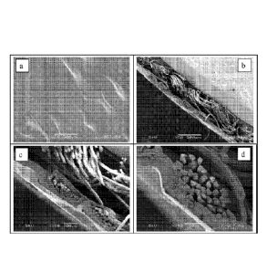
FIG. la is a scanning electron microscopy image (x50) of a top view of a
fabric substrate
laminated with a 2-mil polymeric film
FIG. lb is a scanning electron microscopy image (x50) of a cross section of a
fabric substrate
laminated with a 2-mil polymeric film
FIG. lc is a scanning electron microscopy image (x150) of a cross section of a
fabric
substrate laminated with a 2-mil polymeric film
CA 02774945 2012-03-21
WO 2011/037760
PCT/US2010/048336
FIG. Id is a scanning electron microscopy image (x350) of a cross section of a
more dense
fabric substrate laminated with a 2-mil polymeric film
FIG. 2a is a scanning electron microscopy image (x50) of a top view of a more
dense fabric
substrate laminated with a 1-mil polymeric film
FIG. 2b is a scanning electron microscopy image (x50) of a cross section of a
more dense
fabric substrate laminated with a 1-mil polymeric film
FIG. 2c is a scanning electron microscopy image (x150) of a cross section of a
more dense
fabric substrate laminated with a 1-mil polymeric film
FIG. 2d is a scanning electron microscopy image (x350) of a cross section of a
more dense
fabric substrate laminated with a 1-mil polymeric film
FIG. 3a is a scanning electron microscopy image (x50) of a cross section of a
non-woven
substrate laminated with a 2-mil polymeric film
FIG. 3b is a scanning electron microscopy image (x150) of a cross section of a
non-woven
substrate laminated with a 2-mil polymeric film
FIG. 3c is a scanning electron microscopy image (x350) of a cross section of a
non-woven
substrate laminated with a 2-mil polymeric film
FIG. 4 is a graph showing the correlation of the hemostasis of the inventive
device as a
function of substrate' thicknesses and their corresponding density. The legend
for the
numbers on the graph is displayed in Table 4.
DETAILED DESCRIPTION OF THE INVENTION
Applicants discovered a certain hemostatic composite structure described more
fully below
that utilizes a fabric or non-woven material as a substrate, where the fabric
or non-woven
substrate comprises fibers prepared from a biocompatible and biodegradable
polymer(s) and
6
CA 02774945 2012-03-21
WO 2011/037760 PCT/US2010/048336
a continuous, non-porous polymer film layer. The substrate surface opposite
the polymer film
layer is applied to the wound surface. The composite structure described below
possesses
properties suitable for use as a hemostat, e.g. strength, and flexibility. The
hemostatic
composite structure of the present invention provides and maintains effective
hemostasis
when applied to a wound requiring hemostasis. Effective hemostasis, as used
herein, is the
ability to control and/or abate capillary, venous, or arteriole bleeding
within an effective time,
as recognized by those skilled in the art of hemostasis.
The composite structure described below provides improved hemostasis, meaning
decreasing
the time to achieve hemostasis, which has great clinical significance. It will
be shown that the
present invention provides much improved hemostasis rates over conventional
hemostats.
The composite structure described below exhibits better handling properties
for surgical
applications and settings. Many fabric or non-woven based hemostats do not
have ideal
handling characteristics as they wrinkle and fold during surgical procedures
especially in the
presence of blood or other fluids. The substrate/film composites of the
present invention
minimize such behavior. Additionally, the presence of film improves the
mechanical strength
and pliability of the fabric or non-woven substrate based materials, enhancing
their suitability
for use in laparoscopic procedures. In laparoscopic procedures, the composite
is expected to
be pushed through the trocar and sprung open into the body cavity more easily
than either the
substrate or film components individually.
The composite structure described below exhibit greater propensity and/or
ability to stay in
place during surgical procedures relative to existing hemostatic devices. For
example, some
fabric based products when used in multiple layers, or those in non-woven form
may
disintegrate or their parts may migrate during the application process. A
substrate/film
composite architecture of the present invention helps to maintain the physical
integrity of the
hemostatic materials, so it cannot fall prematurely to pieces, curve, or
migrate during the
procedure. Another advantage of the composite structure is that the device can
be sutured in
place.
The composite structure device of the present invention also provides for the
potential to use
the film component for additional surgical functionality, such as to provide
tissue support, to
help in wound healing and/or to act as delivery carrier for bioactive agents.
7
As noted above, hemostatic composite structure of the present invention
comprise a fabric or
non-woven substrate on the first, wound contacting surface of the hemostatic
composite
structure, laminated with a continuous, non-porous polymer-based film on
second surface of
the hemostatic composite structure. Substrate as used herein refers to the
component of the
hemostatic composite structure which is in direct contact to the wound
surface. The substrates
utilized in the present invention may be fabric/woven or nonwoven that
provides form and
shape and mechanical reinforcement necessary for use in hemostatic composite
structures. In
addition, the substrates are made of materials having hemostatic properties
and be
bioabsorbable.
Bioabsorbable, "Biodegradable" and "bioabsorbable- as used herein refer to a
material that
is broken down spontaneously and/or by the mammalian body into components,
which are
consumed or eliminated in such a manner as not to interfere significantly with
wound healing
and/or tissue regeneration, and without causing any significant metabolic
disturbance.
Polymers useful in preparing the fabric or non-woven substrates in hemostatic
composite
structure of the present invention include, without limitation, collagen,
calcium alginate,
chitin, polyester, polypropylene, polysaccharides, polyacrylic acids,
polymethacrylic acids,
polyamines, polyimines, polyamides, polyesters, polyethers, polynucleotides,
polynucleic
acids, polypeptides, proteins, poly (alkylene oxide), polyalkylenes,
polythioesters,
polythioethers, polyvinyls, polymers comprising lipids, and mixtures thereof.
Preferred fibers
comprise oxidized regenerated polysaccharides, in particular oxidized
regenerated cellulose.
Preferably, oxidized polysaccharides are used to prepare wound dressings of
the present
invention. More preferably, oxidized cellulose is used to prepare fabrics used
in wound
dressings of the present invention. The cellulose either may be carboxylic-
oxidized cellulose,
or may be aldehyde-oxidized cellulose, each as defined and described herein.
Even more
preferably, oxidized regenerated cellulose is used to prepare fabric
substrates used in wound
dressings of the present invention. Regenerated cellulose is preferred due to
its higher degree
of uniformity versus cellulose that has not been regenerated. Regenerated
cellulose and a
detailed description of how to make regenerated oxidized cellulose is set
forth in U.S. Patent
No. 3,364,200 and U.S. Patent No. 5,180,398. As such, teachings concerning
regenerated
8
CA 2774945 2017-08-11
oxidized cellulose and methods of making same are well within the knowledge of
one skilled
in the art of hemostatic wound dressings.
Substrates, or fabrics utilized in conventional hemostatic wound dressings,
such as Surgicel
absorbable hemostat; Surgicel Nu-Knit absorbable hemostat; and Surgicel
Fibrillar
absorbable hemostat; all available from Johnson & Johnson Wound Management
Worldwide,
a division of Ethicon, Inc., Somerville, N.J., a Johnson & Johnson Company, as
well as
Oxycei absorbable cellulose surgical dressing from Becton Dickinson and
Company,
Morris Plains, N.J., all may be used in preparing wound dressings according to
the present
invention. In certain embodiments, wound dressings of the present invention
are effective in
providing and maintaining hcmostasis in cases of severe bleeding. As used
herein, severe
bleeding is meant to include those cases of bleeding where a relatively high
volume of blood
is lost at a relatively high rate. Examples of severe bleeding include,
without limitation,
bleeding due to arterial puncture, liver resection, blunt liver trauma, blunt
spleen trauma,
aortic aneurysm, bleeding from patients with over-anticoagulation, or bleeding
from patients
with coagulopathies, such as hemophilia. Such wound dressings allow a patient
to ambulate
quicker than the current standard of care following, e.g. a diagnostic or
interventional
endovascular procedure.
The fabric substrates utilized in the present invention may be woven or
nonwoven, provided
that the fabric possesses the physical properties necessary for use in
hemostatic wound
dressings. A preferred woven fabric has a dense, knitted structure that
provides form and
shape for the hemostatic wound dressings. Such fabrics are described in U.S.
Patent No.
4,626,253, U.S. Patent No. 5,002,551, and U.S. Patent No. 5,007,916.
The nonwoven substrates may be produced by melt-blown, electrospinning, needle
punched
methods and they can be preferably made from absorbable polymers. More
specifically,
absorbable nonwoven fabric is comprised of fibers that are not derived from
cellulosic
materials, such as comprising aliphatic polyester polymers, copolymers, or
blends thereof
The aliphatic polyesters are typically synthesized in a ring opening
polymerization of
monomers including, but not limited to, lactic acid, lactide (including L-, D-
, meso and D, L
mixtures), glycolic, acid, glycolide, c-caprolactone, p-dioxanone (1,4-dioxan-
2-one), and
trimethylene carbonate (I,3-dioxan-2-one). Examples of non-woven substrates
are described
9
CA 2774945 2017-08-11
in published U.S. patent application No. 2009/0104276 and published U.S.
patent application
No. 2006/0258995.
Other methods known for the production of nonwoven fabrics may be utilized and
include
such processes as air laying, wet forming and stitch bonding.
The thickness of the substrate ranges from about 0.05 to 2 mm, preferably from
0.25 to 0.75
mm. The thickness is measured according to ASTM method (D1777-64)
conventionally used
for the textile industry in general and non-woven in particular. The fabric
density of the
substrate ranges from about 0.05 to 0.6 g/cm3; preferably from about 0.15 to
0.5 g/em3. The
fabric density is defined as the ratio of the fabric's base weight to the
fabric's thickness. Base
weight is defined as the weight of the 1 cm by 1 cm square piece fabric.
Other fabric constructions which produce equivalent physical properties may,
of course, be
utilized in the manufacture of the improved fabric or non-woven substrate and
hemostatic
composite structure of the present invention, and such constructions will be
apparent to those
skilled in the art.
As noted above, hemostatic composite structure of the present invention
comprise a
continuous, non-porous polymer film laminated on the surfaces of the fabric or
non-woven
substrate of the second and the wound opposing surface of the hemostatic
composite
structure. Having a polymeric film on the second and wound opposing surface
provide
additional mechanical barriers to prevent the blood leaking from the wound
once hemostasis
is initially achieved. The preferred polymeric films according to the
invention are fully
amorphous or semi-crystalline absorbable polymers of relatively low melting
point
temperature (below 120 C, more preferably less than 110 C) allowing the use of
low
processing temperatures, which greatly help in keeping the substrate materials
free of
degradation. Also, polymer films of the current invention need to have
relatively low (around
room temperature 25 C or below) glass transition temperatures as measured by
differential
scanning colorimetry for the hemostatic composite to be soft, pliable and
conformable to the
tissue or body contour.
CA 2774945 2017-08-11
The polymers used to prepare the laminated film in wound dressings of the
present invention
are preferably biocompatible synthetic absorbable polymers. More preferably,
the polymers
of the current inventions are fully amorphous (06 crystallinity) or low
melting semi-
crystalline polymers to allow processing (lamination) conducted at relatively
low
temperatures for purposes as described above. This is important because ORC-
based
substrates can degrade during exposure at higher temperatures for instance,
100 C for the
time duration of lamination process. Even more preferably, the polymer films
need to have
relatively low glass transition temperatures (e.g. room temperature or lower)
to be soft,
flexible, elastic, to drape and conform well to the body and tissues. Even
more preferably the
polymer films needs to absorb/hydrolyze relatively quickly; for instance,
about two to four
weeks, which is slightly longer than the absorption rate of ORC-based
substrate, but still fast
to aid in patient comfort and to limit possible long-term infections. Finally,
in case polymer
films are laminated onto ORC-based substrate, polymer films of the current
invention needs
to exhibit minimal degradation upon gamma or e-beam irradiation procedures at
sufficient
levels, such as about 10-40 kGy, to sterilize the composite structure and
optionally the
associated packaging.
The thickness of the film can vary and does not appear to have a significant
effect on
hemostasis performance. Nonetheless, if the film is too thin, the improvement
in mechanical
strength of the composite structure relative to the substrate alone is
negligible. On the other
hand, if the film layer is too thick, the composite structure is too stiff and
difficult to handle.
Applicants found that a preferred polymer film thickness ranges from 0.5 to 2
mils (1 mil =
in/1000).
Preferred polymers used to laminate the substrate include, the polymers and
copolymers of
poly (ethylene diglycolate) (PEDG), poly (ethoxyethylene diglycolate) (PEEDG),
glycolide,
lactide, p-dioxanone, caprolactone, trimethylene carbonate and derivatives of
any of the
above. Examples of such absorbable polymers are taught in published US Patent
Application
No. 2009/0118241, published U.S. patent application No. 2009/0104276,
published U.S.
Patent No. 2008/0103284, and published U.S. patent application No.
2007/0149640 Al.
The first absorbable nonwoven fabric is attached to the second absorbable
woven or knitted
fabric, either directly or indirectly. For example, the polymer film may be
incorporated into
11
CA 2774945 2017-08-11
CA 02774945 2012-03-21
WO 2011/037760 PCT/US2010/048336
the absorbable woven or knitted fabric via thermal lamination (calendaring),
needle
punching, embossing or by chemical or thermal bonding. More preferably, the
hemostatic
composite device of the current invention may be made, for example, by
contacting an one
side of the substrate (ORC or nonwoven) with a film, and heating the substrate
and the film
so that a portion of the substrate is adhered to the film component.
More specifically, a hemostatic composite device of the current invention can
be prepared
utilizing a lamination system having a metal roller with a nominal diameter of
8 inches and a
heating capability of is up to170 C. The rotating speed of the metal roller
can vary from 1 to
feet per minute. The lamination system also included a soft face polyurethane
pressure
roller with a durometer of 40 and a pressure loading of up to 150 pounds per
linear foot. One
side of a film can be covered with a first silicone based release paper while
the other side of
the film can be placed in contact with the one side of a substrate. A second
release paper was
placed on the top side of the substrate to keep the components from sticking
to the rollers of
the lamination system. The first release paper/film/substrate/second release
paper structure
can be placed into the lamination system with the metal roller set to a
temperature of 50-
120 C and running at 1 to 2 feet per minute. Meanwhile, the pressure roller
can be set to
apply a load of 70 pounds per linear inch displaced across the face of the
pressure roller, with
the first release paper contacting the heated metal roller, which can forced
the small portion
of the film surface to migrate into the substrate. See, for instance, SEM
Images of various
hemostatic composites in Figures 1-3.
Generally, higher temperatures and/or slower roller speed allow more of the
film to penetrate
into the substrates, making the adherence much stronger. When an ORC substrate
is used, it
is important to keep the metal roller temperature as low as possible to avoid
degradation of
ORC component. Therefore, fully amorphous, or semi-crystalline film with low
melting point
and relatively low glass transition temperature as discussed above are
preferable to use for
this procedure.
In certain embodiments of the invention, the hemostatic composite structure
may further
include a hemostatic agent, or other biological or therapeutic compounds,
moieties or species,
including drugs and pharmaceutical agents as described in more detail herein
below. The
agents may be bound within the polymeric matrix, as well as to the fabric
surfaces and/or
within the fabric. The agents may be bound by chemical or physical means,
provided that
12
CA 02774945 2012-03-21
WO 2011/037760 PCT/US2010/048336
they are bound such that they do not migrate from the wound dressing upon
contact with
blood in the body. The hemostatic agent may be dispersed partially or
homogenously through
the fabric and/or the polymeric matrix. In some embodiments of the invention,
the hemostatic
agents, or other biological or therapeutic compounds, moieties or species,
e.g. drugs, and
pharmaceutical agents, may be "acid-sensitive", meaning that they may be
degraded or
denatured by, or otherwise detrimentally affected by acidic pH, such as is
provided by
conventional carboxylic-oxidized hemostatic wound dressings.
Hemostatic agents that may be used in hemostatic composite structure according
to the
present invention include, without limitation, procoagulant enzymes, proteins
and peptides,
can be naturally occurring, recombinant, or synthetic, and may be selected
from the group
consisting of prothrombin, thrombin, fibrinogen, fibrin, fibronectin,
heparinase, Factor X/Xa,
Factor VIINIIa, Factor IX/IXa, Factor XI/XIa, Factor XII/XIIa, tissue factor,
batroxobin,
ancrod, ecarin, von Willebrand Factor, collagen, elastin, albumin, gelatin,
platelet surface
glycoproteins, vasopressin and vasopressin analogs, epinephrine, selectin,
procoagulant
venom, plasminogen activator inhibitor, platelet activating agents, synthetic
peptides having
hemostatic activity, derivatives of the above and any combination thereof.
Preferred
hemostatic agents used in the present invention are thrombin, fibrinogen and
fibrin.
Such hemostatic composite structure of the present invention comprises
hemostatic agents,
including but not limited to thrombin, fibrinogen or fibrin, in an amount
effective to provide
rapid hemostasis and maintain effective hemostasis in cases of severe
bleeding. If the
concentration of the hemostatic agent in the wound dressing is too low, the
hemostatic agent
does not provide an effective proagulant activity to promote rapid clot
formation upon
contact with blood or blood plasma. The agents may be incorporated into either
the substrate
or film components.
The laminated hemostatic composite structure described herein may be used as
an adjunct to
primary wound closure devices, such as arterial closure devices, staples, and
sutures, to seal
potential leaks of gasses, liquids, or solids as well as to provide
hemostasis. For example, the
multilaycred dressing may be utilized to seal air from tissue or fluids from
organs and tissues,
including but not limited to, bile, lymph, cerebrospinal fluids,
gastrointestinal fluids,
interstitial fluids and urine. The laminated hemostasis device described
herein has additional
medical applications and may be used for a variety of clinical functions,
including but not
13
CA 02774945 2012-03-21
WO 2011/037760 PCT/US2010/048336
limited to tissue reinforcement and buttressing, i.e., for gastrointestinal or
vascular
anastomoses, approximation, i.e., to connect anastomoses that are difficult to
perform (i.e.
under tension), and tension releasing. The dressing may additionally promote
and possibly
enhance the natural tissue healing process in all the above events. This
dressing can be used
internally in many types of surgery, including, but not limited to,
cardiovascular, peripheral-
vascular, cardio-thoracic, gynecological, neuro-and general surgery. The
dressing may also
be used to attach medical devices (e.g. meshes, clips and films) to tissues,
tissue to tissue, or
medical device to medical device.
Hemostatic composite structure of the present invention is best exemplified in
the figures
prepared by scanning electron microscope. The samples were prepared by cutting
1-cm2
sections of the dressings by using a razor. Micrographs of both the first
surface and opposing
second surface, and cross-sections were prepared and mounted on carbon stubs
using carbon
paint. The samples were gold-sputtered and examined by scanning electron
microscopy
(SEM) under high vacuum at 4 Ky. The SEM images of different substrate/polymer
film
combinations are shown in Figures 1-3.
While the following examples demonstrate certain embodiments of the invention,
they are
not to be interpreted as limiting the scope of the invention, but rather as
contributing to a
complete description of the invention.
Example 1: (First stage of the polymer film starting material) Synthesis of
Hydroxy
Terminated Poly (ethylene diglycolate) (PEDG)
A twin-agitated reactor with intermeshing patterned blades equipped with a
condenser is
employed to prepare a polycondensation product of diglycolic acid and ethylene
glycol using
dibutyltin oxide as catalyst. After charging the reactor with 7.0 kg of
diglycolic acid, 9.7 kg
of ethylene glycol and 1.30 grams of dibutyltin oxide catalyst, the pressure
in reactor is lower
to 1 Ton or less and held overnight. The next day, the vacuum is released with
dry
nitrogen/argon. Vessel oil temperature was set to 170 C, condenser water was
set to 1-2
GPM, and the upper/lower condenser heats is set to 95 C/50 C. The agitator is
set at 30 RPM
in reverse rotation. When the temperature in the reactor reached 150 C, the
agitator speed is
increased to 75 RPM and switched to forward rotation. The reaction is carried
out at 170 C
for a couple hours until approximately all water is distilled and/or first
traces of ethylene
14
CA 02774945 2012-03-21
WO 2011/037760 PCT/US2010/048336
glycol appeared in the distillate. At this point the first nitrogen/argon
stage is completed;
pressure is lowered gradually to full vacuum in steps while the temperature of
the batch is
maintained at 175-180 C. Using Brookfield melt viscometer, a viscosity of the
hydroxy end-
capped polymer is checked periodically to ensure the end product of specific
molecular
weight. After sufficient reaction time spent under vacuum (68 hours, final
vacuum reading
150-200 mTorr) the reaction is stopped and the material sent for analysis. It
was a fully
amorphous, colorless viscous liquid with a glass transition temperature of
about 0-2 C.
Weight average molecular weight is 19,000 g/mol; the resin exhibited an
inherent viscosity
(IV) of 0.62 dL/g, as determined in HFIP at 25 C at a concentration of 0.1
g/dL. The resin is
kept in the reactor under nitrogen/argon until the next, copolymerization
stage.
Example 2: (Second stage of the polymer film starting material) The
Copolymerization of
an a,co-Dihydroxy Poly (ethylene diglycolate) Homopolymer with Glycolide,
PEDG/Gly
The hydroxy terminated poly (ethylene diglycolate) (PEDG) remained in the
reactor (7.7 kg)
was reacted with glycolide monomer (10.3 kg) in the second stage via ring-
opening
polymerization. The reactor is equipped with a melt tank reservoir allowing
glycolide
monomer to be added in a liquid state. Before charging glycolide, a vacuum of
less than 1
Torr is kept overnight to remove any residual moisture. The next day, the
resin is heated to
about 150 C, at which point the molten glycolide monomer is transferred from
the melt tank
with agitation. Agitator mixing is continued (20 RPM) and the batch
temperature raised to
150 C until full mixing is achieved. In situ, a real-time Fourier transform
near-infrared probe
is used to confirm complete mixing of components before the addition of the
catalyst,
Stannous Octoate (1.12 ml of toluene solution, glycolide to catalyst level
240,000:1).
Temperature is then increased to 210 C and the reaction was continued for
another two
hours. A half an hour before discharging, a vacuum is pulled slowly (step by
step) to remove
any residual monomer. The discharged copolymer is fully amorphous, with a
colorless to
slightly yellow tint, and a glass transition temperature of 25.5 C. Weight
average molecular
weight was 35,000 g/mol and an inherent viscosity of 1.09 dL/g, as determined
in HFIP at
25 C at a concentration of 0.1 g/dL, was recorded. Composition is confirmed by
NMR to be
42/58 by weight poly (ethylene diglycolate-co-glycolide). Melt index
measurements revealed
MI = 0.152 g/10min 150 C using load of 3700 grams.
CA 02774945 2012-03-21
WO 2011/037760 PCT/US2010/048336
The discharged copolymer resin is kept in the freezer until the grinding step.
After grinding,
the resin is placed in port-a-vacs (capacity 4-5 kg) and stored under vacuum
in the
refrigerator cabin (temperature set at 10 C). After two weeks under vacuum,
the resin is
ready for further processing (extrusion).
Example 3: Film Extrusion of PEDG/Gly 42/58 wt.% copolymer
Film extrusion of the copolymer described in Example 2 is performed on Davis-
Standard
Extruder (Model KN125, Pawcatuck, CT, USA) using a 6-inch die with die gap of
6 mils.
Extruder temperature ranged from 125 C in Barrel Zone 1 to 150 C in Barrel
Zone 3, with
the sheet die temperature set at 155 C. Extruder pressure (barrel) is
controlled between 2000
and 2500 psig. Screw rotation speed varied from 7.5 to 17.9 rpm. Upstream,
middle, and
downstream rolls are all kept at ambient conditions with Silicone based
release paper
employed to prevent the extruded, warm film of sticking to rolls.
Extruded films with the thicknesses of 1 and 2 mills are kept in-between
released paper and
stored under the vacuum.. Unless specified, there is one layer of substrate
used in the
hemostatic composite structure.
Example 4: Preparation of Hemostatic Composite Structures having ORC
Substrates and
PEDG/Gly 42/58 wt% copolymer composites
Films made from PEDG/Gly 42/58 wt.% copolymer resin having thickness of 1 and
2 mil are
laminated on a variety of ORC based substrates, available from Ethicon Inc.,
under the
tradename of Surgicel Classic , (Examples 4A) and (4A'; 2 layers), Surgicel
NuKnit ,
(Example 4B), Surgicel Fibrillar , (Example 4C) , as well as a nonwoven
construct made
from ORC (Example 4D) using J. J. Jenkins (Matthews, NC, USA) heating set of
Godets
with the nipping roll combination. Laminations are successfully done at
various Godet's
temperatures ranging from 50 to 90 C. Fully amorphous copolymer films allow
the use of
low processing temperatures, which greatly help in keeping the ORC materials
free of
degradation. The roll speed used is generally 1 FPM for 2-mil films and 2 FPM
for 1-mil
films. Produced composites exhibit excellent handling properties, and no
delamination of
films are observed in any of the prepared combinations. SEM images presented
in Figures 1-
3 show films embedded (melted) into the portions of fibers on the surface of
fabrics making
16
CA 02774945 2012-03-21
WO 2011/037760 PCT/US2010/048336
the very strong bond. The largest improvement in handling properties are
observed for
Example 4A' with 2-mil film ¨ no delamination of the second layer Or wrinkling
of the fabric
is observed; in the case of Example 4C, ¨ no disintegration, or breaking up of
individual parts
of fabric was noted since the film keeps them together effectively. Also, in
the case of wet
environment, the side laminated with film can be easily handled since the film
surface is not
sensitive to moisture/water presence. After lamination procedure, film/ORC
substrate
composites are placed in-between silicone release paper and stored in the
vacuum chamber
until further use.
Example 5: Preparation of Hemostatic Composite Structures having non-ORC
substrates and
PEDG/Gly 42/58 wt% copolymer composites
Various non-ORC substrates are laminated using PEDG/Gly 42/58 film as a top-
coat. These
non-woven substrates include combination substrate, poly (glycolide-co-
lactide) (PLGA,
g1yc01ide90/lactidel0 mol/mol) nonwoven Fabric needled-punched with ORC fabric
as
described in published U.S. patent application No. 2006/0258995, (Examples 5A
and 5A'),
poly (glycolide-co-lactide) (PLGA, g1yco1ide90/lactide10 molimol) nonwoven
Fabric,
(Example 5B) and melt blown non-woven 25/75 e-caprolactone/glycolide
copolymer, as
described in published U.S. patent application No. 2009/0104276 having two
different
thicknesses (Examples 5C and 5C'), and a Surgifoam, absorbable gelatin sponge
(Example
5D). The lamination conditions in all these cases are the same to those in
Example 4 as
described above. Good handling with no delamination is observed in all of the
non-ORC
composites.
Example 6: Preparation of Hemostatic Composite Structures having ORC
substrates and
PDS film Composites
Films made from undyed poly (p-dioxanone) PDS resins having thickness of 0.8
mil are
laminated on a variety of ORC based substrates, available from Ethicon Inc.,
under the
tradename of Surgicel Classic , (Examples 6A) and (6A'; 2 layers)õ and
Surgicel NuKnit ,
(Example 6B), Laminations are successfully done at roll temperature of 120 C.
This
processing temperature is higher than in the case of fully amorphous films
described in
previous examples (Examples 4 and 5) because PDS film is semi-crystalline
material with the
17
CA 02774945 2012-03-21
WO 2011/037760 PCT/US2010/048336
melting point of about 110 C. The roll speed used for lamination of 0.8-mil
undyed PDS film
is kept at 2 FPM. Produced composites exhibit good handling properties,
especially under dry
conditions. In the case of wet environment, the film side can be easily
handled since the film
surface is not sensitive to water presence. However, the film compliance in
the wet field is
not as good as in the case of PEDG/Gly 42/58 film. Due to its semi-crystalline
morphology,
the PDS film tends to curve slightly upon application. PDS film/ORC composites
are placed
in-between silicone release paper and stored in the vacuum chamber until
further use.
Example 7: Evaluation of Hemostatic Composite Structures having Film/ORC
Substrates
and Film/non-ORC Substrates using swine linear incision spleen model
Linear incision on a standard swine spleen model, 1.5 cm long and 3 mm deep is
used to
generate hemostasis data for various test articles prepared as described in
Examples 4-6. The
depth of each wound is kept constant by clamping the scalper blade in a pair
of needle
holders at the appropriate depth. The first wound at the distal end of the
spleen serves as a
negative control and was permitted to bleed for a minimum 10 minutes to
demonstrate the
bleeding potential of an untreated wound. The second wound is made
approximately 1 cm
proximal to the first incision. This and the 10-18 subsequent incisions (the
number depending
on the size of the pig) per each test animal are used as the test incisions.
After the incision is created, the test articles (approximately 1.5 cm x 2.5
cm) are applied
with slight pressure using gauze over the incision line and a stopwatch was
started. At the end
of tamponade time of 2 minutes, the pressure is released. The gauze is removed
and wound
inspected for any sign of active bleeding. The procedure is repeated following
approximately
30 seconds intervals until the bleeding (hemorrhage) completely stopped. The
time of the last
release of pressure is recorded as the time to achieve hemostasis. Each test
articles, in most
cases, are applied to total 3 or 4 respective wounds.
The hemostatic composite structures having film/ORC and film/non-ORC are
placed onto the
wound with the substrate contacting the wound and with the film side opposing
to the wound.
The time of achieving hemostasis is recorded along with general observation
noted on
handling characteristics and ability of test articles to stay in place after
the procedure is
completed. The summary of hemostasis results on test articles composed of film
laminated on
ORC is provided in Table 1 below.
18
CA 02774945 2012-03-21
WO 2011/037760 PCT/US2010/048336
The hemostasis results on test articles composed of film laminated on
combination substrate
(PLGA nonwoven Fabric needled-punched with ORC Fabric), 5A and 5A', and those
laminated on exclusively non-ORC substrates (5B, 5C, 5C' and 5D) are presented
in Tables 2
and 3, respectively.
Table 1. Hemostasis data on linear incision spleen model for different
Hemostatic
Composite Structures having various ORC substrates laminated with absorbable
top-coat films
with PEDG / Gly Copolymer Top Film with
PDS Top Fihn
Substrate Alone
2-mil Film 1-mil Film 0.8-mil Film
Hemostasis Hemostasis Hemostasis
ORC Hemostasis time Reduction Reduction Reduction
time time time
Substrate (min:sec) Time* Time* Time*
(min:sec) (min:sec) (min:sec)
>10:00 5:12
Example 4A (different studies 3:22 3:50 >61%
on spleen model) 2:45
7:30 2:00
Example 4A' 6:49 7:05 3:00 2:40 62%
7:03 2:50
4:37 2:00 2:00 2:00
4:45 2:00 4:30 2:00
Example 4B _____ 4:25 __ 2:15 49% 2:50 ___ 36% 3:20 25%
2:00
4:02 2:58 2:00
7:45
3:50 to 5:40 4:30
Example 4D (different studies 2:50 3:20 13-41%
on spleen model) 2:46
3:58 5:50
Example 4C 4:30 4:40 5:48 4:35 ¨ 0%
5:30 2:50
Control: 6:00
2-mil 5:00
5:15
PEDG/Gly
4:45
film alone
19
CA 02774945 2012-03-21
WO 2011/037760
PCT/US2010/048336
* The percent reduction in hemostasis time as compared to the Hemostat without
a top-
coat.
Table 2. Hemostasis data on linear incision spleen model for Combination
Substrate
without biologics and the patch with top-coat film combination.
Combination Substrate with PEDG / Gly Copolymer Top Film
Alone 2-mil Film 1-mil Film
Hemostasis
Hemostasis time Hemostasis time Reduction
Substrate time
Reduction Time
(min:sec) (mm: sec) Time
(mm: sec)
Example 5A 3:20 to 4:00 2:00
(lamination on (from different studies on 2:20 2:30
25-38%
ORC side) spleen model)
3:10
2:00
Example 3:20 to 4:00
2:00
5A'(lamination (from different
studies on 2:14 33-44%
on PLGA side) spleen model) 2:54
2:00
CA 02774945 2012-03-21
WO 2011/037760 PCT/US2010/048336
Table 3. Hemostasis data on linear incision spleen model for non-ORC
substrates with
those containing top-coat film addition.
with PEDG / Gly Copolymer Top Film
Substrate Alone
2-mil Film 1-mil Film
Hemostasis Hemostasis
Non-ORC Hemostasis time Reduction
time time Reduction Time
Substrates (min:sec) Time
(mm: see) (nun: see)
PLGA-based 5:20
2111111 thick (from different study
2:43 2:43 54%
nonwoven patch on the same spleen
(5B) model)
PLGA-based
2mm thick
nonwoven patch N/A 5:00 5:00 The same as the film
alone
Film side on the
wound (5B)
2:50 2:00
2:00 2:00
Example 5C 2:45 2:25 13%
3:00
3:15
3:05
2:00 3:00
Example 5C' 2:00 2:45 2:00 2:45 0%
3:00 2:00
9:15
Example 5D (from different study
2:00 2:00 17%
on the same spleen
model)
We have unexpectedly discovered that film/substrate composites with a single
and double
layer of substrates require significantly less time to achieve hemostasis than
in the cases when
the single or double layer substrates, or 2-mil PEDG/Gly 42/58 film are used
alone. As
indicated in Table 1, the thickness of the film appears not to affect the
hemostasis data as
both 1-mil and 2-mil thick film laminated on substrates produce significant
improvement.
Replacing the PEDG/Gly 42/58 film with a different absorbable polymer film,
such as Poly
(p-dioxanone), PDS produces the same decrease in the hemostasis time.
21
CA 02774945 2012-03-21
WO 2011/037760 PCT/US2010/048336
In addition to hemostasis improvement, hemostatic composites structures having
substrates
laminated with PEDG/Gly 42/58 film exhibit much better handling
characteristics and ability
to stay in place compared to the substrates or s the film when used alone.
On the other hand, PEDG/Gly 42/58 film laminated onto a much thicker ORC
substrate e.g.
Example 4C, show no significant reduction in hemostasis time when compared
with the
substrate alone, indicating that the thickness of ORC layer may play an
important role in the
hemostasis performance of the devices of the present invention.
The trend of significantly faster hemostasis is also observed for the film
laminated
ORC/PLGA Combination substrate presented in Table 2. Placing the film on
either side of
the substrate (ORC or PLGA non-woven) produced comparable results.
Finally, a series of non-ORC substrates including needle punched PLGA fiber
with a gradient
in fabric density (the lamination procedure was identical to those in Example
5), melt blown
nonwoven 8-Cap/Gly 25/75 copolymer having two different thicknesses, and
SURGIFOAM,
absorbable gelatin sponge are also examined with top-coat lamination (see
Table 3). Except
for the thicker and denser melt blown nonwoven 8-Cap/Gly 25/75 substrate, all
of them show
faster hemostasis than the corresponding substrates without top-coat film.
Example 8: Determination of base weight and thicknesses of ORC and non-ORC
substrates
In order to characterize and describe various substrates used to prepare
composites of the
current invention, we decide to measure their base weight expressed in grams
per square
centimeters and the fabrics' thicknesses.
For the base weight measurements, the samples are cut into 1 cm by 1 cm pieces
and
weighted by an analytical balance. The thickness is measured by ASTM method
("Standard
test for thickness of textile materials; Option 1", D1777) with the foot
(probe) diameter of 1.1
inch and the pressure of 0.6 psi. Dividing the Base Weight, BW (g/cm2) with
the Thickness,
T (cm) we obtain the density value for our substrates, which is another
important parameter
in characterizing the laminated film composites. If a substrate is too thick
regardless of
density, the top-coat film will not have any effect on the hemostasis time. In
addition, if a
22
CA 02774945 2012-03-21
WO 2011/037760
PCT/US2010/048336
substrate is relatively thick and dense the effect of top-coat film will be
also negligible. The
measurements of fabric base weight and thicknesses are shown in Table 4.
Table 4. Base weights and thicknesses of ORC and non-ORC substrates
Base Weight Density,
BW/T
Substrate ID Thickness (cm)
(g/cm2) (g/cm3)
1- Surgicel Original
one layer 0.010 0.025 0.39
(Example 4As)
2- Surgicel Original
two layers 0.020 0.049 0.40
(Example 4A's)
3 - Surgicel Nu-Knit
0.023 0.045 0.51
(Example 4Bs)
4 - Surgicel non-woven
0.011 0.061 0.16
(Example 4Ds)
- Surgicel Fibrillar
0.027 0.33 0.08
(Example 4Cs)
6 - ORC/PLGA patch
0.021 0.14 0.14
(Example 5As)
7 - PLGA based patch
0.025 0.14 0.18
(Example 5B)
8¨ MB nonwoven
c-Cap/Gly 25/75
0.015 0.47 0.32
thinner substrate
(Example 5C)
9¨ MB nonwoven
e-Cap/Gly 25/75
0.030 0.90 0.33
thicker substrate
(Example 5C')
23
CA 02774945 2012-03-21
WO 2011/037760 PCT/US2010/048336
The plot of fabric thickness versus fabric density is displayed in Figure 4.
The two
substrates that failed to produce positive hemostasis effect are marked 5 and
9 as described
in Table 4.
24