Note: Descriptions are shown in the official language in which they were submitted.
CA 02775907 2012-03-28
WO 2011/042888 PCT/1B2010/054557
1
Separator device, deposition device and system for handling of somatic
plant embryos
Background to the invention
General introduction to problem area
Somatic embryogenesis in plants is a process in which somatic embryos are
formed from an initial explant being a cell in a plant tissue. The somatic
embryos formed are genetically identical copies of the plant providing the
initial
explant. The process of somatic embryogenesis thereby offers a tool to obtain
large numbers of genotypically identical plants for multiplication of selected
genotypes of commercial interest, for conservation of endangered species or
for generating genetically uniform plant material for research purposes.
Physiological background to the procedures related to the problem
To produce plants from somatic embryos of conifers, a multi-step procedure is
applied to meet the physiological needs of the different stages of development
as described below and shown in Figure 1. Initiation of somatic embryogenesis
starts with induction of somatic embryos from an initial explant, typically an
immature zygotic embryo, on a solidified culture medium containing plant
growth regulator. Somatic embryos continue to form, typically on the same
composition culture medium, and a proliferating embryogenic culture form. At
the proliferating stage, several of the key features generally regarded as
beneficial for the process of somatic embryogenesis process, take place: (i)
the mass propagation of genotypically identical propagules through unlimited
multiplication of immature embryogenic tissue; (ii) cryogenic storage of
proliferating embryos substantiates an virtually eternal store of clones, i.e.
a
clone bank is established, (iii) transgenic modification of the immature
somatic
embryo allow for large scale propagation of genetically improved propagules.
At the next step in the procedure, the proliferating somatic embryo is
subjected
CA 02775907 2012-03-28
WO 2011/042888
PCT/1B2010/054557
2
to a growth medium that triggers embryo development to progress into the
maturation stage. Conversion from proliferation to maturation only occurs in a
fraction of the proliferating embryos in the culture. Low conversion rates are
encountered more frequently in genotypes from recalcitrant conifer species,
but are common in all conifer species as well as other plant species. The
manual labour needed to collect embryos increase with the decrease in
conversion rate, and thereby the cost and risk of contamination and other
inaccuracies. Low conversion rate from proliferation to maturation is a major
bottleneck for commercial large scale applications of somatic embryogenesis
procedures. For germination, mature somatic embryos are subjected to
different culture regimes to induce root- and shoot formation, in a number of
different steps; desiccation, sucrose treatment, red light induction, and blue
light stimulation. Thereafter, germinated embryos deemed appropriately
developed are transferred to a compost material and gradually transferred to
an environment ex vitro during which the sucrose content is reduced. The
different treatments during germination into a plant requires repeated manual
handling of individual germinants and plants adding a considerable cost to the
overall procedure.
Production of plants from somatic embryos
The prior art procedure for producing plants from somatic embryos requires
manual handling at several steps making the procedure time consuming,
expensive and inaccurate.
For conifer species, standard procedures used involve several steps when
manual handling is required. The general procedure is outlined in Fig. 1 (see
e.g. von Arnold S, Clapham D. Spruce embryogenesis. 2008. Methods Mol
Biol. 2008;427:31-47; Belmonte M F, Donald G, Reid D M, Yeung E C and
Stasolla C. 2005. Alterations of the glutathione redox state improve apical
meristem structure and somatic embryo quality in white spruce (Picea glauca).
J Exp Bot, Vol. 56, No. 419, pp. 2355-2364).
CA 02775907 2012-03-28
WO 2011/042888
PCT/1B2010/054557
3
There are four steps that rely on manual handling to obtain a small plant from
the mature somatic embryo as seen in Figure 1. The first manual interaction is
when [1] the mature embryo is isolated from immature embryos (120), and
placed horizontally in a plastic container under sterile conditions; the
second
[2] occur after 3- 7 days of resting (130), then mature embryo is transferred
to
a gelled culture medium for initiation of germination processes. The
germinated somatic embryo will under appropriate culture medium
composition and light conditions initiate roots (140). The third manual
transfer
[3] is when the germinant having a small root formed is transferred to an
upright position with the root partially immersed in liquid germination media
(150). The fourth [4] and final transfer is when the germinated embryos has a
tap root and small lateral roots, then it is transferred into a solid
substrate in a
pot for further plant formation (160).
Table 1. List of designations pertaining to Figure 1.
Item Designation
100 Mature embryo
101 Crown of a mature embryo
102 Foot of a mature embryo
103 Width of crown of a mature embryo
104 Length of a mature embryo
120 Maturation phase
130 Resting phase
140 Germination phase
150 In vitro plant formation phase
160 Ex vitro plant formation phase
CA 02775907 2012-03-28
WO 2011/042888 PCT/1B2010/054557
4
In the hitherto available method for producing plants from somatic embryos the
embryos are picked out manually from the immature embryogenic tissue. This
is time-consuming and ineffective. Prior art US06684564 and US7568309
teach automation of the manual transfers by replacing human eye, human arm
and tweezers with vision systems, conveyer belts and automated robotic arms
with suction tips to pick up the embryos from porous conveyer belts to deposit
in a destination much like a human does the same. However, this approach,
analogous to automating the wing motion of a bird by robotic wings for flight,
is
too complex and impractical for several reasons, as elaborated below. It
would therefore be desirable to provide a simple and practical way to make the
separation and deposition of the embryos faster and more efficient. The
mature somatic embryos produced are initially glued together with immature
embryogenic tissue in clusters. This makes the process more complex, as
embryos have to first be separated from the immature embryogenic tissue in
the cluster by breaking up the cluster. Prior art does not teach how to
breakup
the embryogenic clusters in an automated manner.
Breaking up the clusters by a Disperser
In the patent application PCT/US09/39981 a method of rapidly breaking up the
clusters is disclosed based on suspending the said clusters in liquid medium,
such as water, and forcing the clusters into at least one dispersion sequence
where the clusters of embryogenic mass are exposed to flow-dynamic forces
causing the breakup of the clusters and dispersion of individual embryos.
Segregation of embryos by a Separator
When embryogenic mass is dispersed in liquid according to the above-
presented method, a mixture of immature and mature embryos and immature
embryogenic tissue are suspended in the liquid medium. In many applications,
it is highly desirable to segregate and collect the embryos from the dispersed
embryogenic mass prior to processing further downstream. For example, if the
intention is to image and analyze the shape and condition of the embryos,
such as in Harrell et al., 1993 (Computers and Electronic in Agriculture, 9),
it is
CA 02775907 2012-03-28
WO 2011/042888 PCT/1B2010/054557
highly desirable to only have embryos suspended in liquid without any of the
immature embryogenic tissue in order to avoid obscuring the image. The tasks
of image recognition and analysis become more difficult and tedious if the
image contains more objects than just the embryo. Furthermore, the task of
5 image processing will be more time consuming with adverse impact on the
processing and the conversion rates. Thus there is a need for methods and
means for an effective separator to segregate and selectively remove and
guide only the embryos from the dispersed embryogenic mass into a separate
flow stream in a rapid and efficient manner. Having only individual embryos in
a flow stream would facilitate further processing of the embryos which may
include digital imaging of the individual embryos, image analysis and
characterization of the embryos including identification and control of embryo
orientation prior to deposition into an appropriate substrate for germination
and
plant production.
It is an object of the invention to provide an automated means for gently
segregating and separating dispersed somatic embryos from the immature
embryogenic tissue and guiding the collected embryos into a separate stream
of liquid in a rapid and efficient manner.
Embryo deposition means
The prior art methods to make plants from somatic embryos require intensive
manual handling, and are therefore expensive for plant production. Attempts
to automate the steps used in the manual operation have failed due to the
complex devices developed to automate the manual transfer and delivery of
embryos by means of moving parts such as conveyer belts and elaborate
robotic arms. For example, the prior art documents US7568309 and US
6,684,564 teach means of transferring the embryos into an artificial seed by
means of a porous conveyer belt and moving robotic arms equipped with
suction tips to pick up the embryo from the conveyer belt and to deposit the
embryo by means of a movable robotic arm attached to a rail into an artificial
seed. Such processes require many moving parts such as pulleys and motors
CA 02775907 2012-03-28
WO 2011/042888 PCT/1B2010/054557
6
to drive the conveyer belt, suction device(s) to vacuum excess liquid from the
embryo, and elaborate robotic arm assembly movably attached to a rail with
precision control to locate and pick the embryo from the conveyer belt. The
embryo being a small and delicate object, the robotic arm must have sensitive
and precise means of picking and carrying the embryos without damaging it.
As explained in US 6,684,564, the conveyer belt must stop moving when an
embryo is detected in order for the embryo to be imaged and picked up by
mechanical means of a robotic arm. A conveyer belt that has to move and stop
each time an embryo is detected creates an inherently inefficient process. In
general, the prior art teaches an approach requiring many moving parts
including the conveyer belts and the robotic arm assemblies making the
current state of the art to be impractical.
Thus, one object of the invention is to provide an advantageous method and
device for delivering an embryo to a desired embryo receiver, not requiring
any
pulleys, conveyer belts, robotic arms or such devices with moving parts.
System
It is another object of the invention to provide a system for processing plant
somatic embryos performing the separation process and at least one
additional process step of the entire process from a bioreactor to a planted
propagule, providing cost-effective means for handling and large-scale
production of plants from somatic embryogenesis.
Definitions
For purposes herein, the terms somatic embryo, embryo and plant somatic
embryo are used interchangeably. The terms refer to plant embryos derived
from somatic tissue of a plant, whether mature or immature.
The term embryogenic mass refers collectively to the plant material consisting
of immature embryogenic tissue, or mature embryos and immature
embryogenic tissue, present in the liquid or solid culture of somatic embryos.
CA 02775907 2012-03-28
WO 2011/042888 PCT/1B2010/054557
7
The term immature embryogenic tissue refers to all material other than
embryos that are in the embryogenic mass. The term tissue is being used here
in an unconventional manner consisting of largely undifferentiated cells and
should not to be confused with the normal reference to plant tissue with
specialized cells.
The terms embryogenic clusters, embryo clusters or clusters, are used
interchangeably. The term refers to assemblies of plant embryogenic mass
held together as a continuous solid material of finite size on solid medium or
in
liquid medium.
Norway spruce is a spruce species with the Latin name Picea abies native to
Europe.
The orthogonal directions in polar coordinates are given by axial (z), radial
(r)
and angular (or azimuthal) (0 ) directions. These directions correspond to the
central axis of a cylinder which is normal to the circular cross-sectional of
the
cylinder. The radial and angular directions point along the radius and normal
to
the radius on the cross-sectional surface respectively.
Axisymmetric flow refers to flow inside a tube where the cross-sectional
surface of the tube is always circular, and therefore, there is symmetry with
respect to the axis of the tube. In other words, nothing changes along the
angular (or azimuthal) direction.
Pressure gradient refers to the rate of variation of pressure with respect to
a
given axial direction.
Axial, radial, angular pressure gradient refers to variations in pressure (p)
in
the axial, radial, and angular directions shown respectively in mathematical
terms as partial derivatives
öz ar ae
The term Vortex (plural Vortices), as used here, is a term referred to a flow
that
possesses vorticity with a spinning or swirling motion around a central axis.
CA 02775907 2012-03-28
WO 2011/042888 PCT/1B2010/054557
8
Vortex flow can be categorized as free (irrotational) vortex or forced
(rotational)
vortex.
As used here, the term Vorticity in mathematical terms, is the curl of the
velocity vector field; therefore, it is a vector quantity with magnitude and
direction. In other terms, the value of vorticity at a point in the flow is
related to
rate of rotation of the fluid particles at a point in the flow field.
The term Free vortex, as used here, refers to a vortex flow where the fluid
particles retain their orientation while the flow rotates around an axis
(i.e.,
vorticity is zero) everywhere in the flow except near the central axis (where
in
mathematical terms, a singularity exists). Placing a hypothetical arrow moving
with the fluid particles, the arrow continues to point in the same direction
while
it rotates around the axis with the flow. An ideal irrotational sink vortex
could be
an example of a free vortex.
The term sink vortex as used here refers to the actual flow field produced in
.. the vicinity of a drainage region, said drainage could be by any means
including natural drainage directed downward by gravity or drainage in any
direction induced by pressure differential or other means.
The term Forced vortex, as used here, refers to a vortex flow where the fluid
moves in a solid-body rotation; meaning that there is no shear in the flow and
therefore the vorticity is constant everywhere and equal to 2ç, where c is the
rate of rotation. A hypothetical arrow pointing to the axis of rotation and
attached to the fluid particles in a Forced vortex continues to point to the
axis
of rotation while rotating around the axis.
Cotyledon a part of a plant embryo (100) that becomes the embryonic first
leaves of a seedling. The cotyledon is located at one end of a plant embryo
opposite to the end where roots will eventually form (foot (102)). When there
are several cotyledons, the may form a structure referred to as a crown (101).
Diameter of the crown refers to the diameter of a crown structure at its
widest
(103).
CA 02775907 2012-03-28
WO 2011/042888 PCT/1B2010/054557
9
Length of a plant embryo refers to the linear distance from the tip of the
root
end to the tip of the cotyledon end measured along the longitudinal axis of
the
embryo (104).
The terms tube, channel and flow channel are used interchangeably. The
terms are used without specific reference to any particular geometric shape of
the cross-section, unless specifically stated otherwise.
The terms fluid dynamics and hydrodynamics are used interchangeably and
refer to the same physical principles of flow of fluids.
Strain is the geometrical measure of deformation representing the relative
displacement between points in the material body; it is represented as the
ratio
or percentage of deformation in relation to the original dimension.
Normal strain defines the ratio or percentage amount of stretch or
compression along material line elements (ratio of the deformation to the
original length in the direction of the deformation).
Shear strain defines the ratio or percentage amount of deformation relative to
the original dimension associated with the sliding of material plane layers
over
each other.
Extensional strain is a normal strain where the element stretches.
Axially extensional strain is an element that stretches along the axial
direction.
Radially extensional strain is an element that stretches along the radial
direction.
Compressional strain is a normal strain where the element contracts.
Axially compressional strain refers to deformation of an element that
contracts
along the axial direction.
Radially compressional strain refers to deformation of an element that
contracts along the radial direction.
Rate of Stain is the change in strain with respect to time
CA 02775907 2012-03-28
WO 2011/042888 PCT/1B2010/054557
Hydraulic diameter, Ph, is a term used to characterize flow in noncircular
tubes
and channels. By definition, it is given by Ph, = 4 A / S where A is the cross-
sectional area of the noncircular tube or channel and S is the wetted
perimeter
of the cross-section.
5 Mean velocity in a channel is defined as the volumetric flow rate divided
by the
cross-sectional area of the channel.
Contraction ratio is defined as the ratio of the mean velocity at the outlet
to the
mean velocity at the inlet in a channel.
Mean stress is the stress that is averaged over a surface.
10 Mean rate of strain is the rate of strain averaged over a surface.
Dynamic viscosity of a fluid is the ratio of shear stress to rate of shear
strain, a
constant for a Newtonian fluid. Water, glycerin, silicone oil are examples of
Newtonian fluids.
Rate of strain profile is a profile showing the variation of the rate of
strain.
Unit of length in millimetre is abbreviated as "mm".
Unit of rate of strain as reciprocal second is abbreviated as "1/s".
In general, a flow with higher average rate of strain will impose higher
average
stress on a particle (or embryo) or on a cluster of particles (or cluster of
embryos) suspended in the fluid.
The terms boundary layer, viscous boundary layer, and thin boundary layer
are used interchangeably to mean the boundary layer formed by a forced
rotating flow inside the circular container with or without the presence of a
sink
boundary layer.
Brief description of the drawings
Figure 1 Illustrates a general process of producing somatic plant embryos.
CA 02775907 2012-03-28
WO 2011/042888 PCT/1B2010/054557
ii
Figure 2. illustrates the construction of certain details of a certain
embodiment
of a separator device of the invention.
Figure 3. Top and bottom view of a certain embodiments of a separator device
of the invention
Figure 4. Shows the liquid level in the inner separator container (5), before
operation (A) and during operation (B).
Figure 5. Illustrates different alternative embodiments of the outlets of the
feed
tube (14a, 14b, 14c).
Figure 6. Illustrates different alternative embodiments for the rotating means
(18a, 18b).
Figure 7. Illustrates an embodiment of a separator device of the invention
adapted for continuous use. A and B illustrate alternative means to regulate
the flow through the conduit (7).
Figure 8. Illustrates the secondary flow associated with the boundary layer
during operation.
Figure 9. Illustrates an embodiment of an automated system disclosing the
units of operation.
Figure 10. Illustrates a fully integrated automated system comprising units of
the invention as verified in the experimental part.
Figure 11. Illustration of the deposition for germination and the germination
unit.
Figure 12. Illustrates the deposition of oriented embryos.
Figure 13. Illustration of an axisymmetric disperser unit.
Figure 14. Illustration of a non-axisymmetric disperser unit.
Figure 15 a -f. Illustration of the detector-sorter-orienting unit.
CA 02775907 2012-03-28
WO 2011/042888
PCT/1B2010/054557
12
Table 2. List of designations pertaining to the Figures.
1 House Frame
2 Top support structure
3 Bottom support structure
4 Outer container
Separator container
6 Bottom wall of separator container
7 Conduit of separator container
8 Sensor
9 Feed conduit
Axial centre of separator container (5)
11 Hollow shaft
12 Hollow-shaft motor
13 Upstream container
14 Outlet of feed conduit (9)
Liquid barrier of outer container
16 Opening in the outer container
17 Outer container draining tube
18 Rotation means
19 Base block
Boundary layer
21 Fluid level in the separator container at start
22 Fluid level in the separator container during operation
Secondary outlet
Third outlet from container (5)
31 Separator container (5) draining tube
32 Extraction tube
33 Bottom end of Extraction tube
34 Hole in top support structure
Linear actuator
Valve, such as a pinch, gate, drop-through rotary or needle
36 valve, or a set of such valves (optional)
CA 02775907 2012-03-28
WO 2011/042888
PCT/1B2010/054557
13
37 Draining valve (optional)
38 Controlling unit (optional)
50 Inner diameter of a circular embodiment of an conduit (7)
Inner diameter of a circular embodiment of a separator
51 container (5)
52 Inner diameter of an embodiment of rotating means 18 a
53 Diameter of an embodiment of rotating means 18 b
54 Height of separator container (5)
200 Bioreactor
205 System for processing somatic plant embryos
210 Extraction of embryogenic clusters
215 Transfer of embryogenic clusters
220 Disperser
225 Transfer of dispersed embryogenic mass
230 Separator
233 Transfer of Immature embryogenic tissue
235 Transfer of separated embryos
240 Dilutor
245 Transfer of Diluted embryos
247 Sorter reservoir
249 Test section
250 Detector-Sorter-Orienting System
255 Transfer of oriented embryos
260 Deposition of oriented mature embryos in embryo receivers
263 Fluid and rejected embryos
265 Transfer of Accepted mature embryos
270 Germination
280 Nursery
290 Dilution fluid
300 Plate with embryo containers
305 Perforations in the plate
CA 02775907 2012-03-28
WO 2011/042888
PCT/1B2010/054557
14
310 Rejection reservoir
312 Embryo collector
313 Step motor/switch
315 Linear actuator of the x-/y-table
320 Substrate
325 Cavity in substrate
330 Open space between embryo containers
335 Connectors between embryo containers
340 Embryo container
345 Perforations in embryo container
350 Narrow hole
360 length of the free jet
365 Outlet
370 Straight section of tube before outlet 365
375 Flow direction of fluid and oriented embryos
380 Tube diameter
381 Encapsulating liquid
382 Encapsulating liquid delivery jet
383a Oriented embryo inside the delivery jet
383b Embryo inside an unstable delivery jet
385 Substantially stable delivery jet
386 Substantially unstable delivery jet
387 Vessel delivering the encapsulating liquid (tube)
388 Inner delivering tube
401 Segment of an axisymmetric channel
402 Segment of an axisymmetric channel
403 to 440 Dimensions according to Table 5
441 Connector tube
481 Segment of a non-axisymetric channel
481a Cross section of 481
482 Segment of a non-axisymetric channel
482a Cross section of 482
442 to 490 Dimensions according to Table 6
CA 02775907 2012-03-28
WO 2011/042888
PCT/1B2010/054557
501 Fluid inlet
502 Inlet tube
503 Fluid outlet
504 Outlet tube
505 Reservoir tube
506 Reservoir device
507 Intersection
508 Inlet valve (optional)
509 Outlet valve (optional)
510 Orientation detector
511 Reservoir tube detector (optional)
512 Outlet tube detector (optional)
518 Flow direction
519 Three-way intersection valve (optional)
521 Secondary destination plate (optional)
522 Secondary outlet tube (optional)
523 Secondary intersection (optional)
524 Liquid drainage (optional)
530 Inlet/outlet openings of the intersection valve
531 Intersection valve house
532 Intersection valve rotor
533 Intersection valve rotor flow channel
534 Diameter of inlet/outlet
540 x, y-movable table device (optional)
541 Device for x, y-moving the outlet tube (504) (optional)
550 Three way valve at secondary intersection (523) (optional)
560 Tube air inlet/outlet to reservoir device (optional)
561 Air inlet/outlet (optional)
562 Air filter (optional)
CA 02775907 2012-03-28
WO 2011/042888
PCT/1B2010/054557
16
Table 5. List of dimension designations pertaining to Fig 13.
Cross section Inner diameter Preferred Inner diameter
position [mm] for Norway Spruce
(403) 3.0 - 10.0 9.0 - 9.5
(404) 2.0 - 9.0 5.0 - 5.5
(405) 3.0 - 10.0 9.0 - 9.5
(406) 2.0 - 9.0 4.75 -
5.0
(407) 3.0 - 10.0 9.0 - 9.5
(408) 2.0 - 9.0 4.0 -
4.25
(409) 3.0 - 10.0 9.0 - 9.5
(410) 2.0 - 9.0 5.5 - 6.0
(411) 2.0 - 9.0 5.75 - 6.0
(412) 1,0 - 8.0 3.25 - 3.5
(413) 2.0 - 9.0 5.75 - 6.0
(414) 1,0 - 8.0 3.0 - 3.25
(415) 2.0 - 9.0 5.75 - 6.0
(416) 1,0 - 8.0 2.5 - 2.75
(417) 2.0 - 9.0 5.75 - 6.0
(418) 1,0 - 8.0 2.5 - 2.75
(419) 2.0 - 9.0 5.75 - 6.0
(420) 2.0 - 9.0 5.75 - 6.0
Length on details Length [mm]
(421) 30,0
(422) 10,0
(423) 30,0
(424) 5,0
(425) 30,0
(426) 5,0
(427) 20,0
(428) 10,0
(429) 20,0
CA 02775907 2012-03-28
WO 2011/042888
PCT/1B2010/054557
17
(430) 30,0
(431) 5,0
(432) 30,0
(433) 5,0
(434) 30,0
(435) 5,0
(436) 30,0
(437) 5,0
(438) 20,0
(439) 10,0
(440) 10,0
CA 02775907 2012-03-28
WO 2011/042888 PCT/1B2010/054557
18
Table 6. List of dimension designations pertaining to Fig 14
Exemplified inner cross-section dimensions
Shape of Inner Black arrow side
inner dimensions [mm] Width side
section [mm] (483) - (490) [mm]
Alt. 1 Alt. 2 Alt. 1 Alt. 2 Alt. 1 Alt. 2
(442) circular 9,5 9,5
(483)
(443) Rectangular (483) 5,0 4,75
9.5 9,5
(444) circular 9,5 9,5
(445) Rectangular (484) 9,5 (484) 9,5
5,0 4,25
(446) circular 9,5 9,5
(485)
(447) Rectangular (485) 5,0 3,75
9,5 9,5
(448) circular 9,5 9,5
(449) Rectangular (486) 9.5 (486) 9,5
5,0 3,5
(450) circular 9,5 9,5
(451) circular 6,0 6,0
(452) circular 6,0 6,0
(487)
(453) Rectangular (487) 3,5 3,25
6,0 6,0
(454) circular 6,0 6,0
(455) Rectangular (488) 6,0 (488) 6,0
3,5 3,25
(456) circular 6.0 6,0
(489)
(457) Rectangular (489) 3,5 2,75
6,0 6,0
(458) circular 6.0 6,0
(459) Rectangular (490) 6,0 (490) 6,0
3,5 2,75
(460) circular 6,0 6,0
CA 02775907 2012-03-28
WO 2011/042888 PCT/1B2010/054557
19
Summary of the invention
In the first aspect, the invention provides a device for separating fluid-
suspended embryos and immature embryogenic tissue from each other
comprising:
a) separator container (5), which during operation contains fluid
having a density lower than the density of the embryos to be
separated, said container being essentially cylindrical in shape,
having an essentially flat bottom wall (6), an essentially vertical axis
and comprising a fluid conduit (7) in communication with the inside
of the container, located at the axial region of the bottom wall (6);
b) means of inducing an axisymmetric rotating flow in the fluid
relative to the bottom wall (6), whereby during operation:
i) a viscous boundary layer (20) is created at the bottom
wall (6);
ii) a radial pressure gradient is created in the separator
container (5);
C) means of introducing the fluid-suspended embryos and
immature embryogenic tissue to be separated into the separator
container (5) at a location away from the bottom wall (6), whereby
during operation:
i) the embryos sediment faster than the immature
embryogenic tissue;
ii) the embryos enter the viscous boundary layer (20)
while the immature embryogenic tissue remains
substantially outside the viscous boundary layer (20);
iii) the embryos entering the viscous boundary layer
(20) are drawn into the axial region of the bottom wall
(6) and into the conduit (7); and
CA 02775907 2012-03-28
WO 2011/042888 PCT/1B2010/054557
d) means of collecting embryos from said conduit (7);
whereby the embryos collected are essentially separated from immature
embryogenic tissue.
Preferably, the device of the first aspect is a device wherein the conduit (7)
is
5 placed and dimensioned such that embryos drawn into the axial region of
the
bottom wall (6) during operation enter into the conduit (7) by gravitational
sedimentation.
Preferably, the device of the first aspect is a device wherein the means
collecting embryos comprise means of collecting the embryos from the conduit
10 (7) without substantially altering the volume of fluid in the container.
Preferably, the device of the first aspect is a device wherein the means of
removing the embryos from the conduit (7) without altering the
volume of fluid in the container comprise a valve or a set of valves
(36).
15 Preferably, the device of the first aspect is a device wherein
i) the device comprises means of draining fluid from said conduit
(7), whereby during operation a sink vortex is created at the axial
region of the bottom wall (6); and
ii) the means of collecting embryos comprise means of collecting
20 the embryos from the sink vortex in the fluid drained from the
conduit (7).
Preferably, the device of the first aspect is a device wherein the device is
adapted for batchwise operation and further comprises means of collecting the
embryos selectively during a time period after the sedimentation of the
embryos has occurred but before the immature embryogenic tissue has had
time to sediment.
Preferably, the device of the first aspect is a device wherein the device is
adapted for continuous operation and further comprises a separator container
(5) comprising a second outlet (25) at the top of the separator container (5)
CA 02775907 2012-03-28
WO 2011/042888 PCT/1B2010/054557
21
and means of feeding fluid into the separator container (5) at a rate
exceeding
the rate of fluid flow from the conduit (7), preferably by a factor in the
range of
2-100, more preferably by a factor in the range of about 5-20.
Preferably, the device of the first aspect is a device wherein the second
outlet
(25) is implemented by means of a separator container (5) which is open at the
top.
Preferably, the device of the first aspect is a device wherein the means of
collecting embryos from the sink vortex comprise means of collecting the fluid
exiting the conduit (7).
Preferably, the device of the first aspect is a device wherein the device
additionally comprises means of replacing the fluid in the separator container
(5).
Preferably, the device of the first aspect is a device wherein the device
comprises means of draining fluid from the axial region of bottom wall (6)
during operation, comprising a conduit extended from above or any other
direction to the proximity of the axial region of the bottom wall (6).
The device of the first aspect may preferably comprise a separator container
(5) having diameter in the range of 5-30 cm, more preferably 10-25 cm, 10-25
cm or 18-22 cm, most preferably about 20 cm.
The device of the first aspect may preferably comprise a fluid conduit (7)
having an area of 0.01%-10%, more preferably 0.01-1 %, even more
preferably 0.1-0.15%, and most preferably about 0.125% of the area of the
bottom wall (6).
The device of the first aspect may preferably comprise a means of inducing an
axisymmetric rotating flow resulting in a rotational speed in the range of 5-
1200
rpm, more preferably 30 to 360 rpm in the fluid.
The device of the first aspect may preferably comprise a means of inducing an
axisymmetric rotating flow comprising a rotating disk- or cylinder-shaped
object.
CA 02775907 2012-03-28
WO 2011/042888
PCT/1B2010/054557
22
The device of the first aspect may preferably comprise a means of introducing
embryos and immature embryogenic tissue located at an axial location near
the surface of the fluid present during operation.
The device of the first aspect may preferably comprise a means of maintaining
a static fluid height being 0.1-10 times, more preferably 0.8-2 times the
diameter of the separator container (5).
In a second aspect of the invention, a method of separating fluid-suspended
embryos from immature embryogenic tissue is provided, comprising the steps
of:
a) providing a suitable separator container (5), said container
containing fluid having a density lower than of the embryos to be
separated, being essentially cylindrical in shape, having an
essentially flat bottom wall (6) and an essentially vertical axis;
b) inducing an axisymmetric rotating flow in the fluid relative to the
bottom wall (6), thus:
i) creating a viscous boundary layer (20) at the bottom
wall (6); and
ii) creating a radial pressure gradient in the separator
container (5);
c) introducing the fluid-suspended embryos and immature
embryogenic tissue to be separated into the fluid present in the
separator container (5) at a location away from the bottom wall (6),
thus:
i) sedimenting the embryos faster than the immature
embryogenic tissue;
ii) allowing the embryos to enter the viscous boundary
layer (20) while not allowing the immature embryogenic
tissue to enter the viscous boundary layer (20);
CA 02775907 2012-03-28
WO 2011/042888 PCT/1B2010/054557
23
iii) drawing the embryos entering the viscous boundary
layer (20) into the axial region of the bottom wall (6);
and
d) collecting embryos from said axial region of the bottom wall (6),
whereby the embryos collected are essentially separated from immature
embryogenic tissue.
Preferably, the device provided in step a) above further comprises a conduit
(7) in communication with the fluid in the container at the axial region of
the
bottom wall (6) during operation and the method further comprises the steps of
creating a sink vortex at the axial region of the bottom wall (6) by draining
fluid
from said conduit (7); and collecting embryos from said axial region of the
bottom wall (6) in the fluid drained from the conduit (7).
Preferably, the device provided in step a) above further comprises a suitable
separator container (5) further comprising a conduit (7) in communication with
the fluid in the container at the axial region of the bottom wall (6) during
operation, wherein the conduit (7) is placed and dimensioned such that
embryos drawn into the axial region of the bottom wall (6) during operation
enter into the conduit (7) by gravitational settlement; and the method further
comprises collecting embryos from said conduit (7). More preferably, the
method further comprises the step of modulating the sedimentation velocity of
the embryos in the conduit (7) by means of inducing fluid flow through the
conduit (7) into the separator container (5).
The method of the second aspect may preferably be adapted for batchwise
operation and further comprises selectively collecting the embryos during a
time period after the sedimentation of the embryos has occurred but before the
immature embryogenic tissue has had time to sediment. The method of the
second aspect adapted for batchwise operation may preferably additionally
comprise the step of replacing the fluid in the separator container (5) after
processing a batch of embryos with fresh fluid.
CA 02775907 2012-03-28
WO 2011/042888 PCT/1B2010/054557
24
The method of the second aspect may preferably be adapted for continuous
operation such that it comprises feeding fluid into the separator container
(5) at
a rate exceeding the rate of fluid flow from the conduit (7), preferably by a
factor in the range of 1.1-1000, more preferably 2-100, even more preferably
.. 2-50, 2-30, 3-20 or 5-15, most preferably about 10.
The method of the second aspect may preferably comprise that the collection
of embryos is performed by allowing the embryos to enter the fluid exiting the
conduit (7), and collecting the fluid containing embryos.
In a third aspect, a method for depositing a fluid-suspended plant somatic
embryo in an embryo receiver while maintaining the orientation of the embryo
is provided, comprising the steps of:
i) Providing a suitable embryo receiver (340) with means of
draining fluid (345), (350) from the receiver;
ii) Providing a flow channel dimensioned such that the embryos
may travel with the fluid flowing though the channel but are
restricted to travelling either in a crown-first or crown-last
orientation by dimensional constraints, said flow channel having an
outlet (365) wherein said flow channel comprises a flow channel
section immediately upstream of the outlet (365) having a straight
section (370) with length at least equal to the largest cross-
sectional inside dimension of the flow channel (380);
iii) Placing an embryo (preferably an embryo having the desired
orientation) in the flow channel; and
iv) Forming a free jet (385 and 386) of fluid emanating from the
outlet (365), aligning said free jet with the embryo receiver (340)
and depositing the embryo from the flow channel into the receiver
by using said free jet as a carrier means.
The method of the third aspect preferably further comprises the steps of:
i) Determining the orientation of the embryo in the flow channel;
CA 02775907 2012-03-28
WO 2011/042888 PCT/1B2010/054557
ii) In case the orientation does not match the desired orientation,
directing the embryo away from the embryo receiver (340); and
iii) In case the orientation does match the desired orientation,
directing the embryo into the embryo receiver (340).
5 In a fourth aspect of the invention, a device for depositing fluid-
suspended
plant somatic embryos while maintaining the orientation of the embryo is
provided comprising:
i) a flow channel dimensioned such that the embryos may travel
with fluid flowing though the channel but are restricted to travelling
10 either in a crown-first or crown-last orientation by dimensional
constraints, said flow channel emanating to an outlet (365);
ii) an embryo receiver (340); and
iii) means of forming a free jet of fluid emanating from the outlet
(365), wherein the free jet is during operation aligned with an
15 embryo receiver (340) thus allowing embryos suspended in the
fluid to be deposited in the embryo receiver;
wherein the means of forming a free jet comprise a flow channel
section immediately upstream of the outlet (365) having a straight
section (370) with length at least equal to the largest cross-
20 sectional inside dimension of the flow channel (380).
The device according to the fourth aspect may preferably be designed such
that the length of the straight section is at least 10 times the largest cross-
sectional inside dimension of the flow channel (380).
The device according to the fourth aspect may preferably be designed such
25 that the outlet tip (365) is positioned during operation one to three
flow channel
diameters from the embryo receiver (340).
The device according to the fourth aspect may preferably be designed such
that the embryo receiver (340) has an opening for depositing the embryo
CA 2775907 2017-05-10
81535961
26
having a smallest dimension of at least 10 % larger than the largest cross-
sectional diameter of the embryo (103) to be deposited.
The device according to the fourth aspect may preferably further comprise
means stabilising the jet delivering the embryos comprising means of
encapsulating (387) the jet delivering the embryos in another fluid jet with a
larger diameter.
In a fifth aspect of the invention, a system for processing plant embryos
suspended in a fluid is provided, comprising a separator device (230)
according to the first aspect of the invention, and at least one of the
following:
a) a disperser unit (220) to disperse the embryos and the embryogenic
tissue suspended in a fluid, located upstream of the separator device,
and optionally a bioreactor (200), as embryo source, located upstream
of the disperser unit (220);
b) orientation and sorting unit (250) for orienting and sorting the embryos
suspended in a fluid, located downstream of the separator device (230);
and optionally a deposition device (260), preferably located downstream
of the orientation and sorting unit (250).
In a sixth aspect of the invention, a system comprising a deposition device
(260) according the fourth aspect of the invention, and one or more of the
following:
a) a disperser unit (220) to disperse the embryos and the embryogenic
tissue suspended in a fluid, and optionally a bioreactor (200), as embryo
source, located upstream of the disperser unit (220);
b) a separator device (230) according to the first aspect of the invention,
located downstream of the disperser unit (220);
c) orientation and sorting unit (250) for orienting and sorting the embryos
suspended in a fluid, located downstream of the separator device (230)
and upstream of the deposition device(260).
81535961
26a
In another aspect of the present invention, there is provided a method of
separating
fluid-suspended plant embryos from immature embryogenic tissue comprising the
steps of:
a) providing a suitable separator container, said container containing fluid
having a density lower than the embryos to be separated, being
cylindrical in shape, having a flat bottom wall and a vertical axis, and
further comprising a conduit in communication with the fluid in the
container at the axial region of the bottom wall during operation and
means for inducing an axisymmetric rotating flow being a rotating object
positioned inside the container;
b) creating a sink vortex at the axial region of the bottom wall by draining
fluid from said conduit; and
inducing an axisymmetric rotating flow in the fluid relative to the bottom
wall by the rotation means, thus:
i) creating a viscous boundary layer at the bottom wall; and
ii) creating a radial pressure gradient in the separator
container;
C) introducing the fluid-suspended embryos and immature embryogenic
tissue to be separated into the fluid present in the separator container at
a location away from the bottom wall, thus:
i) sedimenting the embryos faster than the immature
embryogenic tissue;
ii) allowing the embryos to enter the viscous boundary layer
while not allowing the immature embryogenic tissue to enter
the viscous boundary layer;
iii) drawing the embryos entering the viscous boundary layer
into the axial region of the bottom wall; and
CA 2775907 2018-09-24
1
,
,
81535961
26b
d) collecting embryos from said axial region of the bottom wall in the fluid
drained from the conduit,
whereby the embryos collected are separated from immature embryogenic tissue.
In another aspect of the present invention, there is provided a device for
separating
fluid-suspended plant embryos and immature embryogenic tissue from each other
comprising:
a) separator container, which during operation contains fluid having a
density lower than the density of the embryos to be separated, said
container being cylindrical in shape, having a flat bottom wall, a vertical
axis and comprising a fluid conduit in communication with the inside of
the container, located at the axial region of the bottom wall;
b) means of inducing an axisymmetric rotating flow in the fluid relative to
the bottom wall being a rotating object positioned inside the container,
whereby during operation:
i) a viscous boundary layer is created at the bottom wall;
ii) a radial pressure gradient is created in the separator
container;
c) means of introducing the fluid-suspended embryos and immature
embryogenic tissue to be separated into the fluid present in the separator
container at a location away from the bottom wall, whereby during
operation:
i) the embryos sediment faster than the immature
embryogenic tissue;
ii) the embryos enter the viscous boundary layer while the
immature embryogenic tissue remains outside the viscous
boundary layer;
1
CA 2775907 2018-09-24
81535961
26c
iii) the embryos entering the viscous boundary layer are
drawn into the axial region of the bottom wall and into the
conduit; and
d) means of collecting embryos from said conduit;
whereby the embryos collected are separated from immature embryogenic tissue.
Detailed Description of the invention
CA 2775907 2018-09-24
CA 02775907 2012-03-28
WO 2011/042888 PCT/1B2010/054557
27
The somatic embryos produced on solid or liquid medium in petri dish or
biorectors are initially glued together by immature embryogenic tissue into
embryogenic clusters or lumps normally up to 50 mm or sometimes larger
diameter. Prior art teaches methods of picking individual embryos either
manually by tweezers or automatically by conveyer belts and robotic arms and
placing the embryo in an artificial seed. Prior art does not teach how to
rapidly
and efficiently breakup the embryogenic clusters and separating the mature
embryos and placing the mature and viable embryos each in the right
orientation in an individual container, which could be an artificial seed or
otherwise, in a matter of seconds. To provide efficient means for large-scale
production of plants from somatic plant embryos, an automated means for
rapidly and inexpensively separating the mature embryos from the said
embryogenic clusters and rapidly depositing the mature embryos in the correct
orientation into an appropriate substrate for germination is required. To do
so
requires four major operational steps, as disclosed herein.
Firstly, a gentle dispersion of the clusters of somatic embryos into
individual
embryos detached from the immature embryogenic tissue, which is
advantageously performed while suspended in a liquid medium.
Secondly, the process involves segregating and separating the embryos from
the immature embryogenic tissue now dispersed but still mixed together in the
liquid medium. The second step is useful, for example, in order to provide an
optically clear access to the embryos suspended in a transparent liquid
medium without the presence of the embryogenic tissue. The need for such
optical access is to establish the level of maturity and suitability of the
embryos
for germination and plant production.
Thirdly, the process involves identification of the orientation of the mature
embryos prior to deposition and correction of an undesired orientation, which
is done while the embryos are still suspended in liquid medium.
Fourthly, the process involves deposition of the mature embryos with the right
orientation into an appropriate substrate for germination and plant formation.
81535961
28
Effective combination of the above four steps in a fluid dynamics-based
automated system capable of rapidly and efficiently transporting the embryos
through each step, as disclosed herein, provides a means for efficient large-
scale production of plants from somatic embryos.
An embodiment of the fluid dynamics-based automated system for rapid and
efficient dispersion, separation, sorting and orientation, and deposition of
plant
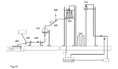
embryos in a substrate for germination is disclosed in Figure 10. The
automated system involves several innovative steps, as disclosed, in the order
of process outlined in the Figure 10, and outlined herein:
1.. The disperser unit (220) by reference to patent application
PCT/US09/39981, and also disclosed herein,
2. The separator unit (230), as disclosed herein,
3. The detector and orienting unit (250) by reference to patent
application PCT/US09/39982, and also disclosed herein
4. The deposition device for germination (260), as disclosed herein.
1. Disperser unit
Detailed description of a disperser unit for means to disperse the embryogenic
cluster in liquid medium is disclosed in patent application PCT/US09/39981.
Disperser unit of the system
PCT/US09/39981 relates to methods and devices for gently dispersing
clusters of somatic plant embryos into individual embryos and immature
embryogenic tissue useful in the system of the invention.
A method of dispersion of clusters of plant embryos suspended in a liquid
medium into individual plant embryos is disclosed in PCT/US09/39981, said
method including at least one dispersion sequence, which comprises the
following steps:
CA 2775907 2018-09-24
CA 02775907 2012-03-28
WO 2011/042888 PCT/1B2010/054557
29
i) subjecting the clusters of embryos to fluid dynamics forces
causing axially extensional strain and radially compressional strain;
ii) subjecting the clusters of embryos to fluid dynamics forces
causing axially compressional strain and radially extensional strain
from fluid dynamics forces;
repeating said steps in sequence until the individual embryos are
separated from each other.
Preferably, the strength of the extensional and compressional strains
increases with each repeated sequence.
A disperser for separating individual embryos contained in clusters of embryos
is also disclosed in PCT/US09/39981, comprising a flow channel including at
least one constriction, such that clusters of embryos flowing through the flow
channel are first subjected to axially extensional strain and radially
compressional strain, and then to axially compressional strain and radially
extensional strain from fluid dynamics forces.
Preferably, the flow channel comprises at least two constrictions, each
constriction having an inner diameter, which is equal to or smaller than the
inner diameter of the constriction immediately up-stream of thereof.
Preferably, the flow channel includes an intermediate portion having a
constant
cross-section, between each constriction.
Preferably, each intermediate portion has an inner diameter, which is equal to
or smaller than the inner diameter of the intermediate portion immediately up-
stream of thereof.
Preferably, each intermediate portion may have a length at least equal to the
clusters of embryos to be dispersed. Preferably, the length of each
intermediate portion is in the interval from 2.5 mm to 60 mm, more preferably
from about 5 mm to about 30 mm. The number of constrictions may be 3-100,
preferably 5-20, most preferably about 10. Preferably, the constrictions have
a
CA 02775907 2012-03-28
WO 2011/042888 PCT/1B2010/054557
cross-sectional area in the interval from 0.75 to 1300 mm2, more preferably in
the interval from 3 to 32 mm2.
The flow channel may have axisymmetric cross-section. The flow channel may
have an essentially circular or oval cross-section.
5 At least part of the flow channel may have a non-axisymmetric cross-
section
such as a rectangular cross-section. The cross-section of each non-
axisymmetric constriction, having a maximal dimension, may preferably be
oriented such that the maximal dimension of each constriction is rotated,
preferably at least 30 , more preferably about 90 in relation to maximal
10 dimension of the next non-axisymmetric constriction in sequence. The
cross-
section of each constriction may represent a rectangle, having a first and a
second side, wherein the first side is longer than the second side, and the
constrictions are oriented such that first side of each constriction is
perpendicular to the first side of the next constriction in sequence having a
15 rectangular cross-section.
The advantages of the method and the device of dispersion disclosed in
P0T/US09/39981 include:
(1) Not requiring moving parts, and therefore being robust
(2) Being naturally applicable to a continuous flow system thereby not
20 requiring operation in batch mode
(3) Being gentle to the embryos
(4) Being fast; the dispersion using the device requires only a few seconds to
disperse hundreds of embryos
(5) The device being compact and completely enclosed allows easy
25 .. sterilization.
CA 02775907 2012-03-28
WO 2011/042888 PCT/1B2010/054557
31
2. Separator unit
Device for segregating and separating somatic embryos from
embryogenic tissue
Once an embryogenic cluster is dispersed in liquid, it creates a mixture of
mature embryos, immature embryos and immature embryogenic tissue
suspended in liquid. The somatic embryos have to be segregated and
separated from the immature embryogenic tissue for further processing and
planting. In an automated system it is highly desirable to perform this
separation process rapidly, efficiently and without causing any interruption
to
the continuous operation of the system. Such a separator device of the
invention is disclosed herein.
The separator device and method of the invention takes advantage of the
difference in the drag coefficient of embryos as opposed to the immature
embryogenic tissue (hereinafter "tissue" or "embryogenic tissue") in the
suspension, to provide a device and method of separating somatic embryos
from embryogenic tissue.
General construction and operation of the device
To facilitate the theoretical discussion that will follow a brief general
description
of a device of the invention is presented below.
The separator device of the invention has a preferably essentially cylindrical
separator container (5) containing fluid medium during operation, and means
of inducing an axisymmetric rotating flow in the fluid within the separator
container (5). There is a bottom wall (6) which is preferably essentially
flat. The
fluid medium has a density that is lower than that of the embryos and tissue
to
be separated. The fluid medium may preferably be water.
The fluid medium inside the separator container (5) is induced to rotate (in
relation to the bottom wall (6)) around the axial centre of the separator
container (5), with essentially homogenous rotational velocity within the
entire
separator container (5), except near a thin boundary layer forming on the
CA 02775907 2012-03-28
WO 2011/042888 PCT/1B2010/054557
32
surface of the bottom wall (6) where the rotational velocity rapidly
approaches
zero at the bottom wall (6). Due to the rotation of the fluid, the pressure
increases along the radial direction everywhere in the separator container
(5),
including inside the thin boundary layer at the bottom wall (6), by
substantially
homogenous amount. More precisely, the pressure increases in the radial
direction such that the rate of change of pressure with respect to radial
distance from the centre axis (10) is independent of the axial location and
quadratically dependent on rate of rotation and linearly dependent on radial
distance from centre axis.
There is a conduit (7) at the axial region (centre) of the bottom wall (6)
from
which somatic embryos can be removed and collected..
The embryos mixed with immature embryogenic tissue (hereafter tissue) are
introduced into the fluid in the separator container (5), at sufficient
distance
away from the bottom wall (6). The embryos and tissue quickly entrain and
disperse in the rotating fluid with the centrifugal force pushing them outward
and the inward pressure force pushing them inward. The two forces counteract
each other and the embryos and tissue eventually find a balanced orbit to
rotate around. However, the embryos will sediment faster than the tissue while
rotating. The tissue having a larger relative drag will remain substantially
more
entrained and suspended rotating along the balanced orbit and exhibits a
much lower rate of sedimentation. Upon sedimenting towards the bottom, the
embryos enter said boundary layer while the tissue remains substantially
outside the boundary layer for reasons elaborated below. Upon entering the
boundary layer, the rate of rotation of the embryos will slow down
substantially.
The inward pressure force, however, remains substantially the same. Thus, for
embryos inside the boundary layer (20), the inward pressure force becomes
dominant over the centrifugal force pushing the embryos towards the centre;
consequently the embryos are pushed into the axial region of the bottom wall
(6) and into the conduit (7) once they have reached the boundary layer. The
.. embryos may then be collected from the conduit (7) using a collection
means,
CA 02775907 2012-03-28
WO 2011/042888 PCT/1B2010/054557
33
e.g. by collecting fluid exiting the conduit (7) (see more detailed
description of
collection means below).
The tissue having a larger surface to volume ratio than the embryo is under
higher drag and therefore, substantially more susceptible to being entrained
in
the primary rotating flow than the embryo. When the tissue approaches the
boundary layer at the bottom of the container, it is easily swept back into
the
rotating flow due to the higher drag.
A key feature of the invention is the creation of the thin boundary layer on
the
bottom wall (6) surface beneath an axisymmetrically rotating fluid such that
the
embryos can sediment and enter the boundary layer while the tissue having a
larger drag cannot enter the boundary layer because of repeated entrainment
in the primary rotating flow. The (i) segregation of the embryos from the
tissue
at the boundary layer due to drag differential and (ii) the imbalance of the
pressure force relative to the centrifugal force inside this boundary layer
are
important features that in combination provide unique and effective means to
rapidly segregate and separate the embryos from tissue. Further (iii) presence
of a sink vortex may be a useful feature of the invention, as further
elaborated
below. Utilization of this combinatorial effect is one feature that clearly
distinguishes the device of the invention from known devices such as
hydroclones or cyclone cleaners.
Flow-dynamic considerations with regard to the method and the device for
separating somatic embryos from embryo genic mass
The invention is based on creating a combination of effects that result in
segregating the embryos from the immature embryogenic tissue.
Said effects comprise:
a) creating a viscous boundary layer at the bottom wall (6), which is
achieved by inducing axisymmetric rotating flow in the fluid (in relation to
the bottom wall (6)) using rotation means (18);
CA 02775907 2012-03-28
WO 2011/042888 PCT/1B2010/054557
34
b) directing only the embryos to enter the said viscous boundary layer,
which is achieved by that the tissue has a much lower rate of
sedimentation and much higher drag;
c) creating a radial pressure gradient in the separator container (5), which
is achieved by the centrifugal effect of the rotation;
d) optionally, creating a sink vortex near the centre of the bottom wall (6),
which is achieved by draining fluid from the conduit (7) during operation.
Consequently embryos will be separated from the tissue and directed into the
axial region (centre) of the bottom wall (6) and into the conduit (7), where
they
.. may subsequently be collected using a collection means, e.g. simply by
collecting fluid exiting the conduit (7). The theoretical basis and
practicalities
for said effects is discussed below.
When the rotation means is not active, a Free vortex (see Definitions) can be
created in the separator container (5) by draining fluid through the conduit
(7).
In this case, the free-surface of the fluid will be nearly levelled except
near the
centre where the fluid surface bends sharply downward. The sink flow creates
a Free vortex throughout the separator container (5) and forms a sink
boundary layer at the bottom surface. The fluid particles outside the sink
boundary layer move along a nearly circular pathline created by balance of the
outward direction centrifugal force on said fluid particles and the inward
direction force due to radial pressure gradient in the flow in the separator
container (5). The fluid particles inside the sink boundary layer rotate at
relatively slower rate of rotation than the fluid particles outside the said
layer;
consequently, the fluid particles inside the sink boundary layer enter the
sink
due to lack of sufficient centrifugal force to keep said fluid particles in a
nearly
circular pathline as compared to the fluid particles outside the sink boundary
layer. The inward force due to the pressure gradient wins over the outward
centrifugal force.
The sink vortex is very efficient in capturing and draining the particles that
are
close to the centre, as the angular velocity increases rapidly as the particle
CA 02775907 2012-03-28
WO 2011/042888 PCT/1B2010/054557
moves toward the centre. However, in a sink vortex, the fluid particles not
near
the centre experience a slow moving vortex with decreasing angular velocity.
Recall, outside the singularity at the centre, the angular (or tangential)
velocity,
defined as v6, in a sink vortex decreases nearly as Elr where r is the radius
5 from the centre axis (10). Therefore, the flow is moving very slowly as
moving
away from the centre. This slow motion, allows the embryos and the immature
embryogenic tissue to sediment to the sink boundary layer and drain into the
conduit (7) together.
A key to this invention is to create a flow condition to separate the embryos
10 from the immature embryogenic tissue prior to them entering the conduit
(7).
Combination of Free and Forced vortex
This may be achieved by combining the above effects of a Free vortex with the
effects of a Forced vortex in a fluid-filled separator container (5) by:
a) creating a Free vortex by draining fluid from the conduit (7) ; and
15 simultaneously
b) creating a Forced vortex by inducing axisymmetric rotating flow in the
fluid using a rotation means (18).
Key phenomena arising from the combination of the Forced vortex and the
Free vortex are explained in the following paragraphs.
20 In essence, certain embodiments of the invention take advantage of the
common feature of the two vortex flows, which is the formation of a relatively
thin viscous boundary layer at the bottom wall (6) of the separator container
(5).
The shear stress exerted on the liquid by the rotation means (18) creates a
25 Forced vortex superimposed on the Free vortex of the sink flow. Recall
in the
Forced vortex, where the fluid rotates nearly in solid-body rotation, the
vorticity
cz is constant and the angular velocity is given by vo = 0.5rgz. Therefore,
the
angular velocity increases linearly with r as moving away from the centre.
Near the centre, the flow is governed largely by the Free vortex of the sink
CA 02775907 2012-03-28
WO 2011/042888
PCT/1B2010/054557
36
flow, where away from the centre, the flow is primarily governed by the Forced
vortex creating the ideal situation to control the movement of the embryos and
the immature embryogenic tissue in a desired manner. The fluid rotating
around the central axis (10) with a centripetal acceleration in the -r
direction
feels a (reactive) centrifugal force in the +r direction. If the said
centrifugal
force is balanced by the positive radial pressure gradient, defined by ¨
"or
exerting a force on the particle toward the centre of the rotation (that is in
the
-r direction), then a buoyant particle will rotate around the centre axis (10)
indefinitely in a circular path.
As the embryos and the immature embryogenic tissue material have a density
greater than the fluid medium used (e.g. water), upon introduction into the
separator container (5), they move generally outward into the flow field with
increasing angular velocity in now the Forced vortex flow. The immature
embryogenic tissue having a higher relative fluid drag force compared to the
embryos, become more entrained into the rotating fluid than the embryos.
Therefore, the embryos follow a spiral pathline around the separator container
(5) and gradually toward the bottom wall (6), while the immature embryogenic
tissue also takes on a spiral path but with a much slower settlement velocity.
Because the embryogenic tissue is relatively larger in surface area to volume
ratio, it will not enter the thin boundary layer as it easily gets entrained
back
into the primary rotating flow. In effect, the embryos enter the viscous
boundary layer at the bottom in relatively short time; order of seconds or
less,
where the immature embryogenic tissue continues to get re-entrained back in
the primary flow because of its larger surface area to volume ratio.
Once the embryos are inside the boundary layer, their angular velocity is
reduced substantially while the positive radial pressure gradient remains
substantially the same forcing the embryos inward towards the centre and into
the sink vortex region. Once in the sink vortex region, the embryos may be
collected, e.g. in the fluid entering the conduit (7).
CA 02775907 2012-03-28
WO 2011/042888
PCT/1B2010/054557
37
Forced vortex with zero or negligible Free vortex
As outline above, a combination of a Free and a Forced vortex can be used to
create flow conditions for separating embryos from embryogenic mass.
There is a reciprocal relation between the rate of liquid drainage through the
conduit (7) and the purity achieved. By "purity" in this context is meant the
effectiveness of separation of the immature embryogenic tissue from the
embryos; higher purity means less embryogenic mass per embryo.
The lower the rate of liquid drainage through the conduit (7) (down to and
including zero flow) the higher the purity achieved. Thus, in certain
embodiments, it may be preferable that the Free vortex (sink vortex) is
completely or substantially absent, to facilitate achieving high purity.
In absence of a sink vortex, the physical process due to rotation of the fluid
inside the separator container (5), i.e. the boundary layer formation at the
bottom plate (6), and the entrapment and inward pressure on the embryos to
migrate toward the axial center of the separator container (5) inside the
boundary layer at the bottom plate (6) remain exactly the same.
The difference from eliminating the sink vortex (with zero or almost zero
liquid
drainage) is that the embryos do not enter conduit (7) by a combination of
fluid
convection and gravitational settlement, but by gravitational settlement
alone.
.. In other words, the mechanisms outlined above will force the embryos to the
axial region of the bottom plate (6). Once the embryos reach the axial center
of the bottom plate (6), where the angular (azimuthal) and radial fluid
velocity
is essentially zero, if the drainage rate and the axial velocity are also
zero,
then this center point is just a 'stagnation' point, and the embryo will have
enough time at the center to settle into the conduit (7). Once inside the
conduit (7), the embryo will settle gradually (e.g. -10 cm per second for the
embryos used the experiments).
CA 02775907 2012-03-28
WO 2011/042888 PCT/1B2010/054557
38
However, the speed of separation gets slower with lower rates of liquid
drainage through the conduit (7). Thus, in applications requiring high speed
of
separation, the combination of Free and Forced vortex may be preferred.
Viscous boundary layer
A feature of great interest is the thin axially-symmetric viscous boundary
layer
forming at the bottom wall (6) of the separator container (5). The viscous
boundary layer is referred to a layer of fluid from the surface of the bottom
wall
(6) at z = 0 to the region designated as the edge of the boundary layer (20)
at
z =8 where the fluid velocity becomes substantially the same as the Forced
vortex. In this flow, the boundary layer thickness is substantially dependent
on
the fluid kinematic viscosity and the rate of rotation of the rotation means
(18).
One can get a good approximation of the boundary layer flow at the bottom of
the separator container (5) by considering the momentum conservation for
Newtonian viscous fluid flow (Navier-Stokes equations) for axisymmetric solid-
body rotation of fluid above a stationary flat plate. The solution to the said
flow
is provided in the fluid mechanics literature (e.g., H. Schlichting, Boundary
Layer Theory, McGraw Hill Series in Mechanical Engineering, 1979) and will
not be reproduced here. We use the same solution to provide a good
approximation to the flow inside the separator container (5) due to the
rotation
of the rotation means (18). This solution is valid in the boundary layer at
the
bottom wall (6) except at the centre and the outer edge near the solid wall of
the separator container (5). One of the important aspects of the invention is
Op based on the pressure gradient, ,
,inside the boundary layer being
vz
substantially negligible considering a sufficiently thin layer. In other
words, the
radial pressure gradient inside and outside of the boundary layer will be
Op
substantially the same, as given by ,= p re where c is the rate of rotation of
or
the fluid medium. It is clear that this quantity is positive everywhere in the
fluid
CA 02775907 2012-03-28
WO 2011/042888 PCT/1B2010/054557
39
in the container and therefore, the pressure increases with distance from the
central axis of the separator container (5).
The increase in pressure with radius r results in formation of a parabolic
variation in liquid height at the free surface of the fluid medium.
Substantially
the same radial pressure gradient exists inside the boundary layer forcing the
fluid to move away from the walls of the separator container (5) and towards
the central axis of the separator container (5). The magnitude of the pressure
inside the boundary layer depends on the height of the fluid inside the
separator container (5).
The height of the fluid in the container during operation (54) should be about
the same as the diameter (51) of the container, preferably the height is 0.6-
1.4
times the diameter, more preferably 0.8-1.2 times. Deviations from the ideal
fluid height during operation may be tolerable but would result in suboptimal
operation.
However, the pressure gradient and consequently the fluid velocity inside the
boundary layer are dependent on the variation in the height of the fluid with
respect to radius, and not the amount of liquid inside the separator container
(5). As the rate of rotation increases, the pressure gradient increases and
the
inward velocity of the fluid and the entrained embryos in the boundary layer
towards the centre increases. However, the boundary layer thickness, given by
decreases with increase in rate of rotation to a point where the
thickness becomes less that the size of an embryo. The efficiency of the
invention to separate the embryos from the immature embryogenic tissue is
dependent on several parameters that will be discussed below. A key
parameter is the speed with which the embryos can be directed into the axial
region of the bottom wall (6) where they can be collected.
Some of the important parameters in this respect are the boundary layer
thickness, 6 , the radial component of velocity, vr , the ratio of the angular
velocity to the radial velocity, ''' , as well as the axial velocity, vz .
Here we
vr
CA 02775907 2012-03-28
WO 2011/042888 PCT/1B2010/054557
shall provide some quantitative values for each of these parameters based on
rate of rotation and radial position. Although this information can be
provided in
a general form by giving the appropriately scaled quantities based on the
similarity variables in the boundary layer equations, for the purpose of
5 providing a specific example for a preferred embodiment in this section,
we
shall provide the results in dimensional form and based on specific preferred
devices. Based on the teachings contained herein, the skilled person will be
able to construct a functional separator device with parameters optimized for
the specific type of embryos to be separated, as well as other relevant
10 considerations.
Effects of rate of rotation
The separator container being used as a non-limiting exemplary embodiment
has a diameter of 200 mm (51) and the exemplary liquid height is 200 mm
(54). Using this exemplary separator container, we shall illustrate the effect
of
15 the rate of rotation of the fluid, in the units of rotation per minute
(rpm), on the
boundary layer thickness and the flow inside the boundary layer. Table 3
provides the values of the radial, axial and angular components of the fluid
velocity and the ratio of the radial to axial and angular velocity inside the
boundary layer from the bottom wall (6) at z = 0 to 3 mm for rate of rotation
15
20 rpm at radial position r = 7 mm. The boundary layer thickness for this
case is
6.38 mm where by definition vo is about 98% of the free-stream angular
velocity of about 110 mm/s.
Table 3. Radial, axial and angular components of the fluid velocity and the
ratio of the radial to axial and radial to angular velocity inside the
boundary
25 layer from the bottom wall at z =0 to the edge of the boundary layer at
z =6.4
mm for rate of rotation =15 rpm at radial position r=7 mm.
z (mm) v1 (mm/s) v, (mm/s) vo (mm/s)
v, v0
0.0 0 0.00 0.0
0.4 0.0 0.24 -157 42.2 -0.91
CA 02775907 2012-03-28
WO 2011/042888
PCT/1B2010/054557
41
0.8 -38.3 0.78 -67 80.9 -0.65
1.2 -52.6 1.38 -36 111.4 -0.44
1.6 -49.4 1.88 -19 131.1 -0.28
2.0 -36.1 2.19 -9 139.9 -0.14
2.4 -19.4 2.32 -2 139.8 -0.03
2.8 -4.0 2.29 3 133.9 0.05
2.8 7.3 2.29 3 133.9 0.05
3.2 13.5 2.42 6 125.5 0.11
3.6 15.1 2.01 8 117.0 0.13
4.0 13.3 1.84 7 110.1 0.12
4.4 9.7 1.71 6 105.7 0.09
4.8 5.5 1.62 3 103.7 0.05
5.2 1.8 1.58 1 103.4 0.02
5.6 -0.9 1.59 -1 104.8 -0.01
6.0 -2.5 1.60 -2 106.6 -0.02
6.4 -2.9 1.63 -2 108.4 -0.03
In the 2 mm layer adjacent to the bottom surface, the flow has a strong inward
secondary flow relative to the primary flow, as the two velocity ratios show
in
the table. The upward component of velocity is relatively much smaller than
the radially inward and angular component of velocity. Therefore, the embryos
once inside the boundary layer will be effectively swept to the centre in a
matter of few seconds. If we substantially increase the rate of rotation, the
radial velocity and the ratios vary favourably, however, the boundary layer
thickness will also decrease substantially, as shown in Table 4. Here, only
the
rate of rotation has increased from 15 rpm to 120 rpm.
Table 4. Radial, axial and angular components of the fluid velocity and the
ratio of the radial to axial and radial to angular velocity inside the
boundary
layer from the bottom wall at z = 0 to 2.3 mm for rate of rotation c =120 rpm
at
radial position r=7 mm.
CA 02775907 2012-03-28
WO 2011/042888 PCT/1B2010/054557
42
v v
z (mm) yr (mm/s) I), (mm/s) vo (mm/s)vz v0
0.0 0.0 0.00 0.0
0.1 -306.7 0.69 -445 337.3 -0.91
0.3 -421.2 2.21 -190 646.9 -0.65
0.4 -395.5 3.90 -101 891.4 -0.44
0.6 -289.1 5.32 -54 1048.9 -0.28
0.7 -155.0 6.19 -25 1119.0 -0.14
0.8 -31.8 6.56 -5 1118.4 -0.03
1.0 58.3 6.49 9 1071.6 0.05
1.1 107.9 6.84 16 1003.9 0.11
1.3 120.6 5.67 21 935.9 0.13
1.4 106.4 5.21 20 881.1 0.12
1.6 77.2 4.83 16 845.4 0.09
1.7 44.0 4.59 10 829.2 0.05
1.8 14.3 4.47 3 827.5 0.02
2.0 -7.4 4.50 -2 838.3 -0.01
2.1 -19.6 4.52 -4 852.6 -0.02
2.3 -23.6 4.61 -5 867.1 -0.03
The velocity of the inward secondary flow has increased substantially forcing
the embryos to reach the centre of the boundary layer more rapidly. However,
the boundary layer thickness now is only 2.3 mm, and the region with the
effective secondary stream is limited to about 0.8 mm above the plate for the
120 rpm case compared to 2.4 mm in the 15 rpm case.
The two examples above show that based on the size of the embryos and for
specific separator container (5) dimensions, the rate of rotation can be used
as
a control parameter to adjust the boundary layer thickness and optimize the
efficiency in separating and transferring the embryos into the axial region of
the bottom wall (6).
CA 02775907 2012-03-28
WO 2011/042888 PCT/1B2010/054557
43
Sink vortex considerations
A sink vortex can be generated in the separator container (5) by draining
fluid
through the conduit (7) when the area of the conduit (7) at the axial region
of
the bottom wall (6) of the separator container (5) is much smaller than the
area
of bottom wall (6) of the separator container (5). An alternative means of
creating a sink vortex is by extending a suction tube from the top to the
centre
of the bottom wall (6) the separator container (5). Without loss of
generality,
and for the sake of clarity in explanation, we shall limit our explanation to
the
case where the conduit (7) is a round perforation with a diameter much smaller
than the diameter of the bottom wall (6), positioned at centre of the bottom
wall
(6). In this case, the flow leaving through the conduit (7) takes on a
rotating
secondary motion forming a free vortex flow. When the conduit (7) diameter is
less than 10% of the diameter of the bottom wall (6), then the effect of the
vertical boundary can be neglected and the sink vortex forms freely. The
characteristics of the sink vortex are an abrupt depression in the free
surface
of the liquid (22), and formation of a sink boundary layer (20) at the bottom
of
the separator container (5) around the conduit (7).
The secondary flow associated with a sink vortex in the separator container
(5)
is illustrated in Figure 8. Note the primary flow is the rotating flow around
the
central axis not shown in this figure. The portion of the secondary flow that
remains outside the boundary layer does not enter the conduit (7) at the
bottom wall (6). This can be illustrated by the following example: if we
insert a
long needle attached to a syringe in the flow, as long as the tip of the
needle is
outside the boundary layer, any dye injected by the needle will continue to
.. rotate around the centre axis and some dye will follow the path of the
secondary flow shown in Figure 8. Only if the tip of the needle is held very
close to the bottom surface will the dye enter the bottom hole with the
draining
fluid. Thus, only the fluid and any object that enters the boundary layer will
enter the hole.
CA 02775907 2012-03-28
WO 2011/042888 PCT/1B2010/054557
44
Since the dispersed embryogenic tissue has a large average drag because of
the large surface to volume ratio, it will not enter the thin boundary layer
as it
quickly gets entrained back into the secondary flow outside of the boundary
layer. However, the embryos will enter the boundary layer and follow the
inward motion of the fluid into the conduit (7). It is this combination of the
secondary motion of the fluid and the boundary layer that creates an easy and
rapid way to separate the embryos from the embryogenic tissue into a
separate stream of liquid leaving the separator container (5) through the
conduit (7).
Separator container (5)
The device of the invention comprises a separator container (5) that is fluid-
filled during operation. Ideally, the separator container (5) is cylindrical
in
shape and has a flat bottom wall (6). Also ideally, the axis (10) of the
cylinder
is vertical, and the flat bottom is perpendicular to the axis. Although
deviations
from the ideal parameters result in less optimal operation, the device may
nevertheless be operated with a certain level of deviations from the ideal
geometry. The tolerances will vary from case to case.
Preferably, the separator container (5) is axially symmetric. Any deviations
of
axial symmetry will result in disturbances in the flow and consequently in
lower
efficiency of separation.
The angle of the flat bottom surface is also important for efficient
separation of
the embryos. A flat bottom wall (6) perpendicular to the axis (10 or z) of the
separator container (5) is preferred.
If the bottom wall (6) angle is positive (upward), then the embryos have to
travel axially upward (z increases) and radially inward (r decreases) through
the boundary layer. This reduces the efficiency of separation because the
embryo being heavier than the fluid tends to settle not to climb.
If the angle of the flat bottom surface is negative (downward), this will
adversely impact the pressure gradient inside the boundary layer. That is the
CA 02775907 2012-03-28
WO 2011/042888 PCT/1B2010/054557
positive radial pressure gradient which is forcing the embryos inside the
boundary layer towards the centre will reduce in magnitude exerting less
inward force on the embryos. The angle at which the radial pressure gradient
becomes so adversely impacted as to not have any impact on the embryos
5 depends on the speed of rotation and the boundary layer characteristics.
In
general, assuming solid body rotation of the fluid with angular velocity in an
open separator container (5), if the downward slope of the flat bottom surface
at every point is such that, ¨dz =c2 where g is gravity, then the pressure
dr g
gradient along the bottom surface will be very small everywhere on the surface
10 except near the conduit (7) in cases where the sink vortex exists. This
case
will be a very inefficient geometry for embryo separation. If the slope is
even
larger, that is the bottom wall (6) is at a higher negative angle downward,
then
there will be an adverse pressure gradient along the surface.
The separator container (5) comprises a fluid conduit (7) located at the axial
15 region of the bottom wall (6). Preferably, the fluid conduit (7)
comprises a
perforation of the bottom wall (6) located at the axis region (preferably the
centre) of the separator container (5). The conduit (7) may also comprise a
tube extended from the top of the separator container (5) to the axial region,
preferably the center, of the bottom wall (6). The conduit (7) may also
20 comprise a tube extended to the axial region, preferably the center, of
the
bottom wall (6) from any other direction.
The ratio of the conduit (7) area to the container bottom wall (6) area is
important. The conduit (7) area should be relatively small (preferably less
than
10% of the bottom wall (6) area).
25 The container may optionally comprise a second fluid outlet (25) near
the top
of the separator container (5). The second outlet (25) may be implemented by
the separator container (5) not having a top wall whereby excess fluid may
leave the separator container (5) though overflow.
CA 02775907 2012-03-28
WO 2011/042888 PCT/1B2010/054557
46
The separator container (5) may also optionally comprise a third fluid outlet
(16) at the bottom of the separator container (5) for draining fluid out of
the
separator container (5).
Dimensions of the separator container (5) may vary in specific embodiments,
and as discussed above, the rate of rotation in the fluid may be used as a
conveniently adjustable control parameter not requiring physical modifications
to the device.
Means of inducing ax/symmetric rotating flow in the separator container (5)
The terms means of inducing axisymmetric rotating flow in the separator
container (5), rotation means or rotating device are used interchangeably
throughout this disclosure.
The rotation means (18) may be a rotating object positioned inside the
container, rotating in the liquid. The object may be connected to a motor (12)
though a shaft (11). The object may also be induced to rotate by application
of
magnetic fields from the outside of the separator container (5).
When a solid object that is immersed in the liquid medium is rotating, the
fluid
particles immediately adjacent to the solid surface of the object stick to the
surface (referred to as no-slip in fluid mechanics) and rotate with the
surface.
The fluid particles at the surface exert a frictional force (more accurately,
shear
stress) on the adjacent fluid particles which in turn propagate further away
from the surface and into the fluid body. The said fluid shear stress in turn
forces a rotating motion on the entire fluid inside the separator container
(5).
Given sufficient time, the motion of the fluid takes on a substantially solid
body
rotation, except near the outer solid boundaries and bottom wall (6) of the
separator container (5) and the vicinity of the central axis (10), as
discussed
above.
Preferably, the rotating object exerts its effect on the fluid mainly though
shear
stress as described above, which results in minimal additional vortices beyond
CA 02775907 2012-03-28
WO 2011/042888 PCT/1B2010/054557
47
the desired ones. Thus, said rotating object is preferably disk- (18b) or
cylinder-shaped (18a).
Another preferred implementation of rotation means (18) is to rotate the fluid
inside the separator container (5) by rotating the entire separator container
(5)
relative to the bottom wall (6). The only difference is that the in this case,
there
is no boundary layer on the vertical surface of the separator container (5)
boundary. However, a boundary layer with substantially same characteristics
will exist at the bottom wall (6).
The desired rate of rotation for the fluid (relative to the bottom wall (6) to
be
achieved is determined as discussed above, and using the teachings above, a
rotation means (18) can be constructed and operated by the skilled person in a
manner to achieve the desired rate of rotation.
Means of draining fluid from the axial region of the bottom wall (6) during
operation
The device may comprise means of draining fluid through the fluid conduit (7)
at the bottom wall (6) of the separator container (5). The means of draining
fluid may simply comprise providing the fluid in the separator container (5)
an
opportunity to flow though the conduit (7) by gravity, but may also be
implemented using a pump. Preferably the means is controllable, e.g. by being
equipped with a valve (36) or by using a controllable pump. By draining is
herein meant any manner of extraction of fluid though the conduit (7) from the
separator container (5), be it gravity-driven or driven by a pressure
gradient, a
pump or other means.
Means of collecting the separated embryos
Means of collecting the separated embryos may comprise that the conduit (7)
is placed and dimensioned such that embryos drawn into the axial region of
the bottom wall (6) during operation enter into the conduit (7) by
gravitational
sedimentation. Preferably, the above means of collecting embryos further
CA 02775907 2012-03-28
WO 2011/042888 PCT/1B2010/054557
48
comprise means of collecting the embryos from the conduit (7) without
substantially altering the volume of fluid in the container. Such means have
the
advantage of eliminating the sink vortex in applications where maximal purity
is desired. Examples of above collecting means include a rotating gate valve
arranged such that the embryos may sediment into the valve as well as out
from the valve upon rotating the valve gate. Provided that all conduits around
the gate valve are fluid-filled, no net flow results from collecting the
embryos.
Another example comprises a first and a second valve arranged such that
i) when the first valve is open and the second is closed the embryos
may sediment into the conduit (7) towards the second valve;
ii) when the first valve is closed and the second is open, the embryos
may sediment further into the conduit (7) beyond the second valve.
Such arrangement also allows collection of embryos from the conduit (7)
without any volumetric flow.
A further preferred exemplary means of collecting embryos comprises
providing a tube intersection (having at least first, second and third
connections) to the conduit (7), arranged as specified below (see Figure 7 A
for an illustration of an exemplary embodiment).
The third connection comprises an outlet for fluid.
The conduit (7) is attached to the second connection. The intersection
comprises a bottom surface, arranged such that embryos sedimenting in the
conduit (7) will preferentially sediment on the bottom surface through the
second connection.
Further, the means of collecting embryos comprises means of providing fluid
flow from the first connection of the intersection, such that the embryos
sedimented in the intersection bottom surface are swept into the third
connection, from which they can readily be collected in the outflowing fluid.
The means of providing fluid flow preferably provides (during operation)
adjusted fluid flow such that the pressure of the fluid flow provides a fluid
CA 02775907 2012-03-28
WO 2011/042888 PCT/1B2010/054557
49
pressure in the intersection counteracting the pressure in the conduit (7).
Preferably, the fluid flow is adjusted such that there is zero or essentially
zero
net flow in the conduit (7). In some cases it may be preferable the fluid flow
is
adjusted such that there is net flow in the conduit (7) in the direction of
the
.. separator container (5). In such cases however, the net flow in the conduit
(7)
should not be so large as to induce such upward drag on the sedimenting
embryos that the sedimentation stops.
Having net flow in the conduit (7) towards the separator container (5) has the
advantage it is possible to that any immature embryogenic mass that might
have entered in the conduit (7) may be blown back into the separator container
(5). Recall that the immature embryogenic mass is exhibits larger drag and its
sedimentation is therefore more easily counteracted than the sedimentation of
the embryos.
On the other hand it may be preferable to adjust the fluid flow from the first
connection such that a net flow from the separator container (5) takes place
in
the conduit (7), if it is desired to speed up the separation processing at the
cost of purity.
The skilled person will appreciate that adjusting the fluid flow to provide
suitable flow in the conduit (7) may be implemented in a large number of
.. equivalent ways. Parameters that can be adjusted include the fluid column
height in the conduit (7) and separator container (5), flow rate from the
first
connection, and flow rate through the third connection.
Means of collecting the separated embryos may also be implemented by
drawing fluid though the conduit (7) and collecting fluid flowing out of the
.. conduit (7). Alternatively, suction from a separate optional fluid outlet
may be
used to draw and collect the embryos from the axial region of the bottom wall
(6). In embodiments where operation includes drawing fluid though the conduit
(7) it is preferable that the means of collecting the separated embryos
comprises means of selectively collecting a fraction of the fluid exiting the
CA 02775907 2012-03-28
WO 2011/042888 PCT/1B2010/054557
conduit (7). Such fraction collection means may be manually controlled or
automatically controlled e.g. simply by timing.
The collection means may also comprise a sensor (8) which detects the
presence or absence of an embryo in fluid exiting though the conduit (7). The
5 sensor (8) may comprise a photo sensor. Photo sensors for use with the
invention may be in principle any of the many photo sensors known in the art
suitable for the purpose. Examples of suitable sensors include but are not
limited to those based on one or more optical beam(s) including laser beam(s),
induction sensors, sonic sensors including ultrasonic sensors. Input from the
10 sensor (8) may be used to direct the fraction collection means to
selectively
collect a fraction of the allowing fluid containing a predetermined
concentration of embryos.
Means of introducing the fluid-suspended embryos and immature embryo genic
15 tissue into the separator container (5)
The fluid-suspended embryos and immature embryogenic tissue may be
introduced into the separator container (5) either intermittently (batchwise)
or
continuously. The particulars of each manner of operating are disclosed
separately below.
20 The means of introduction is preferably a feed conduit (9) suitable for
delivering the fluid-suspended embryos and tissue, preferably to the axial
region of the separator container (5), and preferably close to the surface of
the
fluid. The site of introduction is not very crucial, as long as the
introduction site
is located at least some distance away from the bottom wall (6) to allow
25 .. sufficient distance for the embryos to separate from the tissue. The
introduction means may be integrated into a shaft of the rotation means (18);
e.g. a hollow shaft (11) may be used as a feed conduit (9) to introduce the
embryos and tissue.
CA 02775907 2012-03-28
WO 2011/042888 PCT/1B2010/054557
51
Separator device and methods adapted for batch operation
In many cases, the embryogenic clusters being processed have to be fed into
a dispersion stage batchwise from Petri dish or a bioreactor. For a separator
device located downstream of the disperser unit this will result in uneven
inflow
of embryos. In this case, the liquid medium carrying the dispersed embryo and
immature embryogenic tissue will be rich in embryos for a short period of time
(referred to as "Rich" period) and then depleted in embryos for a time period.
During the period between the separation of one batch of embryos and before
introduction of the subsequent batch (e.g. during exchanges of the source for
the embryogenic clusters being processed), there will be very few or no
embryos present in the separator container (5) (referred to as "Depleted"
period). Because it is beneficial to minimize the amount of immature
embryogenic tissue inside the separator container (5), during the Depleted
period the fluid medium in the separator container may preferably be replaced.
Thus, a device of the invention adapted for batch operation preferably
comprises means of replacing the fluid in the separator container (5).
The device may optionally comprise a sensor (8) monitoring the fluid exiting
the embryo collection means. The sensor (8) may be used to monitor the
transport of the embryos and to detect the occurrence of a depleted stream.
Once the sensor (8) does not detect embryos for a designated period of time,
it may signal the start of the Depleted period which causes the means of
replacing the fluid in the separator container (5) to perform a fluid
replacement
operation.
The triggering of a fluid replacement operation may also be done manually or
by timing the fluid replacement to occur a predetermined time period after the
introduction of the latest batch of embryos.
The fluid replacement operation may be performed in several ways. The
examples below should not be construed as limiting in any way, and features
of found in the different examples may be freely combined.
CA 02775907 2012-03-28
WO 2011/042888 PCT/1B2010/054557
52
For instance, pure medium may be injected in the separator container (5)
though the means of introducing the fluid-suspended embryos. An optional
valve (36) may be used to block any flow through the conduit (7) at the bottom
wall (6), whereby the fluid will overflow though the second outlet (25), thus
rapidly replacing the contents of the container with pure medium.
Alternatively, the device may optionally comprise a third outlet (30) at the
extreme bottom of the separator container (5) equipped with a second valve
(37), optionally operationally coupled to the first valve (36) at the conduit
(7) at
the bottom wall (6). During the fluid replacement operation, the second valve
(37) will open and, optionally, the first valve (36) will close simultaneously
allowing the fluid to exit from the third outlet (30) thus depleting the
separator
container (5) the fluid medium containing of immature embryogenic tissue
material.
In yet another alternative, the means of replacing the fluid comprise an
extraction tube (32) attached to a vertically oriented linear actuator (35)
which
be lowered to extract the liquid containing the immature embryogenic tissue
from the separator container (5) to extract the fluid. Once the liquid is
completely extracted, the extraction tube may be raised with the outlet (33)
above the liquid level.
Alternatively, the fluid in the separator container (5) may also be drained
though the conduit (7).
Once the liquid with immature embryogenic tissue is sufficiently drained,
fresh
fluid medium will be provided into the separator container (5) for the next
batch
of embryogenic mass to be processed.
Separator device and method adapted for continuous operation
If the source of the embryogenic clusters being processed is continuous, the
method and device of this invention can be used in a continuous operational
mode for embryo separation. The device of the invention adapted for
CA 02775907 2012-03-28
WO 2011/042888 PCT/1B2010/054557
53
continuous operation preferably comprises means of continuously replacing
the fluid in the separator container (5).
Preferably the means of continuously replacing the fluid in the separator
container (5) comprise means of providing a flow rate of fresh medium to the
separator container (5) (e.g. through the feed conduit (9)), that is much
larger
than the flow rate from the conduit (7), with excess flow containing the
immature embryogenic tissue leaving the separator container (5), e.g. through
the second outlet (25) and/or through a third outlet (30).
The factor by which the inward flow rate is greater than the outward flow rate
though the conduit (7) can be decided depending on the level of desired
separation efficiency. Higher relative inward flow rate will result in greater
purification factor. In principle, the amount of remaining contaminating
tissue
mixed in the separated embryos will be inversely related to the factor by
which
the inward flow rate is greater than the outward flow rate though the conduit
(7).
It is preferable that the inward flow rate exceeds the outward flow rate
though
the conduit (7) by a factor of about 3, 5, 10, 15, 20 or more. It may also be
preferable that the inward flow rate exceeds the outward flow rate by a factor
in the range of of 1.1-1000, 2-100, 2-50, 2-20, 3-20, 5-15, 5-10, or 8-12. In
certain embodiments, it is preferred that the outward flow rate through the
conduit (7) equals zero or is substantially zero.
In the case of continuous operation mode, it is preferable that the embryos
are
continuously collected from the separator container (5) from the conduit (7).
Other continuous means of collecting the embryos are also possible.
Control unit
The control unit (38) has computational and storage capabilities, and can be
provided as one physical unit, or alternatively as a plurality of logically
interconnected units. The control unit (38) may be implemented in many ways.
CA 02775907 2012-03-28
WO 2011/042888 PCT/1B2010/054557
54
For instance, the control unit (38) could be an ordinary commercially
available
personal computer or a specifically tailored microprocessor-controlled control
unit.
Means of controlling other units and receiving input from other units can be
implemented in many ways, wired and wireless. For instance, the control unit
may comprise a D/A converter input-output unit capable of producing analogue
electric signals that can be transmitted through wires. The signals sent by
the
control unit (38) could be digital such as via serial port, parallel port, USB
port,
Firewire (IEEE1394) or similar wired signals. Alternatively, the signals could
be
wireless though acoustic, optical, infrared or radiofrequency signals. For
example, the Bluetooth or wireless LAN technologies could be used to transmit
the signals from the control unit (38) to the components to be controlled.
It should be noted that the control unit (38) comprises logic for performing
the
functionality of the separation device. This functionality may be implemented
by means of a software or computer program. The control unit (38) may also
comprise storage means or a memory unit for storing the computer program
and processing means or a processing unit, such as a microprocessor, for
executing the computer program. The storage means may also be readable
storage medium separated from, but connected to the control unit (38). When,
in the above, it is described that the separator device or the deposition
device
performs a certain function it is to be understood that the control unit (38)
in
the separator device or the deposition device uses the processing means to
execute a certain part of the program which is stored in the storage means.
Separator with two or more containers
Two or more separator devices as described above can be connected in series
to achieve higher purity of embryos. For instance, a first separator device
with
high outward flow rate through the conduit (7) (resulting in high speed but
low
purity) may be used upstream, and the output of the first separator device may
be directly fed into a second separator device with slower operation but
higher
purification factor. Alternatively, a slower separation device with higher
CA 02775907 2012-03-28
WO 2011/042888
PCT/1B2010/054557
purification factor could be used as the upstream device and a faster one
downstream. Combinations of more than two separator devices are also
possible and may be preferred for applications where purity is of utmost
importance.
5
Method for separating somatic embryos from embryogenic tissue
The invention also relates to a method of segregating and separating fluid-
suspended embryos from immature embryogenic tissue. The method entails
the use of a suitable fluid-filled container, preferably a separator container
(5)
10 as described above.
The method is based on utilizing a combination of effects that in concert
effectively and rapidly cause segregation and separation of embryos from the
immature embryogenic tissue. The method comprises the following:
a) inducing a forced axisymmetric rotating flow in the separator
15
container (5), described above, relative to the bottom wall (6) and optionally
(b) creating a sink vortex at the bottom of the separator container (5) by
draining the fluid from the conduit (7) at or near the centre of the bottom
wall
(6).
The forced rotating flow is such that a thin boundary layer is formed at the
20 bottom wall. In cases involving the use of the optional sink vortex,
there is an
additional boundary layer (sink boundary layer) forming near the draining
conduit (7) due to the sink vortex. The method also comprises having a
positive pressure gradient in the separator container (5). Said pressure
gradient is created by the rotating fluid. The method comprises the
utilization
25 of the characteristics of a boundary layer so formed beneath a rotating
fluid,
optionally in combination of a sink vortex, as elaborated herein. The rotating
fluid creates a positive pressure gradient with substantially equal magnitude
inside and outside the boundary layer. Outside the boundary layer, the objects
rotate with the flow approaching an equilibrium orbit where the inward force
CA 02775907 2012-03-28
WO 2011/042888 PCT/1B2010/054557
56
due to the positive pressure gradient balances the centrifugal force due to
the
rotation of the object. This balance between the centrifugal force and the
force
due to pressure gradient does not exist inside the boundary layer, since the
rate of rotation of the objects decreases substantially inside the boundary
layer. The method is additionally based on a key difference in the
hydrodynamic feature of the embryo with respect to the immature embryogenic
tissue. The drag force on an embryo is substantially less than the drag force
on immature embryogenic tissue due to the large difference in the surface to
volume ratio. Due to this difference, the embryos settle and easily enter the
boundary layer; however, the immature embryogenic tissue also settles but
does not enter the boundary layer. Instead, the immature embryogenic tissue
continues to remain entrained in the primary rotating fluid. The embryo inside
the boundary layer is initially pushed toward the centre axis by the
dominating
force due to the positive pressure gradient. When approaching the centre, the
.. embryo may be quickly trapped inside the sink vortex (if present) and may
easily be collected therefrom. Alternatively, the embryos are simply collected
from the axial region of the bottom wall (6).
The method thus comprises segregating the embryos from the embryogenic
tissue at the boundary layer (20) and optionally also separating the embryos
into a liquid stream free of immature embryogenic tissue by the presence of
the sink vortex.
Preferably, the conduit (7) outward (from the separator container (5)) flow
rate
is zero or essentially zero, and the embryos enter the conduit (7) by
gravitational settlement. Inducing an inward flow in the conduit (7) may be
preferable as it may further improve purity by pushing away embryogenic
mass from the conduit (7). However, the upward drag force on the embryo due
to the inward liquid velocity in conduit (7) should not exceed the weight of
the
embryo minus the weight of the displaced liquid by the embryo (buoyancy
effect); otherwise, the embryo will move inward in conduit (7).
81535961
57
Method adapted for batch operation
The method may be adapted for batchwise operation, in which case it further
comprises collecting the embryos by selectively collecting embryos from the
axial region of the bottom wall (6) during a time period after introduction
and
.. after the sedimentation of the embryos has occurred, but before the
immature
embryogenic tissue has had time to sediment. Preferably, the batchwise
method also comprises the step of replacing the fluid medium in the separator
container (5) prior to introduction of the subsequent batch.
.. Method adapted for continuous operation
The method may be adapted for continuous operation, in which case it further
comprises using a separator container (5) with a second outlet (25) at the top
of the separator container (5), feeding fluid into the separator container (5)
at a
rate exceeding the rate fluid flow from the conduit (7). Preferably, the
embryos
are collected from the conduit, preferably in the fluid exiting the conduit
(7).
3. Orientation and sorting unit
Detailed description of the method and device for detecting, sorting and
orienting means for deposition of mature embryos in the correct orientation in
an appropriate substrate is disclosed in patent application PCT/US09/39982.
The orientation and sorting unit disclosed in PCT/US09/39982 is useful as a
stand-alone method or in combination with the disperser and separator means
in the automated system of the present invention. PCT/US09/39982 provides
.. an apparatus for detection and automated orientation of plant embryos such
as
somatic plant embryos. An apparatus having additional capability of sorting
acceptable embryos from other objects is also provided.
Apparatus of PCT/US09/39982 for automatic orienting of plant embryos
, suspended in a liquid flowing though the apparatus comprises:
CA 2775907 2018-09-24
CA 02775907 2012-03-28
WO 2011/042888
PCT/1B2010/054557
58
a) flow channels for the liquid comprising liquid inlet (501) of an
inlet tube
(502), liquid outlet (503) of an outlet tube (504), reservoir tube (505)
connected to a reservoir device (506), said reservoir device comprising
means for generating positive liquid pressure in relation to the liquid
pressure at outlet (503), means for accommodating liquid flowing in as
well as means of providing liquid for outward flow, wherein the inlet tube
(502), the outlet tube (504) and the reservoir tube (505) are connected at
an intersection (507), and wherein the flow channels are dimensioned
such that embryos may travel with the liquid flowing though the channels
but are restricted to travelling either in a crown-first or crown-last
orientation by dimensional constraints without a possibility to change
orientation while travelling through any of the said tubes unless the
change in orientation occurs as disclosed further below;
b) flow direction means (518) comprising means of:
i) directing the flow from the inlet (501) to the outlet (503);
ii) directing the flow from the inlet (501) to the reservoir device (506);
and
iii) directing the flow from the reservoir device (506) to the outlet (503);
c) detector(s) comprising an orientation detector (510) placed in the
inlet
tube (502), wherein the orientation detector (510) comprises means of
determining the orientation of an embryo passing though the inlet tube
(502);
d) control unit (38) for steering the flow of the liquid in the flow
channels
comprising means of receiving input from the orientation detector (510)
and means controlling the flow direction means (518) such that:
i) in a
default position, when no embryo is detected by the orientation
detector (510), the flow direction means (518) is controlled such that
the flow of the liquid in the flow channels is directed from the inlet
(501) to the outlet (503);
CA 02775907 2012-03-28
WO 2011/042888 PCT/1B2010/054557
59
ii) when an embryo having an orientation matching a predetermined
orientation is detected by the orientation detector (510), the flow
direction means (518) is controlled such that the flow of the liquid in
the flow channels is directed from the inlet (501) to the outlet (503);
iii) when an embryo having an orientation opposite to the predetermined
orientation is detected by the orientation detector (510), the flow
direction means (518) is controlled such that the flow of the liquid in
the flow channels is directed from the inlet (501) toward the reservoir
device (506) so that the embryo enters the reservoir tube (505), after
which the flow direction means (518) is controlled such that the flow
of the liquid in the flow channels is directed from the reservoir device
(506) to the outlet (503) so that the embryo enters the outlet tube
(504), after which the flow direction means (518) is controlled such
that the flow of the liquid in the flow channels is directed from the inlet
(501) to the outlet (503);
whereby all embryos suspended in the liquid exiting from the outlet (503) will
have an orientation matching the predetermined orientation.
Apparatus additionally capable of sorting acceptable embryos from other
objects is also disclosed in PCT/US09/39982 comprising:
a) flow direction means (518) additionally comprise means of directing the
flow into either an embryo receiver (340) or a secondary destination
(521);
b) the orientation detector (510) additionally comprises means of
separating
acceptable embryo from other objects;
c) control unit (38) comprises additional means of controlling the flow
direction means (518) such that:
i) in a default position, when no object is detected by the
orientation
detector (510), the flow direction means (518) is controlled such that
the flow of the liquid in the flow channels is directed from the inlet
CA 02775907 2012-03-28
WO 2011/042888 PCT/1B2010/054557
(501) to the outlet (503) and the outlet flow is directed into the
secondary destination (521);
ii) when an object other than an acceptable embryo is detected by the
orientation detector (510), the flow direction means (518) is controlled
5 such that the flow of the liquid in the flow channels is directed from
the inlet (501) to the outlet (503) and the outlet flow is directed into
the secondary destination (521);
iii) when an acceptable embryo having an orientation matching a
predetermined orientation is detected by the orientation detector
10 (510), the flow direction means (518) is controlled such that the flow
of the liquid in the flow channels is directed from the inlet (501) to the
outlet (503) and the outlet flow is directed into the embryo receiver
(340);
iv) when an acceptable embryo having an orientation opposite to the
15 predetermined orientation is detected by the orientation detector
(510), the flow direction means (518) is controlled such that the flow
of the liquid in the flow channels is directed from the inlet (501) to the
reservoir device (506) so that the embryo enters the reservoir tube
(505), after which the flow direction means (518) is controlled such
20 that the flow of the liquid in the flow channels is directed from the
reservoir device (506) to the outlet (503) so that the embryo enters
the outlet tube (504), after which the flow direction means (518) is
controlled such that the flow of the liquid in the flow channels is
directed from the inlet (501) to the outlet (503) and the outlet flow is
25 directed into the embryo receiver (340);
whereby all acceptable embryos suspended in the liquid exiting from the outlet
(3) will be directed into the embryo receiver (340) and will have an
orientation
matching the predetermined orientation and whereby other objects are sorted
into the secondary destination (521).
CA 02775907 2012-03-28
WO 2011/042888 PCT/1B2010/054557
61
The apparatus of PCT/US09/39982 may more specifically be characterised by
that:
a) the flow direction means comprise an inlet valve (508) placed in the
inlet
tube (502) and outlet valve (509) placed in the outlet tube (504), wherein
said valves provide means of controlling the flow in the flow channels by
opening and closing in response to control signals;
b) the control unit (38) comprises means of controlling the valves (508)
and
(509) such that:
i) in a default position, when no embryo is detected by the orientation
detector (510), the inlet valve (508) is open and the outlet valve (509)
is open, whereby the flow of the liquid in the flow channels is directed
from inlet (501) to outlet (503);
ii) when an embryo having an orientation matching a predetermined
orientation is detected by the orientation detector (510), the inlet valve
(508) remains open and the outlet valve (509) remains open, whereby
the flow of the liquid in the flow channels remains directed from inlet
(501) to outlet (503); and
iii) when an embryo having an orientation opposite to the predetermined
orientation is detected by the orientation detector (510), the inlet valve
(508) remains open and the outlet valve (509) is closed, whereby the
flow of the liquid in the flow channels is directed from inlet (501) to the
reservoir device (506) so that the embryo enters the reservoir tube
(505), after which the inlet valve (508) is closed and the outlet valve
(509) is opened whereby the flow of the liquid in the flow channels is
directed from the reservoir (506) to outlet (503) so that the embryo to
enters the outlet tube (504), after which the inlet valve (508) is
opened and the outlet valve (509) remains open, whereby the flow of
the liquid in the flow channels is again directed from inlet (501) to
outlet (503) as in the default position;
CA 02775907 2012-03-28
WO 2011/042888
PCT/1B2010/054557
62
whereby all embryos suspended in the liquid exiting from the outlet (503) will
have an orientation matching the predetermined orientation.
The apparatus of PCT/US09/39982 may more specifically comprise one or
more of the following:
(a)an additional reservoir tube detector (511) comprising means
of detecting the presence or absence of an embryo in reservoir
tube (505), in which case the control unit (38) comprises
means of receiving input from the reservoir tube detector (311)
to determine when an embryo has entered the reservoir tube
(505) by waiting for the reservoir tube detector (511) to detect
the presence and the location of an embryo in the reservoir
tube (505); and/or
(b)an additional outlet tube detector (12) comprising means of
detecting the presence or absence of an embryo in the outlet
tube (504), in which case the control unit (38) comprises
means of receiving input from the outlet tube detector (512) to
determine when an embryo has entered the outlet tube (504)
by waiting for the outlet tube detector (512) to detect the
presence and location of an embryo in the outlet tube (504);
and/or
One or more cases when an object or an embryo is to enter a particular
location may be determined by a predetermined timing based on a constant
flow rate of the liquid flowing though the apparatus of PCT/US09/39982.
The reservoir device (506) may comprise a liquid container open to
atmospheric pressure containing liquid having a surface level higher relative
to
the outlet (503) such that the hydrostatic pressure is sufficient to provide
liquid
flow in the flow channels from the reservoir device (506) to outlet (503) when
the flow direction means are set accordingly.
CA 02775907 2012-03-28
WO 2011/042888 PCT/1B2010/054557
63
The reservoir device (506) may preferably have a much larger horizontal
cross-sectional area compared to the cross-sectional area of the reservoir
tube
(505), such that the level of liquid inside reservoir (506) is substantially
constant during operation.
The valves (508) and/or (509) may be solenoid pinch valves.
The orientation detector (510) preferably comprises a digital imaging means
and computerized image analysis means.
The orientation and sorting unit disclosed in PCT/US09/39982 provides at
least the following advantages:
- Planting the embryo in the correct orientation
- Low cost
- Accurate orienting, and facilitating sorting of viable embryos from
other
objects
- Imaging and characterization of each somatic embryo is made possible
- Fast processing of large numbers of embryos
- Gentle handling of somatic embryos in liquid phase increases
conversion rate of mature embryos to germinated embryos
- Efficient apparatus allows for sufficient yield of mature embryos also
from cell lines that are only producing limited numbers of mature
embryos
4. Deposition for germination
Method of depositing embryos for germination by a free jet
The deposition method of the present invention is based on delivering an
embryo, processed in any manner prior to such delivery, to an embryo receiver
CA 02775907 2012-03-28
WO 2011/042888 PCT/1B2010/054557
64
(340) by means of a free jet of fluid. The method has the advantage of not
requiring any moving mechanical parts such as conveyer belts or robotic arms.
A method of depositing a plant somatic embryo in an embryo receiver (340) is
disclosed, comprising the steps of:
i) providing a fluid-suspended plant embryo in a desired orientation;
ii) providing a suitable embryo receiver;
iii) optionally, providing means to stabilize the free jet of fluid carrying
the
embryo
iv) introducing the plant embryo into the embryo receiver (340) using means of
a free jet of fluid while maintaining the desired orientation.
The means to stabilize the free jet of fluid carrying the embryo may be used
to
counteract any liquid jet instability that may occur, as shown to occur in
some
cases as discussed below
The method may preferably further comprise using means of generating a free
jet of fluid having a flow channel (387) with an essentially linear straight
section immediately upstream of the outlet of the means. The straight section
(370) has a length of at least one inside diameter (380) of the flow channel,
but
preferably 10 times the inside diameter of the flow channel.
Device for depositing embryos
The embryo delivery device and the embryo receiver are capable of
transporting and holding the embryo, respectively, while preserving the
orientation of the embryo. Although the preferred fluid medium carrying the
embryo here is liquid, more preferably water, other liquid or gaseous fluids
such as air are also within the scope of this invention.
The device of the invention comprises means of forming a free jet (360) of
fluid
emanating from the outlet (365) of a flow channel, wherein the free jet is
aligned with an embryo receiver (340) with features outlined further below.
CA 02775907 2012-03-28
WO 2011/042888 PCT/1B2010/054557
The flow channel section immediately upstream of the point of outlet (365) has
an essentially linear straight section (370) with length at least equal to one
largest inside cross-sectional dimension (380), but preferably 10 times the
largest inside cross-sectional dimension of the flow channel. The straight
5 section of the flow channel (370) immediately upstream of the outlet
(365)
allows for the jet of fluid leaving the outlet to be substantially
unidirectional with
substantially parallel streamlines without secondary streams.
Without limiting the variations in design, it is preferred that the outlet tip
of the
nozzle (365) to be at least one largest inside cross-sectional dimension and
10 preferable about three largest inside cross-sectional dimensions above
the
inlet or the top surface of the embryo receiver. In other words, the free jet
(360)
leaving the outlet (365) has a preferable length that is one to three times
the
largest inside cross-sectional dimension. When the outlet (365) tip is closer
than one tube largest inside cross-sectional dimension to the inlet of the
15 embryo receiver, the liquid overflow may disturb the jet and the embryo
delivery. When the tip is further than three tube largest inside cross-
sectional
dimensions away from the receiver, it is more difficult to keep the jet stable
and
precisely aligned with the receiver, and the embryo may have enough time to
rotate and slightly change orientation (386) causing problems when entering
20 the receiver, as illustrated in Figure 12.
In the case of liquid jets, which are preferred in this invention, depending
on
the length of the free jet and the mean velocity of the free jet, the surface
tension of the liquid and the radius of the liquid jet, the free jet may
become
unstable and form a corrugated shape (386) which could lead to jet breakup.
25 This phenomena, known in the field of hydrodynamic instability as
Rayleigh
instability (see Hydrodynamic Instability by Drazin and Reed; ISBN 0 521
28980 7, 1987), results in free liquid jet breakup given enough time. For
example, in a preferred embodiment of the embryo deposition device, where
the largest inside cross-sectional dimension is about 4.2 mm (that is, the
30 diagonal of a 3x3 mm square cross-section of the channel), the preferred
length of the free jet is about 12.5 mm and the hydraulic diameter of the
round
CA 02775907 2012-03-28
WO 2011/042888 PCT/1B2010/054557
66
jet is about 3.4 mm. It can be shown based on linear stability analysis that
the
corrugated form of the disturbance leading to such instability and jet breakup
will grow by an exponential factor e, that is by a factor of about 2.7 times,
in 2
ms. For a jet speed of about 0.5 m/s, the growth of the disturbance by a
factor
of 2.7 will occur in about 1 mm length of the free jet. Therefore, the jet
after 10
mm could start to show large disturbances. The means to stabilize the jet
disclosed herein as part of the deposition device (260), is based on the
knowledge of hydrodynamic instability and the parameters that are included in
the growth of the primary destabilizing mode in the free jet.
The reason for the instability of a free liquid jet is the effect of the
liquid surface
tension and the liquid jet curvature. A slight disturbance causing an
axisymmetric deformation of the jet slightly decreases the radius of curvature
of the jet increasing the effect of surface tension which in turn further
forces
the radius to decrease magnifying the effect of the disturbance forcing the
jet
into a corrugated shape which eventually breaks up the jet. This phenomena
is clearly visible with the free jet leaving a nozzle. As flow rate decreases,
the
instability and the liquid breakup comes closer and closer to the tube outlet
(365). For cases where it is necessary to have a slow free liquid jet
delivering
the embryo into the receiver, this instability becomes large enough to cause
the embryo to rotate inside the free liquid jet (386), and not be deposited
properly inside the embryo receiver container (340).
The primary mode of instability grows exponentially as est where
P/'
-0 5
S = 0.34 r P , and here in this growth rate equation, r, p and 0- are
respectively the radius, density and surface tension of the liquid jet. If
this
.. instability prevails, the free liquid jet delivery system of this invention
will not be
reliable in terms of consistently delivering the embryo into the container and
preserving the orientation of the embryo.
For slow liquid delivery jets, the jet may be stabilized by essentially
removing
the free surface of the delivery jet (381), that is the interface between the
liquid
CA 02775907 2012-03-28
WO 2011/042888 PCT/1B2010/054557
67
and air, by encapsulating the jet (382) inside another liquid jet with larger
diameter (381). This in effect removes the air surrounding the delivery jet
(that
is the jet containing the embryo) and in effect replaces a smaller diameter
jet
with a larger diameter jet. Since as shown by the growth rate equation above,
the rate of growth of the disturbance depends on radius of the jet to the
power
of -3/2; when radius increases from 4.2 mm to 8.4 mm, the rate of growth of
the instability decreases by about a factor of 3. This results in a
substantially
more stable delivery jet. Since the instabilities first appear on the free-
surface
of the jet, in the encapsulated delivery jet, the outer liquid layer becomes
unstable first leaving the inner delivery jet substantially stable long enough
for
slow delivery jets to remain substantially stable and straight preserving the
orientation of the embryo at the point of delivery to the container of the
embryo
receiver.
In many situations when the embryo and the embryo delivery jet have to move
much slower (that is much less than 0.5 m/s), a typical free liquid jet 12 mm
or
longer with about 3 mm diameter will be unstable and the delivery operation
may fail to preserve the orientation of the embryo being deposited into the
container of the embryo receiver. A much unstable jet with corrugated shape
(386) may not even remain straight enough to deliver the embryo inside the
container. An optional means to stabilize the jet in this situation is to
encapsulate the delivery jet (382) with an outer fluid jet (381) by placing
the
delivery tube (388) inside an outer tube (387) with a sealed joint. By
adjusting
the flow rate in the outer tube (387), the inner delivery jet (381) will be
substantially more stable and therefore provides a more reliable means of
depositing the embryo to the embryo receiver in a manner to preserve the
orientation of the embryo (383a).
Although different liquids can be used as the inner and outer fluids, it is
preferred to use the same liquid for the inner and outer fluid in order to
eliminate an interface between the delivery jet (383) and the encapsulating
jet
(381). When using the same liquid, no interfacial tension exists between the
delivery and the encapsulating jets.
CA 02775907 2012-03-28
WO 2011/042888 PCT/1B2010/054557
68
Without limitations, it is preferred to match the mean velocity of the
delivery
and encapsulating jets such that the fluids in each jet travel substantially
with
the same velocity. It should be noted that any viscous fluid moving adjacent
to
a stationary solid surface will have a zero velocity at the solid surface, the
no-
slip principle in fluid dynamics. Therefore, the liquid immediately adjacent
to
the inner and outer solid surfaces of the inner tube (388) will have a zero
velocity. At the tube outlet (365), the velocity at the interface between the
delivery jet and the encapsulating jet increases from zero right at the outlet
toward reaching the mean free jet velocity. In order to avoid creating extra
shear in between the delivery and the encapsulating jets, it is preferred to
adjust the flow rate of the encapsulating jet in order to have the same mean
velocity in the encapsulating jet as in the delivery jet. In practice, this
can be
achieved by calculating the mean velocity in the encapsulating jet based on
the flow rate and the cross-sectional area of the encapsulating jet (381).
Thus, the deposition device may optionally comprise means increasing the
stability of the delivery jet comprising means of encapsulating the delivery
jet
delivering the embryos in another jet with a substantially larger diameter.
Embryo receiver
An embryo receiver of the invention can be a container or any growing
chamber for receiving an embryo, such as an artificial seed or a germination
container, as long as the embryo receiver (340) is constructed such as to
receive and hold the embryo while preserving the orientation of the embryo.
In the construction of an embryo receiver (340), the physical dimensions of
the
receiver, the properties of the substrate as well as sufficient draining
capacity
(when using liquid to deposit the embryos) are important.
The dimensions of the receiver must be large enough to accommodate the
embryo and small enough to preserve the desired orientation. The embryo
receiver also must have an initial opening diameter at least equal to the
largest
CA 02775907 2012-03-28
WO 2011/042888 PCT/1B2010/054557
69
cross-sectional diameter of the embryo, but preferably at least 10% larger to
allow the embryo to enter with the free jet. The receiver for use with liquid
jet
carrier requires means of draining excess carrier liquid that enters the
receiver
together with the embryo. Preferably, the receiver is perforated (345) to
.. achieve the drainage. Preferably, at least one of the perforations (345) is
located at the bottom of the receiver to most fully drain excess fluid from
the
container. Preferably, the embryo receiver has a cylindrical or conical shape.
A
conical-shaped receiver with the inlet diameter at least two times larger than
the largest cross-sectional diameter of the embryo, and with a flat perforated
bottom with a radius of at most the smallest diameter of the embryo and an
axial length of at least two times the length of an embryo would be one of the
preferred shapes of the container. Such shape of the container is preferred
because it allows the excess liquid to pass and drain freely while the embryo
is
being deposited first unconstrained from the inlet side of the container and
then more restrained when reaching the bottom of the container; such that
preserving the orientation and keeping the embryo in a substantially vertical
orientation.
The substrate (320) needs to be sufficiently rigid as to keep the embryo in
the
desired orientation, yet flexible enough to allow development.
The fluid travelling through a flow channel (350) without the embryo has a
volumetric fluid flow rate equal to the average speed of the fluid multiplied
by
the cross-sectional area of the tube. In one embodiment, the tube is made of
glass with 3 mm by 3 mm square cross-section at the inside. The average
speed of the liquid is 50 cm/s. Therefore, the volumetric flow rate is 4.5
ml/s.
.. With the embryo suspended in liquid through the tube, the embryo is
substantially sliding over a lubricating thin film of liquid adjacent to the
glass
wall allowing easy and smooth translation of the embryo with reduced drag on
the embryo as opposed to the embryo being in dry contact with the glass wall.
There is always a small amount of drag on the embryo, and therefore, the
.. embryo moves slightly slower than the average velocity of the fluid inside
the
flow channel. If the inside diameter of the flow channel does not change along
CA 02775907 2012-03-28
WO 2011/042888 PCT/1B2010/054557
the flow stream, then the velocity of the embryo will remain substantially
constant, and it is simple to accurately predict the timing for delivering
each
successive embryo into the designated embryo receiver by means of a fluid
stream being forced through a flow channel by a substantially constant
5 pressure head or pressure gradient. Therefore, it is preferred that the
shape
and the cross-sectional area of the delivery tube remain substantially
constant.
In case of liquid, the constant pressure head can be provided by keeping a
reservoir of fluid at an appropriate height to get a constant pressure head
sufficient to move the stream of liquid and the embryos through the tube with
10 substantially constant and steady velocity. In one setup, a tube with a
3x3 mm
square cross-section and two ninety degree bends required 0.12 m of liquid
head to move the embryos at the speed of about 0.5 m/s through a 1.2 m
section of the tube. Although it is preferred to deliver the embryo vertically
in
the direction of gravity into the embryo receiver, it is also possible and at
times
15 may be advantageous to force the fluid free jet and embryo to enter an
embryo
receiver in a direction other than vertical and downward. A jet of fluid
leaving a
nozzle at 0.5 m/s remains substantially straight and substantially stable for
a
distance of 10 mm or more as it leaves the tube outlet (365). Therefore, it is
feasible to deposit the embryo in a direction other than vertical-downward
into
20 an embryo receiver (340).
5. System
Without loss of generality, we use somatic embryogenesis as an example of
production from in vitro cultured plant propagules. Somatic embryogenesis
25 technology for mass propagation of plants has been limited because of
the
requirement for tedious manual operations. The methods to make plants from
somatic embryos require intensive manual handling, and are therefore expensive
for plant production. The underlying reason for this is the tedious and time-
consuming laboratory procedure for manually selecting, separating, sorting,
30 orienting and planting of the embryos during maturation, germination,
and plant
CA 02775907 2012-03-28
WO 2011/042888 PCT/1B2010/054557
71
formation processes. It is an object of this invention to provide the method
and
devices for an automated system capable of selecting and depositing mature
embryos that satisfy a set of defined criteria to germinate and form plants in
with
minimal human interference. With this automated system, the processes of
.. selecting and separating the mature embryos from the cluster of embryos
produced
in petri dish or in a liquid bioreactor, sorting, orienting and depositing the
embryos
for germination can be completed in a matter of seconds.
Each unit of the invention disclosed herein or by reference may be used as a
stand-alone unit but is preferably an integrated part of a larger system for
automation of mass propagation of large-scale production of in vitro cultured
plant propagules.
The system of the invention comprises one or more of the following
components:
a) disperser unit (220) to disperse the embryos and the embryogenic tissue
suspended in a fluid (located upstream of the separator device) and
optionally also a bioreactor, as embryo source, located upstream of the
disperser,
b) separator device ,(230) located downstream of the disperser unit, to
effectively separate the mature embryos into a stream of liquid medium
without most of the immature embryogenic tissue.
c) sorting and orienting unit, (250) located downstream of the separator
device for imaging and sorting the embryos according to set criteria and
identifying the orientation of the embryos for deposition further
downstream.
d) deposition device, (260) located downstream of the sorting and orienting
unit, for deposition of the selected and oriented mature embryos into an
embryo receiver (340) for germination and root formation.
CA 02775907 2012-03-28
WO 2011/042888 PCT/1B2010/054557
72
The separator unit of the invention may be used as a stand-alone unit or
preferably combined and integrated with the disperser unit disclosed in
P0T/US09/39982 as a disperser-separator system for rapid extraction of the
mature embryos from the embryogenic cluster produced in a petri dish on solid
medium or from a bioreactor with liquid medium. In this case, the outlet of
the
last disperser tube is attached to the feed conduit (9) of the separator, as
illustrated in Figure 10. The embryogenic clusters dispersed in the disperser
(220) are immediately guided into the separator container (5) through the feed
conduit (9) of the separator while the fluid in the separator container is
rotating
by the rotation means (18). The embryos dispersed with the embryogenic
mass in the separator container settle more rapidly and enter the boundary
layer (20) at the bottom plate (6) of the separator container. Upon entrance
in
the boundary layer, the embryos rapidly follow a converging spiral path to the
centre of the bottom plate (6) and enter the conduit (7). This combination of
the disperser and separator units in a sub-system provides the means to
rapidly and effectively separate embryos from the embryogenic clusters
produced in petri dish on solid medium or in bioreactors in liquid medium into
a
stream of liquid containing only embryos. As illustrated in Figure 12, the
said
stream of liquid with only embryos can be combined with other units, as
disclosed herein, to image and analyse the characteristics of the embryos and
the orientation of the embryos for either deposition in a suitable plate for
germination or reject the embryos that do not meet the set criteria for
acceptable embryos.
The criteria for acceptable embryos depend on the type of embryos being
processes.
As an example, for Norway spruce, the criteria includes but are not limited to
having embryos with clear cotyledon, elongated tale and total length that is
at
least two times larger than the average diameter of the embryo's cross-
sectional plane. The shape must be relatively straight and not too curved.
CA 02775907 2012-03-28
WO 2011/042888 PCT/1B2010/054557
73
To evaluate each embryo against the set criteria for production of plants in a
fast and cost-effective manner, the embryos separated into a liquid stream
with
only embryos leaving the conduit (7) of the separator unit (230) will be
immediately guided into the orientation and sorting unit (250), with a liquid
conduit guiding individual embryos through a test section (249), as shown in
Figure 12. In the test section, the embryo is detected by optical sensors and
imaging systems. The image of the embryo is examined based on image
analysis. The characteristics of the embryo are then tested against a set of
criteria for "acceptable" embryos which would most likely germinate and form
plants with further processing. If the embryo passing through the test section
is
not acceptable, that is it does not satisfy the set criteria, and is rejected.
The
embryo orientation is also determined and changed is not correct.
The correctly oriented and acceptable embryos are passed on in a liquid
conduit to the deposition device (260) and deposited in embryo receivers
(340). In one embodiment, an XY table will position a singulareactor plate
such
that the next empty embryo receiver container in this plate is directly and
precisely under the outlet nozzle (365) deposition device long enough for the
acceptable embryo to be deposited into the container. As soon as the embryo
leaves the outlet of the deposition device and is deposited in the container
of
the singulareactor plate, the XY table will position the singulareactor plate
such
that the liquid leaving the exit nozzle (365) will be deposited outside of the
embryo receiving containers (300) and into the reject section (310), as shown
in Figure 11. Also, rejected embryos are directed outside of the embryo
receiving containers.
Each time an acceptable embryo is detected and deposited, the computer
software may record the image of the embryo, the sequential number, the
position in the plate where the embryo has been deposited, the date and time
of deposit, and a unique code for the embryo. Once the embryo receiver is
full,
it may be transported manually and installed in the respective docking station
for germination and root formation.
CA 02775907 2012-03-28
WO 2011/042888 PCT/1B2010/054557
74
System without orientation module
During tests of the system with the orientation module (250) it was noted that
the orienting step could be rate-limiting in relation to deposition. The when
the
embryo had to be re-oriented the placement into the embryo container (340)
took about four times longer than the embryo deposition time for an embryo
with correct initial orientation.
In order to speed up the system the orientation module (250) was removed
and instead an alternative type embryo collector (312) was constructed. The
embryo collector (312) is shown in figure 16 (A side view, B top view). The
embryo collector is controlled by step motor/switch (313) and the controlling
unit (38) in such a way that when an embryo is indentified with the
orientation
detector (510) located upstream the outlet (365) the step motor/switch (313)
can position the embryo collector(312) according to the orientation of a
passing embryo.
The orientation detector (510) and the controlling unit (38) may for example
be
an imaging system interfaced with a computer identifies the orientation of the
passing embryo. If the identified embryo has the tail/root first it will let
the
embryo pass to the embryo container (340), if the embryo has the head first,
the step motor/switch (313) will position the embryo collector (312) such that
the embryo is collected. After collection the embryo is returned back to the
system, either to the separator (230) or to the dilutor (240). With this set
up the
system performed well and the speed of correctly deposited embryos was
increased by a factor of two compared to a system with orientation module
(250).
The examples below should be construed as non-limiting.
Examples
Example 1: Separation
CA 02775907 2012-03-28
WO 2011/042888 PCT/1B2010/054557
Several clusters of embryogenic mass of Norway Spruce (cell line 06:28:05)
with average hydraulic diameter of the cross-section taken from the mid-
section of the cluster ranging from 5 mm to 30 mm were collected from a
periodically and partially immersed bioreactor and fed into a disperser of the
5 P0T/U509/39981. An in vitro culture container as disclosed in W09625484
was used as the bioreactor
Upon passage through the disperser, the embryos fully dispersed in water,
were fed through the feed conduit (9) and into the separator container (5).
The
rotation means (a disk) was rotating at the rate of 120 rpm for more than 20
10 minutes with the separator container (5) full of water prior to the
injection of the
clusters of embryogenic mass into the disperser. It is preferred to have the
rotation means running for more than few minutes to eliminate the initial
transients due to starting of the motor with a stationary liquid. A 500 mm
glass
flask was placed under the conduit (7) to collect the material leaving the
15 separator container (5). A plastic tube attached to the conduit (7)
glass tube
was attached to another glass tube (glass tube 1) inserted through the rubber
cork pressure-fitted on the top of the flask and extended to about 5 mm from
the bottom of the flask. Another glass tube (glass tube 2) was pressure-fitted
through the rubber cork of the flask extending all the way to the middle
height
20 of the flask. A plastic tube was attached to the top of the glass tube 2
and
guided into a reservoir placed at an elevation much below the bottom of the
flask.
Initially the plastic tube connecting the conduit (7) tube to the top of glass
tube
1 was pinched shut so no flow could go through the conduit (7) tube. Shortly
25 prior to the injection of the dispersed embryos in the separator
container (5),
the said tube initially pinched was opened so liquid could flow through the
conduit (7) glass tube as dictated by the pressure head in the separator
container (5). It was observed that the flow rate of the liquid through the
conduit (7) was initially higher (as expected) until the liquid level in the
flask
30 reached the bottom tip (inlet) of the glass tube 2. A valve downstream
of the
conduit (7) (at the top) of the glass tube 2 was adjusted to keep the liquid
level
CA 02775907 2012-03-28
WO 2011/042888 PCT/1B2010/054557
76
stationary inside the flask. Upon injection of the cluster of the embryogenic
mass in the disperser, the fully dispersed embryogenic mass leaving the
disperser entered the separator container (5) rapidly being entrained in the
flow. In about 3 minutes, the plastic tube connecting the conduit (7) tube to
glass tube 1 was pinched shut, and the motor and the flow was stopped. Also
the valve downstream of glass tube 2 was closed. Since the bottom of tube 2
is about 50 mm above the bottom of the flask, and the embryos in the flask
settle to the bottom of the flask, only liquid from the top portion of the
flask is
removed by glass tube 2 due to the pressure head difference in the flask. The
liquid and suspended material inside the separator container (5), the flask
and
the reservoir downstream of the flask were collected and inspected carefully.
Of the total of 52 embryos collected in the entire system, 45 were in the
flask
and 8 in the separator container (5) and none in the reservoir downstream of
the glass tube 2. Repetition of the experiments with this system provided
similar results with the ratio of recovered embryos being about 60 to 85%.
The rotating means (18) in the system of this example was made with
stereolithography and attached to a hollow shaft of the motor. It was observed
that the rotating means was slightly 'wobbling' , that is not being precisely
axi-
symmetric, creating a rather periodic travelling wave at the liquid surface.
The
other reason for this slight deviation from the ideal axi-symmetric flow is
the
imprecision in the diameter and circular shape of the glass separator
container
(5) used in the experiments. In general, if the separator container (5) and
the
rotating means (18) and the entire system is machined precisely to create an
ideal axi-symmetric flow, the efficiency in collecting the embryos is expected
to
increase.
Example 2: Deposition
The deposition device of the invention, as shown in Figure 11, comprises an
x/y table with linear actuators (315) and a Singulareactor plate with 20 or
more
containers, each container for one individual embryo. The linear actuators
(315) are attached to a control unit (38).
CA 02775907 2012-03-28
WO 2011/042888 PCT/1B2010/054557
77
The embryo receiver is fixed on top of the x/y table in a precisely known
position, such that the linear actuator software can accurately position the
plate. The embryo receiver plate comprises a plurality of containers,
preferably 50 or more, in a grid formation, preferable rectangular grid
formation, with a specific centre-to-centre spacing, preferably about 25 mm.
The container can have any shape appropriate for the application, preferably
conical shape with a flat bottom, as shown in Figure 11A. The wall and bottom
of the container are perforated (305) to allow liquid medium to enter and
drain
freely into and out of the container. The embryo receiver plate contains a
reservoir section (310) with an inlet (330) and an outlet (524), as shown in
Figure 11B. The entire system with the XY table are placed inside a hood to
keep the entire system sterile. The embryo receiver plate can also be
equipped with an airtight cover to be able to transfer under sterile
condition.
Example 3: Separation with two separation units
In order to get pure and well separated mature embryos two separator (230)
units were connected in series. The outlet tube (7) from the first separation
unit
was connected to the feed conduit (9) on the second separation unit. With this
set up the amount of immature embryogenic tissue was reduced by 95 % or
more.
Example 4 Separation with no or minute sink vortex
There is a clear relation between the rate of liquid drainage through conduit
(7)
and the purity of the embryos being separated. By "purity" in this context is
meant the effectiveness of separation of the immature embryogenic tissue
from the embryos; higher purity means less embryogenic mass per embryo.
The slower the rate of liquid drainage through the conduit (7), the slower the
rate of separation and the more effective is the separation process in terms
of
purity.
Based on these observations, a device was made which has almost zero liquid
flow rate from conduit (7) and let the embryos settle down through this outlet
and continuously collect the embryos.
CA 02775907 2012-03-28
WO 2011/042888
PCT/1B2010/054557
78
In this mode, the rate of embryo settlement is based on the number density of
the embryos inside the separator container and the rate of rotation of the
fluid.
In one experiment it was shown that when about 75 to 100 embryos inside the
container, the rate of embryo separation (almost zero flow rate through the
outlet) is in the order of 1 embryo per second at rotation rate of 150 rpm.