Note: Descriptions are shown in the official language in which they were submitted.
CA 02776090 2012-05-04
ROTATIONAL THROMBECTOMY WIRE COUPLER
BACKGROUND
Technical Field
This application relates to a rotational thrombectomy wire for clearing
thrombus from
native vessels and grafts.
Background of Related Art
There have been various attempts to break up clots and other obstructing
material in
grafts or native vessels. One approach is through injection of thrombolytic
agents such as
urokinase or streptokinase. These agents, however, are expensive, require
lengthier hospital
procedures and create risks of drug toxicity and bleeding complications as the
clots are broken.
Other approaches to breaking up clots involve mechanical thrombectomy devices.
For
example, U.S. Patent No. 5,766,191 discloses a cage or basket composed of six
memory wires
that expand to press against the inner lumen to conform to the size and shape
of the lumen. This
multiple wire device is expensive and can be traumatic to the graft, possibly
causing damage,
since as the basket rotates, the graft is contacted multiple times by the
spinning wires. Other
risks associated with the basket include the possibility of catching onto the
graft itself and tearing
the graft as well as catching and tearing the suture at the anastomotic site.
Additionally, the
basket can become filled with a clot which would then require time consuming
withdrawal of the
basket, cleaning the basket and reinserting it into the lumen. This device
could be traumatic if
used in the vessel, could denude endothelium, create vessel spasms and has the
potential for
basket and drive shaft fracture.
U.S. Patent No. 6,090,118, discloses a wire rotated to create a standing wave
to break-up
or macerate thrombus. The single wire is less traumatic than the
aforedescribed basket device
since it minimizes contact with the graft wall while still effectively
mechanically removing
thrombotic material.
U.S. Patent No. 7,037,316 discloses another example of a rotational
thrombectomy wire
for breaking up clots in grafts. The thrombectomy wire has a sinuous shape at
its distal end and
is contained within a sheath in a substantially straight non-deployed
position. When the sheath is
1
CA 02776090 2012-05-04
retracted, the distal portion of the wire is exposed to enable the wire to
return to its non-linear
sinuous configuration. The wire is composed of two stainless steel wires wound
side by side
with an elastomeric tip at the distalmost end. Actuation of the motor causes
rotational movement
of the wire, creating a wave pattern, to macerate thrombus. Thus, it provides
the additional
advantages of increased reliability and consistency in creating the wave
pattern since the wave
pattern created by the standing wave of the `118 patent will depend more on
the rotational speed
and the stiffness of the wire. Additionally, the sinuous configuration enables
creation of a wave
pattern at a lower rotational speed.
Although the sinuous wire of the `316 patent is effective in proper clinical
use to
macerate thrombus in dialysis grafts, it is not best suited for use in native
vessels. US patent
publication no. US 2006/0106407 (now U.S. Patent No. 7,819,887), discloses a
thrombectomy
wire better suited for use in native vessels (and can also be used for deep
vein thrombosis and
pulmonary embolisms).
In neurovascular thrombectomy procedures, the thrombectomy wire needs to
navigate
tortuous vessels. That is, the wire is inserted through the femoral artery and
then must navigate
small and tortuous vessels as it is advanced to the smaller cerebral arteries
of the brain. Within
the brain, the carotid and vertebrobasilar arteries meet to form the circle of
Willis. From this
circle, other arteries, e.g., the anterior cerebral artery, the middle
cerebral artery and the posterior
cerebral artery, arise and travel to various parts of the brain. Clots formed
in these cerebral
arteries can cause stroke and in certain instances death of the patient.
Due to the size and curves of the vessels en route to the cerebral arteries
from the femoral
artery, as well as the size and structure of cerebral arteries themselves,
access is difficult. If the
thrombectomy device is too large then navigation through the small vessels,
which can be as
small as 1 mm, would be difficult. Also, if the device is too stiff, then it
can damage the vessel
walls during insertion. On the other hand, if the device is too flexible, it
will lack sufficient
rigidity to be advanced around the vessel curves and can be caught in the
vessel. Consequently,
it would be advantageous to provide a thrombectomy device for breaking
cerebral clots that
strike the optimal balance of flexibility and stiffness, thus effectively
having the insertability of a
tracking guidewire while enabling high speed rotation to effectively macerate
clots without
damaging vessels.
2
CA 02776090 2012-05-04
SUMMARY
The present disclosure provides in one aspect an assembly for breaking up
vascular
thrombus or other obstructive material. The assembly comprises a motor housing
having a
motor contained therein, a motor shaft extending from the motor, a first
housing, a rotational
thrombectomy wire and a second housing. The first housing is positioned at a
distal end of the
motor shaft and has a first magnet positioned therein recessed from a distal
edge of the first
housing. The distal edge of the first housing has a first plurality of teeth.
A second housing is
positioned at a proximal end of the thrombectomy wire and has a second magnet
positioned
therein recessed from a proximal edge of the second housing. The proximal edge
of the second
housing has a second plurality of teeth intermeshing with the first plurality
of teeth when the
wire is coupled to the motor shaft. The first and second magnets provide an
attractive force
between the first and second housings to intermesh the first plurality of
teeth and the second
plurality of teeth, the first and second plurality of teeth slipping when a
torque of the motor shaft
exceeds a predetermined value.
The distal end of the thrombectomy wire can be non-linear in configuration. In
some
embodiments, the non-linear distal end of the wire can be J-shaped in
configuration; in other
embodiments, the non-linear distal end of the wire can be sinuous shaped.
The assembly can further include an introducer sheath having a lumen wherein
the
thrombectomy wire is slidable within the lumen.
The first and second housings are preferably removably coupled.
In one embodiment, the first housing includes a first gap and the second
housing includes
a second gap, the first magnet axially movable within the first gap as the
first housing rotates and
the second magnet axially movable in the second gap as the second housing
rotates. A first plug
can be provided to close the first gap and a second plug can be provided to
close the second gap.
Preferably, the distal edge of the first housing forms a wavy pattern and the
proximal
edge of the second housing forms a wavy pattern.
In accordance with another aspect of the disclosure, an assembly for breaking
up vascular
thrombus or other obstructive material is provided comprising a motor housing
having a motor
contained therein, a motor shaft extending from the motor, a first housing
positioned at a distal
end of the motor shaft, a rotational thrombectomy wire, and a second housing
positioned at a
proximal end of the thrombectomy wire. The first housing has a first magnet
positioned therein
3
CA 02776090 2012-05-04
and the second housing has a second magnet positioned therein. The first and
second magnets
provide an attractive force for the first and second housings. A cover forms a
clutch positioned
over an end of one of the first and second housings.
In some embodiments, the first magnet flares a distal end of the first housing
when
inserted therein to provide frictional engagement. In some embodiments, the
cover is in the form
of a disc, the disc being formed of a polymeric material and forming a clutch.
In some
embodiments, the polymeric disc is a latex sheet of material. In some
embodiments, the cover is
composed of a material that wears away after a period of use.
The assembly can further include a sheath, wherein exposure of the wire from
the sheath
enables a distal portion of the wire to assume a non-linear configuration.
In some embodiments, operatively coupling the motor to the thrombectomy wire
occurs
prior to inserting the thrombectomy wire through the sheath. In other
embodiments, operatively
coupling the motor to the thrombectomy wire occurs subsequent to inserting the
thrombectomy
wire through the sheath.
In some embodiments, a vacuum can be provided to remove particles from the
vessel.
The thrombectomy wire in some embodiments can be inserted into the cerebral
artery. In
some embodiments, the thrombectomy wire is inserted into the circle of Willis.
BRIEF DESCRIPTION OF THE DRAWINGS
Preferred embodiment(s) of the present disclosure are described herein with
reference to
the drawings wherein:
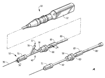
Figure 1 is a perspective view of a first embodiment of a thrombectomy
apparatus of the
present invention;
Figure IA is a perspective view of an alternate embodiment of the apparatus;
Figure 2 is an exploded view of the proximal portion of the thrombectomy
apparatus of
Figure 1;
Figure 2A is a perspective view of one embodiment of the motor housing
attachable to
the thrombectomy wire;
Figure 2B is an exploded view of the motor housing of Figure 1 showing the
components
for operatively connecting the motor to the thrombectomy wire;
Figure 2C is a side view in partial cross-section of the coupler of Figure 2B;
4
CA 02776090 2012-05-04
Figure 2D is a perspective view of the coupler of Figure 2C;
Figure 2E is a side view in partial cross section illustrating the connection
of the internal
components of the motor housing;
Figure 2F is a side view showing the wire operatively connected to the motor
shaft by the
coupler of Figure 2C;
Figure 3 is a side view in partial cross-section of the apparatus of Figure 1;
Figure 3A is longitudinal cross-sectional view taken along line 3A-3A of
Figure 1;
Figure 4 is a side view of the apparatus of Figure 1 showing the rotational
wire in a non-
linear position corresponding to a position exposed from the introducer
sheath;
Figure 4A is an enlarged view of the distal portion of one embodiment of the
thrombectomy wire having a sinuous configuration;
Figure 4B is an enlarged view of the distal portion of an alternate embodiment
of the
thrombectomy wire having a J-tip configuration;
Figure 5 is a longitudinal cross-sectional view of the distal portion of the
thrombectomy
wire of the apparatus of Figure 1;
Figure 6 is an anatomical view showing select cerebral arteries;
Figure 7 is a front anatomical view showing select cerebral arteries,
including the circle
of Willis;
Figure 8 illustrates insertion of a guide catheter through the femoral artery
and into the
cerebral artery over a tracking guidewire;
Figure 9 is a view similar to Figure 8 illustrating withdrawal of the tracking
guidewire;
Figure 9A is a perspective view illustrating attachment of the RHV to the
introducer
catheter;
Figure 10 illustrates insertion of the RHV and introducer catheter through the
guide
catheter and into the circle of Willis;
Figure IOA is a perspective view illustrating insertion of the introducer
sheath into the
RHV;
Figure IOB is a perspective view illustrating attachment of the connector tube
to the
introducer sheath;
Figure IOC is a perspective view of another introducer catheter;
5
CA 02776090 2012-05-04
Figure IOD is a side view showing attachment of the RHV and introducer
catheter of
Figure 1OC;
Figure 11 illustrates insertion of the thrombectomy wire of Figure 1 into the
RHV and
through the introducer catheter, and continued advancement of the wire from
the introducer
catheter so the distal portion of the wire is positioned in the circle of
Willis;
Figure 12 is a side view in partial cross section similar to Figure 2E showing
an alternate
embodiment of a coupler for coupling the thrombectomy wire to the motor;
Figure 13 is a perspective view of the coupler of Figure 12;
Figure 14 is a cross-sectional view of the coupler of Figure 13 shown within
the motor
housing coupling the motor shaft to the thrombectomy wire;
Figure 15 is a perspective view of an alternate embodiment of the coupler for
coupling
the thrombectomy wire to the motor;
Figure 16 is a front view of the housing of Figure 15 for receiving the motor
shaft; and
Figure 17 is a cross-sectional view of the coupler of Figure 15 shown within
the motor
housing coupling the motor shaft to the thrombectomy wire.
DETAILED DESCRIPTION OF PREFERRED EMBODIMENTS
Referring now in detail to the drawings where like reference numerals identify
similar or
like components throughout the several views, Figure 1 illustrates a first
embodiment of the
thrombectomy apparatus of the present invention.
The thrombectomy apparatus of Figure 1 is designated generally by reference
numeral
10. With reference to Figures 1 and 2, the apparatus includes a motor housing
12, a rotational
thrombectomy wire 30, a rotating hemostatic valve (RHV) 40, an introducer
sheath 60 and a
telescoping tube or tubular connector 80. The RHV 40 is connectable to an
introducer catheter
100 discussed below in conjunction with the method of use. The introducer
sheath 60 is
insertable into the RHV 40 to facilitate insertion of the thrombectomy wire 30
through the
introducer catheter 100.
The thrombectomy apparatus or assembly 10 disclosed herein provides a
rotational
thrombectomy wire as a separate unit from a catheter. That is, the
thrombectomy wire 30 is
provided as a separate unit insertable through the RHV 40 which has a distal
end 52 connected to
a proximal end of the introducer catheter 100 to access the surgical site. The
introducer sheath
6
CA 02776090 2012-05-04
60 aids insertion of the thrombectomy wire 30 into the RHV 40 and through the
introducer
catheter 100, with the walls of the introducer sheath 60 maintaining the non-
linear distal end of
the wire 30 in a substantially straightened (substantially linear)
configuration as it enters the
RHV 40.
Additionally, the thrombectomy wire 30 of the present invention can be slid
within the
introducer sheath 60 and introducer catheter 100 prior to connection to the
motor, if desired.
This can aid introduction and manipulation of the wire 30 since it is less
cumbersome and of
lighter weight than if the motor housing 12 was attached during manipulation
of the wire.
However, it is also contemplated that the wire 30 could be attached to the
motor housing 12 prior
to insertion through the introducer sheath 60, RHV 40 and the introducer
catheter 100 and thus
the wire 30 would be slidable within the introducer sheath 60 (and introducer
catheter 100) with
the motor housing 12 attached. Thus, the motor housing 12 can be attached to
the wire at a
desired time prior to or during the procedure.
Turning to the specific components of the thrombectomy apparatus 10, and with
reference to Figures 1-4, the motor housing 12, which also forms a handle
portion, has two
identical housing halves 13a, 13b. A motor 14 is seated within recess 14a of
housing half 13a
and the opposing recess of housing half 13b and has a motor drive shaft 15
extending therefrom.
Tabs 15b (Figure 3) help secure the motor 14 within the housing 12. A gear
reducer (not shown)
could optionally be provided to reduce by way of example the rotational speed
of the motor 52
from 15,000 rpm to 1500 rpm, 750 rpm, 150 rpm, etc. One or more batteries 16,
such as a 3 Volt
battery, is positioned in recess 17a of housing half 13a and the opposing
recess of housing half
13b for powering the motor 14. The battery(s) 16 can be contained within a
compartment in the
housing 12 accessible by removing a battery door. The motor drive shaft 15
connects to a
proximal end of the thrombectomy wire 30 by various couplings, such as for
example a snap fit
wherein cap 31 is frictionally fit within the lumen 15a of the motor drive
shaft 15. Various other
types of connections are also contemplated. A printed circuit board can also
be provided within
the housing 30 and is designated by reference numeral 18.
Motor housing 12 includes a distal tubular portion 22 having a tab in the form
of a ring
24 which fits within a groove in the tube connector 80, best shown in Figure 3
to connect the
motor housing 12 to tube connector 80 described below.
7
CA 02776090 2012-05-04
Switch 19 extends though recess 21 in housing half 13a and in a corresponding
recess in
housing half 13b. A potentiometer (not shown) can optionally be wired to the
motor to enable
dialing the motor speed up or down to adjust the rotational speed of the
thrombectomy wire 30 to
adjust for various procedures and/or clot locations and sizes. In a preferred
embodiment, the
potentiometer is used as a two terminal variable resistor, i.e. a rheostat, by
not connecting the
third terminal. In this manner, in the initial position, the motor speed is at
the desired minimum
and rotation of a knob (or in alternate embodiments sliding of a knob)
progressively increases the
motor speed. Thus, the on/off switch 19 extending from the housing 12 is
electrically connected
to the motor 15 to turn on the motor 15 to activate the apparatus, i.e. rotate
the wire 30.
Turning to the other components illustrated in Figures 2-4, rotating
hemostatic valve
(RHV) 40 is connectable to an introducer catheter 100 (see Figure 9A). A
conventional
introducer catheter can be utilized or alternatively a specially designed
catheter for use with the
apparatus of the present invention. As is standard, the RHV 40 is rotatable
with respect to the
catheter 100 to alter the orientation of the side arm 56.
Side arm 56 extends from the tubular portion 46 and has a port 57 for
introduction of
fluids and/or application of vacuum as described below. Luer lock is provided
at the distal end
52 of RHV 40 to connect to the introducer catheter as threads 51a of rotation
knob 51
threadingly engage proximal threads of the introducer catheter 100. Tube
extension 48 fits
within the lumen of the introducer catheter 100 when attached. Washers 49a,
49b help to provide
a seal against fluid flow.
Tubular portion 46 of RHV 40 includes a lumen 55 extending therethrough to
slidably
receive the tubular portion 62 of the introducer sheath 60. Proximal cap 58 at
proximal end 54
has internal threads 59 to threadingly attach to external proximal threads 47
for attachment of the
cap 58 to the RHV 40. Further, a crush ring 43 and distal ring 44 are seated
within the internal
lumen 55 of the tubular portion 46. Thus, as cap 58 is tightened on RHV 40 by
rotation, it
compresses rings 43 and 44 against the tubular portion 62 of introducer sheath
60 extending
therethrough to connect the introducer sheath 60 to the RHV 40 (see Fig. 3A).
A proximal seal
45 can also be provided. Flange 46a on the proximal end 54 of RHV 40 interacts
with lip 58a of
cap 58 to allow loosening of cap 58 to release introducer sheath 60 without
cap 58 detaching
from RHV 40.
8
CA 02776090 2012-05-04
Side arm 56 of RHV 40 has a lumen 53 in fluid communication with lumen 55 of
tubular
portion 46. Fluids such as imaging dye can be injected through the arm 56,
flowing through the
lumens 53 and 55, i.e. through the space between the outer wall of the
introducer sheath 60 and
the inner wall of lumen 55 and then through the space between the thrombectomy
wire 30 the
inner wall of the introducer catheter 100 and, exiting a distal opening 103
(Fig 10) in the
introducer catheter 100 to flow into the vessel. This imaging dye can be used
to provide an
indication that fluid flow has resumed in the vessel.
The side arm 56 can also be used for vacuum to suction particles detached from
the
vessel by the rotational wire 30. The particles would flow into the distal
opening 103 of the
introducer catheter 100 and through the space between the wire 30 and the
inner wall of the
introducer catheter 100, then exiting through lumen 53 and port 57 into a
suction tube (not
shown).
It should also be appreciated that the guide catheter 150 discussed in
conjunction with the
method of use can also have a side ann for injection of fluid (see e.g., side
arm 152 of Figure 8).
In the alternate embodiment of Figure 1 A, the RHV 40' does not have a side
arm. In this
embodiment, a guide catheter with a side arm can be used for injection and
suction. Otherwise
the components are identical to the components of Figure 1 and for
convenience, the
corresponding components are labeled with "prime" designations e.g.,
rotational knob 51', cap
58', introducer sheath 60', connector tube 80' and locking cap 83'.
The tubular portion 62 of introducer sheath 60, as noted above, extends
through the
lumen 55 of RHV 40 and terminates either within RHV 40 or at a proximal
portion of the lumen
of the introducer catheter 100. The tubular portion 62 preferably has a
stiffness greater than the
stiffness of the thrombectomy wire 30 to maintain the wire 30 in a
straightened position during
passage of wire 30 into the RHV 40 for subsequent passage through the lumen of
the introducer
catheter 100 to the surgical site.
Proximal end 65 of introducer sheath 60 is attachable to connector tube 80.
Preferably,
the enlarged proximal end 65 has a threaded flange 67 as shown in Figure 3A to
threadingly
engage the internal threads 85 on the distal cylindrical locking cap 83 at the
distal end 82 of
tubular connector 80. A valve can be provided within the distal end 82 of the
connector tube 80
in addition or instead of a valve in a proximal end 65 of the introducer
sheath 60 to seal escape of
fluid to improve the vacuum through the side arm 56.
9
CA 02776090 2012-05-04
Note the tube 80 and introducer sheath 60 can alternatively be provided as one
unit,
attached together and positioned over the thrombectomy wire 30. However, in
alternative
embodiments, the wire 30 is inserted through the introducer sheath 60 and
manipulated through
the introducer catheter 100 to the surgical site. Once positioned, the
connector tube 80 is then
threadingly attached at the distal end 82 to the introducer sheath 60 as noted
above and at a
proximal end 84 to the motor housing 12. In this version, the connector tube
80 can be
positioned over the wire 30 prior to insertion of the wire 30 through
introducer sheath 60 or after
insertion through the sheath 60. The wire 30 can be packaged with the sheath
60 and the tube 80
positioned thereover, or packaged apart from the sheath 60 and tube 80.
Proximal end 84 of connector tube 80 is configured for attachment to the motor
housing
12 by an external ring 24 on tip 22 of motor housing 12. Ring 24 is seated
within an internal
groove of connector tube 80, as shown in Figure 3, to provide a snap fit.
Other types of
attachment are also contemplated. The proximal end of the wire 30 is attached
to the drive shaft
of the motor 14. In one embodiment, end cap 31 of wire 30 is snap fit within
opening 15a in
15 motor shaft 15. Other ways to attach the wire 30 and motor shaft 15 are
also contemplated such
as a bayonet mount for example.
As can be appreciated, by having a detachable motor housing 12, different
handles with
different motor speeds and/or different batteries can be utilized by
attachment to the wire 30.
This can even be achieved during the same surgical procedure.
In some embodiments, the housing can be detached, sterilized and reused after
recharging
of the battery or replacing the battery.
In some embodiments, as an alternative to direct connection to the motor
shaft, the
proximal end of wire 30, after insertion to the surgical site or prior to
insertion, can be attached at
a proximal end to a coupler tube which is connected to a gear reducer. The
connection of the
motor and thrombectomy wire can be a friction fit, a magnetic coupling or a
twist connect, e.g. a
bayonet connection, by way of example, such as that shown in co-pending patent
application
serial no. 13/095,329, filed April 27, 2011.
Figures 2A-2F show an alternative mechanism for operatively connecting the
thrombectomy wire and motor. Motor housing 210 is composed of two housing
halves 212a,
212b which form the handle of the apparatus. Seated within the recess 213 in
motor housing 210
is motor 214 electrically connected to two batteries 216. Switch 218 extends
through opening
CA 02776090 2012-05-04
220 in motor housing 210 for access by the user. Attached to motor shaft 222,
which extends
distally from motor 214, is magnetic coupler 230 for magnetic coupling of the
thrombectomy
wire to the motor housing 210. Electrical wire 226 electrically connects
switch 218 to post 214a
of motor 214. Wire 229 connects the switch 218 to the positive terminal of
battery 216 and wire
228 connects the negative terminal of battery 216 to motor post 214b.
The magnetic coupler includes a tube or housing 230, preferably made of PVC,
although
other materials are also contemplated. Tube 230 has a proximal portion 234
which receives
motor shaft 222 and a distal portion 236. A first magnet 242 is positioned in
the distal portion
236 of the tube 230, and due to its transverse dimension being larger than the
transverse
dimension of tube 230, forces the tube 230 to flare outwardly into flared
portion 233, thereby
providing a tight frictional fit. A disc 240, which can be made of a polymeric
or of other
material, but is preferably in the form of a Latex sheet, is provided over the
distal edge 238 of
tube 230 to maintain the first magnet 242 within the tube 230. The disc 240
functions as a clutch
for torque transfer from the motor 214 to the thrombectomy wire 30. The motor
shaft 222,
extending distally from motor 214, extends into the proximal end of the tube
226 and is
frictionally engaged thereto.
A second magnet is contained in housing 246 which is attached to the proximal
end of the
thrombectomy wire 30 by gluing, overmolding, or other attachment methods. When
desired to
attach the thrombectomy wire 30 to the motor housing 210, the thrombectomy
wire 30 is inserted
into the reduced diameter portion 217 of motor housing 214 until the magnetic
attraction
between the second magnet and first magnet 242 maintains a magnetic
connection. In this
manner, when motor 214 is actuated by switch 218, motor shaft 222 rotates to
thereby rotate
magnetically coupled thrombectomy wire 30. Note the torque is transferred to
the wire 30 due to
the disc 240 functioning as a clutch.
As noted above, the disc 240 can be in the form of a polymeric sheet. The
sheet can be
designed to wear off after a period of time, thus wearing away the clutch,
resulting in the loss of
the ability to transfer torque. In this way, over-use of the apparatus can be
prevented, and the
apparatus can advantageously be designed for one time use in a single
procedure.
An alternative embodiment for coupling the motor to the thrombectomy wire is
illustrated
in Figures 12-14. In this embodiment, housing 330 has a proximal portion 334
which frictionally
receives the motor shaft 222 and a distal portion 336. The distalmost edge 338
is in a wavy
11
CA 02776090 2012-05-04
pattern forming a toothed design. A first magnet 340 is positioned in the
distal portion 336,
recessed from the distalmost edge 338.
A second housing 350 is attached to the proximal end of the thrombectomy wire
30. The
second housing 350 has a distal portion 352 to frictionally receive the wire
30 and a proximal
portion 354. The proximalmost edge 358 is in a wavy pattern forming a toothed
design
configured to mate with the toothed design at the distalmost edge 338 of
housing 330. A second
magnet 360 is positioned in the proximal portion 354, recessed distally from
the proximalmost
edge 358. In this manner, first and second magnets 340, 360 do not come into
contact but
provide an attractive coupling force to attach the wire 30 and motor shaft 222
of motor 214.
The first plurality of teeth 337 of first housing 330 intermesh with the
second plurality of
teeth 357 of the second housing 350 so that upon rotation of the motor shaft
222, the coupled
housings 330, 350 rotate. Due to the interaction of the teeth 337 of housing
330 with the teeth
357 of housing 350, rotation of housing 330 causes housing 350 to rotate which
thereby rotates
the wire 30 attached to housing 350. These housings 330, 350 operate as a
clutch mechanism.
That is, if during use, the torque of the motor shaft 222 exceeds a preset
value, indicating for
example that the wire is caught on material in the vessel, the teeth 337, 357
of the housings 330,
350, slip such that housing 330 rotation no longer rotates housing 350. Due to
the spacing of
magnets 340, 360 from each other, as a result of their mounting within the
recess or pockets of
the respective housings 330, 350, the force at which the housings (clutch)
slip is entirely
dependent on the interaction of the teeth. That is, this coupling design forms
a clutch which
when the torque of the motor shaft exceeds a predetermined value, the teeth
slip so the teeth are
no longer operably intermeshed. Thus, the torsional load at which the coupling
slips depends on
the friction between the teeth, thereby relying solely on the coefficient of
friction of the housing
materials and the angle/geometry of the teeth. Slippage occurs when torsional
force is greater
than frictional force and the magnetic force holding the housings together. If
the magnets were
in direct contact, the frictional engagement of the magnets in addition to the
interaction of the
teeth would affect the slippage point. By relying solely on the teeth, the
design is simplified.
The press-fit of the magnets into the recessed pockets also facilitates
manufacture.
In the alternate embodiment of Figures 15 and 16, the housings 430, 450, are
similar to
housings 330, 350 and have distalmost and proximalmost edges 438, 458,
respectively, which
are in a wavy pattern forming teeth 437, 457, which intermesh to rotate the
second housing 450
12
CA 02776090 2012-05-04
as the first housing 430 is rotated by the rotating motor shaft 222. However,
in this embodiment,
spherical magnets are provided within a gap in the housings 430, 450 to allow
movement, e.g.,
rolling, of the magnets.
More specifically, housing 430 has a proximal portion 434 which receives the
motor shaft
222 and a distal portion 436. The distalmost edge 438 is in a wavy pattern
forming a toothed
design. A first substantially spherical magnet 440 is positioned in the distal
portion 436 in an
internal cavity 433, recessed proximally from the distalmost edge 438. The
internal cavity 433
forms a gap 435 proximal of magnet 440. A plug 439 is press fit in a proximal
opening of the
cavity 433 to secure the magnet 440 within the cavity 433. The motor shaft 222
can be mounted
in a proximal opening in plug 439 such as by an interference fit. The magnet
440 can move
within the gap 435. In this manner, as the housing 430 rotates, the magnet 440
does not rotate
with the housing 430 and can float or roll within the gap 435.
A second housing 450 is attached to the proximal end of the thrombectomy wire
30. The
second housing 450 has a distal portion 454 to frictionally receive the wire
30 and a proximal
portion 452. The proximalmost edge 458 is in a wavy pattern forming a toothed
design
configured to mate with the toothed design at the distalmost edge 438 of
housing 430. A second
substantially spherical magnet 460 is positioned in the proximal portion 452,
recessed distally
from the proximalmost edge 458. The housing 450 has an internal cavity 453
forming a gap 455
distal of magnet 460. A plug 459 is press fit in a proximal opening of the
cavity 453 to secure
the magnet 460 within the cavity 453. The thrombectomy wire 30 can be mounted
in a distal
opening of plug 459 such as by an interference fit. The magnet 460 can move
within the gap
455. In this manner, as the housing 450 rotates, the magnet 460 does not
rotate with the housing
and can float or roll within the gap 455. Note as with the embodiment of
Figures 13 and 14, the
first and second magnets 440, 460 do not come into contact but provide an
attractive coupling
force to attach the wire 30 and motor 214. The placement of the magnets in
recessed pockets has
the advantages described above.
The teeth 437, 457, of the respective housings 430, 450 intermesh so that upon
rotation of
the motor shaft 222, the attached housing 430 rotates. Due to the interaction
of the teeth 437 of
housing 430 with the teeth 457 of housing 450, rotation of housing 430 causes
housing 450 to
rotate which thereby rotates the wire 30 attached to housing 450. During such
rotation, magnets
440, 460 can move, e.g., float or roll, within the gaps 433, 453 of housings
430, 450,
13
CA 02776090 2012-05-04
respectively. The gaps can be sufficiently large relative to the magnets to
enable the magnets to
freely float therein, i.e., not only move axially but move in three
dimensions. These housings
430, 450, as in the embodiment of Figures 13 and 14, operate as a clutch
mechanism. If during
use, the torque of the motor shaft exceeds a preset value, indicating that the
wire is caught on a
vessel, the teeth 437., 457 of the housings 430, 450, respectively, slip such
that housing 430
rotation no longer rotates housing 450. Due to the spacing of magnets 440, 460
from each other,
as a result of their mounting within recess of the respective housing 430,
450, the force at which
the housings (clutch) slip is entirely dependent on the interaction of the
teeth 437, 457. That is,
as in the embodiment of Figures 13 and 14, this coupling design forms a clutch
which when the
torque of the motor shaft exceeds a predetermined value, it causes the teeth
437, 457 to slip so
the teeth are no longer operably intermeshed. Thus, the torsional load at
which the coupling slips
depends on the friction between the teeth, thereby relying solely on the
coefficient of friction of
the housing materials and the angle/geometry of the teeth. Slippage occurs
when torsional force
is greater than frictional force and the magnetic force holding the housings
together. If the
magnets were in direct contact, the frictional engagement of the magnets in
addition to the
interaction of the teeth would affect the slippage point. By relying solely on
the teeth, the design
is simplified. The press-fit of the magnets into the recessed pockets also
facilitates manufacture.
Note the step of operatively coupling the thrombectomy wire to the motor
housing 210
using any of the foregoing coupling embodiments can occur prior to the step of
inserting the
thrombectomy wire through the introducer sheath and catheter. Alternatively,
the step of
operatively coupling the thrombectomy wire to the motor housing 210 using any
of the foregoing
embodiments can occur subsequent to the step of inserting the thrombectomy
wire through the
introducer sheath and catheter.
Figure 5 illustrates the thrombectomy wire 30 of the present invention. The
wire 30 has a
distal coiled tip 91. In preferred embodiments, the distal coiled tip (and
underlying cable) is
angled with respect to the longitudinal axis. Figure 4A shows the wire of
Figure 5 forming a
sinuous shape. In Figure 4B, an alternative embodiment of the wire is
illustrated, wherein the
wire 130 forms a J-tip which creates a standing wave upon rotation. In the J-
tip configuration,
due to the angle, when the wire is rotated by the motor at sufficient speed at
least one vibrational
node is formed. Details of this creation of a standing wave are described in
U.S. Patent No.
6,090,118.
14
CA 02776090 2012-05-04
In the embodiment of Figure 4A, the wire 30 forms a substantially sinuous
shape,
resembling a sine curve. More specifically, wire 30 of Figure 4A has a
substantially linear
portion extending through most of its length, from a proximal region, through
an intermediate
region, to distal region 36. At the distal region 36, wire 30 has a sinuous
shape in that as shown
it has a first arcuate region 33 facing a first direction (upwardly as viewed
in the orientation of
Figure 4A) and a second arcuate region 35, spaced longitudinally from the
first arcuate region
33, facing a second opposite direction (downwardly as viewed in the
orientation of Figure 4A).
These arcuate regions 33, 35 form "peaks" to contact vascular structure as the
wire 30 rotates.
This angled (non-linear) distal portion includes a coiled portion with a
covering material to block
the interstices of the coil as discussed below. Note in a preferred
embodiment, the amplitude of
the proximal wave (at. region 33) is smaller than the amplitude of the distal
wave (at region 35),
facilitating movement in and out of the catheter.
When the wire 30 is fully retracted within the introducer catheter 100 (as in
Figure 3), the
curved regions of the wire 30 are compressed so the distal region 36 is
contained in a
substantially straight or linear non-deployed configuration. When the
introducer catheter 100 is
retracted by proximal axial movement (see the arrow of Figure 4), or the wire
is advanced with
respect to the introducer catheter 100 or the wire 30 and catheter 100 are
both moved in the
respective distal and proximal directions, the distal region 36 of the wire 30
is exposed to enable
the wire 30 to return to its non-linear substantially sinuous configuration
shown in Figure 4A
(and Figure 4) for rotation about its longitudinal axis within the lumen of
the vessel.
Thus, as can be appreciated, the wire 30 is advanced within the introducer
catheter 100
which is attached at its proximal end to the distal end of the RHV 40. When at
the desired site,
the wire 30 and introducer catheter are relatively moved to expose the wire 30
to assume its non-
linear shape for motorized rotational movement to break up thrombotic material
on the vessel
wall. If a J-tip wire, such as wire 130, is utilized, the wire 130 can be
rotated within the
introducer catheter to re-orient the wire 130.
The flexible tubular portion 62 of the introducer sheath 60 can optionally
contain one or
more braided wires embedded in the wall to increase the stiffness. Such
braided wires would
preferably extend the length of the sheath.
In an embodiment of the coiled tip being composed of shape memory material,
the
memorized configuration is sinuous or s-shaped as in Figure 4A. In the state
within the
CA 02776090 2012-05-04
introducer catheter 100, the wire is in a substantially linear configuration.
This state is used for
delivering the wire to the surgical site. When the wire is exposed to warmer
body temperature,
the tip transforms to its austenitic state, assuming the s-shaped memorized
configuration.
Alternatively, the coiled tip of the wire can be compressed within the wall of
the introducer
catheter and when released, assumes its shape memorized non-linear shape. The
coiled tip can
alternatively be a radiopaque coil/polymer pre-shaped to an "S".
Details of the wire 30 will now be described with reference to Figure 5. These
details are
the same for wire 130, the only difference being that instead of the distal
coiled tip being sinuous
shaped in the deployed position, the distal tip is in J-configuration. Note it
is also contemplated
that in an alternate embodiment the distal tip can be substantially straight
(substantially linear) in
both the covered and deployed (exposed) position. For convenience, details
will be discussed
with reference to wire 30. Wire 30 has a core 32 having a proximal portion 34
(see Fig. 2) and a
distal portion 37. Transition region 38 of core 32 is tapered distally so that
the diameter of the
distal portion 37 of core 32 is less than the diameter of the proximal portion
34. A uniform
diameter portion 37a extends distal of tapered portion 37. The taper can be
formed by removing
a coating, such as a PTFE coating, placed over the core 32 and a grinding of
the core 32. In one
embodiment, the core 32 is a solid material made of a nickel titanium alloy,
although other
materials are also contemplated. The core 32 can also be formed from a
hypotube with a tapered
body attached, e.g. welded, to the distal end of the hypotube.
The core 32 is connected to a cable 90. The cable 90 can be formed of a
plurality of
wires twisted together such as a 1x19 wire for example. The twisted wires can
be surrounded by
additional wires or a sheath. The core 32 is tapered to accommodate connection
to cable 90.
Hypotube 92 is placed over the distalmost end of the core 32 (the uniform
diameter portion 37a)
and the proximalmost end of the cable 90 and is attached thereto by a number
of methods,
including but not limited to, laser welding, soldering or crimping. The
hypotube 92 thereby
forms a coupler for joining the core 32 and cable 90 as these components are
positioned within
the hypotube 92. The hypotube can have a diameter of about .010 inches,
although other
dimensions are contemplated.
The cable 90 in one embodiment has a variable stiffness such that the proximal
portion
94 is stiffer, e.g., has a tighter braid, than a distal portion 96 to increase
the flexibility of the
distal portion 96. In other embodiments, the cable 90 is of uniform stiffness.
The cable 90 can
16
CA 02776090 2012-05-04
be of substantially uniform diameter. Various covering materials, e.g.,
coating, jackets and/or
shrink wraps, can be used as an alternative or in addition to vary the
stiffness of the cable 90.
A torque tube 97 is positioned over the cable 90. The torque tube 97 extends
from a
tapered region of the core 32, terminating at the distal coil 91. The torque
tube 97 can be
soldered at (proximal) end 97a to the core 32 and at distal end 97b to the
cable 90. The torque
tube 97 can also be attached, e.g., soldered or laser welded, to a proximal
end of the coil.
A polymer coating(s) and/or jacket(s) can be placed over the torque tube 97 to
cover the
interstices in the cable 90 and provide a smooth surface. In one embodiment, a
PTFE shrink
wrap tubing 98 is placed over the torque tube 97 and over a portion of the
core 32, preferably
extending over the tapered transition region 38 of core 32 to terminate at a
proximal end adjacent
the uniform diameter region of the core 32. At a distal end, the shrink wrap
98 terminates at the
end where the torque tube 97 terminates.
Coiled tip 91 is positioned over a distal portion of the cable 90, and
preferably over the
distal tip. The coil tip 91 in one embodiment is composed of a soft and
malleable material such
as platinum and has a uniform pitch and diameter. The distalmost tip of the
cable 90 can have a
laser welded ball to which the coil 91 is welded to enhance retention of the
coil 91 and cable 90.
The coiled tip region has a substantially sinuous configuration. In an
alternate embodiment, the
coiled tip region has a J-tip configuration, as shown for example in Figure
4B. The coiled tip
region can alternatively have a substantially linear configuration in the
deployed/uncovered
position. In each of these embodiments, preferably a covering such as a
jacket, shrink wrap or
coating covers the coil 91. In a preferred embodiment, a polyamide such as a
nylon or Pebax
covering 99 is heat fused over the coil 91, to melt into the interstices. In
some embodiments, a
heat shrink tubing 99a, such as FEP, is placed over the heat fused nylon
coating. The covering
99, and heat shrink tubing 99a, terminate adjacent a distal end of the torque
tube 97 and adjacent
a distal end of the shrink wrap 98.
By way of example only, the components of wire 30 can have the approximate
dimensions set forth in the table below. It should be understood that these
dimensions are being
provided by way of example as other dimensions are also contemplated. These
are also
approximate values.
APPROXIMATE OUTER APPROXIMATE
COMPONENT
DIAMETER LENGTH
17
CA 02776090 2012-05-04
Core 32 (proximal non tapered portion) .016 inches 139.5 cm
Core tapered portion .016 inches to .0095 inches 4.35 inches
Distal coil 91 .016 inches 3.0 inches
Torque tube 97 .013 inches 8.0 inches
Shrink tube 98 .014 inches 10.35 inches
Cable 90 .010 inches 8.2 inches
The covering material, e.g. coating, jackets, and or shrink wraps, helps to
prevent
bending or knotting of the wire which could otherwise occur in native vessels.
The covering also
increases the torsional strength of the wire and also strengthens the wire to
accommodate spasms
occurring in the vessel. The coating also blocks the interstices of the coil
91 to provide a less
abrasive surface. The various coating and/or jackets and/or shrink wrap can be
made of PET,
Teflon, Pebax, polyurethane or other polymeric materials. The material helps
to prevent the
native vessel from being caught in the coil 90 and reduces vessel spasms.
The use of the thrombectomy apparatus 10 will now be described. The use, by
way of
example, is shown and described with respect to the embodiment of Figure 1
with the sinuous tip
of Figure 4, it being understood that the wire embodiment of Figure 4B would
be utilized in a
similar manner. It is also shown for use in the cerebral arteries but use in
other vessels is also
contemplated.
An access sheath (not shown) is inserted into the vessel and then a guidewire,
e.g. .035 or
.038 inches in diameter, and a guide catheter 150 are inserted through the
sheath and advanced
through the vasculature. The guidewire is removed and a smaller diameter
guidewire G, e.g.
.014 inch diameter, and the introducer catheter 100, are inserted through the
guide catheter 150
and access sheath with the guidewire G in the femoral artery F and located via
imaging. The
introducer catheter 100 is advanced to the desired site through the vascular
system into the
cerebral arteries A, for example through the Circle of Willis C (see FIGS. 6,
7 and 8). Once at
the site, the guidewire G is withdrawn as shown in Figure 9. Note the
introducer catheter 100 is
preferably inserted with the RHV 40 attached. That is, the tubular portion 46
of the RHV 40 is
inserted through the introducer catheter 100 (see Figure 10) and attached
thereto by rotation of
cap 51 as shown in Figure 9A. In the alternate embodiment of Figures IOC and
10D, RHV 40 is
attached to thread 124 of the winged luer fitting of introducer catheter 120
by rotation of cap 51
18
CA 02776090 2012-05-04
and/or winged handle 122. Note in an alternate embodiment, instead of the RHV
40 attached
prior to introduction of the introducer catheter 100 through the guide
catheter 150, it can be
attached after introduction of catheter 100 through guide catheter 150.
The introducer sheath 60 is inserted through the RHV 40, and attached to the
RHV 40 by
rotation of cap 58 as shown in Figure 1OA. The thrombectomy wire 30 is
inserted through the
lumen of the introducer sheath 60, through the lumen of the RHV 40 and into
the lumen of the
introducer catheter 100. The introducer catheter 100 extends from the guide
catheter 150 as
shown in Figure 10, but the wire 30 remains inside the introducer catheter
100. The distal end of
the wire 30 is then exposed from the introducer catheter 100 at the target
surgical site by relative
movement of the wire 30 and introducer sheath 100. Note the wire 30 can be
attached to the
motor drive shaft 15 at this point or can be attached before exposed or at any
other time in the
procedure such as prior to insertion of the wire 30 through the introducer
sheath 60. Attachment
is achieved by connection of the connector tube 80 to the introducer sheath 60
(see Figure lOB)
and attachment of the proximal end of the connector 80 to the motor housing 12
or by other
methods, such as a magnetic coupling as described above. The wire 30 extends
through the
connector tube and attachment of the wire 30 (which extends through connector
80) to the motor
drive shaft 15. As noted above, alternatively, the connector tube 80 can be
connected to the
introducer sheath 60 prior to attachment to the motor housing 12, or
alternatively connected after
the wire 30 is at the surgical site and exposed from the introducers sheath.
The alternate
embodiments described herein for coupling the wire to the motor shaft could
also be utilized.
With the wire 30 exposed from the introducer catheter 100, switch 19 on
housing 12 is
actuated to turn on the motor thereby causing wire 30 to rotate about its
longitudinal axis to
break up/macerate thrombus.
The macerated particles can be removed by suction through side arm 56 of RHV
40 as
the particles travel in the space between wire 30 and introducer catheter 100
and RHV 40. The
introducer catheter 100 can optionally have a side port(s) and/or the guide
catheter 150 can
optionally have a side port(s) such as side port 152 for aspirating the small
macerated particles in
addition to or alternative to side arm 56 of RHV 40.
The delivery sheath can include a balloon to block blood flow and allow
aspiration in the
blocked space.
19
CA 02776090 2012-05-04
While the above description contains many specifics, those specifics should
not be
construed as limitations on the scope of the disclosure, but merely as
exemplifications of
preferred embodiments thereof. Those skilled in the art will envision many
other possible
variations that are within the scope and spirit of the disclosure as defined
by the claims appended
hereto.