Note: Descriptions are shown in the official language in which they were submitted.
SURFACE-ASSISTED HEMAGGLUTINATION AND
HEMAGGLUTINATION INHIBITION ASSAYS
BACKGROUND TO THE INVENTION
[0002] Hemagglutinin proteins expressed on the surface of many viruses,
such as influenza.
rubella, smallpox, and others, agglutinate red blood cells (erythrocytes).
This effect provides the
basis for virus titration using hemagglutination (HA) assays. Specific
attachment of antibodies to
epitopes of the hemagglutinins responsible for attachment to the erythrocytes
blocks binding of
the virus particles to erythrocytes. This effect provides the basis for
hemagglutination inhibition
(HAI or HI) assays.
[0003] Hemagglutination assays and hemagglutination inhibition assays were
introduced into
medical and virology practice more than 60 years ago (Salk (1944) J. Immunol.
49, 87-98). Since
that time, they have become important tools for measuring concentrations and
strengths of viral
cultures, the efficacy of the anti-viral immunization, and for studying the
neutralizing capacity of
virus-specific antibodies.
[0004] Two decades later, attempts were made to develop the method to a
universal standard
(Hierholzer et al. (1969) Applied Mierobiol. 18, 824-833). However, the
protocol for HAI assays
kept undergoing minor modifications (e.g., Cross (2002) Seminars in Avian and
Exotic Pet
- 1 -
CA 2776405 2017-07-11
CA 02776405 2012-04-02
WO 2011/050027 PCT/US2010/053322
Medicine 11, 15-18; Hubby et al. (2007) Vaccine 25, 8180-8189; Wang et al.
(2008) Vaccine 26
3626-3633: Noah et al. (2009), Clinical and Vaccine Immunology 16, 558 - 566),
while
preserving the core elements intact: observation of the agglutination in the
solution volume, and
visual detection of hemagglutination or hemagglutination inhibition.
[0005] In classical HA / HAI assays, the antigen (e.g., live or inactivated
virus), either as is,
or pre-incubated with an anti-serum or antibody of interest, is mixed with a
suspension of
purified erythrocytes, such as human group 0 erythrocytes, or avian, equine,
or murine
erythrocytes, depending on the type of the virus and objective of the study.
After incubation of
the mixture in V- or U-bottomed microwells, the major visual effect can be two-
fold:
= If antiserum is absent or unable to effectively block the attachment of
the virus to
erythrocytes. the virus particles link the erythrocytes into a dispersed three-
dimensional
semi-transparent gel, referred to as a "halo."
= If the virus is effectively blocked or absent, then the erythrocytes
precipitate to the
bottom of the well, forming the characteristic bright pellet, or "button."
[0006] Using avian erythrocytes, the agglutination effect can be observed
(optionally) by
inability of the agglutinated erythrocytes to flow down the V-surface of the
tilted plates.
[0007] To determine the concentration or strength of a viral culture in the
HA
hemagglutination assay, the sample is subjected to two-fold serial dilutions,
until the
agglutination vanishes. To determine the efficacy of the antiserum or tested
antibody in the HAI
assay, the serum sample is similarly subjected to serial dilution, until
agglutination appears. The
last dilution on the "borderline" between agglutination / non-agglutination is
called the HA or
HAT titer.
- 2 -
CA 02776405 2012-04-02
WO 2011/050027 PCT/US2010/053322
[0008] HA and HAI assays are used for the study of immune response to a
multitude of
different pathogenic viruses, including adenoviruses, enteroviruses,
reoviruses, myxoviruses,
poxviruses, and flaviviruses, which cause a wide spectrum of human and animal
illnesses, from
influenza and rubella to smallpox and Dengue hemorrhagic fever (e.g., Hatgi et
al. (1966)Am. J.
Trop. Med. Hyg. 15, 601-610; Hierholzer et al. (1969) Applied Microbiol. 18,
824-833; Cross
(2002) Seminars in Avian and Exotic Pet Medicine 11, 15-18; Hubby et al.
(2007) Vaccine 25,
8180-8189; Wang et al. (2008) Vaccine 26, 3626-3633). Thus, HA and HAT tests
remain major
tools in modern virology (WHO Manual on Animal Influenza Diagnosis and
Surveillance,
WHO/CDS/CSR/NCS2002.5 Rev. 1.). Significant improvements to the assays could
be of
widespread benefit.
[0009] The virtues of HA / HAT assays, based on erythrocytes, inspired the
development of
various versions of agglutination / agglutination inhibition tests, based on
latex microbeads
coated with various antigens and affinity ligands, including hemagglutinins
and virus particles
(Ko et al. (1999), J. Clin. Pathol. 52, 770-772; Xu et al. (2005), J. Clin.
Microbiol., 43, 1953-
1955). These methods, although fast and reliable, do not provide greater
sensitivity to sera or
antibody solutions than the classical HAT.
[0010] An objective of the invention presented here was the development of
a functional
assay of enhanced sensitivity that would use real target cells (e.g.,
erythrocytes), rather than
synthetic particles, and would stay as close as possible to the well-proven
and widely accepted
classical HA / HAI. On the other hand, basic principles illustrated in
embodiments of the current
invention could be applied to latex bead agglutination methods to increase
their sensitivity and
informational capacity.
- 3 -
CA 02776405 2012-04-02
WO 2011/050027 PCT/US2010/053322
[0011] While robust, uncomplicated and reliable, HA and HAI assays lack
adequate
sensitivity in the cases of some conditions, such as measles, yellow fever,
and polyoma
(Chapaeain et al. (2006) Virology J, 3, 3-5; Fujino et al. (2007) J.
Virological Methods 142, 15-
20; Niedrig et al. (1999) Trop. Med. Int. Health 4, 67-71). Further,
assessments of agglutination
are typically performed by the human eye, which can become a source of
subjective evaluation.
[0012] In addition to the inadequate sensitivity of the standard HA / HAI
assays with many
viruses, as mentioned above, the development of modem in vitro systems for
high-throughput
analysis of immune responses, such as the MIMIC system, described in US
2005/0282148,
required improved sensitivity in methods of evaluating functionality of
antibody immune
responses. The MIIVIIC system is based on cultures of human immune-competent
cells
developed in a 96-well format, which limits the achievable concentrations and
total quantities of
the antigen-specific antibodies generated in the system.
[0013] Thus, there is a continuing need for functional assays with improved
sensitivity,
including those based on hemagglutination.
BRIEF SUMMARY OF THE INVENTION
[0014] Hemagglutination (HA) and hemagglutination inhibition (HAI)
functional assays
remain important instruments of analysis of virus-cell interaction and
protective efficacy of
virus-specific antibodies and sera. However, the classical protocols of HA and
HAI demonstrate
limited sensitivity towards some viruses and require significant volumes of
viruses and the tested
sera or antibodies, which can constitute an obstacle when experimenting with
scarce or precious
materials. The latter is especially important when analyzing the samples
obtained from the in
vitro systems that model immune responses, such as, for example, the MIMIC
system.
- 4 -
CA 02776405 2012-04-02
WO 2011/050027 PCT/US2010/053322
[0015] Embodiments of the present invention include a new method for the
functional
characterization of viruses and virus-specific antibodies and sera, the
Surface-Assisted
Hemagglutination / Hemagglutination Inhibition functional assay, "SA-HA /
HAI."
Embodiments of the present invention demonstrate sensitivity of the SA-HA
assays to various
influenza viruses 7-200 times higher than the traditional HA assay, and
sensitivity of the SA-
HAT assay to influenza-specific antibodies 7-50 times higher than in the
traditional HAT,
depending on the types of viruses and erythrocytes used.
[0016] Additionally, a version of the SA-HAI assay that uses U-bottom EL1SA
plates makes
it possible to determine the relative contributions of low affinity and high
affinity functional
antibodies in the HAT titer, which is technically impossible using classical
HAI.
[0017] There are three major concepts in the foundation of the current
invention
embodiments:
1) Transferring of the hemagglutination reaction from solution to the
activated surface of
specially coated (opsonized) plates, or (alternatively) ELISA plates.
2) Using photo-registration of the agglutination micro-patterns, digital image
processing,
calculation of a numerical Hemagglutination Parameter that reflects the degree
of
agglutination in every well of the plate and the mathematical computation of
the titers.
3) Using advanced standardization of the HAT assay that effectively reduces
variability in
the HAT data.
[0018] The enhancement in sensitivity allows analysis of experimental
samples of low
concentrations and saves precious materials, such as convalescent sera and
viruses. The SA-HA /
- 5 -
HAI assays can use the same types of erythrocytes as normally used in the
traditional HA / HAI
assays: human, mammalian, and avian.
[0019] Characterizing the relative contributions of low- and high-affinity
virus-specific and
functional (potentially protective) antibodies in the HAI titer makes the
ELISA plate version of
the SA-HAI assay a valuable tool that provides deeper insight in the
properties of humoral
immune responses.
[0020] The SA-HA / HAT assay results can be evaluated visually, in the
manner similar to the
classical HA / HAI assays. However, visual evaluation lacks the precision
necessary in high-
sensitivity experiments and it is obviously prone to human errors, due to
differences in
perceptions by different operators. Photo-registration and digital processing
of the SA-I IA / HAI
images increases the precision of the method and eliminates such subjectivity.
The image
processing can be performed in line with photo registration and in real time.
The SA-I IA / IlAl
method can be performed in a high-throughput mode and allows automation.
[0021] In a first specific embodiment, the present invention is directed a
SA-HA assay. In
particular, the method can be used to determine whether virus is present in a
sample, as well as to
quantify the amount of virus in the sample. In one aspect, the method
comprises: (a) incubating
a target object and a sample suspected of containing a virus in a coated well
of a culture plate
under conditions permitting agglutination of the target object by the virus,
wherein the well is
coated with a protein and/or a lectin and (b) detecting agglutination of the
target object on the
surface of the well bottom of (a), thereby determining presence of virus in a
sample.
100221 In a second, related embodiment, the present invention is again
directed to a SA-HA
assay. This method can also be used to determine whether virus is present in a
sample, as well as
to quantify the amount of virus in the sample. In one aspect, the method
comprises: (a)
- 6 -
CA 2776405 2017-07-11
CA 02776405 2012-04-02
WO 2011/050027 PCT/US2010/053322
incubating a sample suspected of containing a virus in an activated well of a
culture plate, (b)
adding a target object to the well of (a) under conditions permitting
agglutination of the target
object by the virus, and (c) detecting agglutination of the target object on
the surface of the well
bottom of (b), thereby determining presence of virus in a sample.
[0023] In another aspect, the second embodiment further comprises adding a
blocking agent
to the well of (a), prior to adding the target object. While the skilled
artisan will readily
recognize suitable blocking agents that may be used in the method, 2% BSA in
PBS is a suitable
blocking agent.
[0024] In each of these embodiments, the virus is a DNA virus, an RNA
virus, or a retrovirus.
Further, the target object is cells or microspheres. Examples of suitable
microspheres include
latex microspheres and other microspheres that can be readily bound by virus
and agglutinated.
In one aspect, the microspheres are latex microspheres coated with a receptor
that binds with the
virus. Examples of suitable cells include erythrocytes, lymphocytes,
epithelial cells, and
endothelial cells. In one aspect, the erythrocytes are avian erythrocytes or
mammalian
erythrocytes, such as human erythrocytes. The cells may be human group 0
erythrocytes. The
cells may also be erythrocytes present at a concentration of below 0.1%
hematocrit. In another
aspect, the lymphocytes are avian lymphocytes or mammalian lymphocytes, such
as human
lymphocytes.
[0025] In the noted first specific embodiment, the well is opsonized by
coating the well with a
protein or a lectin. In one aspect, the protein is bovine serum albumin or
human serum albumin.
[0026] In the noted second embodiment, the plate comprising an activated
well is an ELISA
plate. In one aspect, the ELISA plate has U-shaped wells. In another aspect,
the ELISA plate
has V-shaped wells.
- 7 -
CA 02776405 2012-04-02
WO 2011/050027 PCT/US2010/053322
[0027] In each of the first and second embodiments, the assays may be
performed such that
the results of the noted methods simply indicate whether virus is present in
the sample or not.
The presence of a halo form of agglutination indicates the presence of virus,
while the presence
of a pellet form of agglutination indicates the absence of the virus. However,
both embodiments
may include further steps of quantifying the amount of agglutination detected.
Such information
can be directly correlated with the amount of virus in the sample, where the
amount of the target
object is held constant. A specific determination of the amount of virus in
the sample can further
be made by comparing the quantified amount of agglutination to a range of
quantified
agglutination values previously determined for known amounts of the virus in
samples, where
the amount of the target object is held constant. The amount of agglutination
detected can be
quantified by quantifying two-dimensional agglutination patterns created by
the agglutinated
target objects on the surface of the well bottom of the plate. Such
quantifying can be peiformed
visually or it can be performed using digital photo registration and digital
image processing. The
digital image processing can include calculation of a numerical agglutination
parameter that
reflects the degree of agglutination. The agglutination parameter is a ratio
of the size of the
image area containing the two-dimensional agglutination patterns on the
surface of the well
bottom and the average pixel intensity of the agglutination patterns in the
area.
[0028] In preferred aspects, the agglutination is detected in the first and
second embodiments
at a sensitivity increased by at least about 10 times compared to performing
the methods in a
non-opsonized or non-activated well under conditions that provide
agglutination in the well
volume rather than on the surface of the well bottom.
[0029] In a third specific embodiment, the present invention is directed to
a SA-HAI assay.
In particular, the method can be used to determine whether virus is present in
a sample, to
- 8 -
quantify the amount of virus in the sample, and to determine functional
binding activity of a
particular antibody. In particular aspect, the method can be used to
determining functional
binding activity of a particular antibody and it comprises: (a) incubating an
agglutinating factor
with an antibody in a coated well of a culture plate, wherein the well is
coated with a protein
and/or a lectin and (b) adding a target object to the well of (a) under
conditions permitting
agglutination of the target object by the agglutinating factor, and (c)
detecting agglutination of
the target object on the surface of the well bottom of (b), wherein when
agglutination detected in
(c) is less than agglutination detected in the absence of the antibody, the
antibody is determined
to have functional binding activity.
[0030] In a fourth, related embodiment, the present invention is again
directed to a SA-HA!
assay. In particular, the method can be used to determine whether virus is
present in a sample, to
quantify the amount of virus in the sample, and to determine functional
binding activity of a
particular antibody. In particular aspect, the method can be used to
determining functional
binding activity of a particular antibody and it comprises: (a) incubating an
agglutinating factor
in an activated well of a culture plate, (b) adding an antibody to the well of
(a), (c) adding a
target object to the well of (b) under conditions permitting agglutination of
the target object by
the agglutinating factor, and (d) detecting agglutination of the target object
on the surface of the
well bottom of (c). wherein when agglutination detected in (d) is less than
agglutination detected
in the absence of the antibody, the antibody is determined to have functional
binding activity.
[0031] In another aspect, the fourth embodiment further comprises washing
the well of the
plate of (c) with a buffered wash solution after adding the antibody to the
well. While the skilled
artisan will readily recognize suitable wash solutions that may be used in the
method. 0.25%
BSA + 0.25% OVA in PBS is a suitable wash solution.
- 9 -
CA 2776405 2017-07-11
CA 02776405 2012-04-02
WO 2011/050027 PCT/US2010/053322
[0032] In another aspect, the fourth embodiment may also further comprise
adding a blocking
agent to the well of (a), prior to adding the antibody. While the skilled
artisan will readily
recognize suitable blocking agents that may be used in the method, 2% BSA in
PBS is a suitable
blocking agent.
[0033] In each of these embodiments, the agglutinating factor is a factor
selected from the
group consisting of a virus, a virus-like particle, a bacterium, and a
protein. In one aspect, the
agglutinating factor is a virus. Suitable viruses include DNA viruses, RNA
viruses, and
retroviruses. Further, the target object is cells or microspheres. Examples of
suitable
microspheres include latex microspheres and other microspheres that can be
readily bound by
virus and agglutinated. In one aspect, the microspheres are latex microspheres
coated with a
receptor that binds with the virus. Examples of suitable cells include
erythrocytes, lymphocytes,
epithelial cells, and endothelial cells. In one aspect, the erythrocytes are
avian erythrocytes or
mammalian erythrocytes, such as human erythrocytes. The cells may be human
group 0
erythrocytes. The cells may also be erythrocytes present at a concentration of
below 0.1%
hematocrit. In another aspect, the lymphocytes are avian lymphocytes or
mammalian
lymphocytes, such as human lymphocytes.
[0034] In each of these embodiments, the antibody used in the method may be
any antibody
that has the potential to a2glutinize the target objects. The antibody may be
used in the methods
as serum comprising the antibody. In one aspect, the serum is a human serum.
In another
aspect, the serum is an animal serum. In a further aspect, the antibody may be
used in the
methods as an experimental fluid comprising the antibody, such as MIMIC
supernatant.
[0035] In the noted first specific embodiment, the well is opsonized by
coating the well with a
protein or a lectin. In one aspect, the protein is bovine serum albumin or
human serum albumin.
- 10 -
CA 02776405 2012-04-02
WO 2011/050027 PCT/US2010/053322
[0036] In the noted second embodiment, the plate comprising an activated
well is an ELISA
plate. In one aspect, the ELISA plate has U-shaped wells. In another aspect,
the ELISA plate
has V-shaped wells.
[0037] In each of the third and fourth embodiments, the presence of a halo
form of
agglutination indicates the antibody has binding activity, while the presence
of a pellet form of
agglutination indicates the antibody does not have binding activity. Thus, the
methods of these
embodiments can provide a simple "yes/no" answer to the question of whether
the antibody has
binding activity. However, both embodiments may include further steps of
quantifying the
amount of agglutination detected. Such information can be directly correlated
with the binding
affinity of the antibody for the target object in the sample, such as a virus.
Agglutination may be
measured by quantifying two-dimensional agglutination patterns created by the
agglutinated
target objects on the surface of the well bottom of the plate. Such
quantifying can be performed
visually or it can be performed using digital photo registration and digital
image processing. The
digital image processing can include calculation of a numerical agglutination
parameter that
reflects the degree of agglutination. The agglutination parameter is a ratio
of the size of the
image area containing the two-dimensional agglutination patterns on the
surface of the well
bottom and the average pixel intensity of the agglutination patterns in the
area. The methods of
the third and fourth embodiments can further comprise determining a relative
contribution of
high affinity antibodies to the agglutination detected in (d) by comparing the
value detected in
(d) to a value detected in (d) where the well was not washed with a buffered
wash solution after
adding the antibody to the well.
[0038] In preferred aspects, the agglutination is detected in the third and
fourth embodiments
at a sensitivity increased by at least about 10 times compared to performing
the methods in a
- 11 -
CA 02776405 2015-10-26
non-opsonized or non-activated well under conditions that provide
agglutination in the well
volume rather than on the surface of the well bottom.
BRIEF DESCRIPTION OF THE FIGURES AND TABLES
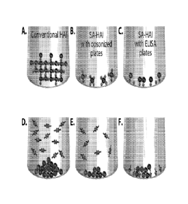
[0039] Figure 1. Patterns of erythrocytes in classical HA / HAI and surface-
assisted HA / HAI
experiments. Left column: Classical HA / HAI. Central column: Surface-assisted
I IA / IIAI with
polypropylene plates soaked with opsonizing solutions. Right column: Surface-
assisted HA /
HAI with U-bottom ELISA plates pre-coated with the virus. A. - Erythrocytes
develop a 3D
"lattice" with unblocked viruses (formation of a "halo"). B.- Erythrocytes
anchor to the
activated walls of the U-shaped well via unblocked viruses (formation of a
"micro-halo"). C. -
Erythrocytes anchor to the unblocked viruses pre-attached to the walls of the
U-shaped well
(formation of a "micro-halo"). D.- Erythrocytes sediment to the center of the
U-shaped well,
unobstructed by viruses blocked with Abs (formation of a "button"). E. -
Erythrocytes sediment
to the center of the well, unobstructed by viruses blocked with Abs (formation
of a "micro-
button"). F. - Erythrocytes sediment to the center of the well unobstructed by
viruses blocked
with Abs (formation of a "micro-button"). =
[0040] Figures 2A-B. Images of surface-assisted HA / HAI experiment with
opsonized plates.
A. SA-HA experiment with human group 0 erythrocytes and turkey erythrocytes.
Images of the
wells with micro-halos and micro-buttons were enlarged from the panel photo-
registered in the
AID EL [SPOT reader (AID ELRO4 AID GmbH, Germany). The two vertical columns of
wells
on the left of the figure show the formation of micro-halos and micro-button
at different virus
dilutions. Circled are dilutions selected for inhibition assays. The single
well shown in the
bottom center of the figure is a "button" from a classical HAI for comparison.
The magnified
- 12-
CA 02776405 2015-10-26
images of the four wells to the right were registered using AID ELISPOT reader
(Cell
Technology Inc., MD). B. SA-HAI experiment with human group 0 erythrocytes,
Solomon
Islands HIN1 influenza virus, and sera from the donors immunized for influenza
in season 2007
/ 2008. SA-HAI titers were determined visually. Circled arc dilutions selected
as SA_HAI titers.
Circling between boxes designates selection of the intermediate dilution.
[0041] Figure 3. Algorithm of digital processing of hemagglutination
patterns. A. General
scheme of the algorithm. B. Transformation of the hemagglutination images
during image
processing.
[0042] Figure 4. Flowcharts for digital processing of SA-HA / HA! plates.
A. Selection of the
image processing variables, single-well mode. B .General processing algorithm
for a single
image. C. Single plate processing mode. D. Multiples plate processing mode. E.
Plate layout and
curve fit data analysis.
[0043] Figures 5A-C. Typical SA-HA1 plate layout. A. Primary image of a
typical plate (a
real plate presented). Sera samples S108, Sill -S114 were placed in duplicates
in the columns
(vertical layout). Columns 11 and 12 contain a standard serum (shown is serum
1410 from a donor
immunized for influenza in the 2009 / 2010 season). B. Processed image of the
same plate.
Squared with red dash line are the wells containing the standard at the
dilution 1:3200, selected
as the Standard Linked Dilution (LSD). C. HAP values determined for the same
plate. Squared
with red dash line are Linked HAP values (LHAP).
[0044] Figures 6A-B. Using Linked Hemagglutination Parameter in calculation
of SA-HAI
titer. A. Correcting of the LHAP value in the process of splining of the
titration curve. The
corrected LHAP is 318.B. Determination of the SA-HAI titer for the sample S112
(i.e., finding
- 13-
CA 02776405 2015-10-26
the dilution factor corresponding to the spline-corrected LH.AP value). The
titer is 5045. Shown
is an illustrative procedure, performed manually in Excel editor.
[0045] Figure 7. Determination of the SA-HA titer of influenza H1N1 virus.
Virus:
A/Solomon Islands/3/2006 [HIN1]. Erythrocytes: Turkey. SA-HAI mode: Opsonized
plates.
Shown is an illustrative procedure performed manually in Excel editor. The SA-
HAI titer was
found to be ¨10090. The classical HA titer was found to be = 240 (data not
shown).
[0046] Figure 8. Comparison of Full Volume and Low Volume modes of
classical HAI assay.
Virus: A/Brisbane/59/2007 [H1N1], BPL-inactivated CDC standard. Erythrocytes:
Turkey. Full
Volume Mode: Component aliquots 30 uL, final volume 90 tit, U-bottom
polystyrene 96-well
plate (image made with a digital camera). Low Volume Mode: Component aliquots
7 tit, final
volume 21 tiL, V-bottom polypropylene 96-well plate (image made with an
ELISPOT plate
reader).
[0047] Figures 9A-B. A. - Comparing classical HAI and SA-HAI titers for
donor sera using
human erythrocytes and Solomon Islands H1N I virus. SA-HAI assays were
performed using
opsonized plates. Donor pre- and post-vaccination sera: From 15 donors
immunized for
influenza in the season 2007 / 2008. Erythrocytes: Human group 0. Virus:
A/Solomon
Islands/3/2006 [1-11N 1], BPL-inactivated standard from the US Centers for
Disease Control and
Prevention, Atlanta, GA. B.¨ Dataset used in A.
[0048] Figures 10A-B. A. - Comparing classical HAI titers determined with
human
erythrocytes and SA-HAI titers with turkey erythrocytes. Experiments were
performed with pre-
and post-vaccination sera from 15 donors immunized for influenza in the season
2007 / 2008.
SA-HAI assays were performed using opsonized plates. Erythrocytes: classical
HAI - human
- 14-
CA 02776405 2015-10-26
group 0; SA-HAT - turkey. Virus: A/Solomon Islands/3/2006 [HIN1], BPL-
inactivated standard
from CDC. B. ¨ Dataset used in A.
[0049] Figure 11. Comparing classical HAI titers determined with human
erythrocytes and
SA-HAI titers using guinea pig erythrocytes. SA-HAI assays were performed
using opsonizal
plates. The experiment was performed with pre- and post-vaccination sera from
three donors
immunized for influenza in the season 2007 / 2008. Erythrocytes: classical HAI
- human group
0; SA-HAI - guinea pig. Virus: A/Solomon Islands/3/2006 [H1N1], BPL-
inactivated standard
from CDC.
[0050] Figures 12A-C. Comparing classical HA titers (A.) and SA-HA titers
(B.) for influenza
viruses in allantoic fluids. SA-HA assays were performed using ELISA plates
lmmulux HB
immunoassay microplates (Dynex Technologies, catalog # 1011). Classical HA
assays were
performed in Low Volume mode using V-bottom polypropylene plates.
Erythrocytes: Turkey.
Viruses: Mice allantoic fluids, year 2010. C. ¨ Datasets used in A. and B.
[0051] Figure 13. Comparing classical HAI and SA-HAI titers using turkey
erythrocytes and
H1N1 influenza virus in allantoic fluids. Virus: A/Brisbane/59/2007 [H1N I].
Other experimental
conditions: As in Figure 12.
[0052] Figure 14. Comparing classical HAI and SA-HAI titers using turkey
erythrocytes and
H3N2 influenza virus in allantoic fluids. Virus: A/Wisconsin/67/2005 [H3N2].
Other
experimental conditions: As in Figures 12 and 13.
[0053] Figures 15A-B. SA-HAI analysis of cross-protection against swine flu
with seasonal
influenza vaccine 2009 / 2010. SA-HAI assays were performed using opsonized
plates. Sera:
From 27 donors immunized with the anti-influenza Fluvirin vaccine in season
2009 / 2010, pre-
and post-vaccinated. Viruses: A/Brisbane/59/2007 [HIN I [ (4.) and
A/California/7/2009 [H1N1]
- 15 -
CA 02776405 2015-10-26
(B.), BPL-inactivated standards from CDC. Erythrocytes: Turkey. Protective SA-
HAI level ¨640
was derived as a product of the protective titer accepted in the classical HAI
(-64) and the factor
of sensitivity enhancement in the SA-HAI assays (-10-fold).
[0054] Figure 16. SA-HAI analysis of samples from in vitro MIMIC setups
immunized with
vaccine and recombinant antigen. SA-HA assays were performed using ELISA
plates Immulux
HB immunoassay microplatcs (Dynex Technologies, catalog/4 1011). MIMIC setups
based on
immune cells from human donors were immunized in vitro with the Fluvirin-2010
seasonal
vaccine and with recombinant H1 hemagglutinin that originated from the
A/California/7/2009
[H1N1] "Swine Flu" virus (Protein Sciences). Erythrocytes: Turkey. Virus in
the SA-HAI assay:
A/California/7/2009 [H1N11, BPL-inactivated standards from CDC.
[0055] Figure 17. Testing affinity profile of potentially protective
antibodies in human sera
(A.) and MIMIC samples (B.) with SA-I IA1 assay. Human sera: From donors
immunized with
Fluvirin in season 2009 / 2010. MIMIC sample: From MIMIC setup made using
immune cells
from donor /1654, year 2010. PS antigens: Recombinant H1 hemagglutinin derived
from
A/California/7/2009 [HINI] "Swine Flu" virus (Protein Sciences). __
-15a-
CA 02776405 2012-04-02
WO 2011/050027 PCT/US2010/053322
DETAILED DESCRIPTION OF THE INVENTION
[0056] As used herein, the term "antibody" is used in the broadest sense
and encompasses
monoclonal antibodies, polyclonal antibodies, chimeric antibodies, humanized
antibodies, single-
chained antibodies, and antibody fragments (e.g., Fab, F(ab'), Fv) from
various mammalian and
avian species. Antibodies useful in the methods of the invention have the
shared characteristic of
a potential for having functional binding activity for an agglutinating
factor, such as a virus, or a
target object, such as an erythrocyte. Thus, the antibodies have the potential
for binding and
causing agglutination. Reference to a "potential" simply means that the
antibodies are of a type
known to have such a characteristic. It will not be known until after the
antibodies are assayed
whether they do, in fact, have functional binding activity for an
agglutinating factor or a target
object, or whether they can bind and cause agglutination.
[0057] As used herein, an "agglutinating factor" is a molecule that has the
potential to
agglutinate the target objects of the present invention. Agglutinating factors
include viruses,
virus-like particles, bacteria, proteins. Suitable viruses include DNA
viruses, RNA viruses, and
retroviruses. Specific viruses include adenoviruses, enteroviruses,
reoviruses, myxoviruses
(including the influenza viruses), poxviruses, and flaviviruses.
[0058] As discussed above, viruses may be detected and/or quantitated using
the methods of
the present invention. Viruses that may be detected and/or quantitated using
the methods include
any virus that have the potential to form an agglutination with target
objects, such as
erythrocytes. Suitable viruses include DNA viruses, RNA viruses, and
retroviruses. Specific
viruses include adenoviruses, enteroviruses, reoviruses, myxoviruses
(including the influenza
viruses), poxviruses, and flaviviruses.
- 16 -
CA 02776405 2012-04-02
WO 2011/050027 PCT/US2010/053322
[0059] The target objects used in the methods of the invention are those
that can agglutinate
upon binding with the agglutinating factors of the present invention. The
particular identity of
the target object is not critical, as long as the characteristics of the
object permit consistent,
reproducible results in the methods of the present invention. Suitable target
objects include cells
and microspheres. The cells may be a population of one particular cell type,
such as
erythrocytes, lymphocytes, epithelial cells, and endothelial cells. An
exemplary population is a
population of erythrocytes. The source of the erythrocytes is not particular
important, as long as
the cells have the potential to form an agglutination in the presence of an
agglutinating factor
such as a virus. Suitable erythrocytes include avian erythrocytes, such as
chicken erythrocytes
and turkey erythrocytes, and mammalian erythrocytes, such as human
erythrocytes, guinea pig
erythrocytes, mouse erythrocytes, rat erythrocytes, bovine erythrocytes,
equine erythrocytes, goat
erythrocytes and sheep erythrocytes. Human erythrocytes may be from a donor of
any blood
group, such as group A erythrocytes, group B erythrocytes, group AB
erythrocytes, and group 0
erythrocytes. Examples of suitable microspheres include latex microspheres and
other
microspheres that can be readily bound by virus and agglutinatized. In one
aspect, the
microspheres are latex microspheres coated with a receptor that binds with the
virus.
[0060] In certain aspects, erythrocytes may be used as the target objects,
and the
concentration of the erythrocytes can be selected such that they are present
in a well of a plate at
a concentration of below about 0.01% hematocrit, below about 0.05% hematocrit,
below about
0.1% hematocrit, below about 0.15% hematocrit, or below about 0.2% hematocrit.
[0061] As discussed above, the methods of the present invention may be
practiced using
culture plates, such as tissue culture plates, where the wells have been
opsonized by coating the
well with a protein or a lectin. The wells may be opsonized by inserting a
solution comprising
- 17 -
CA 02776405 2012-04-02
WO 2011/050027 PCT/US2010/053322
one or more proteins, and/or one or more lectins into the well, and allowing
the proteins and/or
lectins to attach to the surface of the well. The solution can then be removed
from the well, and
the well can optionally be washed. Suitable proteins include bovine serum
albumin and human
serum albumin. Serum albumin from other mammalian species may be used as well,
such as
from goat, horse, pig, rabbit, mouse and rat. As further discussed above, the
methods of the
present invention may be practiced using culture plates where the wells are
activated. Plates
having wells with such a characteristic include plates commercially available
for use in ELISA
assays. While the shape of the wells used in the methods of the present
invention may vary
depending on the particular steps being used, plates having U-shaped wells and
plates having V-
shaped wells are particularly useful.
[0062] As used herein, a "sample" refers to any type of material of
biological origin
including, but not limited to, a cell, fluid, tissue, or organ isolated from a
subject, including, for
example, blood, plasma, serum, fecal matter, urine, semen, bone marrow, bile,
spinal fluid,
lymph fluid, samples of the skin, external secretions of the skin,
respiratory, intestinal, and
genitourinary tracts, tears, saliva, milk, blood cells, organs, or biopsies.
[0063] As discussed above, agglutination is detected in the first and
second embodiments at a
sensitivity higher than that achieved when the methods are performed in non-
opsonized or non-
activated wells and under conditions that provide agglutination in the well
volume rather than on
the surface of the well bottom. The sensitivity is increased by at least about
7 times, 8 times, 9
times, 10 times, 20 times, 30 times, 40 times, 50 times, 60 times, 70 times,
80 times, 90 times,
100 times, 150 times, or even 200 times, or more. Similarly, agglutination is
detected in the third
and fourth embodiments at a sensitivity higher than that achieved when the
methods are
- 18 -
CA 02776405 2012-04-02
WO 2011/050027 PCT/US2010/053322
performed in non-opsonized or non-activated wells. The sensitivity is
increased by at least about
7 times, 8 times, 9 times, 10 times, 20 times, 30 times, 40 times, or even 50
times, or more.
Decreasing the concentrations of viruses and erythrocytes to increase
sensitivity of HA / HAI
assay
[0064] The concentrations of virus and erythrocytes used in the HA / HAT
assay dictate
sensitivity of the method. For higher sensitivity, the concentration of the
virus used in the assay
should be reduced as much as possible. While this statement is self-
explanatory for the
sensitivity to the virus itself in HA mode, a lower concentration of the virus
used in the HAI
mode would also result in lower concentrations of antibodies in the
experimental fluids
necessary for blocking attachment of the virus to the erythrocytes, which is
equivalent to
increasing sensitivity to the tested sera and antibody solutions.
[0065] However, reducing the virus concentration in the classical HA / HAI
assays is limited
by need to discriminate between agglutination and non-agglutination of the
erythrocytes by the
virus. Specifically, in HA assays the titer determined for the virus is equal
to the virus dilution
showing the borderline between agglutination and non-agglutination. In fact,
the HA titer
manifests the virus concentration below which no functional observation is
possible within the
given assay. This is why the concentration of the virus in the HAT assay is
normally maintained
four times higher than the virus titer (Hierholzer et al. (1969) Applied
Microbiol. 18, 824-833;
WHO Manual on Animal Influenza Diagnosis and Surveillance,
WHO/CDS/CSR/NCS2002.5 Rev. 1).
- 19 -
CA 02776405 2012-04-02
WO 2011/050027 PCT/US2010/053322
[0066] In classical HA / HAI, the agglutination / non-agglutination
discriminating signal is
the formation of a halo of the erythrocytes glued into the spatial lattice by
virus particles, or a
button of precipitated erythrocytes.
[0067] It would seem that the virus concentration could be lowered further
if the
corresponding concentration of the erythrocytes could be decreased as well.
This pathway,
however, is also limited within the classical HA / HAT method, because at
erythrocyte
concentrations below ¨0.1-0.2% hematocrit (HCT), formation of the spatial
lattice of
erythrocytes glued by virus particles becomes impossible.
[0068] Thus, to overcome physical limitations of the classical HA / HAT
assay, development
of new alternative methods was necessary.
Principles of the surface-assisted HA / HAI method
[0069] The method was developed in two versions, considered separately
below.
Surface-assisted HA / HAI using opsonized 96-well plates
[0070] While exploring decreased erythrocyte and virus concentrations in
the classical HA /
HAT assay, new effects were revealed. Specifically, if U-bottom plates, such
as 96-well U-
bottom plates, were used and pre-soaked with certain opsonizing solutions,
such as solutions of
bovine or human serum albumin (BSA, HSA), then the erythrocytes bearing
influenza viruses
attached to their surfaces anchored to the opsonized surface of the well upon
precipitation and
stayed attached, forming a two-dimensional "micro-halo," as opposed to the
three-dimensional
halo in the classical HA / HAT (Fig. 1; central and left panels). When the
virus was absent or
blocked with virus-specific antibody, the precipitating erythrocytes were not
able to anchor to the
opsonized surface and gradually concentrated near the center of the well
bottom, due to
- 20 -
CA 02776405 2012-04-02
WO 2011/050027 PCT/US2010/053322
Brownian motion, thus forming a "micro-button." Importantly, these effects
were observed at
concentrations of erythrocytes 20-50 times lower, and influenza viruses 30-600
times lower than
in the classical HAT, depending on the virus strains.
[0071] Characteristic patterns of the Surface-Assisted HA / HAT (SA-HA /
HAI) using
opsonized plates are presented in Figure 2. In all examples of SA-HA / HAT
experiments with
opsonized plates presented in the current application, opsonization was
performed with high-
grade BSA solution, 2% in PBS / NaN3 saline. Other opsonizing solutions can be
used, for
example solutions of glycoproteins, such as lectins.
Suiface-assisted HA / HAI using ELISA plates
[0072] In another embodiment of the present invention, an alternative
version of the SA-HA /
HAT was developed based on 96-well ELISA plates (Fig. 1; right columns).
Specifically, U-
bottom ELISA plates (for example, Immulux HB from Dynex, catalog # 1011, or
ImmunoGrade
plates from BrandTech Scientific, catalog #781724) were first coated with
influenza virus and
then blocked with 2% high grade BSA in a manner similar to a regular ELISA
protocol.
Erythrocytes applied to such plates anchored to the virus particles already
attached to the ELISA
surface, forming a two-dimensional "micro-halo" that looked quite similar to
that observed in the
experiments with opsonized plates. Application of anti-virus sera or
antibodies on the top of the
pre-attached viruses abolished the anchoring of erythrocytes to the attached
viruses, and
precipitating erythrocytes gradually concentrated near the center of the well,
forming a
"micro-button," similar to that described for the SA-HAI using opsonized
plates (Fig. 1; right
panel).
Digital image processing and analysis
- 21 -
CA 02776405 2012-04-02
WO 2011/050027 PCT/US2010/053322
[0073] Results of the SA-HA / HAI experiments can be evaluated visually, in
a manner
similar to the classical HA / HAI. However, using image processing makes such
evaluation more
precise and significantly reduces the subjectivity of operator.
HA / HAI image acquisition
[0074] Digital images of HA / HAI assay wells can be recorded using an
automated imaging
system capable of taking high-resolution images of individual wells on a 96-
well plate. Adequate
systems are available commercially, such as those designed for EliSpot assay
analysis, that can
acquire the digital images necessary for HA / HAI image analysis. For example,
images of HA /
HAI assay wells can be recorded on an AID ELISPOT plate reader and stored in
JPEG format at
1088 x 1036 resolution and 24-bit color depth are adequate for HA / HAT
analysis. The image
acquisition software included with these systems is typically full-featured in
terms of camera and
translation stage control, well selection, and file management; however the
included software is
intended for ELISPOT analyses and is not capable of proper quantification of
HA or HAT titers.
Thus, these types of imagers are useful only for their image acquisition
capabilities, and the
recorded HA / HAT well images were processed using software developed
specifically for
determining the HA / HAI titers, as described below.
Concept of the hemagglutination parameter, HAP
[0075] To quantify and compare hemagglutination patterns in different
wells, a numeric
parameter was devised that was able to characterize the degree of
agglutination of the
erythrocytes used in the assay. The hemagglutination patterns formed by
erythrocytes in the SA-
HA / HAT assay can be "buttons," "halos," or intermediate between the two. Two
main
properties of such patterns are evident: the area over which erythrocytes
attach to the well
surface and the density of erythrocytes per area unit. These properties were
used to calculate a
- 22 -
CA 02776405 2012-04-02
WO 2011/050027 PCT/US2010/053322
numeric value, called the Hemagglutination Parameter (HAP) which is
proportional to the degree
of agglutination observed in the given well. The HAP parameter can be defined
as
HAP = <R> / <I> (1),
where <R> is the average distance of a pixel from the center of the
hemagglutination pattern
(HA spot), and <I> is the average intensity of the pixels in the area. The HAP
is minimal for the
"button" pattern and maximal for the "halo" pattern. The hemagglutination
titration curves in the
SA-HA or SA-HAI assays can thus be presented as sets of the HAP values linked
to the serial
dilutions of the virus or sera, respectively, depending on the type of assay.
Curve fitting and
curve dissecting applied to such a dataset allows precise determination of the
titration point.
Thus, development and use of the numerical Hemagglutination Parameter
transferred the
analysis of the HA or HAT assays from subjective visual evaluation to a
precise mathematical
calculation. The principles of the calculation of the Hemagglutination
Parameter (HAP) can be
applied to similar calculations of Agglutination Parameters for the target
cells other than
erythrocytes, or for the target objects other than cells, such as latex beads.
Image Processing Algorithm
[0076] The objective of the HA / HAT image processing algorithm is to
separate the
hemagglutination pattern from the rest of the well image and to then determine
the HAP value.
The algorithm developed for the SA-HA / HAT assay is illustrated by the
flowchart in Figure 3A.
The process is as follows.
[0077] First, an image of the well is obtained as described above. The well
image is then
cropped to primarily encompass the central pattern-containing portion of the
well. The cropped
image is converted to negative grayscale and then the contrast is adjusted to
fill the entire
intensity spectrum and enhance the hemagglutination spot. Intensity
thresholding is applied to
- 23 -
CA 02776405 2012-04-02
WO 2011/050027 PCT/US2010/053322
remove pixels not corresponding to erythrocytes. The image is then segmented
using either edge
detection or color segmentation algorithms to isolate the erythrocyte pattern.
Edge detection, as
illustrated in Figure 3B, required converting of the color image to a binary
(black and white)
image and segmentation using a binary gradient mask, followed by dilation,
hole filling, and
image erosion. The resulting segmented binary image represents the HA spot
pixels, with all
background pixels eliminated. The grayscale image is then mapped onto the
segmented binary
image, giving an intensity image of the HA spot. The average distance from the
spot centroid
<R> and average intensity of the spot <I> are calculated from the intensity
image, and finally the
HAP value is calculated as the ratio (1) above.
Single Well Image Processing Mode (Pre-Processing)
[0078] The user has control over some of the analysis variables and must
either choose their
values prior to image processing or accept default values. These variables
include, for example,
threshold intensity, crop area, and image segmentation type. Once these values
are selected they
will be applied to all wells in a batch, be it an entire plate or multiple
plates. To optimize the
detection of the HA! HAT pattern a priori, the software has a single-well
processing mode that
allows a user to experiment with the analysis variables on a single well image
before applying
them to an entire batch. The process is illustrated in the flowchart in Figure
4A. A user selects
the single-well analysis mode, loads a single-well image and then sets the
analysis variables. The
image is then processed using the algorithm illustrated by flowcharts in
Figures 3A and 4B, and
the resulting HAP value is displayed, as well as the processed image, showing
the detected
hemagglutination pattern. The user then accepts the values and continues with
batch processing
or iteratively adjusts the variables and re-processes the image until
acceptable values are
established. Typically, values are chosen to provide proper detection of the
HA / HAI pattern for
- 24 -
CA 02776405 2012-04-02
WO 2011/050027 PCT/US2010/053322
the two control cases: the No Virus case in which a micro-button is formed in
the well bottom
and the No Sera case in which a micro-halo is formed. Because these cases
represent the
hemagglutination extremes, proper detection of their patterns increases the
likelihood of proper
detection of the hemagglutination patterns for all wells in the batch.
Tull Plate Image Processing Mode
[0079] Once pre-processing is complete and acceptable variable values are
found, an entire
plate can be processed, as illustrated by the flowchart in Figure 4C. A user
selects the single-
plate analysis mode and then loads a directory which contains images of wells
from the plate.
The software verifies that the images are valid for processing and then
normalizes the
background intensity for all images in the folder by sampling each image
around the well
periphery and normalizing all wells to the maximum average intensity found.
The HA / HAI well
images are then batch-processed by applying the algorithm presented in Figure
4B to each well
serially until all wells have been processed. Once a well is processed, its
HAP value is readily
determined and the results are automatically written to a data file in XML
format along with its
calculated background value and the analysis variables. Screenshots of the
original and
processed plate images are also saved along with an Excel file containing the
formatted HA
parameter values for each well.
Multiple Plate Batch Processing Mode
[0080] Multiple-plate batch processing mode allows processing of multiple
plates at once
using the same settings for each plate. This mode requires minimal user
interaction and is
intended for high-throughput image analysis. The process is illustrated by the
flowchart in Figure
4D. The user first performs a pre-process analysis to determine proper
variable values and then
selects batch plate analysis mode. A directory containing multiple plate
directories is then
- 25 -
CA 02776405 2012-04-02
WO 2011/050027 PCT/US2010/053322
selected and each plate directory is processed serially, similar to the full-
plate processing mode.
The process continues until all plate directories have been processed. If a
directory has been
processed previously, the software will check for any missing data files, such
as screenshots or
Excel files and either re-create them if an XML file is present or re-process
the plate in its
entirety.
Web-Based Curve-Fitting and Data Analysis Application
[0081] After a SA-HA / HAT plate is processed, the titration curves need to
be curve-fitted to
determine their titration points using the Linked Standard Dilution / Linked
HAP value method,
LSD / LHAP (below). To address high-throughput analysis of the serial assays,
a web-based
automated curve fitting and database interface application was developed. The
application can
automatically curve-fit the SA-HA / HAT titration curves using a weighted five-
parameter
logistic equation, find the titration point using the LSD / LHAP method and
then catalog the
results into a central database that can be accessed by multiple users
simultaneously. The
software comprises four main modules: a plate designer module, a file upload
module, a curve-
fitting module and a database module. As illustrated in Figure 4E, the user
first creates a virtual
plate in the plate designer module and defines the HA / HAT plate layout
including all reagent
and cell information for each well such as name, type, concentration or
dilution, and lot or donor
number. The virtual HA / HAI plate is stored in the database and can be linked
to actual plate
data. After a plate is image-processed, the user then uploads the XML file to
the database using
the file upload module and links the actual data to the corresponding virtual
plate. At this point,
the plate is well-defined in the database and ready for curve-fitting. The
curve-fitting module
automatically fits a five-parameter logistic equation to the defined sample
and standard curves on
the plate and a user-defined linked dilution is applied to find the titration
point for each sample
- 26 -
CA 02776405 2012-04-02
WO 2011/050027 PCT/US2010/053322
on the plate. The titration point is stored in the database and made available
to users through the
database module, which supports complex queries for data mining, plotting and
dataset creation.
The database module can export plots or datasets for use in reports or further
analysis using other
software.
Concept of Linked Standard Dilution and Linked Hemagglutination Parameter, LSD
/ LHAP
[0082] The concept described below is another important element of the SA-
HAI method,
along with transferring the agglutination reaction from the solution to the
activated surface, and
with digital image processing and computation of the numerical
hemagglutination parameter.
HA and HAI assays are prone to significant variability, caused by instability
of the virus
agglutinating capacity, changes in temperature, and variation in the quality
of erythrocytes. To
increase reproducibility and stability of the SA-HA / HAT data, an advanced
standardization
protocol named Linked Standard Dilution / Linked Hemagglutination Parameter
(LSD / LHAP)
was developed.
[0083] Using the LSD / LHAP approach, every SA-HAI plate is organized in a
strictly
standardized way (Fig. 5). The samples are serially diluted in the columns 1
to 10 of the 96-well
format, in duplicate. Columns 11 and 12 are occupied with a serum selected as
a standard for all
the SA-HAI plates that are going to be used within a project. Wells All and
Al2 contain no
virus and no serum (double negative control), and wells H11 and H12 contain
virus but no serum
(single negative control). The standard serum is serially diluted six times
starting from the wells
B11 and B12, down to the wells Gil and G12, in such a manner that wells
containing the
standard demonstrate the whole dynamic range of the HAT titration, from a
clear micro-button to
a clear micro-halo. A Linked Standard Dilution (LSD) is selected in the middle
of the standard
titration array in such a way that the hemagglutination pattern would be
between halo and button,
- 27 -
CA 02776405 2012-04-02
WO 2011/050027 PCT/US2010/053322
as it takes place for wells Ell and E12 containing the standard serum (#10)
diluted 1:3200, as
shown in Figure 5.
[0084] The HAP value that is determined for the chosen LSD is named Linked
HAP, or
LHAP. The final LHAP value can differ to a certain extent from the numerical
values determined
for the LSD wells, because the titration curve is set using a splining
procedure that smoothes
random scattering of the datapoints (Fig. 6A). The LSD parameter is kept the
same for all the
setups performed in a whole study. The LHAP value found for the standard sera
in each plate is
used as a titration target for all the tested samples in the plate (Fig. 6B).
This means that the final
objective for the software that processes the HAI titration curves of the
tested sera for a given
SA-HAI plate is the calculation of sample dilutions that would provide the HAP
values equal to
the LHAP value determined for the standard serum in the plate.
[0085] If, for example, the room temperature, the quality of erythrocytes,
or the virus
agglutination capacity, e.g., changes during a long-term study, the
hemagglutination patterns in
the LSD wells of the standard serum would change accordingly (for example,
become closer to a
button if the agglutinating capacity decreases), and the corresponding LHAP
would change as
well (for this example, decreases). However, the corresponding HAP values in
the wells with the
tested sera will shift in the same direction as the LHAP (for this example,
decrease). As a result,
because the software uses the changed LHAP as the updated titration target,
the resulting
SA-HAI titers of the tested sera will remain practically unchanged.
Testing affinity profile of functional antibodies in SA-HAI assay with ELISA
plates
[0086] The classical HAI assay integrates neutralizing effects of
functional antibodies having
different affinities. Other immunosorptive methods, such as ELISA are able to
characterize only
- 28 -
CA 02776405 2012-04-02
WO 2011/050027 PCT/US2010/053322
antibodies of relatively high affinity. The reason for such difference is that
in the classical HAI
the complexes of antibodies with the virus do not pass through the procedures
of sample removal
and washing, which constitutes a backbone of the majority of immunosorptive
assays, including
ELISA. In the classical HAT, even antibodies of relatively low affinity can
demonstrate high
titers, provided that the antibodies are present in the serum in large
quantities.
In the SA-HAI with ELISA plates of the present invention, sera samples can be
removed from
the wells after incubation with the virus that is pre-attached to the plates
and before application
of erythrocytes. This actually triggers dissociation of lower affinity
antibodies from the virus
particles and removal of those antibodies from the reaction volume, which
affects the observed
titers.
[0087] SA-HAT experiments with and without post-incubation removal of the
sera samples
and washing wells with PBS / NaN3 saline to examine the effects of low
affinity Abs on the HAT
titer showed that the difference between titers obtained in the two modes can,
in general, be 2-5-
fold, and even higher for some samples (Fig. 17; the titers obtained with
sample removing and
well washing are always lower). Notably, manipulations with the sample volumes
do not affect
the density of the viruses attached to the SA-HAI plates, as was demonstrated
in separate ELISA
tests (data not shown) Also important is that decreasing the virus density
after washing the wells
would increase the observed sera titers due to depletion of the agglutinating
capacity in the
wells; the effect opposite to what was observed in the experiments. Performing
the SA-HAI
assay with ELISA plates with and without removal of sera samples and washing
wells can help
to characterize the relative contribution of high- and low-affinity functional
antibodies in the
humoral immune response.
- 29 -
CA 02776405 2012-04-02
WO 2011/050027 PCT/US2010/053322
[0088] As stated above, hemagglutination (HA) and hemag2lutination
inhibition (HAI)
functional assays remain important instruments of analysis of virus-cell
interaction and efficacy
of virus-specific antibodies and sera. However, the classical protocols of HA
and HAT
demonstrate limited sensitivity towards many viruses and require significant
volumes of virus
samples, erythrocytes, sera, and antibodies.
[0089] Embodiments of the present invention comprise a new method for the
functional
characterization of viruses and virus-specific antibodies and sera, the
Surface-Assisted
Hemagglutination / Hemagglutination Inhibition functional assay, the "SA-HA /
HAT" assays.
Embodiments of the present invention demonstrate sensitivity of the SA-HA
assay to various
influenza viruses about 7-200 times higher than the traditional HA, and
sensitivity of the SA-
HAT assay to influenza-specific antibodies about 10-50 times higher than in
the traditional HAI,
depending on the type of the virus and the type of erythrocytes used.
[0090] This enhancement in sensitivity allows analysis of low concentration
experimental
samples, and saves precious materials, such as convalescent sera and viruses.
The SA-HA / HAT
can use the same types of erythrocytes as the traditional HA / HAI: human,
mammalian, and
avian.
[0091] Performing the SA-HAI assay in the mode that uses ELISA plates
allows
determination of the relative contributions of low- and high-affinity
functional antibodies in the
HAT titer, which is technically impossible in the classical HAI assay. This
makes the ELISA
mode of the SA-HAI assay a valuable tool that can provide deeper insight into
the quality of
protective humoral immune responses.
[0092] The SA-HA / HAI assay results can be evaluated visually, in a manner
similar to
classical HA / HAI assays. However, visual evaluation lacks adequate precision
for high-
- 30 -
CA 02776405 2012-04-02
WO 2011/050027 PCT/US2010/053322
sensitivity experiments and it is prone to human errors, due to differences in
perception of
different operators. Photo-registration and digital processing of the SA-HA /
HAT images
increases the precision of the method and eliminates the subjectivity of
visual evaluation.
Introduction of the numerical Hemagglutination Parameter (HAP) that reflects
the degree of
agglutination in every well of the SA-HA or SA-HAI plate changes the analysis
of the HA or
HAT assays from a subjective visual evaluation to precise mathematical
processing of titration
curves.
[0093] Introduction of the advanced standardization concept using Linked
Standard Dilution
and Linked Hemagglutination Parameter of the standard significantly decreased
the variability of
the SA-HAI data. Image processing and computation of the SA-HA and SA-HAI
titers can be
performed in-line with photo registration and in real time. The SA-HA / HAI
method can be
performed in a high-throughput mode and allows automation.
Examples
[0094] In all the examples below, the major solvent used for all components
of the assay,
such as media, tested sera, antibody samples, viruses and erythrocytes was PBS
saline containing
0.1% of sodium azide NaN3 (PBS /NaN3). Sodium azide was added to protect the
saline from
bacterial or yeast contamination.
Example 1. SA-HA / HAI method using opsonized plates
[0095] Protocol of the SA-HA / HAT using opsonized U-shaped 96-well plates
(U-bottom
96-well format plate, clear polystyrene, Coming # 3795).
-31 -
CA 02776405 2012-04-02
WO 2011/050027 PCT/US2010/053322
Processing of erythrocytes
[0096] Types of erythrocytes used in the SA-HA / HAI experiments were: human
group 0,
turkey, chicken, guinea pig, horse. Human erythrocytes were acquired from
Florida Blood Bank
or via internal blood donations at Vaxdesign Corp. Turkey, chicken, guinea pig
and horse
erythrocytes were purchased from Rockland Immunochemicals as suspensions in
citrate buffer.
Aliquotting and storage
[0097] The erythrocytes were aliquotted immediately after receiving by 1.0
mL in microfuge
vials and stored at 4 C until further use. The normal storage time was no
longer than 3 weeks for
human, turkey, horse and chicken erythrocytes, and no longer than 1 week for
guinea pig
erythrocytes.
Washing and re-suspending
a. The vial containing the erythrocytes was centrifuged (600g, 2 min).
b. The supernatant + the top layer of the cells were aspirated carefully
using a 1-mL pipette.
PBS / NaN3 (1 mL) was added to the pellet. The cells were re-suspended by slow
back-
and-forth pipettine.
c. Steps a-b were repeated.
d. The cells were again centrifuged (600g, 2 mm). The supernatant + the top
layer of the
cells were aspirated using a 1-mL pipette, and after that 1 mL of 0.5% BSA in
PBS /
NaN3 was added to the pellet. The cells were re-suspended by slow back-and-
forth
pipetting, taking care to produce no bubbles.
e. The cells were again centrifuged (900g, 5 min). The supernatant + the
top layer of the
cells were aspirated using a 1-mL pipette. The residual pellet represented the
100% HCT
- 32-
CA 02776405 2012-04-02
WO 2011/050027 PCT/US2010/053322
stock. Typically, the final pellet constituted ¨1/2 to ¨i/3 of the initial
quantity of the
erythrocytes.
The pellet was stirred slowly to make it homogeneous. An appropriate aliquot
of the
pellet was suspended in 0.5% BSA in PBS / NaN3. Unless otherwise specified,
the final
concentration should be 0.05% HCT for human, horse and turkey erythrocytes and
0.025% HCT for guinea pig erythrocytes.
[0098] The processing of erythrocytes described above was performed anew
for each day of
experiments. Any leftovers of the processed erythrocytes were disposed of
after the experiment.
Processing of the virus
[0099] The following BPL-inactivated virus standards were obtained from the
U.S. Centers
for Disease Control and Prevention, Atlanta, Georgia (CDC):
A/Brisbane/59/2007 [H1N1]
A/New Caledonia/20/99 [HIN1]
A/Solomon Islands/3/2006 [H1N1]
A/Wisconsin/67/2005 [H3N2]
[00100] These virus samples were stored undiluted in 1.5-mL microfuge vials at
4 C. During
the processing, the microfuge vial with the virus was stirred vigorously in a
bench vortex for 30
s; no sonication was used. Afterwards, the vial was centrifuged (400g, 5 mm).
The necessary
aliquot of the supernatant was taken out and diluted in 0.5% BSA / PBS / NaN3.
This processing
was performed anew for each day of experiments.
BPL-inactivated virus standard of A/California/7/2009 [H1N1] from the CDC or
American Type
Culture Collection (ATCC)
- 33 -
CA 02776405 2012-04-02
WO 2011/050027 PCT/US2010/053322
[00101] Due to the increased instability of the A/California/7/2009 WIN]]
virus sample, it
was aliquotted immediately after receiving in 0.5-mL portions, frozen and
stored at -80 C until
further use. For a serial of the SA-HA / HAI experiments, a frozen aliquot of
this virus was
thawed at room temperature and diluted in 4 mL of PBS / NaN3 saline. The
diluted sample was
sonicated on ice using Sonic Dismembrator, Model 500 from Fisher Scientific,
catalog #15-338-
550 at 12% power level, five times for 50 s. After sonication, 0.5 mL of 99%
glycerin was
admixed to the solution. Afterwards, the virus solution was further aliquotted
by 0.25 mL, and
those secondary aliquots were either used immediately or frozen and stored at -
80 C for further
use. After thawing, those secondary aliquots could be used immediately without
further
processing.
Influenza viruses in mice allantoic
[00102] The following influenza viruses in mice allantoic fluids were obtained
from Sanofi
Pasteur:
A/Brisbane/59/2007 [H1N1]
A/New Caledonia/20/99 [H1N1]
A/Solomon Islands/3/2006 [H1N1]
A/Wisconsin/67/2005 [H3N2]
B/Malaysia/2506/2004
B/Florida/4/2006
[00103] Due to instability of the agglutinating capacity of these samples,
they were aliquotted
into 0.05 mL portions immediately after receiving, frozen and stored at -80 C
until further use.
Before the experiment, the aliquots were thawed at room temperature, diluted
as necessary in
PBS / NaN; and used immediately on the same day.
- 34 -
CA 02776405 2012-04-02
WO 2011/050027 PCT/US2010/053322
Typical SA-HA experiment using opsonized plates
[00104] For most SA-HA experiments, the plate layout was horizontal (i.e., the
placement and
serial dilution of the samples performed from left to right).
Blocking / opsonization of the plate
[00105] The plate was filled with 2% BSA in PBS / NaN3, 160 [1.1, per well,
and incubated at
4 C in a planar plate shaker, ¨600 rpm for at least 40 min.
Pre-filling with media
[00106] The plate was flicked off and tapped upside down on a clean paper
towel. The plate
was filled with 0.5% BSA in PBS / NaN3 , 40 !IL per well.
Filling with virus
[00107] Unless specified otherwise, the virus initial dilution was 1:50 or
1:100.
[00108] Pre-diluted virus was added to the wells of the column 1, 40 p.L per
well, thus
becoming diluted 2 times. Dual serial dilutions of the virus were made from
left to right, from
the column 1 to the column 11 using an 8-channel 200- 1i1_, pipetter and
transferring by 40 p L per
channel in every pass. Back-and-forth pipetting in each column was used to mix
solutions
properly, not less than six pipettings per pass, producing no bubbles. The
last 40-p L portion
taken from the column 11 was discarded. The column 12 contained no virus.
Adding solution
[00109] 0.5% BSA in PBS / NaN3 was added to all the wells, 40 1i1_, per well,
not touching the
menisci. For this, the pipette tips were leaned on the top part of the well.
Mixing in a planar plate shaker
- 35 -
CA 02776405 2012-04-02
WO 2011/050027 PCT/US2010/053322
[00110] The plate was placed in the plate shaker, such as Digital mini
vortexer IKA MS3 from
IKA Works, Wilmington, NC, and a short (-5 s) mixing at 1000 rpm was performed
three times.
After mixing, erythrocytes were added.
Filling with erythrocytes and incubation
[00111] Erythrocytes processed and diluted as described above were added to
all the wells, 40
uL per well, the plate was again subjected to short mixing in the plate shaker
at 800 rpm, and
then incubated with shaking at 500 rpm for 30 min. Afterwards, the plate was
left still on the
bench for 2-4 h, depending on the type of erythrocytes used in the assay, to
allow erythrocytes to
precipitate and form the hemagglutination patterns.
Plate reading
[00112] The plate could be read and analyzed visually, or using photo-
registration in a short-
focus photo reader, such as an ELISPOT plate reader, AID ELRO4 AID GmbH,
Germany, with
subsequent digital processing of the patterns of hemagglutination, as
described above, and the
SA-HA titer determined as a midpoint of the HA titration (Fig. 7).
Typical SA- HAI experiment using opsonized plates
[00113] For most SA-HAI experiments, the plate layout was vertical (i.e., the
placement and
serial dilution of the samples were performed from the row A to the row H of
the plate).
[00114] The virus titer determined in the previous SA-HA assay was used to
calculate the virus
dilution to be used in the SA-HAI assay:
(SA-HAT assay virus dilution) = 4 x (SA-HA titer) (2)
Blocking of the plate
[00115] As described above for the typical SA-HA assay.
- 36 -
CA 02776405 2012-04-02
WO 2011/050027 PCT/US2010/053322
Pre-filling with media
[00116] As described above for the typical SA-HA assay.
Filling with sera or antibody solutions
[00117] Pre-dilutions of the tested sera were usually from ¨1:100 to ¨1:800,
depending on the
expected immune response. Pre-dilutions of MIMIC samples were usually ¨1:1 to
¨1:10,
depending on the expected antibody levels.
[00118] Pre-diluted sera or MIMIC samples were placed in the wells of the row
A, 40 L per
well. Dual serial dilutions of the samples were performed from row A to row G
or H using a 12-
channel 200-pt pipetter by transferring 40 L from the wells of the previous
row to the next
row. The technique of the dilution is the same as described above for the
typical SA-HA assay.
Adding virus
[00119] Virus diluted according to the results of the SA-HA test as specified
above was added
to all the wells, 40 p L per well, except for the No Virus negative control
wells, without touching
the menisci. For this, the pipette tips were leaned on the top part of the
wells.
Mixing with a planar plate shaker.
[00120] As described above for the typical SA-HA assay.
Incubation with virus
[00121] The plate was incubated in the planar shaker at 4 C (refrigerator) or
at room
temperature (on the bench) at 500 rpm, covered, for ¨1-2 h, depending on the
virus type. After
the incubation, erythrocytes were added.
Filling the plates with erythrocytes and incubation
[00122] As described above for the typical SA-HA assay.
Plate reading
- 37 -
CA 02776405 2012-04-02
WO 2011/050027 PCT/US2010/053322
[00123] As described above for the typical SA-HA assay. The digital processing
of the
hemagglutination patterns is described in details above.
Example 2. SA-HA / HAI method using ELISA plates
[00124] Protocol of the SA-HA / HAI assays using ELISA U-shaped 96-well plates
(Immulux
HB from Dynex, catalog # 1011 or ImmunoGrade BRANDPlates from BrandTech
Scientific,
catalog #781724).
Processing of erythrocytes
[00125] As described above. in Example 1.
Processing of the viruses
[00126] The processing was similar to the described above in the Example 1 for
the SA-HA /
HAT assays with opsonized plates, except that the final solvent used for
viruses before
application to the plates was BSA / NaN3 saline.
Typical SA-HA experiment using ELISA plates
[00127] For most SA-HA experiments, the plate layout was horizontal (i.e., the
placement and
serial dilution of the samples performed from left to right).
Pre-filling with saline
[00128] All the wells were filled with PBS / NaN3, 50 iL per well.
Filling with virus
- 38 -
CA 02776405 2012-04-02
WO 2011/050027 PCT/US2010/053322
[00129] The filling and serial dilution techniques and the typical layouts
were similar to that
described above in the Example 1, the protocol for SA-HA / HAI using opsonized
plates. The
filling aliquots were 50 pL per well.
Attaching virus to the plate
[00130] The plate was incubated overnight at 4 C (refrigerator), on a planar
plate shaker at
¨500 rpm.
Blocking
[00131] The plate was flicked off and tapped upside down on a clean paper
towel, filled with
2% BSA in PBS / NaN3, 200 iL per well, and incubated at 4 C on a planar plate
shaker, ¨400
rpm for at least 2 h.
Filling with erythrocytes and incubation
[00132] Erythrocytes diluted in the (0.25% BSA + 0.25% OVA) / PBS / NaN3 were
added by
50 luL per well. Typical concentrations of erythrocytes were 0.01-0.025% HCT.
Afterwards, the
plate was left still on the bench for ¨2-4 h., depending on the type of
erythrocytes, to allow
erythrocytes to precipitate and form the hemagglutination patterns.
Plate reading
[00133] As described in Example 1.
Typical SA- HAI experiment using ELISA plates
[00134] For most SA-HAI experiments, the plate layout was vertical (i.e., the
placement and
serial dilution of the samples performed from top to bottom).
[00135] The virus titer determined in the SA-HA assay is used to calculate the
virus dilution to
be used in the SA-HAI assay as shown in the formula (2) above.
- 39 -
CA 02776405 2012-04-02
WO 2011/050027 PCT/US2010/053322
killing with virus
[00136] The plate was filled with the virus chosen for the experiment and
diluted according to
the results of the previously set SA-HA test in PBS / NaN1, 50 p,L per well.
Attaching virus to the plate
[00137] As described above.
Blocking the plate
[00138] As described above.
Pre-filling with media
[00139] The plate was flicked off and tapped on a clean paper towel, and
filled with (0.25%
BSA + 0.25% OVA) / PBS / NaN3, 50 sL per well.
Filling with sera or antibody solutions
[00140] The conditions and technique for placement and serial dilution of the
tested sera or
antibody solutions was similar to that described in Example 1 for the typical
SA-HAI experiment
with opsonized plates, except that:
a. The solvent for the samples was (0.25% BSA + 0.25% OVA) / PBS / NaN3
b. The aliquot volume was 50 L.
Incubation with sera or antibody samples
[00141] As described above for Example 1 with the opsonized plate.
Optional emptying of the plate and washing, or preserving of the sample
without washing
[00142] There are two different versions of the SA-HAT assay with ELISA
plates: with and
without emptying and washing the plate with PBS / NaN3. The specific features
and difference
between the two versions are described above.
[00143] Emptying was performed by flicking the plate off and tapping on a
clean paper towel.
- 40 -
CA 02776405 2012-04-02
WO 2011/050027 PCT/US2010/053322
Washing was performed with PBS / NaN3, 200 pL per well, using an 8- or 12-
channel 200-4
pipetter.
Adding erythrocytes, No Emptying / No Washing version.
[00144] Erythrocytes at a concentration 10 times higher than the desired final
concentration
were added by 5.0 4 to the wells containing 50-4 samples well by squirting
from a 12-
channel 200-4 pipetter. The volumes were squirted in the wells rather than
slowly squeezed
out. The pipetter was washed with PBS / NaN3 before every addition of
erythrocytes to every
row. After adding erythrocytes to all wells, the plate was subjected to short
mixing via the planar
plate shaker. Typical final concentrations of erythrocytes were ¨0.01-0.025%
HCT.
Adding erythrocytes, Emptying / Washing version.
a. After emptying / washing, the plate was filled with (0.25% BSA + 0.25%
OVA) / PBS /
NaN3 solution, 50 4 per well, and the erythrocytes added in the manner
described
above.
b. Alternatively, the emptied / washed plate was filled with erythrocytes
diluted to the final
concentration of ¨0.01-0.025% HCT, 50 4 per well.
Final incubation with erythrocytes
[00145] The plate was kept still and covered on the bench for ¨2-4 h,
depending on the type of
erythrocytes used in the assay to allow erythrocytes to precipitate and form
the hemagglutination
patterns.
Plate reading
As described in Example 1.
Example 3. Classical HA / HA! assay
- 41 -
CA 02776405 2012-04-02
WO 2011/050027 PCT/US2010/053322
[00146] The protocol for the classical HA / HAI assays were versions of the HA
/ HAI
protocols updated by Hierholzer et al. (1969) Applied Microbiol. 18, 824-833.
A few details of
the assay are important:
= The major solvent used for dilution of virus samples, sera, antibody
samples and
erythrocytes was PBS / NaN3 saline.
= The protocols for processing of erythrocytes and viruses remained the
same as described
above, except that only PBS / NaN3 saline was used as a solvent or diluent in
all cases
and on all stages of the procedures.
= The classical HA and HAI assays were performed in two functionally
equivalent modes:
Full Volume Mode and Low Volume Mode, which used 30-50-pL filling aliquots in
the
U-bottom plates and 7-pL filling aliquots in the V-bottom plates,
respectively. Both
modes gave equivalent results concerning HAI titers for the tested sera (Fig.
8). Low
Volume Mode was developed to allow comparative experiments with scarce virus
and
sera samples.
= The final concentration (after adding to the wells and mixing) of
erythrocytes used in the
classical HA / HAT assays was maintained at 0.5% HCT.
Classical HA assay
[00147] For most HA assays, we used a horizontal layout (i.e., the serial
dilution of the tested
virus was performed from left to right).
[00148] Typical starting pre-dilution of the virus samples were 1:5 or 1:10.
a. The plate was filled with the filling aliquots of PBS / NaN3 saline (30-
50-pL in the Full
Volume Mode or 7- L in the Low Volume Mode).
- 42 -
CA 02776405 2012-04-02
WO 2011/050027 PCT/US2010/053322
b. One filling aliquots of the pre-diluted virus were added to the wells of
Column 1.
c. Dual serial dilution was performed from Column 1 to Column 11 using an 8-
channel
200-pt pipetter. The last portion taken from Column 11 was discarded.
d. One filling aliquot of PBS / NaN3 saline was added to all wells.
e. One filling aliquot of erythrocytes at 1.5% HCT in PBS / NaN3 saline was
added to all
wells, and a short mixing was perfonned in a planar plate shaker.
f. The plate was incubated still on the bench for ¨30-60 min in the Full
Volume Mode, or
¨15-30 min in the Low Volume Mode. depending on the type of erythrocytes, to
allow
formation of the hemagglutination patterns (buttons and halos).
g. In the Full Volume Mode, the plate was read visually, and the HA titer
was determined as
a borderline between the halo and button area, left to right. In the Low
Volume Mode, the
plate was read visually as described above, or using photo registration,
digital image
processing, and computation of the HA titer, as described above.
Classical HAI assay
[00149] For most HAI assays, we used a vertical layout (i.e., the serial
dilution of the tested
sera was performed from the row A to the row G or H of the plate). The virus
titer determined in
the HA assay was used to calculate the virus dilution to be used in the HAI
assay as shown in the
formula (2) above.
[00150] Typical starting pre-dilutions of the tested sera samples were 1:5 or
1:10. MIMIC
samples were not tested using the classical HAI assay due to lack of
sensitivity.
a. The plate was filled with the filling aliquots of PBS / NaN3 saline (30-
50- L in the Full
Volume Mode or 7-pt in the Low Volume Mode).
- 43 -
CA 02776405 2012-04-02
WO 2011/050027 PCT/US2010/053322
b. Filling aliquots of the tested sera samples were added in the wells of
row A.
c. Dual serial dilution from Row A to Row G or H was performed using a 12-
channel 200-
!IL pipetter. The last portion taken from Row G or Row H was discarded.
d. One filling aliquot of the virus pre-diluted to the started
concentration as specified above
was added to all of the wells, except for the negative control No Virus wells,
and a short
mixing was performed in a planar plate shaker.
e. The plate was incubated for ¨1 h on the bench in the planar plate shaker
at 400-600 rpm.
f. One filling aliquot of erythrocytes at 1.5% HCT was added to all wells,
and a short
mixing was performed in a planar plate shaker.
g. The plate was incubated still on the bench for ¨30-60 min in the Full
Volume Mode, or
¨15-30 mm in the Low Volume Mode, depending on the type of erythrocytes, to
allow
formation of the hemagglutination patterns (buttons and halos).
h. In the Full Volume Mode, the plate was read visually, and the HA titer
was determined as
a borderline between the halo and button area, left to right. In the Low
Volume Mode, the
plate was read visually as described above, or using photo registration,
digital image
processing, and computation of the HA titer, as described above.
Example 4. Comparison of the classical HAI and SA-HAI performed with human
erythrocytes
[00151] A panel of 30 pre- and post-vaccination sera from 15 donors immunized
for influenza
in the season 2007 / 2008 was tested in the classical HAT assay and the SA-HAI
assays /
opsonized plates using human group 0 erythrocytes. The classical HA / HAT
assays and the SA-
HA / HAT assays were performed as specified above. In the classical HAT assay,
sera samples
- 44 -
CA 02776405 2012-04-02
WO 2011/050027 PCT/US2010/053322
were tested in quadruplicate, and in the SA-HAI assay in duplicates. The virus
used in the
experiment was A/Solomon Islands/3/2006 [H1N11, BPL-inactivated standard from
CDC. The
final concentration of human erythrocytes in the wells of the classical HAI
was 0.5% HCT, and
the final virus dilution in the wells was 1:120. For the SA-HAI, the
corresponding numbers were
0.017% HCT and 1:12000. The comparison of the classical HAI and SA-HAI with
opsonized
plates and human erythrocytes is presented in Figure 9 and in the data table
presented with the
figure. Correlation between the two methods was good, and the SA-HAI
demonstrated sensitivity
enhancement ¨23-fold over the classical method, as seen from the averaged
titer ratio presented
in the data table and from the slope of the correlative scattered plot (Fig.
9).
Example 5. Comparison of the classical HAI performed with human erythrocytes
and SA-
HAI performed with turkey erythrocytes
[00152] Comparative experiments similar to those described above were
performed using the
SA-HAI protocols with opsonized plates and turkey erythrocytes. The final
concentrations of the
erythrocytes and the virus used were the same as above. The results are
presented in Figure 10
and in the data table presented with the figure.
[00153] In this case, as in the previous example, the correlation between the
two methods was
good, and the SA-HAI assay demonstrated sensitivity enhancement ¨21-27-fold
versus the
classical method, as seen from the averaged titer ratio and from the slope of
the correlative
scattered plot.
Example 6. Comparing classical HAI titers determined with human erythrocytes
and SA-
HAI titers using guinea pig erythrocytes
- 45 -
CA 02776405 2012-04-02
WO 2011/050027 PCT/US2010/053322
[00154] Pre- and post-vaccination sera from three donors immunized for
influenza in the
season 2007 / 2008 were tested in the classical HAI assay using human group 0
erythrocytes and
the SA-HAI assay with opsonized plates using guinea pig erythrocytes, as
specified above. The
virus was A/Solomon Islands/3/2006 [H1N1], BPL-inactivated standard from CDC.
The final
concentration of guinea pig erythrocytes and final virus dilutions in the SA-
HAI assay were
0.0083% HCT and 1:10000, respectively. The results presented in Figure 11 and
in the data table
presented with the figure demonstrated good correlation with the classical HAI
method and
sensitivity enhancement of ¨30-50-fold, judged from the averaged titer ratio
and from the slope
of the correlative scattered plot.
Example 7. Comparing classical HA titers and SA-HA titers for influenza
viruses in
allantoic fluids
[00155] The classical HA assay was performed in Low Volume mode, as described
above, and
the SA-HA assay was performed with ELISA plates in the year 2010 using virus
samples in mice
allantoic fluids, as listed above. The results presented in Figure 12 and in
the data table placed
beside the figure demonstrate ¨7- to ¨200-fold enhancement of sensitivity in
the SA-HA assay
versus the classical HA assay, depending on the virus type.
Example 8. Comparing classical HAI and SA-HAI titers using turkey erythrocytes
and
H1N1 influenza virus in allantoic fluids
[00156] The classical HAT assay was performed in Low Volume mode, as described
above,
and the SA-HAT assay was performed with ELISA plates in the year 2010 using
virus samples in
mice allantoic fluids. The virus was A/Brisbane/59/2007 [H1N1]. The final
concentration of
- 46 -
CA 02776405 2012-04-02
WO 2011/050027 PCT/US2010/053322
turkey erythrocytes and final virus sample dilution in the SA-HAI assay were
0.025% HCT and
1:5280. The results presented in Figure 13 and in the data table placed beside
the figure
demonstrate good correlation between the two assays and a sensitivity
enhancement of ¨7-fold,
judged from the averaged titer ratio and from the slope of the correlative
scattered plot.
Example 9. Comparing classical HAI and SA-HAI titers using turkey erythrocytes
and
H3N2 influenza virus in allantoic fluid
[00157] The classical HAI assay was performed in Low Volume mode, as described
above,
and the SA-HAI assay was performed with ELISA plates in the year 2010 using
virus samples in
mice allantoic fluids. The virus was A/Wisconsin/67/2005 [H3N2]. The final
concentration of
turkey erythrocytes and final virus dilution in the SA-HAI assay were 0.025%
HCT and 1:1600.
The results presented in Figure 14 and data table placed beside the figure
demonstrate a
sensitivity enhancement of ¨30-fold, judged from the averaged titer ratio. Low
numbers of
available samples did not allow building a comprehensive dual scattered plot
as in the previous
experiments.
Example 10. SA-HAI analysis of cross-protection against Swine Flu with
seasonal influenza
vaccine 2009 / 2010
[00158] Sera from 27 donors immunized with anti-influenza Fluvirin vaccine in
season 2009 /
2010 were tested using SA-HAI assay with opsonized plates and turkey
erythrocytes. The H1
component of Fluvirin vaccine was derived from the A/Brisbane/59/2007 [Hl N1]
virus. The
objective of the study was to estimate the capacity of the seasonal vaccine to
protect from the
newly appeared pandemic threat of A/California/7/2009 [1-11N11 Swine Flu
virus. Accordingly,
- 47 -
CA 02776405 2012-04-02
WO 2011/050027 PCT/US2010/053322
the SA-HAI assays of the donor sera were performed using A/Brisbane/59/2007
[H1N1] and
A/California/7/2009 [1-11N11 BPL-inactivated virus standards from the CDC, in
the opsonized
plate mode. The results of sera screening are shown in Figure 15. While the
efficacy of the
vaccine against the seasonal virus was high, as can be seen by the difference
between the pre-
and post-vaccine titers, the cross-activity of the immunized sera against the
new Swine Flu virus
was significantly lower the level that is required for protection. The
protection level for SA-HAT
titers was estimated ¨640 to compare with the level ¨64 accepted for the
classical HAI assays.
The estimated sensitivity enhancement of the assay towards the classical HAT
was ¨10 (data not
shown).
[00159] Thus, seasonal vaccination would not be expected to protect from
infection, although
perhaps it could alleviate the severity of the illness, a conclusion that
confirmed the results of
internal studies performed at the CDC (Hancock et. al. (2009)N. Engl. J. Med.
2009, 361, 1945 -
1952).
Example 11. SA-HAI analysis of samples from in vitro MIMIC setups immunized
with
vaccine and recombinant antigen
[00160] MIMIC setups based on immune cells from human donors were immunized
in vitro
with Fluvirin-2010 seasonal vaccine and with recombinant H1 hema2glutinin
originated from
A/California/7/2009 [H1N1] "Swine Flu" virus (Protein Sciences, Meriden, CT).
The MIMIC
supernatants were tested with SA-HAT assay using ELISA plates. The results of
the screening in
Figure 16 demonstrate significant increase of virus-specific and potentially
neutralizing
antibodies after vaccination, in comparison with the No Antigen samples. At
the same time,
challenging the MIMIC system with vaccine generally demonstrated higher
efficacy than
- 48 -
CA 02776405 2012-04-02
WO 2011/050027 PCT/US2010/053322
challenging with the recombinant antigen, which may have been due to the
effective adjuvant
used in the vaccine formulation.
Example 12. Testing affinity profile of potentially protective antibodies in
human sera and
MIMIC samples with SA-HAI assay
[00161] Sera from donors immunized with Fluvirin in season 2009 / 2010 and a
sample from
MIMIC system challenged with various influenza antigens were tested for
relative contribution
of high and low affinity antibodies in the HAI titer. The virus used in the
assay was
A/Brisbane/59/2007 [H1N1], BPL-inactivated standard from the CDC. Data
presented in Figure
17 demonstrate significant impact of low affinity antibodies in the assays
where sera samples
were not removed from the SA-HAI plate after incubation over the pre-attached
virus, and the
wells were not washed. Relative contribution to the SA-HAI titers from the
antibodies that defied
dissociation from the virus during removal of the sera samples and washing was
never larger
than 30%, as judged from comparison of the titers determined for the wells
where samples were
removed and the wells washed and the wells where samples were not removed.
The experiment demonstrates the importance of affinity profiling for
potentially protective
antibodies elicited by vaccination. High HAI titers do not necessarily
manifest high protective
capacity. Washing of the wells with PBS / NaN3 does not decrease the density
of the pre-attached
viruses (checked using virus-specific ELISA; data not shown). Also, it is easy
to demonstrate
that decreased virus density after washing the wells would increase the
observed sera titers due
to depletion of the agglutinating capacity in the wells; the opposite of what
was observed in the
experiments.
- 49 -
101621 Other
embodiments of the invention will be apparent to those skilled in the art from
consideration of the specification and practice of the invention disclosed
herein. It is intended
that the specification and examples be considered as exemplary only, with the
true scope of the
invention being indicated by the following claims.
- 50 -
CA 2776405 2017-07-11