Note: Descriptions are shown in the official language in which they were submitted.
81596506
1
HEART PUMP APPARATUS AND A SYSTEM FOR ASSISTING
THE HEART OF A HUMAN PATIENT
FIELD OF INVENTION
The present invention relates generally to a heart pump apparatus and a system
for
assisting the heart of a human patient and more particularly to a heart pump
apparatus and a system that provide the human heart apparatus with additional
pumping capacity. The invention also relates to a method of assisting the
heart of a
human patient.
BACKGROUND
There are prior art implanted heart help pumps which include a turbine. These
heart
help pumps are implanted either in a blood vessel or in a heart chamber. All
these
pumps have a centre axis about which turbine vanes are provided. These vanes
propeller blood as the centre axis rotates, thus assisting the heart with the
work of
pumping blood through the blood vessel system of the patient.
SUMMARY OF THE INVENTION
One object with the present invention is to achieve a heart pump apparatus and
a
system for assisting the heart of a human patient that at least partially
eliminates
those drawbacks that are associated with devices according to the state of the
art.
Further, an object of the present invention is to provide an apparatus, a
system, and a
method for assisting the heart of a human patient that are uncomplicated in
the
design, and/or easy to produce, and/or simple to adapt to the heart of a human
patient, and/or cost-efficient.
These objects have been reached with a heart pump apparatus comprising a
turbine
pump for assisting the heart of a human patient, according to the present
invention as
defined in the appended claim 1, and further reached with systems and methods
according to the following independent claims.
The invention is based on the realization that a turbine without a centre axis
would
improve the capacity of the heart help pump apparatus.
CA 2776421 2020-01-24
CA 02776421 2012-04-02
WO 2010/042008
PCT/SE2009/000445
2
One advantage is that the present invention may decrease the accumulation of
fat in
the heart. A further advantage with the present invention is that the
turbulence of the
flow of blood in the heart can be decreased. The turbine pump of the present
invention is adapted to create a laminar flow.
According to a first aspect of the present invention there is provided a heart
pump
apparatus for assisting the heart of a human patient. The heart pump apparatus
comprising a turbine pump, a part of which is adapted to be placed in a blood
stream
in the human patient to provide the heart of the human patient with additional
pumping capacity. The turbine pump is a centre axis free turbine pump.
In one embodiment, the turbine pump comprises a rotating body and a stator.
The
rotating body is adapted to be placed in a blood stream. The stator is
preferably
adapted to be placed outside the blood stream and opposite the rotating body.
The
stator can be a part of an electrically controlled arrangement, which includes
elements for receiving current to increase or decrease a magnetic field
created at the
stator, for providing rotation of the rotating body or not, by creating a
magnetic field
between the poles of the stator.
According to one embodiment, at least two rotating bodies can be implanted in
sequence in the same blood vessel. If there are two rotating bodies in
sequence, one
can be adapted to rotate clockwise and the following other rotating body can
be
adapted to rotate counter clockwise, or vice versa.
The rotating body is provided with blades placed internally in the rotating
body, the
blades can be of different design and configuration.
The present invention also relates to a turbine pump system for assisting the
heart of
a human patient, comprising a heart pump apparatus according to claim 1. The
system
can comprise a rotating body adapted to be placed in said blood stream.
Further, the
system can comprise a stator adapted to be placed outside the blood vessel and
opposite the rotating body.
CA 02776421 2012-04-02
WO 2010/042008
PCT/SE2009/000445
3
The present invention also relates to a method, or an operation method, of
surgically
placing a rotating body of a turbine pump and a stator of a turbine pump,
respectively,
as described above, in a patient via a laparoscopic thoracic approach.
The present invention also relates to an operation method for surgically
placing a
rotating body of a turbine pump, as described above, in a patient.
The present invention also relates to an operation method for surgically
placing a
stator of a turbine pump, as described above, in a patient.
The present invention also relates to a method, or an operation method, of
surgically
placing a rotating body of a turbine pump and a stator of a turbine pump,
respectively,
in a patient via a laparoscopic abdominal approach.
The present invention also relates to a method according to any of the
operation
methods or methods of surgically placing a rotating body and/or a stator as
mentioned above, wherein said energy source is using energy, direct or
indirect, from
an external energy source, supplying energy non-invasively, without any
penetration
through the patient's skin to power the rotating body of a turbine pump.
According to another aspect of the invention, the turbine pump according to
the
present invention can be adapted in a left ventricular assist device (LVAD).
According to a further aspect of the invention, the turbine pump system
comprises a
fixation of the heart pump apparatus to a structure of the human body
comprising
bone.
According to an additional aspect of the invention, the system comprises at
least one
switch implantable in the patient for manually and non-invasively controlling
the
device.
In another preferred embodiment, the system comprises a wireless remote
control for
non-invasively controlling the device.
81596506
4
In a preferred embodiment, the system comprises a hydraulic operation device
for operating
the apparatus.
In one embodiment, the system comprises a motor or a pump for operating the
apparatus.
Additional preferred features, advantages and favourable embodiments of the
invention, are
evident from the dependent claims, and also in the following from description
of the
embodiments.
According to one aspect of the present invention, there is provided a heart
pump apparatus
for assisting the heart of a human patient, the heart pump apparatus
comprising a centre
axis free turbine pump comprising: a rotating body adapted to be placed in the
blood stream
of the patient to provide the heart of the human patient with additional
pumping capacity;
and a stator adapted to be placed outside a blood vessel and opposite the
rotating body
placed in the blood stream.
According to another aspect of the present invention, there is provided a
turbine pump system
for assisting the heart of a human patient, comprising a heart pump apparatus
for assisting
the heart of a human patient, the heart pump apparatus comprising a turbine
pump adapted
to be placed in a blood stream in the human patient to provide the heart of
the human patient
with additional pumping capacity, wherein said turbine pump is a centre axis
free turbine; a
stator adapted to be placed outside the blood vessel and opposite the rotating
body.
According to still another aspect of the present invention, there is provided
a turbine pump
system for assisting the heart of a human patient, comprising a heart pump
apparatus for
assisting the heart of a human patient, the heart pump apparatus comprising a
turbine pump
adapted to be placed in a blood stream in the human patient to provide the
heart of the
human patient with additional pumping capacity, wherein said turbine pump is a
centre axis
free turbine; a stator adapted to be placed outside the blood vessel and
opposite the
rotating body; and a wireless energy-transmission device for non-invasively
energizing
implantable energy consuming components of the device with wireless energy.
CA 2776421 2020-01-24
81596506
4a
BRIEF DESCRIPTION OF DRAWINGS
The invention is now described, by way of example, with reference to the
accompanying
drawings, in which:
Fig. 1 schematically shows in a side view the principle of a turbine pump
according to an
embodiment of the present invention placed inside the aorta of a human
patient.
Fig. 2a-b schematically shows, in perspective views and in cross-sectional
views, a stator and
a rotating body of the turbine pump, according an embodiment of the present
invention, the
rotating body provided with blades of different design and configuration.
Fig. 3a-c schematically shows in a side view the principle of a turbine pump
according to
another embodiments of the present invention placed in a blood vessel in a
human patient.
Fig. 4 is an overview of the body of a patient having an implanted heart pump
according to
the invention.
Fig. 4b-4e shows different steps of operation methods for placing the device
according to the
invention.
Fig. 5 is a sectional view of a clot removal device according to the
invention.
Fig. 6 is a cross sectional view of the clot removal device of Fig. 5 taken
along the line
before a cleaning operation.
CA 2776421 2020-01-24
=
81596506
4b
the electric circuit adapted to vary first time intervals between successive
leading and trailing
edges and/or second time intervals between successive trailing and leading
edges and/or
amplitude of the electrical pulses to vary the power of the transmitted
wireless energy, the
energy receiver receiving the transmitted wireless energy having a varied
power.
According to a further aspect of the present invention, there is provided a
heart pump
apparatus for assisting the heart of a human patient, the heart pump apparatus
comprising a
turbine pump adapted to be placed in a blood stream in the human patient to
provide the
heart of the human patient with additional pumping capacity, wherein said
turbine pump is a
centre axis free turbine pump, and said turbine pump comprises at least two
rotating bodies
that can be implanted in sequence in the same blood vessel, wherein one
rotating body is
adapted to rotate clockwise and the following other rotating body is adapted
to rotate
counter clockwise, or vice versa, and wherein said turbine pump defines a
longitudinal
centre and is adapted to accelerate a blood fluid flow in a direction parallel
to said
longitudinal centre.
According to yet a further aspect of the present invention, there is provided
a heart pump
apparatus for assisting the heart of a human patient, the heart pump apparatus
comprising: a
center axis free turbine pump comprising: a rotating body being center axis
free, and being
adapted to be placed in the blood stream of the patient to provide the heart
of the human
patient with additional pumping capacity, and having a longitudinal center in
line with and
surrounded by the blood stream from inlet to outlet of the rotating body, and
a plurality of
separate blades mounted internally in the rotating body, the blades extending
radially internally
in the rotating body in relation to the outgoing bloodstream, each blade
having a first end
attached to the rotating body and a second outer free end, which second free
end is placed at a
distance from the centre of the rotating body a centre axis free rotating
body, the plurality of
blades' second outer free ends surrounding the axis free center of the
rotating body.
According to still a further aspect of the present invention, there is
provided a heart pump
apparatus for assisting the heart of a human patient, the heart pump apparatus
comprising:
CA 2776421 2018-12-04
,
81596506
4c
a turbine pump comprising; first and second rotating bodies that are center
axis free, and
being adapted to be placed in the blood stream of the patient to provide the
heart of the
human patient with additional pumping capacity, each body having a
longitudinal centre in
line with and surrounded by the blood stream, each body containing a plurality
of blades
placed internally in the rotating body, the plurality of blades extending
radially internally in
the rotating body in relation to the outgoing blood stream, wherein one of the
first and
second rotating bodies is adapted to rotate clockwiseand the following other
of the first and
second rotating bodies is adapted to rotate counter clockwise, or vice versa,
ensuring
laminar blood flow.
According to another aspect of the present invention, there is provided a
heart pump
apparatus for assisting the heart of a human patient, the heart pump apparatus
comprising:
a turbine pump comprising: a rotating body being center axis free, adapted to
be placed in
the blood stream of the patient to provide the heart of the human patient with
additional
pumping capacity, and having a substantially straight longitudinal centre in
line with the
blood stream from inlet to outlet, a plurality of blades placed internally in
the rotating body,
the plurality of blades being configured and extending radially internally in
the rotating body
in relation to the outgoing blood stream, so as to cause movement of the blood
stream
through a plurality of pathways between the plurality of blades open towards
the axis free
substantially straight longitudinal centre line.
According to yet another aspect of the present invention, there is provided a
heart pump
apparatus for assisting the heart of a human patient, the heart pump apparatus
comprising:
a turbine pump comprising: a rotating body being center axis free, adapted to
be placed in
the blood stream of the patient to provide the heart of the human patient with
additional
pumping capacity, and having a substantially straight longitudinal centre in
line with the
blood stream from inlet to outlet, a plurality of blades placed internally in
the rotating body,
the plurality of blades being configured and extending radially internally in
the rotating body
in relation to the outgoing blood stream, so as to cause movement of the blood
stream
CA 2776421 2019-07-02
81596506
4d
through a plurality of pathways between the plurality of blades open towards
the axis free
substantially straight longitudinal centre line.
According to another aspect of the present invention, there is provided a
turbine pump
system for assisting the heart of a human patient, comprising a heart pump
apparatus for
assisting the heart of a human patient, the heart pump apparatus comprising a
turbine pump
adapted to be placed in a blood stream in the human patient to provide the
heart of the
human patient with additional pumping capacity, wherein said turbine pump is a
centre axis
free turbine; a stator adapted to be placed outside the blood vessel and
opposite the
rotating body.
.. According to still another aspect of the present invention, there is
provided a turbine pump
system for assisting the heart of a human patient,comprising a heart pump
apparatus for
assisting the heart of a human patient, the heart pump apparatus comprising a
turbine pump
adapted to be placed in a blood stream in the human patient to provide the
heart of the
human patient with additional pumping capacity, wherein said turbine pump is a
centre axis
free turbine; a stator adapted to be placed outside the blood vessel and
opposite the
rotating body; and a wireless energy-transmission device for non-invasively
energizing
implantable energy consuming components of the device with wireless energy.
According to yet another aspect of the present invention, there is provided a
heart pump
apparatus for assisting the heart of a human patient, the heart pump apparatus
comprising a
turbine pump adapted to be placed in a blood stream in the human patient to
provide the
heart of the human patient with additional pumping capacity, wherein said
turbine pump is a
centre axis free turbine pump a fixation member adapted to fixate the heart
pump apparatus
to a structure of the human body comprising bone.
BRIEF DESCRIPTION OF DRAWINGS
The invention is now described, by way of example, with reference to the
accompanying
drawings, in which:
CA 2776421 2019-07-02
81596506
4e
Fig. 1 schematically shows in a side view the principle of a turbine pump
according to an
embodiment of the present invention placed inside the aorta of a human
patient.
Fig. 2a-b schematically shows, in perspective views and in cross-sectional
views, a stator and
a rotating body of the turbine pump, according an embodiment of the present
invention, the
rotating body provided with blades of different design and configuration.
Fig. 3a-c schematically shows in a side view the principle of a turbine pump
according to
another embodiments of the present invention placed in a blood vessel in a
human patient.
Fig. 4 is an overview of the body of a patient having an implanted heart pump
according to
the invention.
Fig. 4b-4e shows different steps of operation methods for placing the device
according to the
invention.
Fig. 5 is a sectional view of a clot removal device according to the
invention.
Fig. 6 is a cross sectional view of the clot removal device of Fig. 5 taken
along the line
before a cleaning operation.
CA 2776421 2019-07-02
CA 02776421 2012-04-02
WO 2010/042008
PCT/SE2009/000445
Fig. 7 is a sectional view of the clot removal device of Fig. 5 taken along
the line IV-IV.
Fig. 8 is a sectional view similar to that of Fig. 5 showing blood clots
before a clot
removal operation.
Fig. 9 is a sectional view similar to that of Fig. 5 during a first step of a
clot removal
5 operation.
Fig. 10 is a sectional view similar to that of Fig. 5 during a second step of
a clot
removal operation.
Fig. 11 is a sectional view similar to that of Fig. 5 during a third step of a
clot removal
operation.
Fig. 12 is a cross sectional view similar to that of Fig. 6 but during a
cleaning operation.
Fig. 13 is a sectional view of the clot removal device of Fig. 11 taken along
the line X-X
showing a clot ejection piston before ejection of clots.
Fig. 14 is a view similar to that of Fig. 12 but after ejection of clots.
Fig. 15 shows a fixation system.
Fig. 16 shows a fixation system.
Fig. 17 shows a fixation system.
Fig. 18 shows a fixation system.
Fig. 19 shows a fixation system.
Fig. 20 shows a fixation system.
Fig. 21 shows a frontal view of the sternum of a human patient, with a
fixating system
applied.
Fig. 22 shows a frontal view of the rib cage of a human patient, with a
fixating system
applied.
CA 02776421 2012-04-02
WO 2010/042008
PCT/SE2009/000445
6
Fig. 23 shows a frontal view of the rib cage of a human patient, with a
fixating system
applied.
Fig. 24 shows a frontal view of the rib cage of a human patient, with a
fixating system
applied.
Fig. 25 shows a frontal view of the rib cage of a human patient, with a
fixating system
applied.
Fig. 26 shows a lateral view of the vertebral column of a human patient, with
a fixating
system applied.
Fig. 27 shows a lateral view of the vertebral column of a human patient, with
a fixating
system applied.
Fig. 28 shows a frontal view of a part of the vertebral column of a human
patient, with
a fixating system applied.
Fig. 29 illustrates a system for treating a disease, wherein the system
includes an
apparatus of the invention implanted in a patient.
Figs. 30-44 schematically show various embodiments of the system for
wirelessly
powering the apparatus shown in Fig. 1.
Fig. 45 is a schematic block diagram illustrating an arrangement for supplying
an
accurate amount of energy used for the operation of the apparatus shown in
Fig. 1.
Fig. 46 schematically shows an embodiment of the system, in which the
apparatus is
operated with wire bound energy.
Fig. 47 is a more detailed block diagram of an arrangement for controlling the
transmission of wireless energy used for the operation of the apparatus shown
in Fig.
1.
Fig. 48 is a circuit for the arrangement shown in Fig. 29, according to a
possible
implementation example.
CA 02776421 2012-04-02
WO 2010/042008
PCT/SE2009/000445
7
Figs. 49-55 show various ways of arranging hydraulic or pneumatic powering of
an
apparatus implanted in a patient.
Figs. 56 ¨ 65 shows flow chars of operation methods.
DETAILED DESCRIPTION OF EMBODIMENTS
In the following a detailed description of embodiments of the present
invention will
be given. In the drawing figures, like reference numerals designate identical
or
corresponding elements throughout the several figures. It will be appreciated
that
these figures are for illustration only and are not in any way restricting the
scope of
the invention. Thus, any references to direction, such as "up" or "down", are
only
.. referring to the directions shown in the figures. Also, any dimensions etc.
shown in the
figures are for illustration purposes.
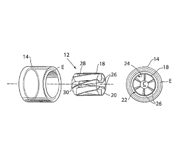
As evident from Figs. land 3, it is shown a turbine pump 10 of a heart pump
apparatus comprising a turbine wheel 12 and a suitable stator 14. Fig.1 shows
the
turbine pump 10 placed in a blood stream 16, such as in the aorta, in a human
patient.
The turbine pump 10 of the present invention provides the heart 3 of the human
patient with additional pumping capacity. The turbine pump 10 can also be
adapted to
be placed in the heart, in the pulmonary artery, or in the blood stream in the
abdominal aorta of the human patient. Consequently, Fig.3a shows the turbine
pump
10 placed in a general blood stream 16, such as a blood vessel 17, in the
human
patient. 132. The heart pump apparatus according to claim 6 or 120, wherein
the
heart pump further comprises confining elements adapted to confine said
rotating
body in the longitudinal extension of the artery in which it is placed.
Further, fig. 1 shows a confining portion 801 of the longitudinal extension of
the
artery. The confining portion 801 confines the rotating body in the artery in
which it is
placed. This eliminates the risk of the rotating body passing further into the
heart.
According to other embodiments the rotating body is confined in the artery by
the
magnetic coupling supplying the propulsion to the rotating body.
CA 02776421 2012-04-02
WO 2010/042008
PCT/SE2009/000445
8
Figs. 2a-b shows embodiments of the turbine pump 10. The turbine wheel 12 is
preferably in the form of a longitudinal rotating body 18. The rotating body
18 has a
cylindrical shape defined by an external cylindrical wall 20 with an outer
surface 22
and an inner surface 24. Internally the rotating body 18, blades 26 are
provided. The
blades 26 are extending radially or non-radially from the inner surface 24.
The rotating
body 18 have a longitudinal centre C in line with the blood stream 16. The
respective
blades 26 are not extending from the inner surface 24 all away to the centre C
of the
rotating body 18. In that respect, the respective blades 26 have a first end
28 attached
to the rotating body 18 and a second outer free end 30, which second free end
30 is
placed at a distance from the centre C of the rotating body 18. Hence, the
centre C of
the rotation body 18 is preferably a void space extending in the longitudinal
direction
L of the rotating body 18. Consequently, the turbine pump 10 according to the
present
invention is a centre axis free turbine pump.
The internal blades 26 arranged on the inner surface 24 of the rotating body
18 could
have the purpose of eliminating friction generated by the blood that flows
through
the inside of the rotation body 18. The blades 26 can also make the external
wall 20 of
the rotating body 18 more stiffened.
As evident from Figs. 2a-b, the rotating body 18 can be provided with blades
26 of
different design and configuration. The incidence angles and the attack angles
of the
2 D blades 26 are variable so that they can be arranged to yield the
highest possible
efficiency.
The turbine pump 10 comprises a device for rotation of the rotating body 18.
The
device for rotation of the rotating body 18 can be a part of an electrically
controlled
arrangement.
As a device for rotation of the rotating body, the stator 14 of the turbine
pump is
preferably provided. The stator 14 is preferably adapted to be placed outside
the
blood vessel 17 and opposite the rotating body 18. The electrically controlled
arrangement includes elements E, shown with phantom lines in Figs. 2a-b, for
receiving current to increase or decrease a magnetic field created at the
stator 14, for
CA 02776421 2012-04-02
WO 2010/042008
PCT/SE2009/000445
9
providing rotation of the rotating body 18, by creating a magnetic field
between the
poles of the stator 14, that is provided for increasing or decreasing the
rotation of the
rotating body 18. Consequently, a turbine pump system for assisting the heart
of a
patient is provided, comprising a rotating body 18 and a stator 14.
According to an embodiment, as evident from Fig. 3b, at least two rotating
bodies 18',
18" can be implanted in sequence in the same blood vessel. If there are two
rotating
bodies in sequence, one 18' can be adapted to rotate clockwise (see arrow R1)
and
the following other rotating body 18" can be adapted to rotate counter
clockwise (see
arrow R2), or vice versa.
Fig. 3c shows an embodiment of the heart pump device showing the drive unit,
in
which the stator 802 and rotor 18 is placed on the outside of the artery. The
stator
802 and rotor 803 is confined in a housing 805 and is separated from the
artery by a
protective sheet 804 placed between the rotor 803 and artery, the protective
sheet is
preferably a thin plastic sheet providing a smooth surface for the rotor 803
to slide
against. The rotor 803 rotates magnetic elements 809 which in turn rotates
magnetic
elements 810 of the rotating body 18 placed inside of the artery, by the
magnetic
elements 809 of the rotor 803 being in magnetic connection with the magnetic
elements 810 of the rotor 18. In some embodiments the magnetic elements 809 of
the rotor, is the rotor 803.
The present invention also relates to a method of surgically placing a
rotating body 18
of a turbine pump 10 in a patient via a laparoscopic thoracic approach, the
method
comprising the steps of: inserting a needle or a tube like instrument into the
thorax of
the patient's body; using the needle or a tube like instrument to fill the
thorax with
gas thereby expanding the thoracic cavity; placing at least two laparoscopic
trocars in
the patient's body; inserting a camera through one of the laparoscopic trocars
into the
thorax; inserting at least one dissecting tool through one of said at least
two
laparoscopic trocars and dissecting an intended placement area of the patient;
placing
the rotating body 18 in any part of the blood stream in the thorax; and
connecting a
source of energy for powering the device.
CA 02776421 2012-04-02
WO 2010/042008
PCT/SE2009/000445
The present invention also relates to an operation method for surgically
placing a
rotating body 18 of a turbine pump 10 in a patient, the method comprising the
steps
of: cutting the patient's skin; opening the thoracic cavity; dissecting a
placement area
where to place the rotating body 18 inside a blood stream in the heart 3, or
the aorta
5 4 or inside the pulmonary artery of the human patient; placing the a
rotating body 18
in the placement area; and connecting a source of energy for powering the
device.
The present invention also relates to a method of surgically placing a
rotating body 18
of a turbine pump 10 in a patient via a laparoscopic abdominal approach, the
method
comprising the steps of: inserting a needle or a tube like instrument into the
abdomen
10 of the patient's body; using the needle or a tube like instrument to
fill the abdomen
with gas thereby expanding the abdominal cavity; placing at least two
laparoscopic
trocars in the patient's body; inserting a camera through one of the
laparoscopic
trocars into the abdomen; inserting at least one dissecting tool through one
of said at
least two laparoscopictrocars and dissecting an intended placement area of the
patient; placing the rotating body 18 in the blood stream in the abdominal
aorta; and
connecting a source of energy for powering the device.
The present invention also relates to an operation method for surgically
placing a
rotating body 18 of a turbine pump 10 in a patient, the method comprising the
steps
of: cutting the patient's skin; opening the abdominal cavity; dissecting a
placement
area where to place the rotating body 18 in region of the abdominal aorta;
placing the
a rotating body in the blood stream in the abdominal aorta; and connecting a
source
of energy for powering the device.
The present invention also relates to an operation method for surgically
placing a
rotating body 18 of a turbine pump 10 and stator 14 of a turbine pump 10 in a
patient,
via a laparoscopic thoracic approach, the method comprising the steps of:
inserting a
needle or a tube like instrument into the thorax of the patient's body; using
the
needle or a tube like instrument to fill the thorax with gas thereby expanding
the
thoracic cavity; placing at least two laparoscopic trocars in the patient's
body;
inserting a camera through one of the laparoscopic trocars into the thorax;
inserting
at least one dissecting tool through one of said at least two laparoscopic
trocars and
CA 02776421 2012-04-02
WO 2010/042008
PCT/SE2009/000445
11
dissecting an intended placement area in the vascular system of the patient;
placing
the rotating body 18 in any part of the blood stream in the thorax, inside a
blood
stream of the blood vessel in the heart 3, or the aorta 4 or inside the
pulmonary artery
of the patient; placing the a stator 14 in the placement area, outside the
blood stream
of the blood vessel, outside the heart 3, or the aorta 4 or outside the
pulmonary
artery of the patient, placing said stator 14 on the outside of said rotating
body 18,
supplying wireless energy to said rotating body 18 causing rotating movement
of said
rotating body 18; and connecting a source of energy for powering said stator.
The present invention also relates to an operation method for surgically
placing a
rotating body 18 of a turbine pump 10 and stator 14 of a turbine pump 10 in a
patient,
the method comprising the steps of: cutting the patient's skin; opening the
thoracic
cavity; placing the rotating body 18 in any part of the blood stream in the
thorax,
inside a blood stream of the blood vessel in the heart 3, or the aorta 4 or
inside the
pulmonary artery of the patient; placing the a stator 14 in the placement
area, outside
the blood stream 16 of the blood vessel 17, outside the heart 3, or the aorta
40r
outside the pulmonary artery of the patient, placing said stator 14 on the
outside of
said rotating body 18, supplying wireless energy to said rotating body 18
causing
rotating movement of said rotating body 18; and connecting a source of energy
for
powering said stator.
The present invention also relates to an operation method for surgically
placing a
rotating body 18 of a turbine pump and stator 14 of a turbine pump 10 in a
patient,
via a laparoscopic abdominal approach, the method comprising the steps of:
inserting
a needle or a tube like instrument into the abdomen of the patient's body;
using the
needle or a tube like instrument to fill the thorax with gas thereby expanding
the
abdominal cavity; placing at least two laparoscopic trocars in the patient's
body;
inserting a camera through one of the laparoscopic trocars into the abdomen;
inserting at least one dissecting tool through one of said at least two
laparoscopic
trocars and dissecting an intended placement area in the region of the
abdominal
aorta of the patient; placing the rotating body 18 inside the blood stream 16
in the
abdominal aorta of the patient, placing the a stator 14 in the placement area,
outside
CA 02776421 2012-04-02
WO 2010/042008
PCT/SE2009/000445
12
the blood stream of the abdominal aorta, placing said stator 14 on the outside
of said
rotating body, supplying wireless energy to said rotating body 18 causing
rotating
movement of said rotating body 18; and connecting a source of energy for
powering
said stator.
The present invention also relates to an operation method for surgically
placing a
rotating body 18 of a turbine pump 10 and stator 14 of a turbine pump 10 in a
patient,
the method comprising the steps of: cutting the patient's skin, opening the
abdominal
cavity; placing the rotating body inside the blood stream 16 in the abdominal
aorta of
the patient, placing the a stator 14 in the placement area, outside the blood
stream of
.. the abdominal aorta, placing said stator 14 on the outside of said rotating
body 18,
supplying wireless energy to said rotating body 18 causing rotating movement
of said
rotating body 18; and connecting a source of energy for powering said stator.
The present invention also relates to a method according to any of the
operation
methods or methods of surgically placing a rotating body 18 and/or a stator 14
as
mentioned above, wherein said energy source is using energy, direct or
indirect, from
an external energy source, supplying energy non-invasively, without any
penetration
through the patient's skin to power the rotating body of a turbine pump 10.
According to another aspect, the turbine pump 10 according to the present
invention
can be adapted in a left ventricular assist device (LVAD). The LVAD is a
surgically
implanted, mechanical pump-type device, which helps maintain the pumping
ability of
a damaged heart. According to the state of the art, a tube pulls blood from
the left
ventricle into a pump (VAD), see Fig. 4. The pump then sends blood into the
aorta.
This effectively helps the weakened ventricle. There exists various kinds of
pumps
(VAD) on the market. For instance, there is one pump that contains a metal
plate that
pushes on a plastic blood sac, forcing the blood out of the sac. The metal
plate is
driven by a miniature electric motor. According to the present invention, the
rotating
body of the turbine pump can be arranged in a tube that pulls blood from the
aorta,
and sends back blood into the aorta.
CA 02776421 2012-04-02
WO 2010/042008
PCT/SE2009/000445
13
Fig. 3c The heart pump according to claim 119, wherein said second part is
adapted to
be a rotor, being cylindrical and placed outside said blood vessel, when
implanted,
adapted to be in magnetic connection with said rotating body such that said
rotating
body follows the rotations of said second part.
Fig.4 shows a patient 1 having an implanted heart pump 2, here illustrated a
pump
(VAD) of a left ventricular assist device (LVAD). As mentioned above,
according to one
aspect of the present invention, the implanted heart pump 2 can be the turbine
pump
10, as shown with phantom lines in Fig. 4, according to one embodiment of the
present invention. The implanted heart pump 2 is connected to the left
ventricle 3a of
the patient's heart 3 by means of a first tube 2a. The heart pump 2 is also
connected
to the aorta, generally designated 4, of the patient 1 by means of a second
tube 2b. In
this way, during operation the heart pump supplements or replaces the blood
pumping operation of the patient's heart 3.
Fig. 4b shows an operation method for surgically placing a rotating body 18 of
a
turbine pump 10 in an artery of a patient, via a laparoscopic inguinal
approach, the
method comprising the steps of: inserting a tube like instrument into the
femoral
artery FA of the patient's body and using the instrument to guide said
rotating body
18 through the femoral artery FA to the aorta A and releasing the rotating
body 18
inside of the aorta A. Thereafter the method comprises the step of placing a
drive unit
814, at least partially encircling the aorta A. The drive unit 814 can be
placed in a
thoracic approach, by opening the thorax of the patient, or in an abdominal
approach,
reaching the heart 3 of the patient through the thoracic diaphragm.
Fig. 4c shows a frontal view of the human patient after the operation has been
performed and the incisions in the inguinal region 820 and the thorax 821 has
been
closed using sutures or staples. The drive unit 814 (here shown in phantom
lines) is
placed at least partially encircling the aorta A in proximity to the heart 3.
Fig. 4d shows an operation method for surgically placing a rotating body 18 of
a
turbine pump 10, via a laparoscopic inguinal approach. The method comprises
the
steps of: inserting a tube like instrument into the femoral artery FA of the
patient's
CA 02776421 2012-04-02
WO 2010/042008 PCT/SE2009/000445
14
body, using the instrument to guide the rotating body 18 through the femoral
artery
FA to the abdominal aorta AA. After the rotating body 18 has been guided to
the
abdominal artery AA through the femoral artery FA the rotating body 18 is
released
inside the artery. The drive unit 814 is inserted through an incision in the
abdomen
and placed at least partially encircling the abdominal artery AA, such that
the drive
unit 814 is placed in magnetic contact with the rotating body 18.
Fig. 4e shows a frontal view of the patient after the incisions in the
inguinal area 820
and the incisions in the thorax 821 has been closed using sutures or staples.
The drive
unit 814 (here shown in phantom lines) are placed partially encircling the
abdominal
artery AA in the abdomen.
A blood clot removal device 100 according to the invention is shown provided
in the
second tube 2b of the heart pump 2, i.e., in the tube leading to the aorta 4
of the
patient 1. This means that part of the blood flow passageway provided by the
second
tube 2b is replaced by a blood flow passageway in the blood clot removal
device 100.
The blood clot removal device 100 is thus an artificial device insertable in
an artificial
blood vessel of the patient. The function of the clot removal device is to
remove any
blood clots in the blood transported by the second tube 2b. These blood clots
are
preferably moved to a place free inside the body of the patient. However, they
could
alternatively be collected in a collecting volume, such as a bag 100a
connected to the
blood clot removal device 100 for subsequent removal or storage. A preferred
storage
capacity of the bag 100a can be more than 100 milliliters, for example. The
blood clot
removal device is an artificial device but could be inserted directly into a
blood vessel
of the patient or connected between two ends of a blood vessel.
The clot removal device is preferably insertable in a blood flow passageway of
the
patient via surgery and is placed in the patient's abdomen or thorax or
cephalic or
neck region or retroperitoneal or any limb of the patient.
The design of a first preferred embodiment of the blood clot removal device
100 will
now be described in detail, with reference to Figs. 5-7. Fig. 5 shows a
sectional view
wherein the blood clot removal device 100 is provided in the blood flow
passageway
CA 02776421 2012-04-02
WO 2010/042008 PCT/SE2009/000445
provided by the second tube 2b. A filter 112 is provided across the blood flow
passageway 114 formed in a housing 111 with the function of stopping potential
blood clots brought forward in the second tube 2b by the blood flow, indicated
by
arrows in the figure. In this preferred embodiment, the filter 112 comprises a
plurality
5 of preferably equally spaced strips 112a of some suitable material, such
as
biocompatible metal or plastic. These strips 112a are preferably arranged
mutual
parallel.
The distance between two adjacent strips is small enough to stop any blood
clots.
Thus, the distance is preferably less than 2 millimeters, and even more
preferably less
10 than 1.0 millimeters, but if the goal is to protect the brain from
larger clots only the
distance could be larger. Although the blood flow passageway 114 in the
preferred
embodiment has an essentially square cross-sectional shape, it will be
realized that it
can take any suitable shape, such as rectangular or circular.
By providing a plurality of strips 112a as a filter across the blood flow
passageway 114,
15 a laminar blood flow is achieved downstream of the filter, which is
advantageous in a
blood clot preventing perspective. The blood flow configuration can be further
enhanced by giving the plurality of strips 112a a desired cross-sectional
shape,
although the rectangular shape shown in Fig. 7 will be adequate for most
purposes.
A first piston 116 is provided movable in a direction essentially
perpendicular to the
direction of the blood flow passageway 114, i.e., essentially perpendicular to
the
direction of the blood flow. This first piston 116 is driven by some suitable
actuator
means, such as pressurized air, a solenoid arrangement, an electrical servo
motor or
the like. A motor could be used to build up a stored power that could be
released very
fast, one example being a spring. In the preferred embodiment, pressurized air
acts as
the actuator means, since by latching the piston by means of a suitable
latching means
for the piston, building up the air pressure, and subsequently releasing the
piston,
very high speed of the piston is achieved, with enables short cleaning times
of the
filter.
CA 02776421 2012-04-02
WO 2010/042008 PCT/SE2009/000445
16
The outer end portion of the first piston 116, i.e., the end portion facing
the blood
flow passageway 114, is essentially flush with the wall of the blood flow
passageway
in a non-active state of the blood clot removal device 100. Also, the outer
end portion
is provided with a concave portion or recess 116a (exaggerated in the figures)
in order
to act as a blood clot capturing means, as will be explained below.
The strike range of the first piston 116 is such that it extends all way
across the blood
flow passageway 14, as will be explained below with reference to Figs. 8-11. A
number
of channels 116b corresponding to the number of strips 112a is provided in the
first
piston 16 to accommodate the strips when the first piston is in an extended
position.
The first piston 116 is also provided with a plurality of through holes 117 in
the
direction of the blood flow passageway. These through holes will allow blood
to flow
through the blood flow passageway also during a cleaning operation, as will be
explained below with reference to Fig. 12.
A second piston 118 is provided across the blood flow passageway 114 from the
first
piston 116. Also this second piston 118 is movable in a direction essentially
perpendicular to the direction of the blood flow passageway 114 and is biased
in the
direction thereof by means of a spring 118a, for example. Likewise, the outer
end
portion of the second piston is provided with a recess 18b similar to the
recess 116a of
the first piston 116.
The first and second pistons 116, 118, are sealed to the housing 111 by means
of a
respective sealing 120, such as an 0 sealing.
A preferred embodiment of the method according to the invention will now be
described with reference to Figs. 8-11, showing different operational steps of
the
above-described device. Fig. 8 is a view similar to that of Fig. 5. However,
this figures
shows the blood clot removal device 100 during operation, wherein blood clots,
generally designated 122, have assembled on the filter 112.
In Fig. 9, the first piston 116 has moved linearly from the retracted starting
position
shown Fig. 8 to an extended position, wherein the outer end portion thereof is
in
CA 02776421 2012-04-02
WO 2010/042008 PCT/SE2009/000445
17
contact with the second piston 118. Due to the recess 116a in the outer end of
the
first piston 116, the blood clots 122 have been assembled in the recess 116a,
whereby
they have been brought with the first piston 116 during the movement thereof.
In the
step shown in Fig. 9, the blood clots are confined in the recess 116a between
the first
and second pistons 116, 118.
By moving the first piston 116 an additional distance from the position shown
in Fig. 9,
the second piston 118 is pushed against the force of the spring 118a to a
fully
retracted position, see Fig. 10. The plurality of strips 112a is in this
position fully
received in a respective channel 116b in the first piston. It is seen that the
outer ends
of the first and second pistons define an unobstructed cavity in which the
blood clots
are confined. It is thereby possible to remove these by some suitable means.
One such
means could be a third piston 124, which is movable in a direction
perpendicular to
both the direction of the blood flow passageway 114 and the direction of
movement
of the first and second pistons 116, 118. This third piston, the movement of
which
could be controlled by means of pressurized air, a solenoid, an electric motor
etc.,
scrapes off the blood clots collected by the first piston 116 and moves them
to a place
outside of the blood clot removal device 100 and the blood flow passageway
114.
Fig. 12 shows a side view of the first piston 116 in a fully extended
position, i.e.,
corresponding to the view of Fig. 11. It is here seen that in this position
the through
holes 117 will be aligned with the blood flow passageway 114, thereby allowing
blood
to flow therethrough also during cleaning of the filter 112.
Fig. 13 shows a cross-sectional view taken along line X-X of Fig. 11. It is
here seen that
the third piston 124 collects the blood clots 122 during a downward movement,
indicated by an arrow in the figure. The clots are ejected from the blood clot
removal
device 100 when the third piston 124 has reached its lower end position, shown
in Fig.
14.
Again with reference to Fig. 10, it will be realized that pressurized air can
be used for
ejecting the collected blood clots from the cavity formed by the first piston
116 and
the second piston 118.
CA 02776421 2012-04-02
WO 2010/042008 PCT/SE2009/000445
18
Fig. 15-28 shows the fixation of a heart pump apparatus to a structure of the
human
body comprising bone 240. The structure could be the sternum, a part of the
rib cage,
comprising one or more ribs or a part of the vertebral column comprising at
least one
vertebra. According to one embodiment the heart pump apparatus 10 is fixated
to the
structure of the human body comprising bone 240 trough a fixating member 241
said
fixating member could comprise a plate 242 which is in contact with the
structure of
the human body comprising bone 240. The heart pump apparatus 10 could also be
fixated to the structure of the human body comprising bone 240 using a second
fixating member 241b which also could comprise a plate 242b in which in turn
could
be in contact with the structure of the human body comprising bone 240.
Fig. 15 shows an embodiment where a heart pump apparatus 10 is fixated to a
structure of the human body comprising bone 240. The structure could be the
sternum, a part of the rib cage comprising one or more ribs or a part of the
vertebral
column structure comprising at least one vertebra. According to the embodiment
the
heart pump apparatus 10 comprises a first fixating member 241a comprising a
plate
242a and a second fixating member 241b comprising a plate 242b. The first and
second fixating members are attached to each other using through-going screws
243
placed from the anterior side A of the structure of the human body comprising
bone
240. An alternative embodiment could comprise screws placed from the posterior
side
P of the structure of the human body comprising bone 240. The first fixating
member
241a and the second fixating member 241b clamp the structure of the human body
comprising bone 240. The fixating member 241a could be in contact with a
connecting
arm 244 which in turn could be in contact with a heart pump device.
Fig. 16 shows an embodiment where the heart pump apparatus 10 is fixated to a
structure of the human body comprising bone 240 using only one fixating member
241a comprising a plate 242a. The structure could be the sternum, a part of
the rib
cage comprising one or more ribs or a part of the vertebral column structure
comprising at least one vertebra. Through-going screws 243 is placed form the
anterior side A the structure of the human body comprising bone 240 and
fixated in
CA 02776421 2012-04-02
WO 2010/042008
PCT/SE2009/000445
19
the plate 242a. An alternative embodiment could comprise screws placed from
the
posterior side P of the structure of the human body comprising bone 240 in
which
case the screws could be fixated in nuts placed in connection with the
structure of the
human body comprising bone, or fixated in directly in the bone of the
structure of the
human body comprising bone 240. The fixating member 241a could be in contact
with
a connecting arm 244 which in turn could be in contact with a heart pump
device.
Fig. 17 shows an embodiment where the heart pump apparatus 10 is fixated to a
structure of the human body comprising bone 240. The structure could be the
sternum, a part of the rib cage comprising one or more ribs or a part of the
vertebral
column comprising at least one vertebra. According to the embodiment the heart
pump apparatus 10 comprises a first fixating member 241a comprising a plate
242a
and a second fixating member 241b comprising a plate 242b. The first and
second
fixating members are attached to each other using through-going screws 243
placed
from the posterior side P of the structure of the human body comprising bone
240.
The screws are fixated to nuts 245 placed on the anterior side of the
structure
comprising bone 240. An alternative embodiment could comprise screws placed
from
the anterior side A of the structure of the human body comprising bone 240, in
which
case the nuts is placed on the posterior side P of the structure comprising
bone 240.
The first fixating member 241a and the second fixating member 241b clamp the
structure of the human body comprising bone 240. The fixating member 241a
could
be in contact with a connecting arm 244 which in turn could be in contact with
a heart
pump device.
Fig. 18 shows an embodiment where the heart pump apparatus 10 is fixated to a
structure of the human body comprising bone 240 using only one fixating member
241a comprising a plate 242a. The structure could be the sternum, a part of
the rib
cage comprising one or more ribs or a part of the vertebral column structure
comprising at least one vertebra. Screws 243 that fixates the fixating member
to the
structure of the human body comprising bone is placed form the posterior side
P the
CA 02776421 2012-04-02
PCT/SE2009/000445
WO 2010/042008
structure of the human body comprising bone 240. The screws fixates the
fixating
member to both the posterior and the anterior cortex of the structure of the
human
body comprising bone 240, however it is conceivable that the screws are
fixated only
to the anterior or posterior cortex. An alternative embodiment could comprise
screws
5 placed from the anterior side A of the structure of the human body
comprising bone
240, in which case the fixating member 241a is placed on the anterior side A
of the
structure of the human body comprising bone 240.
Fig. 19 shows an embodiment where the heart pump apparatus 10 is fixated to a
10 structure of the human body comprising bone 240 using one fixating
member 241b
comprising a plate 242b, and one fixating member 241a without a plate. The
structure
could be the sternum, a part of the rib cage comprising one or more ribs or a
part of
the vertebral column structure comprising at least one vertebra. Screws 243
that
fixates the fixating members 241a,b to the structure of the human body
comprising
15 bone 240 is placed form the anterior side A of the structure of the
human body
comprising bone 240 and fixated in the fixating member 241a. The first
fixating
member 241a and the second fixating member 241b clamp the structure of the
human body comprising bone 240. The fixating member 241a could be in contact
with
a connecting arm 244 which in turn could be in contact with a heart pump
device.
Fig. 20 shows an embodiment where the heart pump apparatus 10 is fixated to a
structure of the human body comprising bone 240 using one fixating member 241b
comprising a plate 242b, and one fixating member 241a without a plate. The
structure
could be the sternum, a part of the rib cage comprising one or more ribs or a
part of
the vertebral column structure comprising at least one vertebra. Screws 243
that
fixates the fixating members 241a,b to the structure of the human body
comprising
bone 240 is placed form the posterior side P of the structure of the human
body
comprising bone 240 and fixated in the plate 242b of the fixating member 241b.
The
first fixating member 241a and the second fixating member 241b clamp the
structure
of the human body comprising bone 240. The fixating member 241a could be in
CA 02776421 2012-04-02
WO 2010/042008 PCT/SE2009/000445
21
contact with a connecting arm 244 which in turn could be in contact with a
heart
pump device.
Fig. 21 shows an embodiment where the heart pump apparatus 10 is adapted to be
fixated to the sternum 250 of a human patient. The device is fixated using a
fixating
member 241b which is fixated to the sternum using screws 243. However the
heart
pump apparatus could be fixated to the sternum 250 of a human patent using any
of
the ways to place the fixating members described previously.
Fig. 22 shows an embodiment where the heart pump apparatus 10 is adapted to be
fixated to two ribs 251, 252. A fixating member 241 comprising a plate 242b is
fixated
with screws adapted to fixate the fixating member to the cortex of the ribs.
Fig. 23 shows an embodiment where the heart pump apparatus 10 is adapted to be
fixated to two ribs 251, 252. A first plate 242a is provided on the posterior
side of the
rib cage, whereas a second plate 242b is provided in the anterior side of the
rib cage.
Screws 243 penetrate the ribs and fixates the first plate 242a to the second
plate
242b. The tightening of the screws creates a clamping effect of the ribs
251,251 and
provides the fixation of the heart pump apparatus 10.1n another embodiment
(not
shown) he screws 243 are placed between the ribs 251,252 and that ways
provides a
clamping effect of the ribs 251,252.
Fig. 24 shows an embodiment where the heart pump apparatus 10 is adapted to be
fixated to one rib 252. A plate 242a is provided on the posterior side of the
rib cage
and screws 243 are provided from the outside thereof, penetrating the rib 252
and
fixating the plate 242a to the rib 252.
Fig. 25 shows an embodiment where the heart pump apparatus 10 is adapted to be
fixated to one rib 252 using cord or band 254, this way there is no need to
penetrate
the rib 252. However the heart pump apparatus could be fixated to the ribcage
of a
human patent using any of the ways to place the fixating members described
previously.
CA 02776421 2012-04-02
WO 2010/042008 PCT/SE2009/000445
22
Fig. 26 shows an embodiment where the heart pump apparatus 10 is adapted to be
fixated to a vertebra 255 of the vertebral column. A fixating member 241 is
fixated to
the vertebra 255 using screws 243. The heart pump apparatus further comprises
a
connecting connecting arm 244 that connects the heart pump apparatus 10 to the
fixating member 241.
Fig. 27 shows an embodiment where the heart pump apparatus 10 is adapted to be
fixated to two vertebras 255, 256 of the vertebral column. A fixating member
241 is
fixated to the two vertebras 255, 256 using screws 243. The heart pump
apparatus
further comprises a connecting connecting arm 244 that connects the heart pump
apparatus 10 to the fixating member 241.
Fig. 28 shows an embodiment where the heart pump apparatus is adapted to be
fixated to a vertebra 255 of the vertebral column by clamping said vertebra
255. Two
fixating members 241a, 241b is placed on two sides of the vertebra and an
attachment comprising screws 243 clamps the vertebra between the first and
second
fixating members 241a,b. The heart pump apparatus further comprises a
connecting
connecting arm 244 that connects the heart pump apparatus 10 to the fixating
member 241.
In all of the above mentioned embodiments the means of attachment could be
replaced with other mechanical attachments or an adhesive. Other mechanical
attachments suitable could be: pop-rivets, nails, staples, band or cord. The
mechanical
fixating members could be of a metallic or ceramic material. Suitable metallic
materials could be titanium or surgical steel.
Fig. 29 illustrates a system for treating a disease comprising an apparatus 10
of the
present invention placed in the abdomen of a patient. An implanted energy-
transforming device 1002 is adapted to supply energy consuming components of
the
apparatus with energy via a power supply line 1003. An external energy-
transmission
device 1004 for non-invasively energizing the apparatus 10 transmits energy by
at
CA 02776421 2012-04-02
WO 2010/042008 PCT/SE2009/000445
23
least one wireless energy signal. The implanted energy-transforming device
1002
transforms energy from the wireless energy signal into electric energy which
is
supplied via the power supply line 1003.
The implanted energy-transforming device 1002 may also comprise other
components, such as: a coil for reception and/or transmission of signals and
energy,
an antenna for reception and/or transmission of signals, a microcontroller, a
charge
control unit, optionally comprising an energy storage, such as a capacitor,
one or more
sensors, such as temperature sensor, pressure sensor, position sensor, motion
sensor
etc., a transceiver, a motor, optionally including a motor controller, a pump,
and other
parts for controlling the operation of a medical implant.
The wireless energy signal may include a wave signal selected from the
following: a
sound wave signal, an ultrasound wave signal, an electromagnetic wave signal,
an
infrared light signal, a visible light signal, an ultra violet light signal, a
laser light signal,
a micro wave signal, a radio wave signal, an x-ray radiation signal and a
gamma
radiation signal. Alternatively, the wireless energy signal may include an
electric or
magnetic field, or a combined electric and magnetic field.
The wireless energy-transmission device 1004 may transmit a carrier signal for
carrying the wireless energy signal. Such a carrier signal may include
digital, analogue
or a combination of digital and analogue signals. In this case, the wireless
energy
signal includes an analogue or a digital signal, or a combination of an
analogue and
digital signal.
Generally speaking, the energy-transforming device 1002 is provided for
transforming
wireless energy of a first form transmitted by the energy-transmission device
1004
into energy of a second form, which typically is different from the energy of
the first
form. The implanted apparatus 10 is operable in response to the energy of the
second
form. The energy-transforming device 1002 may directly power the apparatus
with
the second form energy, as the energy-transforming device 1002 transforms the
first
CA 02776421 2012-04-02
WO 2010/042008 PCT/SE2009/000445
24
form energy transmitted by the energy-transmission device 1004 into the second
form
energy. The system may further include an implantable accumulator, wherein the
second form energy is used at least partly to charge the accumulator.
Alternatively, the wireless energy transmitted by the energy-transmission
device 1004
may be used to directly power the apparatus, as the wireless energy is being
transmitted by the energy-transmission device 1004. Where the system comprises
an
operation device for operating the apparatus, as will be described below, the
wireless
energy transmitted by the energy-transmission device 1004 may be used to
directly
power the operation device to create kinetic energy for the operation of the
apparatus.
The wireless energy of the first form may comprise sound waves and the energy-
transforming device 1002 may include a piezo-electric element for transforming
the
sound waves into electric energy. The energy of the second form may comprise
electric energy in the form of a direct current or pulsating direct current,
or a
combination of a direct current and pulsating direct current, or an
alternating current
or a combination of a direct and alternating current. Normally, the apparatus
comprises electric components that are energized with electrical energy. Other
implantable electric components of the system may be at least one voltage
level
guard or at least one constant current guard connected with the electric
components
of the apparatus.
Optionally, one of the energy of the first form and the energy of the second
form may
comprise magnetic energy, kinetic energy, sound energy, chemical energy,
radiant
energy, electromagnetic energy, photo energy, nuclear energy or thermal
energy.
Preferably, one of the energy of the first form and the energy of the second
form is
non-magnetic, non-kinetic, non-chemical, non-sonic, non-nuclear or non-
thermal.
The energy-transmission device may be controlled from outside the patient's
body to
release electromagnetic wireless energy, and the released electromagnetic
wireless
energy is used for operating the apparatus. Alternatively, the energy-
transmission
CA 02776421 2012-04-02
WO 2010/042008 PCT/SE2009/000445
device is controlled from outside the patient's body to release non-magnetic
wireless
energy, and the released non-magnetic wireless energy is used for operating
the
apparatus.
The external energy-transmission device 1004 also includes a wireless remote
control
5 having an external signal transmitter for transmitting a wireless control
signal for non-
invasively controlling the apparatus. The control signal is received by an
implanted
signal receiver which may be incorporated in the implanted energy-transforming
device 1002 or be separate there from.
10 The wireless control signal may include a frequency, amplitude, or phase
modulated
signal or a combination thereof. Alternatively, the wireless control signal
includes an
analogue or a digital signal, or a combination of an analogue and digital
signal.
Alternatively, the wireless control signal comprises an electric or magnetic
field, or a
combined electric and magnetic field.
The wireless remote control may transmit a carrier signal for carrying the
wireless
control signal. Such a carrier signal may include digital, analogue or a
combination of
digital and analogue signals. Where the control signal includes an analogue or
a digital
signal, or a combination of an analogue and digital signal, the wireless
remote control
preferably transmits an electromagnetic carrier wave signal for carrying the
digital or
analogue control signals.
Fig. BO illustrates the system of Fig. 29 in the form of a more generalized
block
diagram showing the apparatus 10, the energy-transforming device 1002 powering
the apparatus 10 via power supply line 1003, and the external energy-
transmission
device 1004, The patient's skin 1005, generally shown by a vertical line,
separates the
interior of the patient to the right of the line from the exterior to the left
of the line.
Fig. 31 shows an embodiment of the invention identical to that of Fig. 30,
except that
a reversing device in the form of an electric switch 1006 operable for example
by
polarized energy also is implanted in the patient for reversing the apparatus
10. When
CA 02776421 2012-04-02
PCT/SE2009/000445
WO 2010/042008
26
the switch is operated by polarized energy the wireless remote control of the
external
energy-transmission device 1004 transmits a wireless signal that carries
polarized
energy and the implanted energy-transforming device 1002 transforms the
wireless
polarized energy into a polarized current for operating the electric switch
1006. When
the polarity of the current is shifted by the implanted energy-transforming
device
1002 the electric switch 1006 reverses the function performed by the apparatus
10.
Fig. 32 shows an embodiment of the invention identical to that of Fig. 30,
except that
an operation device 1007 implanted in the patient for operating the apparatus
10 is
1 G provided between the implanted energy-transforming device 1002 and the
apparatus
10. This operation device can be in the form of a motor 1007, such as an
electric
servomotor. The motor 1007 is powered with energy from the implanted energy-
transforming device 1002, as the remote control of the external energy-
transmission
device 1004 transmits a wireless signal to the receiver of the implanted
energy-
transforming device 1002.
Fig. 33 shows an embodiment of the invention identical to that of Fig. 30,
except that
it also comprises an operation device is in the form of an assembly 1008
including a
motor/pump unit 1009 and a fluid reservoir 1010 is implanted in the patient.
In this
case the apparatus 10 is hydraulically operated, i.e. hydraulic fluid is
pumped by the
motor/pump unit 1009 from the fluid reservoir 1010 through a conduit 1011 to
the
apparatus 10 to operate the apparatus, and hydraulic fluid is pumped by the
motor/pump unit 1009 back from the apparatus 10 to the fluid reservoir 1010 to
return the apparatus to a starting position. The implanted energy-transforming
device
1002 transforms wireless energy into a current, for example a polarized
current, for
powering the motor/pump unit 1009 via an electric power supply line 1012.
Instead of a hydraulically operated apparatus 10, it is also envisaged that
the
operation device comprises a pneumatic operation device. In this case, the
hydraulic
fluid can be pressurized air to be used for regulation and the fluid reservoir
is replaced
by an air chamber.
CA 02776421 2012-04-02
PCT/SE2009/000445
WO 2010/042008
27
In all of these embodiments the energy-transforming device 1002 may include a
rechargeable accumulator like a battery or a capacitor to be charged by the
wireless
energy and supplies energy for any energy consuming part of the system.
As an alternative, the wireless remote control described above may be replaced
by
manual control of any implanted part to make contact with by the patient's
hand most
likely indirect, for example a press button placed under the skin.
Fig. 34 shows an embodiment of the invention comprising the external energy-
transmission device 1004 with its wireless remote control, the apparatus 10,
in this
case hydraulically operated, and the implanted energy-transforming device
1002, and
further comprising a hydraulic fluid reservoir 1013, a motor/pump unit 1009
and an
reversing device in the form of a hydraulic valve shifting device 1014, all
implanted in
the patient. Of course the hydraulic operation could easily be performed by
just
changing the pumping direction and the hydraulic valve may therefore be
omitted.
The remote control may be a device separated from the external energy-
transmission
device or included in the same. The motor of the motor/pump unit 1009 is an
electric
motor. In response to a control signal from the wireless remote control of the
external
energy-transmission device 1004, the implanted energy-transforming device 1002
powers the motor/pump unit 1009 with energy from the energy carried by the
control
signal, whereby the motor/pump unit 1009 distributes hydraulic fluid between
the
hydraulic fluid reservoir 1013 and the apparatus 10. The remote control of the
external energy-transmission device 1004 controls the hydraulic valve shifting
device
1014 to shift the hydraulic fluid flow direction between one direction in
which the
fluid is pumped by the motor/pump unit 1009 from the hydraulic fluid reservoir
1013
to the apparatus 10 to operate the apparatus, and another opposite direction
in which
the fluid is pumped by the motor/pump unit 1009 back from the apparatus 10 to
the
hydraulic fluid reservoir 1013 to return the apparatus to a starting position.
Fig. 35 shows an embodiment of the invention comprising the external energy-
transmission device 1004 with its wireless remote control, the apparatus 10,
the
CA 02776421 2012-04-02
WO 2010/042008 PCT/SE2009/000445
28
implanted energy-transforming device 1002, an implanted internal control unit
1015
controlled by the wireless remote control of the external energy-transmission
device
1004, an implanted accumulator 1016 and an implanted capacitor 1017. The
internal
control unit 1015 arranges storage of electric energy received from the
implanted
energy-transforming device 1002 in the accumulator 1016, which supplies energy
to
the apparatus 10. In response to a control signal from the wireless remote
control of
the external energy-transmission device 1004, the internal control unit 1015
either
releases electric energy from the accumulator 1016 and transfers the released
energy
via power lines 1018 and 1019, or directly transfers electric energy from the
implanted energy-transforming device 1002 via a power line 1020, the capacitor
1017,
which stabilizes the electric current, a power line 1021 and the power line
1019, for
the operation of the apparatus 10.
The internal control unit is preferably programmable from outside the
patient's body.
In a preferred embodiment, the internal control unit is programmed to regulate
the
apparatus 10 according to a pre-programmed time-schedule or to input from any
sensor sensing any possible physical parameter of the patient or any
functional
parameter of the system.
In accordance with an alternative, the capacitor 1017 in the embodiment of
Fig. 7
10may be omitted. In accordance with another alternative, the accumulator 1016
in
this embodiment may be omitted.
Fig. 36 shows an embodiment of the invention identical to that of Fig. 63,
except that
a battery 1022 for supplying energy for the operation of the apparatus 10 and
an
electric switch 1023 for switching the operation of the apparatus 10 also are
implanted in the patient. The electric switch 1023 may be controlled by the
remote
control and may also be operated by the energy supplied by the implanted
energy-
transforming device 1002 to switch from an off mode, in which the battery 1022
is not
in use, to an on mode, in which the battery 1022 supplies energy for the
operation of
the apparatus 10.
CA 02776421 2012-04-02
WO 2010/042008 PCT/SE2009/000445
29
Fig. 37 shows an embodiment of the invention identical to that of Fig. 69,
except that
an internal control unit 1015 controllable by the wireless remote control of
the
external energy-transmission device 1004 also is implanted in the patient. In
this case,
the electric switch 1023 is operated by the energy supplied by the implanted
energy-
transforming device 1002 to switch from an off mode, in which the wireless
remote
control is prevented from controlling the internal control unit 1015 and the
battery is
not in use, to a standby mode, in which the remote control is permitted to
control the
internal control unit 1015 to release electric energy from the battery 1022
for the
operation of the apparatus 10.
Fig. 38 shows an embodiment of the invention identical to that of Fig. 37,
except that
an accumulator 1016 is substituted for the battery 1022 and the implanted
components are interconnected differently. In this case, the accumulator 1016
stores
energy from the implanted energy-transforming device 1002. In response to a
control
signal from the wireless remote control of the external energy-transmission
device
1004, the internal control unit 1015 controls the electric switch 1023 to
switch from
an off mode, in which the accumulator 1016 is not in use, to an on mode, in
which the
accumulator 1016 supplies energy for the operation of the apparatus 10. The
accumulator may be combined with or replaced by a capacitor.
Fig. 39 shows an embodiment of the invention identical to that of Fig. 38,
except that
a battery 1022 also is implanted in the patient and the implanted components
are
interconnected differently. In response to a control signal from the wireless
remote
control of the external energy-transmission device 1004, the internal control
unit 1015
controls the accumulator 1016 to deliver energy for operating the electric
switch 1023
to switch from an off mode, in which the battery 1022 is not in use, to an on
mode, in
which the battery 1022 supplies electric energy for the operation of the
apparatus 10.
Alternatively, the electric switch 1023 may be operated by energy supplied by
the
accumulator 1016 to switch from an off mode, in which the wireless remote
control is
prevented from controlling the battery 1022 to supply electric energy and is
not in
CA 02776421 2012-04-02
WO 2010/042008
PCT/SE2009/000445
use, to a standby mode, in which the wireless remote control is permitted to
control
the battery 1022 to supply electric energy for the operation of the apparatus
10.
It should be understood that the switch 1023 and all other switches in this
application
3 should be interpreted in its broadest embodiment. This means a
transistor, MCU,
MCPU, ASIC, FPGA or a DA converter or any other electronic component or
circuit that
may switch the power on and off. Preferably the switch is controlled from
outside the
body, or alternatively by an implanted internal control unit.
10 Fig. 40 shows an embodiment of the invention identical to that of Fig.
36, except that
a motor 1007, a mechanical reversing device in the form of a gear box 1024,
and an
internal control unit 1015 for controlling the gear box 1024 also are
implanted in the
patient. The internal control unit 1015 controls the gear box 1024 to reverse
the
function performed by the apparatus 10 (mechanically operated). Even simpler
is to
15 switch the direction of the motor electronically. The gear box
interpreted in its
broadest embodiment may stand for a servo arrangement saving force for the
operation device in favour of longer stroke to act.
Fig. 41 shows an embodiment of the invention identical to that of Fig. 40
except that
20 the implanted components are interconnected differently. Thus, in this
case the
internal control unit 1015 is powered by the battery 1022 when the accumulator
1016, suitably a capacitor, activates the electric switch 1023 to switch to an
on mode.
When the electric switch 1023 is in its on mode the internal control unit 1015
is
permitted to control the battery 1022 to supply, or not supply, energy for the
25 operation of the apparatus 10.
Fig. 42 schematically shows conceivable combinations of implanted components
of
the apparatus for achieving various communication options. Basically, there
are the
apparatus 10, the internal control unit 1015, motor or pump unit 1009, and the
30 external energy-transmission device 1004 including the external wireless
remote
control. As already described above the wireless remote control transmits a
control
CA 02776421 2012-04-02
WO 2010/042008 PCT/SE2009/000445
31
signal which is received by the internal control unit 1015, which in turn
controls the
various implanted components of the apparatus.
A feedback device, preferably comprising a sensor or measuring device 1025,
may be
implanted in the patient for sensing a physical parameter of the patient. The
physical
parameter may be at least one selected from the group consisting of pressure,
volume, diameter, stretching, elongation, extension, movement, bending,
elasticity,
muscle contraction, nerve impulse, body temperature, blood pressure, blood
flow,
heartbeats and breathing. The sensor may sense any of the above physical
parameters. For example, the sensor may be a pressure or motility sensor.
Alternatively, the sensor 1025 may be arranged to sense a functional
parameter. The
functional parameter may be correlated to the transfer of energy for charging
an
implanted energy source and may further include at least one selected from the
group
of parameters consisting of; electricity, any electrical parameter, pressure,
volume,
diameter, stretch, elongation, extension, movement, bending, elasticity,
temperature
and flow.
The feedback may be sent to the internal control unit or out to an external
control
unit preferably via the internal control unit. Feedback may be sent out from
the body
via the energy transfer system or a separate communication system with
receiver and
transmitters.
The internal control unit 1015, or alternatively the external wireless remote
control of
the external energy-transmission device 1004, may control the apparatus 10 in
response to signals from the sensor 1025. A transceiver may be combined with
the
sensor 1025 for sending information on the sensed physical parameter to the
external
wireless remote control. The wireless remote control may comprise a signal
transmitter or transceiver and the internal control unit 1015 may comprise a
signal
receiver or transceiver. Alternatively, the wireless remote control may
comprise a
signal receiver or transceiver and the internal control unit 1015 may comprise
a signal
transmitter or transceiver. The above transceivers, transmitters and receivers
may be
used for sending information or data related to the apparatus 10 from inside
the
patient's body to the outside thereof.
CA 02776421 2012-04-02
WO 2010/042008 PCT/SE2009/000445
32
Where the motor/pump unit 1009 and battery 1022 for powering the motor/pump
unit 1009 are implanted, information related to the charging of the battery
1022 may
be fed back. To be more precise, when charging a battery or accumulator with
energy
feed back information related to said charging process is sent and the energy
supply is
changed accordingly.
Fig. 43 shows an alternative embodiment wherein the apparatus 10 is regulated
from
outside the patient's body. The system 1000 comprises a battery 1022 connected
to
the apparatus 10 via a subcutaneous electric switch 1026. Thus, the regulation
of the
apparatus 10 is performed non-invasively by manually pressing the subcutaneous
switch, whereby the operation of the apparatus 10 is switched on and off. It
will be
appreciated that the shown embodiment is a simplification and that additional
components, such as an internal control unit or any other part disclosed in
the present
application can be added to the system. Two subcutaneous switches may also be
used. In the preferred embodiment one implanted switch sends information to
the
internal control unit to perform a certain predetermined performance and when
the
patient press the switch again the performance is reversed.
Fig. 44 shows an alternative embodiment, wherein the system 1000 comprises a
hydraulic fluid reservoir 1013 hydraulically connected to the apparatus. Non-
invasive
regulation is performed by manually pressing the hydraulic reservoir connected
to the
apparatus.
The system may include an external data communicator and an implantable
internal
data communicator communicating with the external data communicator. The
internal communicator feeds data related to the apparatus or the patient to
the
external data communicator and/or the external data communicator feeds data to
the
internal data communicator.
Fig. 45 schematically illustrates an arrangement of the system that is capable
of
sending information from inside the patient's body to the outside thereof to
give
CA 02776421 2012-04-02
WO 2010/042008 PCT/SE2009/000445
33
feedback information related to at least one functional parameter of the
apparatus or
system, or related to a physical parameter of the patient, in order to supply
an
accurate amount of energy to an implanted internal energy receiver 1002
connected
to implanted energy consuming components of the apparatus 10. Such an energy
receiver 1002 may include an energy source and/or an energy-transforming
device.
Briefly described, wireless energy is transmitted from an external energy
source 1004a
located outside the patient and is received by the internal energy receiver
1002
located inside the patient. The internal energy receiver is adapted to
directly or
indirectly supply received energy to the energy consuming components of the
apparatus 10 via a switch 1026. An energy balance is determined between the
energy
received by the internal energy receiver 1002 and the energy used for the
apparatus
10, and the transmission of wireless energy is then controlled based on the
determined energy balance. The energy balance thus provides an accurate
indication
of the correct amount of energy needed, which is sufficient to operate the
apparatus
10 properly, but without causing undue temperature rise.
In Fig. 45 the patient's skin is indicated by a vertical line 1005. Here, the
energy
receiver comprises an energy-transforming device 1002 located inside the
patient,
preferably just beneath the patient's skin 1005. Generally speaking, the
implanted
energy-transforming device 1002 may be placed in the abdomen, thorax, muscle
fascia (e.g. in the abdominal wall), subcutaneously, or at any other suitable
location.
The implanted energy-transforming device 1002 is adapted to receive wireless
energy
E transmitted from the external energy-source 1004a provided in an external
energy-
transmission device 1004 located outside the patient's skin 1005 in the
vicinity of the
implanted energy-transforming device 1002.
As is well known in the art, the wireless energy E may generally be
transferred by
means of any suitable Transcutaneous Energy Transfer (TET) device, such as a
device
including a primary coil arranged in the external energy source 1004a and an
adjacent
secondary coil arranged in the implanted energy-transforming device 1002. When
an
electric current is fed through the primary coil, energy in the form of a
voltage is
induced in the secondary coil which can be used to power the implanted energy
CA 02776421 2012-04-02
WO 2010/042008 PCT/SE2009/000445
34
consuming components of the apparatus, e.g. after storing the incoming energy
in an
implanted energy source, such as a rechargeable battery or a capacitor.
However, the
present invention is generally not limited to any particular energy transfer
technique,
TET devices or energy sources, and any kind of wireless energy may be used.
The amount of energy received by the implanted energy receiver may be compared
with the energy used by the implanted components of the apparatus. The term
"energy used" is then understood to include also energy stored by implanted
components of the apparatus. A control device includes an external control
unit
1004b that controls the external energy source 1004a based on the determined
energy balance to regulate the amount of transferred energy. In order to
transfer the
correct amount of energy, the energy balance and the required amount of energy
is
determined by means of a determination device including an implanted internal
control unit 1015 connected between the switch 1026 and the apparatus 10. The
internal control unit 1015 may thus be arranged to receive various
measurements
obtained by suitable sensors or the like, not shown, measuring certain
characteristics
of the apparatus 10, somehow reflecting the required amount of energy needed
for
proper operation of the apparatus 10. Moreover, the current condition of the
patient
may also be detected by means of suitable measuring devices or sensors, in
order to
provide parameters reflecting the patient's condition. Hence, such
characteristics
and/or parameters may be related to the current state of the apparatus 10,
such as
power consumption, operational mode and temperature, as well as the patient's
condition reflected by parameters such as; body temperature, blood pressure,
heartbeats and breathing. Other kinds of physical parameters of the patient
and
functional parameters of the device are described elsewhere.
Furthermore, an energy source in the form of an accumulator 1016 may
optionally be
connected to the implanted energy-transforming device 1002 via the control
unit
1015 for accumulating received energy for later use by the apparatus 10.
Alternatively
or additionally, characteristics of such an accumulator, also reflecting the
required
amount of energy, may be measured as well. The accumulator may be replaced by
a
CA 02776421 2012-04-02
PCT/SE2009/000445
WO 2010/042008
rechargeable battery, and the measured characteristics may be related to the
current
state of the battery, any electrical parameter such as energy consumption
voltage,
temperature, etc. In order to provide sufficient voltage and current to the
apparatus
10, and also to avoid excessive heating, it is clearly understood that the
battery should
5 be charged optimally by receiving a correct amount of energy from the
implanted
energy-transforming device 1002, i.e. not too little or too much. The
accumulator may
also be a capacitor with corresponding characteristics.
For example, battery characteristics may be measured on a regular basis to
determine
10 the current state of the battery, which then may be stored as state
information in a
suitable storage means in the internal control unit 1015. Thus, whenever new
measurements are made, the stored battery state information can be updated
accordingly. In this way, the state of the battery can be "calibrated" by
transferring a
correct amount of energy, so as to maintain the battery in an optimal
condition.
Thus, the internal control unit 1015 of the determination device is adapted to
determine the energy balance and/or the currently required amount of energy,
(either
energy per time unit or accumulated energy) based on measurements made by the
above-mentioned sensors or measuring devices of the apparatus 10, or the
patient, or
an implanted energy source if used, or any combination thereof. The internal
control
unit 1015 is further connected to an internal signal transmitter 1027,
arranged to
transmit a control signal reflecting the determined required amount of energy,
to an
external signal receiver 1004c connected to the external control unit 1004b.
The
amount of energy transmitted from the external energy source 1004a may then be
regulated in response to the received control signal.
Alternatively, the determination device may include the external control unit
1004b.
In this alternative, sensor measurements can be transmitted directly to the
external
control unit 1004b wherein the energy balance and/or the currently required
amount
of energy can be determined by the external control unit 1004b, thus
integrating the
above-described function of the internal control unit 1015 in the external
control unit
CA 02776421 2012-04-02
WO 2010/042008 PCT/SE2009/000445
36
1004b. In that case, the internal control unit 1015 can be omitted and the
sensor
measurements are supplied directly to the internal signal transmitter 1027
which
sends the measurements over to the external signal receiver 1004c and the
external
control unit 1004b. The energy balance and the currently required amount of
energy
can then be determined by the external control unit 1004b based on those
sensor
measurements.
Hence, the present solution according to the arrangement of Fig. 45 employs
the feed
back of information indicating the required energy, which is more efficient
than
previous solutions because it is based on the actual use of energy that is
compared to
the received energy, e.g. with respect to the amount of energy, the energy
difference,
or the energy receiving rate as compared to the energy rate used by implanted
energy
consuming components of the apparatus. The apparatus may use the received
energy
either for consuming or for storing the energy in an implanted energy source
or the
.. like. The different parameters discussed above would thus be used if
relevant and
needed and then as a tool for determining the actual energy balance. However,
such
parameters may also be needed per se for any actions taken internally to
specifically
operate the apparatus.
The internal signal transmitter 1027 and the external signal receiver 1004c
may be
implemented as separate units using suitable signal transfer means, such as
radio, IR
(Infrared) or ultrasonic signals. Alternatively, the internal signal
transmitter 1027 and
the external signal receiver 1004c may be integrated in the implanted energy-
transforming device 1002 and the external energy source 1004a, respectively,
so as to
convey control signals in a reverse direction relative to the energy transfer,
basically
using the same transmission technique. The control signals may be modulated
with
respect to frequency, phase or amplitude.
Thus, the feedback information may be transferred either by a separate
communication system including receivers and transmitters or may be integrated
in
the energy system. In accordance with the present invention, such an
integrated
CA 02776421 2012-04-02
WO 2010/042008 PCT/SE2009/000445
37
information feedback and energy system comprises an implantable internal
energy
receiver for receiving wireless energy, the energy receiver having an internal
first coil
and a first electronic circuit connected to the first coil, and an external
energy
transmitter for transmitting wireless energy, the energy transmitter having an
external second coil and a second electronic circuit connected to the second
coil. The
external second coil of the energy transmitter transmits wireless energy which
is
received by the first coil of the energy receiver. This system further
comprises a power
switch for switching the connection of the internal first coil to the first
electronic
circuit on and off, such that feedback information related to the charging of
the first
coil is received by the external energy transmitter in the form of an
impedance
variation in the load of the external second coil, when the power switch
switches the
connection of the internal first coil to the first electronic circuit on and
off. In
implementing this system in the arrangement of Fig. 45, the switch 1026 is
either
separate and controlled by the internal control unit 1015, or integrated in
the internal
control unit 1015. It should be understood that the switch 1026 should be
interpreted
in its broadest embodiment. This means a transistor, MCU, MCPU, ASIC FPGA or a
DA
converter or any other electronic component or circuit that may switch the
power on
and off.
To conclude, the energy supply arrangement illustrated in Fig. 45 may operate
basically in the following manner. The energy balance is first determined by
the
internal control unit 1015 of the determination device. A control signal
reflecting the
required amount of energy is also created by the internal control unit 1015,
and the
control signal is transmitted from the internal signal transmitter 1027 to the
external
signal receiver 1004c. Alternatively, the energy balance can be determined by
the
external control unit 1004b instead depending on the implementation, as
mentioned
above. In that case, the control signal may carry measurement results from
various
sensors. The amount of energy emitted from the external energy source 1004a
can
then be regulated by the external control unit 1004b, based on the determined
energy
balance, e.g. in response to the received control signal. This process may be
repeated
CA 02776421 2012-04-02
WO 2010/042008 PCT/SE2009/000445
38
intermittently at certain intervals during ongoing energy transfer, or may be
executed
on a more or less continuous basis during the energy transfer.
The amount of transferred energy can generally be regulated by adjusting
various
transmission parameters in the external energy source 1004a, such as voltage,
current, amplitude, wave frequency and pulse characteristics.
This system may also be used to obtain information about the coupling factors
between the coils in a TET system even to calibrate the system both to find an
optimal
place for the external coil in relation to the internal coil and to optimize
energy
transfer. Simply comparing in this case the amount of energy transferred with
the
amount of energy received. For example if the external coil is moved the
coupling
factor may vary and correctly displayed movements could cause the external
coil to
find the optimal place for energy transfer. Preferably, the external coil is
adapted to
calibrate the amount of transferred energy to achieve the feedback information
in the
determination device, before the coupling factor is maximized.
This coupling factor information may also be used as a feedback during energy
transfer. In such a case, the energy system of the present invention comprises
an
implantable internal energy receiver for receiving wireless energy, the energy
receiver
having an internal first coil and a first electronic circuit connected to the
first coil, and
an external energy transmitter for transmitting wireless energy, the energy
transmitter having an external second coil and a second electronic circuit
connected
to the second coil. The external second coil of the energy transmitter
transmits
wireless energy which is received by the first coil of the energy receiver.
This system
further comprises a feedback device for communicating out the amount of energy
received in the first coil as a feedback information, and wherein the second
electronic
circuit includes a determination device for receiving the feedback information
and for
comparing the amount of transferred energy by the second coil with the
feedback
information related to the amount of energy received in the first coil to
obtain the
coupling factor between the first and second coils. The energy transmitter may
regulate the transmitted energy in response to the obtained coupling factor.
CA 02776421 2012-04-02
WO 2010/042008 PCT/SE2009/000445
39
With reference to Fig. 46, although wireless transfer of energy for operating
the
apparatus has been described above to enable non-invasive operation, it will
be
appreciated that the apparatus can be operated with wire bound energy as well.
Such
.. an example is shown in Fig. 46, wherein an external switch 1026 is
interconnected
between the external energy source 1004a and an operation device, such as an
electric motor 1007 operating the apparatus 10. An external control unit 1004b
controls the operation of the external switch 1026 to effect proper operation
of the
apparatus 10.
Fig. 47 illustrates different embodiments for how received energy can be
supplied to
and used by the apparatus 10. Similar to the example of Fig. 45, an internal
energy
receiver 1002 receives wireless energy E from an external energy source 1004a
which
is controlled by a transmission control unit 1004b. The internal energy
receiver 1002
may comprise a constant voltage circuit, indicated as a dashed box "constant
V'' in the
figure, for supplying energy at constant voltage to the apparatus 10. The
internal
energy receiver 1002 may further comprise a constant current circuit,
indicated as a
dashed box "constant C" in the figure, for supplying energy at constant
current to the
apparatus 10.
The apparatus 10 comprises an energy consuming part 10a, which may be a motor,
pump, restriction device, or any other medical appliance that requires energy
for its
electrical operation. The apparatus 10 may further comprise an energy storage
device
10b for storing energy supplied from the internal energy receiver 1002. Thus,
the
supplied energy may be directly consumed by the energy consuming part 10a, or
stored by the energy storage device 10b, or the supplied energy may be partly
consumed and partly stored. The apparatus 10 may further comprise an energy
stabilizing unit 10c for stabilizing the energy supplied from the internal
energy
receiver 1002. Thus, the energy may be supplied in a fluctuating manner such
that it
.. may be necessary to stabilize the energy before consumed or stored.
CA 02776421 2012-04-02
WO 2010/042008 PCT/SE2009/000445
The energy supplied from the internal energy receiver 1002 may further be
accumulated and/or stabilized by a separate energy stabilizing unit 1028
located
outside the apparatus 10, before being consumed and/or stored by the apparatus
10.
Alternatively, the energy stabilizing unit 1028 may be integrated in the
internal energy
5 receiver 1002. In either case, the energy stabilizing unit 1028 may
comprise a constant
voltage circuit and/or a constant current circuit.
It should be noted that Fig. 45 and Fig. 47 illustrate some possible but non-
limiting
implementation options regarding how the various shown functional components
and
10 .. elements can be arranged and connected to each other. However, the
skilled person
will readily appreciate that many variations and modifications can be made
within the
scope of the present invention.
Fig. 48 schematically shows an energy balance measuring circuit of one of the
15 .. proposed designs of the system for controlling transmission of wireless
energy, or
energy balance control system. The circuit has an output signal centered on
2.5V and
proportionally related to the energy imbalance. The derivative of this signal
shows if
the value goes up and down and how fast such a change takes place. If the
amount of
received energy is lower than the energy used by implanted components of the
20 .. apparatus, more energy is transferred and thus charged into the energy
source. The
output signal from the circuit is typically feed to an A/D converter and
converted into
a digital format. The digital information can then be sent to the external
energy-
transmission device allowing it to adjust the level of the transmitted energy.
Another
possibility is to have a completely analog system that uses comparators
comparing the
25 .. energy balance level with certain maximum and minimum thresholds sending
information to external energy-transmission device if the balance drifts out
of the
max/min window.
The schematic Fig. 48 shows a circuit implementation for a system that
transfers
30 .. energy to the implanted energy components of the apparatus of the
present invention
from outside of the patient's body using inductive energy transfer. An
inductive
CA 02776421 2012-04-02
PCT/SE2009/000445
WO 2010/042008
41
energy transfer system typically uses an external transmitting coil and an
internal
receiving coil. The receiving coil, L1, is included in the schematic Fig. 31;
the
transmitting parts of the system are excluded.
The implementation of the general concept of energy balance and the way the
information is transmitted to the external energy transmitter can of course be
implemented in numerous different ways. The schematic Fig. 48 and the above
described method of evaluating and transmitting the information should only be
regarded as examples of how to implement the control system.
CIRCUIT DETAILS
In Fig. 48 the symbols Y1, Y2, Y3 and so on symbolize test points within the
circuit. The
components in the diagram and their respective values are values that work in
this
particular implementation which of course is only one of an infinite number of
possible design solutions.
Energy to power the circuit is received by the energy receiving coil L1.
Energy to
implanted components is transmitted in this particular case at a frequency of
25 kHz.
The energy balance output signal is present at test point Y1.
Those skilled in the art will realize that the above various embodiments of
the system
could be combined in many different ways. For example, the electric switch
1006 of
Fig. 31 could be incorporated in any of the embodiments of Figs. 34-40, the
hydraulic
valve shifting device 1014 of Fig. 34 could be incorporated in the embodiment
of Fig.
33, and the gear box 1024 could be incorporated in the embodiment of Fig. 32.
Please
observe that the switch simply could mean any electronic circuit or component.
The embodiments described in connection with Figs. 45, 47 and 48 identify a
method
and a system for controlling transmission of wireless energy to implanted
energy
CA 02776421 2012-04-02
WO 2010/042008 PCT/SE2009/000445
42
consuming components of an electrically operable apparatus. Such a method and
system will be defined in general terms in the following.
A method is thus provided for controlling transmission of wireless energy
supplied to
implanted energy consuming components of an apparatus as described above. The
wireless energy E is transmitted from an external energy source located
outside the
patient and is received by an internal energy receiver located inside the
patient, the
internal energy receiver being connected to the implanted energy consuming
components of the apparatus for directly or indirectly supplying received
energy
thereto. An energy balance is determined between the energy received by the
internal energy receiver and the energy used for the apparatus. The
transmission of
wireless energy E from the external energy source is then controlled based on
the
determined energy balance.
.. The wireless energy may be transmitted inductively from a primary coil in
the external
energy source to a secondary coil in the internal energy receiver. A change in
the
energy balance may be detected to control the transmission of wireless energy
based
on the detected energy balance change. A difference may also be detected
between
energy received by the internal energy receiver and energy used for the
medical
device, to control the transmission of wireless energy based on the detected
energy
difference.
When controlling the energy transmission, the amount of transmitted wireless
energy
may be decreased if the detected energy balance change implies that the energy
balance is increasing, or vice versa. The decrease/increase of energy
transmission may
further correspond to a detected change rate.
The amount of transmitted wireless energy may further be decreased if the
detected
energy difference implies that the received energy is greater than the used
energy, or
vice versa. The decrease/increase of energy transmission may then correspond
to the
magnitude of the detected energy difference.
CA 02776421 2012-04-02
WO 2010/042008 PCT/SE2009/000445
43
As mentioned above, the energy used for the medical device may be consumed to
operate the medical device, and/or stored in at least one energy storage
device of the
medical device.
When electrical and/or physical parameters of the medical device and/or
physical
parameters of the patient are determined, the energy may be transmitted for
consumption and storage according to a transmission rate per time unit which
is
determined based on said parameters. The total amount of transmitted energy
may
also be determined based on said parameters.
When a difference is detected between the total amount of energy received by
the
internal energy receiver and the total amount of consumed and/or stored
energy, and
the detected difference is related to the integral over time of at least one
measured
electrical parameter related to said energy balance, the integral may be
determined
for a monitored voltage and/or current related to the energy balance.
When the derivative is determined over time of a measured electrical parameter
related to the amount of consumed and/or stored energy, the derivative may be
determined for a monitored voltage and/or current related to the energy
balance.
The transmission of wireless energy from the external energy source may be
controlled by applying to the external energy source electrical pulses from a
first
electric circuit to transmit the wireless energy, the electrical pulses having
leading and
trailing edges, varying the lengths of first time intervals between successive
leading
and trailing edges of the electrical pulses and/or the lengths of second time
intervals
between successive trailing and leading edges of the electrical pulses, and
transmitting wireless energy, the transmitted energy generated from the
electrical
pulses having a varied power, the varying of the power depending on the
lengths of
the first and/or second time intervals.
CA 02776421 2012-04-02
WO 2010/042008 PCT/SE2009/000445
44
In that case, the frequency of the electrical pulses may be substantially
constant when
varying the first and/or second time intervals. When applying electrical
pulses, the
electrical pulses may remain unchanged, except for varying the first and/or
second
time intervals. The amplitude of the electrical pulses may be substantially
constant
when varying the first and/or second time intervals. Further, the electrical
pulses may
be varied by only varying the lengths of first time intervals between
successive leading
and trailing edges of the electrical pulses.
A train of two or more electrical pulses may be supplied in a row, wherein
when
applying the train of pulses, the train having a first electrical pulse at the
start of the
pulse train and having a second electrical pulse at the end of the pulse
train, two or
more pulse trains may be supplied in a row, wherein the lengths of the second
time
intervals between successive trailing edge of the second electrical pulse in a
first pulse
train and leading edge of the first electrical pulse of a second pulse train
are varied.
When applying the electrical pulses, the electrical pulses may have a
substantially
constant current and a substantially constant voltage. The electrical pulses
may also
have a substantially constant current and a substantially constant voltage.
Further, the
electrical pulses may also have a substantially constant frequency. The
electrical
pulses within a pulse train may likewise have a substantially constant
frequency.
The circuit formed by the first electric circuit and the external energy
source may have
a first characteristic time period or first time constant, and when
effectively varying
the transmitted energy, such frequency time period may be in the range of the
first
characteristic time period or time constant or shorter.
A system comprising an apparatus as described above is thus also provided for
controlling transmission of wireless energy supplied to implanted energy
consuming
components of the apparatus. In its broadest sense, the system comprises a
control
device for controlling the transmission of wireless energy from an energy-
transmission
device, and an implantable internal energy receiver for receiving the
transmitted
wireless energy, the internal energy receiver being connected to implantable
energy
CA 02776421 2012-04-02
WO 2010/042008 PCT/SE2009/000445
consuming components of the apparatus for directly or indirectly supplying
received
energy thereto. The system further comprises a determination device adapted to
determine an energy balance between the energy received by the internal energy
receiver and the energy used for the implantable energy consuming components
of
5 the apparatus, wherein the control device controls the transmission of
wireless energy
from the external energy-transmission device, based on the energy balance
determined by the determination device.
Further, the system may comprise any of the following:
10 - A primary coil in the external energy source adapted to transmit the
wireless energy
inductively to a secondary coil in the internal energy receiver.
- The determination device is adapted to detect a change in the energy
balance, and
the control device controls the transmission of wireless energy based on the
detected
15 energy balance change
- The determination device is adapted to detect a difference between energy
received
by the internal energy receiver and energy used for the implantable energy
consuming
components of the apparatus, and the control device controls the transmission
of
20 wireless energy based on the detected energy difference.
- The control device controls the external energy-transmission device to
decrease the
amount of transmitted wireless energy if the detected energy balance change
implies
that the energy balance is increasing, or vice versa, wherein the
decrease/increase of
25 energy transmission corresponds to a detected change rate.
- The control device controls the external energy-transmission device to
decrease the
amount of transmitted wireless energy if the detected energy difference
implies that
the received energy is greater than the used energy, or vice versa, wherein
the
30 decrease/increase of energy transmission corresponds to the magnitude of
said
detected energy difference.
CA 02776421 2012-04-02
WO 2010/042008 PCT/SE2009/000445
46
- The energy used for the apparatus is consumed to operate the apparatus,
and/or
stored in at least one energy storage device of the apparatus.
3 - Where electrical and/or physical parameters of the apparatus and/or
physical
parameters of the patient are determined, the energy-transmission device
transmits
the energy for consumption and storage according to a transmission rate per
time unit
which is determined by the determination device based on said parameters. The
determination device also determines the total amount of transmitted energy
based
on said parameters.
- When a difference is detected between the total amount of energy received by
the
internal energy receiver and the total amount of consumed and/or stored
energy, and
the detected difference is related to the integral over time of at least one
measured
electrical parameter related to the energy balance, the determination device
determines the integral for a monitored voltage and/or current related to the
energy
balance.
- When the derivative is determined over time of a measured electrical
parameter
related to the amount of consumed and/or stored energy, the determination
device
determines the derivative for a monitored voltage and/or current related to
the
energy balance.
- The energy-transmission device comprises a coil placed externally to the
human
body, and an electric circuit is provided to power the external coil with
electrical
pulses to transmit the wireless energy. The electrical pulses have leading and
trailing
edges, and the electric circuit is adapted to vary first time intervals
between
successive leading and trailing edges and/or second time intervals between
successive
trailing and leading edges of the electrical pulses to vary the power of the
transmitted
.. wireless energy. As a result, the energy receiver receiving the transmitted
wireless
energy has a varied power.
CA 02776421 2012-04-02
WO 2010/042008 PCT/SE2009/000445
47
- The electric circuit is adapted to deliver the electrical pulses to remain
unchanged
except varying the first and/or second time intervals.
- The electric circuit has a time constant and is adapted to vary the first
and second
time intervals only in the range of the first time constant, so that when the
lengths of
the first and/or second time intervals are varied, the transmitted power over
the coil
is varied.
- The electric circuit is adapted to deliver the electrical pulses to be
varied by only
varying the lengths of first time intervals between successive leading and
trailing
edges of the electrical pulses.
- The electric circuit is adapted to supplying a train of two or more
electrical pulses in
.. a row, said train having a first electrical pulse at the start of the pulse
train and having
a second electrical pulse at the end of the pulse train, and
- the lengths of the second time intervals between successive trailing edge of
the
second electrical pulse in a first pulse train and leading edge of the first
electrical
pulse of a second pulse train are varied by the first electronic circuit.
- The electric circuit is adapted to provide the electrical pulses as pulses
having a
substantially constant height and/or amplitude and/or intensity and/or voltage
and/or
current and/or frequency.
- The electric circuit has a time constant, and is adapted to vary the first
and second
time intervals only in the range of the first time constant, so that when the
lengths of
the first and/or second time intervals are varied, the transmitted power over
the first
coil are varied.
CA 02776421 2012-04-02
WO 2010/042008 PCT/SE2009/000445
48
- The electric circuit is adapted to provide the electrical pulses varying the
lengths of
the first and/or the second time intervals only within a range that includes
the first
time constant or that is located relatively close to the first time constant,
compared to
the magnitude of the first time constant.
Figs. 49-52 show in more detail block diagrams of four different ways of
hydraulically
or pneumatically powering an implanted apparatus according to the invention.
Fig. 49 shows a system as described above with. The system comprises an
implanted
apparatus 10 and further a separate regulation reservoir 1013, a one way pump
1009
and an alternate valve 1014.
Fig. 50 shows the apparatus 10 and a fluid reservoir 1013. By moving the wall
of the
regulation reservoir or changing the size of the same in any other different
way, the
adjustment of the apparatus may be performed without any valve, just free
passage
of fluid any time by moving the reservoir wall.
Fig. 51 shows the apparatus 10, a two way pump 1009 and the regulation
reservoir
1013.
Fig. 52 shows a block diagram of a reversed servo system with a first closed
system
controlling a second closed system. The servo system comprises a regulation
reservoir
1013 and a servo reservoir 1050. The servo reservoir 1050 mechanically
controls an
implanted apparatus 10 via a mechanical interconnection 1054. The apparatus
has an
expandable/contactable cavity. This cavity is preferably expanded or
contracted by
supplying hydraulic fluid from the larger adjustable reservoir 1052 in fluid
connection
with the apparatus 10. Alternatively, the cavity contains compressible gas,
which can
be compressed and expanded under the control of the servo reservoir 1050.
The servo reservoir 1050 can also be part of the apparatus itself.
CA 02776421 2012-04-02
WO 2010/042008 PCT/SE2009/000445
49
In one embodiment, the regulation reservoir is placed subcutaneous under the
patient's skin and is operated by pushing the outer surface thereof by means
of a
finger. This system is illustrated in Figs 53a-c. In Fig. 53a, a flexible
subcutaneous
regulation reservoir 1013 is shown connected to a bulge shaped servo reservoir
1050
by means of a conduit 1011. This bellow shaped servo reservoir 1050 is
comprised in a
flexible apparatus 10. In the state shown in Fig. 53a, the servo reservoir
1050 contains
a minimum of fluid and most fluid is found in the regulation reservoir 1013.
Due to the
mechanical interconnection between the servo reservoir 1050 and the apparatus
10,
the outer shape of the apparatus 10 is contracted, i.e., it occupies less than
its
maximum volume. This maximum volume is shown with dashed lines in the figure.
Fig. 53b shows a state wherein a user, such as the patient in with the
apparatus is
implanted, presses the regulation reservoir 1013 so that fluid contained
therein is
brought to flow through the conduit 1011 and into the servo reservoir 1050,
which,
thanks to its bellow shape, expands longitudinally. This expansion in turn
expands the
apparatus 10 so that it occupies its maximum volume, thereby stretching the
stomach
wall (not shown), which it contacts.
The regulation reservoir 1013 is preferably provided with means 1013a for
keeping its
shape after compression. This means, which is schematically shown in the
figure, will
thus keep the apparatus 10 in a stretched position also when the user releases
the
regulation reservoir. In this way, the regulation reservoir essentially
operates as an
on/off switch for the system.
An alternative embodiment of hydraulic or pneumatic operation will now be
described
with reference to Figs. 54 and 55a-c. The block diagram shown in Fig. 54
comprises
with a first closed system controlling a second closed system. The first
system
comprises a regulation reservoir 1013 and a servo reservoir 1050. The servo
reservoir
1050 mechanically controls a larger adjustable reservoir 1052 via a mechanical
interconnection 1054. An implanted apparatus 10 having an
expandable/contactable
cavity is in turn controlled by the larger adjustable reservoir 1052 by supply
of
CA 02776421 2012-04-02
WO 2010/042008 PCT/SE2009/000445
hydraulic fluid from the larger adjustable reservoir 1052 in fluid connection
with the
apparatus 10.
An example of this embodiment will now be described with reference to Fig. 55a-
c.
5 .. Like in the previous embodiment, the regulation reservoir is placed
subcutaneous
under the patient's skin and is operated by pushing the outer surface thereof
by
means of a finger. The regulation reservoir 1013 is in fluid connection with a
bellow
shaped servo reservoir 1050 by means of a conduit 1011. In the first closed
system
1013, 1011, 1050 shown in Fig. 55a, the servo reservoir 1050 contains a
minimum of
10 .. fluid and most fluid is found in the regulation reservoir 1013.
The servo reservoir 1050 is mechanically connected to a larger adjustable
reservoir
1052, in this example also having a bellow shape but with a larger diameter
than the
servo reservoir 1050. The larger adjustable reservoir 1052 is in fluid
connection with
15 the apparatus 10. This means that when a user pushes the regulation
reservoir 1013,
thereby displacing fluid from the regulation reservoir 1013 to the servo
reservoir
1050, the expansion of the servo reservoir 1050 will displace a larger volume
of fluid
from the larger adjustable reservoir 1052 to the apparatus 10. In other words,
in this
reversed servo, a small volume in the regulation reservoir is compressed with
a higher
20 force and this creates a movement of a larger total area with less force
per area unit.
Like in the previous embodiment described above with reference to Figs. 53a-c,
the
regulation reservoir 1013 is preferably provided with means 1013a for keeping
its
shape after compression. This means, which is schematically shown in the
figure, will
25 thus keep the apparatus 10 in a stretched position also when the user
releases the
regulation reservoir. In this way, the regulation reservoir essentially
operates as an
on/off switch for the system.
Fig. 56 shows a flow chart of an operation method comprising the steps of:
30 1. Inserting a needle or tube like instrument into the thorax of the
patient's body.
CA 02776421 2012-04-02
WO 2010/042008 PCT/SE2009/000445
51
2. Using the needle or tube like instrument to fill the thorax with gas,
thereby
expanding the thoracic cavity.
3. Placing at least two laparoscopic trocars in the patient's body.
4. Inserting a camera through one of the laparoscopic trocars into the
thorax.
5. Inserting a dissecting tool through one of the laparoscopic trocars and
dissecting an intended placement area.
6. Placing the rotating body in any part of the blood stream in the thorax.
7. Connecting a source of energy for powering the device.
The method of the flow chart is also described with reference to fig. 4b. The
area of
dissection can of course be varied since the placing of the drive unit should
be
performed such that the drive unit is placed in magnetic connection with the
rotating
body 18. The following descriptions with reference to flow charts of figs 57¨
65 also
describes operational methods with slight variations. However, the steps of
these
methods could be exchanged within or between the methods to adapt the method
ti a
particular procedure.
Fig. 57 shows a flow chart of an operation method comprising the steps of:
1. Cutting the skin of a patient.
2. Opening the thoracic cavity.
3. Dissecting a placement area where to place the rotating body inside a blood
stream in the heart, or the aorta or inside the pulmonary artery of the
patient.
4. Placing the rotating body in the placement area, and
5. Connecting a source of energy for powering the device.
Fig. 58 shows a flow chart of an operation method comprising the steps of:
CA 02776421 2012-04-02
WO 2010/042008 PCT/SE2009/000445
52
1. Inserting a needle or tube like instrument into the abdomen of the
patient's
body.
2. Using the needle or tube like instrument to fill the abdomen with gas,
thereby
expanding the abdominal cavity.
3. Placing at least two laparoscopic trocars in the patient's body.
4. Inserting a camera through one of the laparoscopic trocars into the
abdomen.
5. Inserting a dissecting tool through one of the laparoscopic trocars and
dissecting an intended placement area.
6. Placing the rotating body in any part of the blood stream in the
abdominal
aorta.
7. Connecting a source of energy for powering the device.
Fig. 59 shows a flow chart of an operation method comprising the steps of:
1. Cutting the skin of a patient.
2. Opening the abdominal cavity.
3. Dissecting a placement area where to place the rotating body in the region
of
the abdominal aorta of the patient.
4. Placing the rotating body in the blood stream in the abdominal aorta, and
5. Connecting a source of energy for powering the device.
Fig. 60 shows a flow chart of an operation method comprising the steps of:
1. Inserting a needle or tube like instrument into the thorax of the patient's
body.
2. Using the needle or tube like instrument to fill the thorax with
gas, thereby
expanding the thoracic cavity.
3. Placing at least two laparoscopic trocars in the patient's body.
CA 02776421 2012-04-02
WO 2010/042008 PCT/SE2009/000445
53
4. Inserting a Camera through one of the laparoscopic trocars into the
thorax.
5. Inserting at least one dissecting tool through one of the laparoscopic
trocars
and dissecting an intended placement area in the vascular system of the
patient.
6. Placing the rotating body in any part of the blood stream in the thorax,
inside a
blood stream of the blood vessel in the heart, or the aorta or inside the
pulmonary artery of the patient.
7. Placing a stator in the placement area, outside the blood stream of the
blood
vessel, outside the heart, or the aorta or outside the pulmonary artery of the
patient, placing said stator on the outside of the rotating body, supplying
wireless energy to the rotating body causing rotating movement of the
rotating body.
8. Connecting a source of energy for powering the stator.
Fig. 61 shows a flow chart of an operation method comprising the steps of:
1. Cutting the skin of a patient.
2. Opening the abdominal cavity.
3. Placing the rotating body in any part of the blood stream in the thorax,
inside a
blood stream of the blood vessel in the heart, or the aorta or inside the
pulmonary artery of the patient.
4. Placing a stator in the placement area, outside the blood stream of the
blood
vessel, outside the heart, or the aorta or outside the pulmonary artery of the
patient, placing said stator on the outside of the rotating body, supplying
wireless energy to the rotating body causing rotating movement of the
rotating body.
5. Connecting a source of energy for powering the stator.
CA 02776421 2012-04-02
WO 2010/042008 PCT/SE2009/000445
54
Fig. 62 shows a flow chart of an operation method comprising the steps of:
1. Inserting a needle or tube like instrument into the abdomen of the
patient's
body.
2. Using the needle or tube like instrument to fill the abdomen with gas,
thereby
expanding the abdominal cavity.
3. Placing at least two laparoscopic trocars in the patient's body.
4. Inserting a camera through one of the laparoscopic trocars into the
abdomen.
5. Inserting at least one dissecting tool through one of the laparoscopic
trocars
and dissecting an intended placement area in the region of the abdominal
aorta of the patient.
6. Placing the rotating body inside the blood stream in the abdominal aorta
of the
patient.
7. Placing a stator in the placement area, outside the blood stream of the
abdominal aorta, placing the stator on the outside of the rotating body,
supplying wireless energy to the rotating body causing rotating movement of
the rotating body.
8. Connecting a source of energy for powering the stator.
Fig. 63 shows a flow chart of an operation method comprising the steps of:
1. Cutting the skin of a patient.
2. Opening the abdominal cavity.
3. Placing the rotating body in any part of the blood stream in the
abdominal
aorta of the patient.
4. Placing a stator in the placement area, outside the blood stream of the
abdominal aorta, placing the stator on the outside of the rotating body,
CA 02776421 2012-04-02
WO 2010/042008 PCT/SE2009/000445
supplying wireless energy to the rotating body causing rotating movement of
the rotating body.
5. Connecting a source of energy for powering the stator.
Fig. 64 shows a flow chart of an operation method comprising the steps of:
5 1. Inserting a tube like instrument into the femoral artery of the
patient's body.
2. Using the instrument to guide the rotating body through the femoral
artery to
the aorta.
3. Releasing the rotating body inside of the aorta.
4. Placing a drive unit on the outside of the aorta such that the drive
unit is
10 placed in magnetic contact with the rotating body.
Fig. 65 shows a flow chart of an operation method comprising the steps of:
1. Inserting a tube like instrument into the femoral artery of the
patient's body.
2. Using the instrument to guide the rotating body through the femoral
artery to
the abdominal aorta.
15 3. Releasing the rotating body inside of the abdominal aorta,
4. Placing a drive unit on the outside of the aorta such that the drive
unit is
placed in magnetic contact with the rotating body.
The step of placing the drive unit on the outside of the aorta, in any of the
embodiments herein, could be performed via an abdominal diaphragm approach
20 entering through the abdomen and further through the thoracic diaphragm
of the
patient.
Embodiments of a turbine pump have been described. A person skilled in the art
realizes that these could be varied within the scope of the appended claims.