Note: Descriptions are shown in the official language in which they were submitted.
CA 02776437 2012-04-02
WO 2011/039374
PCT/EP2010/064718
DIAGNOSTIC METHOD AND APPARATUS FOR PREDICTING POTENTAL
PRESERVED VISUAL ACUITY
Field of the Invention
[001] This invention relates to the field of ophthalmology, and in particular
to a
diagnostic method and apparatus for predicting potential preserved visual
acuity in
patients with impaired vision.
Backgruund of the Invention
[002] There are many reasons for patients to incur visual impairment, and in
such
cases it is important for the ophthalmologist to be able to determine the
visual
function and predict the visual outcome after treatment. For example, it would
be
futile for a patient to undergo surgical intervention if the condition of the
retina is such
that no possibility of improvement exists.
[003] One common cause of visual impairment is macular oedema. Macular
oedema results from abnormal accumulation of fluid in the central retina and
indicates compromised function in one or both of the blood retinal barriers.
It is a
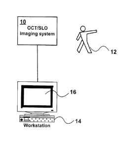
common sequel of many ocular conditions and the main cause of visual loss in
diabetic retinopathy.
[004] Any abnormal pooling of extracellular fluid may result in displacement
of the
spatial relationships between retinal neuronal components. Small amounts of
fluid
may lead to an increase in overall retinal thickness, whilst larger amounts
may give
rise to cell free spaces as seen in cystoid macular oedema
[005] It is known to predict visual acuity by measuring macular thickness.
However,
qualitative analysis of data describing the relationship between central
macular
thickness (CMT) and visual acuity shows that the correlation between CMT and
visual acuity is only moderate that CMT is only able to predict 16.6% of
visual acuity.
CA 02776437 2012-04-02
WO 2011/039374 PCT/EP2010/064718
Summary of the Invention
[006] It is an object of the invention to provide a method of predicting
potential
visual acuity with improved performance.
[007] According to the present invention there is provided a diagnostic method
wherein the potential preserved visual acuity in the retina of a patient is
determined
from the amount of tissue connecting the inner and outer plexiform layers
remaining
in the retina.
[008] The inventors have shown that there is a good correlation between the
amount of remaining tissue connecting the inner and outer plexiform layers and
potential visual acuity. The reason for this correlation is believed to be
that the retinal
tissue within these layers provides axonal connections within the retina
between the
photoreceptors and ganglion cells, and the measure of preserved axonal
connections
is a good indicator of preserved visual acuity.
[009] In one embodiment, a series of coronal images are taken at different
distances from the fovea. These provide a series of concentric rings (which
may or
may not be circular) of different radii surrounding the fovea. The amount of
tissue
remaining between the plexiform layers computed from an analysis of the image
dataset. The inventors have found that connecting tissue in the region between
about
1000 ¨2000 microns from the fovea is the most effective predictor of preserved
visual acuity.
[0010] The plane in which the measurements are taken may be the minimum
detectable plane between the inner and outer plexiform layers. In this
context, it will
be understood that the term plane is used to define a layer having a finite
thickness
following a planar contour.
=
[0011] The plane in which the measurements are taken may be shaped to follow
the
contour of a predefined surface, such as the retinal surface or retinal
pigment
epithelium layer.
2
CA 02776437 2015-12-14
[0012] Additionally, the level of visual acuity can be determined from an
analysis of
the sizes, for example the minimum sizes, of Muller fibers and/or bipolar
cells
connecting the inner and outer plexiform layers.
[0013] The measurements are preferably taken with a suitable imaging and
processing system such as Optical Coherence Tomography (OCT), ultrasound, or
confocal. A preferred system uses a combined OCT/confocal system that allows
the
OCT c.'nd confocal images to be displayed with a pixel for pixel
correspondence on a
computer screen.
[0014] In another aspect the invention provides a diagnostic apparatus for
determining the potential preserved visual acuity in the retina of a patient,
comprising
an imaging system for imaging the retinal tissue between the inner and outer
plexiform layers; and a processor for computing the potential preserved visual
acuity
based on the amount of said retinal tissue remaining between said inner and
outer
plexiform layers.
[0015] The processor may select the layer and position to measure the amount
of
connecting tissue automatically, and may make an indirect measurement of the
amount of tissue connecting the inner and outer plexiform layers.
In one aspect, there is provided a method of determining the potential
preserved visual acuity in a retina of a patient, the method comprising:
using an imagery system to create an images dataset representing the
amount of tissue connecting the inner and outer plexiform layers in the
retina;
measuring the amount of tissue connecting the inner and outer plexiform
layers in the retina from the images dataset;
processing the measured amount of tissue to determine if the measured
amount of tissue is within a predetermined range; and
3
CA 02776437 2015-12-14
if the amount of tissue measured is within the predetermined range,
displaying an image representing the presence of visual acuity in the retina
of the
patient.
In another aspect, there is provided an apparatus for determining the
potential preserved visual acuity in a retina of a patient, the apparatus
comprising:
an imagery system for creating an images dataset representing the amount of
tissue connecting the inner and outer plexiform layers in the retina and for
measuring
the amount of tissue connecting the inner and outer plexiform layers in the
retina
from the images dataset;
a processor for determining if the measured amount of tissue is within a
predetermined range; and
a display for displaying an image representing the presence of visual acuity
in
the retina of the patient, if the amount of tissue measured is within the
predetermined
range.
In another aspect, there is provided a computer program product comprising
a storage medium having stored therein instructions for determining the
potential
preserved visual acuity in a retina of a patient, the instructions comprising:
using an imagery system to create an images dataset representing the
amount of tissue connecting the inner and outer plexiform layers in the
retina;
measuring the amount of tissue connecting the inner and outer plexiform
layers in the retina from the images dataset;
determining if the amount of tissue measured is within a predetermined
range; and
if the amount of tissue measured is within the predetermined range,
outputting a display image representing the presence of visual acuity in the
retina of
the patient.
3a
CA 02776437 2015-12-14
Brief Description of the drawings
[0016] The invention will now be described in more detail, by way of example
only,
with reference to the accompanying drawings, in which:-
[0017] Figure 1 shows light microscopy and optical coherence tomography images
of a human retina affected by cystoid macular oedema (CMO).
[0018] Figure 2 is a scanning electron microscopy image of cystoid macular
oedema.
[0019] Figure 3A is a macular thickness map of a patient with macular oedema
representing subfield mean thicknesses as from ETDRS study;
,
3b
CA 02776437 2012-04-02
WO 2011/039374
PCT/EP2010/064718
[0020] Figure 3B is a grayscale coronal OCT scan with superimposed grid
dividing
the macula in 5 areas of increasing eccentricity (radii:500p, 1000p, 1500p,
2000p,
2500p).
[0021] Figure 4 shows scatter plots of the relationship between a) Central
macular
thickness versus LogMAR VA (r5=0.407*); b) Tissue integrity within circle 1
versus
LogMAR VA (rs=-0.832*); c) Tissue integrity within circle 2 versus LogMAR VA
(rs=-
0.841*); d) Tissue integrity within circle 3 versus LogMAR VA (rs=-0.624*); e)
Tissue
integrity within circle 4 versus LogMAR VA (rs=-0.277*); and f) Tissue
integrity within
circle 5 versus LogMAR VA (rs=-0.134*).
[0022] Figure 5 shows the variation in R2 values representing the association
between visual acuity and retinal spared tissue at increasing eccentricity as
well as
visual acuity and central macular thickness (CMT);
[0023] Figure 6 is a scatter plot, with line of equality, for measured versus
predicted
LogMAR visual acuity using the linear regression model;
[0024] Figure 7 is a Bland-Altman mean-difference plot demonstrating agreement
between measured and predicted LogMAR visual acuity;
[0025] Figure 8 is a Bland-Altman mean-difference plot showing bias (solid
line) and
upper and lower limits of agreement (dashed line); and
[0026] Figure 9 is a block diagram showing an apparatus in accordance with one
embodiment of the invention.
Detailed Description of the Invention
[0027] Observations from histology and optical coherence tomography (OCT), as
shown in Figure 1, give a false impression of multiple cysts delineated by
tissue
structures in the Z-plane of the retina. However, scanning electron
microscopy, as
shown in Figure 2, shows that more commonly a single cystic space is present
within
which a number of structures extend from the inner to the outer retina. Such
structures consist of columns of Muller's fibers together with the axonal
elements of
4
CA 02776437 2012-04-02
WO 2011/039374 PCT/EP2010/064718
bipolar cells passing between the two plexiform layers. Empirical studies have
demonstrated that the two plexiform layers together with the outer limiting
membrane
form a physical resistance barrier to fluid movements. Thus, extracellular
fluid may
be contained within layers defined by these resistance barriers. In diabetic
retinopathy, cystic spaces may occur either between the inner and the outer
plexiform layers or between the outer limiting membrane and the outer
plexiform
layer. In the former location, there is a potential to displace bipolar cells
leading to
cell loss or compromised function, while in the latter, only photoreceptor
cells are at
risk.
[0028] Given the fundamental role of bipolar cells as the sole communication
pathway between photoreceptors and ganglion cells, any loss of connectivity
between these cells will compromise visual function. It therefore follows that
the more
the retinal thickness increases, the more such axons will be stretched. As a
consequence some will break. This phenomenon is believed to explain the
mechanism underlying the apparent relationship between increasing retinal
thickness
and decreasing visual acuity.
[0029] By contrast, those bipolar cells whose axons are closely adjacent to
Muller's
fibers will have a greater chance of surviving displacement because of the
greater
physical strength and support provided by the adjacent Muller's fibers.
[0030] The inventors have shown that a useful indicator of the visual acuity
and
potential visual outcome in eyes with macular oedema is to analyze the
residual
volume of tissue passing between the two plexiform layers, as only such areas
would
allow passage of bipolar axons between photoreceptors and ganglion cells. The
impact of photoreceptor-ganglion cell connectivity on visual acuity further
depends
upon the location of surviving axons within the central visual field. An
optimal
measurement of potential function would be an evaluation of the number of
vertical
elements passing between the plexiform layers, their diameter and eccentricity
from
the fovea.
,
CA 02776437 2012-04-02
WO 2011/039374
PCT/EP2010/064718
Experimental results
[0031] Patients with macular oedema were prospectively recruited from both
diabetic
and uveitic outpatient clinics over a period of nine months. The study
involved a
baseline assessment of visual function, ophthalmoscopy and OCT imaging at a
single timepoint. Patient information was anonymized at the time of patient
recruitment to allow independent data analysis.
[0032] Inclusion criteria for the study were a clinical diagnosis of cystoid
macular
oedema (CMO), confirmed either by OCT alone or by OCT and fundus fluorescein
angiography (FFA) at the time of enrolment. For each patient either one or
both eyes
were included in the study.
[0033] Patients with coexisting ocular pathologies were excluded. Exclusion
criteria
included the presence of media opacity affecting the quality of the OCT scan
and
angiographic or clinical evidence of ischaemic maculopathy.
[0034] Each patient underwent a complete anterior segment examination by slit-
lamp biomicroscopy and best corrected visual acuity assessment using a LogMAR
chart at 3 meters distance. All patients were then dilated using Phenylephrine
2.5%
and Tropicamide 1% and examined by indirect fundoscopy with a 78D lens. In the
diabetic patients, fluorescein angiography was required as one of the
inclusion
criteria of the study in order to assess retinal circulation and to allow
exclusion of
patients with subclinical foveal ischaemia.
[0035] Optical coherence tomography was carried out using a Spectrum-Oil
spectral domain OCT/SLO system (Spectral OCTSLO model E, Spectrum-
Ophthalmic Technologies Inc., Toronto, Canada). This device is an optical
imaging
system, combining a Confocal Scanning Ophthalmoscope and Optical Coherence
Tomography. Both the confocal fundus SLO image and the OCT image are
generated through the same optics and displayed simultaneously on the computer
screen with pixel to pixel correspondence. The system uses light generated
from an
infrared broadband super luminescent diode (SLD) with a wavelength between
6
CA 02776437 2012-04-02
WO 2011/039374
PCT/EP2010/064718
790nm and 950nm. Cross-sectional images of the retina along the x-y plane (B-
Scan), such as single line, radial and raster scans, could be obtained as well
as
coronal images within the z plane (C-scan). The setup is shown in Figure 9,
where
the OCT/SLO unit 10 was used to image the eye of patient 12. The results were
processed in computer 14 and displayed on screen 16.
[0036] A state of the art Fourier domain OCT device, such as described above,
and
designed for ophthalmic use can typically capture axial data over 2mm in depth
(measured in air) at 20,000 lines per second. The scan pattern across the
retina can
be arranged in a grid of 200 by 200 points with a spacing between lines and
rows of
around 25 microns. This scan pattern is centered on the fovea with a capture
time of
2 seconds. The imaging depth is defined by several parameters, but the useful
depth
for use in this analysis is at least 1.5mm measured in air.
[0037] Automated methods to adjust for patient movement during acquisition can
be
employed on the dataset prior to analysis. This can make use of the fast
acquisition
and small spacing between lines and rows, but can take the form of other
simultaneous acquisition streams.
[0038] Analysis of the 3D dataset is achieved by segmenting the dataset
through the
retina in the coronal plane to view tissue present at a level between the
inner and
outer plexiform layers of the retina. In this embodiment the segmentation
thickness
would be around 10 microns (the resolution of the system in the axial
direction).
[0039] From this segmented view (image), the image is processed to a 2 bit
image
so that each pixel is either turned into a one or zero (where a one would be
tissue
and a zero, none or oedema). The amount of remaining tissue within a 1mm
radius
of the anatomical fovea is measured and this used as a predictor of the visual
acuity
that could be potentially achieved after treatment and resolution of the
oedema.
[0040] In preliminary studies, scans were obtained using three different modes
of
operation. First, a series of 24 radial scans over 360 degrees were
automatically
initiated intersecting at the centre of the patient's fixation. Secondly, a
single scan
7
CA 02776437 2012-04-02
WO 2011/039374
PCT/EP2010/064718
mode was selected whose orientation and location within the fundus was
determined
by the operator. Thirdly, the system was used to generate a raster scan of the
macula from the superior to the inferior arcade with 64 scans, again centred
by the
patient's fixation.
[0041] Three dimensional views of the macula were obtained by selecting the
topography mode where images were viewed as surface maps and these were
extracted manually by slicing the 3-D picture using the device based image
analysis
software. The topography scan also allowed the operator to extract information
about
retinal thickness in different areas of the posterior pole by using the ETDRS
macular
grid. Coronal scans (C-scans) were fundamental to the present study and
obtained
by selecting the mid point between the ganglion cell layer and the innermost
aspect
of the outer plexiform layer, in most cases to mid depth of the cysts. In
practice this
was obtained by adjusting the section plane to an appropriate level parallel
with the
retinal surface in the B scan displayed on the x-axis of the coronal image
(Figure 3A).
[0042] The image analysis system extracted three datasets, namely 1) the
number
of columns of tissue present 2) their cross-sectional area at their narrowest
point 3)
and their eccentricity from the foveal centre. Data were collected from a
series of
concentric rings of 500 pm, 1000 pm, 1500 pm, 2000 pm, 2500 pm radii
respectively
(Figure 3B). Within each ring, the area of surviving tissue as opposed to
cystic space
was extracted by first processing the data to compress the grayscale such that
tissue
became white and the oedema black.
[0043] Next, the data were processed to count the number of white pixels
present
within each ring, thus giving a measure proportionate to the potential number
of
connections passing between the two plexiform layers. The number of pixels of
spared tissue within each annulus was converted to an area in mm2 by scaling
the
ratio of the number of pixels of spared tissue to the total number of pixels
in the
annulus by the area in mm2 of the annulus.
8
CA 02776437 2012-04-02
WO 2011/039374
PCT/EP2010/064718
[0044] This study had three primary outcomes: 1) Best corrected Log MAR visual
acuity; 2) retinal tissue integrity evaluated as number of pixels
corresponding to the
tissue component between cystic spaces and observable at increasing
eccentricities
from the fovea in segmented images of OCT/SLO coronal scans; and 3) central
macular thickness measurement obtained from the OCT/SLO retinal thickness map.
[0045] A linear regression model was developed to assess if the amount of glia
could be used to predict visual acuity. A data set of 129 eyes were randomized
and
split into two data sets, one of 100 eyes and the other of 29 eyes.
[0046] The first group of 100 eyes was used to determine the linear regression
model. The remaining 29 eyes were used to test the validity of the model in
predicting visual acuity. All outcome variables were entered into a stepwise
linear
regression model with logMAR visual acuity as the outcome variable. LogMAR is
a
commonly used scale for measuring visual acuity.
[0047] The criterion for entry into the model was P=0.05 and P=0.10 for
removal.
Stepwise linear regression is an extension of simple linear regression where
the
dependent variable is predicted by a linear equation involving one outcome or
independent variable and a constant. In stepwise linear regression, multiple
variables
can be linearly combined in the model. They are entered automatically by the
statistics software provided they make a statistically significant improvement
in the
model.
[0048] The remaining 29 eyes were used to test the model by assessing the
agreement between the predicted and measured visual acuity using the Bland-
Altman method. A total of 81 participants enrolled, 36 males and 45 females.
The
average age was 63 years (range 26-87 years).
[0049] Most patients (73%, 59 subjects) underwent fluorescein angiography,
whereas in the remaining group an angiographic study could not be performed
(27%,
22 subjects) due to previously documented adverse reaction to the dye (9
subjects),
refusal to the investigation (8 subjects) or technical difficulty to obtain a
satisfactory
9
CA 02776437 2012-04-02
WO 2011/039374 PCT/EP2010/064718
venous access (7 subjects). Typical patient contact time was 40 minutes of
which
only 5 minutes were required for OCT imaging.
[0050] The distribution of spared retinal tissue values in the concentric
annuli varied
with eccentricity. Tissue integrity date in ring 1, 2 and 3 were not normally
distributed
(p=n,001; 0.001; and 0.02 respectively), whereas data related to tissue
integrity
within rings 4 and 5 displayed a normal distribution (p=0.093 and p=0.2,
respectively). Values for central macular thickness showed a normal
distribution
(p=0.059), whereas LogMAR visual acuity values were not normally distributed.
The
relationship between tissue integrity at increasing eccentricity and LogMAR
visual
acuity was significant at the 0.01 level (2 tailed) in circles 1 (r=0.832), 2
(r=0.841), 3
(R=0.624), 4 (R=0.277). The correlation was significant at 0.05 level (2
tailed) in ring
(r=0.152).
[0051] Qualitative analysis of the data shows a clear linear relationship
between the
amount of spared tissue within rings 1 and 2 and Log MAR visual acuity. The
relationship between spared tissue in ring 3 and visual acuity was less clear
and
became even less apparent in rings 4 and 5 (Figure 4).
[0052] The linear regression model demonstrated that measures of tissue
integrity
derived from rings 1 and 2 predicted up to 74% and 75% of visual acuity
respectively.
The r2 values for rings 3, 4 and 5 were significantly lower as shown in the
following
table.
Table
Variable R P (2 tailed) R2% R2
CMT 0.047 <0.001 16.6% 0.14
Tissue spared in ring 1 (5000 -0.832 <0.001 69.2% 0.74
Tissue spared in ring 2 (10000 -0.841 <0.001 70.7% 0.75
Tissue spared in ring 3 (1500) -0.624 <0.001 38.9% 0.43
Tissue spared in ring 4 (20000 -0.277 0.001 7.7% 0.09
Tissue spared in ring 5 (25001a) -0.133 0.132 1.8% 0.092
Comparative example
CA 02776437 2012-04-02
WO 2011/039374
PCT/EP2010/064718
The scatter plots shown in Figure 5 represent the relationship between central
macular thickness and visual acuity (a) and between spared retinal tissue at
increasing eccentricities and visual acuity (b - f) for all 129 eyes.
[0053] Qualitative analysis of data describing the relationship between
central
macular thickness (CMT) and visual acuity showed a very weak link as shown in
Figure 5. The correlation between CMT and LogMAR visual acuity was moderate
(r=0.407). The regression analysis of CMT versus visual acuity demonstrates
that
CMT predicts only 16% of visual acuity.
[0054] The linear regression model was given by
log MAR = 1.089 + 0.252 x CMT - 2.140 x T1- 0.854 x T2 (1)
where CMT is the central macular thickness in mm and T1 and T2 are the areas
of
tissue sparing in mm2 in rings 1 and 2 respectively. This model has an R2
value of
80.7% indicating that equation (1) explained over 80.7% of the variation in
LogMAR
visual acuity. It was noteworthy that the most predictive variable was T2 and
this
alone could predict 74.4% of the variation in LogMAR visual acuity.
[0055] Figure 6 shows a scatter plot of the measured Log MAR visual acuity
plotted
against the estimated LogMAR visual acuity. A line of equality is shown along
which
all data points would be expected to lie in presence of perfect agreement.
This was
not the case, as would be expected from most clinical measures.
[0056] Figure 7 shows the Bland-Altman mean-difference plot for the data. It
shows
that there is a relationship between the mean LogMAR visual acuity and the
differences (r=-0.44; P=0.016). This means that the bias changes with visual
acuity
and that the limits of agreement will be underestimated for good visual acuity
(small
values of LogMAR visual acuity) and overestimated for poor visual acuity (high
values of LogMAR visual acuity).
[0057] Figure 8 shows the change in bias and limits of agreement with mean
LogMAR acuity. A more accurate value for the limits of agreement is 0.17 log
units
11
CA 02776437 2012-04-02
WO 2011/039374
PCT/EP2010/064718
at a LogMAR visual acuity of 0.5 although this changes slightly with the mean
measured value.
[0058] The above results also demonstrate that there is a strong correlation
between
visual acuity in patients with cystoid macular oedema and the volume of tissue
passing between the two plexiform layers in the central retina as determined
by OCT.
It is the first time that a predictive measure of visual performance has been
derived
from imaging of macular oedema.
[00Cq1 The results demonstrate that good visual acuity only occurred in those
patients with an adequate volume of tissue running between the inner and the
outer
plexiform layers in the central 1000-2000p of retina (Figure 4 and 5). Given
that
foveal cones have inner connecting fibers that may be up to 500pm in length,
foveolal cones may connect to bipolar cells displaced 500pm radially from the
inner
and outer segments. Thus this lateral displacement of connections between
foveal
cones and ganglion cells explains the dependency of visual acuity on the
tissue
integrity in both rings 1 and 2.
[0060] While there was still reasonable correlation within ring 3, which is
believed to
be due to signals derived from photoreceptors at the extreme edges of the
fovea,
correlation was lost within rings 4 and 5. In these locations, although large
amounts
of tissue volume may be spared, the connectivity is predominantly with
extrafoveal
photoreceptors.
[0061] From the linear regression model, it appears that a minimum of 50% of
preserved retinal tissue within ring 1 is necessary in order to maintain a
visual acuity
of 0.4 LogMAR or better (Figure 4, scatter plot b), whereas at least 70% of
the retinal
tissue within ring 2 is necessary for a level of visual acuity of 0.4 LogMAR
or better
(Figure 4, scatter plot c).
[0062] Even though the total number of bipolar axons traversing the space
between
the plexiform layers may be significantly reduced, both horizontal and
amacrine cells
12
CA 02776437 2012-04-02
WO 2011/039374
PCT/EP2010/064718
will contribute to image processing and VA by integrating signals over a
number of
photoreceptor cells and ganglion cells respectively.
[0063] The apparent correlation between the increase in retinal thickness and
the
decrease in visual acuity in accordance with prior art methods may be
explained by
the present results whereby increase in thickness will be associated with
increase in
loss in viable axons. The more direct approach to assessing neuronal survival
in the
present invention would also explain why the correlation values are so much
better.
[0064] The above results establish retinal tissue integrity as a measure of
preserved
axonal connections and indicator of visual function. The strength of the
relationship
between preserved tissue and visual function, as expected, decreases at
increasing
eccentricities from the centre of the fovea.
[0065] The ability to determine the potential visual outcome for patients
prior to the
commencement of any treatment trial is highly beneficial in that it will allow
exclusion
of those individuals who could not in anyway benefit from intervention.
[0066] It will be appreciated that the invention could be implemented in
software, and
as such the invention also extends to a computer program product comprising a
storage medium having stored thereon instructions which when executed on a
general purpose computer process a dataset obtained from a 3D ophthalmic
imaging
system, such as an OCT/confocal system, to derive therefrom the amount of
tissue
connecting the inner and outer plexiform layers remaining in the retina.
13