Note: Descriptions are shown in the official language in which they were submitted.
CA 02777934 2012-04-16
WO 2011/050069 PCT/US2010/053386
PROXIMITY MEDIATED ASSAYS FOR DETECTING ONCOGENIC
FUSION PROTEINS
CROSS-REFERENCE TO RELATED APPLICATIONS
[0001] The present application claims priority to U.S. Provisional Application
No.
61/253,393, filed October 20, 2009, U.S. Provisional Application No.
61/305,084, filed
February 16, 2010, U.S. Provisional Application No. 61/327,487, filed April
23, 2010, and
U.S. Provisional Application No. 61/383,037, filed September 15, 2010, the
disclosures of
which are herein incorporated by reference in their entirety for all purposes.
BACKGROUND OF THE INVENTION
[0002] Fusion proteins, also known as chimeric proteins, are proteins created
through the
joining of two or more genes which originally encode separate proteins.
Translation of this
fusion gene results in a single polypeptide with functional properties derived
from each of the
original proteins. Chimeric mutant proteins occur when a large-scale mutation,
typically a
chromosomal translocation, creates a novel coding sequence containing parts of
the coding
sequences from two different genes. Naturally-occurring fusion proteins are
important in
cancer, where they function as oncoproteins.
[0003] The BCR-ABL fusion protein is a well-known example of an oncogenic
fusion
protein. It is considered to be the primary oncogenic driver of chronic
myelogenous
leukemia (CML), but is also associated with acute lymphoblastic leukemia
(ALL). In fact,
the cytogenetic hallmark of CML is the Philadelphia chromosome (Ph), which
results in the
formation of the BCR-ABL fusion gene encoding a 210 kDa protein. Indeed, the
resulting
BCR-ABL fusion protein is an active tyrosine kinase that is critical to the
pathogenesis of
CML. Although imatinib (Gleevec ) is currently the first line therapy for
newly diagnosed
patients with CML, about 20-25% of patients do not achieve durable complete
cytogenetic
responses. Studies have shown that the reactivation of BCR-ABL signaling in
the presence
of continued imatinib treatment is the major cause of resistance. In the
majority of patients,
reactivation results from mutations in the BCR-ABL kinase domain which impair
imatinib
binding and lead to the induction of drug resistance. As such, the measurement
of BCR-ABL
activity finds utility in predicting response to therapy with tyrosine kinase
inhibitors such as
imatinib as well as in identifying patients who develop resistance to such
inhibitors.
CA 02777934 2012-04-16
WO 2011/050069 PCT/US2010/053386
[0004] At present, methods available for detecting BCR-ABL activity rely on
measuring
phosphorylated CRKL (pCRKL), a BCR-ABL substrate. For example, La Rosee et al.
(Haematologica, 93:765-9 (2008)) describes a Western blot analysis of total
leukocyte lysates
to determine the level of pCRKL as a surrogate of BCR-ABL activity (see also,
Hochhaus et
al., Leukemia, 16:2190-6 (2002); White et al., J Clin. Oncol., 25:4445-51
(2007)). Similarly,
Khorashad et al. (Haematologica, 94:861-4 (2009)) describes a flow cytometry-
based method
of measuring the level of pCRKL to evaluate BCR-ABL activity (see also,
Hamilton et al.,
Leukemia, 20:1035-9 (2006)). However, these methods lack the specificity and
sensitivity
that is required for determining the presence or level of BCR-ABL activity in
a sample
because they are single antibody assays which rely on detecting the
phosphorylation of a
surrogate protein. Accordingly, specific and sensitive methods are needed to
detect BCR-
ABL activity, as well as the activity of other oncogenic fusion proteins, for
diagnostic,
prognostic, and therapeutic purposes. The present invention satisfies this
need and provides
related advantages as well.
BRIEF SUMMARY OF THE INVENTION
[0005] The present invention provides antibody-based arrays for detecting the
activation
state and/or total amount of one or a plurality of oncogenic fusion proteins
in a biological
sample such as whole blood (e.g., a lysate prepared from isolated rare
circulating cells or
leukocytes) or tumor tissue (e.g., a fine needle aspirate) and methods of use
thereof. In
certain instances, the activation state and/or total amount of oncogenic
fusion protein(s)
present in a sample can be measured in combination with one or a plurality of
signal
transduction molecules. The compositions and methods of the present invention
have the
advantages of specificity associated with enzyme-linked immunosorbent assays,
sensitivity
associated with signal amplification, and high-throughput multiplexing
associated with
microarrays.
[0006] In one aspect, the present invention provides a method for determining
the level or
activation state of an oncogenic fusion protein, the method comprising:
(a) contacting a cellular extract with a first binding moiety specific for a
first
domain of a first full-length protein under conditions suitable to transform
the first full-length
protein present in the cellular extract into a complex comprising the first
full-length protein
and the first binding moiety, wherein the first domain of the first full-
length protein is absent
from a corresponding oncogenic fusion protein comprising a second, different
domain of the
first full-length protein fused to a first domain of a second, different full-
length protein;
2
CA 02777934 2012-04-16
WO 2011/050069 PCT/US2010/053386
(b) removing the complex from step (a) from the cellular extract to form a
cellular extract devoid of the first full-length protein;
(c) contacting the cellular extract from step (b) with a second binding moiety
specific for the second, different domain of the first full-length protein
under conditions
suitable to transform the oncogenic fusion protein present in the cellular
extract into a
complex comprising the oncogenic fusion protein and the second binding moiety;
and
(d) determining the level or activation state of the complex from step (c),
thereby determining the level or activation state of the oncogenic fusion
protein.
[00071 In particular embodiments, determining the level or activation state of
an oncogenic
fusion protein includes measuring a (e.g., concentration) level of expression
and/or activation
(e.g., phosphorylation) of an oncogenic fusion protein (e.g., in a cellular
extract).
[00081 In preferred embodiments, steps (c) and (d) of the method of the
present invention
comprise an enzyme-linked immunosorbent assay (ELISA), a flow cytometry assay,
a tag-
sorting assay, or a proximity dual detection assay as described herein.
[00091 In one particular embodiment of the proximity dual detection assay, the
present
invention provides a method for determining the level or activation state of
an oncogenic
fusion protein, the method comprising:
(a) incubating a cellular extract with a dilution series of capture antibodies
specific for the oncogenic fusion protein to form a plurality of captured
oncogenic fusion
proteins, wherein the capture antibodies are restrained on a solid support,
wherein the
oncogenic fusion protein comprises a first domain corresponding to a first
protein and a
second domain corresponding to a second, different protein, and wherein the
capture
antibodies are specific for the first domain of the fusion protein;
(b) incubating the plurality of captured oncogenic fusion proteins with at
least
two types of detection antibodies specific for the second domain of the
oncogenic fusion
protein to form a plurality of detectable captured oncogenic fusion proteins,
wherein the
detection antibodies comprise:
(1) a plurality of activation state-independent antibodies labeled with a
facilitating moiety, and
(2) a plurality of activation state-dependent antibodies labeled with a first
member of a signal amplification pair,
wherein the facilitating moiety generates an oxidizing agent which
channels to and reacts with the first member of the signal amplification pair;
3
CA 02777934 2012-04-16
WO 2011/050069 PCT/US2010/053386
(c) incubating the plurality of detectable captured oncogenic fusion proteins
with a second member of the signal amplification pair to generate an amplified
signal; and
(d) detecting the amplified signal generated from the first and second members
of the signal amplification pair.
[0010] In another particular embodiment of the proximity dual detection assay,
the present
invention provides a method for determining the level or activation state of
an oncogenic
fusion protein, the method comprising:
(a) incubating a cellular extract with a dilution series of capture antibodies
specific for the oncogenic fusion protein to form a plurality of captured
oncogenic fusion
proteins, wherein the capture antibodies are restrained on a solid support,
wherein the
oncogenic fusion protein comprises a first domain corresponding to a first
protein and a
second domain corresponding to a second, different protein, and wherein the
capture
antibodies are specific for the first domain of the fusion protein;
(b) incubating the plurality of captured oncogenic fusion proteins with at
least
two types of detection antibodies to form a plurality of detectable captured
oncogenic fusion
proteins, wherein the detection antibodies comprise:
(1) a plurality of activation state-independent antibodies labeled with a
facilitating moiety, wherein the activation state-independent antibodies are
specific for the
first domain of the fusion protein, and
(2) a plurality of activation state-dependent antibodies labeled with a first
member of a signal amplification pair, wherein the activation state-dependent
antibodies are
specific for the second domain of the fusion protein,
wherein the facilitating moiety generates an oxidizing agent which
channels to and reacts with the first member of the signal amplification pair;
(c) incubating the plurality of detectable captured oncogenic fusion proteins
with a second member of the signal amplification pair to generate an amplified
signal; and
(d) detecting the amplified signal generated from the first and second members
of the signal amplification pair.
[0011] In another aspect, the present invention provides a method for
optimizing therapy
and/or reducing toxicity in a subject having cancer and receiving a course of
therapy for the
treatment of the cancer, the method comprising:
(a) isolating cancer cells after administration of an anticancer drug;
(b) lysing the isolated cells to produce a cellular extract;
4
CA 02777934 2012-04-16
WO 2011/050069 PCT/US2010/053386
(c) measuring a level of expression and/or activation of an oncogenic fusion
protein in the cellular extract using an assay described herein; and
(d) comparing the measured level of expression and/or activation of the
oncogenic fusion protein to a level of expression and/or activation of the
oncogenic fusion
protein measured at an earlier time during the course of therapy; and
(e) determining a subsequent dose of the course of therapy for the subject or
whether a different course of therapy should be administered to the subject
based upon the
comparison from step (d).
[0012] In certain embodiments, one or more signal transduction molecules
present in the
cellular extract are detected in addition to one or more oncogenic fusion
proteins. Examples
of signal transduction molecules include, without limitation, receptor
tyrosine kinases, non-
receptor tyrosine kinases, tyrosine kinase signaling cascade components,
and/or substrates for
the one or more oncogenic fusion proteins (e.g., BCR-ABL substrates). In some
instances,
the signal transduction molecules are detected using the methods described
herein, except
that, depending on the assay, either two antibodies (i.e., the capture
antibody and the
detection antibody) or three antibodies (i.e., the capture antibody and both
detection
antibodies) are directed to the same protein. In other instances, the signal
transduction
molecules are detected using any method known to one of skill in the art. In
particular
embodiments, one or more of the signal transduction molecules present in the
cellular extract
are detected in conjunction with one or more oncogenic fusion proteins using
the assays (e.g.,
immunoassays) described herein.
[0013] In certain embodiments, the present invention also provides kits for
performing the
proximity dual detection assays described herein, comprising: (a) a dilution
series of one or a
plurality of capture antibodies restrained on a solid support, wherein the
capture antibodies
are specific for one or more analytes of interest (e.g., oncogenic fusion
proteins or signal
transduction molecules); and (b) a plurality (e.g., at least two types) of
detection antibodies
for each analyte of interest. The kits can optionally further comprise other
reagents such as,
for example, the first and second members of the signal amplification pair.
[00141 Other objects, features, and advantages of the present invention will
be apparent to
one of skill in the art from the following detailed description and figures.
5
CA 02777934 2012-04-16
WO 2011/050069 PCT/US2010/053386
BRIEF DESCRIPTION OF THE DRAWINGS
[0015] Figure 1 shows one embodiment of the assay format of the present
invention, which
relies on the co-localization of two additional detector antibodies linked
with enzymes for
subsequent channeling events per each target oncogenic fusion protein bound.
[0016] Figure 2 shows schematically the application of the arrays of the
invention for drug
selection throughout the course of cancer treatment.
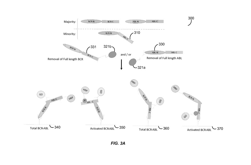
[0017] Figures 3 A-D show various embodiments of the assay format of the
present
invention for detecting expression and activation levels of oncogenic fusion
proteins such as
BCR-ABL.
[0018] Figure 4 shows the BCR-ABL signal in K562 cells after removal of free
BCR.
[0019] Figure 5 shows the BCR signal in K562 cells after removal of free BCR.
[0020] Figure 6 shows the detection of total and phosphorylated levels of BCR-
ABL in
K562 cells.
[0021] Figure 7 shows the phosphorylation level ("Phospho BCR-ABL") and total
amount
("Total BCR-ABL") of BCR-ABL detected in K562 human chronic myelogenous
leukemia
cells with or without depletion of free BCR using BCR C-terminal antibody-
coupled beads.
[0022] Figure 8 shows the phosphorylated BCR-ABL signal in K562 cells with or
without
removal of free BCR using BCR C-terminal antibody-coupled beads. In
particular, Figure
8A provides a microarray comparison of the phosphorylated BCR-ABL signal
detected in
K562 cell lysates with or without removal of full-length BCR ("Non-beads
treated" = BCR
not removed versus "Beads treated" = BCR removed with beads containing an
antibody
specific for the C-terminus of full-length BCR conjugated thereto). Figure 8B
provides a
graphical depiction of the microarray data with Relative Fluorescence Units
(RFU) as a
function of cell number.
[0023] Figure 9 shows the total BCR-ABL signal in K562 cells with or without
removal of
free BCR using BCR C-terminal antibody-coupled beads. In particular, Figure 9A
provides a
microarray comparison of the total BCR-ABL signal detected in K562 cell
lysates with or
without removal of full-length BCR ("Non-beads treated" = BCR not removed
versus "Beads
treated" = BCR removed with beads containing an antibody specific for the C-
terminus of
full-length BCR conjugated thereto). Figure 9B provides a graphical depiction
of the
microarray data with Relative Fluorescence Units (RFU) as a function of cell
number.
6
CA 02777934 2012-04-16
WO 2011/050069 PCT/US2010/053386
[0024] Figure 10 shows the removal of free full-length BCR from an extract of
K562 cells
after contacting the cellular extract with BCR C-terminal antibody-coupled
beads. In
particular, Figure IOA provides a microarray comparison of the total BCR
signal detected in
K562 cell lysates with or without removal of full-length BCR ("Non-beads
treated" = BCR
not removed versus "Beads treated" = BCR removed with beads containing an
antibody
specific for the C-terminus of full-length BCR conjugated thereto). Figure I
OB provides a
graphical depiction of the microarray data with Relative Fluorescence Units
(RFU) as a
function of cell number.
[0025] Figure 11 shows that free full-length BCR and ABL proteins, but not BCR-
ABL
fusion protein, are present in white blood cells (WBCs).
[0026] Figure 12 shows that the free full-length BCR present in WBCs inhibited
the
phospho BCR-ABL signal in K562 cell extracts when such K562 cell extracts were
spiked
with WBC extracts.
[0027] Figure 13 shows the total BCR-ABL signal in K562 cells spiked with WBC
extracts
after removal of free BCR using BCR C-terminal antibody-coupled beads. In
particular,
Figure 13A shows that the free BCR signal was saturated when the K562 cell
extracts were
spiked with WBC extracts. After treatment with BCR C-terminal antibody-coupled
beads,
the free BCR was removed. Figure 13B shows that the BCR-ABL signal was not
changed
with or without beads treatment in the same experiment.
[0028] Figure 14 shows that the BCR-ABL inhibitor imatinib (Gleevec ) dose-
dependently
inhibited activation (i.e., phosphorylation), but not expression (i.e., total
levels), of BCR-
ABL protein in K562 cells.
[0029] Figure 15 shows that the BCR-ABL inhibitor nilotinib (Tasigna ) dose-
dependently
inhibited activation (i.e., phosphorylation), but not expression (i.e., total
levels), of BCR-
ABL protein in K562 cells.
[0030] Figure 16 shows that the BCR-ABL inhibitor dasatinib (Sprycel ) dose-
dependently
inhibited activation (i.e., phosphorylation), but not expression (i.e., total
levels), of BCR-
ABL protein in K562 cells.
[0031] Figure 17 shows that CRKL is both present and activated (i.e.,
phosphorylated) in
K562 cells.
[0032] Figure 18 shows that CRKL is present in A431 human epidermoid carcinoma
cells
and is activated (i.e., phosphorylated) upon EGF treatment.
7
CA 02777934 2012-04-16
WO 2011/050069 PCT/US2010/053386
[0033] Figure 19 shows that CRKL is present in T47D human ductal breast
epithelial tumor
cells but is not activated (i.e., phosphorylated) upon EGF treatment.
[0034] Figure 20 shows that CRKL is present in T47D cells and is activated
(i.e.,
phosphorylated) at low levels upon heregulin (HRG) treatment.
[0035] Figure 21 shows that CRKL is present in MCF-7 human breast
adenocarcinoma
cells and is activated (i.e., phosphorylated) at low levels upon heregulin
(HRG) treatment.
[0036] Figure 22 illustrates the presence of activated (i.e., phosphorylated)
CRKL in the
white blood cells (WBCs) of different donors.
[0037] Figure 23 illustrates that JAK2 is activated (i.e., phosphorylated) in
K562 cells and
A431 cells.
[0038] Figure 24 illustrates that phosphorylated BCR-ABL can be detected and
measured
in cell lysates prepared from K562 cells isolated from blood using anti-CD45
magnetic beads.
[0039] Figure 25 illustrates that total BCR-ABL levels were not changed when
an antibody
directed to the C-terminus of native full-length BCR (which C-terminal domain
is not present
in BCR-ABL) was spotted on the same slide in the same pad as an antibody
directed to the N-
terminal region of BCR-ABL.
[00401 Figure 26 illustrates that the free native BCR signal detected with an
N-terminal-
specific BCR antibody was reduced when an antibody directed to the C-terminus
of native
BCR was spotted on the same slide in the same pad.
DETAILED DESCRIPTION OF THE INVENTION
1. Introduction
[0041] Hematological malignancies are the types of cancer that affect blood,
bone marrow,
and lymph nodes. As the three are intimately connected through the immune
system, a
disease affecting one of the three will often affect the others as well. For
example, although
lymphoma is technically a disease of the lymph nodes, it often spreads to the
bone marrow
and the blood. Chromosomal translocations, which create fusion proteins having
novel
coding sequences containing parts of the coding sequences from two different
genes, are a
common cause of these diseases, but are a less common cause of solid tumors.
As such, it is
important to identify the presence and/or activity of oncogenic fusion
proteins associated with
hematological malignancies in order to provide the appropriate prognosis and
treatment for
patients with these types of cancer.
8
CA 02777934 2012-04-16
WO 2011/050069 PCT/US2010/053386
[0042] For example, the BCR-ABL fusion protein is associated with chronic
myelogenous
leukemia (CML) as well as acute lymphoblastic leukemia (ALL). In particular,
the BCR-
ABL protein is an active tyrosine kinase that is critical to cancer
pathogenesis. Although
imatinib (Gleevec) is currently the first line therapy for newly diagnosed
patients with CML,
about 20-25% of patients do not achieve durable complete cytogenetic
responses. Studies
have shown that the reactivation of BCR-ABL kinase activity in the presence of
continued
imatinib treatment is the major cause of resistance. As such, the measurement
of BCR-ABL
activity finds utility in predicting response to therapy with tyrosine kinase
inhibitors such as
imatinib as well as in identifying patients who develop resistance to such
inhibitors.
[0043] The present invention provides methods for detecting the activation
state and/or
total amount of one or a plurality of fusion proteins (alone or in combination
with one or a
plurality of signal transduction molecules) in isolated cells using an
antibody-based array
assay system. Cellular extracts prepared from isolated leukocytes, circulating
cells, or other
cell types are particularly useful in the methods described herein. In some
embodiments, the
multiplex, high-throughput immunoassays of the present invention can detect
the activation
state of one or more oncogenic fusion proteins and/or signal transduction
molecules at the
single cell level. In fact, signal transduction molecules such as EGFR can be
detected with a
sensitivity of about 100 zeptomoles and a linear dynamic range of from about
100 zeptomoles
to about 100 femtomoles. As such, single-cell detection of the activation
state of one or more
oncogenic fusion proteins and/or signal transduction molecules facilitates
cancer prognosis
and diagnosis as well as the design of personalized, targeted therapies.
[0044] Figure 1 illustrates an exemplary proximity dual detection assay of the
present
invention in which an oncogenic fusion protein such as BCR-ABL is bound to a
capture
antibody and two detection antibodies (i.e., an activation state-independent
antibody and an
activation state-dependent antibody). The capture antibody 1 binds the BCR
portion of the
fusion protein independent of its activation state. While the activation state-
independent
antibody 2 binds the ABL portion of the fusion protein independent of its
activation state, the
activation state-dependent antibody 3 binds the ABL portion of the fusion
protein dependent
of its activation state (e.g., the activation state-dependent antibody will
only bind an activated
form of BCR-ABL having a phosphorylated residue). The activation state-
independent
antibody is labeled with a facilitating moiety 4 ("Enzyme A") and the
activation state-
dependent antibody is labeled with a first member of a signal amplification
pair 5 ("Enzyme
B"). Binding of both detection antibodies to the ABL portion of the fusion
protein brings the
facilitating moiety within sufficient proximity to the first member of the
signal amplification
9
CA 02777934 2012-04-16
WO 2011/050069 PCT/US2010/053386
pair such that a signal generated by the facilitating moiety can channel to
the first member of
the signal amplification pair, resulting in the generation of a detectable
and/or amplifiable
signal. Various methods for proximity channeling are described herein and are
also known in
the art including, but not limited to, FRET, time-resolved fluorescence-FRET,
LOCI, etc. An
advantage of proximity channeling, as used in the methods of the present
invention, is that a
single detectable signal is generated for only those analytes (e.g., fusion
proteins or signal
transduction molecules) that have bound all three antibodies, resulting in
increased assay
specificity, lower background, and simplified detection.
[0045] As explained in greater detail herein, to evaluate potential anticancer
therapies for
an individual patient, the isolated cells can be incubated with one or more
anticancer drugs at
varying doses. Growth factor stimulation can then be performed for a few
minutes (e.g.,
about 1-5 minutes) or for several hours (e.g., about 1-6 hours). The
differential activation of
signaling pathways with and without anticancer drugs can aid in the selection
of a suitable
cancer therapy at the proper dose for each individual patent. Cells can also
be isolated from a
patient during anticancer drug treatment and stimulated with one or more
growth factors to
determine whether a change in therapy should be implemented. As such, Figure 2
shows that
the methods of the present invention advantageously assist the clinician in
providing the right
anticancer drug at the right dose at the right time for every patient.
II. Definitions
[0046] As used herein, the following terms have the meanings ascribed to them
unless
specified otherwise.
[0047] The term "cancer" includes any member of a class of diseases
characterized by the
uncontrolled growth of aberrant cells. The term includes all known cancers and
neoplastic
conditions, whether characterized as malignant, benign, soft tissue, or solid,
and cancers of all
stages and grades including pre- and post-metastatic cancers. Non-limiting
examples of
different types of cancer include hematological malignancies (e.g., leukemia,
lymphoma);
osteogenic sarcomas (e.g., Ewing sarcoma); soft tissue sarcomas (e.g.,
Dermatofibrosarcoma
Protuberans (DFSP), rhabdomyosarcoma); other soft tissue malignancies,
papillary thyroid
carcinomas; prostate cancer; gastric cancer (e.g., stomach); breast cancer;
lung cancer (e.g.,
non-small cell lung cancer); digestive and gastrointestinal cancers (e.g.,
colorectal cancer,
gastrointestinal stromal tumors, gastrointestinal carcinoid tumors, colon
cancer, rectal cancer,
anal cancer, bile duct cancer, and small intestine cancer); esophageal cancer;
gallbladder
cancer; liver cancer; pancreatic cancer; appendix cancer; ovarian cancer;
renal cancer (e.g.,
CA 02777934 2012-04-16
WO 2011/050069 PCT/US2010/053386
renal cell carcinoma); cancer of the central nervous system; skin cancer;
choriocarcinomas;
and head and neck cancers. As used herein, a "tumor" comprises one or more
cancerous
cells.
[0048] A "hematological malignancy" includes any type of cancer that affects
the blood,
bone marrow, and/or lymph nodes. Examples of hematological malignancies
include, but are
not limited to, leukemia, lymphoma, and multiple myeloma. Non-limiting
examples of
different kinds of leukemia include chronic myelogenous leukemia (CML), acute
lymphoblastic leukemia (ALL), chronic lymphocytic leukemia (CLL), acute
myelogenous
leukemia (AML), and large granular lymphocytic leukemia. Subtypes of CML
include, e.g.,
chronic monocytic leukemia. Subtypes of ALL include, e.g., precursor B-cell
acute
lymphoblastic leukemia, pro-B-cell acute lymphoblastic leukemia, precursor T-
cell acute
lymphoblastic leukemia, and acute biphenotypic leukemia. Subtypes of CLL
include, e.g., B-
cell prolymphocytic leukemia. Subtypes of AML include, e.g., acute
promyelocytic
leukemia, acute myeloblastic leukemia, and acute megakaryoblastic leukemia.
Examples of
different kinds of lymphoma include, but are not limited to, Hodgkin's
lymphoma (four
subtypes) and non-Hodgkin lymphoma, such as, e.g., small lymphocytic lymphoma
(SLL),
diffuse large B-cell lymphoma (DLBCL), follicular lymphoma (FL), mantle cell
lymphoma
(MCL), hairy cell leukemia (HCL), marginal zone lymphoma (MZL), Burkitt's
lymphoma
(BL), post-transplant lymphoproliferative disorder (PTLD), T-cell
prolymphocytic leukemia
(T-PLL), B-cell prolymphocytic leukemia (B-PLL), Waldenstrom's
macroglobulinemia (also
known as lymphoplasmacytic lymphoma), and other NK- or T-cell lymphomas.
[0049] The term "analyte" includes any molecule of interest, typically a
macromolecule
such as a polypeptide, whose presence, amount, and/or identity is determined.
In certain
instances, the analyte is a cellular component of a cancerous cell, preferably
an oncogenic
fusion protein or a signal transduction molecule.
[0050] The term "transform" or "transforming" includes a physical and/or
chemical change
of an analyte or sample to extract the analyte or to change or modify the
analyte as defined
herein. As used herein, an extraction, a manipulation, a chemical
precipitation, an ELISA, a
complexation, an immuno-extraction, a physical or chemical modification of the
analyte or
sample to measure a level or concentration or activation state of an analyte
all constitute a
transformation. In other words, as long as the analyte or sample is not
identical before and
after the transformation step, the change or modification is a transformation.
it
CA 02777934 2012-04-16
WO 2011/050069 PCT/US2010/053386
[0051] As used herein, the term "dilution series" is intended to include a
series of
descending concentrations of a particular sample (e.g., cell lysate) or
reagent (e.g., antibody).
A dilution series is typically produced by a process of mixing a measured
amount of a
starting concentration of a sample or reagent with a diluent (e.g., dilution
buffer) to create a
lower concentration of the sample or reagent, and repeating the process enough
times to
obtain the desired number of serial dilutions. The sample or reagent can be
serially diluted at
least 2, 3, 4, 5, 6, 7, 8, 9, 10, 15, 20, 25, 30, 35, 40, 45, 50, 100, 500, or
1000-fold to produce
a dilution series comprising at least 2, 3, 4, 5, 6, 7, 8, 9, 10, 11, 12, 13,
14, 15, 16, 17, 18, 19,
20, 25, 30, 35, 40, 45, or 50 descending concentrations of the sample or
reagent. For
example, a dilution series comprising a 2-fold serial dilution of a capture
antibody reagent at
a 1 mg/ml starting concentration can be produced by mixing an amount of the
starting
concentration of capture antibody with an equal amount of a dilution buffer to
create a 0.5
mg/ml concentration of the capture antibody, and repeating the process to
obtain capture
antibody concentrations of 0.25 mg/ml, 0.125 mg/ml, 0.0625 mg/ml, 0.0325
mg/ml, etc.
[0052] The term "superior dynamic range" as used herein refers to the ability
of an assay to
detect a specific analyte in as few as one cell or in as many as thousands of
cells. For
example, the immunoassays described herein possess superior dynamic range
because they
advantageously detect a particular oncogenic fusion protein or signal
transduction molecule
of interest in about 1-10,000 cells (e.g., about 1, 5, 10, 25, 50, 75, 100,
250, 500, 750, 1000,
2500, 5000, 7500, or 10,000 cells) using a dilution series of capture antibody
concentrations.
[0053] The term "fusion protein" or "chimeric protein" includes a protein
created through
the joining of two or more genes which originally encode separate proteins.
Such gene
fusions are typically generated when a chromosomal translocation replaces the
terminal
exons of one gene with intact exons from a second gene. This creates a single
gene which
can be transcribed, spliced, and translated to produce a functional fusion
protein. In
particular embodiments, the fusion protein is an oncogenic fusion protein,
i.e., a fusion
protein involved in oncogenesis. Examples of oncogenic fusion proteins
include, but are not
limited to, BCR-ABL, DEK-CAN, E2A-PBX1, RARa-PML, IREL-URG, CBFf3-MYH11,
AMLl-MTG8, EWS-FLI, LYT-10-Cal, HRX-ENL, HRX-AF4, NPM-ALK, IGH-MYC,
RUNX1-ETO, TEL-TRKC, TEL-AML1, MLL-AF4, TCR-RBTN2, COLIAI-PDGF, E2A-
HLF, PAX3-FKHR, ETV6-NTRK3, RET-PTC, TMRSS-ERG, and TPR-MET.
[0054] The term "signal transduction molecule" or "signal transducer" includes
proteins
and other molecules that carry out the process by which a cell converts an
extracellular signal
or stimulus into a response, typically involving ordered sequences of
biochemical reactions
12
CA 02777934 2012-04-16
WO 2011/050069 PCT/US2010/053386
inside the cell. Examples of signal transduction molecules include, but are
not limited to,
receptor tyrosine kinases such as EGFR (e.g., EGFRIHER-1/ErbB1, HER-
2/Neu/ErbB2,
HER-3/ErbB3, HER-4/ErbB4), VEGFR-1/FLT-1, VEGFR-2/FLK-1/KDR, VEGFR-3/FLT-4,
FLT-3/FLK-2, PDGFR (e.g., PDGFRA, PDGFRB), c-Met, c-KIT/SCFR, INSR (insulin
receptor), IGF-IR, IGF-IIR, IRR (insulin receptor-related receptor), CSF-1R,
FGFR 1-4,
HGFR 1-2, CCK4, TRK A-C, MET, RON, EPHA 1-8, EPHB 1-6, AXL, MER, TYRO3, TIE
1-2, TEK, RYK, DDR 1-2, RET, c-ROS, V-cadherin, LTK (leukocyte tyrosine
kinase), ALK
(anaplastic lymphoma kinase), ROR 1-2, MUSK, AATYK 1-3, RTK 106, and truncated
forms of the receptor tyrosine kinases such as p95ErbB2; non-receptor tyrosine
kinases such
as Src, Frk, Btk, Csk, Abl, Zap70, Fes/Fps, Fak, Jak, Ack, and LIMK; tyrosine
kinase
signaling cascade components such as Akt, MAPK/ERK, MEK, RAF, PLA2, MEKK,
JNKK,
JNK, p38, Shc (p66), PI3K, Ras (e.g., K-Ras, N-Ras, H-Ras), Rho, Racl, Cdc42,
PLC, PKC,
p70 S6 kinase, p53, cyclin D1, STAT1, STAT3, PIP2, PIP3, PDK, mTOR, BAD, p21,
p27,
ROCK, IP3, TSP-1, NOS, PTEN, RSK 1-3, JNK, c-Jun, Rb, CREB, Ki67, and
paxillin;
nuclear hormone receptors such as estrogen receptor (ER), progesterone
receptor (PR),
androgen receptor, glucocorticoid receptor, mineralocorticoid receptor,
vitamin A receptor,
vitamin D receptor, retinoid receptor, thyroid hormone receptor, and orphan
receptors;
nuclear receptor coactivators and repressors; and combinations thereof.
[0055] The term "sample" as used herein includes any biological specimen
obtained from a
patient. Samples include, without limitation, whole blood, plasma, serum,
ductal lavage
fluid, nipple aspirate, lymph (e.g., disseminated tumor cells of the lymph
node), bone marrow
aspirate, saliva, urine, stool (i.e., feces), sputum, bronchial lavage fluid,
tears, fine needle
aspirate (e.g., harvested by random periareolar fine needle aspiration), any
other bodily fluid,
a tissue sample (e.g., tumor tissue) such as a biopsy of a tumor (e.g., needle
biopsy) or a
lymph node (e.g., sentinel lymph node biopsy), and cellular extracts thereof.
In some
embodiments, the sample is whole blood or a fractional component thereof such
as plasma,
serum, red blood cells, leukocytes such as peripheral blood mononuclear cells,
and/or rare
circulating cells. In particular embodiments, the sample is obtained by
isolating leukocytes
or circulating cells of a solid tumor from whole blood or a cellular fraction
thereof using any
technique known in the art. In other embodiments, the sample is a formalin
fixed paraffin
embedded (FFPE) tumor tissue sample, e.g., from a solid tumor.
[0056] As used herein, the term "circulating cells" comprises extratumoral
cells that have
either metastasized or micrometastasized from a solid tumor. Examples of
circulating cells
include, but are not limited to, circulating tumor cells, cancer stem cells,
and/or cells that are
13
CA 02777934 2012-04-16
WO 2011/050069 PCT/US2010/053386
migrating to the tumor (e.g., circulating endothelial progenitor cells,
circulating endothelial
cells, circulating pro-angiogenic myeloid cells, circulating dendritic cells,
etc.).
[0057] A "biopsy" refers to the process of removing a tissue sample for
diagnostic or
prognostic evaluation, and to the tissue specimen itself. Any biopsy technique
known in the
art can be applied to the methods and compositions of the present invention.
The biopsy
technique applied will generally depend on the tissue type to be evaluated and
the size and
type of the tumor (i.e., solid or suspended (i.e., blood or ascites)), among
other factors.
Representative biopsy techniques include excisional biopsy, incisional biopsy,
needle biopsy
(e.g., core needle biopsy, fine-needle aspiration biopsy, etc.), surgical
biopsy, and bone
marrow biopsy. Biopsy techniques are discussed, for example, in Harrison 's
Principles of
Internal Medicine, Kasper, et al., eds., 16th ed., 2005, Chapter 70, and
throughout Part V.
One skilled in the art will appreciate that biopsy techniques can be performed
to identify
cancerous and/or precancerous cells in a given tissue sample.
[0058] The term "subject" or "patient" or "individual" typically includes
humans, but can
also include other animals such as, e.g., other primates, rodents, canines,
felines, equines,
ovines, porcines, and the like.
[0059] An "array" or "microarray" comprises a distinct set and/or dilution
series of capture
antibodies immobilized or restrained on a solid support such as, for example,
glass (e.g., a
glass slide), plastic, chips, pins, filters, beads (e.g., magnetic beads,
polystyrene beads, etc.),
paper, membrane (e.g., nylon, nitrocellulose, polyvinylidene fluoride (PVDF),
etc.), fiber
bundles, or any other suitable substrate. The capture antibodies are generally
immobilized or
restrained on the solid support via covalent or noncovalent interactions
(e.g., ionic bonds,
hydrophobic interactions, hydrogen bonds, Van der Waals forces, dipole-dipole
bonds). In
certain instances, the capture antibodies comprise capture tags which interact
with capture
agents bound to the solid support. The arrays used in the assays of the
present invention
typically comprise a plurality of different capture antibodies and/or capture
antibody
concentrations that are coupled to the surface of a solid support in different
known/addressable locations.
[0060] The term "capture antibody" is intended to include an immobilized
antibody which
is specific for (i.e., binds, is bound by, or forms a complex with) one or
more analytes of
interest in a sample such as a cellular extract of leukocytes or rare
circulating cells. In
preferred embodiments, the capture antibody is restrained on a solid support
in an array.
Suitable capture antibodies for immobilizing any of a variety of oncogenic
fusion proteins or
14
CA 02777934 2012-04-16
WO 2011/050069 PCT/US2010/053386
signal transduction molecules on a solid support are available from Upstate
(Temecula, CA),
Biosource (Camarillo, CA), Cell Signaling Technologies (Danvers, MA), R&D
Systems
(Minneapolis, MN), Lab Vision (Fremont, CA), Santa Cruz Biotechnology (Santa
Cruz, CA),
Sigma (St. Louis, MO), and BD Biosciences (San Jose, CA).
[0061] The term "detection antibody" as used herein includes an antibody
comprising a
detectable label which is specific for (i.e., binds, is bound by, or forms a
complex with) one
or more analytes of interest in a sample. The term also encompasses an
antibody which is
specific for one or more analytes of interest, wherein the antibody can be
bound by another
species that comprises a detectable label. Examples of detectable labels
include, but are not
limited to, biotin/streptavidin labels, nucleic acid (e.g., oligonucleotide)
labels, chemically
reactive labels, fluorescent labels, enzyme labels, radioactive labels, and
combinations
thereof. Suitable detection antibodies for detecting the activation state
and/or total amount of
any of a variety of oncogenic fusion proteins or signal transduction molecules
are available
from Upstate (Temecula, CA), Biosource (Camarillo, CA), Cell Signaling
Technologies
(Danvers, MA), R&D Systems (Minneapolis, MN), Lab Vision (Fremont, CA), Santa
Cruz
Biotechnology (Santa Cruz, CA), Sigma (St. Louis, MO), and BD Biosciences (San
Jose,
CA). As a non-limiting example, phospho-specific antibodies against various
phosphorylated
forms of signal transduction molecules such as EGFR, c-KIT, c-Src, FLK-1,
PDGFRA,
PDGFRB, Akt, MAPK, PTEN, Raf, and MEK are available from Santa Cruz
Biotechnology.
[0062] The term "activation state-dependent antibody" includes a detection
antibody which
is specific for (i.e., binds, is bound by, or forms a complex with) a
particular activation state
of one or more analytes of interest in a sample. In preferred embodiments, the
activation
state-dependent antibody detects the phosphorylation, ubiquitination, and/or
complexation
state of one or more analytes such as one or more oncogenic fusion proteins or
signal
transduction molecules. In some embodiments, the phosphorylation of the ABL
kinase
domain of the BCR-ABL fusion protein is detected using an activation state-
dependent
antibody. In other embodiments, the phosphorylation of members of the EGFR
family of
receptor tyrosine kinases and/or the formation of heterodimeric complexes
between EGFR
family members is detected using activation state-dependent antibodies.
[0063] Non-limiting examples of activation states of oncogenic fusion proteins
that are
suitable for detection with activation state-dependent antibodies include
phosphorylated
forms of BCR-ABL, DEK-CAN, E2A-PBX1, RARa-PML, IREL-URG, CBF(3-MYH11,
AML1-MTG8, EWS-FLI, LYT-10-Cal, HRX-ENL, HRX-AF4, NPM-ALK, IGH-MYC,
RUNX1-ETO, TEL-TRKC, TEL-AML1, MLL-AF4, TCR-RBTN2, COL1A1-PDGF, E2A-
CA 02777934 2012-04-16
WO 2011/050069 PCT/US2010/053386
HLF, PAX3-FKHR, ETV6-NTRK3, RET-PTC, TMRSS-ERG, and TPR-MET. Examples of
activation states (listed in parentheses) of signal transduction molecules
that are suitable for
detection with activation state-dependent antibodies include, but are not
limited to, EGFR
(EGFRvIII, phosphorylated (p-) EGFR, EGFR:Shc, ubiquitinated (u-) EGFR, p-
EGFRvIII);
ErbB2 (p95:truncated (Tr)-ErbB2, p-ErbB2, p95:Tr-p-ErbB2, HER-2:Shc,
ErbB2:PI3K,
ErbB2:EGFR, ErbB2:ErbB3, ErbB2:ErbB4); ErbB3 (p-ErbB3, ErbB3:P13K, p-
ErbB3:P13K,
ErbB3:Shc); ErbB4 (p-ErbB4, ErbB4:Shc); c-Met (p-c-Met or c-Met/HGF complex),
ER (p-
ER (S118, S167); IGF-1R (p-IGF-1R, IGF-1R:IRS, IRS:PI3K, p-IRS, IGF-1R:PI3K);
INSR
(p-INSR); KIT (p-KIT); FLT3 (p-FLT3); HGFRI (p-HGFRI); HGFR2 (p-HGFR2); RET (p-
RET); PDGFRa (p-PDGFRa); PDGFRP (p-PDGFRP); VEGFRI (p-VEGFRI,
VEGFRI:PLCg, VEGFR1:Src); VEGFR2 (p-VEGFR2, VEGFR2:PLCy, VEGFR2:Src,
VEGFR2:heparin sulfate, VEGFR2:VE-cadherin); VEGFR3 (p-VEGFR3); FGFRl (p-
FGFR1); FGFR2 (p-FGFR2); FGFR3 (p-FGFR3); FGFR4 (p-FGFR4); Tiel (p-Tiel); Tie2
(p-Tie2); EphA (p-EphA); EphB (p-EphB); NFKB and/or 1KB (p-IK (S32), p-NFKB
(S536),
p-P65:IKBa); Akt (p-Akt (T308, S473)); PTEN (p-PTEN); Bad (p-Bad (S112, S136),
Bad: 14-3-3); mTor (p-mTor (S2448)); p70S6K (p-p70S6K (T229, T389)); Mek (p-
Mek
(S217, S221)); Erk (p-Erk (T202, Y204)); Rsk-1 (p-Rsk-1 (T357, S363)); Jnk (p-
Jnk (T183,
Y185)); P38 (p-P38 (T180, Y182)); Stat3 (p-Stat-3 (Y705, S727)); Fak (p-Fak
(Y576)); Rb
(p-Rb (S249, T252, S780)); Ki67; p53 (p-p53 (S392, S20)); CREB (p-CREB
(S133)); c-Jun
(p-c-Jun (S63)); cSrc (p-cSrc (Y416)); and paxillin (p-paxillin (Yl 18)).
[0064] The term "activation state-independent antibody" includes a detection
antibody
which is specific for (i.e., binds, is bound by, or forms a complex with) one
or more analytes
of interest in a sample irrespective of their activation state. For example,
the activation state-
independent antibody can detect both phosphorylated and unphosphorylated forms
of one or
more analytes such as one or more oncogenic fusion proteins or signal
transduction
molecules.
[0065] The term "nucleic acid" or "polynucleotide" includes
deoxyribonucleotides or
ribonucleotides and polymers thereof in either single- or double-stranded form
such as, for
example, DNA and RNA. Nucleic acids include nucleic acids containing known
nucleotide
analogs or modified backbone residues or linkages, which are synthetic,
naturally occurring,
and non-naturally occurring, and which have similar binding properties as the
reference
nucleic acid. Examples of such analogs include, without limitation,
phosphorothioates,
phosphoramidates, methyl phosphonates, chiral-methyl phosphonates, 2'-O-methyl
ribonucleotides, and peptide-nucleic acids (PNAs). Unless specifically
limited, the term
16
CA 02777934 2012-04-16
WO 2011/050069 PCT/US2010/053386
encompasses nucleic acids containing known analogues of natural nucleotides
that have
similar binding properties as the reference nucleic acid. Unless otherwise
indicated, a
particular nucleic acid sequence also implicitly encompasses conservatively
modified
variants thereof and complementary sequences as well as the sequence
explicitly indicated.
[0066] The term "oligonucleotide" refers to a single-stranded oligomer or
polymer of RNA,
DNA, RNAIDNA hybrid, and/or a mimetic thereof. In certain instances,
oligonucleotides are
composed of naturally-occurring (i.e., unmodified) nucleobases, sugars, and
internucleoside
(backbone) linkages. In certain other instances, oligonucleotides comprise
modified
nucleobases, sugars, and/or internucleoside linkages.
[0067] As used herein, the term "mismatch motif' or "mismatch region" refers
to a portion
of an oligonucleotide that does not have 100% complementarity to its
complementary
sequence. An oligonucleotide may have at least one, two, three, four, five,
six, or more
mismatch regions. The mismatch regions may be contiguous or may be separated
by 1, 2, 3,
4, 5, 6, 7, 8, 9, 10, 11, 12, or more nucleotides. The mismatch motifs or
regions may
comprise a single nucleotide or may comprise two, three, four, five, or more
nucleotides.
[0068] The phrase "stringent hybridization conditions" refers to conditions
under which an
oligonucleotide will hybridize to its complementary sequence, but to no other
sequences.
Stringent conditions are sequence-dependent and will be different in different
circumstances.
Longer sequences hybridize specifically at higher temperatures. An extensive
guide to the
hybridization of nucleic acids is found in Tijssen, Techniques in Biochemistry
and Molecular
Biology--Hybridization with Nucleic Probes, "Overview of principles of
hybridization and
the strategy of nucleic acid assays" (1993). Generally, stringent conditions
are selected to be
about 5-10 C lower than the thermal melting point (T,,,) for the specific
sequence at a defined
ionic strength pH. The T,,, is the temperature (under defined ionic strength,
pH, and nucleic
concentration) at which 50% of the probes complementary to the target
hybridize to the target
sequence at equilibrium (as the target sequences are present in excess, at Tm,
50% of the
probes are occupied at equilibrium). Stringent conditions may also be achieved
with the
addition of destabilizing agents such as formamide. For selective or specific
hybridization, a
positive signal is at least two times background, preferably 10 times
background
hybridization.
[00691 The terms "substantially identical" or "substantial identity," in the
context of two or
more nucleic acids, refer to two or more sequences or subsequences that are
the same or have
a specified percentage of nucleotides that are the same (i.e., at least about
60%, preferably at
17
CA 02777934 2012-04-16
WO 2011/050069 PCT/US2010/053386
least about 65%, 70%, 75%, 80%, 85%, 90%, or 95% identity over a specified
region) when
compared and aligned for maximum correspondence over a comparison window or
designated region as measured using a sequence comparison algorithm or by
manual
alignment and visual inspection. This definition, when the context indicates,
also refers
analogously to the complement of a sequence. Preferably, the substantial
identity exists over
a region that is at least about 5, 10, 15, 20, 25, 30, 35, 40, 45, 50, 75, or
100 nucleotides in
length.
[0070] The term "tyrosine kinase inhibitor" includes any of a variety of
therapeutic agents
or drugs that act as selective or non-selective inhibitors of receptor and/or
non-receptor
tyrosine kinases. Without being bound to any particular theory, tyrosine
kinase inhibitors
generally inhibit target tyrosine kinases by binding to the ATP-binding site
of the enzyme.
Examples of tyrosine kinase inhibitors include, but are not limited to,
imatinib (Gleevec ;
ST1571), nilotinib (Tasigna ), dasatinib (Sprycel ), bosutinib (SKI-606),
gefitinib (Iressa ),
sunitinib (Sutent ; SU11248), erlotinib (Tarceva(@; OSI-1774), lapatinib
(GW572016;
GW2016), canertinib (CI 1033), semaxinib (SU5416), vatalanib
(PTK787/ZK222584),
sorafenib (BAY 43-9006), leflunomide (SU101), vandetanib (ZactimaTM; ZD6474),
derivatives thereof, analogs thereof, and combinations thereof. Additional
tyrosine kinase
inhibitors suitable for use in the present invention are described in, e.g.,
U.S. Patent Nos.
5,618,829, 5,639,757, 5,728,868, 5,804,396, 6,100,254, 6,127,374, 6,245,759,
6,306,874,
6,313,138, 6,316,444, 6,329,380, 6,344,459, 6,420,382, 6,479,512, 6,498,165,
6,544,988,
6,562,818, 6,586,423, 6,586,424, 6,740,665, 6,794,393, 6,875,767, 6,927,293,
and 6,958,340.
One of skill in the art will know of other tyrosine kinase inhibitors suitable
for use in the
present invention. In certain instances, the tyrosine kinase inhibitor is
administered in a
pharmaceutically acceptable form including, without limitation, an alkali or
alkaline earth
metal salt such as an aluminum, calcium, lithium, magnesium, potassium,
sodium, or zinc
salt; an ammonium salt such as a tertiary amine or quaternary ammonium salt;
and an acid
salt such as a succinate, tartarate, bitartarate, dihydrochloride, salicylate,
hemisuccinate,
citrate, isocitrate, malate, maleate, mesylate, hydrochloride, hydrobromide,
phosphate,
acetate, carbamate, sulfate, nitrate, formate, lactate, gluconate,
glucuronate, pyruvate,
oxalacetate, fumarate, propionate, aspartate, glutamate, or benzoate salt.
[0071] The term "incubating" is used synonymously with "contacting" and
"exposing" and
does not imply any specific time or temperature requirements unless otherwise
indicated.
[0072] The terms "complete cytogenetic response", "complete cytogenetic
remission",
"CCyR" and "CCgR" include the clinically accepted criteria defined to be the
absence of
18
CA 02777934 2012-04-16
WO 2011/050069 PCT/US2010/053386
Philadelphia chromosome-positive cells in metaphase among a population of at
least 20 cells
in metaphase, as determined by chromosome banding of cells isolated from bone
marrow. In
certain instances when metaphase cells isolated from bone marrow cannot be
obtained or
evaluated by chromosome banding, the term may be defined as the presence of <
1% BCR-
ABL positive nuclei from that of at least 200 nuclei scored as determined by
interphase
fluorescent in situ hybridization (FISH) of blood cells. The interphase FISH
may be
performed, e.g., with BCR-ABL extrasignal, dual color, dual fusion, or in situ
hybridization
probes. For additional descriptions of these terms, see, e.g., O'Brien et al.,
N. Engl. J. Med.,
348:994-1004 (2003); Hughes et al., N. Eng.l J. Med., 349:1423-1432 (2003);
and Bacarani
et al., J. Clin. Oncol., 27:6041-6051 (2009).
[0073] A "major molecular response", "major molecular remission" or "MMR" is
achieved
when the level of an oncogenic fusion protein such as BCR-ABL decreases by at
least about
2-3 logs over one or more control protein levels such as full-length BCR
and/or full-length
ABL. In certain embodiments, a major molecular response is achieved when the
ratio of
oncogenic fusion protein levels (e.g., BCR-ABL levels) to control protein
levels (e.g., BCR
or ABL levels) during anticancer drug (e.g., tyrosine kinase inhibitor)
therapy is reduced by
at least about 2-3 logs relative to the ratio of the same proteins prior to
anticancer drug
therapy. In contrast to definitions of major molecular response which rely on
the detection of
mRNA transcript levels, the antibody-based proximity dual detection assays of
the present
invention advantageously provide the ability to detect one cancer (e.g., CML)
cell in the
background of about 100,000 cells from a healthy donor, thereby enabling the
determination
of a major molecular response by measuring and comparing BCR-ABL, BCR, and/or
ABL
protein levels. In other embodiments, a major molecular response is a clinical
classification
defined as a reduction of at least 3-logs below a standardized baseline value
on a logarithmic
(base 10) scale in the ratio of BCR-ABL mRNA transcripts to either ABL or BCR
mRNA
transcripts expressed as a percentage of ABL or BCR mRNA transcript levels. In
certain
instances, the level of mRNA transcripts is determined using real-time
quantitative RT-PCR
(qPCR) methods. In further embodiments, a major molecular response is the
condition when
the ratio of BCR-ABL to ABL, BCR or control mRNA transcripts is determined by
qPCR to
be a value defined to be < 0.1% on the international scale (IS). The IS is
anchored to the log
reduction scale established by the participating laboratories in the
International Randomized
Study of Interferon versus STI571 (IRIS) clinical study. See, e.g., Hughes et
al., N. Engl. J
Med., 349:1423-1432 (2003); Hughes et al., Blood, 108:28-37 (2006), Brand et
al., Blood,
112:3330-3338 (2008); and Baccarani et al., J. Clin. Oncol., 27:6041-6051
(2009).
19
CA 02777934 2012-04-16
WO 2011/050069 PCT/US2010/053386
[0074] A "complete molecular response", "complete molecular remission" or
"CMR" is
achieved when the level of an oncogenic fusion protein such as BCR-ABL
decreases by at
least about 3-4 logs over one or more control protein levels such as full-
length BCR and/or
full-length ABL. In certain embodiments, a complete molecular response is
achieved when
the ratio of oncogenic fusion protein levels (e.g., BCR-ABL levels) to control
protein levels
(e.g., BCR or ABL levels) during anticancer drug therapy is reduced by at
least about 3-4
logs relative to the ratio of the same proteins prior to anticancer drug
(e.g., tyrosine kinase
inhibitor) therapy. In contrast to definitions of complete molecular response
which rely on
the detection of mRNA transcript levels, the antibody-based proximity dual
detection assays
of the present invention advantageously provide the ability to detect one
cancer (e.g., CML)
cell in the background of about 1,000,000 cells from a healthy donor, thereby
enabling the
determination of a complete molecular response by measuring and comparing BCR-
ABL,
BCR, and/or ABL protein levels. In other embodiments, a complete molecular
response is a
clinical classification in which BCR-ABL mRNA transcripts are undetectable by
qPCR
and/or nested PCR in at least two consecutive blood samples of adequate
quality as to ensure
the capability to detect a 4.0-4.5-log drop in BCR-ABL mRNA levels. In certain
instances, a
complete molecular response may be defined as a reduction of at least 4.5-logs
below a
standardized baseline value on a logarithmic scale in the ratio of BCR-ABL to
ABL, BCR or
control mRNA transcripts expressed as a percentage. See, e.g., Press et al.,
Blood, 107:4250-
4256 (2006); Muller et al., Leukemia., 23:1957-1963 (2009); and Baccarani et
al., J. Clin.
Oncol., 27:6041-6051 (2009).
[0075] The term "course of therapy" includes any therapeutic approach taken to
relieve or
prevent one or more symptoms associated with a cancer such as a hematological
malignancy
(e.g., leukemia, lymphoma, etc.). The term encompasses administering any
compound, drug,
procedure, and/or regimen useful for improving the health of an individual
with cancer and
includes any of the therapeutic agents described herein. One skilled in the
art will appreciate
that either the course of therapy or the dose of the current course of therapy
can be changed
(e.g., increased or decreased) based upon the expression and/or activation
levels of one or
more oncogenic fusion proteins and/or signal transduction molecules determined
using the
methods of the present invention.
III. Description of the Embodiments
[0076] The present invention provides antibody-based arrays for detecting the
activation
state and/or total amount of one or more oncogenic fusion proteins and/or
signal transduction
molecules in a biological sample such as a cellular extract or lysate. The
present invention
CA 02777934 2012-04-16
WO 2011/050069 PCT/US2010/053386
also provides methods of using such arrays for facilitating cancer prognosis
and diagnosis,
the prediction or identification of resistance to drug treatment, and the
design of personalized,
targeted therapies. In particular embodiments, the compositions and methods of
the present
invention advantageously identify patients who are resistant to therapy with a
tyrosine kinase
inhibitor such as imatinib due to mutations in the target protein kinase
(e.g., BCR-ABL), non-
compliance with the therapeutic regimen, and/or administration of a suboptimal
drug dose.
[00771 In one particular embodiment, the present invention provides assays
such as, e.g.,
immunoassays, for the real-time detection of the level of expression and/or
the degree of
activation (e.g., phosphorylation) of BCR-ABL, substrates thereof, and/or
other signal
transduction molecules in a biological sample such as a cellular extract or
lysate. As such,
the present invention advantageously provides benefits to patients with
hematological
malignancies such as CML who are receiving one or more targeted therapies by
screening
and monitoring them throughout the course of therapy and evaluating whether
they should be
moved to an alternative targeted therapy such as, e.g., nilotinib (Tasigna ),
to effectively
inhibit the target molecule (e.g., BCR-ABL) with minimal toxicity.
[00781 In certain embodiments, the present invention provides a method having
a superior
sensitivity range for the detection of oncogenic fusion proteins such as BCR-
ABL. Current
immunoassays for detecting BCR-ABL proteins in cellular extracts can detect
generally one
BCR-ABL-positive leukemic cell in 10-100,000 normal cells, equivalent to a
detection
sensitivity of 10-0.001% (see, e.g., Jilani et al., Leuk. Res., 32:936-943
(2008); Weerkamp
et al., Leukemia, 23:1106-1117 (2009); Raponi et al., Haematologica, 94:1767-
1770 (2009);
and U.S. Patent Publication No. US 2006/0172345). The proximity assays
described herein
advantageously exhibit increased sensitivity to about 1:100,000-10,000,000
cells (i.e., one
leukemic cell to about 100,000-10,000,000 normal cells; equivalent to a
detection sensitivity
of about 0.001-0.00001%) or about 1:1,000,000-10,000,000 cells (i.e., one
leukemic cell to
about 1,000,000-10,000,000 normal cells; equivalent to a detection sensitivity
of about
0.0001-0.00001%), and include, e.g., about 1:100,000 cells, 1:200,000 cells,
1:300,000 cells,
1:400,000 cells, 1:500,000 cells, 1:600,000 cells, 1:700,000 cells, 1:800,000
cells, 1:900,000
cells, 1:1,000,000 cells, 1:2,000,000 cells, 1:3,000,000 cells, 1:4,000,000
cells, 1:5,000,000
cells, 1:6,000,000 cells, 1:7,000,000 cells, 1:8,000,000 cells, 1:9,000,000
cells, 1:10,000,000
cells, 1:100,000-500,000 cells, 1:100,000-1,000,000 cells, 1:500,000-1,000,000
cells,
1:100,000-5,000,000 cells, 1:500,000-10,000,000 cells, 1:2,000,000-10,000,000
cells,
1:5,000,000-10,000,000 cells, 1:1,000,000-7,500,000 cells, 1:1,000,000-
5,000,000 cells, and
any other ranges therein. Sensitivity of the methods described herein may be
comparable to
21
CA 02777934 2012-04-16
WO 2011/050069 PCT/US2010/053386
or exceed that of standard nucleic acid-based BCR-ABL assays (e.g., qPCR or
nested PCR),
which fall within the detection range of 1 leukemic cell to 10,000-1,000,000
normal cells.
For additional descriptions of the sensitivity range of nucleic acid-based BCR-
ABL assays,
see, e.g., Press et al., Blood, 107:4250-4256 (2006) and Radish JP, Blood,
114:3376-3381
(2009).
[0079] In particular embodiments, the antibody-based proximity assays of the
present
invention advantageously enable a higher degree of sensitivity and/or
specificity in detecting
the presence, level, and/or activation state of oncogenic fusion proteins such
as BCR-ABL
compared to current immunoassays and nucleic acid-based assays for detecting
BCR-ABL,
thereby providing a more accurate determination of response indicators such
as, for example,
a complete cytogenetic response, a major molecular response, a complete
molecular response,
and combinations thereof. As a non-limiting example, current nucleic acid-
based assays are
not sensitive enough to detect very low amounts of BCR-ABL transcripts in a
patient sample,
such that a determination of a complete molecular response using current
nucleic acid-based
assays does not necessarily mean that the patient is cured and no longer has
the BCR-ABL-
mediated disease (e.g., CML).
[0080] In one aspect, the present invention provides a method for determining
the level or
activation state of an oncogenic fusion protein, the method comprising:
(a) contacting a cellular extract with a first binding moiety specific for a
first
domain of a first full-length protein under conditions suitable to transform
the first full-length
protein present in the cellular extract into a complex comprising the first
full-length protein
and the first binding moiety, wherein the first domain of the first full-
length protein is absent
from a corresponding oncogenic fusion protein comprising a second, different
domain of the
first full-length protein fused to a first domain of a second, different full-
length protein;
(b) removing the complex from step (a) from the cellular extract to form a
cellular extract devoid of the first full-length protein;
(c) contacting the cellular extract from step (b) with a second binding moiety
specific for the second, different domain of the first full-length protein
under conditions
suitable to transform the oncogenic fusion protein present in the cellular
extract into a
complex comprising the oncogenic fusion protein and the second binding moiety;
and
(d) determining the level or activation state of the complex from step (c),
thereby determining the level or activation state of the oncogenic fusion
protein.
[0081] In one embodiment, the cellular extract comprises an extract of cells
isolated from a
sample. In certain instances, the sample is selected from whole blood, serum,
plasma, fine
22
CA 02777934 2012-04-16
WO 2011/050069 PCT/US2010/053386
needle aspirate (FNA), urine, sputum, bronchial lavage fluid, tears, nipple
aspirate, lymph,
saliva, and combinations thereof. In another embodiment, the sample is
obtained from a
patient having cancer. In some instances, the cancer may be caused by the
formation of an
oncogenic fusion protein due to a chromosomal translocation in the cancer
cells. Examples
of such cancers include, but are not limited to, a hematological malignancy,
an osteogenic
sarcoma, a soft tissue sarcoma, and combinations thereof. In particular
embodiments, the
hematological malignancy is a leukemia or lymphoma. In one preferred
embodiment, the
leukemia is chronic myelogenous leukemia (CML). In another embodiment, the
isolated
cells from which the cellular extract or lysate is prepared may comprise
circulating tumor
cells, leukocytes, or combinations thereof. In certain embodiments, the
isolated cells are
stimulated in vitro with growth factors. In some instances, the isolated cells
are incubated
with an anticancer drug prior to growth factor stimulation. In other
instances, the isolated
cells are lysed following growth factor stimulation to produce the cellular
extract.
[0082] In some embodiments, the cellular extract is prepared from freshly
collected or
frozen bone marrow or whole blood samples. As a non-limiting example, a whole
blood
sample treated with anticoagulants (e.g., EDTA, heparin and/or acid-citrate-
dextrose(ACD))
is first separated into a plasma or serum fraction and a cellular fraction.
The cellular fraction
may be processed by hypotonic lysis of red blood cells with ammonium chloride
and/or
Ficoll-HyPaque density-gradient centrifugation to isolate leukocytes from a
blood sample.
The isolated cells present in the cellular fraction may be lysed to thereby
transform the
isolated cells into a cellular extract by any technique known in the art, such
as those
described in Raponi et al., Leuk Res, 32:923-43 (2008); Weerkamp et al.,
Leukemia, 23:1106-
1117 (2009); and U.S. Patent Nos. 6,610,498 and 6,686,165.
[0083] In some instances, the isolated leukocytes may be treated with one or
more cell-
permeable protease inhibitors prior to lysis. Cell-permeable protease
inhibitors include, but
are not limited to, diisopropyl flurophosphate (DFP), 4-(2-
aminoethyl)benzenesulfonyl
fluoride hydrochloride (AEBSF), phenylmethanesulfonyl fluoride (PMSF) and
mixtures
thereof. As a non-limiting example, isolated leukocytes are incubated for 10-
30 min on ice in
a buffer containing 20mM AEBSF and 1mM PMSF in PBS. The cells are centrifuged
gently
for 5 min at 520g at 4 C to separate supernatant and the isolated leukocytes.
Treatment of
isolated leukocytes with protease inhibitors is described, e.g., in Weerkamp
et al., Leukemia,
23:1106-1117 (2009).
[00841 In some instances, the isolated leukocytes maybe incubated for up to 30
min on ice
in RIPA lysis buffer containing one or more protease inhibitors. RIPA Buffer
comprises or
23
CA 02777934 2012-04-16
WO 2011/050069 PCT/US2010/053386
consists essentially of 50 mM Tris HCI, pH7.5, 150 mM NaCl, 1% NP40, and 0.1%
sodium
dodecyl sulfate. In certain other instances, RIPA Buffer does not contain
sodium
deoxycholate. Following incubation, the cell mixture is centrifuged at >
18,000g for 1-10
minutes at 4 C to separate the cellular extract and the cell debris. The
cellular extract is
collected. For additional descriptions of cell lysis protocols, see, e.g.,
Raponi et al.,
Haematologica, 94:1767-1770 (2009); Weerkamp et al., Leukemia, 23:1106-1117
(2009);
and U.S. Patent Publication No. US 2006/0172345.
[0085] In some embodiments, plasma is prepared from fresh whole peripheral
blood
samples collected and treated with one or more anticoagulants (e.g., EDTA,
heparin or ACD),
such as described in Jilani et al., Leuk. Res., 32:936-943 (2008). As a non-
limiting example,
blood samples may be separated into a plasma or serum fraction and a cellular
fraction. The
plasma may be stored at -70-80 C until assayed or assayed within 96 hours of
the collection
of blood sample.
[0086] In certain embodiments, the oncogenic fusion protein is selected from
the group
consisting of BCR-ABL, DEK-CAN, E2A-PBX1, RARa-PML, IREL-URG, CBF/3-MYH11,
AML1-MTG8, EWS-FLI, LYT-10-Cal, HRX-ENL, HRX-AF4, NPM-ALK, IGH-MYC,
RUNX 1-ETO, TEL-TRKC, TEL-AML 1, MLL-AF4, TCR-RBTN2, COL 1 A 1-PDGF, E2A-
HLF, PAX3-FKHR, ETV6-NTRK3, RET-PTC, TMRSS-ERG, TPR-MET, and combinations
thereof. In particular embodiments, the oncogenic fusion protein is BCR-ABL.
In certain
instances, the first full-length protein is BCR, the first domain of the first
full-length protein
comprises the carboxyl-terminal region of BCR (BCR-C), and the second,
different domain
of the first full-length protein comprises the amino-terminal region of BCR
(BCR-N). In
certain other instances, the second, different full-length protein is ABL, the
first domain of
the second, different full-length protein comprises the carboxyl-terminal
region of ABL
(ABL-C), and the second, different domain of the second, different full-length
protein
comprises the amino-terminal region of ABL (ABL-N). In one alternative
embodiment, the
first full-length protein is ABL and the second, different full-length protein
is BCR.
[0087] In some embodiments, the activation state is selected from the group
consisting of a
phosphorylation state, ubiquitination state, complexation state, and
combinations thereof. In
one preferred embodiment, the oncogenic fusion protein is BCR-ABL and the
activation state
is a phosphorylation state.
[0088] In other embodiments, the assay methods of the present invention
further comprise
determining the level or activation state of one or more signal transduction
molecules. In
24
CA 02777934 2012-04-16
WO 2011/050069 PCT/US2010/053386
particular embodiments, the one or more signal transduction molecules
comprises a BCR-
ABL substrate such as, e.g., CRKL, JAK2, STAT5, Src, FAK, c-ABL, c-CBL, SHC,
SHP-2,
VAV, BAP-1, and combinations thereof.
[0089] In one particular embodiment, the first binding moiety comprises a
first antibody.
In some instances, the first antibody is attached to a solid support. Non-
limiting examples of
solid supports include glass, plastic, chips, pins, filters, beads, paper,
membrane, fiber
bundles, and combinations thereof. In preferred embodiments, the first
antibody is attached
to a bead (e.g., magnetic bead, polystyrene bead, etc.), and the bead
functions as a depletion
tag to remove the first full-length protein from the cellular extract.
[0090] In another particular embodiment, the second binding moiety comprises a
second
antibody. In some instances, the first antibody is attached to a solid
support. Non-limiting
examples of solid supports include glass, plastic, chips, pins, filters,
beads, paper, membrane,
fiber bundles, and combinations thereof. In one preferred embodiment, the
second antibody
is restrained on a solid support such as a membrane (e.g., nylon,
nitrocellulose, PVDF, etc.)
in an addressable array. In another embodiment, the second antibody is
attached to a bead
(e.g., magnetic bead, polystyrene bead, etc.), wherein the bead may optionally
contain a dye
such as a fluorophore (e.g., a colored bead). In instances where a plurality
of beads is used,
each bead may contain an independently selected dye such as a fluorophore
(e.g., a red or
infrared fluorophore) of differing intensities or with differing excitation
and/or emission
spectra.
[0091] In preferred embodiments, steps (c) and (d) comprise a proximity dual
detection
assay (also known as a Collaborative Proximity ImmunoAssay ("COPIA")) as
described
herein. In other embodiments, steps (c) and (d) comprise an enzyme-linked
immunosorbent
assay (ELISA), a flow cytometry assay, or a tag-sorting assay as described
herein.
[0092] In embodiments where steps (c) and (d) comprise a proximity dual
detection assay,
step (c) may further comprise:
(c') contacting the cellular extract from step (b) with a third binding moiety
and a fourth binding moiety under conditions suitable to transform the
oncogenic fusion
protein present in the cellular extract into a complex comprising the
oncogenic fusion protein
and the second, third, and fourth binding moieties,
wherein the third binding moiety is labeled with a facilitating moiety and is
specific for (e.g., specifically binds to) one or more epitopes present in the
following domains
or sequences: (i) the first domain of the second, different full-length
protein; (ii) the second,
CA 02777934 2012-04-16
WO 2011/050069 PCT/US2010/053386
different domain of the first full-length protein; or (iii) the site of fusion
between the second,
different domain of the first full-length protein and the first domain of the
second, different
full-length protein,
wherein the fourth binding moiety is labeled with a first member of a signal
amplification pair and is specific for the first domain of the second,
different full-length
protein, and
wherein the facilitating moiety generates an oxidizing agent which channels to
and reacts with the first member of the signal amplification pair.
[0093] In embodiments where steps (c) and (d) comprise a proximity dual
detection assay,
step (d) may further comprise:
(d') incubating the complex from step (c') with a second member of the signal
amplification pair to generate an amplified signal; and
(d") detecting the amplified signal generated from the first and second
members of the signal amplification pair.
[0094] In certain embodiments, the cellular extract from step (b) maybe
contacted with a
dilution series of the second binding moiety to form a plurality of complexes
comprising the
oncogenic fusion protein and the second binding moiety. In some embodiments,
the third and
fourth binding moieties may comprise third and fourth antibodies,
respectively. In some
instances, the third and fourth antibodies are both activation state-
independent antibodies. In
such instances, the amplified signal generated from the first and second
members of the
signal amplification pair is correlative to the total amount of the oncogenic
fusion protein. In
other instances, the third antibody is an activation state-independent
antibody and the fourth
antibody is an activation state-dependent antibody. In such instances, the
amplified signal
generated from the first and second members of the signal amplification pair
is correlative to
the amount of activated (e.g., phosphorylated) oncogenic fusion protein.
[0095] In other embodiments, the third binding moiety may be directly labeled
with the
facilitating moiety. In yet other embodiments, the fourth binding moiety is
directly labeled
with the first member of the signal amplification pair. In alternative
embodiments, the fourth
binding moiety is labeled with the first member of the signal amplification
pair via binding
between a first member of a binding pair conjugated to the second detection
antibody and a
second member of the binding pair conjugated to the first member of the signal
amplification
pair. In these embodiments, the first member of the binding pair is biotin
and/or the second
member of the binding pair is streptavidin.
26
CA 02777934 2012-04-16
WO 2011/050069 PCT/US2010/053386
[0096] In further embodiments, the facilitating moiety is glucose oxidase. In
certain
instances, the glucose oxidase and the third binding moiety are conjugated to
a sulfhydryl-
activated dextran molecule. In such instances, the sulfhydryl-activated
dextran molecule has
a molecular weight of about 500kDa. In other instances, the oxidizing agent is
hydrogen
peroxide (H202). In such instances, the first member of the signal
amplification pair is a
peroxidase such as, e.g., horseradish peroxidase (HRP), and/or the second
member of the
signal amplification pair is a tyramide reagent such as, e.g., biotin-
tyramide. In particular
embodiments, the amplified signal is generated by peroxidase oxidization of
the biotin-
tyramide to produce an activated tyramide. In certain instances, the activated
tyramide is
directly detected. In certain other instances, the activated tyramide is
detected upon the
addition of a signal-detecting reagent. Non-limiting examples of signal-
detecting reagents
include a streptavidin-labeled fluorophore and a combination of a streptavidin-
labeled
peroxidase and a chromogenic reagent such as, e.g., 3,3',5,5'-
tetramethylbenzidine (TMB).
[0097] In certain embodiments, the assay methods of the present invention
further
comprise:
(e) contacting the cellular extract with a fifth binding moiety specific for a
second domain of the second, different full-length protein under conditions
suitable to
transform the second, different full-length protein present in the cellular
extract into a
complex comprising the second, different full-length protein and the fifth
binding moiety,
wherein the second domain of the second, different full-length protein is
absent from the
oncogenic fusion protein; and
(f) removing the complex from step (e) from the cellular extract to form a
cellular extract devoid of the second, different full-length protein,
wherein step (e) is performed before, during, or after step (a).
[0098] In one particular embodiment, the fifth binding moiety comprises a
fifth antibody.
In some instances, the fifth antibody is attached to a solid support. Non-
limiting examples of
solid supports include glass, plastic, chips, pins, filters, beads, paper,
membrane, fiber
bundles, and combinations thereof. In preferred embodiments, the fifth
antibody is attached
to a bead (e.g., magnetic bead, polystyrene bead, etc.), and the bead
functions as a depletion
tag to remove the second, different full-length protein from the cellular
extract.
[0099] Figure 3A illustrates an exemplary proximity assay (300) for detecting
the presence
(total level) and/or activation state (phosphorylation level) of an oncogenic
fusion protein
such as BCR-ABL (310). The oncogenic fusion protein encoded by the chimeric
BCR-ABL
gene varies in size, depending on the breakpoint in the BCR gene. The BCR-ABL
fusion
27
CA 02777934 2012-04-16
WO 2011/050069 PCT/US2010/053386
gene is generated by the reciprocal translocation of the ABL gene located on
the long arm of
chromosome 9 and the BCR gene located on the long arm of chromosome 22,
resulting in the
oncogenic fusion gene on chromosome 22q-, also referred to as the Philadephia
chromosome.
See, e.g., Kurzock et al., N. Engl. J. Med. 319:990-8 (1988); Rosenberg et
al., Adv. In Virus
Res. 35:39-81 (1988). Depending on the chromosomal breakpoints that
translocate the ABL
gene onto the N-terminal of the BCR gene, different lengths of BCR-ABL fusion
genes are
formed. It has been determined that the breakpoints in the ABL gene are
distributed over
approximately 200kb between exons lb and a2, and the BCR gene breakpoints are
clustered
within three regions; the major breakpoint (M-BCR) between exons 13-15 (b2-
b4); the minor
breakpoint (m-BCR) between alternative exons 1 and 2 (el and e2); and the
micro breakpoint
(mu-BCR) in intron 19 (el9). Thus, depending on the BCR-ABL gene
rearrangement,
unique BCL-ABL proteins are formed. See, e.g., Konopka et al., Cell 37:1035-42
(1984);
van Dongen, JJM, Leukemia 19:1292-5 (2005).
[01001 The p210 BCR-ABL protein is generated from the b3a2 (e14a2) and/or b2a2
(e13a2) gene transcripts, which are detected in more than 95% of CML cases and
a subset of
ALL. This protein contains 1790 amino acids and is composed of an
oligomerization domain
(OLI) from BCR at the N-terminus, followed by the S/T kinase domain from BCR,
an insert
of amino acid sequence from BCR which is not present in normal BCR, followed
by the SH3,
SH2 and Y kinase domains from ABL as well as the C-terminal proline-rich
domain of ABL.
The p190 BCR-ABL fusion protein is encoded by the el a2 fusion gene transcript
which is
principally associated with Ph-positive ALL. Rare cases of CML are due to a
p190-type
BCR-ABL gene translocation, and in these, the disease tends to have a
prominent monocytic
component, resembling chronic myelomonocytic leukemia (CMML). The p230 BCR-ABL
protein is formed from the e19a2 gene transcript which has been associated
with neutrophilic-
CML, classical CML and AML. See, e.g., Konopka et al., Cell 37:1035-42 (1984);
van
Dongen, JJM, Leukemia 19:1292-5 (2005). Exceptional CML cases have been
described
with BCR breakpoints outside the three defined cluster regions, or with
unusual breakpoints
in ABL (see, e.g., Melo, Baillieres Clin. Haematol., 10:203-22 (1997)). The
proximity assay
described herein is capable of detecting both total and activated levels of a
BCR-ABL protein
encoded by a chimeric BCR-ABL gene having a breakpoint at any location within
the BCR
gene.
[01011 As depicted in Figure 3A, full-length BCR protein can first be removed
from a
patient sample by using a depletion tag specific for the carboxyl-terminal
region of full-
length BCR (331). A non-limiting example of such a depletion tag is a bead to
which an
28
CA 02777934 2012-04-16
WO 2011/050069 PCT/US2010/053386
antibody specific for the carboxyl-terminal region of full-length BCR is
attached (321b).
Once full-length BCR is removed from the sample, a capture antibody specific
for the amino-
terminal region of BCR (BCR-N) is used to capture the BCR-ABL fusion protein.
Once
BCR-ABL is captured via binding between BCR-N and the capture antibody
specific for
BCR-N, the total concentration of BCR-ABL is determined (340), and activated
BCR-ABL
(350) is also measured. In some embodiments, the facilitating moiety (e.g.,
GO) is coupled to
an antibody specific for the carboxyl-terminal region of ABL (ABL-C) and the
first member
of the signal amplification pair (e.g., HRP) is coupled to an antibody
specific for ABL-C at a
different epitope than that recognized by the facilitating moiety-coupled
antibody. In certain
instances, the signal amplification pair-coupled antibody is an activation
state-independent
antibody which binds to ABL-C regardless of its activation state and thereby
measures the
total concentration of BCR-ABL (340). In certain other instances, the signal
amplification
pair-coupled antibody is an activation state-dependent antibody which binds to
phospho-
ABL-C (e.g., pY245, pY412) and thereby measures the concentration of activated
BCR-ABL
(350). In one alternative embodiment, the facilitating moiety (e.g., GO) is
coupled to an
antibody specific for BCR-N at a different epitope than that recognized by the
BCR-ABL
capture antibody and the first member of the signal amplification pair (e.g.,
HRP) is coupled
to an antibody specific for ABL-C. The signal amplification pair-coupled
antibody can be an
activation state-independent antibody or an activation state-dependent
antibody as described
herein.
[0102] In some embodiments, full-length ABL protein can additionally or
alternatively be
removed from the patient sample prior to capture and detection of BCR-ABL
expression
and/or activation by using a depletion tag specific for the amino-terminal
region of full-length
ABL (330). A non-limiting example of such a depletion tag is a bead to which
an antibody
specific for the amino-terminal region of full-length ABL is attached (321 a).
In these
embodiments, once BCR-ABL is captured via binding between ABL-C and a capture
antibody specific for ABL-C, the total concentration of BCR-ABL is determined
(360), and
activated BCR-ABL (370) is also measured. In some embodiments, the
facilitating moiety
(e.g., GO) is coupled to an antibody specific for ABL-C at a different epitope
than that
recognized by the BCR-ABL capture antibody and the first member of the signal
amplification pair (e.g., HRP) is coupled to an antibody specific for BCR-N.
In certain
instances, the facilitating moiety-coupled antibody is an activation state-
independent antibody
which binds to ABL-C regardless of its activation state and thereby measures
the total
concentration of BCR-ABL (360). In certain other instances, the facilitating
moiety-coupled
29
CA 02777934 2012-04-16
WO 2011/050069 PCT/US2010/053386
antibody is an activation state-dependent antibody which binds to phospho-ABL-
C (e.g.,
pY245, pY412) and thereby measures the concentration of activated BCR-ABL
(370).
[0103] In embodiments where the facilitating moiety is GO and the first member
of the
signal amplification pair is HRP, binding of both the GO-coupled antibody and
the HRP-
coupled antibody to the BCR-ABL fusion protein brings the GO moiety within
sufficient
proximity to the HRP moiety such that a signal generated by the GO moiety
(i.e., H202) Can
channel to the HRP moiety, resulting in the generation of a detectable and/or
amplifiable
signal. An advantage of proximity channeling, as used in the methods described
herein, is
that a single detectable signal which correlates to total or activated BCR-ABL
protein levels
is generated only upon the binding of all three antibodies (e.g., capture
antibody, facilitating
moiety-coupled antibody, and signal amplification pair-coupled antibody),
resulting in
increased assay specificity, lower background, and simplified detection.
[0104] An alternative embodiment (400) for detecting the presence (total
level) and/or
activation state (phosphorylation level) of BCR-ABL (410) using junction
antibodies is
shown in Figure 3B. Again, beads (421a and/or 421b) are used to remove full
length BCR
and/or ABL proteins, by, e.g., their carboxyl- and amino-terminus,
respectively. Then, by
capturing the N-terminus portion of BCR-ABL using a specific binding moiety
immobilized
on a solid support, the total concentration of BCR-ABL is determined (440),
and activated
BCR-ABL (450) is also measured. Total BCR-ABL (460) and activated protein
(470) can
also be determined by capturing ABL-C and either using a junction antibody for
measuring
total BCR-ABL levels or using an ABL phophorylation-specific antibody for
measuring
activated BCR-ABL levels.
[0105] In particular, Figure 3B (440) illustrates that the total amount of BCR-
ABL present
in a biological sample such as serum can be detected by contacting the
captured analyte with
(i) a first detection antibody (i.e., junction antibody) specific for the site
or point of fusion
between the amino-terminal region of BCR (BCR-N) and the carboxyl-terminal
region of
ABL (ABL-C), and (ii) a second detection antibody specific for ABL-C. Both
first and
second detection antibodies bind to BCR-ABL independent of its activation
state. The first
detection antibody (i.e., junction antibody) is labeled with glucose oxidase
(GO), and the
second detection antibody is labeled with horseradish peroxidase (HRP).
Binding of both
first and second detection antibodies to the BCR-ABL fusion protein brings the
GO moiety
within sufficient proximity to the HRP moiety such that a signal generated by
the GO moiety
(i.e., H202) can channel to the HRP moiety, resulting in the generation of a
detectable and/or
amplifiable signal.
CA 02777934 2012-04-16
WO 2011/050069 PCT/US2010/053386
[0106] Figure 3B (450) further illustrates that the amount of activated BCR-
ABL present in
a biological sample such as serum can be detected by contacting the captured
analyte with (i)
a first detection antibody (i.e., junction antibody) specific for the site or
point of fusion
between BCR-N and ABL-C, and (ii) a second detection antibody specific for an
activated
(e.g., phosphorylated) form of BCR-ABL. The first detection antibody binds to
BCR-ABL
independent of its activation state, while the second detection antibody binds
to a site of
activation (e.g., phosphorylation) present on the ABL-C domain of the fusion
protein. The
first detection antibody (i.e., junction antibody) is labeled with glucose
oxidase (GO), and the
second detection antibody is labeled with horseradish peroxidase (HRP).
Binding of both
first and second detection antibodies to the BCR-ABL fusion protein brings the
GO moiety
within sufficient proximity to the HRP moiety such that a signal generated by
the GO moiety
(H202) can channel to the HRP moiety, resulting in the generation of a
detectable and/or
amplifiable signal.
[0107] In embodiments where steps (c) and (d) comprise an ELISA, the ELISA may
comprise a sandwich ELISA. Any suitable antibody pair may be used for the
capture and
detecting antibodies in a sandwich ELISA. One of skill in the art will know
and appreciate
how to select an appropriate antibody pair for the assay. Generally, two
antibodies are
selected that bind to the target of interest, e.g., BCR-ABL, at different
epitopes such that the
binding of the first (capture) antibody does not interfere with the second
(detecting) antibody.
In preferred embodiments, the first (capture) antibody binds to the second,
different domain
of the first full-length protein (e.g., BCR-N) and the second (detecting)
antibody binds to the
first domain of the second, different full-length protein (e.g., ABL-C). In
certain
embodiments, the detecting antibody will be conjugated to an enzyme, for
example,
horseradish peroxidase (HRP) or alkaline phophatase (AP), to aid in the
detection of the
complex. In other embodiments, a secondary antibody conjugated to an enzyme
(e.g., HRP
or AP), which binds to the detecting antibody, may be used in the assay.
Generally, the
complex will then be detected by the use of a luminescent substrate, e.g.,
Ultra LITETM (NAG
Research Laboratories); SensoLyte (AnaSpec); SuperSignal ELISA Femto Maximum
Sensitivity Substrate (Thermo Scientific); SuperSignal ELISA Pico
Chemiluminescent
Substrate (Thermo Scientific); and CPSD (disodium 3-(4-methoxyspiro { 1,2-
dioxetane-3,2'-
(5'-chloro)tricyclo[3.3.1.13,7] decan}-4-yl)phenyl phosphate; Tropix, Inc).
The CPSD
substrate can be found in chemiluminescent detection systems, such as, e.g.,
the ELISA-
LightTM System (Applied Biosystems). In one preferred embodiment, the BCR-ABL
sandwich ELISA comprises the use of an anti-BCR-N antibody as the capture
antibody,
31
CA 02777934 2012-04-16
WO 2011/050069 PCT/US2010/053386
wherein the capture antibody is restrained on a solid support such as a
microplate well, and
an HRP-conjugated anti-ABL-C antibody as the detecting antibody, wherein the
detecting
antibody may comprise an activation state-independent antibody or an
activation state-
dependent (e.g., phospho-specific) antibody.
[0108] Figure 3C illustrates exemplary sandwich ELISA embodiments (500) for
detecting
the presence (total level) and/or activation state (phosphorylation level) of
BCR-ABL (510).
Beads (521a and/or 521b) are used to remove full length BCR and/or ABL
proteins, by, for
example, their carboxyl- and N-terminus, respectively. Then, using ELISA and
capturing the
N-terminus portion of BCR-ABL using a specific binding moiety, the total
concentration of
BCR-ABL is determined (540), and activated BCR-ABL (550) is also measured.
Total BCR-
ABL (560) and activated protein (570) can also be determined by capturing ABL-
C and then
contacting the captured BCR-ABL fusion protein with a detection antibody
specific for BCR-
N or a detection antibody specific for the phophorylation site on ABL-C,
respectively, using
ELISA.
[0109] In embodiments where steps (c) and (d) comprise a flow cytometry (FCM)
assay,
the FCM assay may comprise a fluorescence-activated cell sorting (FACS) assay.
Flow
cytometry is a technique for counting and examining microscopic particles,
such as cells, by
suspending them in a stream of fluid and passing them by an electronic
detection apparatus.
Flow cytometry allows simultaneous multiparametric analysis of the physical
and/or
chemical characteristics of up to thousands of particles per second. In flow
cytometry, a
beam of light (usually laser light) of a single wavelength is directed onto a
hydrodynamically-
focused stream of fluid. A number of detectors are aimed at the point where
the stream
passes through the light beam: one in line with the light beam (Forward
Scatter or FSC) and
several perpendicular to it (Side Scatter (SSC) and one or more fluorescent
detectors). Each
suspended particle from about 0.2 to about 150 micrometers passing through the
beam
scatters the ray, and fluorescent chemicals found in the particle or attached
to the particle may
be excited into emitting light at a longer wavelength than the light source.
This combination
of scattered and fluorescent light is picked up by the detectors, and, by
analyzing fluctuations
in brightness at each detector (one for each fluorescent emission peak), it is
then possible to
derive various types of information about the physical and chemical structure
of each
individual particle. FSC correlates with the cell volume and SSC depends on
the inner
complexity of the particle. Some flow cytometers have eliminated the need for
fluorescence
and use only light scatter for measurement, while other flow cytometers form
images of each
individual particle's fluorescence, scattered light, and transmitted light.
32
CA 02777934 2012-04-16
WO 2011/050069 PCT/US2010/053386
10110] FACS is a specialized type of flow cytometry. It provides a method for
sorting a
heterogeneous mixture of particles into two or more containers, one particle
at a time, based
upon the specific light scattering and fluorescent characteristics of each
particle. It is a useful
scientific instrument, as it provides fast, objective and quantitative
recording of fluorescent
signals from individual particles as well as physical separation of particles
of particular
interest. The particle suspension is entrained in the center of a narrow,
rapidly flowing
stream of liquid. The flow is arranged so that there is a large separation
between particles
relative to their diameter. A vibrating mechanism causes the stream of
particles to break into
individual droplets. The system is adjusted so that there is a low probability
of more than one
particle per droplet. Just before the stream breaks into droplets, the flow
passes through a
fluorescence measuring station where the fluorescent character of interest of
each particle is
measured. An electrical charging ring is placed just at the point where the
stream breaks into
droplets. A charge is placed on the ring based on the immediately-prior
fluorescence
intensity measurement, and the opposite charge is trapped on the droplet as it
breaks from the
stream. The charged droplets then fall through an electrostatic deflection
system that diverts
droplets into containers based upon their charge. In some systems, the charge
is applied
directly to the stream, and the droplet breaking off retains charge of the
same sign as the
stream. The stream is then returned to neutral after the droplet breaks off A
FACS assay
may be performed using a FACS Calibur flow cytometer available from BD
Biosciences (San
Jose, CA).
[0111] In certain embodiments, a FACS assay is performed using two antibodies
that bind
to the target of interest, e.g., BCR-ABL, at different epitopes such that the
binding of the first
(capture) antibody does not interfere with the second (detecting) antibody. In
preferred
embodiments, the first (capture) antibody binds to the second, different
domain of the first
full-length protein (e.g., BCR-N) and the second (detecting) antibody binds to
the first
domain of the second, different full-length protein (e.g., ABL-C). In certain
embodiments,
the capture antibodies are attached to beads such as polystyrene beads, and
the beads are
internally dyed with fluorophores. In some embodiments, the detecting antibody
is
conjugated to a fluorophore or an enzyme, to aid in the detection of the
complex. The
detecting antibody may comprise an activation state-independent antibody or an
activation
state-dependent (e.g., phospho-specific) antibody. In other embodiments, a
fluorophore,
enzyme, or other detection moiety, which binds to the detecting antibody, may
be used in the
assay. Generally, the complex may then be detected as described above.
33
CA 02777934 2012-04-16
WO 2011/050069 PCT/US2010/053386
[0112] In embodiments where steps (c) and (d) comprise a tag-sorting assay,
the tag-sorting
assay may comprise a Luminex assay. Luminex assays are available, e.g., from
Invitrogen
Corporation (Carlsbad, CA). In some instances, the tag-sorting assay comprises
a multiplex
Luminex assay format. Generally, two antibodies are selected that bind to the
target of
interest, e.g., BCR-ABL, at different epitopes such that the binding of the
first (capture)
antibody does not interfere with the second (detecting) antibody. In preferred
embodiments,
the first (capture) antibody binds to the second, different domain of the
first full-length
protein (e.g., BCR-N) and the second (detecting) antibody binds to the first
domain of the
second, different full-length protein (e.g., ABL-C). In certain embodiments,
the capture
antibodies are attached to polystyrene beads, and the beads are internally
dyed with red and
infrared fluorophores of differing intensities. In some embodiments, the
detecting antibody is
conjugated to a fluorophore or an enzyme, to aid in the detection of the
complex. The
detecting antibody may comprise an activation state-independent antibody or an
activation
state-dependent (e.g., phospho-specific) antibody. In other embodiments, a
fluorophore,
enzyme, or other detection moiety, which binds to the detecting antibody, may
be used in the
assay. Generally, the complex may then be detected, e.g., by the use of a
detection system
such as a Luminex 100TM or 200TM detection system, wherein the beads can be
read in
single-file by dual lasers for classification and quantification of each
analyte.
[0113] Figure 3D illustrates exemplary flow cytometry and tag-sorting
embodiments (600)
for detecting the presence (total level) and/or activation state
(phosphorylation level) of BCR-
ABL (610). Beads (621a and/or 621b) are used to remove full length BCR and/or
ABL
proteins, by, for example, their carboxyl- and N-terminus, respectively. Then,
a bead specific
for capturing the N-terminus portion of BCR-ABL using a specific capturing
moiety and a
bead specific for the C-terminus of ABL are used. Total protein (640) is
determined by
counting molecules with the two specific tags or colored beads. Similarly, the
activated
amount of protein can be determined by using a bead with a capture antibody
specific for the
phosphorylated portion of ABL and the N-terminus of BCR-ABL. By counting these
two
specific colored beads or tags, the amount of activated BCR-ABL is measured
(650). Total
BCR-ABL (660) and activated protein (670) can also be determined by capturing
the C-
terminus of ABL with a specific capture bead and contacting the captured BCR-
ABL fusion
protein with a bead specific for the BCR-N or a bead specific for the
phosphorylated portion
of ABL. By counting these two specific colored beads or tags, the amount of
total (660) or
activated (670) BCR-ABL protein is measured.
34
CA 02777934 2012-04-16
WO 2011/050069 PCT/US2010/053386
[0114] Non-limiting examples of antibodies suitable for use in the methods for
measuring
BCR-ABL total and/or activated protein levels as illustrated in Figures 3A-3D
include those
set forth in Table 1 below.
Table 1. Exemplary antibodies for the BCR-ABL assays of the present invention.
Target Ab Clone Epitope Vendor
Bcr AF5129 N-terminal R&D
sc-48422 H-5 N-terminal Santa Cruz
1684 EP535Y C-terminal Epitomics
AN AF5414 C-terminal R&D
4G10 phospho-tyrosine Millipore
ab62189 pY245 Abcam
PAB0397 pY245 Novus
ab47315 pY412 Abcam
ab55284 pY412 Abcam
NB 100-92665 pY412 Novus
[0115] Additional examples of antibodies that bind both tumor-specific BCR-ABL
fusion
proteins and either non-oncogenic native full-length BCR or ABL proteins
include, but are
not limited to, the BCR b2-epitope specific monoclonal antibody 7C6 that
recognizes b2a2
p210 BCR-ABL, b3a2 p210 BCR-ABL, p160 BCR, and p130 BCR proteins described in
Dhut et al., Oncogene, 3:561-6 (1988); the SH2 domain-specific antibody 8E9
that
recognizes ela2 p190 BCR-ABL, b2a2, b3a2, and p145 ABL proteins described in
U.S.
Patent Nos. 5,369,008 and 6,610,498; the amino-terminus of BCR-specific
antibody
described in U.S. Patent No. 6,107,457; and the carboxyl-terminus of ABL-
specific (24-11)
mouse monoclonal antibody #SC-23 from Santa Cruz Biotechnology, Inc. (Santa
Cruz, CA).
[0116] Examples of junction antibodies suitable for binding to the site or
point of fusion
within the BCR-ABL chimeric protein include, but are not limited to, the BCR-
ABL b2a2
junction-specific (L99H4) mouse monoclonal antibody #3908 available from Cell
Signaling
Technology, Inc. (Danvers, MA), the p210 BCR-ABL fusion protein-specific
isolated
antibodies described in U.S. Patent Publication No. 20050214301, the BCR-ABL
b3a2
junction-specific polyclonal antibody BP-2 described in van Denderen et al.,
Leukemia,
6:1107-12 (1992), and the BCR-ABL ela2 junction-specific monoclonal antibody
(ER-FP1)
described in van Denderen et al., Leukemia, 8:1503-9 (1994). BCR-ABL junction
antibodies
can be used for the detection of BCR-ABL fusion proteins which are associated
with, but not
limited to, Ph-positive leukemias.
[0117] In certain embodiments, the methods of the present invention provide
for preparing
the cellular extract from freshly collected or frozen bone marrow (e.g., bone
marrow aspirate)
CA 02777934 2012-04-16
WO 2011/050069 PCT/US2010/053386
or whole blood samples by recovering or isolating cells of interest such as
white blood cells
(e.g., chronic myelogenous leukemia (CML) cells), e.g., using magnetic bead
capture with
anti-CD45 antibodies and/or anti-CD15 antibodies, without any wash steps after
cell recovery
or isolation. The cellular extract thus obtained can be analyzed for the level
of expression
and/or activation of one or more oncogenic fusion proteins such as BCR-ABL,
substrates
thereof, pathways thereof, or combinations thereof. Without being bound to any
particular
theory, eliminating the need for any wash steps after cell isolation is
advantageous because
cells of interest can be recovered from blood or bone marrow samples without
changing the
intracellular concentration of an anticancer drug such as a tyrosine kinase
inhibitor. As set
forth in Example 9 below, cell isolation without any wash steps as described
herein is
contrary to the art-accepted practice of washing cells after isolation (e.g.,
washing bead-
bound cells) and provides cellular extracts from recovered cells without
substantial dilution
of an anticancer drug such as a tyrosine kinase inhibitor (e.g., Gleevec
Tasigna Sprycel ,
etc.) inside the cells.
[0118] In alternative embodiments, the methods of the present invention
provide for the
simultaneous detection of the total amount and/or activation state of an
oncogenic fusion
protein (e.g., BCR-ABL) in combination with one or both of the native full-
length proteins
containing sequences or domains found within the oncogenic fusion protein
(e.g., full-length
BCR and/or ABL). In one particular embodiment, the present method enables the
detection
and/or measurement of both total BCR-ABL levels as well as total native full-
length BCR
and/or ABL levels in a biological sample such as a blood or bone marrow
aspirate sample. In
certain embodiments, native protein (e.g., full-length BCR and/or ABL) levels
are determined
along with oncogenic fusion protein (e.g., BCR-ABL) levels in a multiplexed
manner on a
single pad. In these embodiments, native full-length protein can be
advantageously isolated
along with oncogenic fusion protein such that the levels of these molecules
are determined on
the same pad.
[0119] As set forth in Example 10 below, these alternative embodiments to the
methods of
the present invention can be used to detect and/or measure total BCR-ABL
levels as well as
total native full-length BCR or ABL levels, and a ratio of total BCR-ABL
levels to native
full-length BCR or ABL levels can be calculated. In some instances, the ratio
of BCR-ABL
levels to native full-length BCR or ABL levels is calculated to provide a more
accurate
determination of response indicators such as, for example, a major molecular
response
(MMR), a complete molecular response (CMR), a complete cytogenetic response
(CCyR),
and combinations thereof. In other instances, these alternative embodiments to
the methods
36
CA 02777934 2012-04-16
WO 2011/050069 PCT/US2010/053386
of the present invention can be used to monitor changes in the expression of
BCR-ABL with
respect to a control such as native full-length BCR or ABL (e.g., by
calculating a ratio of total
BCR-ABL levels to native full-length BCR or ABL levels) as a function of
therapy (e.g.,
tyrosine kinase inhibitor therapy).
[01201 The methods of the present invention are particularly useful for
determining the
activation (e.g., phosphorylation) status of one or more oncogenic fusion
proteins such as
BCR-ABL in patients at risk of developing, suspected of having, or diagnosed
with a cancer
such as a hematological malignancy (e.g., leukemia, lymphoma, etc.). In
certain instances,
the methods of the present invention aid, assist, or facilitate in the
diagnosis of a cancer in a
subject by measuring activated (e.g., phosphorylated) oncogenic fusion protein
levels (e.g.,
phospho-BCR-ABL levels) to determine whether the subject expresses an
activated form of
the oncogenic fusion protein (e.g., a BCR-ABL-positive patient). In other
embodiments, the
methods of the present invention are performed on a subject already determined
to express an
activated form of the oncogenic fusion protein to optimize therapy, reduce
toxicity, and/or
monitor the efficacy of therapeutic treatment. In one particular aspect of
these embodiments,
the level of activated BCR-ABL protein can be determined in a BCR-ABL-positive
patient
during the course of therapy (e.g., while the patient is on anticancer drug
therapy such as
Gleevec , Tasigna , Sprycel , etc.) to optimize therapy, reduce toxicity,
and/or monitor the
efficacy of therapeutic treatment. In some embodiments, both total and
activated (e.g.,
phosphorylated) oncogenic fusion protein (e.g., BCR-ABL) levels are measured
in
accordance with the antibody-based assays of the present invention and a ratio
of activated to
total oncogenic fusion protein levels (e.g., ratio of phospho/total BCR-ABL
protein levels)
can be calculated and used to evaluate the course of therapy for a subject,
e.g., by comparing
the phospho/total ratio of oncogenic fusion protein levels to a ratio of the
same calculated for
the subject at an earlier time (e.g., at an earlier time while on anticancer
drug therapy or at a
point in time prior to anticancer drug therapy). In certain embodiments, the
ratio of activated
to total oncogenic fusion protein levels (e.g., ratio of phospho/total BCR-ABL
protein levels)
can be calculated with respect to the levels of one or more control proteins
such as, e.g., one
or both of the native full-length proteins containing sequences or domains
found within the
oncogenic fusion protein (e.g., BCR and/or ABL for BCR-ABL fusion protein). In
preferred
embodiments, the total level of the control protein is not affected or
substantially changed by
the anticancer drug therapy.
[01211 The methods of the present invention are also particularly useful for
determining the
activation (e.g., phosphorylation) status of one or more signal transduction
molecules in one
37
CA 02777934 2012-04-16
WO 2011/050069 PCT/US2010/053386
or multiple pathways associated with an oncogenic fusion protein such as BCR-
ABL in
patients at risk of developing, suspected of having, or diagnosed with a
cancer such as a
hematological malignancy (e.g., leukemia, lymphoma, etc.). Exemplary signal
transduction
molecules include BCR-ABL substrates such as, e.g., CRKL, JAK2, STATS, Src,
FAK, c-
ABL, c-CBL, SHC, SHP-2, VAV, BAP-1, and combinations thereof. In certain
instances,
the methods of the present invention aid, assist, or facilitate in the
diagnosis of a cancer in a
subject by measuring activated (e.g., phosphorylated) oncogenic fusion protein
levels (e.g.,
phospho-BCR-ABL levels) and activated (e.g., phosphorylated) signal
transduction molecule
levels (e.g., levels of phospho-CRKL, phospho-JAK2, phospho-STAT5, etc.) to
determine
whether the subject expresses an activated form of the oncogenic fusion
protein (e.g., a BCR-
ABL-positive patient) and/or an activated form of one or more signal
transduction molecules
in the pathway. In other embodiments, the methods of the present invention are
perfonned
on a subject already determined to express an activated form of the oncogenic
fusion protein
to optimize therapy, reduce toxicity, and/or monitor the efficacy of
therapeutic treatment. In
one particular aspect of these embodiments, the levels of activated BCR-ABL
protein and one
or more signal transduction pathway components (e.g., CRKL, JAK2, STAT5, Src,
FAK,
etc.) can be determined in a BCR-ABL-positive patient during the course of
therapy (e.g.,
while the patient is on anticancer drug therapy such as Gleevec , Tasigna ,
Sprycel , etc.) to
optimize therapy, reduce toxicity, and/or monitor the efficacy of therapeutic
treatment. In
some embodiments, both total and activated (e.g., phosphorylated) oncogenic
fusion protein
(e.g., BCR-ABL) levels are measured in accordance with the antibody-based
assays of the
present invention and a ratio of activated to total oncogenic fusion protein
levels (e.g., ratio of
phospho/total BCR-ABL protein levels) can be calculated (e.g., with respect to
the levels of
one or more control proteins) and used to evaluate the course of therapy for a
subject, e.g., by
comparing the phospho/total ratio of oncogenic fusion protein levels to a
ratio of the same
calculated for the subject at an earlier time (e.g., at an earlier time while
on anticancer drug
therapy or at a point in time prior to anticancer drug therapy). In other
embodiments, both
total and activated signal transduction pathway component levels are measured
and a ratio of
activated to total signal transduction pathway component levels (e.g., ratio
of phospho/total
CRKL, JAK2, or STATS protein levels) can be calculated (e.g., with respect to
the levels of
one or more control proteins) and used to evaluate the course of therapy for a
subject, e.g., by
comparing the phospho/total ratio of signal transduction pathway component
levels to a ratio
of the same calculated for the subject at an earlier time (e.g., at an earlier
time while on
anticancer drug therapy or at a point in time prior to anticancer drug
therapy). In certain
instances, the level of expression of an oncogenic fusion protein (e.g., BCR-
ABL) can be
38
CA 02777934 2012-04-16
WO 2011/050069 PCT/US2010/053386
correlated or related to the level of activation (e.g., phosphorylation) of
downstream signal
transduction components such as CRKL, JAK2, STAT5, Src, FAK, etc.
[01221 In one particular aspect, the present invention provides a method for
optimizing
therapy and/or reducing toxicity in a subject having cancer and receiving a
course of therapy
for the treatment of cancer, the method comprising:
(a) isolating cancer cells after administration of an anticancer drug (e.g.,
one
or more tyrosine kinase inhibitors such as Gleevec , Tasigna , Sprycel ,
etc.);
(b) lysing the isolated cells to produce a cellular extract;
(c) measuring a level of expression and/or activation (e.g., phosphorylation)
of
an oncogenic fusion protein in the cellular extract using an assay described
herein; and
(d) comparing the measured level of expression and/or activation of the
oncogenic fusion protein to a level of expression and/or activation of the
oncogenic fusion
protein measured at an earlier time during the course of therapy; and
(e) determining a subsequent dose of the course of therapy for the subject or
whether a different course of therapy should be administered to the subject
based upon the
comparison from step (d).
[0123) In particular embodiments, both the expression level and the activation
level of the
oncogenic fusion protein are measured in the cellular extract, e.g., by
performing one of the
proximity assays described herein. In certain preferred embodiments, the
oncogenic fusion
protein comprises BCR-ABL. In certain other preferred embodiments, the subject
expresses
an activated form of the oncogenic fusion protein. In a particularly preferred
embodiment,
the subject is BCR-ABL-positive (e.g., the subject was determined to have
detectable levels
of phospho-BCR-ABL prior to administration of the anticancer drug).
[0124] In some embodiments, both total and activated (e.g., phosphorylated)
oncogenic
fusion protein (e.g., BCR-ABL) levels are measured in the cellular extract in
accordance with
the antibody-based assays of the present invention and a ratio of activated to
total oncogenic
fusion protein levels (e.g., ratio of phospho/total BCR-ABL protein levels)
can be calculated
and used to evaluate the course of therapy for a subject, e.g., by comparing
the phospho/total
ratio of oncogenic fusion protein levels to a ratio of the same calculated for
the subject at an
earlier time (e.g., at an earlier time while on anticancer drug therapy or at
a point in time prior
to anticancer drug therapy). As illustrated in Example 6 below, the
phospho/total ratio of
oncogenic fusion protein (e.g., BCR-ABL) levels upon anticancer drug
inhibition correlates
with the percent inhibition of the activated (e.g., phosphorylated) oncogenic
fusion protein
(e.g., phospho-BCR-ABL) signal upon anticancer drug treatment (see, e.g.,
Figures 14C,
39
CA 02777934 2012-04-16
WO 2011/050069 PCT/US2010/053386
15C, and 16C). In other embodiments, the ratio of activated to total oncogenic
fusion protein
levels (e.g., ratio of phospho/total BCR-ABL protein levels) can be calculated
with respect to
the levels of one or more control proteins such as, e.g., one or both of the
native full-length
proteins containing sequences or domains found within the oncogenic fusion
protein (e.g.,
BCR and/or ABL for BCR-ABL). In preferred embodiments, the total level of the
control
protein is not affected or substantially changed by the anticancer drug
therapy.
[0125] In certain aspects of the methods described herein for optimizing
therapy, less than
about 50% inhibition of activation (e.g., phosphorylation) of oncogenic fusion
protein (e.g.,
BCR-ABL) levels in a subject indicates a need to increase the subsequent dose
of the course
of therapy or to administer a different course of therapy (e.g., change the
current course of
therapy by switching to a different anticancer drug) in order to prevent or
reduce the risk of
the cancer from relapsing in the subject. As a non-limiting example, in
instances where the
subject is on Gleevec therapy, less than about 50% inhibition of the level of
phosphorylation
of BCR-ABL fusion protein indicates a need to increase the subsequent dose of
Gleevec or
to switch the subject to Tasigna therapy. In certain instances, less than
about 49%, 48%,
47%,46%,45%,44%,43%,42%,41%,40%,35%,30%,25%,20%,15%, 10%, or 5%
inhibition of activation (e.g., phosphorylation) of oncogenic fusion protein
(e.g., BCR-ABL)
levels in a subject indicates a need to increase the subsequent dose of the
course of therapy or
to change the current course of therapy. In some embodiments, the percent
inhibition of
activation of oncogenic fusion protein levels can be determined by calculating
a ratio of
activated to total oncogenic fusion protein levels (e.g., ratio of
phospho/total BCR-ABL
protein levels), optionally with respect to the levels of one or more control
proteins (e.g., full-
length BCR and/or ABL for BCR-ABL), and comparing the phospho/total ratio of
oncogenic
fusion protein levels to a ratio of the same calculated for the subject at an
earlier time (e.g., at
an earlier time while on anticancer drug therapy or at a point in time prior
to anticancer drug
therapy). One skilled in the art will know of suitable higher or lower doses
to which the
current course of therapy can be adjusted such that drug therapy is optimized,
e.g., a
subsequent dose that is at least about 1.5, 2, 2.5, 3, 3.5, 4, 4.5, 5, 5.5, 6,
6.5, 7, 7.5, 8, 8.5, 9,
9.5, 10, 15, 20, 25, 30, 35, 40, 45, 50, or 100-fold higher or lower than the
current dose.
[0126] In certain other aspects of the methods for optimizing therapy, less
than about 50%
inhibition of activation (e.g., phosphorylation) of oncogenic fusion protein
(e.g., BCR-ABL)
levels in a subject indicates a lack of compliance by the subject to the
course of therapy (e.g.,
the subject is not taking the anticancer drug regularly or as directed by a
physician) and/or the
existence of possible side-effects or toxicity associated with the course of
therapy. In these
CA 02777934 2012-04-16
WO 2011/050069 PCT/US2010/053386
embodiments, it is recommended that the current course of therapy be carefully
monitored
(e.g., by a physician or other caregiver) for compliance or that a different
course of therapy
be administered (e.g., the current course of therapy be changed by switching
to a different
anticancer drug) in order to increase compliance and/or to prevent or reduce
the risk of side-
effects. In certain instances, less than about 49%, 48%, 47%, 46%, 45%, 44%,
43%, 42%,
41%, 40%, 35%, 30%, 25%,20%,15%, 10%, or 5% inhibition of activation of
oncogenic
fusion protein levels in a subject indicates a lack of compliance by the
subject to the course of
therapy and/or the existence of possible side-effects or toxicity associated
with the course of
therapy. In some embodiments, the percent inhibition of activation of
oncogenic fusion
protein levels can be determined by calculating a ratio of activated to total
oncogenic fusion
protein levels (e.g., ratio of phospho/total BCR-ABL protein levels),
optionally with respect
to the levels of one or more control proteins (e.g., full-length BCR and/or
ABL for BCR-
ABL), and comparing the phospho/total ratio of oncogenic fusion protein levels
to a ratio of
the same calculated for the subject at an earlier time (e.g., at an earlier
time while on
anticancer drug therapy or at a point in time prior to anticancer drug
therapy).
[01271 In further aspects of the methods described herein for optimizing
therapy, greater
than about 80% inhibition of activation (e.g., phosphorylation) of oncogenic
fusion protein
(e.g., BCR-ABL) levels in a subject indicates that the subject is on the
correct therapy at the
correct dose. In certain instances, greater than about 81%, 82%, 83%, 84%,
85%, 86%, 87%,
88%, 89%, 90%, 91%, 92%, 93%, 94%, 95%, 96%, 97%, 98%, 99%, or 100% inhibition
of
activation (e.g., phosphorylation) of oncogenic fusion protein (e.g., BCR-ABL)
levels in a
subject indicates that the subject is on the correct anticancer drug therapy
at the correct dose.
In some embodiments, the percent inhibition of activation of oncogenic fusion
protein levels
can be determined by calculating a ratio of activated to total oncogenic
fusion protein levels
(e.g., ratio of phospho/total BCR-ABL protein levels), optionally with respect
to the levels of
one or more control proteins (e.g., full-length BCR and/or ABL for BCR-ABL),
and then
comparing the phospho/total ratio of oncogenic fusion protein levels to a
ratio of the same
calculated for the subject at an earlier time (e.g., at an earlier time while
on anticancer drug
therapy or at a point in time prior to anticancer drug therapy).
[01281 In a related aspect, the present invention provides a method for
optimizing therapy
and/or reducing toxicity in a subject having cancer and receiving a course of
therapy for the
treatment of cancer, the method comprising:
(a) isolating cancer cells after administration of an anticancer drug (e.g.,
one
or more tyrosine kinase inhibitors such as Gleevec , Tasigna , Sprycel ,
etc.);
41
CA 02777934 2012-04-16
WO 2011/050069 PCT/US2010/053386
(b) lysing the isolated cells to produce a cellular extract;
(c) measuring a level of expression and/or activation (e.g., phosphorylation)
of
an oncogenic fusion protein and one or more signal transduction molecules in
its pathway in
the cellular extract using an assay described herein; and
(d) comparing the measured level of expression and/or activation of the
oncogenic fusion protein and signal transduction molecules to a level of
expression and/or
activation of the oncogenic fusion protein and signal transduction molecules
measured at an
earlier time during the course of therapy; and
(e) determining a subsequent dose of the course of therapy for the subject or
whether a different course of therapy should be administered to the subject
based upon the
comparison from step (d).
[0129] In particular embodiments, both the expression level and the activation
level of the
oncogenic fusion protein and one or more signal transduction molecules are
measured in the
cellular extract, e.g., by performing one of the proximity assays described
herein. In certain
preferred embodiments, the oncogenic fusion protein comprises BCR-ABL. In
certain other
preferred embodiments, the signal transduction molecules include BCR-ABL
substrates such
as, e.g., CRKL, JAK2, STAT5, Src, FAK, c-ABL, c-CBL, SHC, SHP-2, VAV, BAP-l,
and
combinations thereof. In further embodiments, the subject expresses an
activated form of the
oncogenic fusion protein. Ina particularly preferred embodiment, the subject
is BCR-ABL-
positive (e.g., the subject was determined to have detectable levels of
phospho-BCR-ABL
prior to administration of the anticancer drug).
[0130] In some embodiments, both total and activated (e.g., phosphorylated)
oncogenic
fusion protein (e.g., BCR-ABL) levels and signal transduction pathway
component (e.g.,
CRKL, JAK2, STAT5) levels are measured in the cellular extract in accordance
with the
antibody-based assays of the present invention and a ratio of activated to
total oncogenic
fusion protein levels (e.g., ratio of phospho/total BCR-ABL protein levels)
and a ratio of
activated to total signal transduction pathway component levels (e.g., ratio
of phospho/total
CRKL, JAK2, or STAT5 protein levels) can be calculated and used to evaluate
the course of
therapy for a subject, e.g., by comparing the phospho/total ratio of oncogenic
fusion protein
and signal transduction pathway component levels to a ratio of the same
calculated for the
subject at an earlier time (e.g., at an earlier time while on anticancer drug
therapy or at a point
in time prior to anticancer drug therapy). In other embodiments, the ratio of
activated to total
oncogenic fusion protein levels (e.g., ratio of phospho/total BCR-ABL protein
levels) and the
ratio of activated to total signal transduction molecule levels (e.g., ratio
of phospho/total
42
CA 02777934 2012-04-16
WO 2011/050069 PCT/US2010/053386
CRKL, JAK2, or STAT5 protein levels) can be calculated with respect to the
levels of one or
more control proteins such as, e.g., one or both of the native full-length
proteins containing
sequences or domains found within the oncogenic fusion protein (e.g., BCR
and/or ABL for
BCR-ABL). In preferred embodiments, the total level of the control protein is
not affected or
substantially changed by the anticancer drug therapy.
[0131] In certain aspects of the methods described herein for optimizing
therapy, less than
about 50% inhibition of activation (e.g., phosphorylation) of one, two, three,
four, five, six, or
more oncogenic fusion protein (e.g., BCR-ABL) levels and/or signal
transduction pathway
component (e.g., CRKL, JAK2, STAT5) levels in a subject indicates a need to
increase the
subsequent dose of the course of therapy or to administer a different course
of therapy (e.g.,
change the current course of therapy by switching to a different anticancer
drug) in order to
prevent or reduce the risk of the cancer from relapsing in the subject. As a
non-limiting
example, in instances where the subject is on Gleevec therapy, less than
about 50%
inhibition of the level of phosphorylation of BCR-ABL fusion protein and/or a
signal
transduction pathway component such as CRKL, JAK2, and/or STAT5 indicates a
need to
increase the subsequent dose of Gleevec or to switch the subject to TasignaR
therapy. In
certain instances, less than about 49%, 48%, 47%, 46%, 45%, 44%, 43%, 42%,
41%, 40%,
35%, 30%, 25%, 20%, 15%, 10%, or 5% inhibition of activation (e.g.,
phosphorylation) of
one, two, three, four, five, six, or more oncogenic fusion protein (e.g., BCR-
ABL) levels
and/or signal transduction pathway component (e.g., CRKL, JAK2, STAT5) levels
in a
subject indicates a need to increase the subsequent dose of the course of
therapy or to change
the current course of therapy. In some instances, the percent inhibition of
activation of
oncogenic fusion protein and/or signal transduction molecule levels can be
determined by
calculating a ratio of activated to total oncogenic fusion protein levels
(e.g., ratio of
phospho/total BCR-ABL protein levels) and/or a ratio of activated to total
signal transduction
pathway component levels (e.g., ratio of phospho/total CRKL, JAK2, or STAT5
protein
levels), optionally with respect to the levels of one or more control proteins
(e.g., full-length
BCR and/or ABL for BCR-ABL), and comparing the calculated phospho/total ratio
to a ratio
of the same calculated for the subject at an earlier time (e.g., at an earlier
time while on
anticancer drug therapy or at a point in time prior to anticancer drug
therapy). One skilled in
the art will know of suitable higher or lower doses to which the current
course of therapy can
be adjusted such that drug therapy is optimized, e.g., a subsequent dose that
is at least about
1.5, 2, 2.5, 3, 3.5, 4, 4.5, 5, 5.5, 6, 6.5, 7, 7.5, 8, 8.5, 9, 9.5, 10, 15,
20, 25, 30, 35, 40, 45, 50,
or 100-fold higher or lower than the current dose.
43
CA 02777934 2012-04-16
WO 2011/050069 PCT/US2010/053386
[0132] In certain other aspects of the methods for optimizing therapy, less
than about 50%
inhibition of activation (e.g., phosphorylation) of one, two, three, four,
five, six, or more
oncogenic fusion protein (e.g., BCR-ABL) levels and/or signal transduction
pathway
component (e.g., CRKL, JAK2, STATS) levels in a subject indicates a lack of
compliance by
the subject to the course of therapy (e.g., the subject is not taking the
anticancer drug
regularly or as directed by a physician) and/or the existence of possible side-
effects or
toxicity associated with the course of therapy. In these embodiments, it is
recommended that
the current course of therapy be carefully monitored (e.g., by a physician or
other caregiver)
for compliance or that a different course of therapy be administered (e.g.,
the current course
of therapy be changed by switching to a different anticancer drug) in order to
increase
compliance and/or to prevent or reduce the risk of side-effects. In certain
instances, less than
about 49%,48%,47%,46%,45%,44%,43%,42%,41%,40%,35%,30%,25%,20%,15%,
10%, or 5% inhibition of activation of one, two, three, four, five, six, or
more oncogenic
fusion protein (e.g., BCR-ABL) levels and/or signal transduction pathway
component (e.g.,
CRKL, JAK2, STAT5) levels in a subject indicates a lack of compliance by the
subject to the
course of therapy and/or the existence of possible side-effects or toxicity
associated with the
course of therapy. In some embodiments, the percent inhibition of activation
of oncogenic
fusion protein and/or signal transduction molecule levels can be determined by
calculating a
ratio of activated to total oncogenic fusion protein levels (e.g., ratio of
phospho/total BCR-
ABL protein levels) and/or a ratio of activated to total signal transduction
pathway
component levels (e.g., ratio of phospho/total CRKL, JAK2, or STATS protein
levels),
optionally with respect to the levels of one or more control proteins (e.g.,
full-length BCR
and/or ABL for BCR-ABL), and comparing the calculated phospho/total ratio to a
ratio of the
same calculated for the subject at an earlier time (e.g., at an earlier time
while on anticancer
drug therapy or at a point in time prior to anticancer drug therapy).
[0133] In further aspects of the methods described herein for optimizing
therapy, greater
than about 80% inhibition of activation (e.g., phosphorylation) of one, two,
three, four, five,
six, or more oncogenic fusion protein (e.g., BCR-ABL) levels and/or signal
transduction
pathway component (e.g., CRKL, JAK2, STATS) levels in a subject indicates that
the subject
is on the correct therapy at the correct dose. In certain instances, greater
than about 81%,
82%, 83%, 84%, 85%, 86%, 87%, 88%, 89%, 90%, 91%, 92%, 93%, 94%, 95%, 96%,
97%,
98%, 99%, or 100% inhibition of activation (e.g., phosphorylation) of one,
two, three, four,
five, six, or more oncogenic fusion protein (e.g., BCR-ABL) levels and/or
signal transduction
pathway component (e.g., CRKL, JAK2, STATS) levels in a subject indicates that
the subject
44
CA 02777934 2012-04-16
WO 2011/050069 PCT/US2010/053386
is on the correct anticancer drug therapy at the correct dose. In some
embodiments, the
percent inhibition of activation of oncogenic fusion protein and/or signal
transduction
molecule levels can be determined by calculating a ratio of activated to total
oncogenic fusion
protein levels (e.g., ratio of phospho/total BCR-ABL protein levels) and/or a
ratio of
activated to total signal transduction pathway component levels (e.g., ratio
of phospho/total
CRKL, JAK2, or STATS protein levels), optionally with respect to the levels of
one or more
control proteins (e.g., full-length BCR and/or ABL for BCR-ABL), and then
comparing the
calculated phospho/total ratio to a ratio of the same calculated for the
subject at an earlier
time (e.g., at an earlier time while on anticancer drug therapy or at a point
in time prior to
anticancer drug therapy).
[01341 In other aspects of the methods described herein for optimizing
therapy, activation
of an alternative signal transduction pathway indicates a need to change or
adjust the current
course of therapy (e.g., switch to a different anticancer drug). As a non-
limiting example, in
instances where the subject is on Gleevec therapy, activation (e.g.,
phosphorylation) of an
alternative signal transduction pathway such as Src indicates a need to switch
the subject to
therapy with Sprycel or Tasigna .
10135] In another aspect, the present invention provides a method for
selecting a suitable
anticancer drug for the treatment of a cancer, the method comprising:
(a) isolating cells of a cancer after administration of an anticancer drug, or
prior to incubation with an anticancer drug;
(b) lysing the isolated cells to produce a cellular extract;
(c) determining a level of expression and/or activation (e.g.,
phosphorylation)
of an oncogenic fusion protein in the cellular extract using an assay
described herein; and
(d) determining whether the anticancer drug is suitable or unsuitable for the
treatment of the cancer by comparing the level of expression and/or activation
detected for
the oncogenic fusion protein with a reference expression and/or activation
profile generated
in the absence of the anticancer drug.
[01361 In a preferred embodiment, the method for selecting a suitable
anticancer drug for
the treatment of a cancer comprises:
(a) isolating cells of a cancer after administration of an anticancer drug, or
prior to incubation with an anticancer drug;
(b) lysing the isolated cells to produce a cellular extract;
(c) determining a level of expression and/or activation (e.g.,
phosphorylation)
of an oncogenic fusion protein in the cellular extract using an assay
comprising a dilution
CA 02777934 2012-04-16
WO 2011/050069 PCT/US2010/053386
series of capture antibodies specific for the oncogenic fusion protein,
wherein the capture
antibodies are restrained on a solid support;
(d) comparing the level of expression and/or activation detected for the
oncogenic fusion protein with a reference expression and/or activation profile
generated in
the absence of the anticancer drug; and
(e) indicating that the anticancer drug is suitable for the treatment of the
cancer when the level of expression and/or activation detected for the
oncogenic fusion
protein is changed (e.g., substantially decreased) compared to the reference
expression and/or
activation profile.
[0137] In some embodiments, the methods of the present invention may be useful
to aid or
assist in the selection of a suitable anticancer drug for the treatment of a
cancer such as, e.g.,
a hematological malignancy. In other embodiments, the methods of the present
invention
may be useful for improving the selection of a suitable anticancer drug for
the treatment of a
cancer such as, e.g., a hematological malignancy. In certain embodiments, the
method
further or alternatively comprises the step of indicating that the anticancer
drug is unsuitable
for the treatment of the cancer when the level of expression and/or activation
detected for the
oncogenic fusion protein is not changed (e.g., not substantially decreased)
compared to the
reference expression and/or activation profile. In further embodiments, one or
more signal
transduction molecules present in the cellular extract are detected in
addition to one or more
oncogenic fusion proteins, and the anticancer drug is determined to be
suitable or unsuitable
based on this "molecular profile."
[0138] In yet another aspect, the present invention provides a method for
identifying the
response of a cancer to treatment with an anticancer drug, the method
comprising:
(a) isolating cells of a cancer after administration of an anticancer drug, or
prior to incubation with an anticancer drug;
(b) lysing the isolated cells to produce a cellular extract;
(c) determining a level of expression and/or activation (e.g.,
phosphorylation)
of an oncogenic fusion protein in the cellular extract using an assay
described herein; and
(d) identifying the cancer as responsive or non-responsive to treatment with
the anticancer drug by comparing the level of expression and/or activation
detected for the
oncogenic fusion protein with a reference expression and/or activation profile
generated in
the absence of the anticancer drug.
[0139] In a preferred embodiment, the method for identifying the response of a
cancer to
treatment with an anticancer drug comprises:
46
CA 02777934 2012-04-16
WO 2011/050069 PCT/US2010/053386
(a) isolating cells of a cancer after administration of an anticancer drug, or
prior to incubation with an anticancer drug;
(b) lysing the isolated cells to produce a cellular extract;
(c) determining a level of expression and/or activation (e.g.,
phosphorylation)
of an oncogenic fusion protein in the cellular extract using an assay
comprising a dilution
series of capture antibodies specific for the oncogenic fusion protein,
wherein the capture
antibodies are restrained on a solid support;
(d) comparing the level of expression and/or activation detected for the
oncogenic fusion protein with a reference expression and/or activation profile
generated in
the absence of the anticancer drug; and
(e) indicating that the cancer is responsive to treatment with the anticancer
drug when the level of expression and/or activation detected for the oncogenic
fusion protein
is changed (e.g., substantially decreased) compared to the reference
expression and/or
activation profile.
[01401 In some embodiments, the methods of the present invention may be useful
to aid or
assist in the identification of the response of a cancer such as, e.g., a
hematological
malignancy, to treatment with an anticancer drug. In other embodiments, the
methods of the
present invention may be useful for improving the identification of the
response of a cancer
such as, e.g., a hematological malignancy, to treatment with an anticancer
drug. In certain
embodiments, the method further or alternatively comprises the step of
indicating that the
cancer is non-responsive to treatment with the anticancer drug when the level
of expression
and/or activation detected for the oncogenic fusion protein is not changed
(e.g., not
substantially decreased) compared to the reference expression and/or
activation profile. In
further embodiments, one or more signal transduction molecules present in the
cellular
extract are detected in addition to one or more oncogenic fusion proteins, and
the cancer is
identified as responsive or non-responsive to treatment based on this
"molecular profile."
[01411 In still yet another aspect, the present invention provides a method
for predicting the
response of a subject having cancer to treatment with an anticancer drug, the
method
comprising:
(a) isolating cells of a cancer after administration of an anticancer drug, or
prior to incubation with an anticancer drug;
(b) lysing the isolated cells to produce a cellular extract;
(c) determining a level of expression and/or activation (e.g.,
phosphorylation)
of an oncogenic fusion protein in the cellular extract using an assay
described herein; and
47
CA 02777934 2012-04-16
WO 2011/050069 PCT/US2010/053386
(d) predicting the likelihood that the subject will respond to treatment with
the
anticancer drug by comparing the level of expression and/or activation
detected for the
oncogenic fusion protein with a reference expression and/or activation profile
generated in
the absence of the anticancer drug.
[0142] In a preferred embodiment, the method for predicting the response of a
subject
having cancer to treatment with an anticancer drug comprises:
(a) isolating cells of a cancer after administration of an anticancer drug, or
prior to incubation with an anticancer drug;
(b) lysing the isolated cells to produce a cellular extract;
(c) determining a level of expression and/or activation (e.g.,
phosphorylation)
of an oncogenic fusion protein in the cellular extract using an assay
comprising a dilution
series of capture antibodies specific for the oncogenic fusion protein,
wherein the capture
antibodies are restrained on a solid support;
(d) comparing the level of expression and/or activation detected for the
oncogenic fusion protein with a reference expression and/or activation profile
generated in
the absence of the anticancer drug; and
(e) indicating that the subject will likely respond to treatment with the
anticancer drug when the level of expression and/or activation detected for
the oncogenic
fusion protein is changed (e.g., substantially decreased) compared to the
reference expression
and/or activation profile.
[0143] In some embodiments, the methods of the present invention maybe useful
to aid or
assist in the prediction of a subject's likelihood of responding to treatment
with an anticancer
drug for a cancer such as, e.g., a hematological malignancy. In other
embodiments, the
methods of the present invention maybe useful for improving the prediction of
a subject's
likelihood of responding to treatment with an anticancer drug for a cancer
such as, e.g., a
hematological malignancy. In certain embodiments, the method further or
alternatively
comprises the step of indicating that the subject will not likely respond to
treatment with the
anticancer drug when the level of expression and/or activation detected for
the oncogenic
fusion protein is not changed (e.g., not substantially decreased) compared to
the reference
expression and/or activation profile. In further embodiments, one or more
signal transduction
molecules present in the cellular extract are detected in addition to one or
more oncogenic
fusion proteins, and the likelihood that the subject will respond to treatment
is predicted
based on this "molecular profile."
48
CA 02777934 2012-04-16
WO 2011/050069 PCT/US2010/053386
[0144] In a further aspect, the present invention provides a method for
determining whether
a subject having cancer is resistant to treatment with an anticancer drug, the
method
comprising:
(a) isolating cells of a cancer after administration of an anticancer drug, or
prior to incubation with an anticancer drug;
(b) lysing the isolated cells to produce a cellular extract;
(c) determining a level of expression and/or activation (e.g.,
phosphorylation)
of an oncogenic fusion protein in the cellular extract using an assay
described herein; and
(d) determining whether the subject is resistant or sensitive to treatment
with
the anticancer drug by comparing the level of expression and/or activation
detected for the
oncogenic fusion protein with a reference expression and/or activation profile
generated in
the absence of the anticancer drug or in the presence of the anticancer drug
at an earlier time.
[0145] Ina preferred embodiment, the method for determining whether a subject
having
cancer is resistant to treatment with an anticancer drug comprises:
(a) isolating cells of a cancer after administration of an anticancer drug, or
prior to incubation with an anticancer drug;
(b) lysing the isolated cells to produce a cellular extract;
(c) determining a level of expression and/or activation (e.g.,
phosphorylation)
of an oncogenic fusion protein in the cellular extract using an assay
comprising a dilution
series of capture antibodies specific for the oncogenic fusion protein,
wherein the capture
antibodies are restrained on a solid support;
(d) comparing the level of expression and/or activation detected for the
oncogenic fusion protein with a reference expression and/or activation profile
generated in
the absence of the anticancer drug or in the presence of the anticancer drug
at an earlier time;
and
(e) indicating that the subject is resistant to treatment with the anticancer
drug
when the level of expression and/or activation detected for the oncogenic
fusion protein is not
changed (e.g., not substantially decreased) compared to the reference
expression and/or
activation profile.
[0146] In some embodiments, the methods of the present invention may be useful
to aid or
assist in the identification of a subject having cancer who is resistant to
treatment with an
anticancer drug or in the determination of whether a subject having cancer is
resistant to
treatment with an anticancer drug, wherein the subject has a cancer such as,
e.g., a
hematological malignancy. In other embodiments, the methods of the present
invention may
49
CA 02777934 2012-04-16
WO 2011/050069 PCT/US2010/053386
be useful for improving the identification of a subject having cancer who is
resistant to
treatment with an anticancer drug or the determination of whether a subject
having cancer is
resistant to treatment with an anticancer drug, wherein the subject has a
cancer such as, e.g., a
hematological malignancy.
[0147] In certain embodiments, the method further or alternatively comprises
the step of
indicating that the subject is sensitive to treatment with the anticancer drug
when the level of
expression and/or activation (e.g., phosphorylation) detected for the
oncogenic fusion protein
is changed (e.g., substantially decreased) compared to the reference
expression or activation
profile. Non-limiting examples of reasons why a subject having cancer would be
resistant to
treatment with an anticancer drug include the presence of one or more
mutations in the
oncogenic fusion protein of interest (e.g., BCR-ABL), non-compliance with the
therapeutic
regimen, and/or administration of a suboptimal drug dose. With regard to a
suboptimal drug
dose of the anticancer drug, the method can further comprise the step of
increasing the next
or subsequent dose of the anticancer drug administered to the subject. In
further
embodiments, one or more signal transduction molecules present in the cellular
extract are
detected in addition to one or more oncogenic fusion proteins, and the subject
is identified as
resistant or sensitive to treatment based on this "molecular profile."
[0148] In particular embodiments, the expression (e.g., total) level and/or
activation (e.g.,
phosphorylation) level of the oncogenic fusion protein or signal transduction
molecule is
considered to be "changed" in the presence of an anticancer drug when it is at
least about 5%,
10%, 15%,20%,25%,30%,35%,40%,45%,50%,55%,60%,65%,70%,75%,80%,85%,
90%, or 95% more or less expressed or activated than in the absence of the
anticancer drug.
In one embodiment, the expression (e.g., total) level and/or activation (e.g.,
phosphorylation)
level of the oncogenic fusion protein or signal transduction molecule is
considered to be
"substantially decreased" in the presence of an anticancer drug when it is at
least about 50%,
55%, 60%, 65%, 70%, 75%, 80%, 85%, 90%, or 95% less expressed or activated
than in the
absence of the anticancer drug. In further embodiments, the expression (e.g.,
total) level
and/or activation (e.g., phosphorylation) level of the oncogenic fusion
protein or signal
transduction molecule is considered to be "substantially decreased" in the
presence of an
anticancer drug (1) when there is a change from high or strong expression
and/or activation
of the oncogenic fusion protein or signal transduction molecule without the
anticancer drug to
medium, weak, low, or very weak expression and/or activation of the oncogenic
fusion
protein or signal transduction molecule with the anticancer drug, or (2) when
there is a
change from medium expression and/or activation of the oncogenic fusion
protein or signal
CA 02777934 2012-04-16
WO 2011/050069 PCT/US2010/053386
transduction molecule without the anticancer drug to weak, low, or very weak
expression
and/or activation of the oncogenic fusion protein or signal transduction
molecule with the
anticancer drug.
[0149] In some embodiments, the expression level and/or activation level of
the oncogenic
fusion protein or signal transduction molecule is expressed as a relative
fluorescence unit
(RFU) value that corresponds to the signal intensity for a particular analyte
of interest that is
determined using, e.g., a proximity assay such as the Collaborative Proximity
Immunoassay
(COPIA) described herein. In other embodiments, the expression level and/or
activation
level of the oncogenic fusion protein or signal transduction molecule is
expressed as "-", " ",
"+", "++", "+++", or "++++" that corresponds to increasing signal intensity
for a particular
analyte of interest that is determined using, e.g., a proximity assay such as
COPIA. In some
instances, an undetectable or minimally detectable level of expression or
activation of a
particular analyte of interest that is determined using, e.g., a proximity
assay such as COPIA,
may be expressed as "-" or " ". In other instances, a low level of expression
or activation of
a particular analyte of interest that is determined using, e.g., a proximity
assay such as
COPIA, may be expressed as "+". In yet other instances, a moderate level of
expression or
activation of a particular analyte of interest that is determined using, e.g.,
a proximity assay
such as COPIA, may be expressed as "++". In still yet other instances, a high
level of
expression or activation of a particular analyte of interest that is
determined using, e.g., a
proximity assay such as COPIA, may be expressed as "+++". In further
instances, a very
high level of expression or activation of a particular analyte of interest
that is determined
using, e.g., a proximity assay such as COPIA, may be expressed as "++++".
[0150] In yet other embodiments, the expression level and/or activation level
of the
oncogenic fusion protein or signal transduction molecule is quantitated by
calibrating or
normalizing the RFU value that is determined using, e.g., a proximity assay
such as COPIA,
against a standard curve generated for the particular analyte of interest. In
certain instances, a
computed units (CU) value can be calculated based upon the standard curve. In
other
instances, the CU value can be expressed as "-", " ", "+", "++", "+++", or
"++++" in
accordance with the description above for signal intensity.
[0151] In certain embodiments, the expression or activation level of a
particular analyte of
interest, when expressed as "-", " ", "+", "++", "+++", or "++++", may
correspond to a level
of expression or activation that is at least about 1.5, 2, 2.5, 3, 3.5, 4,
4.5, 5, 5.5, 6, 6.5, 7, 7.5,
8, 8.5, 9, 9.5, 10, 15, 20, 25, 30, 35, 40, 45, 50, or 100-fold higher or
lower (e.g., about 1.5-3,
2-3, 2-4, 2-5, 2-10, 2-20, 2-50, 3-5, 3-10, 3-20, 3-50, 4-5, 4-10, 4-20, 4-50,
5-10, 5-15, 5-20,
51
CA 02777934 2012-04-16
WO 2011/050069 PCT/US2010/053386
or 5-50-fold higher or lower) than a reference expression level or activation
level, e.g., when
compared to a negative control such as an IgG control, when compared to a
standard curve
generated for the analyte of interest, when compared to a positive control
such as a pan-CK
control, when compared to an expression or activation level determined in the
presence of an
anticancer drug, and/or when compared to an expression or activation level
determined in the
absence of an anticancer drug. In some instances, the correlation is analyte-
specific. As a
non-limiting example, a "+" level of expression or activation determined
using, e.g., a
proximity assay such as COPIA, may correspond to a 2-fold increase in
expression or
activation for one analyte and a 5-fold increase for another analyte when
compared to a
reference expression or activation level.
[0152] In particular embodiments, both total and activated (e.g.,
phosphorylated) oncogenic
fusion protein (e.g., BCR-ABL) or signal transduction molecule levels are
measured in the
cellular extract in accordance with the antibody-based assays of the present
invention and a
ratio of activated to total oncogenic fusion protein levels (e.g., ratio of
phospho/total BCR-
ABL protein levels) or a ratio of activated to total signal transduction
molecule levels (e.g.,
ratio of phospho/total CRKL, JAK2, or STATS protein levels) can be calculated
and then
compared to a ratio of the same calculated based upon the reference expression
and activation
profiles generated in the absence of the anticancer drug.
[0153] In some embodiments, the reference expression or activation level of
the oncogenic
fusion protein or signal transduction molecule determined in step (c) is
obtained from a
normal cell such as a non-cancerous cell from a healthy individual not having
a cancer such
as a hematological malignancy. In certain other embodiments, the reference
expression or
activation level of the oncogenic fusion protein or signal transduction
molecule determined in
step (c) is obtained from a tumor cell from a sample (e.g., cellular extract)
from a patient with
a cancer such leukemia or lymphoma.
[01541 In some embodiments, the reference expression or activation level of
the oncogenic
fusion protein or signal transduction molecule determined in step (c) is
obtained from a cell
(e.g., a tumor cell obtained from a patient sample) that is not treated with
the anticancer drug.
In particular embodiments, the cell that is not treated with the anticancer
drug is obtained
from the same sample that the isolated cell (e.g., a test cell to be
interrogated) used to produce
the cellular extract is obtained. In certain instances, the presence of a
lower level of
expression or activation of the oncogenic fusion protein or signal
transduction molecule
compared to the reference expression or activation level indicates that the
anticancer drug is
suitable for the treatment of the cancer (e.g., the tumor has an increased
likelihood of
52
CA 02777934 2012-04-16
WO 2011/050069 PCT/US2010/053386
response to the anticancer drug). In certain other instances, the presence of
an identical,
similar, or higher level of expression or activation of the oncogenic fusion
protein or signal
transduction molecule compared to the reference expression or activation level
indicates that
the anticancer drug is unsuitable for the treatment of the cancer (e.g., the
tumor has a
decreased likelihood of response to the anticancer drug).
[0155] In alternative embodiments, the reference expression or activation
level of the
oncogenic fusion protein or signal transduction molecule determined in step
(c) is obtained
from a cell sensitive to the anticancer drug that is treated with the
anticancer drug. In such
embodiments, the presence of an identical, similar, or lower level of
expression or activation
of the oncogenic fusion protein or signal transduction molecule compared to
the reference
expression or activation level indicates that the anticancer drug is suitable
for the treatment of
the cancer (e.g., the tumor has an increased likelihood of response to the
anticancer drug). In
certain other alternative embodiments, the reference expression or activation
level of the
oncogenic fusion protein or signal transduction molecule determined in step
(c) is obtained
from a cell resistant to the anticancer drug that is treated with the
anticancer drug. In such
embodiments, the presence of an identical, similar, or higher level of
expression or activation
of the oncogenic fusion protein or signal transduction molecule compared to
the reference
expression or activation level indicates that the anticancer drug is
unsuitable for the treatment
of the cancer (e.g., the tumor has a decreased likelihood of response to the
anticancer drug).
[0156] In certain embodiments, a higher level of expression or activation of
the oncogenic
fusion protein or signal transduction molecule determined in step (c) is
considered to be
present in a cellular extract when the expression or activation level is at
least about 1.5, 2,
2.5, 3, 3.5, 4, 4.5, 5, 5.5, 6, 6.5, 7, 7.5, 8, 8.5, 9, 9.5, 10, 15, 20, 25,
30, 35, 40, 45, 50, or 100-
fold higher (e.g., about 1.5-3, 2-3, 2-4, 2-5, 2-10, 2-20, 2-50, 3-5, 3-10, 3-
20, 3-50, 4-5, 4-10,
4-20, 4-50, 5-10, 5-15, 5-20, or 5-50-fold higher) than the reference
expression or activation
level of the corresponding analyte in a cell (e.g., a cancer cell obtained
from a patient sample)
not treated with the anticancer drug, in an anticancer drug-sensitive cell
treated with the
anticancer drug, or in an anticancer drug-resistant cell treated with the
anticancer drug.
[0157] In other embodiments, a lower level of expression or activation of the
oncogenic
fusion protein or signal transduction molecule determined in step (c) is
considered to be
present in a cellular extract when the expression or activation level is at
least about 1.5, 2,
2.5, 3, 3.5, 4, 4.5, 5, 5.5, 6, 6.5, 7, 7.5, 8, 8.5, 9, 9.5, 10, 15, 20, 25,
30, 35, 40, 45, 50, or 100-
fold lower (e.g., about 1.5-3, 2-3, 2-4, 2-5, 2-10, 2-20, 2-50, 3-5, 3-10, 3-
20, 3-50, 4-5, 4-10,
4-20, 4-50, 5-10, 5-15, 5-20, or 5-50-fold lower) than the reference
expression or activation
53
CA 02777934 2012-04-16
WO 2011/050069 PCT/US2010/053386
level of the corresponding analyte in a cell (e.g., a cancer cell obtained
from a patient sample)
not treated with the anticancer drug, in an anticancer drug-sensitive cell
treated with the
anticancer drug, or in an anticancer drug-resistant cell treated with the
anticancer drug.
A. Antibody Arrays
[0158] In one aspect, the present invention provides an array having superior
dynamic
range comprising a plurality of dilution series of capture antibodies specific
for one or more
analytes in a cellular extract, wherein the capture antibodies are restrained
on a solid support.
[0159] In some embodiments, the cellular extract is prepared from a whole
blood, urine,
sputum, bronchial lavage fluid, tear, nipple aspirate, lymph, saliva, and/or
fine needle aspirate
(FNA) sample. As a non-limiting example, a whole blood sample is first
separated into a
plasma or serum fraction and a cellular fraction (i.e., cell pellet). The
cellular fraction
typically contains red blood cells, white blood cells (leukocytes), and/or
circulating cells of a
solid tumor such as circulating tumor cells (CTCs), circulating endothelial
cells (CECs),
circulating endothelial progenitor cells (CEPCs), cancer stem cells (CSCs),
and combinations
thereof. Isolated cells present in the cellular fraction may be lysed to
thereby transform the
isolated cells into a cellular extract by any technique known in the art.
[0160] In some instances, the cellular extract comprises an extract of
circulating cells of a
solid tumor. The circulating cells are typically isolated from a patient
sample using one or
more separation methods including, for example, immunomagnetic separation
(see, e.g.,
Racila et al., Proc. Natl. Acad. Sci. USA, 95:4589-4594 (1998); Bilkenroth et
al., Int. J.
Cancer, 92:577-582 (2001)), microfluidic separation (see, e.g., Mohamed et
al., IEEE Trans.
Nanobiosci., 3:251-256 (2004); Lin et al., Abstract No. 5147, 97th AACR Annual
Meeting,
Washington, D.C. (2006)), FACS (see, e.g., Mancuso et al., Blood, 97:3658-3661
(2001)),
density gradient centrifugation (see, e.g., Baker et al., Clin. Cancer Res.,
13:4865-4871
(2003)), and depletion methods (see, e.g., Meye et al., Int. J. Oncol., 21:521-
530 (2002)).
[0161] In other instances, the cellular extract comprises an extract of
leukocytes such as
granulocytes (polymorphonuclear leukocytes), which include, e.g., neutrophils,
basophils,
and eosinophils; agranulocytes (mononuclear leukocytes), which include, e.g.,
peripheral
blood mononuclear cells such as lymphocytes and monocytes, and macrophages;
and
mixtures thereof. Leukocytes can be isolated from whole blood using any
separation method
known in the art, including, e.g., Ficoll-HyPaque density-gradient
centrifugation, hypotonic
lysis of red blood cells, and the use of density gradient media such as
LymphoprepTM and
PolymorphprepTM (Axis-Shield; Oslo, Norway).
54
CA 02777934 2012-04-16
WO 2011/050069 PCT/US2010/053386
[0162] In certain embodiments, the isolated leukocytes, circulating cells, or
other cells
(e.g., cells obtained from a solid tumor via fine needle aspirate) can be
stimulated in vitro
with one or more growth factors before, during, and/or after incubation with
one or more
anticancer drugs of interest. Stimulatory growth factors include, but are not
limited to,
epidermal growth factor (EGF), heregulin (HRG), TGF-a, PIGF, angiopoietin
(Ang), NRG1,
PGF, TNF-u, VEGF, PDGF, IGF, FGF, HGF, cytokines, and the like. In other
instances, the
isolated cells can be lysed, e.g., following growth factor stimulation and/or
anticancer drug
treatment, to produce the cellular extract (e.g., cell lysate) using any
technique known in the
art. Preferably, the cell lysis is initiated between about 1-360 minutes after
growth factor
stimulation, and more preferably at two different time intervals: (1) at about
1-5 minutes
after growth factor stimulation; and (2) between about 30-180 minutes after
growth factor
stimulation. Alternatively, the cell lysate can be stored at -80 C until use.
Protocols for the
isolation, stimulation, and lysis of circulating cells are described in PCT
Publication No. WO
2008/036802, which is incorporated herein by reference in its entirety for all
purposes.
Protocols for the preparation of tumor cell extracts from tissue, biopsy, or
primary cultures
are described in PCT Publication No. WO 2009/108637, which is incorporated
herein by
reference in its entirety for all purposes.
[0163] In certain embodiments, the anticancer drug comprises an anti-signaling
agent (i.e.,
a cytostatic drug) such as a monoclonal antibody or a tyrosine kinase
inhibitor; an anti-
proliferative agent; a chemotherapeutic agent (i.e., a cytotoxic drug); a
hormonal therapeutic
agent; a radiotherapeutic agent; a vaccine; and/or any other compound with the
ability to
reduce or abrogate the uncontrolled growth of aberrant cells such as cancerous
cells. In some
embodiments, the isolated cells are treated with one or more anti-signaling
agents, anti-
proliferative agents, and/or hormonal therapeutic agents in combination with
at least one
chemotherapeutic agent.
[0164] Examples of anti-signaling agents include, without limitation,
monoclonal
antibodies such as trastuzumab (Herceptin ), alemtuzumab (Campath ),
bevacizumab
(Avastin ), cetuximab (Erbitux ), gemtuzumab (Mylotarg ), panitumumab
(VectibixTM),
rituximab (Rituxari ), and tositumomab (BEXXAR ); tyrosine kinase inhibitors
such as
imatinib mesylate (Gleevec ), nilotinib (Tasigna ), dapatinib (Sprycel ),
bosutinib (SKI-
606), gefitinib (Iressa ), sunitinib (Sutent ), erlotinib (Tarceva ),
lapatinib (GW-572016;
Tykerb ), canertinib (CI 1033), semaxinib (SU5416), vatalanib
(PTK787/ZK222584),
sorafenib (BAY 43-9006; Nexavar ), leflunomide (SU101), and vandetanib
(ZACTIMATM;
ZD6474); and combinations thereof.
CA 02777934 2012-04-16
WO 2011/050069 PCT/US2010/053386
[0165] Exemplary anti-proliferative agents include mTOR inhibitors such as
sirolimus
(rapamycin), temsirolimus (CCI-779), and everolimus (RAD001); Akt inhibitors
such as
1L6-hydroxymethyl-chiro-inositol-2-(R)-2-O-methyl-3-O-octadecyl-sn-
glycerocarbonate, 9-
methoxy-2-methylellipticinium acetate, 1,3-dihydro-l-(1-((4-(6-phenyl-lH-
imidazo[4,5-
g]quinoxalin-7-yl)phenyl)methyl)-4-piperidinyl)-2H-benzimidazol-2-one, 10-(4'-
(N-
diethylamino)butyl)-2-chlorophenoxazine, 3-formylchromone thiosemicarbazone
(Cu(II)C12
complex), API-2, a 15-mer peptide derived from amino acids 10-24 of the proto-
oncogene
TCLI (Hiromura et al., J. Biol. Chem., 279:53407-53418 (2004), KP372-1, and
the
compounds described in Kozikowski et al., J. Am. Chem. Soc., 125:1144-1145
(2003) and
Kau et al., Cancer Cell, 4:463-476 (2003); and combinations thereof.
[0166] Non-limiting examples of chemotherapeutic agents include platinum-based
drugs
(e.g., oxaliplatin, cisplatin, carboplatin, spiroplatin, iproplatin,
satraplatin, etc.), alkylating
agents (e.g., cyclophosphamide, ifosfamide, chlorambucil, busulfan, melphalan,
mechlorethamine, uramustine, thiotepa, nitrosoureas, etc.), anti-metabolites
(e.g., 5-
fluorouracil, azathioprine, 6-mercaptopurine, methotrexate, leucovorin,
capecitabine,
cytarabine, floxuridine, fludarabine, gemcitabine (Gemzar ), pemetrexed
(ALIMTA ),
raltitrexed, etc.), plant alkaloids (e.g., vincristine, vinblastine,
vinorelbine, vindesine,
podophyllotoxin, paclitaxel (Taxol ), docetaxel (Taxotere ), etc.),
topoisomerase inhibitors
(e.g., irinotecan, topotecan, amsacrine, etoposide (VP16), etoposide
phosphate, teniposide,
etc.), antitumor antibiotics (e.g., doxorubicin, adriamycin, daunorubicin,
epirubicin,
actinomycin, bleomycin, mitomycin, mitoxantrone, plicamycin, etc.),
pharmaceutically
acceptable salts thereof, stereoisomers thereof, derivatives thereof, analogs
thereof, and
combinations thereof.
[0167] Examples of hormonal therapeutic agents include, without limitation,
aromatase
inhibitors (e.g., aminoglutethimide, anastrozole (Arimidex ), letrozole
(Femara ), vorozole,
exemestane (Aromasin ), 4-androstene-3,6,17-trione (6-OXO), 1,4,6-
androstatrien-3,17-
dione (ATD), formestane (Lentaron ), etc.), selective estrogen receptor
modulators (e.g.,
bazedoxifene, clomifene, fulvestrant, lasofoxifene, raloxifene, tamoxifen,
toremifene, etc.),
steroids (e.g., dexamethasone), finasteride, and gonadotropin-releasing
hormone agonists
(GnRH) such as goserelin, pharmaceutically acceptable salts thereof,
stereoisomers thereof,
derivatives thereof, analogs thereof, and combinations thereof.
[0168] Non-limiting examples of cancer vaccines include ANYARA from Active
Biotech,
DCVax-LB from Northwest Biotherapeutics, EP-2101 from IDM Pharma, GV 1001 from
56
CA 02777934 2012-04-16
WO 2011/050069 PCT/US2010/053386
Pharmexa, I0-2055 from Idera Pharmaceuticals, INGN 225 from Introgen
Therapeutics and
Stimuvax from Biomira/Merck.
[0169] Examples of radiotherapeutic agents include, but are not limited to,
radionuclides
such as 47Sc, 64Cu, 67Cu, 89Sr, 86y 87y 90y 105 , 111Ag, 111 hi, 117m Sri,
149Pm, 153Sm, 166Ho,
177Lu, 186Re, 188Re, 211At, and 212Bi, optionally conjugated to antibodies
directed against
tumor antigens.
[0170] In particular embodiments, the one or more analytes present in the
cellular extract
comprise one or a plurality of oncogenic fusion proteins, alone or in
combination with one or
a plurality of signal transduction molecules. Non-limiting examples of
oncogenic fusion
proteins and signal transduction molecules of interest are described above.
[0171] In some embodiments, each dilution series of capture antibodies
comprises a series
of descending capture antibody concentrations. In certain instances, the
capture antibodies
are serially diluted at least 2-fold (e.g., 2, 5, 10, 20, 50, 100, 500, or
1000-fold) to produce a
dilution series comprising a set number (e.g., 2, 3, 4, 5, 6, 7, 8, 9, 10, 15,
20, 25, or more) of
descending capture antibody concentrations which are spotted onto the array.
Preferably, at
least 2, 3, 4, 5, or 6 replicates of each capture antibody dilution are
spotted onto the array.
[0172] In other embodiments, the solid support comprises glass (e.g., a glass
slide), plastic,
chips, pins, filters, beads, paper, membrane (e.g., nylon, nitrocellulose,
polyvinylidene
fluoride (PVDF), etc.), fiber bundles, or any other suitable substrate. In a
preferred
embodiment, the capture antibodies are restrained (e.g., via covalent or non-
covalent
interactions) on glass slides coated with a nitrocellulose polymer such as,
for example,
FAST Slides, which are commercially available from Whatman Inc. (Florham
Park, NJ).
[0173] As a non-limiting example, an addressable microarray of the present
invention may
comprise a capture antibody dilution series to determine the activation state
of BCR-ABL in a
cellular extract, in which the capture antibody is directed to the BCR domain
of the BCR-
ABL fusion protein and the detection antibodies (e.g., both the activation
state-dependent and
activation state-independent antibodies) are directed to the ABL domain. In an
alternative
embodiment, both the capture and activation state-independent antibodies are
directed to the
BCR domain of the BCR-ABL fusion protein and the activation state-dependent
antibody is
directed to the ABL domain. The arrays may further comprise a plurality of
different capture
antibodies directed to additional fusion proteins and/or signal transduction
molecules in a
series of descending concentrations (i.e., serial dilutions), wherein the
capture antibodies are
coupled to the surface of the solid support in different addressable
locations.
57
CA 02777934 2012-04-16
WO 2011/050069 PCT/US2010/053386
[0174] One skilled in the art will appreciate that the array can be any
configuration that
allows discrete signals for each of the activated oncogenic fusion proteins
and/or signal
transduction molecules to be detected. For example, the array can be a line or
a grid of
distinct regions (e.g., dots or spots) on the support surface, where each
region contains a
different capture antibody or capture agent (i.e., to bind the capture tag
present on the capture
antibody). The array can be configured for use in methods where the activation
states of a
plurality of oncogenic fusion proteins and/or signal transduction molecules
are detected in a
single, multiplex assay. In various embodiments, the plurality comprises at
least 2, 3, 4, 5, 6,
7, 8, 9, 10, 15, 20, 25, 30, 35, 40, 45, 50, or more oncogenic fusion proteins
and/or signal
transduction molecules. In particular embodiments, the plurality comprises the
BCR-ABL
fusion protein in combination with at least 1, 2, 3, 4, 5, 6, 7, 8, 9, 10, 15,
20, 25, 30, 35, 40,
45, 50, or more additional oncogenic fusion proteins and/or signal
transduction molecules.
B. Proximity Dual Detection Assays
[0175] In particular aspects, the assays of the present invention for
detecting the activation
state of a particular analyte of interest in a cellular extract of leukocytes
or other cell types
such as circulating cells of a solid tumor is a multiplex, high-throughput
proximity (i.e.,
three-antibody) assay having superior dynamic range.
[0176] As a non-limiting example, in situations where the analyte is a single
protein (e.g.,
EGFR), the three antibodies used in the proximity assay can comprise: (1) a
capture antibody
specific for the analyte; (2) a detection antibody specific for an activated
form of the analyte
(i.e., activation state-dependent antibody); and (3) a detection antibody
which detects the total
amount of the analyte (i.e., activation state-independent antibody). The
activation state-
dependent antibody is capable of detecting, for example, the phosphorylation,
ubiquitination,
and/or complexation state of the analyte. The activation state-independent
antibody is
generally capable of detecting both the activated and non-activated forms of
the analyte.
[0177] As another non-limiting example, in situations where the analyte is a
fusion protein
(e.g., BCR-ABL) containing a first domain corresponding to one protein (e.g.,
BCR) that is
fused to a second domain corresponding to another protein (e.g., ABL), the
three antibodies
used in the proximity assay can comprise: (1) a capture antibody specific for
the first domain
of the fusion protein; (2) a detection antibody specific for an activated form
of the second
domain of the fusion protein (i.e., activation state-dependent antibody); and
(3) a detection
antibody which detects the total amount of the fusion protein by specifically
binding to the
second domain of the fusion protein regardless of its activation state (i.e.,
activation state-
58
CA 02777934 2012-04-16
WO 2011/050069 PCT/US2010/053386
independent antibody). The activation state-dependent antibody is capable of
detecting, for
example, the phosphorylation, ubiquitination, and/or complexation state of the
fusion protein.
The activation state-independent antibody is generally capable of detecting
both the activated
and non-activated forms of the fusion protein.
[0178] In a preferred aspect, the present invention provides a method for
performing a
multiplex, high-throughput immunoassay having superior dynamic range, the
method
comprising:
(a) incubating a cellular extract with one or a plurality of dilution series
of
capture antibodies specific for one or more fusion proteins to form a
plurality of captured
fusion proteins, wherein the capture antibodies are restrained on a solid
support, wherein each
fusion protein comprises a first domain corresponding to a first protein and a
second domain
corresponding to a second, different protein, and wherein the capture
antibodies are specific
for the first domain of the fusion proteins;
(b) incubating the plurality of captured fusion proteins with detection
antibodies specific for the second domain of the fusion proteins to form a
plurality of
detectable captured fusion proteins, wherein the detection antibodies
comprise:
(1) a plurality of activation state-independent antibodies labeled with a
facilitating moiety, and
(2) a plurality of activation state-dependent antibodies labeled with a first
member of a signal amplification pair,
wherein the facilitating moiety generates an oxidizing agent which
channels to and reacts with the first member of the signal amplification pair;
(c) incubating the plurality of detectable captured fusion proteins with a
second member of the signal amplification pair to generate an amplified
signal; and
(d) detecting the amplified signal generated from the first and second members
of the signal amplification pair.
[0179] In one alternative aspect, the method for performing a multiplex, high-
throughput
immunoassay having superior dynamic range comprises:
(a) incubating a cellular extract with one or a plurality of dilution series
of
capture antibodies specific for one or more fusion proteins to form a
plurality of captured
fusion proteins, wherein the capture antibodies are restrained on a solid
support, wherein each
fusion protein comprises a first domain corresponding to a first protein and a
second domain
corresponding to a second, different protein, and wherein the capture
antibodies are specific
for the first domain of the fusion proteins;
59
CA 02777934 2012-04-16
WO 2011/050069 PCT/US2010/053386
(b) incubating the plurality of captured fusion proteins with detection
antibodies to form a plurality of detectable captured fusion proteins, wherein
the detection
antibodies comprise:
(1) a plurality of activation state-independent antibodies labeled with a
facilitating moiety, wherein the activation state-independent antibodies are
specific for the
first domain of the fusion proteins, and
(2) a plurality of activation state-dependent antibodies labeled with a first
member of a signal amplification pair, wherein the activation state-dependent
antibodies are
specific for the second domain of the fusion proteins,
wherein the facilitating moiety generates an oxidizing agent which
channels to and reacts with the first member of the signal amplification pair;
(c) incubating the plurality of detectable captured fusion proteins with a
second member of the signal amplification pair to generate an amplified
signal; and
(d) detecting the amplified signal generated from the first and second members
of the signal amplification pair.
[01801 In another alternative aspect, the method for performing a multiplex,
high-
throughput immunoassay having superior dynamic range comprises:
(a) incubating a cellular extract with one or a plurality of dilution series
of
capture antibodies specific for one or more fusion proteins to form a
plurality of captured
fusion proteins, wherein the capture antibodies are restrained on a solid
support, wherein each
fusion protein comprises a first domain corresponding to a first protein and a
second domain
corresponding to a second, different protein, and wherein the capture
antibodies are specific
for the first domain of the fusion proteins;
(b) incubating the plurality of captured fusion proteins with detection
antibodies to form a plurality of detectable captured fusion proteins, wherein
the detection
antibodies comprise:
(1) a plurality of activation state-independent antibodies labeled with a
facilitating moiety, wherein the activation state-independent antibodies are
specific for the
sequence, site, or point of fusion between the first and second domains of the
fusion proteins
(i.e., junction antibodies), and
(2) a plurality of activation state-dependent antibodies labeled with a first
member of a signal amplification pair, wherein the activation state-dependent
antibodies are
specific for the second domain of the fusion proteins,
CA 02777934 2012-04-16
WO 2011/050069 PCT/US2010/053386
wherein the facilitating moiety generates an oxidizing agent which
channels to and reacts with the first member of the signal amplification pair;
(c) incubating the plurality of detectable captured fusion proteins with a
second member of the signal amplification pair to generate an amplified
signal; and
(d) detecting the amplified signal generated from the first and second members
of the signal amplification pair.
[0181] In certain embodiments, one or more signal transduction molecules
present in the
cellular extract are detected in addition to one or more fusion proteins.
Examples of signal
transduction molecules of interest are described above and include, without
limitation,
receptor tyrosine kinases, non-receptor tyrosine kinases, and/or tyrosine
kinase signaling
cascade components. The signal transduction molecules can be detected using
the methods
described herein, except that all three antibodies (i.e., the capture antibody
and both detection
antibodies) are directed to the same protein, or using any method known to one
of skill in the
art. In addition, the signal transduction molecules can be detected using the
single detection
(i.e., two-antibody) assays described in PCT Publication No. WO 2008/036802,
incorporated
herein by reference in its entirety for all purposes. In particular
embodiments, one or more of
the signal transduction molecules present in the cellular extract are detected
in conjunction
with one or more fusion proteins using the immunoassays and arrays described
herein.
[0182] In some instances, the cellular extract is incubated with capture
antibodies already
restrained on a solid support. In other instances, the cellular extract is
first incubated with
capture antibodies in solution and then contacted with a solid support to
immobilize the
captured analytes, e.g., via capture tags present on the capture antibodies
which interact with
capture agents bound to the solid support.
[0183] In some embodiments, the detection antibodies are incubated with
analytes that are
bound to capture antibodies in solution or restrained on a solid support. In
certain instances,
the cellular extract comprising a plurality of analytes is first incubated
with the detection
antibodies in solution and then contacted with capture antibodies in solution
or restrained on
a solid support. In certain other instances, the cellular extract comprising a
plurality of
analytes is first incubated with capture antibodies and detection antibodies
in solution and
then contacted with a solid support to immobilize the antibody-analyte
complexes, e.g., via
capture tags present on the capture antibodies or detection antibodies which
interact with
capture agents bound to the solid support. Prior to the detecting step, the
immobilized
complexes can be washed to remove uncomplexed antibodies, the washed complexes
can be
61
CA 02777934 2012-04-16
WO 2011/050069 PCT/US2010/053386
sequentially released from the support surface, and proximity channeling for
each of the
analytes being assayed can be detected by a suitable method as described
herein.
[0184] In embodiments where the support surface comprises capture agents
restrained in an
array, the incubating step can comprise contacting the cellular extract
comprising a plurality
of analytes in solution with the capture antibodies and detection antibodies,
using an excess
of all three antibodies to drive the reaction to completion. In one variation
of the method, the
resulting antibody-analyte complexes are attached to a solid phase and washed
to remove
unbound antibodies. As depicted in Figure 2 of PCT Publication No. WO
2008/036802,
which is incorporated herein by reference in its entirety for all purposes,
the capture antibody
1 can comprise a capture tag 10. The complexes are attached to a solid phase
12 via a capture
agent 11 that is adhered to the solid phase and binds the capture tag, thereby
immobilizing the
complex. The immobilized complex is washed with a suitable buffer, and then
released from
the solid phase by the addition of a releasing agent 13. The releasing agent
may function by
any mechanism that results in the release of the washed complex. In one
embodiment, the
capture tag comprises a cleavable site that is recognized and cleaved by the
releasing agent.
In another embodiment, depicted in Figure 2, the releasing agent competes with
the capture
tag for binding to the capture agent. For example, the capture agent may be a
first
oligonucleotide that hybridizes with a partially complementary oligonucleotide
(i.e., the
capture tag) attached to the capture antibody; and the releasing agent may be
an
oligonucleotide that is fully complementary to the capture agent, resulting in
strand
displacement and release of the washed complex from the solid phase. Other
examples of
suitable capture tags/capture agents/releasing agents that can be used
include, but are not
limited to, 2,4-dinitrophenol (DNP)/anti-DNP antibody/2,4-DNP lysine; T2/anti-
T3
antibody/T3; ouabain/anti-digoxin antibody/digoxin; and
dethiobiotin/streptavidin/biotin (see,
e.g., Ishikawa et al., J Clin. Lab Anal., 12:98-107 (1998)).
[0185] After the washed complex is released from the solid phase, it is
either: (1)
contacted with a support surface comprising capture molecules restrained in an
array that
specifically bind capture tags on the capture antibody, or (2) dissociated,
and the dissociated
detection antibodies are contacted with a support surface comprising capture
agents that
specifically bind capture tags on the detection antibodies. Figure 2 of PCT
Publication No.
WO 2008/036802 depicts the embodiment where the washed complex is dissociated
and the
dissociated detection antibodies are contacted with the support surface 14.
The support
surface comprises a plurality of capture molecules restrained in an
"addressable" or "zip
code" array. Each distinct region of the array comprises a unique capture
agent 9 that
62
CA 02777934 2012-04-16
WO 2011/050069 PCT/US2010/053386
specifically binds the capture tag 8 present on the activation state-
independent detection
antibody 2 or the activation state-dependent antibody 3, thereby restraining
and organizing
the tagged detection antibodies in the array. In a preferred embodiment, the
capture agents
and capture tags are oligonucleotides that specifically hybridize to each
other. Addressable
arrays comprising oligonucleotide capture molecules are well known in the art
(see, e.g.,
Keramas et al., Lab Chip, 4:152-158 (2004); Delrio-Lafreniere et al., Diag.
Microbiol. Infect.
Dis., 48:23-31 (2004)).
[0186] The presence of the detection antibodies at each distinct region of the
array can be
directly or indirectly detected with a moiety such as a facilitating moiety or
a first member of
a signal amplification pair. Examples of moieties that can be directly
detected include
fluorophores, chromophores, colloidal gold, colored latex, etc. In one
embodiment, both
moieties are independently selected fluorophores. Any pair of fluorophores
that provide a
distinguishable readout while in close proximity to each other can be used,
such as, for
example, Cy3/Cy5, Cy5/phycoerthrin, and the like. Alternatively, if an
oligonucleotide
addressable array is used, both moieties can be the same fluorophore delivered
to different
zip codes. Laser scanning confocal microscopy can be used to detect
fluorophore moieties
that are adhered on the array. In assays where the complexes are released from
the array
prior to detection, such as in strand displacement assays, suitable methods
for detecting the
fluorophore moieties include capillary flow confocal laser induced
fluorescence, nano-HPLC,
micro-capillary electrophoresis, etc.
[0187] In some embodiments, the activation state-independent antibodies
further comprise
a detectable moiety. In such instances, the amount of the detectable moiety is
correlative to
the amount of one or more of the analytes in the cellular extract. Examples of
detectable
moieties include, but are not limited to, fluorescent labels, chemically
reactive labels, enzyme
labels, radioactive labels, and the like. Preferably, the detectable moiety is
a fluorophore
such as an Alexa Fluor dye (e.g., Alexa Fluor 647), fluorescein, fluorescein
isothiocyanate
(FITC), Oregon GreenTM; rhodamine, Texas red, tetrarhodamine isothiocynate
(TRITC), a
CyDyeTM fluor (e.g., Cy2, Cy3, Cy5), and the like. The detectable moiety can
be coupled
directly or indirectly to the activation state-independent antibodies using
methods well known
in the art.
[0188] In certain instances, the activation state-independent antibodies are
directly labeled
with the facilitating moiety. The facilitating moiety can be coupled to the
activation state-
independent antibodies using methods well-known in the art. A suitable
facilitating moiety
for use in the present invention includes any molecule capable of generating
an oxidizing
63
CA 02777934 2012-04-16
WO 2011/050069 PCT/US2010/053386
agent which channels to (i.e., is directed to) and reacts with (i.e., binds,
is bound by, or forms
a complex with) another molecule in proximity (i.e., spatially near or close)
to the facilitating
moiety. Examples of facilitating moieties include, without limitation, enzymes
such as
glucose oxidase (GO) or any other enzyme that catalyzes an oxidation/reduction
reaction
involving molecular oxygen (02) as the electron acceptor, and photosensitizers
such as
methylene blue, rose bengal, porphyrins, squarate dyes, phthalocyanines, and
the like. Non-
limiting examples of oxidizing agents include hydrogen peroxide (H202), a
singlet oxygen,
and any other compound that transfers oxygen atoms or gains electrons in an
oxidation/reduction reaction. Preferably, in the presence of a suitable
substrate (e.g., glucose,
light, etc.), the facilitating moiety (e.g., glucose oxidase, photosensitizer,
etc.) generates an
oxidizing agent (e.g., hydrogen peroxide (H202), single oxygen, etc.) which
channels to and
reacts with the first member of the signal amplification pair (e.g.,
horseradish peroxidase
(HRP), hapten protected by a protecting group, an enzyme inactivated by
thioether linkage to
an enzyme inhibitor, etc.) when the two moieties are in proximity to each
other.
[0189] The preparation of sulfhydryl-modified dextran molecules and their use
in making
conjugates between an antibody and a facilitating moiety such as glucose
oxidase (GO) is
described in PCT Publication No. WO 2009/108637, which is incorporated herein
by
reference in its entirety for all purposes.
[0190] In certain other instances, the activation state-independent antibodies
are indirectly
labeled with the facilitating moiety via hybridization between an
oligonucleotide linker
conjugated to the activation state-independent antibodies and a complementary
oligonucleotide linker conjugated to the facilitating moiety. The
oligonucleotide linkers can
be coupled to the facilitating moiety or to the activation state-independent
antibodies using
methods well-known in the art. In some embodiments, the oligonucleotide linker
conjugated
to the facilitating moiety has 100% complementarity to the oligonucleotide
linker conjugated
to the activation state-independent antibodies. In other embodiments, the
oligonucleotide
linker pair comprises at least one, two, three, four, five, six, or more
mismatch regions, e.g.,
upon hybridization under stringent hybridization conditions. One skilled in
the art will
appreciate that activation state-independent antibodies specific for different
analytes can
either be conjugated to the same, oligonucleotide linker or to different
oligonucleotide linkers.
[0191] The length of the oligonucleotide linkers that are conjugated to the
facilitating
moiety or to the activation state-independent antibodies can vary. In general,
the linker
sequence can be at least about 5, 10, 15, 20, 25, 30, 35, 40, 45, 50, 75, or
100 nucleotides in
length. Typically, random nucleic acid sequences are generated for coupling.
As a non-
64
CA 02777934 2012-04-16
WO 2011/050069 PCT/US2010/053386
limiting example, a library of oligonucleotide linkers can be designed to have
three distinct
contiguous domains: a spacer domain; signature domain; and conjugation domain.
Preferably, the oligonucleotide linkers are designed for efficient coupling
without destroying
the function of the facilitating moiety or activation state-independent
antibodies to which they
are conjugated.
[0192] The oligonucleotide linker sequences can be designed to prevent or
minimize any
secondary structure formation under a variety of assay conditions. Melting
temperatures are
typically carefully monitored for each segment within the linker to allow
their participation in
the overall assay procedures. Generally, the range of melting temperatures of
the segment of
the linker sequence is no greater than 5 C. Computer algorithms (e.g., OLIGO
6.0) for
determining the melting temperature, secondary structure, and hairpin
structure under defined
ionic concentrations can be used to analyze each of the three different
domains within each
linker. The overall combined sequences can also be analyzed for their
structural
characterization and their comparability to other conjugated oligonucleotide
linker sequences,
e.g., whether they will hybridize under stringent hybridization conditions to
a complementary
oligonucleotide linker.
[0193] The spacer region of the oligonucleotide linker provides adequate
separation of the
conjugation domain from the oligonucleotide crosslinking site. The conjugation
domain
functions to link molecules labeled with a complementary oligonucleotide
linker sequence to
the conjugation domain via nucleic acid hybridization. The nucleic acid-
mediated
hybridization can be performed either before or after antibody-analyte (i.e.,
antigen) complex
formation, providing a more flexible assay format. Unlike many direct antibody
conjugation
methods, linking relatively small oligonucleotides to antibodies or other
molecules has
minimal impact on the specific affinity of antibodies towards their target
analyte or on the
function of the conjugated molecules.
[0194] In some embodiments, the signature sequence domain of the
oligonucleotide linker
can be used in complex multiplexed protein assays. Multiple antibodies can be
conjugated
with oligonucleotide linkers with different signature sequences. In multiplex
immunoassays,
reporter oligonucleotide sequences labeled with appropriate probes can be used
to detect
cross-hybridization between antibodies and their antigens in the multiplex
assay format.
[0195] Oligonucleotide linkers can be conjugated to antibodies or other
molecules using
several different methods. For example, oligonucleotide linkers can be
synthesized with a
thiol group on either the 5' or 3' end. The thiol group can be deprotected
using reducing
CA 02777934 2012-04-16
WO 2011/050069 PCT/US2010/053386
agents (e.g., TCEP-HC1) and the resulting linkers can be purified by using a
desalting spin
column. The resulting deprotected oligonucleotide linkers can be conjugated to
the primary
amines of antibodies or other types of proteins using heterobifunctional cross
linkers such as
SMCC. Alternatively, 5'-phosphate groups on oligonucleotides can be treated
with water-
soluble carbodiimide EDC to form phosphate esters and subsequently coupled to
amine-
containing molecules. In certain instances, the diol on the 3'-ribose residue
can be oxidized
to aldehyde groups and then conjugated to the amine groups of antibodies or
other types of
proteins using reductive amination. In certain other instances, the
oligonucleotide linker can
be synthesized with a biotin modification on either the 3' or 5' end and
conjugated to
streptavidin-labeled molecules.
[0196] Oligonucleotide linkers can be synthesized using any of a variety of
techniques
known in the art, such as those described in Usman et al., J. Am. Chem. Soc.,
109:7845
(1987); Scaringe et al., Nucl. Acids Res., 18:5433 (1990); Wincott et al.,
Nucl. Acids Res.,
23:2677-2684 (1995); and Wincott et al., Methods Mol. Bio., 74:59 (1997). In
general, the
synthesis of oligonucleotides makes use of common nucleic acid protecting and
coupling
groups, such as dimethoxytrityl at the 5'-end and phosphoramidites at the 3'-
end. Suitable
reagents for oligonucleotide synthesis, methods for nucleic acid deprotection,
and methods
for nucleic acid purification are known to those of skill in the art.
[0197] The preparation and use of oligonucleotide-conjugated antibodies for
simultaneous
detection of total and phosphorylated analytes is described in PCT Publication
No. WO
2008/036802, which is incorporated herein by reference in its entirety for all
purposes.
[0198] In certain instances, the activation state-dependent antibodies are
directly labeled
with the first member of the signal amplification pair. The signal
amplification pair member
can be coupled to the activation state-dependent antibodies using methods well-
known in the
art. In certain other instances, the activation state-dependent antibodies are
indirectly labeled
with the first member of the signal amplification pair via binding between a
first member of a
binding pair conjugated to the activation state-dependent antibodies and a
second member of
the binding pair conjugated to the first member of the signal amplification
pair. The binding
pair members (e.g., biotin/streptavidin) can be coupled to the signal
amplification pair
member or to the activation state-dependent antibodies using methods well-
known in the art.
Examples of signal amplification pair members include, but are not limited to,
peroxidases
such horseradish peroxidase (HRP), catalase, chloroperoxidase, cytochrome c
peroxidase,
eosinophil peroxidase, glutathione peroxidase, lactoperoxidase,
myeloperoxidase, thyroid
peroxidase, deiodinase, and the like. Other examples of signal amplification
pair members
66
CA 02777934 2012-04-16
WO 2011/050069 PCT/US2010/053386
include haptens protected by a protecting group and enzymes inactivated by
thioether linkage
to an enzyme inhibitor.
[0199] The capture antibodies, activation state-independent antibodies, and
activation state-
dependent antibodies are typically selected to minimize competition between
them with
respect to analyte binding (i.e., all antibodies can simultaneously bind their
corresponding
fusion proteins or signal transduction molecules).
[0200] In one example of proximity channeling, the facilitating moiety is
glucose oxidase
(GO) and the first member of the signal amplification pair is horseradish
peroxidase (HRP).
When the GO is contacted with a substrate such as glucose, it generates an
oxidizing agent
(i.e., hydrogen peroxide (H202)). If the HRP is within channeling proximity to
the GO, the
H202 generated by the GO is channeled to and complexes with the HRP to form an
HRP-
H202 complex, which, in the presence of the second member of the signal
amplification pair
(e.g., a chemiluminescent substrate such as luminol or isoluminol or a
fluorogenic substrate
such as tyramide (e.g., biotin-tyramide), homovanillic acid, or 4-
hydroxyphenyl acetic acid),
generates an amplified signal. Methods of using GO and HRP in a proximity
assay are
described in, e.g., Langry et al., U.S. Dept. of Energy Report No. UCRL-ID-
136797 (1999).
When biotin-tyramide is used as the second member of the signal amplification
pair, the
HRP-H2O2 complex oxidizes the tyramide to generate a reactive tyramide radical
that
covalently binds nearby nucleophilic residues. The activated tyramide is
either directly
detected or detected upon the addition of a signal-detecting reagent such as,
for example, a
streptavidin-labeled fluorophore or a combination of a streptavidin-labeled
peroxidase and a
chromogenic reagent. Examples of fluorophores suitable for use in the present
invention
include, but are not limited to, an Alexa Fluor dye (e.g., Alexa Fluor 555),
fluorescein,
fluorescein isothiocyanate (FITC), Oregon GreenTM; rhodamine, Texas red,
tetrarhodamine
isothiocynate (TRITC), a CyDyeTM fluor (e.g., Cy2, Cy3, Cy5), and the like.
The
streptavidin label can be coupled directly or indirectly to the fluorophore or
peroxidase using
methods well-known in the art. Non-limiting examples of chromogenic reagents
suitable for
use in the present invention include 3,3',5,5'-tetramethylbenzidine (TMB),
3,3'-
diaminobenzidine (DAB), 2,2'-azino-bis(3-ethylbenzothiazoline-6-sulfonic acid)
(ABTS), 4-
chloro- I -napthol (4CN), and/or porphyrinogen.
[0201] In another example of proximity channeling, the facilitating moiety is
a
photosensitizer and the first member of the signal amplification pair is a
large molecule
labeled with multiple haptens that are protected with protecting groups that
prevent binding
of the haptens to a specific binding partner (e.g., ligand, antibody, etc.).
For example, the
67
CA 02777934 2012-04-16
WO 2011/050069 PCT/US2010/053386
signal amplification pair member can be a dextran molecule labeled with
protected biotin,
coumarin, and/or fluorescein molecules. Suitable protecting groups include,
but are not
limited to, phenoxy-, analino-, olefin-, thioether-, and selenoether-
protecting groups.
Additional photosensitizers and protected hapten molecules suitable for use in
the proximity
assays of the present invention are described in U.S. Patent No. 5,807,675.
When the
photosensitizer is excited with light, it generates an oxidizing agent (i.e.,
singlet oxygen). If
the hapten molecules are within channeling proximity to the photosensitizer,
the singlet
oxygen generated by the photosensitizer is channeled to and reacts with
thioethers on the
protecting groups of the haptens to yield carbonyl groups (ketones or
aldehydes) and
sulphinic acid, releasing the protecting groups from the haptens. The
unprotected haptens are
then available to specifically bind to the second member of the signal
amplification pair (e.g.,
a specific binding partner that can generate a detectable signal). For
example, when the
hapten is biotin, the specific binding partner can be an enzyme-labeled
streptavidin.
Exemplary enzymes include alkaline phosphatase, 0-galactosidase, HRP, etc.
After washing
to remove unbound reagents, the detectable signal can be generated by adding a
detectable
(e.g., fluorescent, chemiluminescent, chromogenic, etc.) substrate of the
enzyme and detected
using suitable methods and instrumentation known in the art. Alternatively,
the detectable
signal can be amplified using tyramide signal amplification and the activated
tyramide either
directly detected or detected upon the addition of a signal-detecting reagent
as described
above.
[0202] In yet another example of proximity channeling, the facilitating moiety
is a
photosensitizer and the first member of the signal amplification pair is an
enzyme-inhibitor
complex. The enzyme and inhibitor (e.g., phosphonic acid-labeled dextran) are
linked
together by a cleavable linker (e.g., thioether). When the photosensitizer is
excited with light,
it generates an oxidizing agent (i.e., singlet oxygen). If the enzyme-
inhibitor complex is
within channeling proximity to the photosensitizer, the singlet oxygen
generated by the
photosensitizer is channeled to and reacts with the cleavable linker,
releasing the inhibitor
from the enzyme, thereby activating the enzyme. An enzyme substrate is added
to generate a
detectable signal, or alternatively, an amplification reagent is added to
generate an amplified
signal.
[0203] In a further example of proximity channeling, the facilitating moiety
is HRP, the
first member of the signal amplification pair is a protected hapten or an
enzyme-inhibitor
complex as described above, and the protecting groups comprise p-alkoxy
phenol. The
addition of phenylenediamine and H202 generates a reactive phenylene diimine
which
68
CA 02777934 2012-04-16
WO 2011/050069 PCT/US2010/053386
channels to the protected hapten or the enzyme-inhibitor complex and reacts
with p-alkoxy
phenol protecting groups to yield exposed haptens or a reactive enzyme. The
amplified
signal is generated and detected as described above (see, e.g., U.S. Patent
Nos. 5,532,138 and
5,445,944).
[0204] One skilled in the art will appreciate that binding partners other than
antibodies can
be used to immobilize and/or detect one or more analytes from a cellular
extract in
accordance with the proximity (i.e., three-antibody) assays described herein.
Non-limiting
examples of such binding partners include ligands or receptors of the analyte,
substrates of
the analyte, binding domains (e.g., PTB, SH2, etc.), aptamers, and the like.
[0205] An exemplary protocol for performing the proximity assays described
herein is
provided in Example 1.
[0206] In another embodiment, the present invention provides kits for
performing the
proximity assays described above comprising: (a) a dilution series of a
plurality of capture
antibodies restrained on a solid support; and (b) a plurality of detection
antibodies (e.g.,
activation state-independent antibodies and activation state-dependent
antibodies). In some
instances, the kits can further contain instructions for methods of using the
kit to detect the
activation states of one or a plurality of fusion proteins and/or signal
transduction molecules.
The kits may also contain any of the additional reagents described above with
respect to
performing the specific methods of the present invention such as, for example,
first and
second members of the signal amplification pair, tyramide signal amplification
reagents,
substrates for the facilitating moiety, wash buffers, etc.
IV. Construction of Antibody Arrays
[0207] In certain aspects, the present invention provides antibody-based
arrays for
detecting the activation state of one or a plurality of fusion proteins in a
cellular extract using
a dilution series of capture antibodies restrained on a solid support. The
arrays used in the
assays of the present invention typically comprise a plurality of one or more
different capture
antibodies at a range of capture antibody concentrations that are coupled to
the surface of a
solid support in different addressable locations.
[0208] The solid support can comprise any suitable substrate for immobilizing
proteins.
Examples of solid supports include, but are not limited to, glass (e.g., a
glass slide), plastic,
chips, pins, filters, beads (e.g., magnetic beads, polystyrene beads, etc.),
paper, membranes,
fiber bundles, gels, metal, ceramics, and the like. Membranes such nylon
(BiotransTM, ICN
69
CA 02777934 2012-04-16
WO 2011/050069 PCT/US2010/053386
Biomedicals, Inc. (Costa Mesa, CA); Zeta-Probe , Bio-Rad Laboratories
(Hercules, CA)),
nitrocellulose (Protran , Whatman Inc. (Florham Park, NJ)), and PVDF
(ImmobilonTM,
Millipore Corp. (Billerica, MA)) are suitable for use as solid supports in the
arrays of the
present invention. Preferably, the capture antibodies are restrained on glass
slides coated
with a nitrocellulose polymer, e.g., FAST Slides, which are commercially
available from
Whatman Inc. (Florham Park, NJ).
[02091 Particular aspects of the solid support which are desirable include the
ability to bind
large amounts of capture antibodies, the ability to bind capture antibodies
with minimal
denaturation, and the inability to bind other proteins. Another suitable
aspect is that the solid
support displays minimal "wicking" when antibody solutions containing capture
antibodies
are applied to the support. A solid support with minimal wicking allows small
aliquots of
capture antibody solution applied to the support to result in small, defined
spots of
immobilized capture antibody.
[02101 The capture antibodies are typically directly or indirectly (e.g., via
capture tags)
restrained on the solid support via covalent or non-covalent interactions
(e.g., ionic bonds,
hydrophobic interactions, hydrogen bonds, Van der Waals forces, dipole-dipole
bonds). In
some embodiments, the capture antibodies are covalently attached to the solid
support using a
homobifunctional or heterobifunctional crosslinker using standard crosslinking
methods and
conditions. Suitable crosslinkers are commercially available from vendors such
as, e.g.,
Pierce Biotechnology (Rockford, IL).
[02111 Methods for generating the arrays of the present invention include, but
are not
limited to, any technique used to construct protein or nucleic acid arrays. In
some
embodiments, the capture antibodies are spotted onto an array using a
microspotter, which
are typically robotic printers equipped with split pins, blunt pins, or ink
jet printing. Suitable
robotic systems for printing the antibody arrays described herein include the
PixSys 5000
robot (Cartesian Technologies; Irvine, CA) with ChipMaker2 split pins
(TeleChem
International; Sunnyvale, CA) as well as other robotic printers available from
BioRobics
(Woburn, MA) and Packard Instrument Co. (Meriden, CT). Preferably, at least 2,
3, 4, 5, or 6
replicates of each capture antibody dilution are spotted onto the array.
[02121 Another method for generating the antibody arrays of the present
invention
comprises dispensing a known volume of a capture antibody dilution at each
selected array
position by contacting a capillary dispenser onto a solid support under
conditions effective to
draw a defined volume of liquid onto the support, wherein this process is
repeated using
CA 02777934 2012-04-16
WO 2011/050069 PCT/US2010/053386
selected capture antibody dilutions at each selected array position to create
a complete array.
The method may be practiced in forming a plurality of such arrays, where the
solution-
depositing step is applied to a selected position on each of a plurality of
solid supports at each
repeat cycle. A further description of such a method can be found, e.g., in
U.S. Patent No.
5,807,522.
[02131 In certain instances, devices for printing on paper can be used to
generate the
antibody arrays of the present invention. For example, the desired capture
antibody dilution
can be loaded into the printhead of a desktop jet printer and printed onto a
suitable solid
support (see, e.g., Silzel et al., Clin. Chem., 44:2036-2043 (1998)).
[02141 In some embodiments, the array generated on the solid support has a
density of at
least about 5 spots/cm2, and preferably at least about 10, 20, 30, 40, 50, 60,
70, 80, 90, 100,
110, 120, 130, 140, 150, 160, 170, 180, 190, 200, 210, 220, 230, 250, 275,
300, 325, 350,
375, 400, 425, 450, 475, 500, 550, 600, 650, 700, 750, 800, 850, 900, 950,
1000, 2000, 3000,
4000, 5000, 6000, 7000, 8000 or 9000, or 10,000 spots/cm2.
[02151 In certain instances, the spots on the solid support each represents a
different
capture antibody. In certain other instances, multiple spots on the solid
support represent the
same capture antibody, e.g., as a dilution series comprising a series of
descending capture
antibody concentrations.
[0216] Additional examples of methods for preparing and constructing antibody
arrays on
solid supports are described in U.S. Patent Nos. 6,197,599, 6,777,239,
6,780,582, 6,897,073,
7,179,638, and 7,192,720; U.S. Patent Publication Nos. 20060115810,
20060263837,
20060292680, and 20070054326; and Varnum et al., Methods Mol. Biol., 264:161-
172
(2004).
[0217] Methods for scanning antibody arrays are known in the art and include,
without
limitation, any technique used to scan protein or nucleic acid arrays.
Microarray scanners
suitable for use in the present invention are available from PerkinElmer
(Boston, MA),
Agilent Technologies (Palo Alto, CA), Applied Precision (Issaquah, WA), GSI
Lumonics
Inc. (Billerica, MA), and Axon Instruments (Union City, CA). As a non-limiting
example, a
GSI ScanArray3000 for fluorescence detection can be used with ImaGene software
for
quantitation.
71
CA 02777934 2012-04-16
WO 2011/050069 PCT/US2010/053386
V. Drug Selection and Optimization for Cancer Therapy
[0218] In certain aspects, the present invention provides methods for the
selection of
appropriate therapies to down-regulate or shut down one or more deregulated
signaling
pathways. In certain other aspects, the present invention provides methods for
optimizing
therapy and/or reducing toxicity in a subject having cancer and receiving a
course of therapy
for the treatment of cancer. Thus, the present invention may be used to
facilitate the design
of personalized therapies based on the particular molecular signature provided
by the
collection of activated oncogenic fusion proteins and/or signal transduction
proteins in a
given patient's cancer or tumor.
[0219] Accordingly, in one particular aspect, the present invention provides a
method for
optimizing therapy and/or reducing toxicity in a subject having cancer and
receiving a course
of therapy for the treatment of cancer, the method comprising:
(a) isolating cancer cells after administration of an anticancer drug (e.g.,
one
or more tyrosine kinase inhibitors such as Gleevec , Tasigna , Sprycel ,
etc.);
(b) lysing the isolated cells to produce a cellular extract;
(c) measuring a level of expression and/or activation (e.g., phosphorylation)
of
an oncogenic fusion protein in the cellular extract using an assay described
herein; and
(d) comparing the measured level of expression and/or activation of the
oncogenic fusion protein to a level of expression and/or activation of the
oncogenic fusion
protein measured at an earlier time during the course of therapy; and
(e) determining a subsequent dose of the course of therapy for the subject or
whether a different course of therapy should be administered to the subject
based upon the
comparison from step (d).
[0220] In particular embodiments, both total and activated (e.g.,
phosphorylated) oncogenic
fusion protein (e.g., BCR-ABL) levels are measured in the cellular extract in
accordance with
the antibody-based assays of the present invention and a ratio of activated to
total oncogenic
fusion protein levels (e.g., ratio of phospho/total BCR-ABL protein levels)
can be calculated
and used to evaluate the course of therapy for a subject, e.g., by comparing
the phospho/total
ratio of oncogenic fusion protein levels to a ratio of the same calculated for
the subject at an
earlier time (e.g., at an earlier time while on anticancer drug therapy or at
a point in time prior
to anticancer drug therapy).
72
CA 02777934 2012-04-16
WO 2011/050069 PCT/US2010/053386
[0221] In a related aspect, the present invention provides a method for
optimizing therapy
and/or reducing toxicity in a subject having cancer and receiving a course of
therapy for the
treatment of cancer, the method comprising:
(a) isolating cancer cells after administration of an anticancer drug (e.g.,
one
or more tyrosine kinase inhibitors such as Gleevec , Tasigna , Sprycel ,
etc.);
(b) lysing the isolated cells to produce a cellular extract;
(c) measuring a level of expression and/or activation (e.g., phosphorylation)
of
an oncogenic fusion protein and one or more signal transduction molecules in
its pathway in
the cellular extract using an assay described herein; and
(d) comparing the measured level of expression and/or activation of the
oncogenic fusion protein and signal transduction molecules to a level of
expression and/or
activation of the oncogenic fusion protein and signal transduction molecules
measured at an
earlier time during the course of therapy; and
(e) determining a subsequent dose of the course of therapy for the subject or
whether a different course of therapy should be administered to the subject
based upon the
comparison from step (d).
[0222] In particular embodiments, both total and activated (e.g.,
phosphorylated) oncogenic
fusion protein (e.g., BCR-ABL) levels and signal transduction pathway
component (e.g.,
CRKL, JAK2, STAT5) levels are measured in the cellular extract in accordance
with the
antibody-based assays of the present invention and a ratio of activated to
total oncogenic
fusion protein levels (e.g., ratio of phospho/total BCR-ABL protein levels)
and a ratio of
activated to total signal transduction pathway component levels (e.g., ratio
of phospho/total
CRKL, JAK2, or STAT5 protein levels) can be calculated and used to evaluate
the course of
therapy for a subject, e.g., by comparing the phospho/total ratio of oncogenic
fusion protein
and signal transduction pathway component levels to a ratio of the same
calculated for the
subject at an earlier time (e.g., at an earlier time while on anticancer drug
therapy or at a point
in time prior to anticancer drug therapy).
[0223] In another aspect, the present invention provides a method for
selecting a suitable
anticancer drug for the treatment of a cancer, the method comprising:
(a) isolating cells of a cancer after administration of an anticancer drug, or
prior to incubation with an anticancer drug;
(b) lysing the isolated cells to produce a cellular extract;
(c) determining a level of expression and/or activation (e.g.,
phosphorylation)
of an oncogenic fusion protein in the cellular extract using an assay
described herein; and
73
CA 02777934 2012-04-16
WO 2011/050069 PCT/US2010/053386
(d) determining whether the anticancer drug is suitable or unsuitable for the
treatment of the cancer by comparing the level of expression and/or activation
detected for
the oncogenic fusion protein with a reference expression and/or activation
profile generated
in the absence of the anticancer drug.
[0224] In a preferred embodiment, the method for selecting a suitable
anticancer drug for
the treatment of a cancer comprises:
(a) isolating cells of a cancer after administration of an anticancer drug, or
prior to incubation with an anticancer drug;
(b) lysing the isolated cells to produce a cellular extract;
(c) determining a level of expression and/or activation (e.g.,
phosphorylation)
of an oncogenic fusion protein in the cellular extract using an assay
comprising a dilution
series of capture antibodies specific for the oncogenic fusion protein,
wherein the capture
antibodies are restrained on a solid support;
(d) comparing the level of expression and/or activation detected for the
oncogenic fusion protein with a reference expression and/or activation profile
generated in
the absence of the anticancer drug; and
(e) indicating that the anticancer drug is suitable for the treatment of the
cancer when the level of expression and/or activation detected for the
oncogenic fusion
protein is changed (e.g., substantially decreased) compared to the reference
expression and/or
activation profile.
[0225] In certain instances, the preferred embodiment may further comprise,
i.e., as step
(f), or alternatively comprise, i.e., as step (e), the step of indicating that
the anticancer drug is
unsuitable for the treatment of the cancer when the level of expression and/or
activation
detected for the oncogenic fusion protein is not changed (e.g., not
substantially decreased)
compared to the reference expression and/or activation profile. In other
instances, one or
more signal transduction molecules present in the cellular extract are
detected in addition to
one or more oncogenic fusion proteins, and the anticancer drug is determined
to be suitable or
unsuitable based on this "molecular profile."
[0226] In particular embodiments, both total and activated (e.g.,
phosphorylated) oncogenic
fusion protein (e.g., BCR-ABL) levels are measured in the cellular extract in
accordance with
the antibody-based assays of the present invention and a ratio of activated to
total oncogenic
fusion protein levels (e.g., ratio of phospho/total BCR-ABL protein levels)
can be calculated
and used to determine whether the anticancer drug is suitable or unsuitable
for the treatment
of the cancer, e.g., by comparing the phospho/total ratio of oncogenic fusion
protein levels to
74
CA 02777934 2012-04-16
WO 2011/050069 PCT/US2010/053386
a ratio of the same calculated based upon the reference expression and
activation profiles that
were generated in the absence of the anticancer drug.
[0227] In another aspect, the present invention provides a method for
identifying the
response of a cancer to treatment with an anticancer drug, the method
comprising:
(a) isolating cells of a cancer after administration of an anticancer drug, or
prior to incubation with an anticancer drug;
(b) lysing the isolated cells to produce a cellular extract;
(c) determining a level of expression and/or activation (e.g.,
phosphorylation)
of an oncogenic fusion protein in the cellular extract using an assay
described herein; and
(d) identifying the cancer as responsive or non-responsive to treatment with
the anticancer drug by comparing the level of expression and/or activation
detected for the
oncogenic fusion protein with a reference expression and/or activation profile
generated in
the absence of the anticancer drug.
[0228] In a preferred embodiment, the method for identifying the response of a
cancer to
treatment with an anticancer drug comprises:
(a) isolating cells of a cancer after administration of an anticancer drug, or
prior to incubation with an anticancer drug;
(b) lysing the isolated cells to produce a cellular extract;
(c) determining a level of expression and/or activation (e.g.,
phosphorylation)
of an oncogenic fusion protein in the cellular extract using an assay
comprising a dilution
series of capture antibodies specific for the oncogenic fusion protein,
wherein the capture
antibodies are restrained on a solid support;
(d) comparing the level of expression and/or activation detected for the
oncogenic fusion protein with a reference expression and/or activation profile
generated in
the absence of the anticancer drug; and
(e) indicating that the cancer is responsive to treatment with the anticancer
drug when the level of expression and/or activation detected for the oncogenic
fusion protein
is changed (e.g., substantially decreased) compared to the expression and/or
reference
activation profile.
[0229] In certain instances, the preferred embodiment may further comprise,
i.e., as step
(f), or alternatively comprise, i.e., as step (e), the step of indicating that
the cancer is non-
responsive to treatment with the anticancer drug when the level of expression
and/or
activation detected for the oncogenic fusion protein is not changed (e.g., not
substantially
decreased) compared to the reference expression and/or activation profile. In
other instances,
CA 02777934 2012-04-16
WO 2011/050069 PCT/US2010/053386
one or more signal transduction molecules present in the cellular extract are
detected in
addition to one or more oncogenic fusion proteins, and the cancer is
identified as responsive
or non-responsive to treatment based on this "molecular profile."
[0230] In particular embodiments, both total and activated (e.g.,
phosphorylated) oncogenic
fusion protein (e.g., BCR-ABL) levels are measured in the cellular extract in
accordance with
the antibody-based assays of the present invention and a ratio of activated to
total oncogenic
fusion protein levels (e.g., ratio of phospho/total BCR-ABL protein levels)
can be calculated
and used to identify whether the cancer is responsive or non-responsive to
treatment with the
anticancer drug, e.g., by comparing the phospho/total ratio of oncogenic
fusion protein levels
to a ratio of the same calculated based upon the reference expression and
activation profiles
that were generated in the absence of the anticancer drug.
[0231] In yet another aspect, the present invention provides a method for
predicting the
response of a subject having cancer to treatment with an anticancer drug, the
method
comprising:
(a) isolating cells of a cancer after administration of an anticancer drug, or
prior to incubation with an anticancer drug;
(b) lysing the isolated cells to produce a cellular extract;
(c) determining a level of expression and/or activation (e.g.,
phosphorylation)
of an oncogenic fusion protein in the cellular extract using an assay
described herein; and
(d) predicting the likelihood that the subject will respond to treatment with
the
anticancer drug by comparing the level of expression and/or activation
detected for the
oncogenic fusion protein with a reference expression and/or activation profile
generated in
the absence of the anticancer drug.
102321 Ina preferred embodiment, the method for predicting the response of a
subject
having cancer to treatment with an anticancer drug comprises:
(a) isolating cells of a cancer after administration of an anticancer drug, or
prior to incubation with an anticancer drug;
(b) lysing the isolated cells to produce a cellular extract;
(c) determining a level of expression and/or activation (e.g.,
phosphorylation)
of an oncogenic fusion protein in the cellular extract using an assay
comprising a dilution
series of capture antibodies specific for the oncogenic fusion protein,
wherein the capture
antibodies are restrained on a solid support;
76
CA 02777934 2012-04-16
WO 2011/050069 PCT/US2010/053386
(d) comparing the level of expression and/or activation detected for the
oncogenic fusion protein with a reference expression and/or activation profile
generated in
the absence of the anticancer drug; and
(e) indicating that the subject will likely respond to treatment with the
anticancer drug when the level of expression and/or activation detected for
the oncogenic
fusion protein is changed (e.g., substantially decreased) compared to the
reference expression
and/or activation profile.
[0233] In certain instances, the preferred embodiment may further comprise,
i.e., as step
(f), or alternatively comprise, i.e., as step (e), the step of indicating that
the subject will not
likely respond to treatment with the anticancer drug when the level of
expression and/or
activation detected for the oncogenic fusion protein is not changed (e.g., not
substantially
decreased) compared to the reference expression and/or activation profile. In
other instances,
one or more signal transduction molecules present in the cellular extract are
detected in
addition to one or more oncogenic fusion proteins, and the likelihood that the
subject will
respond to treatment is predicted based on this "molecular profile."
[0234] In particular embodiments, both total and activated (e.g.,
phosphorylated) oncogenic
fusion protein (e.g., BCR-ABL) levels are measured in the cellular extract in
accordance with
the antibody-based assays of the present invention and a ratio of activated to
total oncogenic
fusion protein levels (e.g., ratio of phospho/total BCR-ABL protein levels)
can be calculated
and used to predict whether the subject will have a likelihood of responding
to treatment with
the anticancer drug, e.g., by comparing the phospho/total ratio of oncogenic
fusion protein
levels to a ratio of the same calculated based upon the reference expression
and activation
profiles that were generated in the absence of the anticancer drug.
[0235] In a further aspect, the present invention provides a method for
determining whether
a subject having cancer is resistant to treatment with an anticancer drug, the
method
comprising:
(a) isolating cells of a cancer after administration of an anticancer drug, or
prior to incubation with an anticancer drug;
(b) lysing the isolated cells to produce a cellular extract;
(c) determining a level of expression and/or activation (e.g.,
phosphorylation)
of an oncogenic fusion protein in the cellular extract using an assay
described herein; and
(d) determining whether the subject is resistant or sensitive to treatment
with
the anticancer drug by comparing the level of expression and/or activation
detected for the
77
CA 02777934 2012-04-16
WO 2011/050069 PCT/US2010/053386
oncogenic fusion protein with a reference expression and/or activation profile
generated in
the absence of the anticancer drug or in the presence of the anticancer drug
at an earlier time.
[0236] In a preferred embodiment, the method for determining whether a subject
having
cancer is resistant to treatment with an anticancer drug comprises:
(a) isolating cells of a cancer after administration of an anticancer drug, or
prior to incubation with an anticancer drug;
(b) lysing the isolated cells to produce a cellular extract;
(c) determining a level of expression and/or activation (e.g.,
phosphorylation)
of an oncogenic fusion protein in the cellular extract using an assay
comprising a dilution
series of capture antibodies specific for the oncogenic fusion protein,
wherein the capture
antibodies are restrained on a solid support;
(d) comparing the level of expression and/or activation detected for the
oncogenic fusion protein with a reference expression and/or activation profile
generated in
the absence of the anticancer drug or in the presence of the anticancer drug
at an earlier time;
and
(e) indicating that the subject is resistant to treatment with the anticancer
drug
when the level of expression and/or activation detected for the oncogenic
fusion protein is not
changed (e.g., not substantially decreased) compared to the reference
expression and/or
activation profile.
[0237] In certain instances, the preferred embodiment may further comprise,
i.e., as step
(f), or alternatively comprise, i.e., as step (e), the step of indicating that
the subject is
sensitive to treatment with the anticancer drug when the level of expression
and/or activation
detected for the oncogenic fusion protein is changed (e.g., substantially
decreased) compared
to the reference expression and/or activation profile. In other instances, one
or more signal
transduction molecules present in the cellular extract are detected in
addition to one or more
oncogenic fusion proteins, and the subject is identified as resistant or
sensitive to treatment
based on this "molecular profile."
[0238] In particular embodiments, both total and activated (e.g.,
phosphorylated) oncogenic
fusion protein (e.g., BCR-ABL) levels are measured in the cellular extract in
accordance with
the antibody-based assays of the present invention and a ratio of activated to
total oncogenic
fusion protein levels (e.g., ratio of phospho/total BCR-ABL protein levels)
can be calculated
and used to determine whether the subject is resistant or sensitive to
treatment with the
anticancer drug, e.g., by comparing the phospho/total ratio of oncogenic
fusion protein levels
to a ratio of the same calculated based upon the reference expression and
activation profiles
78
CA 02777934 2012-04-16
WO 2011/050069 PCT/US2010/053386
that were generated in the absence of the anticancer drug or in the presence
of the anticancer
drug at an earlier time.
[0239] In some embodiments, the methods of the present invention may further
comprise
sending or reporting the results of step (d) to a clinician, e.g., an
oncologist or a general
practitioner. In other embodiments, the methods of the present invention may
further
comprise recording or storing the results of step (d) in a computer database
or other suitable
machine or device for storing information, e.g., at a laboratory.
[0240] In some embodiments, the methods of the present invention may further
comprise
the step of obtaining a sample from a subject having cancer from which cells
such as cancer
cells are isolated. The sample may be obtained from a subject either before
anticancer drug
treatment (e.g., prior to incubation with an anticancer drug) or after
administration of an
anticancer drug (e.g., at any time throughout the course of cancer treatment).
Suitable
samples include, but are not limited to, whole blood, plasma, serum, ductal
lavage fluid,
nipple aspirate, lymph (e.g., disseminated tumor cells of the lymph node),
bone marrow
aspirate, saliva, urine, stool (i.e., feces), sputum, bronchial lavage fluid,
tears, fine needle
aspirate (e.g., harvested by random periareolar fine needle aspiration), any
other bodily fluid,
a tissue sample (e.g., tumor tissue) such as a biopsy of a tumor (e.g., needle
biopsy) or a
lymph node (e.g., sentinel lymph node biopsy), and cellular extracts thereof.
In some
embodiments, the sample is whole blood or a fractional component thereof such
as plasma,
serum, red blood cells, leukocytes such as peripheral blood mononuclear cells,
and/or rare
circulating cells. In particular embodiments, the sample is obtained by
isolating leukocytes
or circulating cells of a solid tumor from whole blood or a cellular fraction
thereof using any
technique known in the art. If isolated cells are obtained from a subject who
has not received
treatment with an anticancer drug, the isolated cells may be incubated in
vitro under suitable
conditions with one or a cocktail of anticancer drugs which target one or more
of the analytes
to be detected in step (c).
[0241] In certain embodiments, the cancer is a hematological malignancy (e.g.,
leukemia,
lymphoma), osteogenic sarcoma (e.g., Ewing sarcoma), soft tissue sarcoma
(e.g., DFSP,
rhabdomyosarcoma), other soft tissue malignancy, papillary thyroid carcinoma,
or prostate
cancer. In particular embodiments, the cancer is caused by the formation of an
oncogenic
fusion protein due to a chromosomal translocation in the cancerous cells or
tumor.
[0242] In some embodiments, the isolated cells are stimulated in vitro with
growth factors
as described herein. In other embodiments, the anticancer drug may comprise
one or more of
79
CA 02777934 2012-04-16
WO 2011/050069 PCT/US2010/053386
the therapeutic agents described herein, including but not limited to
monoclonal antibodies,
tyrosine kinase inhibitors, chemotherapeutic agents, hormonal therapeutic
agents,
radiotherapeutic agents, and vaccines.
[0243] In some embodiments, the activation state detected for the oncogenic
fusion protein
present in the cellular extract may be, e.g., a phosphorylation state, a
ubiquitination state, a
complexation state, or combinations thereof. In other embodiments, the solid
support may
comprise, e.g., glass, plastic, chips, pins, filters, beads, paper, membrane,
fiber bundles, and
combinations thereof In yet other embodiments, the capture antibodies are
restrained on the
solid support in an addressable array.
[0244] In certain embodiments, the assay in step (c) comprises:
(i) incubating (e.g., contacting) the cellular extract with the dilution
series of capture
antibodies to form a plurality of captured oncogenic fusion proteins (e.g., to
transform the
oncogenic fusion proteins present in the cellular extract into complexes of
captured
oncogenic fusion proteins comprising the oncogenic fusion proteins and capture
antibodies);
(ii) incubating (e.g., contacting) the plurality of captured oncogenic fusion
proteins
with detection antibodies comprising activation state-independent antibodies
and activation
state-dependent antibodies specific for the same or different domain of the
oncogenic fusion
protein to form a plurality of detectable captured oncogenic fusion proteins
(e.g., to transform
the complexes of captured oncogenic fusion proteins into complexes of
detectable captured
oncogenic fusion proteins comprising the captured oncogenic fusion proteins
and detection
antibodies),
wherein the activation state-independent antibodies are labeled with a
facilitating
moiety, the activation state-dependent antibodies are labeled with a first
member of a signal
amplification pair, and the facilitating moiety generates an oxidizing agent
which channels to
and reacts with the first member of the signal amplification pair;
(iii) incubating (e.g., contacting) the plurality of detectable captured
analytes with a
second member of the signal amplification pair to generate an amplified
signal; and
(iv) detecting the amplified signal generated from the first and second
members of
the signal amplification pair.
[0245] The activation state-independent antibodies may be directly labeled
with the
facilitating moiety or indirectly labeled with the facilitating moiety, e.g.,
via hybridization
between an oligonucleotide conjugated to the activation state-independent
antibodies and a
complementary oligonucleotide conjugated to the facilitating moiety.
Similarly, the
activation state-dependent antibodies may be directly labeled with the first
member of the
CA 02777934 2012-04-16
WO 2011/050069 PCT/US2010/053386
signal amplification pair or indirectly labeled with the first member of the
signal
amplification pair, e.g., via binding between a first member of a binding pair
conjugated to
the activation state-dependent antibodies and a second member of the binding
pair conjugated
to the first member of the signal amplification pair. In certain instances,
the first member of
the binding pair is biotin and the second member of the binding pair is an
avidin such as
streptavidin or neutravidin.
[0246] In some embodiments, the facilitating moiety may be, for example,
glucose oxidase
(GO). In certain instances, the GO and the activation state-independent
antibodies can be
conjugated to a sulfhydryl-activated dextran molecule. The sulfhydryl-
activated dextran
molecule typically has a molecular weight of about 500kDa (e.g., about 250,
300, 350, 400,
450, 500, 550, 600, 650, 700, or 750kDa). In other embodiments, the oxidizing
agent may
be, for example, hydrogen peroxide (H202). In yet other embodiments, the first
member of
the signal amplification pair may be, for example, a peroxidase such as
horseradish
peroxidase (HRP). In further embodiments, the second member of the signal
amplification
pair may be, for example, a tyramide reagent (e.g., biotin-tyramide).
Preferably, the
amplified signal is generated by peroxidase oxidization of biotin-tyramide to
produce an
activated tyramide (e.g., to transform the biotin-tyramide into an activated
tyramide). The
activated tyramide may be directly detected or indirectly detected, e.g., upon
the addition of a
signal-detecting reagent. Non-limiting examples of signal-detecting reagents
include
streptavidin-labeled fluorophores and combinations of streptavidin-labeled
peroxidases and
chromogenic reagents such as, e.g., 3,3',5,5'-tetramethylbenzidine (TMB).
[0247] In certain instances, the HRP and the activation state-dependent
antibodies can be
conjugated to a sulfhydryl-activated dextran molecule. The sulfhydryl-
activated dextran
molecule typically has a molecular weight of about 70kDa (e.g., about 40, 45,
50, 55, 60, 65,
70, 75, 80, 85, 90, 95, or 100kDa).
VI. Production of Antibodies
[0248] The generation and selection of antibodies not already commercially
available for
analyzing the activation states of oncogenic fusion proteins or signal
transduction molecules
in a biological sample such as a cellular extract or lysate in accordance with
the invention can
be accomplished several ways. For example, one way is to express and/or purify
a
polypeptide of interest (i.e., antigen) using protein expression and
purification methods
known in the art, while another way is to synthesize the polypeptide of
interest using solid
phase peptide synthesis methods known in the art. See, e.g., Guide to Protein
Purification,
81
CA 02777934 2012-04-16
WO 2011/050069 PCT/US2010/053386
Murray P. Deutcher, ed., Meth. Enzymol., Vol. 182 (1990); Solid Phase Peptide
Synthesis,
Greg B. Fields, ed., Meth. Enzymol., Vol. 289 (1997); Kiso et al., Chem.
Pharm. Bull.,
38:1192-99 (1990); Mostafavi et al., Biomed. Pept. Proteins Nucleic Acids,
1:255-60, (1995);
and Fujiwara et al., Chem. Pharm. Bull., 44:1326-31 (1996). The purified or
synthesized
polypeptide can then be injected, for example, into mice or rabbits, to
generate polyclonal or
monoclonal antibodies. One skilled in the art will recognize that many
procedures are
available for the production of antibodies, for example, as described in
Antibodies, A
Laboratory Manual, Harlow and Lane, Eds., Cold Spring Harbor Laboratory, Cold
Spring
Harbor, N.Y. (1988). One skilled in the art will also appreciate that binding
fragments or Fab
fragments which mimic (e.g., retain the functional binding regions of)
antibodies can also be
prepared from genetic information by various procedures. See, e.g., Antibody
Engineering:
A Practical Approach, Borrebaeck, Ed., Oxford University Press, Oxford (1995);
and Huse et
al., J Immunol., 149:3914-3920 (1992).
[02491 In addition, numerous publications have reported the use of phage
display
technology to produce and screen libraries of polypeptides for binding to a
selected target
antigen (see, e.g, Cwirla et al., Proc. Natl. Acad. Sci. USA, 87:6378-6382
(1990); Devlin et
al., Science, 249:404-406 (1990); Scott et al., Science, 249:386-388 (1990);
and Ladner et at.,
U.S. Patent No. 5,571,698). A basic concept of phage display methods is the
establishment
of a physical association between a polypeptide encoded by the phage DNA and a
target
antigen. This physical association is provided by the phage particle, which
displays a
polypeptide as part of a capsid enclosing the phage genome which encodes the
polypeptide.
The establishment of a physical association between polypeptides and their
genetic material
allows simultaneous mass screening of very large numbers of phage bearing
different
polypeptides. Phage displaying a polypeptide with affinity to a target antigen
bind to the
target antigen and these phage are enriched by affinity screening to the
target antigen. The
identity of polypeptides displayed from these phage can be determined from
their respective
genomes. Using these methods, a polypeptide identified as having a binding
affinity for a
desired target antigen can then be synthesized in bulk by conventional means
(see, e.g., U.S.
Patent No. 6,057,098).
[02501 The antibodies that are generated by these methods can then be selected
by first
screening for affinity and specificity with the purified polypeptide antigen
of interest and, if
required, comparing the results to the affinity and specificity of the
antibodies with other
polypeptide antigens that are desired to be excluded from binding. The
screening procedure
can involve immobilization of the purified polypeptide antigens in separate
wells of
82
CA 02777934 2012-04-16
WO 2011/050069 PCT/US2010/053386
microtiter plates. The solution containing a potential antibody or group of
antibodies is then
placed into the respective microtiter wells and incubated for about 30 minutes
to 2 hours.
The microtiter wells are then washed and a labeled secondary antibody (e.g.,
an anti-mouse
antibody conjugated to alkaline phosphatase if the raised antibodies are mouse
antibodies) is
added to the wells and incubated for about 30 minutes and then washed.
Substrate is added to
the wells and a color reaction will appear where antibody to the immobilized
polypeptide
antigen is present.
[0251] The antibodies so identified can then be further analyzed for affinity
and specificity.
In the development of immunoassays for a target protein, the purified target
protein acts as a
standard with which to judge the sensitivity and specificity of the
immunoassay using the
antibodies that have been selected. Because the binding affinity of various
antibodies may
differ, e.g., certain antibody combinations may interfere with one another
sterically, assay
performance of an antibody may be a more important measure than absolute
affinity and
specificity of that antibody.
[0252] Those skilled in the art will recognize that many approaches can be
taken in
producing antibodies or binding fragments and screening and selecting for
affinity and
specificity for the various polypeptides of interest, but these approaches do
not change the
scope of the present invention.
A. Polyclonal Antibodies
[0253] Polyclonal antibodies are preferably raised in animals by multiple
subcutaneous (sc)
or intraperitoneal (ip) injections of a polypeptide of interest and an
adjuvant. It may be useful
to conjugate the polypeptide of interest to a protein carrier that is
immunogenic in the species
to be immunized, such as, e.g., keyhole limpet hemocyanin, serum albumin,
bovine
thyroglobulin, or soybean trypsin inhibitor using a bifunctional or
derivatizing agent. Non-
limiting examples of bifunctional or derivatizing agents include
maleimidobenzoyl
sulfosuccinimide ester (conjugation through cysteine residues), N-
hydroxysuccinimide
(conjugation through lysine residues), glutaraldehyde, succinic anhydride,
SOC12, and
R1N=C=NR, wherein R and R1 are different alkyl groups.
[0254] Animals are immunized against the polypeptide of interest or an
immunogenic
conjugate or derivative thereof by combining, e.g., 100 g (for rabbits) or 5
g (for mice) of
the antigen or conjugate with 3 volumes of Freund's complete adjuvant and
injecting the
solution intradermally at multiple sites. One month later, the animals are
boosted with about
1/5 to 1/10 the original amount of polypeptide or conjugate in Freund's
incomplete adjuvant
83
CA 02777934 2012-04-16
WO 2011/050069 PCT/US2010/053386
by subcutaneous injection at multiple sites. Seven to fourteen days later, the
animals are bled
and the serum is assayed for antibody titer. Animals are typically boosted
until the titer
plateaus. Preferably, the animal is boosted with the conjugate of the same
polypeptide, but
conjugation to a different immunogenic protein and/or through a different
cross-linking
reagent may be used. Conjugates can also be made in recombinant cell culture
as fusion
proteins. In certain instances, aggregating agents such as alum can be used to
enhance the
immune response.
B. Monoclonal Antibodies
[0255] Monoclonal antibodies are generally obtained from a population of
substantially
homogeneous antibodies, i.e., the individual antibodies comprising the
population are
identical except for possible naturally-occurring mutations that may be
present in minor
amounts. Thus, the modifier "monoclonal" indicates the character of the
antibody as not
being a mixture of discrete antibodies. For example, monoclonal antibodies can
be made
using the hybridoma method described by Kohler et al., Nature, 256:495 (1975)
or by any
recombinant DNA method known in the art (see, e.g., U.S. Patent No.
4,816,567).
[0256] In the hybridoma method, a mouse or other appropriate host animal
(e.g., hamster)
is immunized as described above to elicit lymphocytes that produce or are
capable of
producing antibodies which specifically bind to the polypeptide of interest
used for
immunization. Alternatively, lymphocytes are immunized in vitro. The immunized
lymphocytes are then fused with myeloma cells using a suitable fusing agent,
such as
polyethylene glycol, to form hybridoma cells (see, e.g., Goding, Monoclonal
Antibodies:
Principles and Practice, Academic Press, pp. 59-103 (1986)). The hybridoma
cells thus
prepared are seeded and grown in a suitable culture medium that preferably
contains one or
more substances which inhibit the growth or survival of the unfused, parental
myeloma cells.
For example, if the parental myeloma cells lack the enzyme hypoxanthine
guanine
phosphoribosyl transferase (HGPRT), the culture medium for the hybridoma cells
will
typically include hypoxanthine, aminopterin, and thymidine (HAT medium), which
prevent
the growth of HGPRT-deficient cells.
[0257] Preferred myeloma cells are those that fuse efficiently, support stable
high-level
production of antibody by the selected antibody-producing cells, and/or are
sensitive to a
medium such as HAT medium. Examples of such preferred myeloma cell lines for
the
production of human monoclonal antibodies include, but are not limited to,
murine myeloma
lines such as those derived from MOPC-21 and MPC-11 mouse tumors (available
from the
84
CA 02777934 2012-04-16
WO 2011/050069 PCT/US2010/053386
Salk Institute Cell Distribution Center; San Diego, CA), SP-2 or X63-Ag8-653
cells
(available from the American Type Culture Collection; Rockville, MD), and
human myeloma
or mouse-human heteromyeloma cell lines (see, e.g., Kozbor, J. Immunol.,
133:3001 (1984);
and Brodeur et al., Monoclonal Antibody Production Techniques and
Applications, Marcel
Dekker, Inc., New York, pp. 51-63 (1987)).
[0258] The culture medium in which hybridoma cells are growing can be assayed
for the
production of monoclonal antibodies directed against the polypeptide of
interest. Preferably,
the binding specificity of monoclonal antibodies produced by hybridoma cells
is determined
by immunoprecipitation or by an in vitro binding assay, such as a
radioimmunoassay (RIA)
or an enzyme-linked immunoabsorbent assay (ELISA). The binding affinity of
monoclonal
antibodies can be determined using, e.g., the Scatchard analysis of Munson et
al., Anal.
Biochem., 107:220 (1980).
[0259] After hybridoma cells are identified that produce antibodies of the
desired
specificity, affinity, and/or activity, the clones may be subcloned by
limiting dilution
procedures and grown by standard methods (see, e.g., Goding, Monoclonal
Antibodies:
Principles and Practice, Academic Press, pp. 59-103 (1986)). Suitable culture
media for this
purpose include, for example, D-MEM or RPMI-1640 medium. In addition, the
hybridoma
cells may be grown in vivo as ascites tumors in an animal. The monoclonal
antibodies
secreted by the subclones can be separated from the culture medium, ascites
fluid, or serum
by conventional antibody purification procedures such as, for example, protein
A-Sepharose,
hydroxylapatite chromatography, gel electrophoresis, dialysis, or affinity
chromatography.
[0260] DNA encoding the monoclonal antibodies can be readily isolated and
sequenced
using conventional procedures (e.g., by using oligonucleotide probes that are
capable of
binding specifically to genes encoding the heavy and light chains of murine
antibodies). The
hybridoma cells serve as a preferred source of such DNA. Once isolated, the
DNA may be
placed into expression vectors, which are then transfected into host cells
such as E. coli cells,
simian COS cells, Chinese Hamster Ovary (CHO) cells, or myeloma cells that do
not
otherwise produce antibody, to induce the synthesis of monoclonal antibodies
in the
recombinant host cells. See, e.g., Skerra et al., Curr. Opin. Immunol., 5:256-
262 (1993); and
Pluckthun, Immunol Rev., 130:151-188 (1992). The DNA can also be modified, for
example,
by substituting the coding sequence for human heavy chain and light chain
constant domains
in place of the homologous murine sequences (see, e.g., U.S. Patent No.
4,816,567; and
Morrison et al., Proc. Natl. Acad. Sci. USA, 81:6851 (1984)), or by covalently
joining to the
CA 02777934 2012-04-16
WO 2011/050069 PCT/US2010/053386
immunoglobulin coding sequence all or part of the coding sequence for a non-
immunoglobulin polypeptide.
[02611 In a further embodiment, monoclonal antibodies or antibody fragments
can be
isolated from antibody phage libraries generated using the techniques
described in, for
example, McCafferty et al., Nature, 348:552-554 (1990); Clackson et al.,
Nature, 352:624-
628 (1991); and Marks et al., J. Mol. Biol., 222:581-597 (1991). The
production of high
affinity (nM range) human monoclonal antibodies by chain shuffling is
described in Marks et
al., BioTechnology, 10:779-783 (1992). The use of combinatorial infection and
in vivo
recombination as a strategy for constructing very large phage libraries is
described in
Waterhouse et al., Nuc. Acids Res., 21:2265-2266 (1993). Thus, these
techniques are viable
alternatives to traditional monoclonal antibody hybridoma methods for the
generation of
monoclonal antibodies.
C. Humanized Antibodies
[02621 Methods for humanizing non-human antibodies are known in the art.
Preferably, a
humanized antibody has one or more amino acid residues introduced into it from
a source
which is non-human. These non-human amino acid residues are often referred to
as "import"
residues, which are typically taken from an "import" variable domain.
Humanization can be
essentially performed by substituting the hypervariable region sequences of a
non-human
antibody for the corresponding sequences of a human antibody. See, e.g., Jones
et al.,
Nature, 321:522-525 (1986); Riechmann et al., Nature, 332:323-327 (1988); and
Verhoeyen
et al., Science, 239:1534-1536 (1988). Accordingly, such "humanized"
antibodies are
chimeric antibodies (see, e.g., U.S. Patent No. 4,816,567), wherein
substantially less than an
intact human variable domain has been substituted by the corresponding
sequence from a
non-human species. In practice, humanized antibodies are typically human
antibodies in
which some hypervariable region residues and possibly some framework region
(FR)
residues are substituted by residues from analogous sites of rodent
antibodies.
[02631 The choice of human variable domains, both light and heavy, to be used
in making
the humanized antibodies described herein is an important consideration for
reducing
antigenicity. According to the so-called "best-fit" method, the sequence of
the variable
domain of a rodent antibody is screened against the entire library of known
human variable-
domain sequences. The human sequence which is closest to that of the rodent is
then
accepted as the human FR for the humanized antibody (see, e.g., Sims et al., I
Immunol.,
151:2296 (1993); and Chothia et al., J. Mol. Biol., 196:901 (1987)). Another
method uses a
86
CA 02777934 2012-04-16
WO 2011/050069 PCT/US2010/053386
particular FR derived from the consensus sequence of all human antibodies of a
particular
subgroup of light or heavy chains. The same FR may be used for several
different humanized
antibodies (see, e.g., Carter et al., Proc. Natl. Acad. Sci. USA, 89:4285
(1992); and Presta et
al., J. Immunol., 151:2623 (1993)).
[0264] It is also important that antibodies be humanized with retention of
high affinity for
the antigen and other favorable biological properties. To achieve this goal,
humanized
antibodies can be prepared by a process of analysis of the parental sequences
and various
conceptual humanized products using three-dimensional models of the parental
and
humanized sequences. Three-dimensional immunoglobulin models are commonly
available
and are familiar to those skilled in the art. Computer programs are available
which illustrate
and display probable three-dimensional conformational structures of selected
candidate
immunoglobulin sequences. Inspection of these displays permits analysis of the
likely role of
the residues in the functioning of the candidate immunoglobulin sequence,
i.e., the analysis of
residues that influence the ability of the candidate immunoglobulin to bind
its antigen. In this
way, FR residues can be selected and combined from the recipient and import
sequences so
that the desired antibody characteristic, such as increased affinity for the
target antigen(s), is
achieved. In general, the hypervariable region residues are directly and
specifically involved
in influencing antigen binding.
[02651 Various forms of humanized antibodies are contemplated in accordance
with the
present invention. For example, the humanized antibody can be an antibody
fragment, such
as a Fab fragment. Alternatively, the humanized antibody can be an intact
antibody, such as
an intact IgA, IgG, or IgM antibody.
D. Human Antibodies
[0266] As an alternative to humanization, human antibodies can be generated.
In some
embodiments, transgenic animals (e.g., mice) can be produced that are capable,
upon
immunization, of producing a full repertoire of human antibodies in the
absence of
endogenous immunoglobulin production. For example, it has been described that
the
homozygous deletion of the antibody heavy-chain joining region (JH) gene in
chimeric and
germ-line mutant mice results in complete inhibition of endogenous antibody
production.
Transfer of the human germ-line immunoglobulin gene array in such germ-line
mutant mice
will result in the production of human antibodies upon antigen challenge. See,
e.g.,
Jakobovits et al., Proc. Natl. Acad. Sci. USA, 90:2551 (1993); Jakobovits et
al., Nature,
87
CA 02777934 2012-04-16
WO 2011/050069 PCT/US2010/053386
362:255-258 (1993); Bruggermann et al., Year in Immun., 7:33 (1993); and U.S.
Patent Nos.
5,591,669, 5,589,369, and 5,545,807.
[02671 Alternatively, phage display technology (see, e.g., McCafferty et al.,
Nature,
348:552-553 (1990)) can be used to produce human antibodies and antibody
fragments in
vitro, using immunoglobulin variable (V) domain gene repertoires from
unimmunized
donors. According to this technique, antibody V domain genes are cloned in-
frame into
either a major or minor coat protein gene of a filamentous bacteriophage, such
as M13 or fd,
and displayed as functional antibody fragments on the surface of the phage
particle. Because
the filamentous particle contains a single-stranded DNA copy of the phage
genome,
selections based on the functional properties of the antibody also result in
selection of the
gene encoding the antibody exhibiting those properties. Thus, the phage mimics
some of the
properties of the B cell. Phage display can be performed in a variety of
formats as described
in, e.g., Johnson et al., Curr. Opin. Struct. Biol., 3:564-571 (1993). Several
sources of V-
gene segments can be used for phage display. See, e.g., Clackson et al.,
Nature, 352:624-628
(1991). A repertoire of V genes from unimmunized human donors can be
constructed and
antibodies to a diverse array of antigens (including self-antigens) can be
isolated essentially
following the techniques described in Marks et al., J. Mol. Biol., 222:581-597
(1991);
Griffith et al., EMBO J., 12:725-734 (1993); and U.S. Patent Nos. 5,565,332
and 5,573,905.
[0268) In certain instances, human antibodies can be generated by in vitro
activated B cells
as described in, e.g., U.S. Patent Nos. 5,567,610 and 5,229,275.
E. Antibody Fragments
[02691 Various techniques have been developed for the production of antibody
fragments.
Traditionally, these fragments were derived via proteolytic digestion of
intact antibodies (see,
e.g., Morimoto et al., J. Biochem. Biophys. Meth., 24:107-117 (1992); and
Brennan et al.,
Science, 229:81 (1985)). However, these fragments can now be produced directly
using
recombinant host cells. For example, the antibody fragments can be isolated
from the
antibody phage libraries discussed above. Alternatively, Fab'-SH fragments can
be directly
recovered from E. coli cells and chemically coupled to form F(ab')2 fragments
(see, e.g.,
Carter et al., BioTechnology, 10:163-167 (1992)). According to another
approach, F(ab')2
fragments can be isolated directly from recombinant host cell culture. Other
techniques for
the production of antibody fragments will be apparent to those skilled in the
art. In other
embodiments, the antibody of choice is a single chain Fv fragment (scFv). See,
e.g., PCT
Publication No. WO 93/16185; and U.S. Patent Nos. 5,571,894 and 5,587,458. The
antibody
88
CA 02777934 2012-04-16
WO 2011/050069 PCT/US2010/053386
fragment may also be a linear antibody as described, e.g., in U.S. Patent No.
5,641,870. Such
linear antibody fragments may be monospecific or bispecific.
F. Bispecific Antibodies
[0270] Bispecific antibodies are antibodies that have binding specificities
for at least two
different epitopes. Exemplary bispecific antibodies may bind to two different
epitopes of the
same polypeptide of interest. Other bispecific antibodies may combine a
binding site for the
polypeptide of interest with binding site(s) for one or more additional
antigens. Bispecific
antibodies can be prepared as full-length antibodies or antibody fragments
(e.g., F(ab')2
bispecific antibodies).
[0271] Methods for making bispecific antibodies are known in the art.
Traditional
production of full-length bispecific antibodies is based on the co-expression
of two
immunoglobulin heavy chain-light chain pairs, where the two chains have
different
specificities (see, e.g., Millstein et al., Nature, 305:537-539 (1983)).
Because of the random
assortment of immunoglobulin heavy and light chains, these hybridomas
(quadromas)
produce a potential mixture of 10 different antibody molecules, of which only
one has the
correct bispecific structure. Purification of the correct molecule is usually
performed by
affinity chromatography. Similar procedures are disclosed in PCT Publication
No. WO
93/08829 and Traunecker et al., EMBO J., 10:3655-3659 (1991).
[0272] According to a different approach, antibody variable domains with the
desired
binding specificities (antibody-antigen combining sites) are fused to
immunoglobulin
constant domain sequences. The fusion preferably is with an immunoglobulin
heavy chain
constant domain, comprising at least part of the hinge, CH2, and CH3 regions.
It is preferred
to have the first heavy chain constant region (CH1) containing the site
necessary for light
chain binding present in at least one of the fusions. DNA encoding the
immunoglobulin
heavy chain fusions and, if desired, the immunoglobulin light chain, are
inserted into separate
expression vectors, and are co-transfected into a suitable host organism. This
provides for
great flexibility in adjusting the mutual proportions of the three polypeptide
fragments in
embodiments when unequal ratios of the three polypeptide chains used in the
construction
provide the optimum yields. It is, however, possible to insert the coding
sequences for two or
all three polypeptide chains into one expression vector when the expression of
at least two
polypeptide chains in equal ratios results in high yields or when the ratios
are of no particular
significance.
89
CA 02777934 2012-04-16
WO 2011/050069 PCT/US2010/053386
[0273] In a preferred embodiment of this approach, the bispecific antibodies
are composed
of a hybrid immunoglobulin heavy chain with a first binding specificity in one
arm, and a
hybrid immunoglobulin heavy chain-light chain pair (providing a second binding
specificity)
in the other arm. This asymmetric structure facilitates the separation of the
desired bispecific
compound from unwanted immunoglobulin chain combinations, as the presence of
an
immunoglobulin light chain in only one half of the bispecific molecule
provides for a facile
way of separation. See, e.g., PCT Publication No. WO 94/04690 and Suresh et
at., Meth.
Enzymol., 121:210 (1986).
[0274] According to another approach described in U.S. Patent No. 5,731,168,
the interface
between a pair of antibody molecules can be engineered to maximize the
percentage of
heterodimers which are recovered from recombinant cell culture. The preferred
interface
comprises at least a part of the CH3 domain of an antibody constant domain. In
this method,
one or more small amino acid side-chains from the interface of the first
antibody molecule
are replaced with larger side chains (e.g., tyrosine or tryptophan).
Compensatory "cavities"
of identical or similar size to the large side-chain(s) are created on the
interface of the second
antibody molecule by replacing large amino acid side-chains with smaller ones
(e.g., alanine
or threonine). This provides a mechanism for increasing the yield of the
heterodimer over
other unwanted end-products such as homodimers.
[0275] Bispecific antibodies include cross-linked or "heteroconjugate"
antibodies. For
example, one of the antibodies in the heteroconjugate can be coupled to
avidin, the other to
biotin. Heteroconjugate antibodies can be made using any convenient cross-
linking method.
Suitable cross-linking agents and techniques are well-known in the art, and
are disclosed in,
e.g., U.S. Patent No. 4,676,980.
[0276] Suitable techniques for generating bispecific antibodies from antibody
fragments are
also known in the art. For example, bispecific antibodies can be prepared
using chemical
linkage. In certain instances, bispecific antibodies can be generated by a
procedure in which
intact antibodies are proteolytically cleaved to generate F(ab')2 fragments
(see, e.g., Brennan
et al., Science, 229:81 (1985)). These fragments are reduced in the presence
of the dithiol
complexing agent sodium arsenite to stabilize vicinal dithiols and prevent
intermolecular
disulfide formation. The Fab' fragments generated are then converted to
thionitrobenzoate
(TNB) derivatives. One of the Fab'-TNB derivatives is then reconverted to the
Fab'-thiol by
reduction with mercaptoethylamine and is mixed with an equimolar amount of the
other
Fab'-TNB derivative to form the bispecific antibody.
CA 02777934 2012-04-16
WO 2011/050069 PCT/US2010/053386
[0277] In some embodiments, Fab'-SH fragments can be directly recovered from
E. coli
and chemically coupled to form bispecific antibodies. For example, a fully
humanized
bispecific antibody F(ab')2 molecule can be produced by the methods described
in Shalaby et
al., I Exp. Med., 175: 217-225 (1992). Each Fab' fragment was separately
secreted from E.
coli and subjected to directed chemical coupling in vitro to form the
bispecific antibody.
[0278] Various techniques for making and isolating bispecific antibody
fragments directly
from recombinant cell culture have also been described. For example,
bispecific antibodies
have been produced using leucine zippers. See, e.g., Kostelny et al., J.
Immunol., 148:1547-
1553 (1992). The leucine zipper peptides from the Fos and Jun proteins were
linked to the
Fab' portions of two different antibodies by gene fusion. The antibody
homodimers were
reduced at the hinge region to form monomers and then re-oxidized to form the
antibody
heterodimers. This method can also be utilized for the production of antibody
homodimers.
The "diabody" technology described by Hollinger et al., Proc. Natl. Acad. Sci.
USA,
90:6444-6448 (1993) has provided an alternative mechanism for making
bispecific antibody
fragments. The fragments comprise a heavy chain variable domain (VH) connected
to a light
chain variable domain (VL) by a linker which is too short to allow pairing
between the two
domains on the same chain. Accordingly, the VH and VL domains of one fragment
are
forced to pair with the complementary VL and VH domains of another fragment,
thereby
forming two antigen binding sites. Another strategy for making bispecific
antibody
fragments by the use of single-chain Fv (sFv) dimers is described in Gruber et
al., J.
Immunol., 152:5368 (1994).
[0279] Antibodies with more than two valencies are also contemplated. For
example,
trispecific antibodies can be prepared. See, e.g., Tutt et al., J Immunol.,
147:60 (1991).
G. Antibody Purification
[0280] When using recombinant techniques, antibodies can be produced inside an
isolated
host cell, in the periplasmic space of a host cell, or directly secreted from
a host cell into the
medium. If the antibody is produced intracellularly, the particulate debris is
first removed,
for example, by centrifugation or ultrafiltration. Carter et al., BioTech.,
10:163-167 (1992)
describes a procedure for isolating antibodies which are secreted into the
periplasmic space of
E. coli. Briefly, cell paste is thawed in the presence of sodium acetate (pH
3.5), EDTA, and
phenylmethylsulfonylfluoride (PMSF) for about 30 min. Cell debris can be
removed by
centrifugation. Where the antibody is secreted into the medium, supernatants
from such
expression systems are generally concentrated using a commercially available
protein
91
CA 02777934 2012-04-16
WO 2011/050069 PCT/US2010/053386
concentration filter, for example, an Amicon or Millipore Pellicon
ultrafiltration unit. A
protease inhibitor such as PMSF may be included in any of the foregoing steps
to inhibit
proteolysis and antibiotics may be included to prevent the growth of
adventitious
contaminants.
[02811 The antibody composition prepared from cells can be purified using, for
example,
hydroxylapatite chromatography, gel electrophoresis, dialysis, and affinity
chromatography.
The suitability of protein A as an affinity ligand depends on the species and
isotype of any
immunoglobulin Fc domain that is present in the antibody. Protein A can be
used to purify
antibodies that are based on human'yl, '2., or y4 heavy chains (see, e.g.,
Lindmark et al., J.
Immunol. Meth., 62:1-13 (1983)). Protein G is recommended for all mouse
isotypes and for
human y3 (see, e.g., Guss et al., EMBO 1, 5:1567-1575 (1986)). The matrix to
which the
affinity ligand is attached is most often agarose, but other matrices are
available.
Mechanically stable matrices such as controlled pore glass or
poly(styrenedivinyl)benzene
allow for faster flow rates and shorter processing times than can be achieved
with agarose.
Where the antibody comprises a CH3 domain, the Bakerbond ABXTM resin (J. T.
Baker;
Phillipsburg, N.J.) is useful for purification. Other techniques for protein
purification such as
fractionation on an ion-exchange column, ethanol precipitation, reverse phase
HPLC,
chromatography on silica, chromatography on heparin SEPHAROSETM,
chromatography on
an anion or cation exchange resin (such as a polyaspartic acid column),
chromatofocusing,
SDS-PAGE, and ammonium sulfate precipitation are also available depending on
the
antibody to be recovered.
[02821 Following any preliminary purification step(s), the mixture comprising
the antibody
of interest and contaminants maybe subjected to low pH hydrophobic interaction
chromatography using an elution buffer at a pH between about 2.5-4.5,
preferably performed
at low salt concentrations (e.g., from about 0-0.25 M salt).
[02831 One of skill in the art will appreciate that any binding molecule
having a function
similar to an antibody, e.g., a binding molecule or binding partner which is
specific for one or
more analytes of interest in a sample, can also be used in the methods and
compositions of
the present invention. Examples of suitable antibody-like molecules include,
but are not
limited to, domain antibodies, unibodies, nanobodies, shark antigen reactive
proteins,
avimers, adnectins, anticalms, affinity ligands, phylomers, aptamers,
affibodies, trinectins,
and the like.
92
CA 02777934 2012-04-16
WO 2011/050069 PCT/US2010/053386
VII. Methods of Administration
[0284] According to the methods of the present invention, the anticancer drugs
described
herein are administered to a subject by any convenient means known in the art.
The methods
of the present invention can be used to select a suitable anticancer drug or
combination of
anticancer drugs for the treatment of a cancer (e.g., a hematological
malignancy) in a subject.
The methods of the invention can also be used to identify the response of a
cancer (e.g., a
hematological malignancy) in a subject to treatment with an anticancer drug or
combination
of anticancer drugs. In addition, the methods of the invention can be used to
predict the
response of a subject having cancer (e.g., a hematological malignancy) to
treatment with an
anticancer drug or combination of anticancer drugs. Furthermore, the methods
of the present
invention can be used to identify a subject having cancer (e.g., a
hematological malignancy)
who is resistant to treatment with an anticancer drug or combination of
anticancer drugs.
Those of skill in the art will appreciate that the anticancer drugs described
herein can be
administered alone or as part of a combined therapeutic approach with
conventional
chemotherapy, radiotherapy, hormonal therapy, immunotherapy, and/or surgery.
[0285] In certain embodiments, the anticancer drug comprises an anti-signaling
agent (i.e.,
a cytostatic drug) such as a monoclonal antibody or a tyrosine kinase
inhibitor; an anti-
proliferative agent; a chemotherapeutic agent (i.e., a cytotoxic drug); a
hormonal therapeutic
agent; a radiotherapeutic agent; a vaccine; and/or any other compound with the
ability to
reduce or abrogate the uncontrolled growth of aberrant cells such as cancerous
cells. In some
embodiments, the subject is treated with one or more anti-signaling agents,
anti-proliferative
agents, and/or hormonal therapeutic agents in combination with at least one
chemotherapeutic
agent. Exemplary monoclonal antibodies, tyrosine kinase inhibitors, anti-
proliferative agents,
chemotherapeutic agents, hormonal therapeutic agents, radiotherapeutic agents,
and vaccines
are described above.
[0286] In some embodiments, the anticancer drugs described herein can be co-
administered
with conventional immunotherapeutic agents including, but not limited to,
immunostimulants
(e.g., Bacillus Calmette-Guerin (BCG), levamisole, interleukin-2, alpha-
interferon, etc.),
immunotoxins (e.g., anti-CD33 monoclonal antibody-calicheamicin conjugate,
anti-CD22
monoclonal antibody-pseudomonas exotoxin conjugate, etc.), and
radioimmunotherapy (e.g.,
anti-CD20 monoclonal antibody conjugated to 1 "In, 90Y, or 131I, etc.).
[0287] Anticancer drugs can be administered with a suitable pharmaceutical
excipient as
necessary and can be carried out via any of the accepted modes of
administration. Thus,
93
CA 02777934 2012-04-16
WO 2011/050069 PCT/US2010/053386
administration can be, for example, oral, buccal, sublingual, gingival,
palatal, intravenous,
topical, subcutaneous, transcutaneous, transdermal, intramuscular, intra
joint, parenteral,
intra-arteriole, intradermal, intraventricular, intracranial, intraperitoneal,
intravesical,
intrathecal, intralesional, intranasal, rectal, vaginal, or by inhalation. By
"co-administer" it is
meant that an anticancer drug is administered at the same time, just prior to,
or just after the
administration of a second drug (e.g., another anticancer drug, a drug useful
for reducing the
side-effects associated with anticancer drug therapy, a radiotherapeutic
agent, a hormonal
therapeutic agent, an immunotherapeutic agent, etc.).
[0288] A therapeutically effective amount of an anticancer drug may be
administered
repeatedly, e.g., at least 2, 3, 4, 5, 6, 7, 8, or more times, or the dose may
be administered by
continuous infusion. The dose may take the form of solid, semi-solid,
lyophilized powder, or
liquid dosage forms, such as, for example, tablets, pills, pellets, capsules,
powders, solutions,
suspensions, emulsions, suppositories, retention enemas, creams, ointments,
lotions, gels,
aerosols, foams, or the like, preferably in unit dosage forms suitable for
simple administration
of precise dosages.
[0289] As used herein, the term "unit dosage form" refers to physically
discrete units
suitable as unitary dosages for human subjects and other mammals, each unit
containing a
predetermined quantity of an anticancer drug calculated to produce the desired
onset,
tolerability, and/or therapeutic effects, in association with a suitable
pharmaceutical excipient
(e.g., an ampoule). In addition, more concentrated dosage forms may be
prepared, from
which the more dilute unit dosage forms may then be produced. The more
concentrated
dosage forms thus will contain substantially more than, e.g., at least 1, 2,
3, 4, 5, 6, 7, 8, 9, 10,
or more times the amount of the anticancer drug.
[0290] Methods for preparing such dosage forms are known to those skilled in
the art (see,
e.g., REMINGTON'SPHARMACEUTICAL SCIENCES, 18TH ED., Mack Publishing Co.,
Easton, PA
(1990)). The dosage forms typically include a conventional pharmaceutical
carrier or
excipient and may additionally include other medicinal agents, carriers,
adjuvants, diluents,
tissue permeation enhancers, solubilizers, and the like. Appropriate
excipients can be tailored
to the particular dosage form and route of administration by methods well
known in the art
(see, e.g., REMINGTON'SPHARMACEUTICAL SCIENCES, supra).
[0291] Examples of suitable excipients include, but are not limited to,
lactose, dextrose,
sucrose, sorbitol, mannitol, starches, gum acacia, calcium phosphate,
alginates, tragacanth,
gelatin, calcium silicate, microcrystalline cellulose, polyvinylpyrrolidone,
cellulose, water,
94
CA 02777934 2012-04-16
WO 2011/050069 PCT/US2010/053386
saline, syrup, methylcellulose, ethylcellulose, hydroxypropylmethylcellulose,
and polyacrylic
acids such as Carbopols, e.g., Carbopol 941, Carbopol 980, Carbopol 981, etc.
The dosage
forms can additionally include lubricating agents such as talc, magnesium
stearate, and
mineral oil; wetting agents; emulsifying agents; suspending agents; preserving
agents such as
methyl-, ethyl-, and propyl-hydroxy-benzoates (i.e., the parabens); pH
adjusting agents such
as inorganic and organic acids and bases; sweetening agents; and flavoring
agents. The
dosage forms may also comprise biodegradable polymer beads, dextran, and
cyclodextrin
inclusion complexes.
[0292] For oral administration, the therapeutically effective dose can be in
the form of
tablets, capsules, emulsions, suspensions, solutions, syrups, sprays,
lozenges, powders, and
sustained-release formulations. Suitable excipients for oral administration
include
pharmaceutical grades of mannitol, lactose, starch, magnesium stearate, sodium
saccharine,
talcum, cellulose, glucose, gelatin, sucrose, magnesium carbonate, and the
like.
[0293] In some embodiments, the therapeutically effective dose takes the form
of a pill,
tablet, or capsule, and thus, the dosage form can contain, along with an
anticancer drug, any
of the following: a diluent such as lactose, sucrose, dicalcium phosphate, and
the like; a
disintegrant such as starch or derivatives thereof; a lubricant such as
magnesium stearate and
the like; and a binder such a starch, gum acacia, polyvinylpyrrolidone,
gelatin, cellulose and
derivatives thereof. An anticancer drug can also be formulated into a
suppository disposed,
for example, in a polyethylene glycol (PEG) carrier.
[0294] Liquid dosage forms can be prepared by dissolving or dispersing an
anticancer drug
and optionally one or more pharmaceutically acceptable adjuvants in a carrier
such as, for
example, aqueous saline (e.g., 0.9% w/v sodium chloride), aqueous dextrose,
glycerol,
ethanol, and the like, to form a solution or suspension, e.g., for oral,
topical, or intravenous
administration. An anticancer drug can also be formulated into a retention
enema.
[0295] For topical administration, the therapeutically effective dose can be
in the form of
emulsions, lotions, gels, foams, creams, jellies, solutions, suspensions,
ointments, and
transdermal patches. For administration by inhalation, an anticancer drug can
be delivered as
a dry powder or in liquid form via a nebulizer. For parenteral administration,
the
therapeutically effective dose can be in the form of sterile injectable
solutions and sterile
packaged powders. Preferably, injectable solutions are formulated at a pH of
from about 4.5
to about 7.5.
CA 02777934 2012-04-16
WO 2011/050069 PCT/US2010/053386
[0296] The therapeutically effective dose can also be provided in a
lyophilized form. Such
dosage forms may include a buffer, e.g., bicarbonate, for reconstitution prior
to
administration, or the buffer may be included in the lyophilized dosage form
for
reconstitution with, e.g., water. The lyophilized dosage form may further
comprise a suitable
vasoconstrictor, e.g., epinephrine. The lyophilized dosage form can be
provided in a syringe,
optionally packaged in combination with the buffer for reconstitution, such
that the
reconstituted dosage form can be immediately administered to a subject.
[0297] A subject can also be monitored at periodic time intervals to assess
the efficacy of a
certain therapeutic regimen. For example, the activation states of certain
oncogenic fusion
proteins and/or signal transduction molecules may change based on the
therapeutic effect of
treatment with one or more of the anticancer drugs described herein. The
subject can be
monitored to assess response and understand the effects of certain drugs or
treatments in an
individualized approach. Additionally, subjects who initially respond to a
specific anticancer
drug or combination of anticancer drugs may become refractory to the drug or
drug
combination, indicating that these subjects have developed acquired drug
resistance. These
subjects can be discontinued on their current therapy and an alternative
treatment prescribed
in accordance with the methods of the present invention.
[0298] In certain aspects, the methods described herein can be used in
conjunction with
panels of gene expression markers that predict the likelihood of cancer
prognosis and/or
recurrence in various populations. These gene panels can be useful for
identifying subjects
who are unlikely to experience recurrence and, thus, unlikely to benefit from
adjuvant
chemotherapy. The expression panels can be used to identify subjects who can
safely avoid
adjuvant chemotherapy, without negatively affecting disease-free and overall
survival
outcomes.
[0299] In addition, in certain other aspects, the methods described herein can
be used in
conjunction with panels of gene expression markers that identify the original
tumors for
cancers of unknown primary (CUP).. These gene panels can be useful in
identifying subjects
with metastatic cancer who would benefit from therapy consistent with that
given to subjects
diagnosed initially with cancer. Suitable systems include, but are not limited
to, the Aviara
CancerTYPE ID assay, an RT-PCR-based expression assay that measures 92 genes
to
identify the primary site of origin for 39 tumor types; and the Pathwork
Tissue of Origin
Test, which measures the expression of more than 1600 genes on a microarray
and compares
a tumor's gene expression "signature" against those of 15 known tissue types."
96
CA 02777934 2012-04-16
WO 2011/050069 PCT/US2010/053386
VIII. Examples
[0300] The following examples are offered to illustrate, but not to limit, the
claimed
invention.
Example 1. Single Cell Detection Using a Proximity Dual Detector Microarray
ELISA
with Tyramide Signal Amplification.
[0301] This example illustrates a multiplex, high-throughput, proximity dual
detector
microarray sandwich ELISA having superior dynamic range that is suitable for
analyzing the
activation states of fusion proteins and signal transduction molecules in
cellular extracts
prepared from isolated cells. In particular embodiments, the proximity assay
is in the format
of an addressable microarray.
1) Capture antibody was printed on a 16-pad FAST slide (Whatman Inc.) with a
serial dilution of from 1 mg/ml to 0.004 mg/ml. Alternatively, a 2-fold
dilution
series of each capture antibody (0.25 mg/ml, 0.125 mg/ml, and 0.0625 mg/ml)
can be used, and double and quadruple spots can be made for each antibody
dilution. For detecting the BCR-ABL fusion protein, an exemplary capture
antibody comprises an anti-BCR monoclonal or polyclonal antibody.
2) After drying overnight, the slide was blocked with Whatman blocking buffer.
3) 80 l of cell lysate was added onto each pad with a 10-fold serial
dilution. The
slide was incubated for two hours at room temperature.
4) After six washes with TBS-Tween, 80 l of detection antibodies for the
proximity assay diluted in TBS-Tween/2% BSAI1% FBS was added to the
slides. For detecting the BCR-ABL fusion protein, exemplary detection
antibodies include: (1) an anti-ABL monoclonal or polyclonal antibody directly
conjugated to glucose oxidase (GO); and (2) a monoclonal or polyclonal
antibody recognizing phosphorylated ABL directly conjugated to horseradish
peroxidase (HRP). The incubation was for 2 hours at room temperature.
5) Alternatively, the detection step can utilize a biotin-conjugate of the
antibody
recognizing phosphorylated ABL. In these instances, after six washes an
additional sequential step of incubation with streptavidin-HRP for 1 hour was
included.
6) Alternatively, the detection step can utilize an oligonucleotide-mediated
GO
conjugate of the anti-ABL antibody. Either the directly conjugated or biotin-
steptavidin (SA) linked conjugate of HRP to the phosphorylated ABL antibody
can be used.
97
CA 02777934 2012-04-16
WO 2011/050069 PCT/US2010/053386
6) For signal amplification, 80 l of biotin-tyramide at 5 4g/ml was added and
reacted for 15 min. The slide was washed six times with TBS-Tween, twice
with 20% DMSO/TBS-Tween, and once with TBS.
7) 80 l of SA-Alexa 555 was added and incubated for 30 min. The slide was
then
washed twice, dried for 5 minutes, and scanned on a microarray scanner
(Perkin-Elmer, Inc.).
[0302] The proximity assay microarray format described herein advantageously
exhibits
very low background (e.g., compared to two-antibody assays) due to the
increased specificity
obtained by detecting the proximity between two detection antibodies.
Example 2. Generation of Activation Profiles for Drug Selection.
[0303] The compositions and methods of the present invention can be applied
for drug
selection for cancer treatment. As a non-limiting example, the present
invention finds utility
in identifying the presence and/or activity of one or a plurality of oncogenic
fusion proteins
associated with hematological malignancies in order to provide the appropriate
treatment for
patients with these types of cancer. A typical protocol entails the generation
of two profiles,
a reference activation profile and a test activation profile, which are then
compared to
determine the efficacy of a particular drug treatment regimen (see, Figure 2).
Reference Activation Profile
[0304] To derive a reference activation profile, a blood sample is obtained
from a patient
having a specific type of cancer (e.g., leukemia such as CML) prior to
anticancer drug
treatment. Tumor cells are isolated from the blood sample using, e.g., any of
the techniques
as described in greater detail herein. The isolated cells can be stimulated in
vitro with one or
more growth factors. The stimulated cells are then lysed to produce a cellular
extract. The
cellular extract is applied to an addressable array containing a dilution
series of a panel of
capture antibodies specific for one or a plurality of oncogenic fusion
proteins (alone or in
combination with one or a plurality of signal transduction molecules) whose
activation states
may be altered in the patient's type of cancer. Single detection or proximity
assays are
performed using the appropriate detection antibodies (e.g., activation state-
independent
antibodies and/or activation state-dependent antibodies) to determine the
activation state of
each analyte of interest. A reference activation profile is thus generated
providing the
activation states of specific analytes such as oncogenic fusion proteins in
the patient's cancer
in the absence of any anticancer drugs.
98
CA 02777934 2012-04-16
WO 2011/050069 PCT/US2010/053386
Test Activation Profile
[0305] To obtain a test activation profile, a second blood sample is obtained
from the
patient having the specific type of cancer (e.g., leukemia such as CML) either
prior to
anticancer drug treatment or after administration of an anticancer drug (e.g.,
at any time
throughout the course of cancer treatment). Tumor cells are isolated from the
blood sample.
If isolated cells are obtained from a patient who has not received treatment
with an anticancer
drug, the isolated cells are incubated with anticancer drugs which target one
or more of the
activated oncogenic fusion proteins and/or signal transduction molecules
determined from the
reference activation profile described above. For example, if it is determined
from the
reference activation profile that the BCR-ABL fusion protein is activated,
then the cells can
be incubated with imatinib (Gleevec ). The isolated cells can then be
stimulated in vitro with
one or more growth factors. The isolated cells are then lysed to produce a
cellular extract.
The cellular extract is applied to the addressable array and single detection
or proximity
assays are performed to determine the activation state of each analyte of
interest. A test
activation profile for the patient is thus generated providing the activation
states of specific
analytes such as oncogenic fusion proteins in the patient's cancer in the
presence of specific
anticancer drugs.
Drug selection
[0306] The anticancer drugs are determined to be suitable or unsuitable for
treatment of the
patient's cancer by comparing the test activation profile to the reference
activation profile.
For example, if drug treatment causes most or all of the oncogenic fusion
proteins and/or
signal transduction molecules to be substantially less activated than in the
absence of the
drugs, e.g., a change from strong activation without the drugs to weak or very
weak
activation with the drugs, then the treatment is determined to be suitable for
the patient's
cancer. In such instances, treatment is either initiated with the suitable
anticancer drug in a
patient who has not received drug therapy or subsequent treatment is continued
with the
suitable anticancer drug in a patient already receiving the drug. However, if
the drug
treatment is deemed unsuitable for treatment of the patient's cancer,
different drugs are
selected and used to generate a new test activation profile, which is then
compared to the
reference activation profile. In such instances, treatment is either initiated
with a suitable
anticancer drug in a patient who has not received drug therapy or subsequent
treatment is
changed to a suitable anticancer drug in a patient currently receiving the
unsuitable drug.
99
CA 02777934 2012-04-16
WO 2011/050069 PCT/US2010/053386
[0307] The protocol described in this example is also useful for identifying
patients who
are resistant to therapy with a tyrosine kinase inhibitor such as imatinib due
to mutations in
the target protein kinase (e.g., BCR-ABL), non-compliance with the therapeutic
regimen,
and/or administration of a suboptimal drug dose.
Example 3. Exemplary Oncogenic Fusion Proteins Associated With Cancer.
[0308) This example provides a non-exhaustive list of translocations in human
tumors that
cause the formation of oncogenic fusion proteins and their associated
neoplasms.
[0309] Burkitt's lymphoma: c-myc gene translocation t(8;14)(g24;g32). The most
common chimeric oncoprotein is c-myc/IGH.
[0310] AML: translocation of a part of chromosome 8 to chromosome 21 The
resulting
chimeric oncoprotein is RUNXI/ETO. Another translocation t(12; l5)(p l3;g25)
results in the
TEL/TrkC (kinase) chimeric oncoprotein.
[0311] CML: Philadelphia chromosome is a translocation which results in
BCR/ABL
(kinase).
[0312] Ewing sarcoma: translocation between chromosomes 11 and 22. The
resulting
chimeric oncoprotein is EWS/FLI (transcription factor).
[0313] ALL: Chimeric oncogenic proteins:
Cytogenetic translocation Molecular genetic abnormality %
_ cryptic t(12;21) TEL/AML1 (kinase) :25.4% =t(1;19)(g23;p13) E2A/PBX (PBX1)
4.8%
t(9;22)(g34;gl 1) BCR/ABL fusion (P 185) 1.6%
t(4;11)(g21;q23) MLL/AF4 fusion 1.6%
t(8;14)(g24;g32) IGH/MYC fusion
t(l1;14)(pl3;gl1) TCR/RBTN2 fusion
[0314] DFSP: Over 95% of DFSP tumors have the chromosomal translocation
t(17;22),
which results in the chimeric oncoprotein COL1A1/PDGF (binds and activates
PDGFR).
[0315] Acute promyelocytic leukemia: a translocation denoted as
t(15;17)(g22;g12). The
resulting chimeric oncoprotein is RARa/PML (transcription complex protein).
100
CA 02777934 2012-04-16
WO 2011/050069 PCT/US2010/053386
[0316] Pro-B-cell acute lymphoblastic leukemia: translocation t(17;19), which
results in
the chimeric oncoprotein E2A/HLF (apoptosis inhibitor).
[0317] Acute pre-B-cell leukemia: translocation t(1;19). The chimeric
oncoprotein is
E2A/Pbx1 (kinase substrate).
[0318] Rhabdomyosarcoma : translocation of t(2:13)(g35;g14), which results in
the
chimeric oncoprotein PAX3/FKHR (transcription factor).
[0319] A soft tissue malignancy of very young children: t(12;15)(p13;g25)
rearrangement
which results in the following chimeric oncoprotein: protein tyrosine kinase
ETV6/NTRK3
(kinase).
[0320] Papillary thyroid carcinoma: the chimeric oncoprotein is RET/PTC
(kinase).
[0321] Prostate cancer: the chimeric oncoprotein is TMRSS/ERG (kinase).
[0322] Additional examples of translocations in human tumors that cause the
formation of
oncogenic fusion proteins and their associated neoplasms:
I ! at,.
her/abl chronic myelogenous leukemia; acute lymphocytic Ieukerlia
dek/can acute myeloid leukemia
C2A/pbxl acute pre-B-cell leukemia
PMURAR acute promyelocytic leukemia
1/erg myeloid leukemia
ire//org B-cell lymphoma
CBFp/MYH11 acute myeloid leukemia
arnIl/mtg8 acute myeloid leukemia
ews/r`Jr Evving sarcoma
lyt-10/Cal B-cell lymphoma
hr.menl acute leukemias
hrx/af4 acute leukemias
NPM/ALK large-cell lymphornas
Adapted from G.M. Cooper, Oncogenes, 2nd ed. Boston and London: Jones and
Bartlett, 1995.
Example 4. Proximity-Mediated Immunoassay for the Detection of Total and
Activated
Levels of Oncogenic Fusion Proteins.
Introduction
[0323] The oncogene BCR-ABL is formed by a translocation of the normal ABL
gene
located at the long arm of chromosome 9 to the N-terminal part of the BCR gene
located at
the long arm of chromosome 22 to form the Philadelphia chromosome. Depending
on where
the translocated ABL gene is spliced onto the N-terminal of the BCR gene,
different lengths
of the BCR-ABL gene products are produced, with the most prevalent being the
p210 BCR-
ABL gene product as the oncogene that causes CML and a subset of ALL. Other
additional
101
CA 02777934 2012-04-16
WO 2011/050069 PCT/US2010/053386
gene products, such as p185 and p230 BCR-ABL, are also produced. In
particular, the p210
BCR-ABL gene product is responsible for causing AML. This protein contains
1790 amino
acids and is composed of an oligomerization domain (OLI) from BCR at the N-
terminus,
followed by the S/T kinase domain from BCR, an insert of amino acid sequence
from BCR
which is not present in normal BCR, followed by the SH3, SH2 and Y kinase
domains from
ABL as well as the C-terminal proline-rich domain of ABL.
Clinical Background
[0324] While most patients with chronic myelogenous leukemia (CML) in the
chronic
phase respond well to imatinib, some patients do not achieve the desirable end
point, and
others may eventually lose response or are intolerant. With regard to 400
mg/day imatinib
(Gleevec) treatment: there is a complete cytogenetic response (CCyR) in 70-80%
of patients;
there is a loss of CCyR at a rate of 4 to 7%/yr in the first 3 years and then
1 to 2%/yr
thereafter; the overall survival rate after 7 years is 90%, the even free
survival rate after 7
years is 81%; and the relapse rate is about 30% in 5 years. There is a better
response to 800
mg/day imatinib (Gleevec) treatment (CCyR = 95%; Relapse Rate = 5%), but such
a dose of
imatinib is also more toxic. Nilotinib has an efficacy similar to 800 mg/day
imatinib, but it is
more toxic than imatinib.
[0325] Complete BCR-ABL inhibition yields better response to Gleevec, but
incomplete
BCR-ABL inhibition results in relapse to Gleevec. Therefore, real-time
detection of the level
of expression and the degree of activation of BCR-ABL would benefit CML
patients under
targeted therapy by adjusting drug dose to effectively inhibit the target
while minimizing
toxicity. For example, those patients with incomplete inactivation with 400
mg/day of
Gleevec could be placed on a higher dose (e.g., 800 mg/day) sooner to ensure
response.
Diagnostic Challenge
[0326] High baseline levels of full-length BCR and ABL make it very difficult
to detect the
relatively low in abundance BCR-ABL fusion protein. In addition, current
assays that detect
BCR-ABL phosphorylation levels do not have the sensitivity to detect
phosphorylation in
clinical blood samples. Furthermore, antibodies against fusion proteins such
as BCR-ABL
are not very specific for multiple variants of the fusion protein.
Novel BCR-ABL Detection Assays
[0327] Figure 3A illustrates an exemplary proximity assay (300) for detecting
the presence
(total level) and/or activation state (phosphorylation level) of an oncogenic
fusion protein
such as BCR-ABL (310). Figure 3B illustrates an alternative embodiment (400)
of the
102
CA 02777934 2012-04-16
WO 2011/050069 PCT/US2010/053386
proximity assays of the present invention for detecting the presence (total
level) and/or
activation state (phosphorylation level) of BCR-ABL (410) using junction
antibodies.
[0328] Non-limiting examples of antibodies suitable for use in the methods for
measuring
BCR-ABL total and/or activated protein levels as illustrated in Figures 3A-3B
include those
set forth in Table 1 above.
Results
[0329] Figure 4 illustrates the BCR-ABL signal in K562 cells (i.e., cells from
a human
chronic myelogenous leukemia cell line) after removal of free full-length BCR
using an
antibody specific for the carboxyl-terminal region of full-length BCR
conjugated to beads in
accordance with the exemplary proximity assay depicted in Figure 3A. In
particular, Figure 4
demonstrates that the BCR-ABL signal is not affected by removal of free BCR
from a patient
sample using the novel proximity assay of the present invention.
[0330] Figure 5 illustrates the BCR signal in K562 cells after removal of free
BCR using an
antibody specific for the carboxyl-terminal region of full-length BCR
conjugated to beads in
accordance with the exemplary proximity assay depicted in Figure 3A. As shown
in Figure
5, a decrease in BCR signal at 10,000 cells demonstrates that only BCR is
removed using the
novel proximity assay of the present invention.
[0331] Figure 6 illustrates the detection of total and phosphorylated levels
of BCR-ABL in
K562 cells using the exemplary proximity assay depicted in Figure 3A.
Conclusion
[0332] This example demonstrates that the exemplary proximity assays depicted
in Figures
3A and 3B for detecting the presence and/or activation state of BCR-ABL is
advantageous
for at least the following reasons: (1) full-length BCR proteins can be
removed from a
patient sample such as blood or bone marrow aspirate using a depletion-tag
specific for the
C-terminal region of BCR; (2) once full-length BCR is removed from the patient
sample,
capture antibodies specific for the N-terminal region of BCR can capture BCR-
ABL fusion
proteins; and (3) once BCR-ABL fusion proteins are captured, their levels of
expression and
activation can be detected with high sensitivity via proximity channeling,
wherein a single
detectable signal which correlates to total or activated BCR-ABL protein
levels is generated
only upon the binding of all three antibodies, resulting in increased assay
specificity, lower
background, and simplified detection.
103
CA 02777934 2012-04-16
WO 2011/050069 PCT/US2010/053386
Example 5. Detection of Total and Activated Levels of BCR-ABL Without
Interference
from Full-Length BCR and/or ABL.
[03331 This example provides additional experimental data demonstrating the
advantages
of the exemplary proximity assays depicted in Figures 3A and 3B for detecting
the presence
(total level) and/or activation state (phosphorylation level) of BCR-ABL. In
one particular
embodiment, full-length BCR proteins are removed from an extract of cells
(e.g., malignant
white blood cells such as leukemia cells) obtained from a patient sample or
from an extract of
cells obtained from a cell line (e.g., a leukemia cell line) using a depletion-
tag specific for the
C-terminal region of BCR. Once full-length BCR is removed from the cellular
extract,
capture antibodies specific for the N-terminal region of BCR can capture BCR-
ABL fusion
proteins. Once BCR-ABL fusion proteins are captured, their levels of
expression and
activation can be detected with high sensitivity via proximity channeling,
wherein a single
detectable signal which correlates to total or activated BCR-ABL protein
levels is generated
only upon the binding of all three antibodies, advantageously resulting in
increased assay
specificity, lower background, and simplified detection.
[03341 Figure 7 illustrates the phosphorylation level ("Phospho BCR-ABL") and
total
amount ("Total BCR-ABL") of BCR-ABL detected in K562 cells (i.e., cells from a
human
chronic myelogenous leukemia cell line) after removal of free full-length BCR
using an
antibody specific for the carboxyl-terminal region of full-length BCR
conjugated to beads in
accordance with the exemplary proximity assay depicted in Figure 3A. In
particular, Figures
7A and 7B show that the phosphorylated and total BCR-ABL signals were not
changed after
contacting an extract of K562 cells with antibodies specific for the C-
terminus of full-length
BCR coupled to beads to remove free BCR protein using the novel proximity
assay of the
present invention. Figure 7C illustrates that free full-length BCR was indeed
removed from
the cellular extract after treatment with BCR C-terminal antibody-coupled
beads.
[03351 Figure 8 provides another illustration of the phosphorylated BCR-ABL
signal in
K562 cells after removal of free BCR using an antibody specific for the
carboxyl-terminal
region of full-length BCR conjugated to beads in accordance with the exemplary
proximity
assay depicted in Figure 3A. In particular, Figure 8A provides a microarray
comparison of
the phosphorylated BCR-ABL signal detected in K562 cell lysates with or
without removal
of full-length BCR ("Non-beads treated" = BCR not removed versus "Beads
treated" = BCR
removed with beads containing an antibody specific for the C-terminus of full-
length BCR
conjugated thereto). Figure 8B provides a graphical depiction of the
microarray data with
Relative Fluorescence Units (RFU) as a function of cell number. These figures
demonstrate
104
CA 02777934 2012-04-16
WO 2011/050069 PCT/US2010/053386
that the phospho BCR-ABL signal was not changed upon removal of full-length
BCR from
an extract of K562 cells using the novel proximity assay of the present
invention.
[03361 Figure 9 provides another illustration of the total BCR-ABL signal in
K562 cells
after removal of free BCR using an antibody specific for the carboxyl-terminal
region of full-
length BCR conjugated to beads in accordance with the exemplary proximity
assay depicted
in Figure 3A. In particular, Figure 9A provides a microarray comparison of the
total BCR-
ABL signal detected in K562 cell lysates with or without removal of full-
length BCR ("Non-
beads treated" = BCR not removed versus "Beads treated" = BCR removed with
beads
containing an antibody specific for the C-terminus of full-length BCR
conjugated thereto).
Figure 9B provides a graphical depiction of the microarray data with Relative
Fluorescence
Units (RFU) as a function of cell number. These figures demonstrate that the
total BCR-ABL
signal was not changed upon removal of full-length BCR from an extract of K562
cells using
the novel proximity assay of the present invention.
[03371 Figure 10 provides another illustration of the removal of free full-
length BCR from
an extract of K562 cells after contacting the cellular extract with BCR C-
terminal antibody-
coupled beads. In particular, Figure 10A provides a microarray comparison of
the total BCR
signal detected in K562 cell lysates with or without removal of full-length
BCR ("Non-beads
treated" = BCR not removed versus "Beads treated" = BCR removed with beads
containing
an antibody specific for the C-terminus of full-length BCR conjugated
thereto). Figure I OB
provides a graphical depiction of the microarray data with Relative
Fluorescence Units (RFU)
as a function of cell number. These figures demonstrate that full-length BCR
was virtually
depleted from the cellular extract when treated with beads containing an
antibody specific for
the C-terminus of full-length BCR. In fact, cellular extracts containing,
e.g., less than 1000
cells, did not produce a BCR signal that was significantly above background (0
cells) when
incubated with BCR C-terminal antibody-coupled beads.
[03381 Figure 11 illustrates that free full-length BCR and ABL proteins, but
not BCR-ABL
fusion protein, are present in white blood cells (WBCs). Figure 12 shows that
the free full-
length BCR present in WBCs inhibited the phospho BCR-ABL signal in K562 cell
extracts
when such K562 cell extracts were spiked with WBC extracts. Figure 13
illustrates the total
BCR-ABL signal in K562 cells spiked with WBC extracts after removal of free
BCR using
an antibody specific for the carboxyl-terminal region of full-length BCR
conjugated to beads.
In particular, Figure 13A shows that the free BCR signal was saturated when
the K562 cell
extracts were spiked with WBC extracts. After treatment with BCR C-terminal
antibody-
105
CA 02777934 2012-04-16
WO 2011/050069 PCT/US2010/053386
coupled beads, the free BCR was removed. Figure 13B shows that the BCR-ABL
signal was
not changed with or without beads treatment in the same experiment.
[0339] In some embodiments, full-length ABL protein can additionally or
alternatively be
removed from the extract of cells (e.g., malignant white blood cells such as
leukemia cells)
obtained from a patient sample or from the extract of cells obtained from a
cell line (e.g., a
leukemia cell line) prior to capture and detection of BCR-ABL expression
and/or activation
by using a depletion tag specific for the amino-terminal region of full-length
ABL. A non-
limiting example of such a depletion tag is a bead to which an antibody
specific for the N-
terminal region of full-length ABL is attached.
Example 6. Detection of Total and Activated Levels of BCR-ABL in Cells Treated
With
Inhibitors of BCR-ABL Kinase Activity.
[0340] This example demonstrates that BCR-ABL inhibitors such as imatinib
(Gleevec ),
nilotinib (Tasigna ), and dasatinib (Sprycel ) dose-dependently inhibited
activation (i.e.,
phosphorylation), but not expression (i.e., total levels), of BCR-ABL protein
in K562 cells
(i.e., cells from a human chronic myelogenous leukemia cell line).
[0341] In particular, Figures 14A and 14B illustrate that the BCR-ABL
inhibitor imatinib
(Gleevec) dose-dependently inhibited activation (i.e., phosphorylation), but
not expression
(i.e., total levels), of BCR-ABL protein in K562 cells. Figure 14C shows the
phospho/total
ratio of BCR-ABL upon imatinib inhibition, which correlates with the percent
inhibition of
the phospho BCR-ABL signal with imatinib treatment.
[0342] Figures 15A and 15B illustrate that the BCR-ABL inhibitor nilotinib
(Tasigna )
dose-dependently inhibited activation (i.e., phosphorylation), but not
expression (i.e., total
levels), of BCR-ABL protein in K562 cells. Figure 15C shows the phospho/total
ratio of
BCR-ABL upon nilotinib inhibition, which correlates with the percent
inhibition of the
phospho BCR-ABL signal with nilotinib treatment.
[0343] Figures 16A and 16B illustrate that the BCR-ABL inhibitor dasatinib
(Sprycel )
dose-dependently inhibited activation (i.e., phosphorylation), but not
expression (i.e., total
levels), of BCR-ABL protein in K562 cells. Figure 16C shows the phospho/total
ratio of
BCR-ABL upon dasatinib inhibition, which correlates with the percent
inhibition of the
phospho BCR-ABL signal with dasatinib treatment.
Example 7. Detection of Total and Activated Levels of the BCR-ABL Substrate
CRKL
in Various Cancer Cell Lines.
106
CA 02777934 2012-04-16
WO 2011/050069 PCT/US2010/053386
[0344] This example demonstrates the detection of total and activated
(phosphorylated)
levels of the BCR-A]3L substrate CRKL as determined by a sandwich ELISA. In
other
embodiments, the presence and/or activation state of a BCR-ABL substrate such
as CRKL
can be measured using a proximity assay such as a Collaborative Proximity
Immunoassay
(COPIA) described in PCT Application No. PCT/US2010/042182, filed July 15,
2010, and
US Patent Publication Nos. 20080261829, 20090035792, and 20100167945, the
disclosures
of which are herein incorporated by reference in their entirety for all
purposes.
[0345] Figure 17 illustrates that CRKL is both present and activated (i.e.,
phosphorylated)
in K562 cells (i.e., cells from a human chronic myelogenous leukemia cell
line). Figure 18
shows that CRKL is present in A431 cells (i.e., cells from a human epidermoid
carcinoma
cell line) and is activated (i.e., phosphorylated) upon EGF treatment. Figure
19 shows that
CRKL is present in T47D cells (i.e., cells from a human ductal breast
epithelial tumor cell
line) but is not activated (i.e., phosphorylated) upon EGF treatment. Figure
20 shows that
CRKL is present in T47D cells and is activated (i.e., phosphorylated) at low
levels upon
heregulin (HRG) treatment. Figure 21 shows that CRKL is present in MCF-7 cells
(i.e., cells
from a human breast adenocarcinoma cell line) and is activated (i.e.,
phosphorylated) at low
levels upon heregulin (HRG) treatment. Figure 22 illustrates the presence of
activated (i.e.,
phosphorylated) CRKL in the white blood cells (WBCs) of patient samples, with
the level of
activation being different between donors.
Example 8. Detection of Total and Activated Levels of the BCR-ABL Substrate
JAK2
in Various Cancer Cell Lines.
[0346] This example demonstrates the detection of activated (phosphorylated)
levels of the
BCR-ABL substrate JAK2 as determined by a sandwich ELISA. In other
embodiments, the
presence and/or activation state of a BCR-ABL substrate such as JAK2 can be
measured
using a proximity assay such as a Collaborative Proximity Immunoassay (COPIA)
described
in PCT Application No. PCT/US2010/042182, filed July 15, 2010, and US Patent
Publication
Nos. 20080261829, 20090035792, and 20100167945, the disclosures of which are
herein
incorporated by reference in their entirety for all purposes.
[0347] Figure 23 illustrates that JAK2 is activated (i.e., phosphorylated) in
K562 cells (i.e.,
cells from a human chronic myelogenous leukemia cell line) and A431 cells
(i.e., cells from a
human epidermoid carcinoma cell line).
Example 9. Isolation of Cells From Blood Without Dilution of Anticancer Drug.
107
CA 02777934 2012-04-16
WO 2011/050069 PCT/US2010/053386
[0348] This example demonstrates the recovery of K562 cells (i.e., cells from
a human
chronic myelogenous leukemia cell line) from blood spiked with K562 cells
using magnetic
bead capture with anti-CD45 antibodies, followed by the preparation of a K562
cell lysate
and determination of the expression and/or activation status of one or more
oncogenic fusion
proteins (e.g., BCR-ABL), substrates thereof, pathways thereof, or
combinations thereof.
This example also demonstrates the recovery of white blood cells from patient
blood samples
using magnetic bead capture with anti-CD45 and/or anti-CD 15 antibodies,
followed by the
preparation of a cell lysate and determination of the expression and/or
activation status of one
or more oncogenic fusion proteins (e.g., BCR-ABL), substrates thereof,
pathways thereof, or
combinations thereof. By eliminating the need for any wash steps after cell
isolation, the
methods described herein are advantageous because cells of interest can be
recovered from
blood without changing the intracellular concentration of an anticancer drug
such as a
tyrosine kinase inhibitor. As such, the methods described in this example are
contrary to the
art-accepted practice of washing cells after isolation (e.g., washing bead-
bound cells), and
provide cell lysates from recovered cells without substantial dilution of an
anticancer drug
such as a tyrosine kinase inhibitor (e.g., Gleevec , Tasigna , Sprycel , etc.)
inside the cells.
K562 Cell Recovery from lml of Blood Using CD45 Dynabeads
1. Prepare Buffer 1
1.1 Add 500mg of BSA to 500ml of PBS
1.2 Add 2m1 of 0.5M EDTA
2. Prepare Blood and Spike with K562 Cells
2.1 Obtain l0mis of whole blood from donor
2.2 Dilute 1:1 dilution of blood with Buffer 1
2.3 Do a cell count of K562 cells using the automated CellCounter
2.4 Add 5e6, 1e6, O.le6, and 0 K562 cells separately into lml of diluted blood
for each
concentration
3. Wash DynaBeads (Invitrogen Cat. No. 111.53D)
3.1 Transfer 100 l beads for every 1 e7 cells in a 1.5m1 eppendorf tube
3.2 Add lml of Buffer 1 and mix gently
3.3 Place the tube on the magnet for 1 min.
3.4 Remove the supernatant
3.5 Remove the tube from the magnet and resuspend in equal amounts of Buffer 1
as
the starting volume of beads transferred
108
CA 02777934 2012-04-16
WO 2011/050069 PCT/US2010/053386
4. Cell Isolation
4.1 Add l00 1 of washed beads to each K562 spiked blood sample.
4.2 Incubate samples on a rotator in a cold room (or room temperature) for 20
min, 2hrs
or 1 hr
4.3 Place samples on magnet and remove supernatant
5. Preparation of Cell Lysate
5.1 Add 1 ml of cold Lysis Buffer to beads bound with 5e6 K562 cells
5.2 Add 500 p1 of Lysis Buffer to beads bound with 1 e6 K562 cells
5.3 Add 100 l of Lysis Buffer to beads bounds with 0.1e6 K562 cells
5.4 Vortex and place on ice for 20'; vortex intermittently
5.5 Centrifuge for 15' on max speed at 4 degrees
5.6 Transfer the supernatant to an eppendorf tube to run microarray, e.g., by
performing
the proximity-mediated immunoassay described herein
CML Cell Isolation From Patient Blood Sample Using CD45 and/or CD 15 Dynabeads
1. Prepare Buffer 1
1.1 Add 500mg of BSA to 5OOml of PBS
1.2 Add 2m1 of 0.5M EDTA
2. Prepare Patient Blood
2.1 Obtain 2-3mls of whole blood from patient using EDTA (or Heparin) as an
anticoagulant
2.2 Add or do not add protease inhibitors
3. Wash DynaBeads (Invitrogen Cat. No. 111.53D)
3.1 Transfer l00 1(200 1, 300 1) beads for every lml of blood sample in a
1.5ml
eppendorf tube
3.2 Add lml of Buffer 1 and mix gently
3.3 Place the tube on the magnet for 1 min.
3.4 Remove the supernatant
3.5 Remove the tube from the magnet and resuspend in l00 1 of Buffer 1 as the
starting
volume of beads transferred
4. Cell Isolation
4.1 Add the 100 l of washed beads to 1 ml of blood sample in a 1.5m1 eppendorf
tube
109
CA 02777934 2012-04-16
WO 2011/050069 PCT/US2010/053386
4.2 Incubate samples on a rotator in a cold room (or room temperature) for 20
min, 2hrs
orlhr
4.3 Place samples on magnet and remove supernatant
5. Preparation of Cell Lysate
5.1 Add 100 .il of Lysis Buffer to beads bounds with cells
5.2 Vortex and place on ice for 20'; vortex intermittently
5.3 Centrifuge for 15' on max speed at 4 degrees
5.4 Transfer the supernatant to an eppendorf tube to run microarray, e.g., by
performing.
the proximity-mediated immunoassay described herein
[0349] Figure 24 illustrates that phosphorylated BCR-ABL can be detected and
measured
in cell lysates prepared from K562 cells isolated from blood using anti-CD45
magnetic beads.
In particular, the 4G10 antibody (Millipore), which binds to the phospho-
tyrosine residue in
the Abl portion of the fusion protein, was used to detect phosphorylated BCR-
ABL levels. A
similar assay can be performed on cell lysates prepared from white blood cells
(e.g., chronic
myelogenous leukemia (CML) cells) isolated from the blood of patient samples
using anti-
CD45 and/or anti-CD 15 magnetic beads to detect and measure the presence
and/or level of
phosphorylated BCR-ABL.
Example 10. Detection of Both Total Oncogenic Fusion Protein and Total Native
Full-
Length Protein Levels in Patient Samples.
[0350] This example demonstrates a method for the simultaneous detection of
the total
amount and/or activation state of an oncogenic fusion protein in combination
with one or
both of the native full-length proteins containing sequences or domains found
within the
oncogenic fusion protein. In one particular embodiment, the present method
enables the
detection and/or measurement of both total BCR-ABL levels as well as total
native full-
length BCR and/or ABL levels in a biological sample such as a blood or bone
marrow
aspirate sample.
[0351] In certain embodiments, native protein levels (e.g., full-length BCR
and/or ABL
levels) are determined along with oncogenic fusion protein levels (e.g., BCR-
ABL levels) in
a multiplexed manner on a single pad. In these embodiments, native full-length
protein can
be advantageously isolated along with oncogenic fusion protein such that the
levels of these
molecules are determined on the same pad.
110
CA 02777934 2012-04-16
WO 2011/050069 PCT/US2010/053386
[0352] Figure 25 illustrates that total BCR-ABL levels were not changed when
an antibody
directed to the C-terminus of native full-length BCR (which C-terminal domain
is not present
in BCR-ABL) was spotted on the same slide in the same pad as an antibody
directed to the N-
terminal region of BCR-ABL. Figure 26 illustrates that the free native BCR
signal detected
with an N-terminal-specific BCR antibody was reduced when an antibody directed
to the C-
terminus of native BCR was spotted on the same slide in the same pad.
[0353] In particular embodiments, the method described in this example can be
used to
detect and/or measure total BCR-ABL levels as well as total native full-length
BCR or ABL
levels, and a ratio of total BCR-ABL levels to native full-length BCR or ABL
levels can be
calculated. In some instances, the ratio of BCR-ABL levels to native full-
length BCR or
ABL levels is calculated to provide a more accurate determination of response
indicators
such as, for example, a major molecular response, a complete molecular
response, a complete
cytogenetic response, and combinations thereof. In other instances, the method
described in
this example can be used to monitor changes in the expression of BCR-ABL with
respect to a
control such as native full-length BCR or ABL (e.g., by calculating a ratio of
total BCR-ABL
levels to native full-length BCR or ABL levels) as a function of therapy
(e.g., tyrosine kinase
inhibitor therapy).
[0354] All publications and patent applications cited in this specification
are herein
incorporated by reference as if each individual publication or patent
application were
specifically and individually indicated to be incorporated by reference.
Although the
foregoing invention has been described in some detail by way of illustration
and example for
purposes of clarity of understanding, it will be readily apparent to those of
ordinary skill in
the art in light of the teachings of this invention that certain changes and
modifications may
be made thereto without departing from the spirit or scope of the appended
claims.
111