Note: Descriptions are shown in the official language in which they were submitted.
CA 02777949 2012-05-17
APPARATUS FOR
FACILITATING UROLOGICAL PROCEDURES
FIELD OF THE INVENTION
This invention relates to the medical art and more particularly to an
apparatus for facilitating
urological procedures.
BACKGROUND OF THE INVENTION
One of the most common urological procedures, both historically and in current
practice,
is the placement of-a catheter in the urethra for the purpose of draining
urine or fluid, to
determine the amount of urine present, to diagnose problems or to maintain
anatomic continuity.
This procedure is performed by inserting the catheter manually while noting
any resistance to
forward movement as shown by a failure of the catheter to slide smoothly into
the urethra. While
most placements proceed without problems, typically about ten percent of
urinary catheter
placements are difficult, causing a substantial burden on the delivery of
effective care through
the healthcare system. The most common problem is tetany, a spasm of the
external urinary
sphincter or stricture of the urethra. Stones, and even clots descending from
the bladder, also
constitute urethral obstructions. In addition, urethral lumen calibers vary
considerably, and
particularly with urethritis, BPH, urethritis stricture disease and prostate
disorders in males. The
cost to the healthcare system, hospitals, clinics and doctors' offices is
substantial. In addition, the
delay in servicing urological catheter patients in a timely manner constitutes
poor medical
efficiency, delivery and control. When difficulty is encountered, the
resulting frustration among
healthcare professionals, especially nurses, physician extenders and physician
assistants, creates
1
CA 02777949 2012-05-17
a very real feeling of ineffectiveness on the part of these healthcare
workers, to say nothing of
the dissatisfaction on the part of the patients caused by the delay and added
discomfort. While
the dollar cost to the healthcare system is not the only concern, such
elements as added labor and
material costs, time delays for patient rectification, excess space and
equipment required,
catheter kit value, nurse technician and physician costs constitute an expense
to the healthcare
system of surprising proportions. The best available current data indicates
about 150,000 urinary
catheter placements are made in the United States per day. Of these, about
15,000 are difficult.
From this data it can be calculated that the cost to the healthcare system for
additional services
by healthcare professionals in the United States is over $700 million dollars
per year. Moreover,
the additional space and equipment amounts to at least $800 million per year
for a total added
cost of about $1.5 billion per year.
Accordingly, an important object of the present invention is to virtually
eliminate these
additional costs, greatly improve patient comfort and satisfaction, as well as
shortening the time
required for catheter placement while adding only a relatively small cost to
the equipment
required.
Another more specific object of the invention is to eliminate or drastically
reduce
problems associated with difficult urethral catheter passage including the
formation of iatrogenic
trauma strictures, urethral bleeding, urethral mucosal lining tears, patient
pain or discomfort, scar
tissue formation, treatment delay, increased infection potential, and
inappropriate use of
antibiotic which may enhance a recalcitrant immune strain modification of the
offending
organism.
A further specific object of the invention is to provide an apparatus and
method for safely
passing a catheter through the urethra of both male and female human patients
with a provision
2
CA 02777949 2012-05-17
for enabling healthcare workers such as nurses and physician's assistants who
are not board
certified urologists to negotiate most obstructions in a safe, efficient and
timely manner without
the need of a cystoscope.
In several kinds of surgical operations, e.g., urological procedures, it is
the current
practice to insert and remove various instruments through the urethra several
times during a
single surgical operation. The repeated insertion and removal of instruments
often requires a
significant amount of force. This can of course traumatize the tissue. It is
therefore another
object of the present invention to eliminate the need for inserting and
removing a series of
surgical instruments by passing them through an opening in the body in a
manner that can cause
discomfort or injure the tissue and in that way reduce the possibility of
bleeding, trauma,
inflammation; infection, false passage, and long-term complications such as
scarring.
In addition, the manipulation of a surgical instrument or other object that is
partially or
completely inserted into the body can also result in damage to the surrounding
tissue. A more
specific object of the invention is to minimize the possibility of damaging
the tissue through
either the manipulation or the repeated insertion and removal of instruments
that have to be used
in succession to complete a surgical operation: For example, in many urologic
procedures a
cystoscope is inserted blindly or under direct vision for evaluation and
diagnosis. The cystoscope
is frequently removed and another instrument then inserted for lavage,
cauterization, extraction
or surgery. A series of such instruments are usually inserted in a logical
sequence. Finally, at the
conclusion of the endoscopic or percutaneous procedure, it is frequently
necessary to insert a
catheter as a percutaneous drain or for drainage of the bladder or as a post-
op drain. The insertion
and removal of each of these other instruments increases the chances for
traumatizing or injuring
surrounding tissue or even creating a false passage and losing access.
Moreover, each time a
3
CA 02777949 2012-05-17
body orifice, i.e., oral cavity, urinary, gastrointestinal tract or other
opening is manipulated, the
potential for bacteremia is increased. In short, tissue trauma can result from
retrograde or
antegrade passage instrumentation or removal of foreign bodies. Moreover, many
endoscopic,
percutaneous or laparoscopic instruments have a relatively small diameter
working channel
which limits the size of biopsy specimens. The small size limits the removal
of such specimens
or foreign bodies by necessitating multiple insertions and withdrawals. This
prolongs the
operation and is an additional source of tissue trauma. Finally, when one
instrument is removed
and replaced by a second instrument, positioning the distal end of the second
instrument is
inexact because there is nothing present to locate the second instrument at a
predetermined stop
point with respect to the position taken by the previous instrument.
While it is known in the art to use a sheath to facilitate the insertion of a
small catheter
into the body, as described 'for example in U.S. patents 4,581,025 and
RE31,855, no provision is
made for accommodating a series of surgical instruments including endoscopes,
cauteries, or
instruments used in removing tissue for biopsy. Moreover, no provision is made
for holding the
patented sheath in place nor is there any provision for introducing anesthetic
or medication.
Accordingly, it is a more specific object of the invention to provide a method
and surgical
instrument that can be placed percutaneously or transurethrally for
facilitating both endoscopic
surgery or cystoscopic procedures so as to ease the successive placement,
manipulation and
removal of various surgical instruments including relatively bulky or rigid
instruments such as
endoscopes, cautery instruments, cold knife scalpel instrument, and biopsy
instruments without
increasing the likelihood of bleeding, trauma, inflammation and long-term
complications.
Another object of the invention is to provide such an instrument with a
provision for holding
itself securely in place during use while permitting introduction of fluids,
e.g., for irrigating the
4
CA 02777949 2012-05-17
tissue or for anesthesia, etc., and for accommodating instruments that are
larger than the lumen
of the working sheath. Yet another object of the invention is to provide a
method for using such
an instrument.
These and other more detailed and specific objects of the present invention
will be better
understood by reference to the following figures and detailed description
which illustrate by way
of example of but a few of the various forms of the invention within the scope
of the appended
claims.
SUMMARY OF THE INVENTION
Briefly, in accordance with one aspect of the invention, a flexible direct
vision viewing
instrument or viewer, e.g., having fiberoptic shaft or cable, is placed within
a urinary catheter
with its tip located at a distal end of the catheter so that the surfaces at
the distal tip of both the
viewing cable and the catheter are aligned, i.e., are flush so as to fit
together in such a way as to
form a smoothly curved end surface for negotiating obstructions as easily as
possible. The cable
is maintained in this position within the catheter by means of a releasable
retainer with the distal
surfaces that comprise the tip of the instrument thus maintained in alignment.
During insertion,
the urethra is viewed by the healthcare worker throughout all or part of the
insertion procedure
by means of the viewer for the purpose of observing and identifying
obstructions that may be
present. Following insertion, the cable is withdrawn while allowing the
catheter to remain in
place within the urethra.
Another aspect of the invention is concerned with a surgical instrument,
comprising a
flexible catheter that acts as a working sheath and obturator as well as a
method for facilitating
endoscopic examination and surgical procedures using such an instrument. The
sheath is an
CA 02777949 2012-05-17
s
elongated self-supporting tube with a lumen of sufficient size to accommodate
other surgical
instruments. During use, the instrument is placed into the body percutaneously
or through a body
passage into a body cavity, e.g., endoscopically through the urethra, trachea,
esophagus or
rectum, or into the peritoneal cavity. A removable obturator provided in the
lumen to facilitate
insertion of the sheath into the body. Once inserted, the obturator can be
removed. Following
this, the sheath is not moved while the operation is being performed. An
inflatable balloon is
preferably provided on the distal end of the sheath to hold it in place and
thereby prevent
retrograde movement. A peripheral duct or channel is also preferably provided
for introducing
lubricants during insertion or anesthetic during the procedure. While the
working sheath remains
in the body, any of various instruments selected by the surgeon, including
instruments that are
larger than the lumen of the sheath, can be inserted and removed by being
passed into the body
through the lumen of the sheath while the sheath remains in a substantially
fixed position, i.e.,
stationary. The sheath thus acts as an artificial protective lining for the
body opening through
which it is passed, e.g., the urethra, gastrointestinal tract, bronchial
tract, or percutaneous
opening. The sheath can be used to introduce anesthetic and optionally
lubricants to reduce
discomfort or pain during insertion. In addition, the sheath can be used, if
desired, to locate the
distal end of any of a series of surgical instruments at a selected stop point
with respect to the
position taken by a preceding instrument. This feature may be very helpful
with procedures
under fluoroscopic (x-ray) guidance.
The invention thus provides a working sheath which can be thought of as a
temporary
and removable artificial tract or liner that is placed through an opening in
the body of the patient
at the beginning of a surgical procedure to facilitate endoscopic evaluation
and treatment of the
urinary tract or other body cavity for minimizing trauma and patient pain.
During use, it allows
6
CA 02777949 2012-05-17
multiple insertions and removals, i.e., the interchange of endoscopic
instruments, catheters,
drains, etc. At its proximal, i.e. exterior end, the lumen of the sheath has
an entry port for
instruments with a removable cap that provides a nipple seal to preclude
backflow of fluid from
the body after the obturator has been removed. The instrument can be placed
into the urethra
blindly with an obturator in the lumen or under direct vision, i.e., with a
fiber-optic scope
extending through the sheath to act as an obturator. In other words, the
obturator itself can
comprise a fiber-optic bundle for illuminating and viewing a body cavity
through the sheath,
both during the insertion of the sheath and thereafter.
THE FIGURES
Fig. 1 is a plan view of an instrument in accordance with the invention.
Fig. 2 is a longitudinal cross-sectional view of the instrument on a larger
scale as it
appears when inserted into a body cavity, in this case through the urethra
into the bladder.
Fig. 2A is a view similar to Fig. 2 with the obturator removed.
Fig. 3 is a partial vertical longitudinal sectional view of the proximal end
of the
instrument in a sealed condition.
Fig. 4 is a vertical cross-sectional view taken on line 4-4 of Fig. 2A
Fig. 5 is a partial perspective view on a larger scale showing a portion of
the instrument
in accordance with the invention.
Fig. 6 is a perspective view of the proximal end of the invention showing the
insertion of
an endoscope through the lumen thereof.
Fig. 7 shows the instrument in place within the female urethra.
Fig. 8 shows the instrument positioned in the male urethra; and
7
CA 02777949 2012-05-17
Fig. 9 is a partial perspective view of an optional form of the instrument
that is adapted to
split open along a separation line during use.
Fig. 10 is a perspective view of another form of the invention showing a
fiberoptic
viewing cable in place within a urinary catheter as it appears just prior to
use.
Fig. 11 is a vertical cross sectional view taken on line 11-11 of Fig. 10 on
an enlarged
scale with the cable removed.
Fig. 12 is an enlarged side elevational view of the invention partly in
section with the tip
of the fiberoptic viewing cable in position for insertion into the urethra.
Fig. 13 is a view similar to Fig. 12 showing the fiberoptic viewing cable with
the tip in a
retracted position.
Fig. 14 is a vertical cross sectional view of the tip of the instrument with
the fiberoptic
viewing cable in the position taken during insertion into the urethra as shown
in Fig. 12 but on an
enlarged scale.
Fig. 15 is a vertical cross sectional view taken on line 15-15 of Fig. 14 and
Fig. 16 is a vertical longitudinal sectional view of the distal end of another
alternative
fiberoptic viewing cable in accordance with a modified form of the invention
as seen in the
retracted position.
DETAILED DESCRIPTION OF THE INVENTION
As mentioned briefly above, the working sheath of the present invention can be
thought
of as a temporary and removable artificial tract device or liner that is
placed
percutaneously or transurethral ly to facilitate endoscopic evaluation and/or
treatment of the
urinary tract and other body cavities by enabling other surgical instruments
to be passed through
8
CA 02777949 2012-05-17
it into the body so as to minimize tissue trauma, discomfort or pain.
Typically the sheath is about
40cm long and has a central lumen that is typically about
6MM (18 French) to 10MM (30 French) in diameter. An inflatable
circumferentially extending
balloon is provided at its distal end. At the proximal end of the sheath are
two tubular extensions
for introducing fluid through longitudinally extending peripheral ducts or
channels to be
described in detail below. One channel is used for expanding a balloon to
retain the sheath in
place in the body. The second channel is used for introducing an anesthetic,
medication or
lubricant. The sheath preferably has a smooth finish with a low coefficient of
friction.
Optionally, a low friction coating can be provided to facilitate placement of
the sheath within the
body. The sheath can be formed from any of various well-known commercially
available
polymeric materials and can be either a Silicone or latex rubber,
polypropylene or polyphylenene
is preferred. When the sheath is formed from highly flexible material, a
relatively stiff obturator
is placed within the sheath to facilitate insertion of the sheath into the
body. The sheath is
inserted only once at the beginning of a procedure and therefore can be
thought of as a single
insertion instrument.
Refer now to the figures, and particularly to Figs. 1-5.
As shown in the figures, the sheath, indicated generally by the numeral 10,
has an
elongated body portion 12 with a distal end 14 and an a proximal end 16.
Inside the sheath 10 is
a lumen or working channel 18 that extends the entire length of the sheath 10
and is provided
with a distal opening 20 at one end and a proximal opening 22 at the opposite
end. It will be
noted that the distal end 14 of the sheath 10 adjacent the opening 20 is
tapered at 21 so that its
outer diameter is progressively reduced proceeding toward the opening 20. The
sheath 10 can
vary in length to suit the application to which it is applied, but in general
it is typically from
9
CA 02777949 2012-05-17
30cm to 50cm in length and is preferably about 40cm in length when it is to be
used for
gynecological procedures. It can be longer, say, 50cm in length, when used in
the male, for
example, in a transurethral resection of a bladder tumor. For transurethral
use, the outside
diameter is typically about 9mm (27 French) and the inside diameter about 5mm
(15 French). It
should be understood that the dimensions presented herein are merely typical
and can be varied
to suit the circumstances in which the instrument is used.
At the distal end 14 of the sheath 10, which can be coated with a hydrophilic
lubricant to
reduce frictional drag during insertion, is provided an inflatable
circumferentially extending
annular balloon 24 formed from a ring of resilient elastomeric material such
as synthetic rubber,
latex rubber or the like, that extends around the sheath 10 adjacent the
distal opening 20. It will
be noted in Fig. 4 that the balloon 24 does not extend entirely around the
sheath 10 but is
provided with ends 24a and 24b that give the balloon 24 a C-shaped
configuration (Fig. 4) for
purposes to be described below. Inflation air or liquid is supplied to the
balloon 24 when
required through a tubular extension 26 at the proximal end 16 of the sheath
10. If the sheath 10
is formed from an elastomer such as rubber, the balloon 24 can be integral
with the sheath.
However, if the sheath 10 is formed from a firm plastic material such as
polypropylene, the
balloon 24 is formed from rubber that is bonded to the outside surface of the
sheath 10, i.e., by
means of a suitable adhesive. The proximal extension 26 has a central passage
28 for inflation air
or liquid which communicates with a longitudinally extending peripheral
channel 29 that has a
distal opening 31 communicating with the interior of the balloon 24. The free
end of the tubular
extension 26 is provided with an inflation port that preferably includes a
Luer lock 30 through
which inflation fluid (gas or liquid) can be introduced and retained until the
Luer lock is opened.
CA 02777949 2012-05-17
The proximal end 16 of the sheath 10 has a second tubular inlet comprising an
extension
32 with a central passage 34 that communicates through a longitudinally
extending peripheral
duct 36 with an opening 38 located a short distance, e.g., 1 or 2cm, proximal
of the balloon 24.
The passage 34 terminates at its free end in an opening 40 that is sealed,
e.g., by means of Luer
lock 42. During use, an anesthetic, lubricant or other fluid can be introduced
through the inlet 40
into the passage 34, the duct 36 and exits through opening 38 into the
urethra, a portion of which
is shown at 44 adjacent to the urinary bladder 46. The anesthetic or lubricant
introduced in this
way during placement in the urethra 44 will allow the sheath 10 to slide
easily through the
urethra 44 and will reduce patient discomfort. Endoscopic procedures thus can
be performed
with topical anesthesia supplied through the opening 38, with minimal sedation
or light general
or spinal anesthesia.
An advantageous feature of the invention is that the topical anesthetic or
medication
supplied through the opening 38, Fig. 2, is retained between the tissue and
the sheath 10, thus
preventing flushing so as to provide longer retention, tissue contact and
effectiveness. Although
the initial discomfort or pain will usually require topical or general
anesthesia at the time of
initial placement, the pain will become attenuated as a result the opening 38
as described. The
instillation of the topical anesthetic can be administered intermittently as
necessary through the
delivery port 38 distal to the retention balloon 24. For example, lidocaine or
other topical
anesthetic solution or gel that is used for patient comfort can be diffused
into the urethra lumen
as the working sheath 10 is inserted. This holds or locks the medication
between the urethral
inner wall and the outer wall of the sheath 10 which will not absorb or allow
the medication to
flush or drop out, thus considerably concentrating and prolonging its
effective pharmakinetic life.
At the proximal end 16 of the sheath 10 is a circumferentially extending
digital grasp ring or
11
CA 02777949 2012-05-17
sleeve 48 which can, if desired, be provided with a non-slip knurled or other
suitable high
friction surface to make the grasp ring easy to hold onto as the sheath 10 is
being manipulated.
The ring 48 is preferably bonded to the sheath 10 by means of a suitable
adhesive but, if desired,
can be integral with the sheath 10. The tubular extension 32 can be used for
any of a variety of
purposes including, but not limited to, lavage, aspiration, irrigation, the
introduction of
medication such as an anesthetic, lubricant or antibiotic, or for other
purposes. The extensions 26
and 32 are preferably about 120 apart when the sheath 10 is viewed from one
end.
In the lumen 18 of the sheath 10 (Figs. 1 and 2) is placed an elongated
generally
cylindrical obturator 50 having a parabolic insertion tip 52 at its distal end
and optionally an
enlarged head 54 at its proximal end, which functions as a handle to enable
the obturator 50 to be
easily inserted and removed from the lumen 18 of the sheath 10. It can be seen
that the tapered
portion 21 of the sheath 10 and the parabolic insertion tip 52 of the
obturator 50 form a smoothly
contoured surface that facilitates introduction of the instrument into the
body, for example during
a transurethral insertion through the urethra 44 into the bladder 46.
Refer now to Fig. 2 which illustrates a preferred form of obturator 50 in
accordance with
the present invention. Although the obturator 50 can be solid, in one form it
comprises a stiff-
walled tube having an interior 60 that is filled with internal supporting
elements such as inert
glass, metal or plastic beads 62. The beads 62 will enable the obturator 50 to
flex at right angles
to its longitudinal axis but will reliably prevent the obturator 50, as well
as the sheath 10, from
collapsing. In this way sufficient stiffness is assured so that the entire
instrument consisting of
the sheath 10 with the obturator 50 in place (Figs. 1 and 2) within the lumen
18 can be inserted
without difficulty through a body opening such as the urethra 44 without
buckling, a problem
sometimes referred to as a "wet noodle" effect wherein the article being
inserted buckles as axial
12
CA 02777949 2012-05-17
force is applied from its outer end during the insertion process. Typically,
the obturator 50 has an
internal diameter of about 4mm and the beads can have it diameter of about
2mm. The obturator
50 is typically formed from a plastic resin such as a polyolefin, e.g.
polyethylene plastic. A head
54 has an extension 54a cemented inside the free end of the interior 60 of the
obturator 50.
Similarly, the insertion tip 52 is provided with an axial cylindrical
extension 52a which is
cemented within the hollow interior 60 of the obturator 50. Following
insertion of the instrument
into the body, e.g., through the urethra 44, the obturator 50 can be removed.
As soon as this is
done, the opening 22 at the proximal end of the sheath 10 can be sealed with a
removable cap 70
formed from rubber or other suitable material with a central cylindrically-
shaped, optionally
barbed axial extension 72 that serves as a plug or nipple seal to prevent the
loss of fluid from the
body.
In another form, the beads 62 are replaced with a fiber-optic bundle 63 (Fig.
2) extending
longitudinally the entire length of the obturator 50 and the insertion tip 52
comprises an optical
lens for viewing and illuminating a body cavity or passage during insertion of
the sheath
containing the obturator, as well as after insertion.
Placed transurethrally, the invention permits accessing the entire lower and
upper urinary
tract by endoscopic instrumentation and offers the operator a spectrum of
diagnostic or
therapeutic options on preferred procedures. When placed percutaneously or
through other body
orifices, e.g., the trachea, esophagus or rectum, into other body cavities, it
offers the same
options. It allows multiple interchange of endoscopic instruments, catheters,
drains, etc.
Under certain circumstances it may be necessary to insert a surgical
instrument,
e.g., 68 as shown in Fig. 9, of a larger diameter than can be accommodated by
the lumen 18 of
the sheath 10. The present invention provides a feature that facilitates the
insertion of such an
13
CA 02777949 2012-05-17
instrument. Refer now to Figs. 4, 5 and 9. As shown in these figures, the
sheath 10 is provided
with a longitudinally extending line of weakness which serves as a separation
line 71 along
which the sheath 10 can split open as shown at 73 (Fig. 9). Thus, when the
oversized instrument
68 is inserted, the pressure produced by the tip 69 of the instrument 68 will
be directed
circumferentially as indicated by the arrows 67 of Fig. 9. This stress will
then split the wall 12 of
sheath 10 open along the separation line 71 progressively proceeding toward
the distal end 14 of
the sheath 10 as the oversized instrument 68 is moved toward the right in Fig.
9. The separation
line 71 can be provided by molding aligned longitudinally extending V-shaped
indentations 74
and 76 into the wall 12 of the sheath 10 or, if desired, only a single
indentation 76 can be used.
Alternatively, when the sheath 10 is formed from an extruded plastic tube
having an axial
molecular orientation, no indentation 74 or 76 is needed. In that case, a tear
can be initiated at a
starting notch or slit 77 provided at 73 in the open end 22 (Fig. 2) and, once
the ring 48 has been
removed, the sheath 10 will continue to split open axially as the oversized
instrument 68 is
forced into the sheath 10. When the splitting at 73 is initiated, a low
coefficient of friction on the
mating surfaces is desirable so that the force is exerted to split the sheath
10 without causing
excessive frictional drag. After the entire sheath 10 is split open, the
balloon 24 is deflated as the
seal with the sheath wall 12 is eliminated. The edges of the sheath 10 where
it is split apart must
not be sharp and therefore will not macerate the lining of the urethra 44
during withdrawal. The
unique splitting open or unzip feature provided by the separation line 71
already described,
which is preferably situated midway between the extensions 26 and 32, allows
placement of an
oversized instrument, catheter or other device of greater outside diameter
than the lumen 18 of
the sheath 10. The unzip feature of the instrument is activated and initiates
a splitting of the
14
CA 02777949 2012-05-17
working sheath 10 as the oversized instrument, drain, catheter or other device
68 is inserted, thus
opening the working sheath 10 completely.
Refer now to Fig. 6 which illustrates how the working sheath 10, after
placement in the
urinary tract via the urethra, male or female as the case may be, facilitates
the insertion of an
endoscopic or fiber-optic instrument 80 of known commercially available
construction with an
optic linkage 82 via camera 85 to a light source 84 for projection to make
possible the inspection
and visible exploration of the entire urinary tract or other body cavity via
percutaneous entry. If
desired, the obturator 50 containing the fiber-optic bundle 63 (Fig. 2) can be
optically connected
at its proximal end with the distal end 81 of the fiber-optic instrument 80 of
Fig. 6 so that
illumination and viewing of a body passage or cavity can be carried out
through the obturator 50.
The invention has numerous benefits and advantages. It offers a passive
assistance to the
operating physician. For example, in urology during endoscopic procedures,
placement in the
urethra provides additional patient safety, reduced tissue trauma, increased
comfort and speed
during the procedure with minimal tissue irritation. It thus offers reduced
healing time and
iatrogenic infection potential.
Moreover, the instrument of the present invention can be made disposable, as
well as
being readily placed and retained in the male or female urethra during use and
is subsequently
easily removed upon procedure completion for discard when in a disposable
form. The
instrument can be fabricated from any suitable biocompatible polymeric
material in compliance
with CDRH as well as FDA standards and specifications. It can be easily
mastered for efficient
use, as minimal training would be indicated.
The obturator 50 can be employed as described hereinabove or, if desired, the
obturator
50 can comprise a commercially available endoscope of suitable known
construction which will
CA 02777949 2012-05-17
then serve as an obturator during placement of the working sheath 10 in the
urethra or other body
or surgical opening in the body. The invention can also be used to access
gastrointestinal or
pulmonary tracts or percutaneously into any body cavity.
The invention provides numerous endoscopic instrument choices: cystoscope,
ureteroscope, future specialized urological endoscopes, electro-cautery
delivery, light and laser
light delivery, and the like. The obturator 50 can be configured for
conformation to male or
female urethra multiple angulations, i.e., can be curved as desired. In
addition, the invention is
applicable in multiple medical specialties including urology, gynecology,
general surgery,
vascular surgery, gastroenterology and pulmonary medicine, radiation
treatment, etc. Since the
invention provides a large working sheath for lavage capability or
instrumentation in the sheath
itself, the fiber-optics that are inserted through the lumen 18 offer better
visualization to the
operator. The Luer locks 30 and 42 limit instrument position shifting and back
flushing. In
addition, larger and/or rigid instruments can be easily passed through the
working sheath 10
without trauma to the urethral tissue, thus offering greatly improved patient
comfort.
Endoscopic and percutaneous procedures prior to the present invention
frequently
required time consuming insertion and removal of instrumentation which
commonly caused
tissue trauma, discomfort and pain as the instrument was passed to the
surgical site. General
anesthesia was frequently necessary because of the discomfort and pain
associated with these
procedures. The invention inherently makes many of these issues moot. Also, as
a result of the
above benefits, the invention reduces the risk of iatrogenic infection,
bacteremia and/or potential
sepsis common with the trauma associated with the insertion and removal of
multiple
instruments endoscopically or percutaneously. Once placed in the urethra, the
sheath prevents
scarring and maceration of urethral tissue when multiple procedures and
instruments are passed.
16
CA 02777949 2012-05-17
This saves a substantial amount of time for the physician and reduces patient
discomfort or pain.
The invention can be adapted to access the upper urinary tract for efficient
diagnosis,
identification and manipulation of bladder and kidney stones of various types
with minimal
discomfort to the patient. Bladder stones and other foreign objects can be
rapidly removed
without tearing the urethral mucosa. The invention also facilitates more
office or outpatient
surgery as it minimizes the need for general anesthesia.
The invention is also useful in laparoscopic bladder suspension procedures so
that the
operator can identify the bladder wall for suture or staple placement. The
cystoscope operator
can direct light on the internal bladder wall and extract a staple or suture
through the urethral exit
route using the invention. Moreover, the invention has a channel 36 built in
that can be used with
a negative pressure for aspiration of laser or electrocautery smoke or vapor
through a filter and
trap system external to the urethra. The negative pressure port 36 for laser
smoke or vapor
removal passes into a filter trap disposal system (not shown), which is
readily available.
The invention also facilitates the withdrawal of debris such as bladder or
kidney stones
which can be removed in larger fragments or by irrigation after being crushed
mechanically or
by lithotriphy (sonically, laser or electrohydraulic energy), either by
aspiration through the lumen
18 or through a catheter (not shown) that is placed within the lumen 18. In
this way debris can be
removed without anesthesia or pain and with minimal tissue trauma using a
rubber catheter (not
shown) placed within the sheath 10. Less postoperative pain, faster healing,
discharge-of
medication and endothelial cells or urine is assured.
Briefly, in removing kidney stones, the procedure is commenced with the
working sheath
in place in the urethra. If desired, the unzip option is invoked to place a
larger diameter
catheter 68 within the sheath 10. Ureteroscopy can be done with a ureteroscope
which can be
17
CA 02777949 2012-05-17
rigid or flexible through the working sheath 10 surrounding the instrument
substantially reduces
patient discomfort and length of the procedure.
The instrument according to the invention is delivered to the physician in a
sealed
package (not shown) with the obturator 50 in place within the lumen 18 of the
working sheath
10. At this point the obturator 50 can be removed and replaced with a
cystoscope or other
suitable instrument that serves as an obturator for insertion if the operator
so chooses. Insertion,
either by the obturator 50 or the cystoscope 80 is made until the inflation
balloon 24 clears the
opening of the bladder 46. This can be determined by reading the depth of the
insertion lines (not
shown) that are printed on the sheath 10 at the urethral entry point. A
lubricant and anesthetic gel
or fluid should be employed in the urethra to facilitate placement with
minimum pain. Upon
inflation of the balloon 24 through the duct 29 with gas or fluid, a slight
retracting pull should be
made to assure that the bladder wall has retained the sheath 10. The check
valve in the Luer lock
30 will maintain the pressure to keep the balloon 24 inflated. The obturator
50 then can be
withdrawn and a cystoscope 80 placed within the sheath 10 to view the inside
of the bladder 46.
This is accomplished by grasping the proximal end 16 of the sheath 10 with the
left forefinger
and thumb by means of the grasp ring 48 and retracting the obturator 50 with
the other hand. The
obturator 50 will have served its role to insert the sheath 10 through the
many curves of the
urethra 44 into the bladder 46. The working sheath 10 is now in place so as to
permit therapeutic
treatment or diagnostic perspective for the operator. The instillation ducts
34, 36 can deliver
solutions, e.g., lidocaine for numbing the tissue, medication, etc., and an
endoscopic device 80
can be introduced to deliver light, e.g., laser energy for viewing or desired
treatment (laser,
cryonic, pyronic, electrocautery) and the like.
18
CA 02777949 2012-05-17
During laparoscopic surgery, in the space of Ritzius (the preperitoneal
cavity), it can be
to the patient's and surgeon's advantage (with respect to visual positive
identification orientation
minimization of urinary tract infections and contamination, the time required
for reinsertion of a
cystoscope, etc.) to have rapid access to an internal view of the bladder
itself, and this is readily
accomplished through the use of the present invention. Staple and suture
penetration of the
bladder wall is visually apparent and a urethral catheter or stent can be
inserted through the
sheath 10. The light provided by an endoscope can be used to transilluminate
and delineate the
bladder or bladder wall during laparoscopy. Enlargements, wall thickening,
vessel enrichment,
which all may herald benign or malignant growth, should be more apparent to
the operator using
the invention. The invention also facilitates the disposal of urine from the
bladder that is
generated during the procedure. One primary advantage of the invention is to
permit rapid
insertion and removal of the cystoscope 80 or other useful instrument during
endoscopic
operative procedures while minimizing the time required to complete the
operation while
reducing cross- contamination and infection generation, all of which are very
advantageous to
the patient by ensuring safety while minimizing the time during which the
patient is at risk.
EXAMPLES
Typical methods of using the instrument in accordance with the invention will
now be
described by way of example.
Non-Therapeutic Use
While the invention can be used therapeutically, it also has several non-
therapeutic
applications including by way of example, draining of the urinary bladder in
the course of a
19
CA 02777949 2012-05-17
physical examination, draining the bladder of a pregnant female, determining
bladder capacity or
urine volume before or after voiding, post-urination bladder retention or for
visual examination
of the urethra or bladder.
Therapeutic uses of the invention will be described in the following examples.
Example 1
The use of the instrument in accordance with the present invention will be
described in
carrying out a transurethral resection of a bladder tumor. This procedure is
usually performed
under a general or spinal anesthetic because of the discomfort/ pain from
passage of the
instrument and also from that which might occur from the resection of the
tumor by electrical
current. However, if one is using laser energy (neodinium YAG or Holmium
lasers, for
example), the patient's intraoperative discomfort/pain is usually less as
these procedures can, in
that case, be performed without general or spinal anesthesia.
First, the working sheath 10 is inserted through the urethra as shown in Fig.
2. The
obturator 50 is then removed. Generally, a rigid cystoscope (usually 20-23
French) is passed
through the lumen 18 of the sheath 10 with a rigid optical lens to evaluate
the urethra and bladder
to determine the number of tumors, their locations, extent of involvement of
the bladder, and
possible involvement of ureteral orifices (where the ureters enter into the
bladder). After the
endoscopic assessment is performed with the rigid scope, it is removed and the
biopsy
instrument can be passed through the lumen 1 S of the instrument 10 to obtain
small biopsies of
the tumor and other locations in the bladder. At this point, to treat the
tumors by resection
(usually electrocautery), the entire rigid cystoscope and scope is removed
from the lumen 18
while the instrument 10 remains in place so that a larger instrument
(resectoscope) can be passed
CA 02777949 2012-05-17
through the lumen 18. It is not uncommon that, prior to the passage of this
larger instrument, it
will be necessary to dilate the urethra with a series of instruments called
`sounds' to at least 28
French or larger depending on the size of the resectoscope chosen (and this is
based generally on
the size of the tumor to be resected).
The resectoscope is passed through the lumen 18 of the sheath 10, either
blindly with its
own obturator in place therein or under direct vision with a rigid fiber-optic
scope in place. The
resection of the tumor or tumors is then undertaken. Eventually, the tumor
fragments will have to
be removed. The tumor fragments may be removed during the procedure if
necessary, or at the
end of the procedure.
Once a satisfactory resection and removal of tumor fragments has been
accomplished, it
is necessary to remove the rigid resectoscope from the lumen 18 and pass a
catheter through the
lumen 18 for temporary irrigation and drainage of the bladder or leave the
sheath 10 in place and
hook up to a drainage bag for irrigation.
Continuous postoperative irrigation may be necessary, as inevitably there will
be
bleeding after the procedure. If the blood is not irrigated out, clots will
form which will result in
obstruction of the catheter or, if the catheter has not been left in the
urethra, resulting in urinary
retention. On occasion, bleeding can be so extensive that clot formation
cannot be prevented
despite irrigation. In these situations, the patient is usually taken back to
the operating room on
an emergency basis and given another general anesthetic so the patient will
not experience severe
pain upon reinsertion of instrumentation such as a resectoscope.
Some of the primary advantages of the invention are the following. First,
depending on
the size of the tumor, the entire procedure may be accomplished under local
anesthetic and
limited sedation because manipulation of the lower urinary tract would be
limited to the initial
21
CA 02777949 2012-05-17
passage of the working sheath 10. This would be quite similar to the office
cystoscopy which
requires generally only topical anesthesia (Iidocaine jelly instilled into the
urethra). Second,
tissue trauma or injury which can cause bleeding or scarring is minimized
because there would
be no repeated trauma to the urethral mucosa lining. Third, if it is necessary
to pass a larger
catheter for irrigation and drainage at the end of the procedure, this can be
done without
significant discomfort or trauma because of the unzip feature of the sheath 10
which allows it to
split at 73 along line 71. Fourth, postoperative bleeding, which may
necessitate a return to the
operating room, may be handled on the floor or, if necessary, in the operating
room without an
anesthetic, as an armamentarium of available endoscopic instruments could be
passed through
this working sheath 10. Fifth, the overall procedure should be less time
consuming because of
the rapidity with which instrumentation can be passed through the working
sheath 10. This
assures safe passage of instrumentation to the operative site (bladder) and
reduced risk of false
passage into the urethra which can occur upon multiple passage of
instrumentation
transurethrally. Finally, economic savings can be realized because of less
time in the operating
room, less or lack of anesthesia, surgeon's time, and disposable
instrumentation.
Example 2
The invention will now be described in conducting a laparoscopic pelvic
reconstruction
procedure, e.g., culpo-suspension, paravaginal repairs, and enterocele
repairs.
First, the working sheath 10 is inserted as shown in Fig. 2. The obturator 50
is then
removed. A Foley catheter is then passed through the lumen 18 of the sheath 10
to keep the
bladder empty, or the sheath 10 itself can be used for this purpose without a
Foley. When the
procedure is performed laparoscopically through the working sheath 10, it is
frequently
22
CA 02777949 2012-05-17
necessary to perform a cystoscopic evaluation with the examination of the
bladder by passing
urethral catheters through the lumen 18 (performed cystoscopically) during or
at the end of the
laparoscopic procedure. The Foley catheter is removed and the cystoscope is
introduced next
through the lumen 18 of the sheath 10.
If it is necessary to perform the evaluation during the laparoscopic
procedure, it is
necessary that the surgeon carry out the procedure from the operative site on
the abdomen to
between the patient's legs and remove a Foley catheter that was placed in the
sheath 10 at the
beginning of the procedure to keep the bladder empty, therefore minimizing
injury rate during
the laparoscopic exposure and dissection necessary to perform the above
reconstructive
procedures. After the Foley has been removed, the surgeon must then pass a
rigid cystoscope
through the lumen 18 of the sheath 10 to inspect the bladder or to pass
ureteral catheters.
The advantages are many if one is able to use the working sheath 10. First,
transurethral
diagnostic and endoscopic procedures are performed without having to remove
the sheath 10
which was placed at the beginning of the procedure for bladder drainage. This
by itself would
decrease intraoperative time. Second, having the surgeon leave the infra-
abdominal operative site
to take a position below is avoided. He can simply pass the flexible scope
through the lumen 18
of the working sheath 10 to inspect the bladder via CRT or directly through a
lens and possibly
even define the bladder transurethrally which is frequently helpful when
performing a
laparoscopic procedure. Third, even ureteral catheterization could be
performed. Actually, the
working sheath 10 gives the surgeon the option of not even having to place the
patient in the
dorsal lithotomy position for these procedures if a flexible scope or
instrumentation is used. The
procedure described also potentially decreases the risk of intraoperative
iatrogenic infections and
concretely procedure times.
23
CA 02777949 2012-05-17
Example-3
The invention will be described in removing kidney stones, Nephrolithiasis
(renal and
ureteral calculi) are quite common urologic problems, requiring emergent
surgical intervention.
When renal or ureteral calculi are causing blockage of the upper urinary
tracts, patients will most
commonly experience severe and disabling colicky pain requiring immediate
emergency medical
attention, including pain management and surgical intervention. No population
is immune from
the renal colic attack as a result of kidney stones (and as will be discussed
later), especially
astronauts who are at even greater risk. When these attacks occur, patients
are initially evaluated
in the emergency room. Once the diagnosis of nephrolithiasis is made, they are
admitted to the
hospital for management and probable surgical intervention if it is unlikely
they will pass the
stone on their own or if pain management is a problem.
First, the working sheath 10 is introduced into the bladder as shown in Fig. 2
and the
obturator 50 is then removed. The surgical procedure will depend on the size
and location of the
stone along with complicating factors. If the stone is in the upper urinary
tract (kidney or upper
two-thirds ureter), it is possible to disintegrate the calculus by extra-
corporeal shock wave
lithotripsy. However, if the stone is causing high-grade obstruction of the
ureter, it will be
necessary to pass a ureteral stent through the lumen 18 of the instrument 10
to relieve the
obstruction and also alleviate the pain. The stent is generally passed
transurethrally through the
lumen 18 after the patient has received a general anesthetic. This is
accomplished by placing the
patient in the dorsal- lithotomy position. A rigid cystoscope is then passed
transurethrally
through the sheath 10 into the bladder. Through the scope, the stent is then
negotiated into the
ureteral orifice and into the upper collecting system, bypassing the calculus.
24
CA 02777949 2012-05-17
Generally, the stent is positioned in the upper collecting system using
fluoroscopic
guidance. Fluoroscopy may not be necessary if one has an accurate pre-
operative measurement
of the ureter, which can be obtained from an excretory urogram. Sometimes, the
surgeon may
elect to extract the calculus rather than passing a stent, and this is done by
removing the
cystoscope and then inserting a ureteroscope. The ureteroscope is then
negotiated through the
ureteral orifice up into the ureter to the point where the calculus is
visualized. A stone basket is
passed through the operating channel of the ureteroscope and the calculus is
extracted under
ureteroscopic guidance. Because of the edema and inflammation from the
calculus itself and
manipulation of the calculus, the ureter will frequently obstruct and this
will cause further pain to
the patient. The most common way of addressing this problem is to place a
ureteral stent after the
calculus ahs been extracted.
The advantages of the working sheath 10 of the present invention are obvious.
They
include a possibility of performing this procedure without a general
anesthetic, less operative
time, and less chance for infection.
The invention can be used in treating a condition that is experienced by
astronauts. The
most common surgical emergency that has plagued astronauts has been the
formation of
nephrolithiasis as a result of being in a weightless environment. The
weightless environment is
quite similar to what is experienced by patients who are immobilized for long
periods of time.
Astronauts and patients who are immobilized experience calcium attrition and
subsequent
increased urinary excretion, of which can result in the formation of renal
calculi. When
astronauts experience renal colic, this is truly a grave situation as they are
unable to perform their
duties. With the increasing distance and duration of space missions and NASA's
manning a space
station, it is problematic to return astronauts to earth for emergency medical
treatment. Since
CA 02777949 2012-05-17
travel in space can lead to renal colic pain, whether here on earth or in
space, the strongest
parenteral pain medication (opiate) is required. These patients also
experience severe nausea
accompanying the pain. NASA is presently trying to come up with solutions to
treat these
episodes in the space station. Administration of general anesthetic in space
is a great challenge as
it requires someone skilled in anesthesia and because administering anesthesia
and I.V. fluids in
space is a difficult problem due to lack of gravitational force requiring
specialized equipment and
techniques. An important advantage of the present invention in this
application is the potential
for allowing treatment of these stones endoscopically without a general
anesthetic.
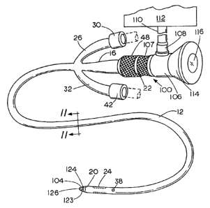
Briefly, in accordance with another aspect of the invention (Figs. 10-16), a
flexible direct
vision viewing instrument or viewer, e.g., having fiberoptic shaft or cable,
is placed within a
urinary catheter with its tip located at a distal end of the catheter so that
the surfaces at the distal
tip of both the viewing cable and the catheter are aligned, i.e., are flush,
thereby fitting together
in such a way as to form a smoothly curved end surface for negotiating
obstructions as easily as
possible. The cable is maintained in this position within the catheter by
means of a releasable
retainer with the distal surfaces that comprise the tip of the instrument thus
maintained in
alignment. During insertion, the urethra is viewed by the healthcare worker
throughout all or part
of the insertion procedure by means of the viewer for the purpose of observing
and identifying
obstructions that may be present. Following insertion, the cable is withdrawn
while allowing the
catheter to remain in place within the urethra. Refer now particularly to
Figs. 10-15 which
illustrate this embodiment of the invention wherein the same numerals
designate corresponding
components already described.
In this embodiment of the invention, the obturator 50 is replaced by a
flexible direct
viewing instrument having a flexible fiberoptical shaft or cable. The viewing
instrument or
26
CA 02777949 2012-05-17
viewer indicated generally by numeral 100, has a cable or shaft portion 102
with a distal tip 104.
The shaft 102 of the viewing cable can be made detachable from the viewing
body 106 so that
the viewing body and eyepiece can be used repeatedly while the plastic shaft
102 can be a single
use item. However, if the entire optical system is constructed to be
sterilized, it can be supplied
as a multi-use unit.
As can be seen in the Figures, the tip 104 of the flexible cable 102 has a
smoothly curved
generally parabolic or rounded end surface. While no precise mathematical
formula for the
curvature of the tip 104 is necessary, it is important that the end of the tip
be rounded or curved
rather than having a sharp point. The end can be thought of as parabolic,
bullet-shaped or dome-
shaped. The numeral 12 in this embodiment represents a catheter rather than a
sheath and can be
structured generally much like a standard urinary catheter. The catheter can
be from about 10-20
French (3-7mm) but is preferably in the range of about 13-15 French (about 4-
5mm). The
catheter 12 and shaft 102 of the instrument 100 is of a standard length, e.g.,
50cm for a male
patient. The catheter 12 is provided with centimeter marks as shown in Fig.
10.
The catheter 12 has a distal opening 20 that is located centrally and in
alignment with the
longitudinal axis thereof. The outside surface of the catheter 12 adjacent to
the opening is tapered
at 21 to provide a smoothly curved contour that tapers centrally toward the
distal tip of the
instrument. In a preferred form of the invention, the shaping and positioning
of the rounded cable
tip 104 and the adjacent tapered contour 21 of the catheter 12 are related to
one another while in
the insertion position so that the curved distal end surfaces are aligned,
i.e., flush, as clearly
shown in Figs. 10, 12 and 14 whereby, together, their surfaces form one
substantially
continuously curved composite tip surface that tapers centrally proceeding
toward the distal end
thereof with a blunt end to facilitate insertion into the urethra and aid in
the negotiation of
27
CA 02777949 2012-05-17
obstructions if any are encountered. It is thus the curvature of the tip of
the cable that includes
the viewing port and light supply together with the tapered end portion 21 of
the catheter which
cooperate to provide the smooth bullet-shaped or dome-shaped surface that
helps the instrument
move easily through the urethra, passing any obstructions that may be present.
As shown in Figs. 10-13, the viewing instrument 100 includes the viewing body
106 that
is provided with a fiberoptic light coupling 108 to which light is introduced
from a fiberoptic
feed cable 110 connected to a suitable light source 1 12 that can comprise any
suitable light for
illumination known to the art. A relatively inexpensive light source is
preferably used, such as a
battery-operated source, e.g., a combination of say a dozen light emitting
diodes (not shown) that
are optically coupled to focus light onto the input end of the feed cable 110.
Other well-known
light sources such as a 300-watt xenon lamp or other light source can be used
but are more
expensive. At the right end of the instrument 100 is an eyepiece 114 having a
viewing lens 116
(Fig. 1). Next to the viewing body 106 is a handgrip 107.
As shown in Figs. 12 and 13, the cable or shaft 102 is provided with a
radially outwardly
extending annular projection which in this case is circular but can be of any
shape desired
and is sized to fitting in either of two longitudinally spaced apart circular
grooves 118 and 120
within the lumen 18 of catheter 12 to serve as a retainer for holding or
locking the shaft 102 of
the instrument in either an extended operating position of Fig. 12 or an
alternate recessed
viewing position shown in Fig. 13 in which the tip 104 is recessed slightly,
e.g., 2mm behind the
opening 20 at the tip of the catheter 12 to provide enhanced viewing under
certain circumstances,
particularly when the urethra is collapsed or when an obstruction is
encountered, and the viewing
tip 104 is pressed tightly against the tissue of the body. Under these
conditions, all that can be
seen through the eyepiece lens 116 is a pink or red color.
When this condition occurs, the operator
28
CA 02777949 2012-05-17
need only withdraw the cable slightly from the operating position of Fig. 12
to the retracted
position of Fig. 13 by pulling outwardly on the handgrip 107. The groove 120
acts to securely
hold the tip 104 in a slightly retracted position so that the surrounding
tissue can be more clearly
seen. Clear saline can also be introduced through the duct 36 for washing away
any blood or
debris or to slightly distend the urethra if desired. Thus, the enlargement
and the cooperating
recesses 118 and 120 in the catheter serve as a simple and reliable retainer
for releasably holding
the viewing tip 104 in the operating position of Fig. 13 in which the tip is
aligned with the
tapered portion at the end of the catheter to form one continuous, smoothly
contoured surface to
ease the instrument through the urethra or, if desired, to hold it in the
retracted position of Fig. 13
for viewing when the tip would otherwise be in contact with the surface of the
urethra or an
obstruction. As soon as the obstruction has been viewed with the tip 104
retracted so that any
difficulty can be more easily observed and understood, the tip can be returned
to the position in
Fig. 12 and the insertion process continued, the smooth surface at the tip 104
enabling any
obstruction to be passed as easily as possible.
Refer now to Figs. 14 and 15 which illustrate the internal structure of the
shaft 102 and
catheter 12 in more detail. As noted above, the shaft 102 of the viewing
instrument 100 is
slideably supported within the lumen 18 of the rubber catheter 12 and can be
slid longitudinally
therein when desired. A lubricant can be provided between the catheter 12 and
the shaft 102 to
allow it to slide more easily. Within the cable or shaft 102 the instruments
are provided "three
flexible fiberoptical illumination bundles 122-124 for supplying light to
illuminate the area in
front of the distal tip 104 of the instrument and a centrally located flexible
coherent fiberoptical
bundle 126 for transmitting the image from the objective lens 126a at the
distal tip of the shaft
102 to the eyepiece lens 116. The circular arrangement fiber bundles 122-124
of Figs. 14 and 15
29
CA 02777949 2012-05-17
provide even distribution of light throughout the field of view. It will be
noted that the opening
20 at the end of the catheter 12 is aligned centrally with the longitudinal
axis of the catheter for
allowing zero degree viewing, i.e., straight ahead, through the lens 126a.
Another form of tip for the cable 102 is shown in Fig. 16. In this embodiment,
the
illumination fiberoptic bundles 122-124 are cut off straight to provide a flat
end surface. The
coherent viewing fibers 126, however, extend further distally. To provide a
curved end surface
for the flexible shaft 102, the end of the shaft is enclosed within a smoothly
contoured, optically
transparent plastic dome 132 as a part of the shaft that can be formed from
any suitable
transparent material, such as glass, but is preferably formed from transparent
plastic resin having
a refractive index approaching optical grade glass and covered on both
surfaces with an
antireflective coating. When the shaft 102 is extended distally to the
operating position, the dome
132 will take the position shown by dotted lines 132a which cooperates with
the tapered surface
20 of the catheter 12 so that the two surfaces join together to provide a
single smooth and
continuous generally bullet-shaped or dome-shaped surface that tapers
centrally proceeding
toward its free end to facilitate insertion to the greatest extent possible.
While not preferred, the invention also contemplates the possibility of
provided steering
cables, e.g., four steering cables within the shaft 102 for the purpose of
turning the tip either
from side to side, or up and down. This modification is not preferred because
of the added cost.
The method of use of the apparatus described in Figs. 10-16 will now be
described. The
catheter 12 and the illumination instrument 100 are packaged separately in
sterile containers. Just
prior to use, the containers are opened and the flexible shaft or cable 102 of
the instrument 100 is
inserted into the lumen 18 of the catheter 12 and releaseably retained in'the
position of Fig. 12 so
that the distal tip of shaft 102 extends slightly through the opening 20 with
the tapered surface 21
CA 02777949 2012-05-17
of the catheter aligned, i.e., flush, with the adjacent surface of the distal
tip 104 of the instrument
100 so that the surfaces form a single continuous, smoothly contoured
composite surface as
described above.
The tip 104 is inserted into and advanced through the urethra. The lighting
system 112 is
energized so that light is transmitted through the feed cable 110 thence
through the fiber bundles
122-124 (Figs. 14 and 15) to illuminate the inside of the urethra. Light from
the illuminated body
tissue of the patient is then carried back from the objective lens 126a
through the coherent fiber
bundle 126 to the eyepiece 114 of the instrument allowing the healthcare
worker, the physician's
assistant, nurse, other healthcare provider or in some cases the patient to
observe the urethra
through the eyepiece lens 116 during insertion. As this is done, any
obstructions that may be
present can be observed through the lens system. In the event the tip 104 of
the viewing cable
becomes pressed up against the wall of the urethra or is for any other reason
in contact with the
surface of body tissue, the operator can then, by placing the fingers of one
hand on the handgrip
107 and the other on the collar 48, withdraw the shaft 102 to the retracted
position of Fig. 13 by
sliding the projection into the groove 120. When this is done, the open tip 20
of the catheter
12 will hold the body tissue surface a short distance away from the objective
lens 126a allowing
the adjacent surface of the body tissue to be more clearly seen. If an
obstruction such as a
stricture, stone or scar tissue is encountered, it can be seen by the
operator, thus enabling the
operator to know the general nature of the obstruction. Most of the time this
will enable the user
even though not a board certified urologist to continue with the insertion of
the catheter and
avoid iatrogenic trauma strictures, urethral bleeding, or other difficulties
such as urethra mucosal
lining tears, patient pain, scar tissue formation, or treatment delays as well
as the increased costs
that result. However, if the nature of the obstruction is not understood or if
further difficulties are
31
CA 02777949 2012-05-17
encountered, the operator will at least have some information available for a
urologist who may
have to finish the intubation. In addition, if the field of vision is
obscured, saline can be
introduced via passage 36 and opening 38 into the urethra for washing away
debris in front of the
objective lens to provide better visibility. The flexible viewing cable 102
also aids in the
insertion of the catheter 12 by filling the lumen 18 so as to prevent the
catheter from collapsing
and as well as giving it a degree of stiffness. However, the catheter 12 can,
if desired, be passed
into the urethra without the viewing instrument 100 in place and the viewing
instrument inserted
later if an obstruction is encountered.
Once the catheter 12 has been inserted completely, fluid is introduced through
the duct 29
causing the distal balloon 24 to inflate conventionally for holding the
catheter in the bladder. The
instrument 100 is then removed. While the main purpose for inserting the
catheter 12 is for
urinary drainage, it is also used to diagnose problems or maintain anatomic
continuity.
Thus, the invention provides a method and apparatus for more efficiently and
safely
passing a urethral catheter into the bladder of a male or female human patient
and is particularly
beneficial in enabling obstructions to be observed directly during incubation.
It is especially well
suited for negotiating obstructions of any sort such as anomalous structures,
scars due to injury
or infection or stones, enlarged prostate due to cancer or AIDS or related
tissue deterioration. A
nurse, physician's assistant, resident or other healthcare worker is able,
using the invention, to
pass a catheter into the bladder for a routine urinary drainage much more
effectively than in the
past since they are able to observe the entire insertion process visually
through the eyepiece of
the instrument as it is being performed. The ability to observe visually what
is taking place
fulfills a long-felt need, since by using the invention it is no longer
necessary to merely shove the
catheter blindly through the urethra; the invention makes it possible to do so
under direct vision.
32
CA 02777949 2012-05-17
Moreover, the relatively simple optical system and viewing cable 102, besides
being flexible so
that it can negotiate curves is much less expensive than a cystoscope which
can cost in the
neighborhood of $5,000.00 to $15,000.00. The use of the invention when applied
in the field of
veterinary medicine may provide an even greater visual advantage than with
human patients
since in that case no verbal communication is possible. The visual advantage
is therefore often of
critical importance.
If, during an insertion, an obstruction is observed, the operator by noting
the nature of
blockage present is better able to steer the tip of the catheter around it. In
most situations the
catheter can be passed the rest of the way into the bladder without further
problems. However, if
a serious blockage is encountered, the loss of time is minimized since the
visualization by the
operator will enable the urologist to pinpoint the blockage location by means
of the centimeter
marks (Fig. 10) indicating the distance from the penile glans or the urethral
meatus, or the
urologist can be told in advance something about the nature of the problem. At
the same time
patient comfort is improved, as is the safety of the procedure by avoiding the
possibility of false
passage or other injury that might otherwise take place. The invention greatly
improves catheter
placement through the urethra because ability to view the procedure as it is
carried out provides
mucli more positive control by the operator.
If desired, the operator can begin intubation by passing the catheter 12
conventionally,
i.e., without the flexible viewing shaft 102 in place and introduce it all or
part way unless a
problem is encountered, and at that point insert the fiberoptic viewing cable
102. The invention
can be used in virtually all urinary catheter placements which are now
inserted blindly, in the
neighborhood of 150,000 insertions per day in the U.S. It is particularly
useful for problematic
insertions, e.g., an injured patient who is suffering from pelvic injury and
is bleeding from the
33
CA 02777949 2012-05-17
penis. Insertion of the catheter under those conditions can be carried out
much more safely with
the present invention because it allows placement under vision. Any blood that
may obscure the
field can be flushed out by introducing saline through the opening 38.
Moreover, any patient
having a relatively high score on the American Urologic Association Symptom
Index can be
more safely intubated using the present invention.
While a so called "three-way" catheter has been shown in the Figures, it will
be
understood that if no retention within the bladder or flushing of the tip is
required, the ducts 29
and 36 can be eliminated.
The invention substantially reduces the burden placed on "physician extenders"
(R.N.'s,
physician assistants and technicians), decreases physician involvement,
alleviating the extreme
shortage of board certified urologists. It also speeds the rectification of
urinary retention in the
bladder, as well as other urinary tract disorders, reduces the chance of
urinary tract infections,
iatrogenic trauma and patient pain. Rapid training of physician extenders can
be accomplished by
utilizing many visual aids currently available, thus raising healthcare
efficiency and reducing
costs by showing the P.E:'s how to negotiate common obstructions.
A teaching model of this invention can be fabricated so that a direct vision
electronic
image from the viewer 100 can be cabled to a CRT or DVD projector in real time
for group
training in medical schools and diverse specialties and subspecialties or
physician extenders. If
desired, an image sequence can be recorded for later replay. This modification
of the invention
can be used for remote relay for diagnosis or diagnosis confirmation via
satellite relay. The two
parties can be miles apart or hundreds of miles apart and faithful electronic
images can be sent.
Currently, most NASA manned missions include a physician. An ER surgeon or
trauma surgeon
34
CA 02777949 2012-05-17
is therefore able to supervise and make medical decisions using the invention.
The physician can
obtain diagnosis confirmation or prime diagnosis in this precision manner.
Moreover, inventory problems with catheter insertion areas such as bedsides,
emergency
rooms, clinics, physicians' offices, ambulances and paramedics, etc. are
minimal as few catheter
sizes utilizing the invention need be maintained to serve the entire adult
male/female population
that might be encountered. The same applies to pediatric patients. Since the
catheter is under
visual control of the placer at all times, false passage and other possible
trauma is avoided by
permitting circumnavigation of virtually any impediment to thereby provide
rapid passage to the
bladder.
Many variations are possible. For example, a void valve (not shown) can be
provided at
the proximal end of the lumen 18 for controlling urine flow from the bladder
46 into a disposable
bag. The term viewer is used broadly herein to include electronic image
transmission from the
viewing port 104 to the eyepiece.
Other variations of the present invention within the scope of the appended
claims will be
apparent to those skilled in the art once the principles described herein are
understood.