Note: Descriptions are shown in the official language in which they were submitted.
CA 2778270 2017-03-29
INTEGRATED HEALTH DATA CAPTURE AND ANALYSIS SYSTEM
[0001]
BACKGROUND OF THE INVENTION
[0002] An epidemic of infectious diseases capable of spreading across a large
region, e.g., a continent
or the entire world, can be hugely costly to societies. Such incidences
include pandemics of influenza,
smallpox, tuberculosis, human immune deficiency virus (HIV), and Severe Acute
Respiratory
Syndrome (SARS). The World Bank estimated in 2008 that a flu pandemic could
cost $3 trillion and
result in a nearly 5% drop in world gross domestic product (GDP). The World
Bank further estimated
that more than 70 million people could die worldwide in a severe pandemic.
Others have estimated that
a flu pandemic could cause an economic recession in the United States, costing
the country at least
$500 billion to $675 billion in the near term. In 2003, SARS disrupted travel,
trade and the workplace
in the Asia Pacific region and cost the region about $40 billion. The SARS
pandemic lasted for six
months, killing at least 1000 of the 8,000 people it infected in 25 countries.
The city of Toronto, CA
was closed to air traffic for several weeks and suffered significant financial
loss.
[0003] In 2009, the spring flu season cost billions of dollars even though it
only lasted only a few
weeks. The 2009-2010 winter flu season is anticipated to start by late August
and could run through
April 2010. Even if working vaccines are available, their supplies are
expected to be limited and cannot
be expected to stop the flu for several months. Economic losses can be
minimized if the flu can be
contained through proactive screening that allows for effective anti-viral
administration and narrowly
targeted quarantines.
[0004] Economic loss due to "avoidance behaviors" is even greater than the
cost of treating flu
victims. The cost includes reducing air travel, avoiding travel to infected
destinations and reducing
consumption of services, such as mass transit, dining out, shopping, etc.
According to the World Bank,
if a flu epidemic approached the 2.5% mortality rates similar to 1918-19 flu,
avoidance behaviors
would cost a region five times more than losses from mortality or work
absenteeism.
SUMMARY OF THE INVENTION
[0005] There is a pressing need for an infrastructure to mitigate the spread
of infectious diseases such
as influenza when it occurs. The present invention meets this need through an
integrated system that
provides real-time sampling, modeling, analysis, and/or recommended
interventions. The system can
identify active cases in an outbreak through pro-active sampling in high risk
locations, such as schools
or crowded commercial areas, and can allow for sampling and quarantine of
surrounding cases to help
eradicate the outbreak. The system can also suggest an appropriate response
for deployment of scarce
resources and predict the impact of such mitigation both in terms of reduction
of mortality and
CA 02778270 2012-04-19
WO 2011/049886 PCT/US2010/053088
morbidity and economic impact. Furthermore, the systems of the present
invention can help the
government provide accurate, more reliable, and timely information that may
reduce unnecessary
avoidance behavior and save billions of dollars.
[0006] In one aspect, the present invention provides a system for modeling a
progression of a disease
within a population, comprising: a static database component comprising static
data related to the
disease and/or the population; a dynamic database component comprising dynamic
data about the
population and individual subjects; and a computer modeling component that is
configured to model the
data in the static database component and the dynamic database component,
thereby modeling the
disease within the population. The disease can be an infectious disease or a
chronic disease.
[0007] In some embodiments, the infectious disease agent or an analyte thereof
comprises an
adenovirus, Bordella pertussis, Chlamydia pneumoiea, Chlamydia trachomatis,
Cholera Toxin, Cholera
Toxin f3, Campylobacter jejuni, Cytomegalovirus, Diptheria Toxin, Epstein-Barr
NA, Epstein-Barr EA,
Epstein-Barr VCA, Helicohacter Pylori, Hepatitis B virus (HBV) Core, Hepatitis
B virus (HBV)
Envelope, Hepatitis B virus (HBV) Surface (Ay), Hepatitis C virus (HCV) Core,
Hepatitis C virus
(HCV) NS3, Hepatitis C virus (HCV) NS4, Hepatitis C virus (HCV) NS5, Hepatitis
A, Hepatitis D,
Hepatitis E virus (HEV) orf2 3 KD, Hepatitis E virus (HEV) orf2 6 KD,
Hepatitis E virus (HEV) orf3
3KD, Human immunodeficiency virus (HIV)-1 p24, Human immunodeficiency virus
(HIV)-1 gp41,
Human immunodeficiency virus (HTV)-1 gp120, Human papilloma virus (HPV),
Herpes simplex virus
HSV-1/2, Herpes simplex virus HSV-1 gD, Herpes simplex virus HSV-2 gG, Human T-
cell leukemia
virus (HTLV)-1/2, Influenza A, Influenza A H3N2, Influenza B, Leishmania
donovani, Lyme disease,
Mumps, M. pneumoniae, M tuberculosis, Parainfluenza 1, Parainfluenza 2,
Parainfluenza 3, Polio
Virus, Respiratory syncytial virus (RSV), Rubella, Rubeola, Streptolysin 0,
Tetanus Toxin, T. pallidum
15 kd, T. pallidum p47, T. cruzi, Toxoplasma, or Varicella Zaster =
[0008] In other embodiments, the disease is an infectious disease comprising a
microrganism, a
microbe, a virus, a bacterium, an archaeum, a protozoan, a protist, a fungus
or a microscopic plant. The
virus can comprise influenza or HIV. The bacterium can comprise mycobacterium
tuberculosis. The
protozoan can comprise malaria.
[0009] In still other embodiments, the disease is a chronic disease or
condition comprising diabetes,
prediabetes, insulin resistance, metabolic disorder, obesity, or
cardiovascular disease.
[0010] The static database component of the invention can include information
about the individuals in
the population. The information about the individuals in the population can
include one or more of age,
race, sex, location, genetic factors, single nucleotide polymorphisms (SNPs),
family history, disease
history or therapeutic history.
[0011] The static database component can also comprise information about the
disease. The
information about the disease can include one or more of virulence,
contagiousness, mode of
transmission, treatment availability, vaccine availability, death rate,
recovery time, cost of treatment,
infectivity, rate of spread, rate of mutation, and past outbreak.
-2-
CA 02778270 2012-04-19
WO 2011/049886 PCT/US2010/053088
[0012] In some embodiments, the data in the dynamic database component is
updated in real-time. In
some embodiments, the data in the dynamic database component comprises an
indication of the disease
state of the individuals in the population. The indication of the disease
state of an individual can be
determined by measuring a biomarker, a physiological parameter, or a
combination thereof.
[0013] When the disease monitored by the invention is influenza, the
biomarker/s can include
hemagglutinin and/or neuraminidase. The hemagglutinin can be selected from the
group consisting of
H1, H2, H3, H4, HS, H6, H7, H8, H9, H10, H11, H12, H13, H14, H15, and H16, and
the
neuraminidase can be selected from the group consisting of N1, N2, N3, N4, and
N5. In some
embodiments, the hemagglutinin comprises H1 and the neuraminidase comprises
Ni. In some
embodiments, the hemagglutinin comprises HS and the neuraminidase comprises
NI.
[0014] The biomarker measured by the invention can be a host antibody. For
example, the biomarker
can be an IgM antibody, an IgG antibody or an IgA antibody against a disease
marker.
[0015] In some embodiments, the biomarker comprises a marker of inflammation.
Such marker of
inflammation can be a cytokine or C-reactive protein. The marker of
inflammation can also be IL-113,
1L-6, 1L-8, 1L-10, or TNFix.
[0016] In some embodiments, the biomarker is measured in a sample of bodily
fluid from the
individual. Exemplary bodily fluids include without limitation blood, plasma,
serum, sputum, urine,
feces, semen, mucous, lymph, saliva, or nasal lavage.
[0017] In some embodiments, the physiological parameter measured by the
invention comprises one or
more of body weight, temperature, heart rate, blood pressure, mobility,
hydration, ECG, or alcohol use.
[0018] The biomarker or physiological parameter can be determined using a
point-of-care device. The
point of care devices can be deployed according to instructions determined by
the computer modeling
component. The point of care device can perform without limitation one or more
of cartridge assays,
real time PCR, rapid antigen tests, viral culture, and immunoassays. The point
of care device can
measure more than one biomarker with more than 30% better accuracy and/or
precision than standard
methods. In some embodiments, the system comprises a plurality of point of
care devices. The point of
care devices can be positioned at one or more of a school, a workplace, a
shopping center, a community
center, a religious institution, a hospital, a health clinic, a mobile unit,
or a home.
[0019] The point of care device can comprise a portable instrument. For
example, the point of care
device can include a portable cartridge. In some embodiments, the cartridge is
configured to accept
reagents for measuring the biomarkers. The biomarkers can be measured
according to a protocol
communicated from the computer modeling component. In some embodiments, the
cartridge is
configured to measure a set of biomarkers from a plurality of bodily fluid
samples.
[0020] The point of care device of the invention can include a graphical user
interface configured for
data entry.
[0021] In some embodiments, the point of care device is configured to
communicate the biomarker or
physiological parameter measurements to the computer modeling component. The
communication can
-3-
CA 02778270 2012-04-19
WO 2011/049886 PCT/US2010/053088
include wireless communication, wired communication, or a combination thereof.
Wireless
communication comprises without limitation WiFi, Bluetooth, Zigbee, cellular,
satellite, and/or
WWAN. The communication can also be performed over a secure interne
communication. In some
embodiments, the point of care device is configured to perform two way
communications with the
computer modeling component.
[0022] In some embodiments of the system of the invention, the modeling
results are updated in real
time when updated dynamic data becomes available, e.g., after the computer
modeling component
receives updated information from a point of care device.
[0023] The computer modeling component can be configured to present the
modeling results to one or
more of healthcare professionals, government agencies and individual human
subjects. The computer
modeling component can also be configured to predict one or more courses of
action based on the
modeling results. The one or more courses of action are ranked according to a
ranking parameter,
including without limitation ranking by financial considerations, number of
affected individuals,
quality-adjusted life year (QALY), and/or quality-adjusted life year (QALY)
per economic cost unit.
[0024] The one or more courses of action comprise a strategy to control the
spread of the disease. The
strategy to control the spread of the disease can include one or more of
household quarantine, individual
quarantine, geographic quarantine, social distancing, hospitalization, school
closure, work place
closure, travel restrictions, public transit closure, therapeutic treatment or
intervention, prophylactic
treatment or intervention, vaccination, provision of protective clothing,
provision of masks, and
additional point-of-care testing. The strategy to control the spread of the
disease can further include
one or more of counseling at risk or affected individuals for behavior
modification, repeated biomarker
and/or physiological measurements, and reward for the individual. Still
further, the strategy to control
the spread of the disease can include one or more of patient triage
recommendations, resource
management, efficacy index for each strategy, costs of each strategy, return
on investment for each
strategy. The strategy to control the spread of the disease can be one or more
of targeted prophylaxis,
blanket prophylaxis, targeted antibiotic prophylaxis, blanket antibiotic
prophylaxis, targeted anti-viral
prophylaxis, blanket anti-viral prophylaxis, targeted vaccination, and blanket
vaccination. The targeted
prophylaxis or vaccination can be targeting the prophylaxis or vaccination to
children between 1-4 yrs
of age, children between 5-14 yrs of age, pregnant women, young adults between
15-30 yrs of age,
first-line medical response workers, individuals identified to at high risk of
mortality, or geriatric
individuals.
[0025] In some embodiments of the invention, the computer modeling component
is configured to
estimate a surveillance strategy based on the modeling results. The
surveillance strategy can include
determining the disease status of an individual or group of individuals using
a point of care device. The
surveillance strategy can be updated when a diseased individual is detected.
In some embodiments, the
updated strategy comprises one or more of testing a household comprising the
diseased individual,
testing a school comprising the diseased individual, and testing a work place
comprising the diseased
-4-
CA 02778270 2012-04-19
WO 2011/049886 PCT/US2010/053088
individual. The updated strategy can further be one or more of quarantine,
prophylaxis or
hospitalization.
[0026] In some embodiments, the computer modeling component comprises a
graphical interface for
displaying modeling results to a user.
[0027] The computer modeling component can include a plurality of nonlinear
ordinary differential
equations, and/or a plurality of parameters. In some embodiments, the computer
modeling component
comprises a learning machine that updates the plurality of parameters when the
static data and/or
dynamic data are updated.
[0028] The model of the data can be configured to include a plurality of
states. In some embodiments,
the plurality of states comprises one or more of: susceptible individuals,
early exposed individuals, late
exposed individuals, early infected individuals, late infected individuals,
recovered individuals,
individuals who died due to the infection and/or associated complications,
asymptomatic individuals,
individuals given therapeutic treatment, individuals given therapeutic
treatment and quarantined,
individuals treated prophylactically, vaccinated individuals, individuals
protected due to vaccination,
early infected individuals who are hospitalized, late infected individuals who
are hospitalized,
susceptible individuals who arc home quarantined, early exposed individuals
who arc home
quarantined, late exposed individuals who are home quarantined, early infected
individuals who are
home quarantined, late infected individuals who are home quarantined,
asymptomatic individuals who
are home quarantined, susceptible individuals quarantined in the whole
neighborhood, early exposed
individuals quarantined in the whole neighborhood, late exposed individuals
quarantined in the whole
neighborhood, early infected individuals quarantined in the whole
neighborhood, late infected
individuals quarantined in the whole neighborhood, asymptomatic individuals
quarantined in the whole
neighborhood, amount of therapeutic drug doses available, amount of antivirals
and/or antibiotics
available to the target population, home quarantined individuals that arc
vaccinated, home quarantined
individuals that are protected due to vaccination, home quarantined
individuals that recovered,
susceptible individuals earmarked by mitigation policies for action, early
exposed individuals
earmarked by mitigation policies for action, late exposed individuals
earmarked by mitigation policies
for action, asymptomatic individuals earmarked by mitigation policies for
action, early infected
individuals earmarked by mitigation policies for action, late infected
individuals earmarked by
mitigation policies for action, prophylactic-treated individuals earmarked by
mitigation policies for
action, vaccinated individuals earmarked by mitigation policies for action,
protected individuals
earmarked by mitigation policies for action, recovered individuals earmarked
by mitigation policies for
action, susceptible individuals earmarked for therapeutic treatment, early
exposed individuals
earmarked for therapeutic treatment, late exposed individuals earmarked for
therapeutic treatment,
asymptomatic individuals earmarked for therapeutic treatment, early infected
individuals earmarked for
therapeutic treatment, late infected individuals earmarked for therapeutic
treatment, susceptible
individuals earmarked for surveillance, early exposed individuals earmarked
for surveillance, late
-5-
CA 02778270 2012-04-19
WO 2011/049886 PCT/US2010/053088
exposed individuals earmarked for surveillance, asymptomatic individuals
earmarked for surveillance,
early infected individuals earmarked for surveillance, late infected
individuals earmarked for
surveillance, prophylactic individuals earmarked for surveillance, vaccinated
individuals earmarked for
surveillance, protected individuals earmarked for surveillance, susceptible
individuals in whole
neighborhood quarantine earmarked by mitigation policies for action, early
exposed individuals in
whole neighborhood quarantine earmarked by mitigation policies for action,
late exposed individuals in
whole neighborhood quarantine earmarked by mitigation policies for action,
asymptomatic individuals
in whole neighborhood quarantine earmarked by mitigation policies for action,
early infected
individuals in whole neighborhood quarantine earmarked by mitigation policies
for action, late infected
individuals in whole neighborhood quarantine earmarked by mitigation policies
for action,
prophylactic-treated individuals in whole neighborhood quarantine individuals
earmarked by mitigation
policies for action, cumulative number of therapeutic doses administered,
cumulative number of
antivirals and/or antibiotics administered, cumulative number of home
quarantined asymptomatic
individuals, cumulative number of home quarantined symptomatic individuals,
cumulative number of
total infected individuals, cumulative number of infected individuals who are
not quarantined,
cumulative number of infected individuals with some action taken, cumulative
number of hospitalized
individuals, and cumulative number of deaths.
[0029] In another aspect, the present invention provides a system for
controlling spread of influenza
within a population, comprising: a static database component comprising static
data related to the
influenza and/or the population; a dynamic database component comprising
dynamic data about the
population; and a computer modeling component that is configured to model the
data in the static
database component and the dynamic database component, thereby modeling the
incidence of the
influenza within the population.
[0030] In still another aspect, the present invention provides a system for
controlling spread of human
immunodeficiency virus (HIV) within a population, comprising: a static
database component
comprising static data related to the HIV and/or the population; a dynamic
database component
comprising dynamic data about the population; a computer modeling component
that is configured to
model the data in the static database component and the dynamic database
component, thereby
modeling the incidence of the HIV within the population.
[0031] In yet another aspect, the present invention provides a system for
controlling spread of hepatitis
within a population, comprising: a static database component comprising static
data related to the
hepatitis and/or the population; a dynamic database component comprising
dynamic data about the
population; and a computer modeling component that is configured to model the
data in the static
database component and the dynamic database component, thereby modeling the
incidence of the
hepatitis within the population.
[0032] In an aspect, the present invention provides a system for controlling
spread of diabetes within a
population, comprising: a static database component comprising static data
related to the diabetes
-6-
CA 2778270 2017-03-29
and/or the population; a dynamic database component comprising dynamic data
about the population;
and a computer modeling component that is configured to model the data in the
static database
component and the dynamic database component, thereby modeling the incidence
of the diabetes within
the population.
[0033]
BRIEF DESCRIPTION OF THE DRAWINGS
[00341
100351 Figure 1 illustrates a simplified model representation.
[0036] Figure 2 illustrates a model representation taking into account various
states and transitions
between states.
[0037] Figure 3 illustrates an assay for H1N1 antigen using sandwich complexes
in four different
configurations.
[0038] Figure 4A illustrates an assay for host anti-virus antibodies. The
figure illustrates a spike
recovery assay for host anti-H1N1 antibodies. Shown is a version using a-HI /
a-N1 configuration.
Figure 4B illustrates direct assays for a-H1N1 antibodies illustrating
sandwich complexes.
100391 Figure 5 illustrates an exemplary device that can be used in the
present invention. The
exemplary devices comprise assay units, reagents unit, and other modular
components.
[0040] Figure 6 illustrates two side-cut away views of the exemplary device
that can be used in the
present invention. The exemplary device comprises cavities in the housing of
the device shaped to
accommodate an assay unit, a reagent unit, and a sample tip.
[0041] Figure 7A demonstrates an exemplary assay unit that comprises a small
tip or tubular
formation. Figure 7B demonstrates an example of a sample tip as described
herein.
[0042] Figures 8A and 8B illustrate two examples of a reagent unit comprising
a cup.
[0043] Figure 9 illustrates a thin film, for example, contamination, within
the tip when a liquid is
expelled and another liquid aspirated.
[0044] Figure 10 demonstrates an example of a system comprising a device and a
fluid transfer device.
[0045] Figure 11 illustrates an exemplary system of the invention comprising a
heating block for
temperature control and a detector.
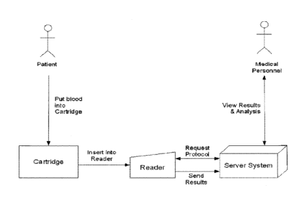
[0046] Figure 12 demonstrates an exemplary a system wherein a patient delivers
blood to a device and
then the device is inserted into a reader.
-7-
CA 02778270 2012-04-19
WO 2011/049886 PCT/US2010/053088
[0047] Figure 13 illustrates the process flow of building a system for
assessing the medical condition
of a patient.
[0048] Figures 14A through 14E demonstrate an example of a plasma separation
method wherein a
whole blood sample has been aspirated into a sample tip and a magnetic reagent
is mixed and
suspended with the sample, then a magnetic field is applied to the whole blood
sample and magnetic
reagent mixture. Separated blood plasma sample can then be distributed into a
well of a device.
[0049] Figure 15 demonstrates an exemplary method of a control assay as
described herein comprising
a known quantity of control analyte.
[0050] Figure 16 illustrates an exemplary embodiment of a Health Shield user
interface.
[0051] Figure 17 illustrates another exemplary embodiment of a Health Shield
use interface.
[0052] Figure 18 illustrates simulation of the 2009 La Gloria outbreak with
and without Health Shield
mitigation policies.
[0053] Figure 19 illustrates diabetes risk prediction visualization.
[0054] Figure 20A illustrates the detection of H1N1 viral particles using a
point of care device. Figure
20B illustrates the detection of H1N1 viral particles using a point of care
device in clinical samples.
[0055] Figure 21 illustrates the detection of host antibodies using a point of
care device.
[0056] Figures 22A illustrates the detection of host antibodies using a point
of care device. Figure 22B
illustrates the dynamic range of host antibody detection using a point of care
device.
[0057] Figure 23 illustrates the detection of human cytokine IL-6 using a
point of care device.
[0058] Figure 24 illustrates the detection of protein-C and C-reactive protein
(CRP) using a point of
care device in a patient undergoing chemotherapy.
[0059] Figure 25 illustrates the detection of glucagon-like peptide 1 (GLP-1)
using a point of care
device.
[0060] Figure 26 illustrates the detection of C-pcptidc, an insulin precursor,
using a point of care
device.
[0061] Figure 27 illustrates the detection of C-peptide using a cartridge
point of care device compared
to a reference detection system (Linco).
[0062] Figure 28A illustrates the measurement of GLP-1 in three human subjects
after feeding. Figure
28B illustrates the measurement of C-peptide over the course of the same
experiment.
[0063] Figure 29 illustrates a calibration curve correlating an assay unit and
a reagent unit for
conducting an assay for VEGFR2.
[0064] Figure 30 illustrates CRP concentration plotted against the assay
signal (photon counts) and the
data fitted to a 5-term polynomial function to generate a calibration
function.
[0065] Figure 31 shows a fit was achieved between a model and the values of
the parameters Smax,
CO.5 and D as described herein.
[0066] Figure 32 displays data according to the dilution used to achieve the
final concentration in an
assay tip.
-8-
CA 02778270 2012-04-19
WO 2011/049886 PCT/US2010/053088
[0067] Figure 33 illustrates the normalized assay response (B/Bmax) is plotted
against the log
normalized concentration (C/CO.5) for relative dilutions: 1:1 (solid line),
5:1 (dashed line), and 25:1
(dotted line).
[0068] Figures 34 and 35 illustrate a similar example as Figure 33 at
different normalized
concentrations.
[0069] Figure 36A shows a spike in IL-6 in septic individuals. Figure 36B
shows a decline in Protein
C in septic individuals.
[0070] Figure 37 shows an increase in 11-6 and 'TNF-a (right panel) in an
individual as the load of
H1N1 influenza increased in the patient (left panel).
DETAILED DESCRIPTION OF THE INVENTION
[0071] In one embodiment, the present invention provides an integrated health
data capture, analysis
and pandemic mitigation solution, referred to herein as the Health Shield
(HS). HS can be used for
infection caused by the influenza virus and other pathogenic agents prone to
endemic or pandemic
spread. Flu outbreaks cost billions of dollars and cannot presently be
completely contained by
vaccination. Economic losses can be minimized if the flu can be contained
through proactive screening
that allows for administration of effective anti-viral agents and narrowly
targeted quarantines. Based on
epidemic models, activating the HS of the invention can reduce spread of the
virus, e.g., by at least
50%, through proactive sampling and containment. The HS can also reduce
unnecessary avoidance
behavior by tracking the virus's spread in real time. Where desired, test
results can be wirelessly
relayed to a server operating HS software. Accordingly, appropriate entities
(e.g., local, regional and
national governments) can be notified with alerts when an event is detected,
thereby allowing for
proactive management of a possible outbreak.
[0072] In further embodiments, the Health Shield infrastructure provides
strategic industrial and
commercial parks as "safe zones," which allow economically important
activities to continue. As a
result, fewer workers will be infected with the virus, and schools and
businesses will be less disrupted.
Pandemic mitigation strategies will maintain productivity to drive economic
growth and preclude
actions prompted by panic..
[0073] The system can comprise an integrated sampling and modeling technology
suite embedded in a
real-time informatics infrastructure. The ability to sample, model, and learn
from data as it is acquired
longitudinally, enables the development of an optimal strategy for the care
and management of disease
both on an individual and population basis. Custom applications can be built
for numerous diseases and
therapeutic areas. The HS infrastructure can also be used to protect a region
from a wide spectrum of
threats beyond infectious disease, including chronic disease and bioterrorism
threats.
I. Health Shield Infrastructure
[0074] The Health Shield provides a system to contain the spread of infectious
diseases through
integrated, automated, and real-time sampling, modeling, analysis, and
recommended interventions.
-9-
CA 02778270 2012-04-19
WO 2011/049886 PCT/US2010/053088
For example, the HS can identify active cases in an outbreak (through pro-
active sampling in high risk
locations, such as schools or crowded commercial areas) and direct the
sampling and defensive
measures, e.g., quarantine, of surrounding cases to mitigate or eradicate the
outbreak. HS algorithms
characterize spread of the epidemic similarly to the case of a forest fire,
where the THS models'
mitigation policy aims to eradicate "hot spots" before a "fire" can take hold
and spread and/or can
create a fire-break around a disease hot spot.
[0075] In some embodiments, the HS comprises two technological components ¨ a
Field System (FS)
and an Operating System (OS) ¨ that can be adapted for management of chronic
diseases to improve
health outcomes and decrease healthcare costs.
(a) Field System (FS)
[0076] The Field System components of the HS can be deployed at various points
of care, including
without limitation a clinic, a community site (e.g., school, community
center), a hospital, a doctor's
office or an individual's home. The FS can also use any number of platforms to
monitor disease, e.g.,
immunoassays, PCR assays, real-time PCR, microorganism plating, etc. The FS
also includes standard
medical equipment, e.g., scales to determine weight, blood pressure devices,
thermometers to measure
temperature, ruler to measure height, etc. In some embodiments, the FS devices
comprise customized
portable, single-use cartridges, as described herein. The FS collects relevant
data in the field, and
transmits the data to the OS.
[0077] In some embodiments, the Field System comprises a measurement device
intended to be
deployed in an area to be monitored. In some embodiments, the FS analyzes
bodily fluid samples, e.g.,
blood from a finger stick, in real-time. The system analyzes the bodily fluids
for evidence of infection
or disease by detecting, e.g., markers of a pathogen, nucleic acids, proteins,
glycoproteins, lipids, or a
combination thereof indicative of a disease condition. In some embodiments,
the FS simultaneously
measures multiple markers including one or more of selected antigens or the
pathogen, antibodies
directed to the pathogen, intracellular or cell surface proteins or
glycoproteins, and cytokines indicative
of the response of an infected subject to a given pathogen, (e.g., a viral
strain or other microorganism).
The system can also collect environmental, demographic, personal and
physiological (e.g. temperature,
blood pressure) information. In some embodiments, such information is
collected through a graphical
touchscreen interface. Individualized content can be analyzed by a remote
system to facilitate
mitigation strategies in real-time.
[0078] In some embodiments, the FS includes cartridges that perform assays on
the bodily fluids. The
devices include without limitation non-significant risk devices, and the
assays can be validated under
appropriate guidelines, e.g., those provided by the U.S. Federal Drug
Administration (FDA) and/or
International Conference on Harmonization (ICH). Cartridges used by the
present invention arc
described in U.S. Patent Application No. 11/389,409 entitled "POINT-OF-CARE-
FLUIDIC SYSTEMS
AND USES THEREOF," U.S. Patent Application No. 11/746,535 entitled "REAL-TIME
-10-
CA 02778270 2012-04-19
WO 2011/049886 PCT/US2010/053088
DETECTION OF INFLUENZA VIRUS," and U.S. Patent Application No. 12/244,723
entitled
"MODULAR POINT-OF-CARE DEVICES, SYSTEMS, AND USES THEREOF" and are described
in
further detail below. The measurement systems can be self-contained and few if
any extra materials are
required to operate them. In some embodiments, the only requirement for an FS
system is a power
source for the instruments. In other embodiments, the power source is provided
with the FS in form of
a battery, generator, solar or other portable power source. The cartridges can
be pre-loaded with the
desired assays and require little or no preparation prior to use. For example,
some or all assay
components can be stored in a refrigerator (e.g., at about 4 degrees C) prior
to deployment.
[0079] The FS platform can run any appropriate assay that is currently
performed in the conventional
laboratory infrastructure. New assays can be rapidly transferred and fully
validated. In some
embodiments, assays that are entirely new to the HS system can be customized
and validated within
less than about three months, two months, one month, 3 weeks, 2 weeks or less
than about 1 week. In
some embodiments, the assays run on HS Systems are validated under FDA ICH
guidelines.
[0080] The Field Systems can be placed at any desired point of care, e.g., an
area suspected or known
to be at risk of infection or disease. Point of care testing (POCT) is defined
by a near-patient testing
system. Exemplary points-of-care include but arc not limited to the home,
clinic, schools, or
commercial centers. In some embodiments, the FS is deployed in mobile units.
Thus, it should be
understood that medical experts are not necessarily required for the testing.
To enable this, the FS can
be engineered to be simple to use and provides all directions for use in a
simple user interface with a
touch screen. In some embodiments, the systems are designed for non-computer
literate individuals to
test themselves in their own homes. In such a setting, the data can be sent to
a remote system, e.g., the
Operating System as described below, where officials or others monitoring the
assays can learn of
positive test results. In some embodiments, the testing and data
upload/analysis are performed in real-
time so that containment measures can be initiated immediately.
[0081] In some embodiments, the systems are deployed in public locations. If
desired, standard public
health employees can be trained to do the testing. in some embodiments, the
systems are designed so
that total training time is minimized at a given site. For example, current
deployment demonstrates that
training should require no longer than half an hour per site, although
supplemental and advanced
training can be performed as appropriate. In some embodiments, trained
individuals can in turn train
others on using the systems. The FS can be successfully used in the home by
patients who have no
medical training ¨ as the testing is designed to be fully automated and uses a
graphical touch-screen
interface on the instrument to walk users through the test process. In some
embodiments, the only steps
required from a user are to: 1) place a sample into the cartridge, e.g.,
sputum or a finger-stick which can
be performed by the user themselves using a disposable single-use lancet just
as used in diabetes
management for glucose monitoring; and 2) insert the cartridge into the
accompanying instrument, as
described in more detail below.
-11-
CA 02778270 2012-04-19
WO 2011/049886 PCT/US2010/053088
[0082] Non-limiting customized cartridge devices for use with the FS of the
invention are described in
U.S. Patent Application No. 11/389,409 entitled "POINT-OF-CARE-FLUIDIC SYSTEMS
AND USES
THEREOF," U.S. Patent Application No. 11/746,535 entitled "REAL-TIME DETECTION
OF
INFLUENZA VIRUS," and U.S. Patent Application No. 12/244,723 entitled "MODULAR
POINT-OF-
CARE DEVICES, SYSTEMS, AND USES THEREOF." Such devices are further detailed
below.
(b) Operating System (OS)
[0083] The data collected from each FS device can be securely transmitted to
the Operating System in
real-time through network connection, e.g., over a broadband, wireless,
satellite or cellular network.
One of skill in the art will appreciate that network communications often
comprise multiple hops, e.g.,
an FS device can connect to a wireless local area network (WLAN) that is
securely connected to the
World Wide Web through broadband landlines.
[0084] In some embodiments, the Operating System includes one or more servers
as are known in the
art and commercially available. Such servers can provide load balancing, task
management, and backup
capacity in the event of failure of one or more of the servers or other
components of the system, to
improve the availability of the OS. A server can also be implemented on a
distributed network of
storage and processor units, as known in the art, wherein the data processing
according to the present
invention reside on workstations such as computers. A server of the OS
component can include a
database and system processer. A database can reside within the server, or it
can reside on another
server system that is accessible to the server. As the information in a
database may contains sensitive
information, a security system can be implemented that prevents unauthorized
users from gaining
access to the database.
[0085] In some embodiments, the Operating System comprises a data engine that
imports data from a
desired source to provide direction for epidemic or pandemic mitigation. The
OS can translate the
source data into a standardized format to be analyzed. In some embodiments,
the data engine is self-
learning and dynamically models a plurality of integrated data sets in real-
time. This OS modeling
approach provides several benefits. For example, the models can be trained to
perform a variety of
calculations, including but not limited to: 1) predicting outcomes for
individuals and populations; 2)
considering the efficacy of proposed intervention strategies for individuals
and populations; and 2)
quantifying the socioeconomic effect of the recommended interventions. In some
embodiments, the OS
is made available to remote users via a remote interface. For example, the
users can access the OS
through a secure online web-portal or the like.
[0086] The OS software portal incorporates automatic modeling in a system that
is constantly learning
from each new data point that is transmitted to the software portal. The
system thereby becomes
increasingly more predictive over time. In some embodiments, Monte Carlo
modeling approaches are
used. Monte Carlo approaches rely on repeated random sampling to compute
results. Monte Carlo
simulation considers random sampling of probability distribution functions as
model inputs to produce
-12-
CA 02778270 2012-04-19
WO 2011/049886 PCT/US2010/053088
hundreds or thousands of possible outcomes instead of a few discrete
scenarios. The results provide
probabilities of different outcomes occurring. In some embodiments, the
solution and refitting/refining
of model parameters sets is achieved by using reverse search and integrated
parameter estimation
techniques. See, e.g., Sheela, 1979 -COMPUTER METHODS IN APPLIED MECHANICS AND
ENGINEERING 19 (1979) 99-106; Moles, et al. 2003 - Genome Res. 2003 13: 2467-
2474; Rodriguez-
Fernandez, et al. BMC Bioinformatics 2006,7:483-500; Barthelmann, et al. 2000 -
Advances in
Computational Mathematics 12: 273-288.
[0087] There is a rich literature surrounding the modeling and simulation of
epidemiological data. The
basis of the McKendrick model is a stochastic process (Birth Process) that
yields a series of differential
equations that can be parameterized, explored, and, eventually, optimized
regarding the control and
spread of the disease. A reasonably straightforward analysis of the process is
given by Chiang, C.L.
1978. An Introduction to Stochastic Processes and Their Applications. Robert
E. Kreiger Publishing
Co, Inc. Huntington, NY. p 517. Once the process is established in a
stochastic space, and
appropriately parameterized, explicit expressions for population moments and
or extinction
probabilities can be derived. If the process is straightforward these
expressions can be modeled and
estimated either in closed form or numerically.
[0088] If the populations are large enough that stochastic variation is small
compared to overall
population sizes and system dynamics one can model the spread and growth of a
disease state using
differential equations systems. For example, a simple SIR model (Susceptible,
Infected, Removed) of
SARS was explored by Choi and Pak, J Epidemiol Community Health. 2003
Oct;57(10):831-5. More
complex models accounting for exposure, the SEIR model, have been explored by
d'Onofrio,
Mathematical Biosciences 179 (2002) 57-72, especially with respect to the
optimization of vaccination
strategies. For influenza in particular, Stilianakis, et al., J Infect Dis.
1998 Apr;177(4):863-73, looked
at particular aspects of drug resistance in the growth and spread of disease.
Other aspects of disease
modeling including spread and diffusion kinetics (FitzGibbon, et al.,
MATHEMATICAL
BIOSCTENCES 128:131-155 (1995)), mathematical and numerical stability (Dwyer,
et al., The
American Naturalist, 150(6): 685-707; Inaba, J. Math. Biol. (1990) 28:411-
434).
[0089] Simulation is a valuable tool in the solution of these complex systems.
There are many models
that lend themselves to simulation solution. See, e.g., Longini, et al., 1984,
Int J. Epidemiology.
13:496-501; O'Neill, 2002. A Tutorial Introduction to Bayesian Inference for
Stochastic Models Using
Markov Chain Monte Carlo Methods. Math Biosci. 180:103-114; Gibson, G.J. 1997.
Investigating
mechanisms of Spatiotemporal Epidemic Spread Using Stochastic Models. Am
Phytopathological
Society. 87:139-146. In particular, see Timpka, et al. (2005) AMIA 2005
Symposium Proceedings.
729-733, with regards to simulating influenza. In some embodiments, the model
of epidemic growth
and spread and the incumbent screening and containment strategies are embedded
into a health
economics model of cost effectiveness. See, e.g., Brandeau, et al. Journal of
Health Economics 22
(2003) 575-598.
-13-
CA 02778270 2012-04-19
WO 2011/049886 PCT/US2010/053088
[0090] A simplified exemplary model representation according to the invention
is shown in Figure 1.
The model can be configured to describe the spread, surveillance, and
mitigation with its attendant cost
effectiveness for epidemic/pandemic policy management. Briefly, an at risk
population is segmented
into various states or conditions (represented by the circles in the Figure),
with flux components
between each state modified by a variety of configurable parameters, including
but not limited to the
rate of infection, the means and granularity of the surveillance mechanism,
and the policy decision at
hand. To aid the policy maker in the decision process, both the out-of-pocket
and societal costs, e.g.,
QALYs, can be calculated by the model and displayed to the policy maker.
[0091] The model illustrated in Figure 1 comprises a system of deterministic
nonlinear ordinary
differential equations. Each node (or state) represents a population of
individuals having similar
phenotypic and disease characteristics, such as their state of infectiousness.
Various states can also
represent individuals in different locations, such as in schools, workplaces,
during hospitalization,
isolated quarantine, or home isolation. A plurality of age groups, e.g., 2, 3,
4, 5, 6, 7, 8, 9, 10, 11, 12,
13, 14, 15, 20, 25, 30, 35, 40, 45, 50 or more age groups, are represented by
modular structure, thus
allowing specification of age-specific characteristics. In some embodiments,
the model takes age into
account in a continuum as opposed to within discrete groups. The arrows shown
connecting the nodes
in the figure indicate flux from one state to another. As described herein,
the model parameters come
from a variety of sources, e.g., literature reports, patient data, prior
outbreaks, and can be estimated
based on data as desired. The model projections capture a range of
possibilities based on the quantified
uncertainties. As the model predictions are implemented, the parameters can be
continuously adjusted
in real time according to the actual results in the field. For example, the
effectiveness of various
mitigation policies might be reassessed and adjusted given real world results
applied to the current,
specific affected populations.
[0092] Those skilled in the art will appreciate that the model shown in Figure
1 can be expanded to
take into account any number of relevant states and parameters. Figure 2 shows
a larger model
representation. Each circle represents a class of individual and each arrow
represents a transition from
one stated to another. Transitions from one state to another can take into
account changes from natural
causes, or from interventions, e.g., therapeutic treatment. The model can also
take into account
transitions that don't involve disease state, e.g., change of social
interaction with various groups. For
example, a quarantined individual may transition from community involvement to
involvement with a
limited number of individuals, e.g., contact being limited to health care
workers or other care-takers.
The model parameters at the outset of an epidemic can be derived from data
from the closest applicable
previous disease outbreak with the closest demographics and type of location
(e.g., a city, a rural area).
The model can be continuously refined by application of data gathered within
the present epidemic to
become progressively better.
[0093] Near the top of Figure 2, a flux from left to right is highlighted by
the row Pi, Si, El E21, Iii,
I2i, R, and D. These states represent a disease spread model comprising states
of prophylactically
-14-
CA 02778270 2012-04-19
WO 2011/049886 PCT/US2010/053088
treated, e.g., with anti-virals (1'1), susceptible individuals (Si), early
exposed individuals (El 1), late
exposed individuals (E2i), early symptomatic infected individuals (Ili), late
symptomatic infected
individuals (l2,), recovered¨and thus potentially immune¨individuals (R,), and
the deceased (Di). An
individual can transition from state E21 to state Ai, which represents the
asymptomatic infectious
subpopulation in the community at hand. An individual can also transition to
state V, which represents
vaccination. From the vaccinated state, an individual can transition to either
a cleared and immune
state, C, or to the ineffective and exposed state, Eli. By taking into account
any number of individuals,
i, the model can capture a population representation of epidemic spread. The
delay criteria, E21 and I2i,
accommodate the time dependent spread of the disease. The segment above the
disease spread model
represents the impact of a policy of treatment and its effects on population
wellness and disease spread,
while the segment below the disease specific spread represents a mitigation
strategy of quarantine. The
model integrates an active, user-defined surveillance strategy and user
defined mitigation strategy with
a cost effectiveness matrix to aid in decision making. In some embodiments,
the model accounts for
sub-optimal disease mitigation. For example, even when a developing disease
hot spot has been
located, there can be logistic delays in getting therapeutic agents to the
area and in implementing
quarantine. These delays can permit further progression of the epidemic
without mitigation. The
model can take such sub-optimal mitigation into account.
[0094] The model equations fonn an Ordinary Differential Equation System
(ODEs) with
appropriately parameterized flux coefficients as defined by the arrows in
Figure 2. The basic form of
the model is given by the vector ODE:
[0095] dX/dt = f(X,t)
[0096] where X is a dimensionalized vector and the function f(X,t) is
represented by a matrix of
mixing parameters and functional interactions as defined in the Figure. In the
model in the figure, there
arc more than 80 dimensions to the dimensionalized vector. One of skilled in
the art will appreciate
that the format and components of the matrix for the function f is derivable
from Figure 2 and the
explanation herein.
[0097] The equation sets represented above are duplicated for each of a
variety of age groups, as
described herein. Consider an example with seven age groups. In the example,
the conglomerate
model of seven sets is replicated for each geopolitical region in a given
geographical region. The
model then can be generalized to account for more wide spread of the disease
in a larger region. For
example, by parameterizing the mixing matrices and resource/cost tables, one
can account for
interregional travel and nationwide surveillance and mitigation strategies.
[0098] A variety of states modeled by the OS and presented in Figures 1 and 2
are shown in Table 1:
-15-
CA 02778270 2012-04-19
WO 2011/049886 PCT/US2010/053088
Table 1: Description and nomenclature for the states used to describe the
outbreak
Variable Name Descnption
Susceptible individuals
El Early exposed individuals
E2 Late exposed individuals
Ii Early infected individuals
12 Late infected individuals
Recovered individuals
Individuals who have died due to the infection and associated complications
A Asymptomatic individuals
Individuals treated with antivirals
Tq individuals treated with antivirals & quarantined
Individuals prophylaxtically treated with antivirals
V Vaccinated individuals
Individuals protected due to vaccination
H1 Early infected individuals who are hospitalized
H2 Late infected individuals who are hospitalized
QS Susceptible individuals who are home quarantined
QE1 Early exposed individuals who are home quarantined
QE2 Late exposed individuals who are home quarantined
QI 1 Early infected individuals who are home quarantined
QI2 Late infected individuals who are home quarantined
QA Asyptomatics who are home quarantined
QS_iso Susceptibles quarantined in the whole neighborhood
QE l_i so Early exposed individuals quarantined in the whole neighborhood
QE2_iso Late exposed individuals quarantined in the whole neighborhood
QI 1 iso Early infected individuals quarantined in the whole neighborhood
Q12_iso Late infected individuals quarantined in the whole neighborhood
QA_iso Asymptomatics quarantined in the whole neighborhood
Dv Amount of drug doses available
Da Amount of antivirals available
Qlv Home quarantined individuals that are vaccinated
Q 1 c Home quarantined individuals that are protected due to
vaccination
Qr Home quarantined individuals that recovered
Sm Susceptibles earmarked by mitigation policies for action
Elm Early exposed individuals earmarked by mitigation policies for
action
E2m Late exposed individuals earmarked by mitigation policies for
action
Am Asymptomatics earmarked by mitigation policies for action
I lm Early infected individuals earmarked by mitigation policies for
action
I2m Late infected individuals earmarked by mitigation policies for
action
Pm Prophylactic-treated individuals earmarked by mitigation policies
for action
Vm Vaccinated individuals earmarked by mitigation policies for
action
Cm Protected individuals earmarked by mitigation policies for action
Rm Recovered individuals earmarked by mitigation policies for action
St Susceptibles earmarked for treatment with antivirals
El t Early exposed individuals earmarked for treatment with antivirals
E2t Late exposed individuals earmarked for treatment with antivirals
At Asymptomatics earmarked for treatment with antivirals
lit Early infected individuals earmarked for treatment with
antivirals
12t Late infected individuals earmarked for treatment with antivirals
Ss Susceptibles earmarked for surveillance
E 1 s Early exposed individuals earmarked for surveillance
-16-
CA 02778270 2012-04-19
WO 2011/049886 PCT/US2010/053088
E2s Late exposed individuals earmarked for surveillance
As Asymptomatics earmarked for surveillance
Ii s Early infected individuals earmarked for surveillance
I2s Late infected individuals earmarked for surveillance
Ps Prophylactic individuals earmarked for surveillance
Vs Vaccinated individuals earmarked for surveillance
Cs Protected individuals earmarked for surveillance
Susceptibles in whole neighborhood quarantine earmarked by mitigation
Sm_iso policies for action
Early exposed individuals in whole neighborhood quarantine earmarked by
Elm iso mitigation policies for action
Late exposed individuals in whole neighborhood quarantine earmarked by
E2m_iso mitigation policies for action
Asymptomatics in whole neighborhood quarantine earmarked by mitigation
Am_iso policies for action
Early infected individuals in whole neighborhood quarantine earmarked by
I lm_iso mitigation policies for action
Late infected individuals in whole neighborhood quarantine earmarked by
I2m_iso mitigation policies for action
Prophylactic-treated individuals in whole neighborhood quarantine
Pm_iso individuals earmarked by mitigation policies for action
ND' Cumulative number of Drug doses administered
N_Da Cumulative number of Antivirals administered
N_QA Cumulative number of home quarantined asymptomatics
N_QS Cumulative number of home quarantined symptomatics
N_TI Cumultative number of total infected individuals
N I Cumulative number of Infected individuals who are not
quarantined
N Idet Cumulative number of Infected individuals with some
action taken
N_H Cumulative number of hospitalized individuals
N_D Cumulative number of deaths
[0099] The model of the invention can be configured to take into account many
characteristics of the
individuals, populations and disease being monitored. In some embodiment, the
force of infection is
taken into account in the model. The force of infection, also termed the
transmission rate, refers to the
rate at which existing infectious individuals transmit the disease to
susceptible individuals. In some
embodiments, each infectious individual is given two attributes: an age-group
j, based on the
individual's age, and a mixing group k, based on the individual's mixing
pattern in the society. Mixing
patterns include without limitations mixing freely with others in society,
e.g., at school or work,
reduced mixing from taking days-off from work due to illness, etc. The force
of infection exerted on
population age-group i by all populations of age-groups j can be computed as
follows:
( k I
= pzflE I co> AkY.e ______________________ (1 e) __
k A )
k j \s Y y
where,
fi is rate of transmission (per day per infectious individual per
susceptible individual)
9 is parameter defining randomness of mixing between different age-
groups: if 0 = 1
-17-
CA 02778270 2012-04-19
WO 2011/049886 PCT/US2010/053088
the interactions are perfectly assortative, if 0 = 0, the interactions are
perfectly
random
pi is relative susceptibility of individuals in age group i
coi is relative infectiousness of infectious individuals of age group j
AA is a weight factor that accounts for the differences in the relative
extent of
potentially transmission-causing interactions between individuals of age-group
i
and those of age-groups j and mixing-groups k
II, is the number of infectious individuals of age-group j
Nk is total number of individuals of age-group j and mixing group k in the
population
N, is total number of individuals of all-age-groups in the population
[00100] In the force of infection equation, the interaction weights Akti are
calculated based upon
1. the time spent by an individual of age-group i in company of individuals
of age-group j and
mixing-group k in different locations such as work, school, home etc
2. the number of individuals of age-group .1 and mixing-group k that come in
potentially
transmission-causing contact with an individual of age-group i
[00101] Of the above parameters, p1, q, Aki , I, N ik can change dynamically
with time as a result
of evolution of the epidemic, imposition of mitigation policies or both.
[00102] The OS model can include a number of mitigation policies that direct
medical decision making
policy when faced with an outbreak. These policies can be modeled for each
particular setting, e.g.,
geographical location and disease or infectious agent, to best take advantage
of the available resources.
Each policy can be imposed with a realistic efficacy/compliance which can be
estimated from historical
data. The model can predict the results of implementing various mitigation
policies, thereby providing
the appropriate individuals with a suggested response. Exemplary non-limiting
mitigation policies are
listed in Table 2:
Table 2: Mitigation Policies Represented in the Model
Community / Public 1. Individual hygiene: hand sanitizer, face masks, etc;
Health Measures 2. Social distancing;
3. Hospital hygiene;
4. School / daycare closure;
5. Workplace closure;
6. Public transportation closure;
7. Household quarantine;
8. Geographical area quarantine: e.g., neighborhood, village, town, city;
-18-
CA 02778270 2012-04-19
WO 2011/049886 PCT/US2010/053088
9. Individual quarantine; or
10. Travel restrictions
Pharmaceutical 1. Targeted prophylaxis, e.g., anti-viral
Prophylaxis (a) Household of an infected individual;
(b) Workplace of an infected individual;
(c) Condition-targeted: individuals with primary conditions; or
(d) Health care workers treating infected individuals
2. Blanket prophylaxis, e.g., anti-viral;
3. Targeted vaccination: single or multiple doses:
(a) Children between 1-4 years of age;
(b) Children between 5-14 years of age;
(c) Pregnant women;
(d) Young adults between 15-30 years of age;
(c) First-line medical response personnel;
(f) Individuals identified at high risk of mortality;
(g) Geriatrics; or
(h) Middle aged individuals between 30-60 years of age.
or
4. Blanket vaccination: single or multiple doses
Treatment 1. Therapeutic administration, e.g., anti-viral;
2. Hospitalization (antibiotic, anti-pyretic, saline, etc); or
3. Antibiotics treatment of quarantined individuals
[00103] In addition to mitigation policies, the OS model can incorporate
results obtained in the field
when performing surveillance with a variety of different technologies. These
include the cartridge
systems described herein, rapid antigen test, immunofluorescence,
immunoassays, real time PCR, viral
culture test, physiological measures, urine and blood workup, etc. The model
includes the
representation of the sensitivity and specificity of each test for samples
from both asymptomatic
individuals and symptomatic individuals. In addition, the turn around time for
the different tests can be
included in the model.
[00104] Depending on each particular system, various forms of surveillance
strategies can be included
in the model. In one embodiment, surveillance comprises the testing of
individuals reporting for testing
voluntarily. The surveillance can also be performed for population-groups
which include, but are not
limited to, the following:
= Children between 1-4 yrs of age
= Children between 5-14 yrs of age
= Pregnant women
-19-
CA 02778270 2012-04-19
WO 2011/049886 PCT/US2010/053088
= Young-adults between 15-30 yrs of age
= First-line medical response workers
= Individuals identified to at high risk of mortality
= Geriatrics
= Middle-aged individuals between 30-60 yrs of age
[00105] Each of these population-groups can be tested using any of the testing
methods or combinations
thereof. Different proportions of asymptomatic individuals and symptomatic
individuals reporting for
voluntary testing can also be accounted for in the model.
[00106] In another embodiment, surveillance includes the testing based on
implementation of any
surveillance policy as defined by the end user. The catalog of surveillance
policies captured by the
model includes without limitation the following:
= Household surveillance: testing of entire household based on index
confirmed case
= School surveillance: testing of school children based on index confirmed
case
= Work place surveillance: testing of employees based on index confirmed
case
For confirmed cases indentified as a result of the surveillance tests,
appropriate action of quarantine,
prophylaxis or hospitalization can be taken.
[00107] In some embodiments, the HS allows for an automated analysis to be
performed using these
methodologies for the selection, parameterization, and/or exploration of an
appropriate epidemic model
to implement the optimal screening and containment strategy. The model can be
modified according to
a cost effectiveness health economics model. In some embodiments, the model is
configured to predict
spread of an infectious pathogen in a heterogeneous human population. The
models can take into
account regional demographics and individual risk factors. As described in
more detail below, in one
embodiment, the model enables evaluation of healthcare mitigation policies,
including without
limitation: a) surveillance/testing strategies; b) hospitalization, home
isolation, and quarantine policies;
c) prophylactic vaccination and treatment policies, e.g., anti-viral therapy;
and d) social distancing
measures such as school and workplace closures.
[00108] In addition to infectious outbreak dynamics, the model can provide
cost assessment as well as
evaluation of the quality adjusted life years (QALY) saved by comparing
alternative mitigation
approaches. The model can be configured to take into account non-economic cost
measures. The
model can be configured to adjust for the cost associated with different
errors, based on economic cost,
temporal costs, or other factors, in order to minimize the cost of the errors
made by a model. For
example, the model may assign a high cost to misdiagnosing an infected
individual so that mitigation
strategies are not put into place. The model could then adjust to favor
avoidance of such errors.
Similarly, a misdiagnosis for a chronic condition may have a lesser cost as
the individual may be tested
again before the disease has progressed very far. In the case of an epidemic,
predictions may not only
relate to an individual's case, but to populations of people in different
regions. Based on large sets of
demographic data, the HS analytic system can be configured to predict risk and
costs optimized for both
-20-
CA 02778270 2012-04-19
WO 2011/049886 PCT/US2010/053088
treatment and assay delivery. For example, locations with lower expected risk
may be sampled less
than locations with greater expected risk.
[00109] The OS has actions built in that are triggered when certain events are
detected. For example,
alerts can be sent to government officials when an infected individual is
detected. Rules can be set to
notify a clinician automatically by phone, email or fax when a case is
detected. The detected individual
and contacts, e.g., family members, co-workers, or anyone who has had contact
with the individual in
the past few days, weeks, months, or years, can also be notified. The mles
that trigger the action can be
customized prior to deployment or during a period of monitoring depending on
the needs of the
situation.
[00110] The OS models also perform sanity and outlier checks on the data
received from the FS. In
some embodiments, actions are taken when variability or noise is identified in
the data. In some
embodiments, an assay for an individual is repeated when outliers are
detected.
[00111] In some embodiments, the OS models can predict outcomes for
individuals and populations. In
some embodiments, the models match predictions ¨ such as response to
infection, optimal treatment
regimen for an individual or population, and projected spread of the virus ¨
to actual historical data,
e.g., data from the spring flu season. In some embodiments, the models
consider the efficacy of
proposed intervention strategies for individuals and populations, including
use of pre-emptive antiviral
therapies, reactive anti-viral therapies, quarantine, hospitalization,
targeted closures and establishment
of "safe zones" in key hotels, restaurants, schools, manufacturing plants and
other locations. The
models can also quantify the socioeconomic effect (in out-of-pocket
expenditures, lives saved, lost days
of productivity, etc.) that the recommended interventions would have had at
the time of each case.
[00112] In some embodiments, the Field Systems and OS are also customized to
provide solutions for
various settings wherein the systems can improve outcomes and reduce the cost
of care. For example,
the FS and OS can provide health monitoring solutions for pharmaceutical and
biotechnology
companies and for consumers.
Deployment of the Health Shield
[00113] In some embodiments, the Health Shield comprises a fully integrated
diagnostic/Patient Health
Record/Electronic Medical Record platform. The deployed Field System devices
can be configured to
be portable, and thus can be deployed in a variety of points-of-care,
including without limitation a
clinic, a community site (e.g., school, community center), a hospital, a
doctor's office or an individual's
home. As described herein, portable FS devices can be configured to wirelessly
connect to a network,
requiring only an optional cable for power. In some embodiments, the network
connection is made to a
web-portal where assay data is sent in real-time. The FS systems can be
deployed in urban
environments near care centers and the same devices can by deployed in remote
settings, e.g., even
where patients live long distances from the nearest medical clinics.
-21-
CA 02778270 2012-04-19
WO 2011/049886 PCT/US2010/053088
[00114] The performance of the FS assays will vary from assay to assay but all
tests are developed with
a goal of high accuracy, e.g., via high specificity and sensitivity. In some
embodiments, the specificity
is greater than about 50%, 55%, 60%, 65%, 70%, 75%, 80%, 85%, 90%, 91%, 92%,
93%, 94%, 95%,
96%, 97%, 98% or greater than about 99%. In some embodiments, the specificity
approaches 100%.
In some embodiments, the sensitivity is greater than about 50%, 55%, 60%, 65%,
70%, 75%, 80%,
85%, 90%, 91%, 92%, 93%, 94%, 95%, 96%, 97%, 98% or greater than about 99%. In
some
embodiments, the sensitivity approaches 100%. The exact performance of an
assay can depend on a
number of factors, including but not limited to the performance of the marker
being detected, the skill
of the user and assay performance inherent in the device. In some embodiments,
the FS systems are
designed to be highly user friendly and require minimal skill to effectively
operate. The time required
for assay performance will also vary based on the use case for deployment.
Each system is fully
customized to best achieve the goals of deployment so all specifications are
set accordingly. In some
embodiments, the assays are run in a matter of minutes, e.g., less than about
30 min, 25 min, 20 min, 15
min, 10 min, 9 min, 8 min, 7 min, 6 min, 5 min, 4 min, 3 min, 2 min, or less
than about 1 minute. In
some embodiments, the HS out-performs current centralized laboratory test
analyses across broad
ranges of tests.
[00115] The assays of the present invention advantageously can examine a set
of markers. In some
embodiments, the assays will measure both antibodies and viral load to provide
enhanced evaluation of
the status of an individual subject. The assays can also be designed to
measure other markers for
infection and response to infection, e.g., cytokine production levels, and
will therefore provide
additional information about the severity of illness, suggest individualized
treatments, and can also
indicate when confirmatory tests are appropriate for a negative initial
screen.
[00116] The system can also be configured to detect infection with mutant or
other strains that are as yet
uncharacterized. Before those strains are identified, spikes in inflammatory
markers can indicate that
an individual is infected with a strain that has not yet been identified,
thereby allowing for potential
rapid containment and identification of the fact the virus is mutating.
Defensive measures (such as
investments in vaccinations) can then be updated accordingly.
[00117] The HS technology is configurable to be simple to use and eliminates
the multiple steps for
data sampling analysis that would otherwise occur under existing situations
(e.g., sample collection,
shipping, remote analysis, decision making). As a result, the HS can provide
greater accuracy and
faster decision by providing real-time field data to a central monitoring
site, e.g., that of a governmental
agency. The system thereby provides the opportunity for optimal healthcare
support and direction. For
example, the FS systems can be located at community friendly sites, such as
pharmacies, schools,
clinics, or recreation centers, so that citizens could easily be tested and/or
treated on a desirable basis,
e.g., to monitor infectious diseases such as flu. In addition, because the
device can be portable,
community workers can visit the elderly and others incapable of traveling, or
make home visits when
infection, e.g., by flu, is suspected. In some embodiments, the data collected
is analyzed on both an
-22-
CA 02778270 2012-04-19
WO 2011/049886 PCT/US2010/053088
individual and population based circumstance. This assay data collected by the
deployed FS devices
can be made available to providers, government officials, hospitals, or the
like.
[00118] When deployed in a region of interest, e.g., a school, community
center, commercial center,
locally, regionally, or nationally, the HS can be used to develop safety
systems for monitoring potential
adverse events and healthcare pandemics. The FS device can also be used in
high screening strategies
where a large number of individuals, e.g., everyone at-risk or suspected to be
at-risk, can be tested on a
routine basis in a preventive manner or in reaction to an outbreak. The data
collected by the FS is
accumulated at the OS, which then aggregates and manages the collective data.
In some embodiments,
the system requires only a small sample of bodily fluid, e.g., a finger stick
of blood, saliva or sputum,
typical safety issues that arise from blood draws are greatly reduced or
eliminated. In some
embodiments, the real time data is used to help select the optimal biomarker
assays for a given
situation. In some embodiments, the analyte set is chosen prospectively as a
sub-set from a large assay
menu. Thus, the ideal assay set appropriate for the early stage of an epidemic
(which might emphasize
antigen detection) can be changed later in the epidemic, e.g., to look for
antibodies that provide
information as to the likely stage of community immunity that may be relevant
to management of
subsequent epidemics.
[00119] When monitoring infectious disease, the Health Shield deployment
strategy can provide
screening and sampling for the at-risk population derived from the minimum
number of expected initial
outbreaks. In some embodiments, the system assumes the same range of cases
that had occurred to
provide real world empirical data for modeling disease spread.
[00120] An index case can potentially infect any number of secondary
individuals. The number of
secondary individuals can depend on any number of factors of the index case,
including but not limited
to age, mobility, living situation, work environment, socialization, and
geographical location. The HS
can model these factors and others to estimate the potential spread of a given
outbreak. In a non-
limiting example, real world data suggests that a typical index case is likely
to infect 50 other
individuals. An exemplary infection pattern may comprise 4 or 5 family members
and 45 or 46 co-
workers, friends, and other people with whom the infected person has come in
contact. In the HS rapid
response model, each index case would require 25 to 50 secondary screens
(regardless of age group) to
prevent the people in contact with the index case from becoming infected and
spreading the virus.
Depending on characteristics of the index case and infective agent, 5, 10, 15,
20, 25, 30, 35, 40, 45, 50,
55, 60, 65, 70, 75, 80, 85, 90, 95 or 100 secondary screens might be required.
In some embodiments,
more than 100 secondary screens may be necessary for an index case.
[00121] In some embodiments, the HS is equipped with an initial quantity of FS
device cartridges, e.g.,
about 1, 2, 3,4, 5, 6, 7, 8, 9, 10, 20, 30, 40, 50, 60, 70, 80, 90, or 100
times the expected number of
index cases. In some embodiments, the system provides about 50 times the
cartridges per expected
number of index cases. Each cartridge can be used to test a bodily fluid
sample, as described herein.
The abundance of cartridges provides on-demand, proactive containment for
pandemic mitigation.
-23-
CA 02778270 2012-04-19
WO 2011/049886 PCT/US2010/053088
Once the infrastructure is activated, the HS provides additional on-demand
shipments as required. This
scheme provides screening and sampling sufficient to cover the at-risk
population surrounding the
index cases.
[00122] Individuals may be provided with a device when procuring a
prescription of drugs by any
common methods, for example, at a pharmacy. The individual may be given a
device in a school, a
work place, or other area of interest. The devices may also be distributed
manually by healthcare
workers. When the device is distributed to an individual, the individual's
contact information,
including without limitation cell phone, email address, text messaging
address, or other means of
wireless communication, may at that time be entered into the databases of the
OS component and
associated with the individual therein. The OS system may include a script or
other program that can
detect when a signal generated from a detection device has not yet been sent
to the OS system, for
example at a given time, and the OS system can then send an alert notifying
the individual to test a
bodily fluid sample.
[00123] Because of the portability and size of the FS components of the Health
Shield, the HS can
become a part of everyday lifestyle for managing disease and potential health
hazards. In some
embodiments, the systems arc placed in homes and at easily available
locations. The real-time data
collection and data analysis provide a rapid pro-active healthcare system to
respond to sudden
outbreaks.
[00124] The HS systems can predict the optimal surveillance measures for
disease management. The
HS system can identify outbreaks as early as possible to track and contain
spread to enable appropriate,
rapid mitigation strategies to be put into place. The model for a given
setting can be optimized to take
into account various factors to provide optimal surveillance and mitigation
strategy. One factor
includes prioritizing testing based on risk factors and symptoms, including
prioritizing testing of
infants, children, pregnant women, medical personnel, high risk individual and
geriatrics. Another
factor includes testing close contacts of index cases, such as targeting
testing at household, schools, and
workplaces where there are confirmed or suspected cases. in addition, the
system as assess the impact
of alternate diagnostic tests based on various factors, such as sensitivity,
specificity, turn around (i.e.,
time to get results from an assay). In some embodiments, the assays performed
comprise one or more
of cartridge assays, real time PCR, rapid antigen tests, viral culture, and
immunoassays. In some
embodiments, a less expensive assay may be used for a large number of
secondary assays to minimize
expense. Based on these data, a smaller number of more expensive, but more
sensitive and specific
assays can be used to test selected individuals.
[00125] When suspected infected individuals are detected by the HS, whether
the individual is
symptomatic or asymptomatic, assays can be performed in the field with the FS
and the results and
location of the subject can be relayed to the OS, e.g., at a central server at
a central monitoring site. At
the monitoring site, the results can be displayed and alerts registered if
appropriate so that containment
efforts, including further deployment and testing of FS components, can be
initiated. In some
-24-
CA 02778270 2012-04-19
WO 2011/049886 PCT/US2010/053088
embodiments, the model contained in the software will automatically suggest
where the disease is likely
to spread and where resources will need to be deployed to contain the disease
and do further in-field
monitoring. The system can contact individuals involved in surveillance, e.g.,
government or
healthcare workers, e.g., by phone, pager, fax, email, text message, or other
rapid form of
communication. In some embodiments, the data and analysis provided by the HS
is provided to
officials and health care professionals, not to individual users. This helps
ensure that medical decision
making is made appropriately.
[00126] An advantage of the Health Shield as described herein is that assay
results from the field
systems can be substantially immediately communicated to any third party that
may benefit from
obtaining the results. For example, once results of a measurement taken by an
FS device are
communicated to the OS, an analyte concentration can be determined at the
Operating System
component and transmitted to an individual or to medical personnel who may
need to take further
action. This might include identification of an index case. The communication
step to a third party can
be performed wirelessly as described herein, and by transmitting the data to a
third party's hand held
device, the third party can be notified of the assay results virtually anytime
and anywhere. Thus, in a
time-sensitive scenario, a patient may be contacted immediately anywhere if
urgent medical action may
be required.
[00127] The systems of the invention can be designed to interface with any
combination of different
Electronic Health Record (EHR) systems and any other relevant databases.
Moreover, the system can
be configured to automatically translate data that currently exists in
different formats into one standard
format. Once the system imports and translates the data, it can centralize the
information into one or
more repositories and pass the imported data through predictive models. In
this manner, the system can
compile and take advantage of multiple data sources to best model the outbreak
and predict appropriate
containment responses. Those models learn from every new data point, becoming
increasingly
predictive over time. In some embodiments, the models recognize patterns that
predict how a given
individual's disease is likely to progress.
[00128] A pilot program can be used to help refine the system parameters. In
some embodiment, an
initial screening and containment strategy is developed. The HS is then
deployed to pilot that model in
a region of interest, e.g., a township, neighborhood, hospital or commercial
area. With this pilot the
robustness of the assumptions underlying the modeling effort can be tested,
and the containment
strategy can be fine tuned. In some embodiments, the fine tuning is performed
automatically by the
learning algorithms of the OS. For example, the modeling software contains
pattern recognition
technologies that allow the algorithms forecasting the spread of the disease
to be continually refined
with every new data-point sent to the software portal. As such, the system
becomes increasingly
predictive over time. In some embodiments, these refinements continue even
after the system is
deployed after the pilot stages.
-25-
CA 02778270 2012-04-19
WO 2011/049886 PCT/US2010/053088
[00129] After a system is developed using historical data, archived samples
and even the pilot phase,
the systems can be placed in strategic locations to begin preventing the
spread of any outbreak.
Because each instrument can process different cartridges that can be
customized for a given disease of
interest, e.g., with a specific strain of influenza that presents concern, the
same systems can be used to
contain and prevent the spread of a virus even if it mutates. In some
embodiments, the cartridges
contain protein-based tests which measure inflammation and response to
infection allowing officials to
recognize severe infection even if the virus mutates, and specific tests for
new viral trains can
immediately be developed and deployed through the existing infrastructure and
instruments. In
addition, the same instruments deployed to monitor infectious disease are
available to then monitor
other health-related issues such as diabetes, obesity, cardiovascular disease
and oncology concerns, e.g.,
cancer therapy. Different cartridges and additional models for the software
can be customized around
the HS systems already in place. Validation data for each application can be
performed prior to
deployment and adjusted prospectively by learning from the incoming data.
[00130] Noncompliance with the recommended treatment can undermine the
efficacy of the
containment strategy of the present invention. As such, in some embodiments
the system of the present
invention can be used to monitor patient compliance and notify the patient or
other medical personnel
of such noncompliance. For example, a patient taking a pharmaceutical agent as
part of medical
treatment plan can take a bodily fluid sample which is assayed as described
herein, but a metabolite
concentration, for example, detected by the system may be at an elevated level
compared to a known
profile that will indicate multiple doses of the pharmaceutical agent have
been taken. The patient or
medical personnel may be notified of such noncompliance via any method
discussed herein, including
without limitation notification via a handheld device such a PDA or cell
phone, or through a third party
such as a healthcare worker who also receives communication of the
noncompliance. Such a known
profile may be located or stored on an external device described herein.
[00131] In an embodiment, the system can be used to identify sub-populations
of patients which are
benefited or harmed by a therapy. In this way, drugs with potential toxicity
can be administered to only
to those who will benefit.
[00132] In terms of pharmaceutical-related adverse events, the Health Shield
systems can be placed in
an individual's residence. In some embodiments, the HS is used to monitor
safety and efficacy of
treatments for acute conditions, e.g., debilitating or life threatening
illnesses, or for chronic conditions.
The FS components can also be placed in central locations such as pharmacies
such that individuals can
be tested when filling prescriptions.
[00133] Case studies have been performed for diabetes, infection, and oncology
considering the needs
of governmental disease management systems as well as healthcare corporations.
One such study was
aimed at a model for preventing and reversing diabetes. The modeled data
demonstrated dramatic cost
savings associated with eliminating the centralized infrastructure for blood
and data analysis of health
information and instead using the systems of the present invention with FS
systems placed at various
-26-
CA 02778270 2012-04-19
WO 2011/049886 PCT/US2010/053088
points of care, including the home environment. The system provided savings in
part by limitation of
shipping costs, reduction of personnel costs associated with running analysis,
reduction of costs
associated with false positives, reduction of time associated with waiting for
results. In various
modeling environments, the HS system would reduce the costs associated with
conventional testing by
greater than an estimated 50%, in addition to the value of time-saved in
acquiring the relevant data.
5. Monitoring Influenza Outbreaks
[00134] In one aspect, the systems of the invention are deployed to monitor
and contain disease
outbreaks. The HS is particularly beneficial in the influenza setting because
containment strategies that
initially rely on mass vaccination programs may not be sufficiently effective
to contain an outbreak.
Influenza A virus strains are categorized according to two proteins found on
the surface of the virus:
hemagglutinin (H) and neuraminidase (N). All influenza A viruses contain these
two surface proteins,
but the structures of these proteins differ between virus strains, due to
rapid genetic mutation in the
viral genome. There are 16 H and 9 N subtypes known in birds, but only a
subset, e.g., H 1, 2 and 3,
and N 1 and 2, are commonly found in humans. The pathogenicity of a strain
varies among subtype.
For example, the H5N1 strain, commonly referred as "avian flu" or "bird flu,"
most commonly affects
birds but a recent outbreak of the strain in humans in Asia killed up to 60%
of those infected.
[00135] Although flu vaccines can help prevent spread, the changing subtypes
and mutations of the flu
makes vaccination only a partial solution. For example, the H1N1 influenza
virus, commonly referred
to as the Swine Flu, is responsible for the 2009 pandemic. Like H5N1, H1N1 can
be virulent in
humans. The United States Center for Disease Control and Prevention (CDC)
maintains information
about the 2009 H1N1 pandemic at www.cdc.gov/H1N1FLU/. The CDC is concerned
that the new
H1N1 flu virus could result in a particularly severe flu season in 2009, e.g.,
through widespread illness,
doctor's visits, hospitalizations and deaths. The first H1N1 vaccine will not
be available before mid-
October at the earliest, and vaccine supplies will not be sufficient to treat
even the most at-risk
populations until later in the fall. As a result, the best way to prevent a
widespread epidemic and public
panic will be to control the virus by preventing its spread, particularly to
those who are at highest risk.
[00136] Some governments have been trying flu containment methods that were
effective with Severe
Acute Respiratory Syndrome (SARS), including screening for fever or
respiratory symptoms.
However, those methods are not sufficiently targeted to contain H1N1. One
problem is the flu victims
can be contagious at least a day before a fever or other symptoms present. In
some embodiments, the
Health Shield of the invention systematically tests not only those who are
symptomatic but also family
members and close work associates. Accordingly, infected individuals can be
treated and isolated
before they have an opportunity to spread the infection widely, reducing the
flu's real and
psychological impact.
[00137] The spread and the death rate from flu in the fall of 2009 would be
mitigating by keeping
patients from flooding emergency rooms for testing and treatment. Potentially
hundreds of millions of
-27-
CA 02778270 2012-04-19
WO 2011/049886 PCT/US2010/053088
dollars can be saved by reducing costly emergency room and hospital visits, by
proper use of
medication, and by reducing virus spread in hospitals. The HS models of the
invention can identify
optimal intervention strategies and timing for administration of appropriate
medication, such as
Tamiflu. These steps can reduce hospital and emergency room visits and allow
people to resume work
more quickly. Eliminating such unnecessary emergency room visits can help
prevent the spread of the
virus and reduce hospitalization and emergency room spending.
[00138] Influenza, e.g., H1N1 and H5N1, can be detected from a bodily fluid,
e.g., a finger-stick of
blood, sputum, saliva, or a combination thereof, using FS point-of-care
instruments. These instruments
can be placed in appropriate locations (e.g., home, schools, restaurants,
primary care units, live-stock
facilities etc.) and can be deployed in many cases without local supporting
infrastructure other than a
power source. The testing can be done rapidly, e.g., in less than about 1, 2,
3, 4, 5, 10, 15, 20, 25, 30,
35, 40, 45, 50, 55 or 60 minutes. In some embodiments, the results from the FS
are reported back to an
OS central monitoring site in real-time. The blood or saliva based assays can
detect influenza by
several methods, including immunodetection by sensitive antibodies of specific
epitopes of the virus
itself, e.g., hemagglutinin and/or neuraminidase. The assays can distinguish
between the various types
of identified influenza strains, e.g., influenza A, influenza B, H5N1, H1N1,
etc. The assays can detect
individual particles of a particular virus strain, even in a background of
differing strains or genetic
variants. The assays can detect biomarkers, viral proteins, coat proteins, and
the like.
[00139] In some embodiments, the assays measure inflammatory markers and
immune response
markers, e.g., cytokines, which allow for clinicians to identify the severity
of infection, the extent of the
acute phase and/or inflammatory reactions of the subject. This can, e.g.,
assist in determining the
proper treatment regimen for an individual. The ability to measure response to
infection allows for
characterization of infection even to strains of viruses that have not yet
been characterized. As those
strains are characterized, specific tests can be customized and added to the
cartridges. Depending on
the assay required, the new tests can be deployed immediately, within days,
within weeks, or within a
matter of months.
[00140] There are currently over 100 cytokines/chemokines whose coordinate or
discordant regulation
is of clinical interest. Exemplary cytokines that can be used in systems and
methods of the invention
include, but are not limited to, BDNF, CREB pS133, CREB Total, DR-5, EGF, ENA-
78, Eotaxin, Fatty
Acid Binding Protein, FGF-basic, granulocyte colony-stimulating factor (G-
CSF), GCP-2, Granulocyte-
macrophage Colony-stimulating Factor GM-CSF (GM-CSF), growth-related oncogene-
keratinocytes
(GRO-KC), HGF, 1CAM-1, 1FN-alpha, 1FN-gamma, the interleukins 1L-10, 1L-11, 1L-
12, 1L-12 p40,
IL-12 p40/p70, IL-12 p70, IL-13, IL-15, IL-16, IL-17, IL-18, IL-lalpha, IL-
lbeta, IL- lra, IL-lra/IL-
1F3, IL-2, IL-3, IL-4, IL-5, IL-6, IL-7, IL-8, IL-9, interferon-inducible
protein (10 IP-10), JE/MCP-1,
keratinocytes (KC), KC/GROa, LIF, Lymphotacin, M-CSF, monocyte chemoattractant
protein-1
(MCP-1), MCP-1(MCAF), MCP-3, MCP-5, MDC, MIG, macrophage inflammatory (MIP-1
alpha),
MIP-1 beta, MIP-1 gamma, MIP-2, MIP-3 beta, OSM, PDGF-BB, regulated upon
activation, normal T
-28-
CA 02778270 2012-04-19
WO 2011/049886 PCT/US2010/053088
cell expressed and secreted (RANTES), Rb (pT821), Rb (total), Rb pSpT249/252,
Tau (pS214), Tau
(pS396), Tau (total), Tissue Factor, tumor necrosis factor-alpha (TNF-alpha),
TNF-beta, TNF-RI, TNF-
RH, VCAM-1, and VEGF. In some embodiments, the cytokine is 1L-12p70, 1L-10, 1L-
1 alpha, 1L-3, IL-
12 p40, IL- lra, IL-12, IL-6, IL-4, IL-18, IL-10, IL-5, eotaxin, IL-16, MIG,
IL-8, IL-17, IL-7, IL-15, IL-
13, IL-2R (soluble), IL-2, LIF/HILDA, IL-1 beta, Fas/CD95/Apo-1, and MCP-1.
[00141] Markers of inflammation that can be used with the systems and methods
of the invention
include ICAM-1, RANTES, MIP-2, MIP-1-beta, MIP-1-alpha, and MMP-3. Further
markers of
inflammation include adhesion molecules such as the integrins a1131, a2131,
a3f31, a401, a5131, a6131,
a7131, a8131, a9131, aV137, a4137, a6134, aD[32, aL[32, aM132, aVI33, aV[35,
aV[36, aV[38, aX132,
aTEL1437, beta-2 integrin, beta-3 integrin, beta-2 integrin, beta-4 integrin,
beta-5 integrin, beta-6
integrin, beta-7 integrin, beta-8 integrin, alpha-1 integrin, alpha-2
integrin, alpha-3 integrin, alpha-4
integrin, alpha-5 integrin, alpha-6 integrin, alpha-7 integrin, alpha-8
integrin, alpha-9 integrin, alpha-D
integrin, alpha-L integrin, alpha-M integrin, alpha-V integrin, alpha-X
integrin, alpha-Hb integrin,
alphalELb integrin; Integrin-associated Molecules such as Beta IG-H3, Melusin,
CD47, MEPE,
CD151, Osteopontin, IBSP/Sialoprotein II, RAGE, IGSF8; Selectins such as E-
Selectin, P-Selectin, L-
Scicctin; and Ligands such as CD34, GlyCAM-1, MadCAM-1, PSGL-1, vitroncctic,
vitroncctin
receptor, fibronectin, vitronectin, collagen, laminin, ICAM-1, ICAM-3, BL-CAM,
LFA-2, VCAM-1,
NCAM, and PECAM. Further markers of inflammation include cytokines such as TFN-
a, TFN-fl, TFN-E,
and -C, IFN-y, IL29, IL28A and IL28B, IL-1, IL-la and 13, IL-2, IL-3, IL-
4, IL-5, IL-6,
IL-7, IL-8, IL-9, IL-10, IL-11, IL-12, IL-13, IL-14, IL-15, IL-16, IL-17, IL-
18, IL-19, IL-20, IL-21, IL-
22, IL-23, IL-24, IL-25, IL-26, IL-27, IL-28, IL-29, IL-30, and TCCR/WSX-1.
Further markers of
inflammation include cytokine receptors such as Common beta chain, IL-3 R
alpha, IL-3 R beta, GM-
CSF R, IL-5 R alpha, Common gamma Chain/IL-2 R gamma, IL-2 R alpha, IL-9 R, IL-
2 R beta, IL-4
R, IL-21 R, IL-15 R alpha, IL-7 R alpha/CD127, IL- lra/IL-1F3, IL-1 R8, IL-1
RI, IL-1 R9, IL-1 Rh,
IL-18 R alpha/IL-1 R5, IL-1 R3/IL-1 R AcP, IL-18 R beta/IL-1 R7, IL-1 R4/ST2
SIGIRR, IL-1 R6/IL-
1 R rp2, IL-11 R alpha, 1L-31 RA, CNTF R alpha, Leptin R, G-CSF R, LIE R
alpha, TL-6 R, OSM R
beta, IFN-alphalbeta RI, IFN-alpha/beta R2, IFN-gamma R1, IFN-gamma R2, IL-10
R alpha, IL-10 R
beta, IL-20 R alpha, IL-20 R beta, IL-22 R, IL-17 R, IL-17 RD, IL-17 RC, IL-
17B R, IL-13 R alpha 2,
IL-23 R, IL-12 R beta 1, IL-12 R beta 2, TCCR/WSX-1, and IL-13 R alpha I.
Further markers of
inflammation include chemokines such as CCL-1, CCL-2, CCL-3, CCL-4, CCL-5, CCL-
6, CCL-7,
CCL-8, CCL-9, CCL-10, CCL-11, CCL-12, CCL -13, CCL-14, CCL-I5, CCL-16, CCL-17,
CCL-18,
CCL-19, CCL-20, CCL-21, CCL-22, CCL-23, CCL-24, CCL-25, CCL-26, CCL-27, CCL-
28, MCK-2,
MIP-2, CINC-1, CINC-2, KC, CINC-3, LIX, GRO, Thymus Chemokine-1, CXCL-1, CXCL-
2, CXCL-
3, CXCL-4, CXCL-5, CXCL-6, CXCL-7, CXCL-8, CXCL-9, CXCL-10, CXCL-11, CXCL-12,
CXCL-
13, CXCL-14, CXCL-15, CXCL-16, CXCL-17, XCL1, XCL2, and Chemerin. Further
markers of
inflammation include chemokine receptors such as CCR-1, CCR-2, CCR-3, CCR-4,
CCR-5, CCR-6,
CCR-7, CCR-8, CCR-9, CCR-10, CXCR3, CXCR6, CXCR4, CXCR1, CXCR5, CXCR2, Chem
R23.
-29-
CA 02778270 2012-04-19
WO 2011/049886 PCT/US2010/053088
Further markers of inflammation include Tumor necrosis factors (TNFs), such as
TNFa, 4-1BB
Ligand/INFSF9, LIGHT/TNFSF14, APRILTINFSF13, Lymphotoxin, BAFF/TNFSF13B,
Lymphotoxin beta/TNFSF3, CD27 Ligand/TNFSF7, 0X40 Ligand/TNFSF4, CD30
Ligand/TNFSF8,
TL1A/TNFSF15, CD40 Ligand/TNFSF5, TNF-alpha/TNFSF1A, EDA, TNF-beta/TNESF1B,
EDA-A2,
TRAIL/TNFSF10, Fas Ligand/TNFSF6, TRANCE/TNFSF11, GITR Ligand/TNFSF18, and
TWEAK/TNFSF12. Further markers of inflammation include TNF Superfamily
Receptors such as 4-
1BB/TNFRSF9, NGF RiTNFRSF16, BAFF R/TNFRSF13C, Osteoprotegerin/TNFRSF11B,
BCMA/TNFRSF17, 0X40/TNFRSF4, CD27/TNFRSF7, RANK/TNFRSF11A, CD30/TNFRSF8,
RELT/TNFRSF19L, CD40/TNFRSF5, TACl/TNFRSF13B, DcR3/TNFRSF6B, TNF RI/TNFRSF1A,
DcTRAIL R1/TNFRSF23, TNF RIT/TNFRSF1B, DcTRAIL R2/TNFRSF22, TRAIL
R1/TNFRSF10A,
DRITNERSF25, TRAIL R2/TNFRSF10B, DR6/TNFRSF21, TRAIL R3/TNFRSF10C, EDAR,
TRAIL R4/TNFRSF10D, Fas/TNFRSF6, TROY/TNFRSF19, GITR/TNFRSF18, TWEAK
R/TNFRSF12, HVEM/TNFRSF14, and XEDAR. Further markers of inflammation include
TNF
Superfamily Regulators such as FADD, TRAF-2, RIP1, TRAF-3, TRADD, TRAF-4, TRAF-
1, and
TRAF-6. Further markers of inflammation include acute-phase reactants and
acute phase proteins.
Further markers of inflammation include TGF-beta superfamily ligands such as
Activins, Activin A,
Activin B, Activin AB, Activin C, BMPs (Bone Morphogenetic Proteins), BMP-2,
BMP-7, BMP-3,
BMP-8, BMP-3b/GDF-10, BMP-9, BMP-4, BMP-10, BMP-5, BMP-15/GDF-9B, BMP-6,
Decapentaplegic, Growth/Differentiation Factors (GDFs), GDF-1, GDF-8, GDF-3,
GDF-9 GDF-5,
GDF-11, GDF-6, GDF-15, GDF-7, GDNF Family Ligands, Artemin, Neurturin, GDNF,
Persephin,
TGF-beta, TGF-beta, TGF-beta 3, TGF-beta 1, TGF-beta 5, LAP (TGF-beta 1),
Latent TGF-beta bpl,
Latent TGF-beta 1, Latent TGF-beta bp2, TGF-beta 1.2, Latent TGF-beta bp4, TGF-
beta 2, Lefty,
MIS/AMH, Lefty-1, Nodal, Lefty-A, Activin RIA/ALK-2, GFR alpha-1/GDNF R alpha-
1, Activin
RIB/ALK-4, GFR alpha-2/GDNF R alpha-2, Activin RBA, GFR alpha-3/GDNF R alpha-
3, Activin
RUB, GFR alpha-4/GDNF R alpha-4, ALK-1, MIS RH, ALK-7, Ret, BMPR-IA/ALK-3, TGF-
beta
RT/ALK-5, BMPR-TB/ALK-6, TGF-beta RTT, BMPR-IT, TGF-beta RITb, Endoglin/CD105,
and TGF-
beta RM. Further markers of inflammation include TGF-beta superfamily
Modulators such as
Amnionless, NCAM-1/CD56, BAMBI/NMA, Noggin, BMP-1/PCP, NOMO, Caronte, PRDC,
Cerberus
1, SKI, Chordin, Smadl, Chordin-Like 1, Smad2, Chordin-Like 2, Smad3, COCO,
Smad4, CRIML
Smad5, Cripto, Smad7, Crossveinless-2, Smad8, Cryptic, SOST, DAN, Latent TGF-
beta bpl, Decorin,
Latent TGF-beta bp2, FLRG, Latent TGF-beta bp4, Follistatin,
TMEFF1/Tomoregulin-1, Follistatin-
like 1, TMEFF2, GASP-1/WFIKKNRP, TSG, GASP-2/WF1KKN, TSK, Gremlin, and
Vasorin. Further
markers of inflammation include EGF Ligands such as Amphiregulin, LRIG3,
Betacellulin,
Neuregulin-1/NRG1, EGF, Neuregulin-3/NRG3, Epigen, TGF-alpha, Epiregulin,
TMEFF1/Tomoregulin-1, HB-EGF, TMEFF2, and LRIG1. Further markers of
inflammation include
EGF R/ErbB Receptor Family, such as EGF R, ErbB3, ErbB2, and ErbB4. Further
markers of
inflammation include Fibrinogen. Further markers of inflammation include SAA.
Further markers of
-30-
CA 02778270 2012-04-19
WO 2011/049886 PCT/US2010/053088
inflammation include glial markers, such as alpha.1-antitrypsin, C-reactive
protein (CRP), a2-
macroglobulin, glial fibrillary acidic protein (GFAP), Mac-1, and F4/80.
Further markers of
inflammation include myeloperoxidase. Further markers of inflammation include
Complement markers
such as C3d, Clq, C5, C4d, C4 bp, and C5a-C9. Further markers of inflammation
include Major
histocompatibility complex (MHC) glycoproteins, such as HLA-DR and HLA-A,D,C.
Further markers
of inflammation include Microglial markers, such as CR3 receptor, MHC I, MHC
II, CD 31, CD11a,
CD1 lb, CD11c, CD68, CD45RO, CD45RD, CD18, CD59, CR4, CD45, CD64, and CD44.
Further
markers of inflammation include alpha.2 macroglobulin receptor, Fibroblast
growth factor, Fc gamma
RI, Fc gamma Rh, CD8, LCA (CD45), CD18, CD59, Apo J, clusterin, type 2
plasminogen activator
inhibitor, CD44, Macrophage colony stimulating factor receptor, MRP14, 27E10,
4-hydroxynonenal-
protein conjugates, licB, NFKB, cPLA 2, COX-2, Matrix metalloproteinases,
Membrane lipid
peroxidation, and ATPase activity. HSPC228, EMP1, CDC42, TLE3, SPRY2, p4OBBP,
HSPC060 and
NAB2, or a down-regulation of HSPA1A, HSPA1B, MAPRE2 and OAS1 expression,
TACE/ADAM17, alpha-1-Acid Glycoprotein, Angiopoietin-1, MIF, Angiopoietin-2,
CD14, beta-
Defensin 2, MMP-2, ECF-L/CHI3L3, MMP-7, EGF, MMP-9, EMAP-II, MSP, EN-RAGE,
Nitric
Oxide, Endothclin-1, Ostcoactivin/GPNMB, FPR1, PDGF, FPRL1, Pcntraxin 3/TSG-
14, FPRL2, Gas6,
PLUNC, GM-CSF, RAGE, S100A10, S100A8, S100A9, HIF-1 alpha, Substance P, TFPI,
TGF-beta 1,
TEMP-1, TIMP-2, TEMP-3, TEMP-4, TLR4, LBP, TREM-1, Leukotriene A4, Hydrolase
TSG-6,
Lipocalin-1, uPA, M-CSF, and VEGF.
[00142] Physiological data for each individual can also be measured and
communicated from the FS
instruments or points-of-care to the OS. Such data can include without
limitation temperature, heart
rate/pulse, blood pressure, oximetric signals, weight, water retention,
plethysmographic signals,
respiratory rate, fat content, water content, blood perfusion, mobility,
posture, bioelectric impedance,
electrocardiogram (ECG), or galvanic skin response.
[00143] In some embodiments, the assays are used to detect host antibodies
against a particular
pathogen or marker. One potential problem when measuring such antibodies is
interference which can
occur in individuals who had flu vaccinations in the past. In such situations,
high influenza antibody
titers in the blood may interfere with the assay. Flu virus mainly replicates
in lungs and therefore may
be detected in, e.g., sputum, nasal lavage and saliva. Therefore, a saliva
based sample can also be
processed in the point-of-care for verification. The hemagglutinin (H antigen)
antigen on the surface of
influenza particles is believed to be instrumental in the entry of the virus
into target cells.
Hemagglutinin can bind red cells and in appropriate conditions causes the
cells to agglutinate.
Accordingly, red cells in blood can act as concentrating agents for the virus.
This phenomenon can be
exploited in assays for the virus since red cells can be concentrated before a
blood sample is analyzed.
Furthermore red cells can be collected (and concentrated) on an appropriate
surface in an assay
cartridge, thereby presenting large amounts of virus for analysis and
detection.
-31-
CA 02778270 2012-04-19
WO 2011/049886 PCT/US2010/053088
[00144] Two key evaluative measures of any medical screening or diagnostic
test are its sensitivity and
specificity, which measure how well the test performs to accurately detect all
affected individuals
without exception, and without falsely including individuals who do not have
the target disease
(predictive value).
[00145] A true positive (TP) result is where the test is positive and the
condition is present. A false
positive (FP) result is where the test is positive but the condition is not
present. A true negative (TN)
result is where the test is negative and the condition is not present. A false
negative (FN) result is where
the test is negative but the condition is not present. In this context:
Sensitivity=TP/(TP+FN);
Specificity=TN/(FP+TN); and Predictive value of a positive=TP/(TP+FP).
[00146] Sensitivity is a measure of a test's ability to correctly detect the
target disease in an individual
being tested. A test having poor sensitivity produces a high rate of false
negatives, i.e., individuals who
have the disease but are falsely identified as being free of that particular
disease. The potential danger
of a false negative is that the diseased individual will remain undiagnosed
and untreated for some
period of time, during which the disease may progress to a later stage wherein
treatments, if any, may
be less effective. An example of a test that has low sensitivity is a protein-
based blood test for HIV.
This type of test exhibits poor sensitivity because it fails to detect the
presence of the virus until the
disease is well established and the virus has invaded the bloodstream in
substantial numbers. In
contrast, an example of a test that has high sensitivity is viral-load
detection using the polymerase chain
reaction (PCR). High sensitivity is achieved because this type of test can
detect very small quantities of
the virus. High sensitivity is particularly important when the consequences of
missing a diagnosis are
high.
[00147] Specificity, on the other hand, is a measure of a test's ability to
identify accurately patients who
are free of the disease state. A test having poor specificity produces a high
rate of false positives, i.e.,
individuals who arc falsely identified as having the disease. A drawback of
false positives is that they
force patients to undergo unnecessary medical treatments with their attendant
risks, emotional and
financial stresses, and which could have adverse effects on the patient's
health. Specificity is important
when the cost or risk associated with further diagnostic procedures or further
medical intervention is
very high.
[00148] In some embodiments, the HS performs multiple assays to improve assay
sensitivity and/or
specificity. For example, the sensitivity and specificity of disease
monitoring can be enhanced. In
some embodiments, multiple bodily samples are assayed for an individual. For
example, saliva and
blood based (finger-stick) tests can be run simultaneously for persons who
have previously been
vaccinated for the flu. Testing multiple samples can increase the chance of
identifying the infection. In
addition, it can be important to control for false negatives to maximize
containment. In some
embodiments, the present invention address false negatives by including tests
for both inflammation
and infection markers on each test cartridge. Where the flu test is negative
but these other markers are
strongly suggestive of flu, confirmatory tests can be included for that
specific subset of patients. A
-32-
CA 02778270 2012-04-19
WO 2011/049886 PCT/US2010/053088
variety of exemplary marker panels, also referred to as test menus, are
disclosed herein for various
disease settings. One of skill will appreciate that the use of multiple assays
and/or physiological
parameters to improve sensitivity and/or specificity is not limited to these
exemplary embodiments but
rather can be an effective technique when monitoring many diseases and
disorders.
[00149] In some embodiments, the HS decentralized detection capability
provided by the FS units can
provide early identification of persons with a confirmed case of flu, i.e., an
"index case," and then
query all close contacts that those individuals so identified. Given such a
network of contacts,
containing epidemic spread ideally requires rapid deployment, identification,
and preemptive action in
an exposed and/or asymptomatically infected population. The HS provides a
system to carry out these
operations and prevent the spread of disease.
[00150] The Health Shield system can be deployed for the surveillance and
containment of an influenza
outbreak. The HS can be deployed in a variety of settings, e.g., at a local,
regional or national level.
The OS for a given setting can use in vilico modeling to simulate various
deployment strategies to best
contain the flu or other condition and can be optimized for each setting. In
some embodiments, the
model comprises an epidemiological model that includes a variety of
appropriate parameters to model
the expected and/or contained outbreak. In some embodiments, the system uses
Monte Carlo
simulations to test a spectrum of screening and containment strategies which
will, in turn, be analyzed
as to cost/benefit ratios, etc. For example, the system can project where and
how to deploy limited
resources, e.g., medical personnel, therapeutic treatments and vaccines. The
OS model can be
preloaded with population and individual specific information for the setting
to be monitored. These
factors include but are not limited to incubation time, connectivity of the
susceptible population,
manner of infection, virulence of the virus, death rates and hospitalization
rates, disease incidence,
transmission mode, infection rate, therapeutic intervention outcomes, vaccine
efficacy, and resistance to
or effectiveness of anti-viral therapies, e.g., Tamiflu. Parameters for the
individuals being monitored
include without limitation age, sex, social contacts (living arrangements,
family, co-workers, etc.), prior
history of disease, general health (e.g., other pre-existing conditions), etc.
Model parameters can be
continuously updated once the system is deployed.
[00151] The FS instruments are deployed to operate in conjunction with the
configured OS. In some
embodiments, the data from the FS are provided to an OS through a software
portal. The remote OS
can then perform the desired calculations. In general, the FS systems are
deployed to selected hotspots.
In some embodiments, the OS model is used to direct the optimal deployment of
the FS instruments.
Optimal and hotspot locations include without limitation areas where people
gather, e.g., shopping
areas, schools and work places. Locations where sick people gather are also
targeted, including without
limitation clinics, pharmacies and hospitals. In some embodiments, FS devices
are deployed to homes,
as described herein.
[00152] Once deployed, the FS systems are used to test the subjects. In some
embodiments, this
includes testing for disease antigens, e.g., viral coat proteins. The analytes
also include host proteins as
-33-
CA 02778270 2012-04-19
WO 2011/049886 PCT/US2010/053088
markers of disease, e.g., immune markers including cytokines, and inflammatory
markers that indicate
an ongoing infection. In detecting infectious disease agents and evaluating
the status and prognosis of
patients, it can be desirable to be able to measure multiple analytes
simultaneously. For example, this
increases the chance of detecting disease as any one single analyte may not be
found at abnormal levels.
Multiple analyte measurements also reduce noise and can make the system more
accurate in disease
monitoring.
[00153] The following table presents an example menu for detection of H1N1
virus, also known as
swine flu:
Table 3
Marker Sample Indication
H1 Blood/Sputum/Saliva/Nasal lavage Infection
Ni Blood/Sputum/Saliva/Nasal lavage Infection
Hl:N1 Blood/Sputum/Saliva/Nasal lavage Infection
IgM anti-H1 Blood Primary response to infection
IgM anti-N1 Blood Primary response to infection
IgG anti-H1 Blood Prior infection
IgG anti-N1 Blood Prior infection
TgA anti-HI Sputum/Saliva/Nasal lavage Prior infection
IgA anti-N1 Sputum/Saliva/Nasal lavage Prior infection
IgG anti-HI:HI Blood Prior + current infection
IgG anti-NI:NI Blood Prior + current infection
Cytokines Blood Acute process
C-Reactive Protein Blood Acute process
[00154] In the table, "Ab:Ag" represents the complex formed between an
antibody (Ab) and an antigen
(Ag). For example, "IgG anti-H1:Hl" represents a complex between host IgG anti-
H1 antibodies and
influenza hemagluttinin H1 antigens. As different influenza strains are
monitored, the menu will be
adjusted accordingly. For example, a menu for monitoring H1N5 virus would
comprise detection of N5
antigen and anti N5 antibodies.
[00155] Detection of IgM versus IgG or IgA antibodies can be used to determine
whether an individual
had a prior exposure to the influenza particles of interest. IgM antibodies
are made rapidly in the days
following infection on the first exposure to an immunogen. When previously
exposed individuals
encounter a second infectious agent having similar or identical antigenic
character, IgG and IgA
antibodies are produced very rapidly. This secondary response is typically
much stronger and more
specific than the original IgM response. In primary infections and in very
severe infections, active
virus is more Rely to be present in blood and to be detectable directly. In
secondary infections, where
-34-
CA 02778270 2012-04-19
WO 2011/049886 PCT/US2010/053088
antibody is present, it will generally be in excess over the antigen and
antigen may be masked to
immunoassay methods. In some embodiments, the complex formed by antigen and
antibody is detected
using a sandwich immunoassay in which one reagent is directed to the antigen
and the other to IgG.
Once a subject produces IgG and IgA antibodies, such may be found in the blood
well after the
infection has resolved.
[00156] As shown in Table 3, the menu can also include one or more cytokines
as a marker of immune
response and/or inflammation. Cytokines of interest include without limitation
IL-113, IL-6, IL-8, IL-10
and TNFa. Cytokines such as these may be produced in large amounts during the
early part of a viral
infection. In some cases, the level of these markers will rise and fall
rapidly. Valuable information as
to patient status and prognosis can be obtained by making serial measurements
of one or more
cytokines. For example, fevers of viral and bacterial origin may be
distinguished by measuring changes
in cytokine levels. A recent study found that "CRP velocity" (CRPv), defined
as the ratio between
blood C-reactive protein on admission to an Emergency Room and the number of
hours since the onset
of fever, can differentiate acute bacterial and non-bacterial febrile
illnesses. Paran et al., C-reactive
protein velocity to distinguish febrile bacterial infections from non-
bacterial febrile illnesses in the
emergency department, Crit Care. 2009;13(2):R50. The study also found that
blood levels of other
acute-phase proteins, such as IL-1, IL-6, and TNF-a, correlated with CRPv.
[00157] The detection levels of influenza markers is shown in Table 4:
Table 4: Threshold or action levels for influenza No-markers
Marker Sample Level
HI Blood/Sputum/Saliva/Nasal lavage ng/mL
Ni Blood/Sputum/Saliva/Nasal lavage ng/mL
Hi :Ni Blood/Sputum/Saliva/Nasal lavage ng/mL
IgM anti-H1 Blood ug/mL
IgM anti-N1 Blood ug/mL
IgG anti-H1 Blood ug/mL
IgG anti-N1 Blood ug/mL
IgA anti-H1 Sputum/Saliva/Nasal lavage ug/mL
TgA anti-N1 Sputum/Saliva/Nasal lavage ug/mL
IgG anti-HI:Hi Blood ug/mL
IgG anti-NI:N1 Blood ug/mL
Cytokines Blood 10 x increase
C-Reactive Protein Blood 10 x increase
[00158] The exemplary markers in Tables 3 and 4 correspond to a menu for
detection of H1N1. The
threshold levels for detecting a certain marker are shown in Table 4. When the
measurements are made
over a time course, the fold increase in a marker, e.g., cytokines or C-
reactive protein, can be detected.
-35-
CA 02778270 2012-04-19
WO 2011/049886 PCT/US2010/053088
Here, a 10 x change is considered indicative of an event. When time course
data for an individual is not
available, the fold-change can be determined by comparing to a reference
threshold. For example, the
detected level of a given marker can be compared to the mean level of the
marker in the general healthy
population. It will be appreciated that different flu strains, e.g., H5N1,
H3N2, etc, can be detected
using appropriate analytical methods.
[00159] An OS recommended course action for influenza when detecting a given
marker is shown in
Table 5.
Table 5: Suspected swine flu action matrix
Marker Sample Indication Action
HI Blood/Sputum/Saliva/Nasal Infection
Quarantine
lavage
Ni Blood/Sputum/Saliva/Nasal Infection
Quarantine
lavage
H1 :N1 Blood/Sputum/Saliva/Nasal Infection
Quarantine
lavage
IgM anti-H1 Blood lo response to Quarantine
infection
1gM anti-Ni Blood lo response to Quarantine
infection
IgG anti-H1 Blood Prior infection None
IgG anti-N1 Blood Prior infection None
IgA anti-H1 Sputum/Saliva/Nasal lavage Prior infection
None
IgA anti-N1 Sputum/Saliva/Nasal lavage Prior infection
None
IgG anti-HI:HI Blood Prior + current Quarantine
infection
IgG anti-N1:N1 Blood Prior + current Quarantine
infection
Cytokines Blood Acute process Quarantine
C-Reactive Protein Blood Acute process Quarantine
Drug resistance Any Viral mutation Special
Virulence gene(s) Any Dangerous viral strain Special
Temperature rise NA Infection Quarantine
[00160]As above, the example in Table 5 highlights H1N 1 swine flu. It will be
appreciated that
different flu strains, e.g., H5N1, H3N2, etc, can be detected using
appropriate analytical methods.
Further, the action will depend on a number of factors, including but not
limited to expected virulence,
transmission, cost of treatment, etc. For example, a quarantine may be
required for a virulent strain but
-36-
CA 02778270 2012-04-19
WO 2011/049886 PCT/US2010/053088
not for a less severe outbreak. The recommended course of action for drug
resistance can depend on
the drug. In the influenza setting, resistance to oseltamivir (Tamiflut) can
be especially important.
Oseltamivir is an orally active antiviral drug that acts as a neuraminidase
inhibitor. The drug slows the
spread of influenza (flu) virus between cells in the body by stopping the new
virus from chemically
cutting ties with its host cell. It can be used for both influenza A and B.
Resistance can be determined
by a number of methods, e.g., a functional assay (culture) or identification
of a genetic marker.
Zanamivir is also used to treat flu infection.
[00161] Specific strains of influenza virus can be detected using a sandwich
assay format. A number of
assay configurations can be used. Figure 3 illustrates assays for H1N1 antigen
illustrating sandwich
complexes in four different assay types. One of skill will understand that a
similar arrangement can be
used to detect other virus strains, e.g., H5N1, H2N3, etc. Shown are the final
reaction products for four
assay configurations for measurement of H1N1 virus (having several copies of
each on the viral
particle). The assays involve: 1) adding sample, e.g., blood, serum, saliva or
nasal lavage, to a capture
surface having an antibody to one of the viral surface antigens (H1, Ni); 2)
adding enzyme-labeled
antibody to one of the surface antigens; and 3) washing the surface to remove
unbound viral particles.
The different assay configurations can detect various particles.
Configurations a-H1 / a-N1 and a-N1 /
a-H1 will measure H1N1 virus, configuration a-H1 / a-HI detects any virus
having H1 antigen, and
configuration a-N1 / a-NI detects any virus having the Ni antigen. A cartridge
system to detect the
assays is described in U.S. Patent Application 11/746,535, filed May 9, 2007
and entitled "REAL-
TIME DETECTION OF INFLUENZA VIRUS."
[00162] Sandwich assays can also be used to detect host antibodies to
influenza strains, e.g., human
antibodies to H1N1 swine flu. A first embodiment of such assay is shown in
Figure 4A. In the figure,
the assay capture phase has antibody to viral antigen attached to a solid
phase. The viral particle
(antigen) can be captured by the solid phase and a detection-reagent, e.g.,
Alkaline-phosphatase labeled
antibody to viral antigen, can be used to detect the host antibodies. This
assay is configured as an
antigen assay. Antibody is detected by spiking viral antigen to the sample,
e.g., bodily fluid such as
blood or plasma, and comparing the assay response with and without added
antigen. Anti-viral
antibodies can be measured by adding (spiking) a known, fixed amount of virus
or viral antigen to the
patient sample. Following incubation, the spiked sample is used in an assay
for viral antigen. If
antibodies are present, the assay will exhibit reduction in measured antigen
(low spike recovery). The
sample dilution or the level of the spiked antigen can be titrated to give a
quantitative value for the
antibody. When antibody to viral antigen is present, there is little or no
signal generated in the absence
of added antigen. There is a reduced (or zero) response when antigen is added
compared with the
response to antigen-negative control samples which were spiked with antigen.
In other words, the
antigen "spike recovery" is low or zero. The amount of antibody can be deduced
from the spike
recovery if it is more than zero. Antibody in the sample can also be titered
by using increasing antigen
spikes until an assay response is obtained. One of skill will appreciate that
the assays can be adapted to
-37-
CA 02778270 2012-04-19
WO 2011/049886 PCT/US2010/053088
detect host antibody to other virus strains, e.g., H5N1. The method can also
be adapted to detect host
antibodies to any appropriate antigen, e.g., to other microbial insults.
[001631 Another configuration to detect host antibodies to influenza viral
particles is shown
schematically in Figure 4B. This is a direct detection method. In this
embodiment, the assay capture
phase has viral antigen attached to a solid phase and uses a detection-reagent
comprised of Alkaline-
phosphatase labeled antibody to human immunoglobulin. As described herein, the
ideotype of the host
antibodies can determine whether the host is naive to the antigen (IgM
antibodies are found) or has had
prior exposure (IgG or IgA antibodies are found). By use of antibodies
specific to immunoglobulin
species, e.g., IgM, IgG, IgA, etc.), the type of antibody can be determined.
The assay involves: 1)
incubating sample with a capture surface to which is bound virus and/or viral
antigen; 2) washing the
surfact to remove unbound IgG, then 3) incubating with an enzyme-labeled anti-
human
immunoglobulin specific for either IgG of IgM; 4) washing to remove unbound
enzyme-labeled
antibody; and 4), incubating with substrate. Figure 4B shows the assay status
after the fourth step.
[001641 The FS systems are used to monitor the analytes and other individual
parameters (blood
pressure, temperature, weight, etc.) over time. In some embodiments, tests are
performed on an
individual on a set schedule, e.g., one or more assays might be performed at
least every 1 h, 2 h, 3 h, 4
h, 5h, 6h, 7h, 8 h, 9h, 10 h, 11 h, 12h, 13 h, 14h, 15h, 16h, 17h, 18h, 19h,
20h, 21 h, 22 h, 23 h,
24 h, 36 h, 2 days, 3 days, 4 days, 5 days, 6 days, 1 week, 10 days, 2 weeks,
3 weeks, 4 weeks, 1 month,
weeks, 6 weeks, 7 weeks, 8 weeks, 2 months, 9 weeks, 10 weeks, 11 weeks, 12
weeks, 3 months, 4
months, 5 months, 6 months, 7 months, 8 months, 9 months, 10 months, 11
months, or at least every
year. The frequency of testing can vary between individuals and between
different diseases. For
example, those considered to be at risk, e.g., school children, the elderly,
health care workers and
physicians, can be tested more frequently. In some embodiments, the OS directs
the frequency of
assays. For example, the OS may identify those at risk at schedule more
frequent testing. Testing can
also be scheduled in real-time or semi-real time. For example, once an index
case is identified, other
individuals in social contact with the index case might be tested immediately
and more frequently
thereafter. In some embodiments, test frequency in increased in a hotspot with
increased risk. In some
embodiments, test frequency is reduced as risk is abated, thereby conserving
resources.
[00165] As noted, a variety of field devices can be used with the systems and
methods of the invention.
The OS can direct an optimal deployment of the FS devices. In some
embodiments, the types of assays
are adjusted over time as the threat changes, e.g., to monitor different
analytes. In some embodiments,
the sample type or types are adjusted over time as the threat changes. In
addition, viral nucleic acid has
been detected in blood using PCR techniques, e.g., real-time PCR. In some
embodiments, multi-sample
type cartridges as described herein are used. These cartridges enable sample
processing and analysis of
a limited number of analytes in more than one sample type, e.g., using one or
more of blood,
concentrated red cells, sputum, saliva, nasal lavage, or other bodily fluid.
In some embodiments, multi-
analyte cartridges as described herein are used. These cartridges can perform
analysis of many analytes
-38-
CA 02778270 2012-04-19
WO 2011/049886 PCT/US2010/053088
on a single sample type. Both types of cartridges can be used in a given
setting as deemed optimal in a
given setting.
[00166] The deployed FS systems are used to test the selected sample types
using the selected assays,
and the results are reported back to the OS system, as described herein. In
evaluating individuals for
possible flu infection, it is advantageous to make a series of measurements
over time. Based on early
measurements, the ideal analyte set may be changed to optimize the information
gathered by the assay
system. Use of such longitudinal measurements permits computation of trends in
analyte levels
indicating trends in the disease processes. In some embodiments, the
longitudinal measurements of the
invention take account of dynamic data from particular individuals along with
population information
gathered in previous epidemics. In some embodiments, the models also adjust
for data from cohorts of
subjects exposed to a current epidemic.
[00167] The OS monitors the incoming data for incidence of infection, and
provides assessment and
containment recommendations when an infection is encountered. When an
infection is observed,
appropriate parties are notified, e.g., individuals, social contacts thereof,
health-care workers, and
government officials. In some embodiments, the course of action recommended by
the OS is used to
contain the spread of the virus. In some embodiments, the course of action
includes providing
therapeutic treatment to an infected individual. In some embodiments,
prophylactic treatment is
administered to those in contact with the infected individual. This might
include vaccination. In some
embodiments, depending on the severity of the outbreak, infected individuals
may be quarantined.
Those having contact with the infected individual can be quarantined as well.
[00168] The FS and OS continue to monitor throughout, and continuously updates
the OS database with
the incoming information. In some embodiments, the OS adjusts the recommended
course of action in
response to the real world measurements. In this manner, the Health Shield of
the present invention
provides dynamic response to the detected outbreak. Once an outbreak has been
contained, the FS
components of the system can be relocated to alternate hotspots, etc.
6. Monitoring Infectious Disease
[00169] It will be appreciated that the systems of the invention as described
above can be employed to
monitor the incidence of a number of infectious diseases in addition to
influenza. For example, the HS
can be deployed to monitor and prevent spread of infectious diseases in areas
where resources are
limited, e.g., rural or remote areas, or developing countries. In some
embodiments, the HS is used to
monitor Acquired Immune Deficiency Syndrome (AIDS), tuberculosis (TB), and/or
malaria. AIDS is a
disease of the human immune system caused by the human immunodeficiency virus
(HIV). HIV is
transmitted through direct contact of a mucous membrane or the bloodstream
with a bodily fluid
containing HIV, such as blood, semen, vaginal fluid, prcseminal fluid, and
breast milk. The disease is
also spread due to sharing of infected syringes used to inject illicit drugs.
AIDS progressively reduces
the effectiveness of the immune system and leaves individuals susceptible to
opportunistic infections
-39-
CA 02778270 2012-04-19
WO 2011/049886 PCT/US2010/053088
and tumors. This weakening of the immune system exacerbates the risks of TB
and malaria.
Tuberculosis is a common and often deadly infectious disease caused by
mycobacteria, e.g.,
Mycobacterium tuberculosis. Tuberculosis resides mostly in the lungs, and is
spread through the air,
when infected individuals cough, sneeze, or spit. Malaria is a vector-borne
infectious disease caused by
protozoan parasites, and is spread by the bite of an infective female
Anopheles mosquito. AIDS, TB
and malaria each kill over a million people a year, mostly in developing
countries. Treatments are
available for these infectious agents, but the cost of treatment varies
widely. TB and malaria treatments
are relatively inexpensive but AIDS treatments can be costly. Drug-resistance
can be an issue for all of
these pathogens.
[00170] In some embodiments, the HS system is deployed to monitor and limit
the spread of infectious
diseases including AIDS, TB and malaria. In some embodiments, this
configuration of the Health
Shield is deployed in developing countries. The general infrastructure can be
manner similar to that
described above for influenza. The data entered into the model can include
phannacokinetic and
pharmacodynamic (PK/PD) data for the various drugs and drug combination
administered for the
diseases. Assays for drug resistance can also be included in the FS systems.
The system may also
gather information about the drug therapy compliance of the individuals. The
system can thereby
estimate the optimal treatment regimen for each individual. Given an
individual's profile, one person
may be treated with drug regimen aimed at aggressively curing or halting
disease progression. Another
individual may be assigned a treatment that is less optimal for achieving
rapid cure, but will have a
higher compliance rate (e.g., fewer treatments, e.g., fewer pills per day) and
ultimately achieve better
long term results for that individual.
[00171] The FS systems can be located in developing hot spots. Hot spots can
include, e.g., areas with
a greater amount of infective mosquitoes, or areas wherein the individuals
have lesser ability to protect
themselves from mosquito bites. In some embodiments, central testing zones may
be constructed
within the hotspots. In some embodiments, individuals without access to power
may have blood
samples taken and/or analyzed in a central lab setting that has the necessary
resources. These labs can
be located at or near the hotspots. In some embodiments, the central labs are
contained in mobile units
that can be moved to the location of the individuals.
[00172] The HS systems of the invention can be configured to provide
strategies and recommendations
for controlling the spread of the disease. Individuals and organizations in a
hotspot or monitored area
can be educated about the disease, e.g., causes, treatments, and methods to
avoid spread. In some
embodiments, the OS models suggest active protective measures. For example, if
the system identifies
an emerging hotspot for TB, extra mosquito nets, bug sprays, insecticides, or
anti-pesticides can be
deployed to that area. Vaccinations or prophylactic treatments can also be
administered. In some
embodiments, the model predicts areas where the infection is most likely to
spread, thus allowing early
or preemptive vaccination in those areas to prevent disease. Infected
individuals or groups of
individuals can be placed under supervision or quarantined. In some
embodiments, individuals are
-40-
CA 02778270 2012-04-19
WO 2011/049886 PCT/US2010/053088
quarantined within their home, a hospital or other care facility.
Additionally, the contacts of an infected
individual, e.g., friends, family and co-workers, can be quarantined or placed
under close monitoring or
surveillance. In some embodiments, the HS system identifies carriers, i.e.,
individuals who carry a
disease but are not symptomatic. For example, about 80% of the population of
Africa tests positive for
tuberculosis. In some embodiments, steps are taken to reduce spread by
carriers. For example, the
carriers can be treated, educated about methods to reduce spread, e.g.,
avoiding exchange of bodily
fluids or hygienic methods, or quarantined as appropriate. The OS system can
provide estimates of the
overall benefits and cost-benefit analysis of various actions to be taken.
[00173] The assays of the FS systems can be designed to measure analytes
specific to the disease or
disease being monitored. Non-limiting examples of analytes measured when
monitoring AIDS, TB and
malaria include HIV virus, HIV viral RNA, IgM antibodies to HIV, IgG
antibodies to HIV, CD4, CD8,
and/or drug treatments. Non-limiting examples of analytes measured when
monitoring TB include TB
antigens, anti-TB antibodies, mycobacterium antibodies and interferon gamma,
which can rise upon
infection. Non-limiting examples of analytes measured when monitoring malaria
include malarial
antigens and anti-malaria antibodies. Various actions that can be taken when
detecting AIDS analytes
include without limitation those actions listed in Table 6.
Table 6: Analyte and action matrix for AIDS
Analyte or analytical indication Interpretation Action
Community
action
Viral RNA Current infection Treat Council
contacts
Low helper T-cell [CD4] (#) Current infection Treat Council
contacts
Low CD4/CD8 ratio Current infection Treat Council
contacts
IgM Antibody to virus Recent infection Initiate treatment
Contact tracing
IgG Antibody to virus Established Treat Council
infection contacts
Protective antibody Subject for research None
Antibody to CMV Risk of blindness Monitor/Treat
None
Antibody to Herpes Virus Risk of severe Monitor/Treat None
herpes
Viral resistance to drug Mutation of virus Change drug
Council
contacts
Viral resistance to drug Mutation of virus Change Council
-41-
CA 02778270 2012-04-19
WO 2011/049886 PCT/US2010/053088
combinations combination contacts
Drug level not optimal Adjust None
Increase in viral RNA level Viral breakout Treat aggressively None
Decrease in CD4 Viral breakout Treat aggressively None
Decrease in CD4/CD8 Viral breakout Treat aggressively None
[00174] In some embodiments, the systems of the invention are used to monitor
chronic, incurable
infectious diseases. Such diseases are over spread by contact with infected
blood and other bodily
fluids. AIDS is currently incurable but individuals with HIV can sometimes
live for decades through
the use of antiviral treatments. Transmission can be reduced by over 80%
through the proper use of
condoms, restriction of sexual partners and abstinence. Hepatitis B and C are
chronic liver diseases
caused by infection with hepatitis B and C virus, respectively. The Health
Shield of the present
invention can be used to monitor the health status of those with hepatitis, in
a similar manner to other
infectious disease as described herein. For example, containment methods at
hot spots can be
implemented, e.g., education and distribution of condoms can be used to halt
the spread of Hepatitis C,
which can be spread through sexual contact. At the individual level, infected
individuals can be
assigned appropriate education and therapy or interventions if the condition
worsens. For example,
liver damage in the late stages of hepatitis can be made worse by alcohol
abuse. Infected individuals
can be educated about such adverse effects of alcohol. Non-limiting examples
of analytes measured
when monitoring hepatitis include hepatitis B viral antigens, hepatitis C
viral antigens, hepatitis B viral
DNA, hepatitis C viral DNA, anti-hepatitis B surface antigen antibodies, anti-
hepatitis C surface
antigen antibodies, anti-hepatitis B core protein antibodies, anti-hepatitis C
core protein antigen
antibodies. Non-limiting examples of analytes measured when monitoring liver
function include
aspartate transaminase (AST) or alanine transaminase (ALT). The AST/ALT ratio
is sometimes useful
in differentiating between causes of liver damage when liver enzymes are
elevated. For example, a
ratio greater than 2.0 is more likely to be associated with alcoholic
hepatitis whereas a ratio less than
1.0 is more likely to be associated with viral hepatitis.
[00175] Those of skill in the art will appreciate that the Health Shield
system can be configured and
adapted for the monitoring and containment of any number of infectious agents
using similar
approaches as described herein. The present invention includes monitoring of
the following non-
limiting infectious agents and analytes thereof: Adenovirus, Bordella
pertussis, Chlamydia pneumoiea,
Chlamydia trachomatis , Cholera Toxin, Cholera Toxin 13, Campylobacter Muni ,
Cytomegalovirus,
Diptheria Toxin, Epstein-Barr NA, Epstein-Barr EA, Epstein-Barr VCA,
Helicobacter Pylori ,
Hepatitis B virus (HBV) Core, Hepatitis B virus (HBV) Envelope, Hepatitis B
virus (HBV) Surface
(Ay), Hepatitis C virus (HCV) Core, Hepatitis C virus (HCV) N53, Hepatitis C
virus (HCV) N54,
Hepatitis C virus (HCV) NS5, Hepatitis A, Hepatitis D, Hepatitis E virus (HEV)
orf2 3 KD, Hepatitis E
virus (HEV) orf2 6 KD, Hepatitis E virus (HEV) orf3 3KD, Human
immunodeficiency virus (HIV)-1
-42-
CA 02778270 2012-04-19
WO 2011/049886 PCT/US2010/053088
p24, Human immunodeficiency virus (HIV)-1 gp41, Human immunodeficiency virus
(HIV)-1 gp120,
Human papilloma virus (HPV), Herpes simplex virus HSV-1/2, Herpes simplex
virus HSV-1 gD,
Herpes simplex virus HSV-2 gG, Human T-cell leukemia virus (HTLV)-1/2,
Influenza A, Influenza A
H3N2, Influenza B, Leishmania donovani, Lyme disease, Mumps, M. pneumoniae, M.
tuberculosis,
Parainfluenza 1, Parainfluenza 2, Parainfluenza 3, Polio Virus, Respiratory
syncytial virus (RSV),
Rubella, Rubeola, Streptolysin 0, Tetanus Toxin, T. pallidum 15 kd, T.
pallidum p47, T. cruzi,
Toxoplasma, and Varicella Zoster.
7. Monitoring Chronic Disease and Treatment Efficacy
[00176] In addition to monitoring infectious disease, the Health Shield makes
it possible to understand
an individual's disease trajectory and his/her response to therapy. Given both
the inherent genetic
variance embedded in the human species and variability of an individual's
environment, the ability to
monitor and track the most informative pathophysiologic factors in a disease
process allows us to
determine whether a therapy is effective. Such monitoring can help make sure
that health care dollars
are spent on treatments and drugs that work. With traditional laboratory
systems, up to 50% of
individuals fail to comply with prescriptions for laboratory testing and as
many as 60% of therapeutic
prescriptions do not have the intended effects. The HS provides greater
compliance via home
deployment and greater drug effectiveness by real-time monitoring of efficacy.
Because the HS
provides for point-of-care testing, it helps facilitate compliance with lab
testing orders.
[00177] In some embodiments, the integrated technologies of the invention are
used to manage chronic
diseases like congestive heart failure. Such monitoring can help improve
quality of life and avoid
costly hospitalizations through pre-emptive action. For diabetic individuals,
the systems can provide
automated counseling that help coordinate and manage life style changes and
reverses the progression
of the disease and prevents (and predicts) complications. By improving
outcomes and allowing for
earlier interventions, significant healthcare savings can be achieved. In some
embodiments, the same
systems can be used to monitor the interactions between drugs for chronic
disease patients taking
multiple therapies. This ability not only prevents adverse drug reactions and
reduces the costs of the
associated complications but also allows potentially life saving drugs to be
used more widely in chronic
disease populations.
[00178] Diabetes mellitus (diabetes) is a condition in which the body either
fails to properly produce or
respond to insulin, a hormone produced in the pancreas that enables cells to
absorb glucose in order to
turn it into energy. In diabetes, the body either fails to properly respond to
insulin, does not make
enough insulin, or both. This causes glucose to accumulate in the blood,
leading to various
complications. Acute complications including hypoglycemia, diabetic
ketoacidosis, or nonketotic
hyperosmolar coma may occur if the disease is not adequately controlled.
Serious long-term
complications include cardiovascular disease, chronic renal failure, retinal
damage and blindness,
several types of nerve damage, and microvascular damage, which may cause
erectile dysfunction and
-43-
CA 02778270 2012-04-19
WO 2011/049886 PCT/US2010/053088
poor wound healing. Poor healing of wounds, particularly of the feet, can lead
to gangrene, and
possibly amputation. In type 1 diabetes, or juvenile diabetes, the body fails
to produce insulin. Presently
almost all persons with type 1 diabetes must take insulin injections. Type 2
diabetes, also known as
adult-onset or late-onset diabetes, results from insulin resistance, a
condition in which cells fail to use
insulin properly, sometimes combined with relative insulin deficiency. About
90% of Americans who
are diagnosed with diabetes have type 2 diabetes. Many people destined to
develop type 2 diabetes
spend many years in a state of pre-diabetes, a condition that occurs when a
person's blood glucose
levels are higher than normal but not high enough for a diagnosis of type 2
diabetes. As of 2009 there
are 57 million Americans who have pre-diabetes.
[00179] Pre-diabetes has been termed "America's largest healthcare epidemic."
Handelsman, Yehuda,
MD. A Doctor's Diagnosis: Prediabetes. Power of Prevention, Vol 1, Issue 2,
2009. High sugar and
high fat diets are causing earlier onset of obesity and diabetes, especially
in wealthy countries. Young
people consume a diet high in sugar and fat and become obese, which can in
turn progress to serious
diseases and disorders, including but not limited to prediabetes, diabetes,
heart disease. In many
environments, easy access to carbonated beverages containing high levels of
sugar and to fat-rich fast
foods promotes this process.
[00180] The HS system of the invention can be used to aid response to the
spread of diabetes. In some
embodiments, the system is used to identify individuals at high risk. in some
embodiments, the system
can identify locations, e.g., geographic locations, communities, school
systems or schools, where the
risk of progression to disease is highest. In a non-limiting example, consider
the HS deployed within a
school. The FS system would be deployed to the school in a manner similar to
that described above for
infectious diseases. In some embodiments, school employees, e.g., a school
nurse, could administer
assays to all students or to a subset of students, e.g., at risk students. The
testing could be performed at
regular intervals, e.g., at least once a school year, at least once a
semester, at least once a quarter, at
least monthly, at least every three weeks, at least every two weeks, or at
least weekly. In some
embodiments, subsets of students could be tested at different intervals. For
example, the entire student
body might be tested at a first frequency, and a subset of the student body,
e.g., those identified at risk
from various factors, e.g., obesity or previous test results, could be
monitored at a second frequency. In
a non-limiting example, the first frequency might be at least once a school
year and the second
frequency might be at least once a semester, at least once a quarter, or at
least monthly. Any similar
scheme where those at risk are tested more frequently can be used.
[00181] The FS systems deployed in the schools can be used to monitor a
variety of analytes which are
indicative of risk or disease, e.g., hormone levels and glucose levels. In
some embodiments, such
analytes are measured in blood. Non-limiting examples of appropriate analytes
that can be measured
by the FS systems include glucose, hemoglobin Ale, insulin, glucagon, glucagon-
like peptide 1 (GLP-
1), the insulin precursor peptide-C, leptin, adiponectin, cholesterol, HDL
cholesterol, LDL cholesterol
and triglycerides. Other physiological data, e.g., body mass, can also be
entered into the system for the
-44-
CA 02778270 2012-04-19
WO 2011/049886 PCT/US2010/053088
OS component of the HS to calculate individual and group risks. The system can
also monitor drug
therapy, by entering a regimen into an individual's health profile, or by
directly detecting drug levels
with the FS. In some embodiments, the system monitors the progression of any
or all of these variables
over time.
[00182] When the HS identifies an individual, e.g., a student, or a
population, e.g., a student body,
having or at risk of developing prediabetes or diabetes, the system can
recommend a course of action.
In the case of a population, the system may issue a warning and/or recommend
action if the population
incidence or risk increases above a threshold level. In some embodiments, the
course of action
comprises counseling to individuals, care takers, or other individuals who can
influence an individual's
lifestyle to mitigate disease or risk thereof. For example, parents or school
officials may be notified.
The system can also recommend therapies or interventions, including exercise,
weight loss, altered
eating habits, etc. For a population, a recommendation might include
population control measures,
including without limitation removal of sugary soft drinks from a school's
premises, healthier cafeteria
menus, and improved physical education.
[00183] Susceptibility to Type II diabetes is not only is this due to poor
lifestyle choices but is affected
by other factors, e.g., genetic factors. In the United States, such variation,
e.g. in the Native American
population and those with significant indigenous ancestry, such as the
Hispanic population, are
potentially at elevated risk. Environmental factors are also potential
factors. The OS model can be
extended to take into account additional factors, including without limitation
genetic and environmental
factors. For example, the model can be configured to include adaptive sampling
based on non-assay
risk measures. Such risk measures include without limitation body weight,
medical history, blood
pressure, family history, activity level, genetic variability, and alcohol
use. The model can also be
configured to adaptive sampling based on FS assay data in conjunction with
geographic, family,
demographic, employment, health care provider, and other data. Similarly, the
system can model
adaptive therapeutic treatment based on outcomes for the individual and for a
population that the
analytics system determines to be similar for the variables that best indicate
risk. The system can also
incorporate visualization that assists a doctor in explaining and clarifying
to the user their risk factors,
and appropriate actions to mitigate risk, e.g., therapeutic and/or
prophylactic treatments and/or
interventions, weight loss, dietary changes, exercise and other lifestyle
changes. Such visualization
might include, e.g., a decision tree or heat map. In some embodiments, the
visualization shows
cumulative risk from additive factors. An exemplary use of a decision tree for
diabetes is presented in
Example 4. Each of these approaches can be applied to the model for diabetes
and other chronic or
infectious diseases.
[00184] In another embodiment, the point-of-care and real-time monitoring
capabilities of the HS can
be used to improve the efficiency of clinical trials. The time-savings impact
of the Health Shield has
been quantified next to conventional testing and data analytics by
pharmaceutical companies.
Modeling studies show that the HS can reduce the clinical trials process by
potentially a number of
-45-
CA 02778270 2012-04-19
WO 2011/049886 PCT/US2010/053088
years and save $100Ms per program. In addition, the data generated can provide
better success and
outcomes for the drugs monitored by defining patient populations and
identifying possible adverse
events in a predictive manner.
[00185] In a separate embodiment, a method of monitoring more than one
pharmacological parameter
useful for assessing efficacy and/or toxicity of a therapeutic agent is
provided. For example, a
therapeutic agent can include any substance that has therapeutic utility
and/or potential. Such
substances include but are not limited to biological or chemical compounds
such as simple or complex
organic or inorganic molecules, peptides, proteins (e.g. antibodies) or a
polynucleotides (e.g. anti-
sense). A vast array of compounds can be synthesized, for example polymers,
such as polypeptides and
polynucleotides, and synthetic organic compounds based on various core
structures, and these can also
be included as therapeutic agents. In addition, various natural sources can
provide compounds for
therapeutic use, such as plant or animal extracts, and the like. It should be
understood, although not
always explicitly stated that the agent is used alone or in combination with
another agent, having the
same or different biological activity as the agents identified by the
inventive screen. The agents and
methods also are intended to be combined with other therapies. For example,
small molecule drugs are
often measured by mass-spectrometry which can be imprecise. ELISA (antibody-
based) assays can be
much more accurate and precise.
[00186] Physiological parameters according to the present invention include
without limitation
parameters such as temperature, heart rate/pulse, blood pressure, and
respiratory rate.
Pharmacodynamic parameters include concentrations of biomarkers such as
proteins, nucleic acids,
cells, and cell markers. Biomarkers could be indicative of disease or could be
a result of the action of a
drug. Pharmacokinetic (PK) parameters according to the present invention
include without limitation
drug and drug metabolite concentration. Identifying and quantifying the PK
parameters in real time
from a sample volume is extremely desirable for proper safety and efficacy of
drugs. If the drug and
metabolite concentrations are outside a desired range and/or unexpected
metabolites are generated due
to an unexpected reaction to the drug, immediate action may be necessary to
ensure the safety of the
patient. Similarly, if any of the pharmacodynamic (PD) parameters fall outside
the desired range during
a treatment regime, immediate action may have to be taken as well.
[00187] Being able to monitor the rate of change of an analyte concentration
or PD or PK parameters
over a period of time in a single subject, or performing trend analysis on the
concentration, PD, or PK
parameters, whether they are concentrations of drugs or their metabolites, can
help prevent potentially
dangerous situations. For example, if glucose were the analyte of interest,
the concentration of glucose
in a sample at a given time as well as the rate of change of the glucose
concentration over a given
period of time could be highly useful in predicting and avoiding, for example,
hypoglycemic events.
Such trend analysis has widespread beneficial implications in drug dosing
regimen. When multiple
drugs and their metabolites are concerned, the ability to spot a trend and
take proactive measures is
often desirable.
-46-
CA 02778270 2012-04-19
WO 2011/049886 PCT/US2010/053088
[00188] A number of other diseases and conditions can be monitored using the
HS system and methods
described herein. For example, the system can be used to monitor and control
spread of a
microorganism, virus, or Chlamydiaceae. Exemplary microorganisms include but
are not limited to
bacteria, viruses, fungi and protozoa. Analytes that can be detected by the
subject method also include
blood-born pathogens selected from a non-limiting group that consists of
Staphylococcus epidermic/is,
Escherichia coli, methicillin-resistant Staphylococcus aureus (MSRA),
Staphylococcus aureus,
Staphylococcus hominis, Enterococcus faecalis, Pseudomonas aeruginosa,
Staphylococcus capitis,
Staphylococcus warneri, Klebsiella pneumoniae, Haemophilus influenzae,
Staphylococcus simulans,
Streptococcus pneumoniae and Candida albicans.
[00189] Other microorganisms that can be detected by the subject method also
encompass a variety of
sexually transmitted diseases selected from the following: gonorrhea
(Neisseria gorrhoeae), syphilis
(Treponena pallidum), clamydia (Clam.yda tracomitis), nongonococcal urethritis
(Ureaplasm
urealyticum), yeast infection (('andida albicans), chancroid (Haemophilus
ducreyi), trichomoniasis
(Trichomonas vagina/is), genital herpes (HSV type I & II), HIV I, HIV II and
hepatitis A, B, C, G, as
well as hepatitis caused by TTV.
[00190] Additional microorganisms that can be detected by the subject methods
encompass a diversity
of respiratory pathogens including but not limited to Pseudomonas aeruginosa,
methicillin¨resistant
Staphlococccus aureus (MSRA), Klebsiella pneumoniae, Haemophilis influenzae,
Staphlococcus
aureus, Stenotrophomonas maltophilia, Haemophilis parainfluenzae, Escherichia
coli, Enterococcus
faecalis, Serratia marcescens, Haemophilis parahaemolyticus, Enterococcus
cloacae, Candida
albicans, Moraxiella catarrhalis, Streptococcus pneumoniae, Citrobacter
freundii, Enterococcus
faecium, Klebsella oxytoca, Pseudomonas fluorscens , Neiseria meningitidis,
Streptococcus pyo genes,
Pneumocystis carinii, Klebsella pneumoniae Legion ella pneumophila,
illycoplasnict pneumoniae, and
Mycobacterium tuberculosis.
[00191] Any number of biomarkers can be detected in a deployed Health Shield.
Listed below are
additional exemplary markers according to the present invention: Theophylline,
CRP, CKMB, PSA,
Myoglobin, CA125, Progesterone, TxB2, 6-keto-PGF-1-alpha, and Theophylline,
Estradiol , Lutenizing
hormone, Triglycerides, Tryptase, Low density lipoprotein Cholesterol, High
density lipoprotein
Cholesterol, Cholesterol, IGFR.
[00192] Exemplary liver markers include without limitation LDH, (LD5), (ALT),
Arginase 1 (liver
type), Alpha-fetoprotein (AFP), Alkaline phosphatase, Alanine
aminotransferase, Lactate
dehydrogenase, and Bilirubin.
[00193] Exemplary kidney markers include without limitation TNFa Receptor,
Cystatin C, Lipocalin-
type urinary prostaglandin D, synthatase (LPGDS), Hepatocyte growth factor
receptor, Polycystin 2,
Polycystin 1, Fibrocystin, Uromodulin, Alanine, aminopeptidase, N-acetyl-B-D-
glucosaminidase,
Albumin, and Retinol-binding protein (RBP).
-47-
CA 02778270 2012-04-19
WO 2011/049886 PCT/US2010/053088
[00194] Exemplary heart markers include without limitation Troponin I (TnI),
Troponin T (TnT), CK,
CKMB, Myoglobin, Fatty acid binding protein (FABP), CRP, D-dimer, S-100
protein, BNP, NT-
proBNP, PAPP-A, Myeloperoxidase (MPO), Glycogen phosphorylase isoenzyme BB
(GPBB),
Thrombin Aetivatable Fibrinolysis Inhibitor (TAFI), Fibrinogen, Ischemia
modified albumin (IMA),
Cardiotrophin-1, and MLC-I (Myosin Light Chain-I).
[00195] Exemplary pancreatic markers include without limitation Amylase,
Pancreatitis-Associated
protein (PAP-1), and Regeneratein proteins (REG).
[00196] Exemplary muscle tissue markers include without limitation Myostatin.
[00197] Exemplary blood markers include without limitation Erythopoeitin
(EPO).
[00198] Exemplary bone markers include without limitation, Cross-linked N-
telopeptides of bone type I
collagen (NTx) Carboxyterminal cross-linking telopeptide of bone collagen,
Lysyl-pyridinoline
(deoxypyridinoline), Pyridinoline, Tartrate-resistant acid phosphatase,
Procollagen type I C propeptide,
Procollagen type I N propeptide, Osteocalcin (bone gla-protein), Alkaline
phosphatase, Cathepsin K,
COMP (Cartillage Oligimeric Matrix Protein), Osteocrin, Osteoprotegerin (OPG),
RANKL, sRANK ,
TRAP 5 (TRACP 5), Osteoblast Specific Factor 1 (OSF-1, Pleiotrophin), Soluble
cell adhesion
molecules, sTiR, sCD4, sCD8, sCD44, and Osteoblast Specific Factor 2 (OSF-2,
Periostin).
[00199] In some embodiments markers according to the present invention are
disease specific.
Exemplary cancer markers include without limitation PSA (total prostate
specific antigen), Creatinine,
Prostatie acid phosphatase, PSA complexes, Prostrate-specific gene-1, CA 12-5,
Carcinoembryonic
Antigen (CEA), Alpha feto protein (AFP) , hCG (Human chorionic gonadotropin),
Inhibin, CAA
Ovarian C1824, CA 27.29, CA 15-3, CAA Breast C1924, Her-2, Pancreatic, CA 19-
9, CAA pancreatic,
Neuron-specific enolase, Angiostatin
DcR3 (Soluble decoy receptor 3), Endostatin, Ep-CAM (MK-1), Free
Immunoglobulin Light Chain
Kappa, Free Immunoglobulin Light Chain Lambda, Herstatin, Chromogranin A,
Adrenomedullin,
Integrin, Epidermal growth factor receptor, Epidermal growth factor receptor-
Tyrosine kinase, Pro-
adrenomedullin N-terminal 20 peptide, Vascular endothelial growth factor,
Vascular endothelial growth
factor receptor, Stem cell factor receptor, c-kit/KDR, KDR, and Midkine.
[00200] Exemplary infectious disease conditions include without limitation:
Viremia, Bacteremia,
Sepsis, and markers: PMN Elastase, PMN elastase/ al-PI complex, Surfactant
Protein D (SP-D), HBVe
antigen, HBVs antigen, Anti-HBVe, Anti-HIV, T-supressor cell antigen, T-cell
antigen ratio, T-helper
cell antigen, Anti-HCV, Pyrogens, p24 antigen, Muramyl-dipeptide.
[00201] Exemplary diabetes markers include without limitation C-Peptide,
Hemoglobin Al c, Glycated
albumin, Advanced glycosylation end products (AGEs), 1,5-anhydroglueitol,
Gastric Inhibitory
Polypeptide, Glucose, Hemoglobin, ANGPTL3 and 4.
[00202] Exemplary inflammation markers include without limitation TNF-a, IL-6,
IL 13, Rheumatoid
factor (RF), Antinuclear Antibody (ANA), acute phase markers including C-
reactive protein (CRP),
Clara Cell Protein (Uteroglobin).
-48-
CA 02778270 2012-04-19
WO 2011/049886 PCT/US2010/053088
[00203] Exemplary allergy markers include without limitation Total IgE and
Specific IgE.
[00204] Exemplary autism markers include without limitation Ceruloplasmin,
Metalothioneine, Zinc,
Copper, B6, B12, Glutathione, Alkaline phosphatase, and Activation of apo-
alkaline phosphatase.
[00205] Exemplary coagulation disorders markers include without limitation b-
Thromboglobulin,
Platelet factor 4, Von Willebrand factor.
[00206] In some embodiments a marker may be therapy specific. COX inhibitors
include without
limitation TxB2 (Cox-1), 6-keto-PGF-1-alpha (Cox 2), 11-Dehydro-TxB- la (Cox-
1).
[00207] Other markers of the present include without limitation Leptin, Leptin
receptor, and
Procalcitonin, Brain S100 protein, Substance P, 8-Iso-PGF-2a.
[00208] Exemplary geriatric markers include without limitation, Neuron-
specific enolase, GFAP, and
S1 00B.
[00209] Exemplary markers of nutritional status include without limitation
Prealbumin, Albumin,
Retinol-binding protein (RBP), Transferrin, Acylation-Stimulating Protein
(ASP), Adiponectin, Agouti-
Related Protein (AgRP), Angiopoietin-like Protein 4 (ANGPTL4, FIAF), C-
peptide, AFABP
(Adipocyte Fatty Acid Binding Protein, FABP4), Acylation-Stimulating Protein
(ASP), EFABP
(Epidermal Fatty Acid Binding Protein, FABP5), Glicentin, Glucagon, Glucagon-
Like Peptide-1,
Glucagon-Like Peptide-2, Ghrelin, Insulin, Leptin, Leptin Receptor, PYY,
RELMs, Resistin, amd sTfR
(soluble Transfen-in Receptor).
[00210] Exemplary markers of Lipid metabolism include without limitation Apo-
lipoproteins (several),
Apo-Al, Apo-B, Apo-C-CII, Apo-D, Apo-E.
[00211] Exemplary coagulation status markers include without limitation Factor
I: Fibrinogen, Factor
II: Prothrombin, Factor III: Tissue factor, Factor IV: Calcium, Factor V:
Proaccelerin, Factor VI, Factor
VII: Proconvertin, Factor VIII:, Anti-hemolytic factor, Factor IX: Christmas
factor, Factor X: Stuart-
Prower factor, Factor XI: Plasma thromboplastin antecedent, Factor XII:
Hageman factor, Factor XIII:
Fibrin-stabilizing factor, Prekallikrein, High-molecular-weight kininogen,
Protein C, Protein S, D-
dimer, Tissue plasminogen activator, Plasminogen, a2-Antiplasmin, Plasminogen
activator inhibitor 1
(PAI1).
[00212] Exemplary monoclonal antibodies include those for EGFR, ErbB2, and
IGF1R.
[00213] Exemplary tyrosine kinase inhibitors include without limitation Abl,
Kit, PDGFR, Src, ErbB2,
ErbB 4, EGFR, EphB, VEGFR1-4, PDGFRb, FLt3, FGFR, PKC, Met, Tie2, RAF, and
TrkA.
[00214] Exemplary Serine/Threoline Kinas Inhibitors include without limitation
AKT, Aurora A/B/B,
CDK, CDK (pan), CDK1-2, VEGFR2, PDGFRb, CDK4/6, MEK1-2, mTOR, and PKC-beta.
[00215] GPCR targets include without limitation Histamine Receptors, Serotonin
Receptors,
Angiotensin Receptors, Adrenoreceptors, Muscarinic Acetylcholine Receptors,
GnRH Receptors,
Dopamine Receptors, Prostaglandin Receptors, and ADP Receptors.
[00216] Because the HS comprises a series of integrated technologies that can
be quickly adapted to
perform additional assays, the system offers a customizable technology package
distinct from other
-49-
CA 02778270 2012-04-19
WO 2011/049886 PCT/US2010/053088
systems presently available. For example, systems that focus on a specific
technology/application will
have difficulty being broadly applied to improve outcomes and reduce
healthcare expenditures across
all diseases.
8. Field System Cartridge Systems
(a) Field System Devices
[00217] Customized cartridge devices for use with the FS of the invention are
described in U.S. Patent
Application No. 11/389,409, filed March 24, 2006 and entitled "POINT-OF-CARE-
FLUIDIC
SYSTEMS AND USES THEREOF," U.S. Patent Application No. 11/746,535, filed May
9, 2007 and
entitled "REAL-TIME DETECTION OF INFLUENZA VIRUS," and U.S. Patent Application
No.
12/244,723, filed October 2, 2008 and entitled "MODULAR POINT-OF-CARE DEVICES,
SYSTEMS, AND USES THEREOF." Further details are provided herein.
[00218] In one embodiment, a FS device for use with the invention comprises a
device for automated
detection of an analyte in a bodily fluid sample comprises an array of
addressable assay units
configured to run a chemical reaction that yields a detectable signal
indicative of the presence or
absence of the analyte. In some embodiments, the device further an array of
addressable reagent units,
each of which is addressed to correspond to one or more addressable assay
units in said device, such
that individual reagent units can be calibrated in reference to the
corresponding assay unit(s) before the
arrays are assembled on the device. In some embodiments, at least one of the
assay units and at least
one of the reagent units are movable relative to each other within the device
such that reagents for
running the chemical reaction are automatically brought to contact with the
bodily fluid sample in the
assay unit. The array of assay units or reagent units can be addressed
according to the chemical
reaction to be run by the configured assay unit.
[00219] In one embodiment, the device is self-contained and comprises all
reagents, liquid- and solid-
phase reagents, required to perform a plurality of assays in parallel. Where
desired, the device is
configured to perform at least 2, 3, 4, 5, 6, 7, 8, 9, 10, 20, 30, 40, 50,
100, 200, 500, 1000 or more
assays. One or more control assays can also be incorporated into the device to
be performed in parallel
if desired.
[00220] The assays can be quantitative immunoassays and can be conducted in a
short period of time.
Other assay type can be performed with a device of the invention including,
but not limited to,
measurements of nucleic acid sequences and measurements of metabolytes, such
as cholesterol and
enzymes such as alaninc aminotransferase. In some embodiments, the assay is
completed in no more
than one hour, preferably less than 30, 15, 10, or 5 minutes. In other
embodiments, the assay is
performed in less than 5 minutes. The duration of assay detection can be
adjusted accordingly to the
type of assay that is to be carried out with a device of the invention. For
example, if needed for higher
sensitivity, an assay can be incubated for more than one hour or up to more
than one day. In some
-50-
CA 02778270 2012-04-19
WO 2011/049886 PCT/US2010/053088
examples, assays that require a long duration may be more practical in other
POC applications, such as
home use, than in a clinical POC setting.
[00221] Any bodily fluids suspected to contain an analyte of interest can be
used in conjunction with
the system or devices of the invention. Commonly employed bodily fluids
include but are not limited to
blood, serum, saliva, urine, gastric and digestive fluid, tears, stool, semen,
vaginal fluid, interstitial
fluids derived from tumorous tissue, and cerebrospinal fluid.
[00222] A bodily fluid may be drawn from a patient and provided to a device in
a variety of ways,
including but not limited to, lancing, injection, or pipetting. As used
herein, the terms subject and
patient are used interchangeably herein, and refer to a vertebrate, preferably
a mammal, more preferably
a human. Mammals include, but are not limited to, murines, simians, humans,
farm animals, sport
animals, and pets. In one embodiment, a lancet punctures the skin and a sample
is collected using, e.g.,
gravity, capillary action, aspiration, or vacuum force. The lancet may be part
of the device, or part of a
system, or a stand alone component. Where needed, the lancet may be activated
by a variety of
mechanical, electrical, electromechanical, or any other known activation
mechanism or any
combination of such methods. In another embodiment where no active mechanism
is required, a patient
can simply provide a bodily fluid to the device, as for example, could occur
with a saliva sample. The
collected fluid can be placed in the sample collection unit within the device.
In yet another
embodiment, the device comprises at least one rnicroneedle which punctures the
skin.
[00223] The volume of bodily fluid to be used with a device is generally less
than about 500 microliters,
typically between about 1 to 100 microliters. Where desired, a sample of 1 to
50 microliters, 1 to 40
microliters, 1 to 30 microliters, 1 to 10 microliters or even 1 to 3
microliters can be used for detecting
an analyte using the device.
[00224] In an embodiment, the volume of bodily fluid used for detecting an
analyte using the subject
devices or systems is one drop of fluid. For example, one drop of blood from a
pricked finger can
provide the sample of bodily fluid to be analyzed with a device, system or
method described herein.
[00225] A sample of bodily fluid can be collected from a subject and delivered
to a device of the
invention as described hereinafter.
[00226] In an embodiment, the arrays of assay and reagent units are configured
to be a set of mix-and-
match components. The assay units can comprise at least one capture surface
capable of reacting with
an analyte from the sample of bodily fluid. The assay unit may be a tubular
tip with a capture surface
within the tip. Examples of tips of the invention are described herein. A
reagent unit typically stores
liquid or solid reagents necessary for conducting an assay that detect a give
analyte. Each individual
assay and reagent unit can be configured for assay function independently. To
assemble a device, the
units can be assembled in a just-in-time fashion for use in integrated
cartridges.
[00227] Separate components, both liquid and solid phase, can be made and then
be tested for
performance and stored. In an embodiment, the assembly of the device is
carried out in on-demand
fashion at a manufacturing location. The device can be modular and include
components such as a
-51-
CA 02778270 2012-04-19
WO 2011/049886 PCT/US2010/053088
housing that is generic for all assays, assay units, such as tips, and reagent
units, such as a variety of
frangible or instrument operable containers that encapsulate liquid reagents.
In some instances, an
assembled device is then tested to define and/or verify calibration (the
relation of the system response
to known analyte levels). Assay devices can be assembled from a library of pre-
manufactured and
calibrated elements on demand. In some embodiments, fluidic pathways within a
device can be simple
and obviate any chance of trapping bubbles and providing an efficient way to
wash away excess labeled
reagents in reagent excess assays such as ELISAs.
[00228] A housing for a FS device of the invention can be made of polystyrene
or another moldable or
machinable plastic and can have defined locations to place assay units and
reagent units. In an
embodiment, the housing has means for blotting tips or assay units to remove
excess liquid. The means
for blotting can be a porous membrane, such as cellulose acetate, or a piece
bibulous material such as
filter paper.
[00229] In some embodiments, at least one of the components of the device may
be constructed of
polymeric materials. Non-limiting examples of polymeric materials include
polystyrene, polycarbonate,
polypropylene, polydimethysiloxanes (PDMS), polyurethane, polyvinylchloride
(PVC), polysulfone,
polymethylmethacrylate (PMMA), acrylonitrile-butadiene-styrene (ABS), and
glass.
[00230] The device or the subcomponents of the device may be manufactured by
variety of methods
including, without limitation, stamping, injection molding, embossing,
casting, blow molding,
machining, welding, ultrasonic welding, and thermal bonding. In an embodiment,
a device in
manufactured by injection molding, thermal bonding, and ultrasonic welding.
The subcomponents of
the device can be affixed to each other by thermal bonding, ultrasonic
welding, friction fitting (press
fitting), adhesives or, in the case of certain substrates, for example, glass,
or semi-rigid and non-rigid
polymeric substrates, a natural adhesion between the two components.
[00231] An exemplary device as described herein is illustrated in Figure 5.
The device 100 is also
sometimes referred to herein as a cartridge 100. The device 100 comprises a
housing 130 with locations
to accommodate assay units 121 and reagent units 103, 122, 124, 125. In the
exemplary embodiment of
Figure 5, assay units 121 occupy a center row of the housing 130 of the device
100. The assay units 121
can optionally include at least one calibration unit 126. In an example, the
assay units 121 are similar to
pipette tips and are referred to as assay tips 121 and the calibration units
126 are referred to as
calibration tips 126 herein, however, the assay units 121 can be of any shape
and size as are
accommodated broadly by a device 100 as described herein. The assay units 121
and calibration units
126 are exemplary assay units 121 and are described in more detail herein. The
assay units 121 in
Figure 5 can comprise a capture surface and are capable, for example, of
performing a chemical
reaction such as nucleic acid assays and immunoassays. The assay units 121 can
be assembled into the
housing according to instructions or the assays that a user wishes to perform
on a sample.
[00232] As shown in Figure 5, the housing of the device 100 can comprise a
sample collection unit 110
configured to contain a sample. A sample, such as a blood sample, can be
placed into the sample
-52-
CA 02778270 2012-04-19
WO 2011/049886 PCT/US2010/053088
collection unit 110. A sample tip 111 (for example, a pipette tip that couples
to a fluid transfer device as
described in more detail herein) can occupy another portion of the housing
130. When an assay is to be
run the sample tip 111 can distribute the sample to pretreatment reagent units
or pretreatment units 103,
104, 105, 106, 107, or assay units 121. Exemplary pretreatment units 103, 104,
105, 106, 107 include
but are not limited to: mixing units 107, diluent or dilution units 103, 104,
and, if the sample is a blood
sample, plasma removal or retrieval units 105, 106. The pretreatment units
103, 104, 105, 106, 107 can
be the same type of unit or different types of units. Other pretreatment units
103, 104, 105, 106, 107 as
are necessary to run a chemical reaction can be incorporated into device 100
as would be obvious to
one skilled in the art with knowledge of this disclosure. The units 103, 104,
105, 106, 107 can contain
various amounts of reagents or diluents, flexible to whatever is needed to run
the assay on the current
cartridge 100.
[00233] Often, the assay units 121 can be manufactured separately from the
housing 130 and then
inserted into the housing 130 with pick-and-place methods. The assay units 121
can fit snugly into the
housing 130 or can fit loosely into the housing 130. In some embodiments, the
housing 130 is
manufactured such that it holds the reagent units 103, 122, 124, 125 and/or
assay units 121 snugly in
place, for example during shipping or manipulation a cartridge. Reagents units
103, 122, 124, 125 arc
shown in Figure 5 that contain a conjugate reagent 122 (for example, for use
with an immunoassay), a
wash reagent 125 (for example, to wash said conjugate from capture surfaces),
and a substrate 124 (for
example, an enzyme substrate). Other embodiments of the device 100 and the
components in the
example in Figure 5 are described herein. Reagent units 103, 122, 124, 125 can
be manufactured and
filled separately from the housing 130 and then placed into the housing 130.
In this way, a cartridge 100
can be built in a modular manner, therefore increasing the flexibility of the
cartridge 100 to be used for
a variety of assays. Reagents in a reagent unit 103, 122, 124, 125 can be
chosen according to the assay
to be run. Exemplary reagents and assays arc described herein.
[00234] A device, such as the example shown in Figure 5, can also comprise
other features as may be
needed to run a chemical reaction. For example, if the assay units 121 are
assay tips 121 as described
herein, the device can comprise tip touch-off pads 112 to remove excess sample
or reagent from an
assay tip 121 or a sample tip 111 after fluid transfer, for example, by a
system as described herein. The
housing 130 can also comprise units or areas 101, 102 within the device 100
for placing a used tip or
unit, for example, in order to avoid cross-contamination of a sample tip 111
or assay unit 121. In Figure
5, the device 100 comprises a sample tip 111 for transferring a sample between
units of the device 100.
The device 100 as illustrated in Figure 5 also comprises a pretreatment tip
113 for transferring a sample
that has been pretreated in a unit of the device 100 to other units of a
device 100 to perform a chemical
reaction. For example, the sample tip 111 can be used to remove a blood sample
from the sample
collection unit 110 and transfer the blood sample to pretreatment units 103,
104, 105, 106, 107 as
described. Red cells can be removed from the blood sample in the pretreatment
units 103, 104, 105,
106, 107 and the pretreatment tip 113 can then be used to collect the blood
plasma from the
-53-
CA 02778270 2012-04-19
WO 2011/049886 PCT/US2010/053088
pretreatment units 103, 104, 105, 106, 107 and transfer the blood plasma to
another pretreatment unit
(for example, a diluent unit) 103, 104, 105, 106, 107 and/or to at least one
assay unit 121. In an
embodiment, a sample tip 111 is the sample collection unit 110. In another
embodiment, the sample
collection unit 110 is similar to a well and is configured to contain a sample
as received by a user.
[00235] Assay units 121 and reagent units 103, 122, 124, 125 as shown in
Figure 5 can be addressable
to indicate the location of the units on the cartridge 100. For example, a
column of the cartridge 100 as
shown in Figure 5 can contain an assay unit 121 to run an assay configured to
detect C-reactive protein,
and the column can contain corresponding reagent units 103, 122, 124, 125 for
that assay in the same
column, wherein the units are addressed to correspond to each other. For
example, the addresses can be
entered and stored in a computer system, and the cartridge 100 can be given a
label, such as a bar code.
When the bar code of the cartridge 100 is scanned for use, the computer system
can send the addresses
of the units to a system, such as those described herein, to transfer the
fluids and run a reaction
according to the addresses entered into the computer. The addresses can be
part of a protocol sent to
operate the system. The addresses can be in any configuration and can be
altered if need be to change
the protocol of running an assay, which in turn can offer a change in assay
protocol or steps to a user of
the cartridge that has not been typically available in prior art POC devices.
In some embodiments, the
housing 130 and units are configured in a 6 by 8 array of units as shown in
Figure 5. The layout of the
units can be of any fonnat, for example, rectangular arrays or random layouts.
A cartridge 100 can
comprise any number of units, for example between 1 and about 500. In some
embodiments, a cartridge
100 has between 5-100 units. As an example as shown in Figure 5, the cartridge
100 has 48 units.
[00236] Two side cut-away views of the exemplary device 200 of Figure 5 are
illustrated in Figures 6A
and 6B. A cavity can be shaped in a housing 220 of a device to accommodate
assay units (for example,
assay tips) 201 in a vertical orientation (housing horizontal) with their
bosses toward the top of the
device 200. As shown in Figure 6, a cavity can also be shaped to accommodate a
reagent unit 210, 212
or a sample collection unit or tip 202. There may be features in the housing
220 to capture the units
precisely and hold them securely. Such features can also be designed to
operate with a mechanism for
moving the tips, such as tip pick-up and drop-off. In another embodiment, the
sample collection unit
comprises a bendable or breakable element that serves to protect a small
collection tube during
shipment and to hold a plunger device in place within a capillary. Also shown
in Figure 6A are two
exemplary embodiments of reagent units 210, 212 as are described herein. The
bottom of the housing
220 can be configured to collect waste liquids, for example, wash reagents
after use that are transferred
back through a hole in the housing 220 to the bottom. The housing 220 can
comprise an absorbent pad
to collect waste fluids. The assay units 201 and sample units 202 can be
positioned to fit through a
cavity of the housing 220 of the device 200 and extend beyond an inner support
structure. The reagent
units 210, 212 fit snugly into the housing as is shown in Figure 6 and do not
extend beyond the inner
support structure. The housing 220 and the areas in which the assay units 201
and reagents units 210,
212 can be held and positioned may adapt a variety of patterns.
-54-
CA 02778270 2012-04-19
WO 2011/049886 PCT/US2010/053088
[00237] In some embodiments, each tip provides for a single assay and can be
paired with or
corresponded to an appropriate reagent, such as required reagents for running
the designated assay.
Some tips provide for control assay units and have known amounts of analyte
bound to their capture
surfaces either in the manufacturing process or during the performance of an
assay. In case of a control
assay unit, the unit is configured to run a control assay for comparison. The
control assay unit may
comprise, for example, a capture surface and analyte that are in a solid or
liquid state.
[00238] In many embodiments, the device holds all reagents and liquids
required by the assay. For
example, for a luminogenic ELISA assay the reagents within the device may
include a sample diluent,
capture surfaces (e.g., three capture antibodies), a detector conjugate (for
example, three enzyme-
labeled antibodies), a wash solution, and an enzyme substrate. Additional
reagents can be provided as
needed.
[00239] In some embodiments, reagents can be incorporated into a device to
provide for sample
pretreatment. Examples of pretreatment reagents include, without limitation,
white cell lysis reagents,
red cell lysis reagents, red cell removal reagents, reagents for liberating
analytes from binding factors in
the sample, enzymes, and detergents. The pretreatment reagents can also be
added to a diluent
contained within the device.
[00240] An individual reagent unit can be configured to receive a movable
assay unit. In some
embodiments, the individual assay unit comprises an open ended hollow
cylindrical element comprising
a capture surface and a reaction cuvette. A cylindrical assay unit can be
referred to as an assay tip
herein. In some embodiments, the individual assay unit is configured to run an
immunoassay. An assay
unit 301 that comprises a small tip or tubular formation is shown in Figure
7A. In some instances, the
tip 301 is configured to provide an interior cylindrical capture surface 311
and a boss 321 capable of
engaging with the housing of device. In some instances, the boss 321 and the
tip 301 is configured to
engage with a mechanism of moving the tip 301 such as a system as described
herein or for example, a
fluid transfer device. An assay tip 301 as shown in Figure 7A can comprise an
opening 331 at the
bottom of the tip. The opening 331 can be utilized for transferring fluids or
reagents in and out of an
assay unit 301. In an embodiment, an assay unit 301 as described is or is
similar to a pipette tip with the
improvement that the assay unit 301 comprises a capture surface 311 configured
to detect an analyte in
a sample.
[00241] The tip 301 can be manufactured by an injection-molded process. In an
embodiment, the tip
301 is made of a clear polystyrene for use with chemiluminescence assays. As
shown in Figure 7A, an
exemplary tip 301 comprises a boss (shown as the larger top half of the tip
301), which can engage with
a housing and can engage, for example, with tapered elements of a fluid
transfer device and/or pipetting
devices so as to form a pressure-tight seal. Also shown in Figure 7A, the
exemplary tip 301 comprises a
smaller cylindrical part. In many embodiments, an assay capture surface is
contained within the smaller
cylindrical part. The assay capture surface can be anywhere within the tip 301
or on the outside of the
tip 301. The surface of the tip 301 can be of many geometries including, but
not limited to, tubular,
-55-
CA 02778270 2012-04-19
WO 2011/049886 PCT/US2010/053088
cubic, or pyramidal. In chemiluminescence and fluorescence-based assays, the
tip 301 can serve as a
convenient means to present the assay product to the assay optics.
[00242] Figure 7B demonstrates an exemplary sample collection unit 302
comprising a sample tip 302.
The sample tip 302 as shown in Figure 7B can also be separate from a sample
collection unit 302 and
used to transfer sample from the sample collection units to other units on a
device as described herein.
The sample tip as shown in Figure 7B comprises a boss 322 as described herein
to couple the tip 302
with a housing of a device and a fluid transfer device. The sample tip 302
also comprises an opening
332 to allow the transfer of fluids or samples in and out of the sample tip.
In some embodiments, the
sample tip 302 is of the same shape as an assay tip 301. In other embodiments
(such as those shown in
Figures 7A and 7B), the sample tip 302 is a different shape than the assay tip
301.
[00243] In an embodiment, one function of a tip is to enable samples and
liquid reagents to be brought
into contact with the capture surface of the assay unit. The movement can
occur by a variety of means
including, but not limited to, capillary action, aspiration, and controlled
pumping. The small size of the
tips enables rapid control of the required temperature for a chemical
reaction. Heat transfer and/or
maintenance can be carried out by simply placing the tip in a temperature
controlled block or chamber.
[00244] In some embodiments, the tip is able to contain about 1 to 40
microliters of fluid. In a further
embodiment, the tip is able to contain about 5 to 25 microliters of fluid. In
an embodiment, the tip
contains 20 microliters of fluid. In some instances, a tip can contain 1
microliter of fluid or less. In
other instances, a tip can contain up to 100 microliters.
[00245] Where desired, the end of the tip can be blotted onto an absorbent
material (for example
incorporated into a disposable cartridge) prior to introduction of the next
assay component to avoid
contamination with a small amount of sample and/or reagent. Due to physical
forces, any liquid drawn
into a subject tip can be held at any desired location with minimal risk of
the liquid draining out, even
when held in a vertical orientation.
[00246] The assay unit (for example, an assay tip) can be coated with assay
capture reagents prior to
use, using similar fluidics as in the assay (for example, controlled capillary
or mechanical aspiration).
[00247] A capture surface (also referred to herein as a reaction site) can be
formed by a binding
antibody or other capture reagents bound covalently or by adsorption to the
assay unit. The surface can
then dried and maintained in dry condition until used in an assay. In an
embodiment, there is a reaction
site for each analyte to be measured.
[00248] In an embodiment, the assay unit can be moved into fluid communication
with the reagent unit
and/or a sample collection unit, such that a reagent or sample can interact
with a reaction site where
bound probes can detect an analyte of interest in the bodily fluid sample. A
reaction site can then
provide a signal indicative of the presence or concentration of the analyte of
interest, which can then be
detected by a detection device described herein.
-56-
CA 02778270 2012-04-19
WO 2011/049886 PCT/US2010/053088
[00249] In some embodiments, the location and configuration of a reaction site
is an important element
in an assay device. Most, if not all, disposable immunoassay devices have been
configured with their
capture surface as an integral part of the device.
[00250] In one embodiment, a molded plastic assay unit is either commercially
available or can be
made by injection molding with precise shapes and sizes. For example, the
characteristic dimension can
be a diameter of 0.05 ¨ 3 mm or can be a length of 3 to 30 mm. The units can
be coated with capture
reagents using method similar to those used to coat microliter plates but with
the advantage that they
can be processed in bulk by placing them in a large vessel, adding coating
reagents and processing
using sieves, holders, and the like to recover the pieces and wash them as
needed.
[00251] The assay unit can offer a rigid support on which a reactant can be
immobilized. The assay unit
is also chosen to provide appropriate characteristics with respect to
interactions with light. For example,
the assay unit can be made of a material, such as functionalized glass, Si,
Ge, GaAs, GaP, 5i02, SiN4,
modified silicon, or any one of a wide variety of gels or polymers such as
(poly)tetrafluoroethylene,
(poly)vinylidenedifluoride, polystyrene, polycarbonate, polypropylene, PMMA,
ABS, or combinations
thereof. In an embodiment, an assay unit comprises polystyrene. Other
appropriate materials may be
used in accordance with the present invention. A transparent reaction site may
be advantageous. In
addition, in the case where there is an optically transmissive window
permitting light to reach an optical
detector, the surface may be advantageously opaque and/or preferentially light
scattering.
[00252] A reactant immobilized at the capture surface can be anything useful
for detecting an analyte of
interest in a sample of bodily fluid. For instance, such reactants include,
without limitation, nucleic acid
probes, antibodies, cell membrane receptors, monoclonal antibodies and
antisera reactive with a
specific analyte. Various commercially available reactants such as a host of
polyclonal and monoclonal
antibodies specifically developed for specific analytes can be used.
[00253] One skilled in the art will appreciate that there are many ways of
immobilizing various
reactants onto a support where reaction can take place. The immobilization may
be covalent or
noncovalent, via a linker moiety, or tethering them to an immobilized moiety.
Non-limiting exemplary
binding moieties for attaching either nucleic acids or proteinaceous molecules
such as antibodies to a
solid support include streptavidin or avidinlbiotin linkages, carbamate
linkages, ester linkages, amide,
thiolester, (N)-functionalized thiourea, functionalized maleimide, amino,
disulfide, amide, hydrazone
linkages, and among others. In addition, a silyl moiety can be attached to a
nucleic acid directly to a
substrate such as glass using methods known in the art. Surface immobilization
can also be achieved via
a Poly-L Lysine tether, which provides a charge-charge coupling to the
surface.
[00254] The assay units can be dried following the last step of incorporating
a capture surface. For
example, drying can be performed by passive exposure to a dry atmosphere or
via the use of a vacuum
manifold and/or application of clean dry air through a manifold.
[00255] In many embodiments, an assay unit is designed to enable the unit to
be manufactured in a high
volume, rapid manufacturing processes. For example, tips can be mounted in
large-scale arrays for
-57-
CA 02778270 2012-04-19
WO 2011/049886 PCT/US2010/053088
batch coating of the capture surface into or onto the tip. In another example,
tips can be placed into a
moving belt or rotating table for serial processing. In yet another example, a
large array of tips can be
connected to vacuum and/or pressure manifolds for simple processing.
[00256] In an embodiment, an assay unit can be operably coupled with a fluid
transfer device. The fluid
transfer device can be operated under automatic control without human
interaction. In assay units
comprising tips, the control of the installed height of a disposable liquid
tip relies on the tapered
interference attachment of the tip to the liquid dispenser. A fluid transfer
device can engage the tip. In
some instances, the immersion length of a tip in liquid to be transferred must
be known to minimize the
liquid contact with the outside of the tip which may be uncontrolled. In order
to couple or adhere a tip
to the fluid transfer device a hard stop can be molded at the bottom of the
tapered connector which
engages the nozzle of the dispenser. An air tight seal can be made by an o-
ring that is half way up the
taper or in the flat bottom of the nozzle. By separating the seal function of
the tip from the controlled
height of the tip both can be separately adjusted. The modular device and
fluid transfer device can
enable many assays to be performed in parallel.
[00257] The reagent units of a device can store reagents that are required to
perform a give chemical
reaction for detecting a given analyte of interest. Liquid reagents can be
dispensed into small capsules
that can be manufactured from a variety of materials including, without
limitation, plastic such as
polystyrene, polyethylene, or polypropylene. In some embodiments, the reagent
units are cylindrical
cups. Two examples of a reagent unit 401, 402 comprising a cup are shown in
Figures 8A and 8B.
Where desired, the units 401, 402 fit snugly into cavities in a housing of a
device. The units 401, 402
can be sealed on the open surface to avoid spilling the reagents 411, 412
onboard. In some
embodiments, the seal is an aluminized plastic and can be sealed to the cup by
thermal bonding. A unit
can be of any shape as is necessary to contain a reagent. For example, a
cylindrical shaped reagent unit
401 is shown in Figure 8A, and the reagent unit contains a liquid reagent 411.
A different shaped
reagent unit 402 is illustrated in Figure 8B also contain a liquid reagent
412. Both exemplary reagent
units 401, 402 comprise optional slight modifications near the top surface
that allow the units 401, 402
to fit snugly into a housing of a device as described herein.
[00258] In many embodiments of the invention the reagent units are modular.
The reagent unit can be
designed to enable the unit to be manufactured in a high volume, rapid
manufacturing processes. For
example, many reagent units can be filled and sealed in a large-scale process
simultaneously. The
reagent units can be filled according to the type of assay or assays to be run
by the device. For example,
if one user desires different assays than another user, the reagent units can
be manufactured accordingly
to the preference of each user, without the need to manufacture an entire
device. In another example,
reagent units can be placed into a moving belt or rotating table for serial
processing.
[00259] In another embodiment, the reagent units are accommodated directly
into cavities in the
housing of a device. In this embodiment, a seal can be made onto areas of
housing surrounding the
units.
-58-
CA 02778270 2012-04-19
WO 2011/049886 PCT/US2010/053088
[00260] Reagents according to the present invention include without limitation
wash buffers, enzyme
substrates, dilution buffers, conjugates, enzyme-labeled conjugates, DNA
amplifiers, sample diluents,
wash solutions, sample pre-treatment reagents including additives such as
detergents, polymers,
chelating agents, albumin-binding reagents, enzyme inhibitors, enzymes,
anticoagulants, red-cell
agglutinating agents, antibodies, or other materials necessary to run an assay
on a device. An enzyme-
labeled conjugate can be either a polyclonal antibody or monoclonal antibody
labeled with an enzyme
that can yield a detectable signal upon reaction with an appropriate
substrate. Non-limiting examples of
such enzymes are alkaline phosphatase and horseradish peroxidase. In some
embodiments, the reagents
comprise immunoassay reagents. In general, reagents, especially those that are
relatively unstable when
mixed with liquid, are confined separately in a defined region (for example, a
reagent unit) within the
device.
[00261] In some embodiments, a reagent unit contains approximately about 5
microliters to about 1
milliliter of liquid. In some embodiments, the unit may contain about 20-200
microliters of liquid. In a
further embodiment, the reagent unit contains 100 microliters of fluid. In an
embodiment, a reagent unit
contains about 40 microliters of fluid. The volume of liquid in a reagent unit
may vary depending on the
type of assay being run or the sample of bodily fluid provided. In an
embodiment, the volumes of the
reagents do not have to predetermined, but must be more than a known minimum.
In some
embodiments, the reagents are initially stored dry and dissolved upon
initiation of the assay being run
on the device.
[00262] In an embodiment, the reagent units can be filled using a siphon, a
funnel, a pipette, a syringe, a
needle, or a combination thereof. The reagent units may be filled with liquid
using a fill channel and a
vacuum draw channel. The reagent units can be filled individually or as part
of a bulk manufacturing
process.
[00263] In an embodiment, an individual reagent unit comprises a different
reagent as a means of
isolating reagents from each other. The reagent units may also be used to
contain a wash solution or a
substrate. In addition, the reagent units may be used to contain a
lurninogenic substrate. In another
embodiment, a plurality of reagents are contained within a reagent unit.
[00264] In some instances, the setup of the device enables the capability of
pre-calibration of assay
units and the reagent units prior to assembly of disposables of the subject
device.
[00265] In an aspect, an FS system of the invention comprises a device
comprising assay units and
reagent units comprising reagents (both liquid and solid phase reagents). In
some embodiments, at least
one of the whole device, an assay unit, a reagent unit, or a combination
thereof is disposable. In a
system of the invention, the detection of an analyte with a device is operated
by an instrument. In most
embodiments, the instrument, device, and method offer an automated detection
system. The automated
detection system can be automated based upon a defined protocol or a protocol
provided to the system
by a user.
-59-
CA 02778270 2012-04-19
WO 2011/049886 PCT/US2010/053088
[00266] In an aspect, a system for automated detection an analyte in a bodily
fluid sample comprises a
device or cartridge, and a detection assembly or detector for detecting the
detectable signal indicative of
the presence or absence of the analyte.
[00267] In an embodiment, the user applies a sample (for example, a measured
or an unmeasured blood
sample) to the device and inserts the device into the instrument. All
subsequent steps are automatic,
programmed either by the instrument (hard wired), the user, a remote user or
system, or modification of
the instrument operation according to an identifier (for example, a bar code
or RFID on the device).
[00268] Examples of different functions of that can be carried out using a
system of the invention
include, but are not limited to, dilution of a sample, removal of parts of a
sample (for example, red
blood cells (RBCs)), reacting a sample in an assay unit, adding liquid
reagents to the sample and assay
unit, washing the reagents from the sample and assay unit, and containing
liquids during and following
use of the device. Reagents can be onboard the device in a reagent unit or in
a reagent unit to assembled
onto the device.
[00269] An automated system can detect a particular analyte in a biological
sample (for example, blood)
by an enzyme-linked immunosorbent assay (ELISA). The system is amenable to
multiplexing and is
particularly suited for detecting an analyte of interest present in a small
volume of a whole blood
sample (for example, 20 microliters or less). The system can also detect
analytes in different dilutions
of a single sample, allowing different sensitivities to be tested on the same
device, when desired. All
reagents, supplies, and wastes can be contained on the device of the system.
[00270] In use, a sample from a subject is applied to the assembled device and
the device is inserted
into an instrument. In an embodiment, an instrument can begin processing the
sample by some
combination of removal of red cells (blood sample), dilution of the sample,
and movement the sample
to the assay unit. In an embodiment with multiplexed assays, a plurality of
assay units is used and a
portion of the sample is moved to individual assay units in sequence or in
parallel. Assays can then be
performed by a controlled sequence of incubations and applications of reagents
to the capture surfaces.
[00271] An exemplary fluid transfer device is comprised of any component
capable of performing
precise and accurate fluid movements. Example of components include, but are
not limited to, pumps to
aspirate and eject accurately known fluid volumes from wells or units of the
device, at least one
translational stage for improving the precision and accuracy of the movement
within the system. The
system also comprises a detector to detect a signal generated by a signal
generator (such as an enzyme
in contact with its substrate) in an assay unit. Detectors include PMTs,
Diodes, CCD and the like. In
the case of absorbance or fluorescence based assays, a light source is used.
For luminescence-based
assays, no light source is needed in the system instrument and a PMT or an
Avalanche photodiode
detector can be employed. Where desired, the instrument has temperature
regulation to provide a
regulated temperature environment for incubation of assays. In an embodiment
of the invention, the
instrument controls the temperature of the device. In a further embodiment,
the temperature is in the
range of about 30-40 degrees Celsius. In some embodiments, the temperature
control by the system can
-60-
CA 02778270 2012-04-19
WO 2011/049886 PCT/US2010/053088
comprise active cooling. In some instances, the range of temperature is about
0-100 degrees Celsius.
For example, for nucleic acid assays, temperatures up to 100 degrees Celsius
can be achieved. In an
embodiment, the temperature range is about 15-50 degrees Celsius. A
temperature control unit of the
system can comprise a thermoelectric device, such as a Peltier device.
[00272] Cartridges, devices, and systems as described herein can offer many
features that are not
available in existing POC systems or integrated analysis systems. For example,
many POC cartridges
rely on a closed fluidic system or loop to handle small volumes of liquid in
an efficient manner. The
cartridges and fluidic devices described herein can have open fluid movement
between units of the
cartridge. For example, a reagent can be stored in a unit, a sample in a
sample collection unit, a diluent
in a diluent unit, and the capture surface can be in an assay unit, wherein in
one state of cartridge, none
of the units are in fluid communication with any of the other units. Using a
fluid transfer device or
system as described herein, the assay units do not have to be in fluid
communication with each other.
This can be advantageous in some settings because each assay chemistry does
not interact physically or
chemically with others to avoid interference due to assay cross talk. The
units can be movable relative
to each other in order to bring some units into fluid communication. For
example, a fluid transfer device
can comprise a head that engages an assay unit and moves the assay unit into
fluidic communication
with a reagent unit.
[00273] The devices and systems herein can provide an effective means for high
throughput and real-
time detection of analytes present in a bodily fluid from a subject. The
detection methods may be used
in a wide variety of circumstances including identification and quantification
of analytes that are
associated with specific biological processes, physiological conditions,
disorders or stages of disorders.
As such, the systems have a broad spectrum of utility in, for example, drug
screening, disease
diagnosis, phylogenetic classification, parental and forensic identification,
disease onset and recurrence,
individual response to treatment versus population bases, and monitoring of
therapy. The subject
devices and systems are also particularly useful for advancing preclinical and
clinical stage of
development of therapeutics, improving patient compliance, monitoring ADRs
associated with a
prescribed drug, developing individualized medicine, outsourcing blood testing
from the central
laboratory to the home or on a prescription basis, and monitoring therapeutic
agents following
regulatory approval or during clinical trials. The devices and systems can
provide a flexible system for
personalized medicine. Using the same system, a device can be changed or
interchanged along with a
protocol or instructions to a programmable processor of the systems to perform
a wide variety of assays
as described. The systems and devices herein offer many features of a
laboratory setting in a desk-top
or smaller size automated instrument. Because of these features, the devices
are particularly well suited
for deployment as FS devices for the HS systems of the invention.
[00274] In some embodiments, an individual be monitored by the HS is provided
with a plurality of
devices to be used for detecting a variety of analytes. An individual may, for
example, use different
fluidic devices on different days of the week. In some embodiments the
software on the external device
-61-
CA 02778270 2012-04-19
WO 2011/049886 PCT/US2010/053088
associating the identifier with a protocol may include a process to compare
the current day with the day
the fluidic device is to be used based on a clinical trial for example. In
another embodiment, the
individual is provided different reagent units and assay units that can be fit
into a housing of a device
interchangeably. In yet another embodiment, as described the individual does
not need a new device for
each day of testing, but rather, the system can be programmed or reprogrammed
by downloading new
instructions from, e.g. an external device such as a server. If for example,
the two days of the week are
not identical, the external device can wirelessly send notification to the
individual using any of the
methods described herein or known in the art to notify them of the proper
device and/or proper
instructions for the system. This example is only illustrative and can easily
be extended to, for example,
notifying a subject that a fluidic device is not being used at the correct
time of day. Using these
methods, the FS devices can be rapidly adjusted as the disease being
monitored. For example, the OS
may direct the FS to immediately assay individuals in contact with an index
case.
[00275] In one embodiment, a cartridge as illustrated in Figure 5 comprises a
variety of assay units and
reagent units. The assay units can comprise a capture surface according to an
analyte to be detected.
The assay units can then be assembled with the rest of the device in a just-in-
time fashion. In many
prior art POC devices, the capture surface is integral to the device and if
the capture surface is incorrect
or not properly formed, the whole device may function improperly. Using a
device as described herein,
the capture surface and/or assay unit can be individually quality controlled
and customized
independently of the reagent units and the housing of the device.
[00276] Reagent units can be filled with a variety of reagents in a similar
just-in-time fashion. This
provides flexibility of the device being customizable. In addition, the
reagent units can be filled with
different volumes of reagents without affecting the stability of a device or
the chemical reactions to be
run within the device. Coupled with a system as described with a fluid
transfer device, the devices and
units described herein offer flexibility in the methods and protocols of the
assays to be run. For
example, a batch of similar devices containing the same reagents can be given
to a community being
monitored by the HS. After a period of monitoring, the OS identifies that the
assay could be optimized
by changing the dilution of the sample and the amount of reagent provided to
the assay unit. As
provided herein, the assay can be changed or optimized by only changing the
instructions to a
programmable processor of the fluid transfer device. For example, the batch of
cartridges in the patient
pool had excess diluent loaded on the cartridge. The new protocol demands four
times as much diluent
as the previous protocol. Due to the methods and systems provided herein, the
protocol can be changed
at the central OS server and sent to all the systems for executing the methods
with the devices without
having to provide new devices to the patient pool. In other words, a POC
device and system as
described herein can offer much of the flexibility of a standard laboratory
practice where excess
reagents and often excess sample are often available. Such flexibility can be
acheived without
compromising the advantages of the POC testing scenario or the capability to
assay small sample
volumes.
-62-
CA 02778270 2012-04-19
WO 2011/049886 PCT/US2010/053088
[00277] In some instances, wherein the units of the cartridge are separate,
devices and systems provide
flexibility in construction of the systems described herein. For example, a
cartridge can be configured to
run 8 assays using an array of assay units and an array of reagent units. Due
to the features of the
cartridge as described herein, the same housing, or a housing of the same
design can be used to
manufacture a cartridge with up to 8 different assays than the previous
cartridge. This flexibility is
difficult to achieve in many other POC device designs because of the closed
systems and fluid channels,
and therefore the devices may not be modular or as easy to assemble as
described.
[00278] Currently, a need exists for detecting more than one analyte where the
analytes are present in
widely varying concentration range, for example, one analyte is in the pg/ml
concentration range and
another is in the ug/ml concentration range. In a non-limiting example, a
viral antigen may be detected
in pg/ml range whereas a host antibody to that antigen is detected in the
ug/ml range. See Table 4. The
system as described herein has the ability to simultaneously assay analytes
that are present in the same
sample in a wide concentration range. Another advantage for being able to
detect concentrations of
different analytes present in a wide concentration range is the ability to
relate the ratios of the
concentration of these analytes to safety and efficacy of multiple drugs
administered to a patient. For
example, unexpected drug-drug interactions can be a common cause of adverse
drug reactions. A real-
time, concurrent measurement technique for measuring different analytes would
help avoid the
potentially disastrous consequence of adverse drug-drug interactions. This can
be useful when rapidly
deploying drugs to control an outbreak.
[00279] Being able to monitor the rate of change of an analyte concentration
and/or or concentration of
pharmacodynamic (PD) or pharmacokinetic (PK) markers over a period of time in
a single subject, or
performing trend analysis on the concentration, or markers of PD, or PK,
whether they are
concentrations of drugs or their metabolites, can help prevent potentially
dangerous situations. For
example, if the HS is being used to monitor diabetes and glucose were the
analyte of interest, the
concentration of glucose in a sample at a given time as well as the rate of
change of the glucose
concentration over a given period of time could be highly useful in predicting
and avoiding, for
example, hypoglycemic events. Such trend analysis has widespread beneficial
implications in drug
dosing regimen. When multiple drugs and their metabolites are concerned, the
ability to spot a trend
and take proactive measures is often desirable.
[00280] Accordingly, the data generated with the use of the subject fluidic
devices and systems can be
utilized for performing a trend analysis on the concentration of an analyte in
a subject.
[00281] Often, multiple assays on the same cartridge may require different
dilutions or pre-treatments.
The range of dilution can be substantial between assays. Many current POC
devices offer a limited
range of dilution and therefore a limited number of assays that can be
potentially carried out on the
POC device. However, a system and/or cartridge as described herein can offer a
large range of
dilutions, e.g., 1:2-1:10,000 due to the ability of the system to serially
dilute a sample. Therefore, a
-63-
CA 02778270 2012-04-19
WO 2011/049886 PCT/US2010/053088
large number of potential assays can be performed on a single cartridge or a
plurality of cartridges
without modifying the detector or reading instrument for the assays.
[00282] In an example, a system as provided herein is configured to run
multiple (e.g., five or more)
different target analyte detection assays. In order to bring the expected
analyte concentration within the
range of detection of an immunoassay as described herein and commonly used in
the POC field, a
sample must be diluted e.g., 3:1, 8:1, 10:1, 100:1, and 2200:1, to run each of
the five assays. Because
the fluid transfer device is able to hold and move fluid within the device,
serial dilutions can be
performed with a system as described herein to achieve these five different
dilutions and detect all five
different target analytes. As described above, the protocol for performing the
assays is also capable of
being adjusted without modifying the device or the system.
[00283] In a laboratory setting with traditional pipetting, typically larger
volumes of sample are used
than in a POC setting. For example, a laboratory may analyze a blood sample
withdrawn from the arm
of a patient in a volume in the milliliter range. In a POC setting, many
devices and users demand that
the process is fast, easy and/or minimally invasive, therefore, small samples
(on the order of a volume
in the microliter range) such as one obtained by a fingerstick) are typically
analyzed by a POC device.
Because of the difference in sample, current POC devices can lose flexibility
in running an assay that is
afforded in a laboratory setting. For example, to run multiple assays from a
sample, a certain minimum
volume can be required for each assay to allow for accurate detection of an
analyte, therefore putting
some limits on a device in a POC setting.
[00284] In another example, a system and/or fluid transfer device as described
herein provides a great
deal of flexibility. For example, the fluid transfer device can be automated
to move an assay unit, an
assay tip, or an empty pipette from one unit of the device to a separate unit
of the device, not in fluid
communication with each other. In some instances, this can avoid cross-
contamination of the units of a
device as described. In other instances, it allows for the flexibility of
moving several fluids within a
device as described into contact with each other according to a protocol or
instructions. For example, a
cartridge comprising 8 different reagent sets in 8 different reagent units can
be addressed and engaged
by a fluid transfer device in any order or combination as is instructed by a
protocol. Therefore, many
different sequences can be run for any chemical reaction to run on the device.
Without changing the
volume of the reagents in the cartridge or the type of reagents in the
cartridge, the assay protocol can be
different or modified without the need for a second cartridge or a second
system.
[00285] For example, an FS worker orders a cartridge with a specific type of
capture surface and
specific reagents to run an assay to detect an analyte (for example, C-
reactive protein (CRP)) in a
sample. The protocol the FS worker originally planned for may require 2
washing steps and 3 dilution
steps. After the FS worker has received the device and system, those at the OS
site responsible for the
deployed FS devices determines that the protocol should have 5 washing steps
and only 1 dilution step.
The devices and systems herein can allow the flexibility for this change in
protocol without having to
reconfigure the device or the system. In this example, only a new protocol or
set of instructions are
-64-
CA 02778270 2012-04-19
WO 2011/049886 PCT/US2010/053088
needed to be sent from the OS component to the programmable processor of the
FS system or the fluid
transfer device.
[00286] In another example, a system as provided herein is configured to run
five different target
analyte detection assays, wherein each assay needs to be incubated at a
different temperature. In many
prior art POC devices, incubation of multiple assays at different temperatures
is a difficult task because
the multiple assays are not modular and the capture surfaces cannot be moved
relative to the heating
device. In a system as described herein, wherein an individual assay unit is
configured to run a chemical
reaction, an individual assay unit can be place in an individual heating unit.
In some embodiments, a
system comprises a plurality of heating units. In some instances, a system
comprises at least as many
heating units as assay units. Therefore, a plurality of assays can be run as a
plurality of temperatures.
[00287] Systems and devices as described herein can also provide a variety of
quality control measures
not previously available with many prior art POC devices. For example, because
of the modularity of a
device, the assay units and reagents units can be quality controlled
separately from each other and/or
separately from the housing and/or separately from a system or fluid transfer
device. Exemplary
methods and systems of quality control offered by the systems and devices
herein are described.
[00288] An FS system as described for use with the invention can run a variety
of assays, regardless of
the analyte being detected from a bodily fluid sample. A protocol dependent on
the identity of the
device may be transferred from the external OS component where it can be
stored to a reader assembly
to enable the reader assembly to carry out the specific protocol on the
device. In some embodiments,
the device has an identifier (ID) that is detected or read by an identifier
detector described herein. The
identifier detector can communicate with a communication assembly via a
controller which transmits
the identifier to an external device. Where desired, the external device sends
a protocol stored on the
external device to the communication assembly based on the identifier. The
protocol to be run on the
system may comprise instructions to the controller of the system to perform
the protocol, including but
not limited to a particular assay to be run and a detection method to be
performed. Once the assay is
performed by the system, a signal indicative of an analyte in the bodily fluid
sample is generated and
detected by a detection assembly of the system. The detected signal may then
be communicated to the
communications assembly, where it can be transmitted to the external device
for processing, including
without limitation, calculation of the analyte concentration in the sample.
[00289] In some embodiments, the identifier may be a bar code identifier with
a series of black and
white or reflective lines or blocks, which can be read by an identifier
detector such as a bar code
reader, which are well known or an Radio-frequency identification (RFID) tag
with an appropriate
detector. Other identifiers could be a series of alphanumerical values,
colors, raised bumps, or any other
identifier which can be located on a device and be detected or read by an
identifier detector. The
identifier detector may also be an LED that emits light which can interact
with an identifier which
reflects light and is measured by the identifier detector to determine the
identity of a device. In some
embodiments the identifier may comprise a storage or memory device and can
transmit information to
-65-
CA 02778270 2012-04-19
WO 2011/049886 PCT/US2010/053088
an identification detector. In some embodiments a combination of techniques
may be used. In some
embodiments, the detector is calibrated by use of an optical source, such as
an LED.
[00290] In an example, a bodily fluid sample can be provided to a device, and
the device can be inserted
into a system. In some embodiments the device is partially inserted manually,
and then a mechanical
switch in the reader assembly automatically properly positions the device
inside the system. Any other
mechanism known in the art for inserting a disk or cartridge into a system may
be used. In some
embodiments, manual insertion may be required.
[00291] In some embodiments a method of automatically selecting a protocol to
be run on a system
comprises providing a device comprising an identifier detector and an
identifier; detecting the
identifier; transferring said identifier to the external OS component of the
systems of the invention; and
selecting a protocol to be run on the system from a plurality of protocols on
external OS component
associated with said identifier.
[00292] In one embodiment, an FS system of the invention for automated
detection of a plurality of
analytes in a bodily fluid sample comprises: a fluidic device (such as those
described herein)
comprising: a sample collection unit configured to contain the bodily fluid
sample; an array of assay
units, wherein an individual assay unit of said array of assay units is
configured to run a chemical
reaction that yields a signal indicative of an individual analyte of said
plurality of analytes being
detected; and an array of reagent units, wherein an individual reagent unit of
said array of reagent units
contains a reagent. The system further comprises a fluid transfer device
comprising a plurality of heads,
wherein an individual head of the plurality of heads is configured to engage
the individual assay unit,
and wherein said fluid transfer device comprises a programmable processor
configured to direct fluid
transfer of the bodily fluid sample from the sample collection unit and the
reagent from the individual
reagent unit into the individual assay unit. For example, an individual assay
unit comprises a reagent
and is configured is to run a chemical reaction with that reagent.
[00293] In some instances, the configuration of the processor to direct fluid
transfer effects a degree of
dilution of the bodily fluid sample in the array of assay units to bring
signals indicative of the plurality
of analytes being detected within a detectable range, such that said plurality
of analytes are detectable
with said system. In an example, the bodily fluid sample comprises at least
two analytes that are present
at concentrations that differ by at least 1, 2, 3, 4, 5, 6, 7, 8, 9, 10, 15,
50, or 100 orders of magnitude. In
an example the bodily fluid sample is a single drop of blood. In an
embodiment, the concentrations of at
least two analytes present in a sample differs by up to 10 orders of magnitude
(for example, a first
analyte is present at 0.1 pg/mL and a second analyte is present at 500 ug/mL.
In another example, some
protein analytes are found at concentrations of greater than 100 mg/mL, which
can extend the range of
interest to about twelve orders of magnitude.
[00294] A degree of dilution of the bodily fluid sample can bring the signals
indicative of the at least
two analytes within the detectable range. In many instances, a system further
comprises a detector, such
as a photomultiplier (PMT). With a photomultiplier, for example, a detectable
range of the detector can
-66-
CA 02778270 2012-04-19
WO 2011/049886 PCT/US2010/053088
be about 10 to about 10 million counts per second. Each count corresponds to a
single photon. In some
instances, PMTs are not 100% efficient and the observed count rate may be
slightly lower than, but still
close to, the actual number of photons reaching the detector per unit time. In
some instances, counts are
measured in about ten intervals of about one second and the results are
averaged. In some
embodiments, ranges for assays are 1000 ¨ 1,000,000 counts per second when
using a PMT as a
detector. In some instances, count rates as low as 100 per second and count
rates as high as 10,000,000
are measurable. The linear response range of PMTs (for example, the range
where count rate is directly
proportional to number of photons per unit time) can be about 1000-3,000,000
counts per second. In an
example, an assay has a detectable signal on the low end of about 200-1000
counts per second and on
the high end of about 10,000-2,000,000 counts per second. In some instances
for protein biomarkers,
the count rate is directly proportional to alkaline phosphatase bound to the
capture surface and also
directly proportional to the analyte concentration. Other exemplary detectors
include avalanche
photodiodes, avalanche photodiode arrays, CCD arrays, super-cooled CCD arrays.
Many other
detectors have an output that is digital and generally proportional to photons
reaching the detector. The
detectable range for exemplary detectors can be suitable to the detector being
used.
[00295] An individual head of a fluid transfer device can be configured to
adhere to the individual assay
unit. The fluid transfer device can be a pipette, such as an air-displacement
pipette. The fluid transfer
device can be automated. For example, a fluid transfer device can further
comprise a motor in
communication with a programmable processor and the motor can move the
plurality of heads based on
a protocol from the programmable processor. As described, an individual assay
unit can be a pipette tip,
for example, a pipette tip with a capture surface or reaction site.
[00296] Often times, in a POC device, such as the systems and devices
described herein, the dilution
factor must be estimated and reasonably precise. For example, in environments
where non-expert users
operate the system there needs to be ways of ensuring accurate dilution of a
sample.
[00297] As described herein, a fluid transfer device can affect a degree of
dilution of a sample to
provide accurate assay results. For example, a programmable fluid transfer
device can be multi-headed
to dilute or serially dilute samples as well as provide mixing of a sample and
diluent. A fluid transfer
device can also provide fluid movement in POC devices.
[00298] As described, the systems and devices herein can enable many features
of the flexibility of
laboratory setting in a POC environment. For example, samples can be collected
and manipulated
automatically in a table top size or smaller device or system. A common issue
in POC devices is
achieving different dilution ranges when conducting a plurality of assays,
wherein the assays may have
significantly different sensitivity or specificity. For example, there may be
two analytes in a sample, but
one analyte has a high concentration in the sample and the other analyte has a
very low concentration.
As provided, the systems and devices herein can dilute the sample to
significantly different levels in
order to detect both analytes. For example, if the analyte is in a high
concentration, a sample can be
serially diluted to the appropriate detection range and provided to a capture
surface for detection. In the
-67-
CA 02778270 2012-04-19
WO 2011/049886 PCT/US2010/053088
same system or device, a sample with an analyte in a low concentration may not
need to be diluted. In
this manner, the assay range of the POC devices and systems provided herein
can be expanded from
many of the current POC devices.
[00299] A fluid transfer device can be part of a system that is a bench-top
instrument. The fluid transfer
device can comprise a plurality of heads. Any number of heads as is necessary
to detect a plurality of
analytes in a sample is envisioned for a fluid transfer device of the
invention. In an example, a fluid
transfer device has about eight heads mounted in a line and separated by a
distance. In an embodiment,
the heads have a tapered nozzle that engages by press fitting with a variety
of tips, such as assay unit or
sample collection units as described herein. The tips can have a feature that
enables them to be removed
automatically by the instrument and disposed into in a housing of a device as
described after use. In an
embodiment, the assay tips are clear and transparent and can be similar to a
cuvette within which an
assay is run that can be detected by an optical detector such as a
photomultiplier tube.
[00300] In an example, the programmable processor of an FS system can comprise
instructions or
commands and can operate a fluid transfer device according to the instructions
to transfer liquid
samples by either withdrawing (for drawing liquid in) or extending (for
expelling liquid) a piston into a
closed air space. Both the volume of air moved and the speed of movement can
be precisely controlled,
for example, by the programmable processor.
[00301] Mixing of samples (or reagents) with diluents (or other reagents) can
be achieved by aspirating
components to be mixed into a common tube and then repeatedly aspirating a
significant fraction of the
combined liquid volume up and down into a tip. Dissolution of reagents dried
into a tube can be
performed in a similar fashion. Incubation of liquid samples and reagents with
a capture surface on
which is bound a capture reagent (for example an antibody) can be achieved by
drawing the appropriate
liquid into the tip and holding it there for a predetermined time. Removal of
samples and reagents can
be achieved by expelling the liquid into a reservoir or an absorbent pad in a
device as described.
Another reagent can then be drawn into the tip according to instructions or
protocol from the
programmable processor.
[00302] In an example as illustrated in Figure 9, a liquid 1111 previously in
a tip 1101 can leave a thin
film 1113 within the tip 1101 when expelled. Therefore, a system can use the
action of the leading (for
example uppermost) portion of the next liquid 1112 to scour the previously
present liquid 1111 from
the tip 1101. The portion of the subsequent liquid contaminated with the
liquid previously present 1113
can be held within the top of the tip 1101 where it does not continue to
interact with the capture surface
1102. The capture surface 1102 can be in a defined area of the tip 1101 such
that the previous liquid
1111 does not react with the capture surface 1102, for example as shown in
Figure 9, the capture
surface 1102 occupies a defined portion of the cylindrical part of the tip
1101 not extending all the way
up to the boss of the tip. In many instances, incubation time is short (for
example 10 minutes) and
separation of the contaminated zone of liquid is relatively large (> 1 mm) so
diffusion or the active
components of the contaminated portion of liquid 1113 does not occur rapidly
enough react with the
-68-
CA 02778270 2012-04-19
WO 2011/049886 PCT/US2010/053088
capture surface 1102 during the incubation. For many high sensitivity assays,
there is a requirement to
remove one reagent or wash the capture surface (for example, a detector
antibody which is labeled with
the assay signal generator). In an example, a fluid transfer device of a
system described herein can
provide washing by adding further removal and aspiration cycles of fluid
transfer, for example, using a
wash reagent. In an example, four wash steps demonstrated that the unbound
detector antibody in
contact with the capture surface is reduced by a factor of better than 106-
fold. Any detector antibody
non-specifically bound to the capture surface (highly undesirable) can also be
removed during this wash
process.
[00303] Extension of the range of an assay can be accomplished by dilution of
the sample. In POC
assay systems using disposable cartridges containing the diluent there is
often a practical limit to the
extent of dilution. For example, if a small blood sample is obtained by
fingerstick (for example, about
20 microliters) is to be diluted and the maximum volume of diluent that can be
placed in a tube is 250
microliters, the practical limit of dilution of the whole sample is about 10-
fold. In an example herein, a
system can aspirate a smaller volume of the sample (for example about 2
microliters) making the
maximum dilution factor about 100-fold. For many assays, such dilution factors
are acceptable but for
an assay like that of CRP (as described in the examples herein) there is a
need to dilute the sample
much more. Separation-based ELISA assays can have an intrinsic limitation in
thee capacity of the
capture surface to bind the analyte (for example equivalent to about a few
hundred ng/ml in the diluted
sample for a typical protein analyte). Some analytes are present in blood at
hundreds of micrograms/mi.
Even when diluted by 100-fold, the analyte concentration may be outside the
range of calibration. In an
exemplary embodiment of a system, device, and fluid transfer device herein,
multiple dilutions can be
achieved by performing multiple fluid transfers of the diluent into an
individual assay unit or sample
collection unit. For example, if the concentration of an analyte is very high
in a sample as described
above, the sample can be diluted multiple times until the concentration of the
analyte is within an
acceptable detection range. The systems and methods herein can provide
accurate measurements or
estimations of the dilutions in order to calculate the original concentration
of the analyte.
[0030411n an embodiment, an FS system as described herein can move a liquid
sample and move an
assay unit. A system can comprise a heating block and a detector. In order to
move a liquid sample, a
system may provide aspiration-, syringe-, or pipette-type action. In an
exemplary embodiment, a fluid
transfer device for moving a liquid sample is a pipette and pipette head
system. The number of pipette
devices required by the system can be adjusted according to the type of
analyte to be detected and the
number of assays being run. The actions performed by the pipette system can be
automated or operated
manually by a user.
[00305] Figure 10 demonstrates an example of a fluid transfer device 520 and
system 500 as described
herein. The fluid transfer device system can move eight different or identical
volumes of liquid
simultaneously using the eight different heads 522. For example, the cartridge
(or device as described
herein) 510 comprises eight assay units 501. Individual assay units 501 are
configured according to the
-69-
CA 02778270 2012-04-19
WO 2011/049886 PCT/US2010/053088
type of assay to be run within the unit 501. Individual assay units 501 may
require a certain volume of
sample. An individual head 522 can be used to distribute a proper amount of
sample to an individual
assay unit 501. In this example, each head 522 corresponds to an addressed
individual assay unit 501.
[00306] The fluid transfer device mechanism 520 can also be used to distribute
reagents from the
reagent units. Different types of reagents include a conjugate solution, a
wash solution, and a substrate
solution. In an automated system, the stage 530 on which the device 510 sits
can be moved to move the
device 510 relative to the positioning of the assay units 501 and heads 522
and according to the steps
necessary to complete an assay as demonstrated in Figure 10. Alternatively,
the heads 522 and tips 501
or the fluid transfer device 520 can be moved relative to the position of the
device 510.
[00307] In some embodiments, a reagent is provided in dry form and rehydrated
and/or dissolved during
the assay. Dry forms include lyophilized materials and films coated on and
adherent to surfaces.
[00308] A FS system can comprise a holder or engager for moving the assay
units or tips. An engager
may comprise a vacuum assembly or an assembly designed to fit snugly into a
boss of an assay unit tip.
For example, a means for moving the tips can be moved in a manner similar to
the fluid transfer device
heads. The device can also be moved on a stage according to the position of an
engager or holder.
[00309] In an embodiment, an instrument for moving the tips is the same as an
instrument for moving a
volume of sample, such as a fluid transfer device as described herein. For
example, a sample collection
tip can be fit onto a pipette head according to the boss on the collection
tip. The collection tip can then
be used to distribute the liquid throughout the device and system. After the
liquid has been distributed,
the collection dip can be disposed, and the pipette head can be fit onto an
assay unit according to the
boss on the assay unit. The assay unit tip can then be moved from reagent unit
to reagent unit, and
reagents can be distributed to the assay unit according to the aspiration- or
pipette-type action provided
by the pipette head. The pipette head can also perform mixing within a
collection tip, assay unit, or
reagent unit by aspiration- or syringe-type action.
[00310] An FS system can comprise a heating block for heating the assay or
assay unit and/or for
control of the assay temperature. Heat can be used in the incubation step of
an assay reaction to
promote the reaction and shorten the duration necessary for the incubation
step. A system can comprise
a heating block configured to receive an assay unit. The heating block can be
configured to receive a
plurality of assay units from a device as described herein. For example, if 8
assays are desired to be run
on a device, the heating block can be configured to receive 8 assay units. In
some embodiments, assay
units can be moved into thermal contact with a heating block using the means
for moving the assay
units. The heating can be performed by a heating means known in the art.
[00311] An exemplary FS system 600 as described herein is demonstrated in
Figure 11. The system 600
comprises a translational stage 630 onto which a device 610 (or cartridge in
this example) is placed
either manually or automatically or a combination of both. The system 600 also
comprises a heating
block 640 that can be aligned with the assay units 611 of the device 610. As
shown in Figure 11, the
device 610 comprises a series of 8 assay units 611 and multiple corresponding
reagent units 612, and
-70-
CA 02778270 2012-04-19
WO 2011/049886 PCT/US2010/053088
the heating block 640 also comprises an area 641 for at least 8 units to be
heated simultaneously. Each
of the heating areas 641 can provide the same or different temperatures to
each individual assay unit
611 according to the type of assay being run or the type of analyte being
detected. The system 600 also
comprises a detector (such as a photomultiplier tube) 650 for detection of a
signal from an assay unit
611 representative of the detection of an analyte in a sample.
[003121111 an embodiment, a sensor is provided to locate an assay unit
relative to a detector when an
assay is detected.
[00313] In an embodiment, the detector is a reader assembly housing a
detection assembly for detecting
a signal produced by at least one assay on the device. The detection assembly
may be above the device
or at a different orientation in relation to the device based on, for example,
the type of assay being
performed and the detection mechanism being employed. The detection assembly
can be moved into
communication with the assay unit or the assay unit can be moved into
communication with the
detection assembly.
[0031411n many instances, an optical detector is provided and used as the
detection device. Non-
limiting examples include a photodiode, photomultiplier tube (PMT), photon
counting detector,
avalanche photo diode, or charge-coupled device (CCD). In some embodiments a
pin diode may be
used. In some embodiments a pin diode can be coupled to an amplifier to create
a detection device with
sensitivity comparable to a PMT. Some assays may generate luminescence as
described herein. In some
embodiments chemiluminescence is detected. In some embodiments a detection
assembly could include
a plurality of fiber optic cables connected as a bundle to a CCD detector or
to a PMT array. The fiber
optic bundle could be constructed of discrete fibers or of many small fibers
fused together to form a
solid bundle. Such solid bundles are commercially available and easily
interfaced to CCD detectors.
[00315] A detector can also comprise a light source, such as a bulb or light
emitting diode (LED). The
light source can illuminate an assay in order to detect the results. For
example, the assay can be a
fluorescence assay or an absorbance assay, as are commonly used with nucleic
acid assays. The
detector can also comprise optics to deliver the light source to the assay,
such as a lens or fiber optics.
[0031611n some embodiments, the detection system may comprise non-optical
detectors or sensors for
detecting a particular parameter of a subject. Such sensors may include
temperature, conductivity,
potentiometric signals, and amperometric signals, for compounds that are
oxidized or reduced, for
example, 02, H207, and 12, or oxidizable/reducible organic compounds.
[00317] A device and system may, after manufacturing, be shipped to the end
user, together or
individually. The device or system of the invention can be packaged with a
user manual or instructions
for use. In an embodiment, the system of the invention is generic to the type
of assays run on different
devices. Because components of the device can be modular, a user may only need
one system and a
variety of devices or assay units or reagent units to run a multitude of
assays in a point-of-care
environment. In this context, a system can be repeatedly used with multiple
devices, and it may be
necessary to have sensors on both the device and the system to detect such
changes during shipping, for
-71-
CA 02778270 2012-04-19
WO 2011/049886 PCT/US2010/053088
example. During shipping, pressure or temperature changes can impact the
performance of a number of
components of the present system, and as such a sensor located on either the
device or system can relay
these changes to, for example, the external device so that adjustments can be
made during calibration or
during data processing on the external device. For example, if the temperature
of a fluidic device is
changed to a certain level during shipping, a sensor located on the device
could detect this change and
convey this information to the system when the device is inserted into the
system by the user. There
may be an additional detection device in the system to perform these tasks, or
such a device may be
incorporated into another system component. In some embodiments information
may be transmitted to
either the system or the external device, such as the OS component of the
invention, or a personal
computer at a local installation. The transmission may comprise wired and/or
wireless connections.
Likewise, a sensor in the system can detect similar changes. In some
embodiments, it may be desirable
to have a sensor in the shipping packaging as well, either instead of in the
system components or in
addition thereto. For example, adverse conditions that would render an assay
cartridge or system invalid
that can be sensed can include exposure to a temperature higher than the
maximum tolerable or breach
of the cartridge integrity such as moisture penetration.
[00318] In an embodiment, the system comprises a communication assembly
capable of transmitting
and receiving information wirelessly from an external device, e.g., the OS
component of the present
invention. Such wireless communication can use, without limitation, Wifi,
Bluetooth, Zigbee, satellite,
cellular or RTM technology. Various communication methods can be used, such as
a dial-up wired
connection with a modem, a direct link such as a TI, ISDN, or cable line. In
some embodiments, a
wireless connection is established using exemplary wireless networks such as
cellular, satellite, or pager
networks, GPRS, or a local data transport system such as Ethernet or token
ring over a local area
network. In some embodiments the information is encrypted before it is
transmitted. In some
embodiments the communication assembly may contain a wireless infrared
communication component
for sending and receiving information. The system may include integrated
graphic cards to facilitate
display of information.
[00319] In some embodiments the communication assembly can have a memory or
storage device, for
example localized RAM, in which the information collected can be stored. A
storage device may be
required if information can not be transmitted at a given time due to, for
example, a temporary inability
to wirelessly connect to a network. The information can be associated with the
device identifier in the
storage device. In some embodiments the communication assembly can retry
sending the stored
information after a certain amount of time.
[00320] In some embodiments an external device, e.g., the OS portal component
of the invention,
communicates with the communication assembly within the reader assembly. An
external device can
wirelessly or physically communicate with the FS system, but can also
communicate with a third party,
including without limitation an individual, medical personnel, clinicians,
laboratory personnel, or others
in the health care industry.
-72-
CA 02778270 2012-04-19
WO 2011/049886 PCT/US2010/053088
[00321] An exemplary method and system is demonstrated in Figure 12. In the
example of Figure 12, a
patient delivers a blood sample to a device as described herein and then the
device is inserted into a
reader, wherein the reader can be desktop system capable of reading an analyte
in the blood sample.
The reader can be a system as described herein. The reader can be a bench-top
or desk-top system and
can be capable of reading a plurality of different devices as described
herein. The reader or system is
capable of carrying out a chemical reaction and detecting or reading the
results of the chemical reaction.
In the example in Figure 12, a reader is automated according to a protocol
sent from an external device
(for example, a server comprising a user interface). A reader can also send
the results of the detection of
the chemical reaction to the server and user interface. In an exemplary
system, the user (for example,
medical personnel such as a physician or researcher) can view and analyze the
results as well as decide
or develop the protocol used to automate the system. Results can also be
stored locally (on the reader)
or on the server system. The server can also host patient records, a patient
diary, and patient population
databases.
[00322] Figure 13 illustrates the process flow of building a system for
assessing the medical condition
of an individual according to an embodiment of the HS system disclosed herein.
The patient inputs
personal data and or measurements from a device, reader, and/or system as
described herein into a
database as may be present on a server, e.g., the OS component. The FS system
can configured to
display the personal data on a patient station display. in some embodiments,
the FS station display is
interactive and the individual can modify inputted data. The OS database
contains data from other
individuals being monitored by the Health Shield. The HS database can also
include data from the other
individuals collected historically from public or private institutions. In
some embodiments, data from
other individuals is internal data from a clinical study.
[00323] Figure 13 also illustrates the flow of data from reader collection
data that includes the data from
the subject to a server that is connected over a public network. The server
can manipulate the data or
can just provide the data to a user station. The patient data may also be
input to the server separately
from the data pertaining to a medical condition that is stored in a database.
Figure 13 also demonstrates
a user station display and the flow of information to medical personnel or a
user. For example, using the
exemplary process flow of Figure 13, a patient at home can input a bodily
fluid sample into a cartridge
of the invention as described herein and place it in a system or reader as
described herein. The patient
can view the data from the system at a patient station display and/or modify
or input new data into the
process flow. The data from the patient can then travel over a public network,
such as the internet, for
example, in an encrypted format, to a server comprising a network interface
and a processor, wherein
the server is located at a central computing hub or in a clinical trial
center. The server can use medical
condition data to manipulate and understand the data from the user and then
send the results over a
public network as described to a user station. The user station can be in a
medical office or laboratory
and have a user station display to display the results of the assay and
manipulation of the patient data to
the medical personnel. In this example, the medical personnel can receive
results and analysis of a
-73-
CA 02778270 2012-04-19
WO 2011/049886 PCT/US2010/053088
sample from a patient from a test that the patient administered in an
alternate location such as the
patient's home. Other embodiments and example of systems and components of
systems are described
herein.
[00324] The OS component of the HS system can store protocols to be run on an
FS system. The
protocol can be transmitted to the communication assembly of a FS system after
the OS has received an
identifier indicating which device has been inserted in the FS system. In some
embodiments a protocol
can be dependent on a device identifier. In some embodiments the OS component
stores more than one
protocol for each field device. In other embodiments patient information on
the external device includes
more than one protocol. In some instances, the OS component stores
mathematical algorithms to
process a photon count sent from a communication assembly and in some
embodiments to calculate the
analyte concentration in a bodily fluid sample.
[00325] Having the FS and OS components of the systems integrated over a
network connection
provides a number of advantages. For example, the information can be
transmitted from the Operating
System back to not only the FS reader assembly, but to other parties or other
external devices, for
example without limitation, a PDA or cell phone. Such communication can be
accomplished via a
wireless network as disclosed herein. In some embodiments a calculated analyte
concentration or other
patient information can be sent to, for example but not limited to, medical
personnel or the patient. In a
non-limiting example, a quarantine notice can be sent to both the infected
individual and to medical
personnel who can put into place the quarantine.
[00326] In some embodiments, the data generated with the use of the subject
devices and systems can
be utilized for performing a trend analysis on the concentration of an analyte
in a subject.
[00327] Another advantage as described herein is that assay results can be
substantially immediately
communicated to any third party who may benefit from obtaining the results.
For example, once the
analyte concentration is determined at the Operating System component, it can
be transmitted to a
patient or medical personnel who may need to take further action. This might
include identification of
an index case. The communication step to a third party can be performed
wirelessly as described
herein, and by transmitting the data to a third party's hand held device, the
third party can be notified of
the assay results virtually anytime and anywhere. Thus, in a time-sensitive
scenario, a patient may be
contacted immediately anywhere if urgent medical action may be required.
[00328] By detecting a device based on an identifier associated with a fluidic
device after it is inserted
in the FS system, the system allows for fluidic device-specific protocols to
be downloaded from an
external device, e.g., the OS component, and run. In some embodiments the OS
component can store a
plurality of protocols associated with the system or associated with a
particular individual or group of
individuals. For example, when the identifier is transmitted to the OS
component, software on the OS
component, such as a database, can use the identifier to identify protocols
stored in the database
associated with the identifier. If only one protocol is associated with the
identifier, for example, the
database can select the protocol and software on the external device can then
transmit the protocol to
-74-
CA 02778270 2012-04-19
WO 2011/049886 PCT/US2010/053088
the communication assembly of the system. The ability to use protocols
specifically associated with a
device allows for any component of a device of the invention to be used with a
single system, and thus
virtually any analyte of interest can be detected with a single system.
[00329] In some embodiments multiple protocols may be associated with a single
identifier. For
example, if it is beneficial to detect from the same individual an analyte
once a week, and another
analyte twice a week, protocols on the external device associated with the
identifier can also each be
associated with a different day of the week, so that when the identifier is
detected, the software on the
external device can select a specific protocol that is associated with the day
of the week. Such
optimized testing can reduce the cost of the HS system by only performing
assays according to an
optimized schedule.
[00330] In some embodiments, an individual is provided with a plurality of
devices to use to detect a
variety of analytes. The individual may, for example, use different devices on
different days of the
week. In some embodiments the software on the Operating System associating the
identifier with a
protocol may include a process to compare the current day with the day the
device is to be used based
on a clinical trial for example. If for example, the two days of the week are
not identical, the Operating
System can wirelessly send notification to the subject using any of the
methods described herein or
known in the art to notify them that an incorrect device is in the system and
also of the correct device to
use that day. This example is only illustrative and can easily be extended to,
for example, notifying a
subject that a device is not being used at the correct time of day.
[00331] The system can also use a networking method of assessing the medical
condition of a subject.
A system of communicating information may or may not include a reader for
reading subject data. For
example, if biomarker data is acquired by a microfluidic point-of-care device,
the values assigned to
different individual biomarkers may be read by the device itself or a separate
device. Another example
of a reader would be a bar code system to scan in subject data that has been
entered in an electronic
medical record or a physician chart. A further example of a reader would
consist of an electronic patient
record database from which subject data could be directly obtained via the
communications network. In
this way, the efficacy of particular drugs can be determined in real-time,
thereby helping to determine
whether a different mitigation strategy should be put into place.
(b) Field System Methods
[00332] The FS devices described herein provide an effective means for real-
time detection of analytes
present in a bodily fluid from a subject. Accordingly, in an embodiment, the
present invention makes
use of a method of detecting an analyte in a bodily fluid sample comprising
providing a blood sample to
a FS device, allowing the sample to react within at least one assay unit of
the device, and detecting the
detectable signal generated from the analyte in the blood sample.
[00333] Figure 5 demonstrates an exemplary embodiment of a FS device
comprising at least one assay
unit and at least one reagent unit. The assay units (for example, designated
as sample tips and calibrator
-75-
CA 02778270 2012-04-19
WO 2011/049886 PCT/US2010/053088
tips in Figure 5) can contain a capture surface and the reagent units can
contain items such as
conjugates, washes, and substrates. The device exemplified in Figure 5 also
comprises a whole blood
sample collection tip, a plasma sample collection tip, a blood input well, a
beads well or plasma
separation well, a tip touch-off or blotting pad, a dilution well, a diluted
plasma sample well or plasma
diluent well, collection tip disposal areas.
[00334] In an embodiment, a method comprises performing an Enzyme-linked
Immunosorbent Assay
(ELISA). In an example, a sample is provided to a sample collection unit of a
device as described
herein. The device is then inserted into a reader system, wherein reader
system detects the type of
cartridge or device that is inserted. The reader system can then communicate
with an external device,
e.g., the OS component of the HS system, to receive a set of instructions or
protocol that allows the
reader system to perform the desired assay or assays of the cartridge. The
protocol can be sent to the
programmable processor of a fluid transfer device of the reader system. In an
example, the fluid transfer
device engages a sample tip of the cartridge and picks up a certain volume of
the sample from the
sample collection unit and moves it to a pretreatment unit where red blood
cells are removed. The
plasma of the sample can then be aspirated into a plasma tip or any assay tip
by the fluid transfer device
according to the protocol. The tip containing the plasma can then pick up a
diluent to dilute the sample
as is necessary for the assays to be run. Many different dilutions can be
carried by using serial dilutions
of the sample. For example, each assay tip or assay unit can contain a sample
of a different dilution.
After the sample is aspirated into an assay unit by the fluid transfer device,
the assay unit can then be
incubated with the sample to allow any target analyte present to attach to the
capture surface.
Incubations as described in this example can be at the system or room
temperature for any period of
time, for example 10 minutes, or can be in a heating device of the system as
described herein. The assay
unit can engage a reagent unit addressed with a reagent corresponding to the
assay to be run in each
individual assay unit that have a capture surface for that assay. In this
example, the first reagent is a
detector solution of an ELISA, for example, comprising a detector antibody
such as a labeled anti-
protein antibody different from that of the capture surface. The detector
solution is then aspirated out of
the assay unit and then a wash solution can be aspirated into the assay unit
to remove any excess
detector solution. Multiple wash steps can be used. The final reagent to be
added is an enzymatic
substrate which causes the bound detector solution to chemiluminesce. In some
embodiments, the
results of the assay are read by a detector of the system while the tip still
contains the assay product. In
other embodiments, the enzymatic substrate is expelled from the assay unit and
the results of the assay
are read by a detector of the system. At each step as described, incubations
can occur as necessary as
described herein. In this example, the entire process after putting the
cartridge into the system is
automated and carried out by a protocol or set of instructions to the
programmable system.
[00335] One exemplary method proceeds with delivering a blood sample into the
blood input well. The
sample can then be picked up by a collection tip and inserted into the plasma
separation well.
Alternatively, the blood can be deposited directly into a well containing a
blood separator. For example,
-76-
CA 02778270 2012-04-19
WO 2011/049886 PCT/US2010/053088
plasma separation can be carried out by a variety of methods as described
herein. In this example,
plasma separation proceeds using magnetizable beads and antibodies to remove
the components of the
blood that are not plasma. The plasma can then be carried by a plasma
collection tip as to not
contaminate the sample with the whole blood collection tip. In this example,
the plasma collection tip
can pick-up a predetermined amount of diluent and dilute the plasma sample.
The diluted plasma
sample is then distributed to the assay units (sample tips) to bind to a
capture surface. The assay units
can be incubated to allow for a capture reaction to be carried out. The assay
unit then can be used to
collect a conjugate to bind with the reaction in the assay unit. The conjugate
can comprise an entity that
allows for the detection of an analyte of interest by a detector, such as an
optical detector. Once
conjugate has been added to the assay unit, the reaction can be incubated. In
an exemplary method
using an exemplary device of Figure 5, a reagent unit containing a wash for
the conjugate is then
accessed by the assay unit (sample tip) to remove any excess conjugate that
can interfere with any
analyte detection. After washing away excess conjugate, a substrate can be
added to the assay unit for
detection. In addition, in the example of Figure 5 and this method, a
calibrator tip assay unit can be
used to carry out all of the methods described in this paragraph except the
collection and distribution of
the sample. Detection and measurements using the calibrator tip assay unit can
be used to calibrate the
detection and measurements of the analyte from the sample. Other processes and
methods similar to
those used in this example are described hereinafter.
[00336] Any bodily fluids suspected to contain an analyte of interest can be
used in conjunction with
the system or devices of the invention. For example, the input well or sample
collection unit in the
example of Figure 5 can collect of contain any type of commonly employed
bodily fluids that include,
but are not limited to blood, serum, saliva, urine, gastric and digestive
fluid, tears, stool, semen, vaginal
fluid, interstitial fluids derived from tumorous tissue liquids extracted from
tissue samples, and
cerebrospinal fluid. In an embodiment, the bodily fluid is blood and can be
obtained by a fingerstick. In
an embodiment, the bodily fluid sample is a blood plasma sample. In another
embodiment, the bodily
fluid sample is an unmodified blood sample.
[00337] A bodily fluid may be drawn from a patient and distributed to the
device in a variety of ways
including, but not limited to, lancing, injection, or pipetting. In one
embodiment, a lancet punctures the
skin and delivers the sample into the device using, for example, gravity,
capillary action, aspiration, or
vacuum force. The lancet may be onboard the device, or part of a reader
assembly, or a stand alone
component. Where needed, the lancet may be activated by a variety of
mechanical, electrical,
electromechanical, or any other known activation mechanism or any combination
of such methods. In
another embodiment where no active mechanism is required, an individual can
simply provide a bodily
fluid to the device, as could occur, for example, with a saliva sample. The
collected fluid can be placed
into a collection well or unit of the device. In some embodiments, there is a
user activated lancet and
sample collecting capillary within the device.
-77-
CA 02778270 2012-04-19
WO 2011/049886 PCT/US2010/053088
[00338] The volume of bodily fluid to be used with a method or device
described herein is generally
less than about 500 microliters, further can be between about 1 to 100
microliters. Where desired, a
sample of 1 to 50 microliters, 1 to 40 microliters, 1 to 30 microliters, 1 to
10 microliters or even 1 to 3
microliters can be used for detecting an analyte using the subject fluidic
device. In an embodiment, the
sample is 20 microliters. A slight excess of sample may be collected over that
required for the assay,
e.g., 1%, 2%, 3%, 4%, 5%, 6%, 7%, 8%, 9%, 10%, 12%, 15%, 20%, 25%, 30%, 35%,
40%, 45%, 50%,
55%, 60%, 65%, 70%, 75%, 80%, 85%, 95%, or 100% extra. In some embodiments,
more than 100%
extra sample volume is collected. For example, when the sample volume required
for the assays is, for
example, 15 uL, the system may use a volume in the range 16 ¨50 uL.
[00339] In an embodiment, the volume of bodily fluid used for detecting an
analyte in the field is one
drop of fluid. For example, one drop of blood from a pricked finger can
provide the sample of bodily
fluid to be analyzed according to the invention.
[00340] In some embodiments, the bodily fluids are used directly for detecting
the analytes present in
the bodily fluid without further processing. Where desired, however, the
bodily fluids can be pre-treated
before performing the analysis with a device. The choice of pre-treatments
will depend on the type of
bodily fluid used and/or the nature of the analyte under investigation. For
instance, where the analyte is
present at low level in a sample of bodily fluid, the sample can be
concentrated via any conventional
means to enrich the analyte. Methods of concentrating an analyte include but
are not limited to drying,
evaporation, centrifugation, sedimentation, precipitation, and amplification.
Where the analyte is a
nucleic acid, it can be extracted using various lytic enzymes or chemical
solutions or using nucleic acid
binding resins following the accompanying instructions provided by
manufacturers. For blood or
plasma samples, the sample may be mixed with an anticoagulant such as EDTA or
heparin. These
agents may conveniently be added from dried form. Where the analyte is a
molecule present on or
within a cell, extraction can be performed using lysing agents including but
not limited to
anticoagulants such as EDTA or heparin, a denaturing detergent such as SDS or
non-denaturing
detergent such as Thesit , sodium deoxycholate, triton X-100, and tween-20.
[00341] In an embodiment, a user collects a sample of bodily fluid with a
syringe. The sample can enter
the syringe through a capillary tube. In an embodiment measuring an analyte in
a blood sample, the
subject performs a fingerstick and touches the outer end of the glass
capillary to the blood so that blood
is drawn by capillary action and fills the capillary with a volume. In some
instances, the sample volume
is known. In some embodiments, the sample volume is in the range of about 5 ¨
20 microliters or other
volume ranges as described herein.
[00342] In another embodiment, a method and system is provided to obtain a
plasma sample
substantially free of red blood cells from a blood sample. When conducting an
assay, the analytes are
often contained in the blood plasma, and the red blood cells can interfere
with a reaction.
[00343] Often, when measuring a blood sample, the analytes of interest are in
the serum or plasma. For
clinical purposes, the final reported concentration of multiple blood tests
often needs to relate to the
-78-
CA 02778270 2012-04-19
WO 2011/049886 PCT/US2010/053088
concentration of blood serum or blood plasma in a diluted sample. In many
cases, blood serum or blood
plasma is the test medium of choice in the lab. Two operations may be
necessary prior to running an
assay, dilution and red blood cell removal. Blood samples vary significantly
in the proportion of the
sample volume occupied by red cells (the hematocrit which varies from about 20
¨ 60%). Furthermore,
in a point-of-care environment when assay systems are operated by non-expert
personnel, e.g., a device
deployed in the home of an individual being monitored by the Health Shield,
the volume of sample
obtained may not be that which is intended. If a change in volume is not
recognized, it can lead to error
in the reported analyte concentrations.
[00344] In related but separate embodiment, the present invention uses a
method of retrieving plasma
from a blood sample comprising mixing a blood sample in the presence of
magnetizable particles in a
sample collection unit, wherein the magnetizable particles comprise an
antibody capture surface for
binding to non-plasma portions of the blood sample, and applying a magnetic
field above a plasma
collection area to the mixed blood sample to effect suspension of the non-
plasma portions of the blood
sample on top of the plasma collection area, thereby retrieving the plasma
from a blood sample.
[00345] In order to process blood samples, the device or system of the
invention may include a
magnetic reagent or object which binds to red cells and enables magnetic
removal of red cells from
plasma. The reagent can be provided in lyophilized form but also can be
present as a liquid dispersion.
A reagent comprised of magnetizable particles (for example, about 1 micrometer
in size) can be coated
with an antibody to a red cell antigen or to some adaptor molecule. In some
embodiments, the reagent
also contains unbound antibodies to red cell surface antigens, which may be
unlabeled or labeled with
an adaptor moiety (such as biotin, digoxigenin, or fluorescein). In an
embodiment analyzing a blood
sample, the red blood cells in a diluted sample co-agglutinate with the
magnetizable particles aided by a
solution phase antibody. Alternatively, a lectin that recognizes a red cell
surface carbohydrate can be
used as a co-agglutination agent. Sometimes, combinations of red cell
agglutinating agents arc used.
Alternatively, a device of the invention can comprise a blood filter, such as
a pad of glass fiber, to aid in
the separation of red blood cells from a sample.
[00346] When blood is mixed with a magnetic reagent, a co-agglutination can
occur in which many, if
not all, of the red cells form a mixed agglutinate with the magnetizable
particles. The reagent
dissolution and mixing process is driven by repeated aspiration using a tip or
collection tip of the
invention or a pipette-Re tip. After the magnetizable mass has formed, the
mass can be separated from
the blood plasma by use of a magnet to hold the mass in place as plasma is
allowed to exit the tip. In an
embodiment, the plasma exits the tip by gravity in a vertical orientation,
while the magnet holds the
mass in place. In another embodiment, the plasma exits the tip by vacuum or
pressure means, while the
mass is held within the tip. The plasma can be deposited into a well, another
collection tip, or assay unit
as described herein.
[00347] An example of a plasma separation method of the invention is
demonstrated in Figures 14A
through 14E. In Figure 14A, a whole blood sample 901 has been aspirated into a
sample tip 910 as
-79-
CA 02778270 2012-04-19
WO 2011/049886 PCT/US2010/053088
described herein, for example in the amount of about 20 microliters. The whole
blood sample 901 is
then deposited into a separation well 920 (for example, a well containing
magnetic beads or particles)
of an example device. Figure 14B illustrates a method of suspending and mixing
a magnetic reagent in
the whole blood sample 902 in a separation well (for example, magnetic bead
particles and free binding
molecules). Figure 14C demonstrates a 10 microliter air slug 930 that can be
used to prevent loss from
the tip 910. The mixed whole blood sample and magnetic reagent 902 are
incubated for several seconds
(for example, 60 to 180 seconds) to allow an agglutination reaction to occur.
[00348] Figure 14D demonstrates the application of a magnetic field 940 to the
whole blood cell and
magnetic reagent mixture 902. The magnetic field 940 can be applied by a
magnetic collar 942 that is
incorporated with a system or with any magnetic means known in the art. The
magnetic field 940
attracts any particles that have adhered to the magnetic reagent. In this way,
the plasma 903, which does
not adhere with the magnetic reagent, can be separated from non-plasma
portions of a whole blood
sample.
[00349] Figure 14E demonstrates a method of distributing a blood plasma sample
903, as separated by
the magnetic reagent described herein, into a well or unit 950 of a device as
described herein. The blood
plasma sample 903 can also be distributed to a collection tip or assay unit,
as well as any other sort of
assay device as obvious to one skilled in the art. In Figure 14E, the magnetic
field 940 is shown to
move with the tip 910 distributing the blood plasma sample 903. In this
example, 5 to 8 microliters of
plasma have been removed from a 20 microliter whole blood sample. 1 to 99% of
a whole blood sample
can be plasma separated using a method described herein. In an embodiment, 25
to 60% of the volume
of the whole blood sample is plasma that can be separated.
[00350] Other exemplary steps of a method as described can be completed. In
order to move the blood
plasma sample to another well or unit, a capillary plasma collection tip
(which can be operated by a
robotic system or any other system of the invention) collects the blood plasma
sample by capillary and
aspiration force. Another step can comprise distributing the plasma sample in
a diluent, and the sample
can then be diluted by the diluent. The diluted blood plasma sample can then
be collected by the
collection tip in a predetermined volume. The diluted blood plasma sample can
then be mixed and
distributed into a well or unit of a device to be distributed to one or a
plurality of assay units of a device
of the invention. The sample can also be distributed into any other type of
device, such as a microtiter
plate, as would be obvious to those skilled in the art.
[00351] The example process demonstrated in Figures 14A through 14E can be
used with other devices
and systems, such as any of the FS devices described herein. For example, a
fluid transfer tip can
contain the agglutinated mass and the plasma could be deposited into a
microtiter plate. Other devices
and systems as would be obvious to those skilled in the art could be utilized
to execute the example
blood plasma separation as disclosed herein.
[00352] The sample of bodily fluid can also be diluted in a variety of other
manners, such as using a
sample collection device capable of dilution. The housing of the sample
collection device can comprise
-80-
CA 02778270 2012-04-19
WO 2011/049886 PCT/US2010/053088
a tube. In the tube, two moveable seals can contain a volume of a diluent. In
a preferable embodiment,
the volume of the diluent is predetermined, e.g., in about the range of 50
microliters to 1 milliliter,
preferably in the range of about 100 microliters to 500 microliters.
[00353] In one embodiment, the FS devices of the invention are used in a
method for automated
detection of a plurality of analytes in a bodily fluid sample comprising:
providing the bodily fluid
sample to a fluidic device, wherein the fluidic device comprises: a sample
collection unit configured to
contain the bodily fluid sample; an array of assay units, wherein an
individual assay unit of said array of
assay units is configured to run a chemical reaction that yields a signal
indicative of an individual
analyte of said plurality of analytes being detected; and an array of reagent
units, wherein an individual
reagent unit of said array of reagent units contains a reagent. The method can
also comprise engaging
the individual assay unit using a fluid transfer device. Continuing the
method, the bodily fluid sample
can be transferred from the sample collection unit to the individual assay
unit using the fluid transfer
device and the reagent from the individual reagent unit can be transferred to
the individual assay unit,
thereby reacting the reagent with the bodily fluid sample to yield the signal
indicative of the individual
analyte of the plurality of analytes being detected. In some embodiments, the
fluid transfer device
comprises a plurality of heads, wherein an individual head of the plurality of
heads is configured to
engage the individual assay unit; and wherein said fluid transfer device
comprises a programmable
processor configured to direct fluid transfer of the bodily fluid sample from
the sample collection unit
and the reagent from the individual reagent unit into the individual assay
unit.
[00354] In some instances, instructions are provided to the programmable
processor, for example, by a
user, an individual, or the manufacturer. Instructions can be provided from an
external device, such as a
personal electronic device or, preferably, from the OS component of the Health
Shield system. The
instructions can direct the step of transferring the bodily fluid sample to
the individual assay unit. For
example, the step of transferring the bodily fluid sample can affect a degree
of dilution of the bodily
fluid sample in the individual assay unit to bring the signal indicative the
individual analyte of the
plurality of analytes being detected within a detectable range. In some
examples, the degree of dilution
of the bodily fluid sample brings the signals indicative of the at least two
individual analytes within a
detectable range as described herein.
[00355] Pattern recognition techniques can be used to determine if the
detection of an analyte or a
plurality of analytes by a method as described herein is within or outside a
certain range. For example,
detectable signals outside the reportable range can be rejected. The certain
range can be established
during calibration of a fluidic device the reagent and assay units. For
example, the range is established
when a device is assembled in a just-in-time fashion.
[00356] In some instances, if the detectable signal of an analyte as detected
with a lower dilution factor
or degree of dilution exceeds that for a higher dilution factor, the lower
dilution result can be identified
as insufficient for computing a quantitative result. In most instances,
concentrations of an analyte in a
sample as derived from signals from samples with different degrees of dilution
get lower as the degree
-81-
CA 02778270 2012-04-19
WO 2011/049886 PCT/US2010/053088
of dilution becomes greater. If this does happen, an assay result can be
verified. The FS devices
described herein provide the flexibility of quality control rules such as
those described that many POC
devices cannot offer. The FS devices described provide many of the quality
control features as would
be expected in a laboratory setting.
[00357] In an embodiment, a sample is diluted in a ratio that is satisfactory
for both high sensitivity and
low sensitivity assays. For example, a dilution ratio of sample to diluent can
be in the range of about
1:10,000 ¨ 1:1. The device can enable a sample to be diluted into separate
locations or extents. The
device can also enable the sample to be subject to serial dilutions. Combining
the use of serial dilution
with the wide dynamic range of detection of luminescence with a PMT provides
for quantitation of
analytes in a range of about 1000,000,000 fold. For example, for protein
biomarkers the range can be
from about 1 pg/mL to 1000 ug/mL.
[00358] In embodiments, a sample containing an analyte for detection can be
moved from a first
location to a second location by aspiration-, syringe-, or pipette-type
action. The sample can be drawn
into the reaction tip by capillary action or reduced atmospheric pressure. In
some embodiments, the
sample is moved to many locations, including an array of assay units of a
device of the invention and
different wells in the housing of a device of the invention. The process of
moving the sample can be
automated by a system of the invention, as described herein.
[00359] The assay units and/or collection tips containing the sample can also
be moved from a first
location to a second location. The process of moving an assay unit or a
collection tip can be automated
and carried out by a user-defined protocol.
[00360] In an embodiment, the assay units are moved to collect reagent from a
reagent unit of the
invention. In many embodiments, movement of an assay unit is automated.
Aspiration-, syringe-, or
pipette-type action can be used to collect reagent from a reagent unit into an
assay unit.
[00361] Once a sample has been added to an assay unit that comprises a capture
surface, the entire unit
can be incubated for a period of time to allow for a reaction between the
sample and the capture surface
of the assay unit. The amount of time needed to incubate the reaction is often
dependent on the type of
assay being run. The process can be automated by a system of the invention. In
an embodiment, the
incubation time is between 30 seconds and 60 minutes. In another embodiment,
the incubation time is
minutes.
[00362] An assay unit can also be incubated at an elevated temperature. In an
embodiment, the assay
unit is incubated at temperature in a range of about 20 to 70 degrees Celsius.
The assay unit can be
inserted into a heating block to elevate the temperature of the assay unit
and/or the contents of the assay
unit.
[00363] In an embodiment of a FS method of the invention, a conjugate is added
to the assay unit after a
sample has been added to the unit. The conjugate can contain a molecule for
labeling an analyte
captured by a capture surface in the assay unit. Examples of conjugates and
capture surface are
described hereinafter. The conjugate can be a reagent contained within a
reagent unit. The conjugate
-82-
CA 02778270 2012-04-19
WO 2011/049886 PCT/US2010/053088
can be distributed to the assay unit by aspiration-, syringe-, or pipette-type
action. Once a conjugate has
been distributed to an assay unit, the assay unit can be incubated to allow
the conjugate to react with an
analyte within the assay unit. The incubation time can be determined by the
type of assay or the analyte
to be detected. The incubation temperature can be any temperature appropriate
for the reaction.
[00364] In another embodiment, a method of calibrating a device for automated
detection of an analyte
in a bodily fluid sample is used with the FS device of the invention. A device
can comprise an array of
addressable assay units configured to run a chemical reaction that yields a
detectable signal indicative
of the presence or absence of the analyte, and an array of addressable reagent
units, each of which is
addressed to correspond to one or more addressable assay units in said device,
such that individual
reagent units are calibrated in reference to the corresponding assay unit(s)
incorporated into a complete
assay device. The final multiplexed device can then be assembled using the
calibrated components,
making the device, and a method and system that utilize the device, modular
components. In some
embodiments, calibration for multiplexed assays is performed as above using
all the assays
simultaneously in a multiplexed assay device.
[00365] Calibration can be pre-established by measuring the performance of
assay reagents, such as
conjugates, before the assay units and reagent unit are assembled in a device
of the invention.
Calibration information and algorithms can be stored on a server linked
wirelessly to the assay system.
Calibration can be performed in advance or retrospectively by assays performed
in replicate systems at
a separate location or by using information obtained when the assay system is
used.
[00366] In an aspect, a control material can be used in a device or system to
measure or verify the
extent of dilution of a bodily fluid sample. For example, another issue of
solid-phase based assays such
as ELISA is that an assay uses a solid-phase reagent that is difficult to
quality control without
destruction of its function. The systems and methods herein provide methods to
determine the dilution
achieved in a POC system using a disposable device with automated mixing
and/or dilution.
[00367] In an embodiment, a method provides retrospective analysis, for
example, by use of the OS
component to analyze data in real time prior to reporting results. For
example, an assay can be
performed and a control assay can be run in parallel to the assay. The control
assay provides a
measurement of an expected dilution of the sample. In some examples, the
control assay can verify the
dilution of the sample and thus, dilution of a sample for the assay or
plurality of assays run within the
system can be considered accurate.
[00368] A method of measuring a volume of a liquid sample can comprise:
reacting a known quantity
of a control analyte in a liquid sample with a reagent to yield a detectable
signal indicative of the
control analyte; and comparing an intensity of said detectable signal with an
expected intensity of said
detectable signal, wherein the expected intensity of said signal is indicative
of an expected volume of
the liquid sample, and wherein said comparison provides a measurement of said
volume of said liquid
sample being measured. In many instances, the control analyte is not present
in said liquid sample in a
detectable amount.
-83-
CA 02778270 2012-04-19
WO 2011/049886 PCT/US2010/053088
[00369] In an embodiment, a method can further comprise verifying the volume
of said liquid sample
when the measurement of the volume of the sample is within about 50% of the
expected volume of the
liquid sample.
[00370] For example, a method utilized an FS device described herein can
further comprise: reacting a
bodily fluid sample containing a target analyte with a reagent to yield a
detectable signal indicative of
the target analyte; and measuring the quantity of the target analyte in the
bodily fluid sample using an
intensity of said detectable signal indicative of the target analyte and the
measurement of said volume
of said liquid sample. The liquid sample and the bodily fluid sample can be
the same sample. In some
embodiments, the control analyte does not react with the target analyte in the
bodily fluid sample,
therefore providing not interacting with detection of the target analyte.
[00371] In some instances, the liquid sample (to be used as a control) and the
bodily fluid sample are
different liquid samples containing the analyte of interest. For example, a
control liquid, such as a
control solution containing a known control analyte level. This type of
control verifies that the assay
chemistry is operating properly.
[00372] A control analyte used to verify correct dilution of a sample can be,
without limitation,
fluorescein-labeled albumin, fluorescein labeled IgG, anti-fluorescein, anti-
digoxigcnin, digoxigcnin-
labeled albumin, digoxigenin-labeled IgG, biotinylated proteins, non-human
IgG. Other exemplary
control analytes can be obvious to one skilled in the art. In an embodiment,
the control analyte does not
occur in a human bodily fluid sample. In some embodiments, the control analyte
is added as a liquid or
in dried form to the sample.
[00373] In a POC system as described herein configured to detect a plurality
of analytes within a
sample, the system can have capabilities to dilute and mix liquids. In many
instances, an automated
system or user can use a control assay to measure the dilution actually
achieved and factor that dilution
into the system calibration. For example, a control analyte can be never found
in the sample of interest
and dried into a reagent unit. The quantity of the dried control analyte can
be known and mixed with a
sample in the reagent unit. The concentration of analyte can be measured to
indicate the volume of
sample and any dilution performed on the sample.
[00374] Examples of control analytes for an immunoassay include, but are not
limited to: fluorescein-
labeled protein, biotinylated protein, fluorescein-labeled, AxlexaTm-labeled,
Rhodamine-labeled, Texas
Red-labeled, immunoglobulin. For example the labeling can be achieved by
having at least two haptens
linked per molecule of protein. In some embodiments, 1-20 haptens are linked
per molecule of protein.
In a further embodiment, 4-10 haptens are linked per molecule of protein. Many
proteins have large
numbers of free amino groups to which the haptens can be attached. In many
instances, hapten-
modified proteins are stable and soluble. Also, haptens such as fluorescein
and Texas Red are
sufficiently large and rigid that antibodies with high affinity can be made
(for example, a hapten is large
enough to fill the antibody binding site). In some embodiments, haptens can be
attached to proteins
-84-
CA 02778270 2012-04-19
WO 2011/049886 PCT/US2010/053088
using reagents, such as fluorescein isothocyanate, and fluorescein carboxylic
acid NHS ester to create
control analytes in which the part recognized by the assay system is the
hapten.
[00375] In some embodiments, a method utilizes dried control analyte. In some
examples, a dried
control analyte avoids dilution of the sample and can make the control analyte
more stable. Dried
control analyte can be formulated so it dissolves rapidly and/or completely on
exposure to a liquid
sample. In some embodiments, a control analyte can be an analyte for which
antibodies with high
affinity. In some instances, a control analyte can be an analyte that has no
cross reaction with any
endogenous sample component. Additionally, for example, the analyte can be
inexpensive and/or easy
to make. In some embodiments, the control analyte is stable over the lifetime
of the device or system
described herein. Exemplary carriers used to create analytes with covalently
linked haptens include
proteins such as, but not limited to: albumin, IgG, and casein. Exemplary
polymer carriers used to
create novel analytes with covalently linked haptens include, but are not
limited to: Dextran, Poly-
vinylpyrolidone. Exemplary excipients used to formulate and stabilize control
analytes include, but are
not limited to: sucrose, salts, and buffers (such as sodium phosphate and tris
chloride).
[00376] A control analyte and method as described herein can be used in a
variety of ways including the
examples described herein. For example, a method can measure a volume of a
sample. In some
embodiments, a method measures dilution or a dilution factor or a degree of
dilution of a sample. In
some instances, a method provides a concentration of the control analyte in a
sample. In a system or
device described herein to detect a plurality of analytes, measurements from a
method herein using a
control analyte can be used to verify or describe measurements of target
analytes. For example, a fluid
transfer device with multiple heads may be used to distribute liquid into a
plurality of assay units,
including a control unit. In some instances, it can be assumed that liquid
amount distributed into the
plurality of units is the same or similar between the individual units. In
some embodiments, a method
described herein with a control analytc can be used to verify that the correct
volume of sample has been
collected or utilized within a device or system. In another embodiment, a
method verifies the correct
volume of diluent has been provided to the sample. Also, the dilution factor
or degree of dilution can
also be verified. In yet another embodiment, a method with a control analyte
verifies the correct volume
of diluted sample has been distributed to the plurality of units.
[00377] Figure 15 demonstrates an exemplary method of a control assay as
described herein comprising
a known quantity of control analyte. A unit 1010 before assembly into a
cartridge can be filled with a
solution 1001 comprising a known mass of control analyte 1002. The liquid of
the solution can be dried
to leave the control analyte 1002 in the unit 1010. The unit 1010 can then be
inserted into a device and
transported for use. When the unit 1010 is used and receives a sample or
diluent 1003, the sample 1003
can be delivered in an expected volume and mixed with the dried control
analyte 1002 within the unit
1010 to create a control solution 1004 with an expected concentration. The
control solution 1004 can be
optionally diluted. In an embodiment, the control analyte 1002 can be detected
in the same manner as a
target analyte in the device. The control analyte concentration in the control
solution 1004 is measured.
-85-
CA 02778270 2012-04-19
WO 2011/049886 PCT/US2010/053088
The measurement of the concentration can be used to calculate the volume of
the sample 1003 added to
create the control solution 1004. In this manner, a user can compare the
measured volume of the sample
1003 with the expected volume of the sample 1003.
[00378] In an example, red blood cells can be removed from a blood sample.
However, if some red
blood cells remain, or red blood cells are not removed from a blood sample, a
method with a control
analyte can be used to correct for effects from red blood cells in the blood
sample. Because hematocrit
can vary significantly (for example, from 20 ¨ 60% of the total volume of a
sample), the quantity of an
analyte in a fixed or expected volume (v) of blood can be a function of the
hematocrit (H expressed
here as a decimal fraction). For example, the quantity of analyte with a
concentration C in plasma is
C*v*(1-H). Thus the quantity for a sample with hematocrit 0.3 is 1.4 times
that for a sample with
hematocrit 0.5. In an exemplary embodiment, undiluted blood can be dispensed
into a device as
described and red cells can be removed. A control analyte concentration in the
plasma fraction can then
be measured to estimate the volume of sample plasma and determine the
hematocrit.
[00379] In some embodiments, unbound conjugate may need to be washed from a
reaction site to
prevent unbound conjugates from producing inaccurate detection. The limiting
step of many
immunoassays is a washing step. The compromise of minimum carryover and high
sensitivity is
dependent on the wash removal of unbound conjugate. The wash step can be
severely limited in a
microtiter plate format due to the difficulty of removing the wash liquid from
a well (for example, by
automatic means). An assay unit device can have a number of advantages in the
way liquids are
handled. An advantage may be an improvement in the signal to noise ratio of an
assay.
[00380] Removal of the conjugate can be difficult to if conjugates are
sticking to the edges of the assay
units of a device if, for example, there is not an excess of a wash solution.
A wash of the conjugate can
occur by either pushing the wash solution from above or drawing the wash
solution up and expelling
the liquid similar to the loading of the sample. The washing can be repeated
as many times as
necessary.
[00381] When using a wash buffer in an assay, the device can store the wash
buffer in reagent units and
the assay unit can be brought into fluid communication with the wash. In an
embodiment, the wash
reagent is able to remove unbound reagent from the assay units by about 99,
99.9, or 99.999% by
washing. In general, a high washing efficiency resulting in a high degree of
reduction of undesired
background signals is preferred. Washing efficiency is typically defined by
the ratio of signal from a
given assay to the total amount of signal generated by an assay with no wash
step and can be readily
determined by routine experimentation. It can be generally preferred to
increase the volume of washing
solution and time of incubation but without sacrificing the signals from a
given assay. In some
embodiments, washing is performed with about 50 ul to about 5000 ul of washing
buffer, preferably
between about 50 ul to about 500 ul washing buffer, for about 10 to about 300
seconds.
[00382] Additionally, it can be advantageous to use several cycles of small
volumes of wash solution
which are separated by periods of time where no wash solution is used. This
sequence allows for
-86-
CA 02778270 2012-04-19
WO 2011/049886 PCT/US2010/053088
diffusive washing, where labeled antibodies diffuse over time into the bulk
wash solution from
protected parts of the assay unit such as the edges or surfaces where it is
loosely bound and can then be
removed when the wash solution is moved from the reaction site.
[00383] In many embodiments, the last step is to distribute an enzymatic
substrate to detect the
conjugate by optical or electrical means. Examples of substrates are described
hereinafter.
[00384] For example, the reagent in the individual reagent unit of a device
herein can be an enzyme
substrate for an immunoassay. In another embodiment, the step of transferring
the substrate reagent
from the individual reagent unit can be repeated after a reaction at the
capture site. For example,
enzymatic substrate is transferred to a reaction site and incubated. After
measuring the assay signal
produced, used substrate can be removed and replaced with fresh substrate and
the assay signal
remeasured. A signal indicative of the individual analyte being can be
detected using a system as
described herein from both the first and the second application of substrate.
The second substrate is
usually the same as the original substrate. In an embodiment, the second
substrate is transferred to a
reaction site from a second reagent unit of a device herein. In another
embodiment, the second substrate
is transferred to a reaction site from the same reagent unit as the original
substrate. Transferring a
second substrate thereby creates a second reaction to yield a second signal
indicative of the individual
analyte. The intensity of the original signal and a second intensity of the
second signal can be compared
to calculate the final intensity of the signal indicative of the individual
analyte and whether the assay
was properly conducted.
[00385] In an embodiment, the intensities of the multiple signals can be used
for quality control of an
assay. For example, if the signals differ by 20%, 30%, 40%, 50%, 60%, 70%,
80%, 90%, 100% or
more, the assay results may be disregarded.
[00386] In an embodiment, a method as described herein comprises re-loading
sample and or detector-
conjugate (enzyme-labeled antibody) and or the enzyme substrate and sample to
rectify or confirm an
assay signal or to use as an internal control. For example, re-use of an assay
tip or unit as described can
be provided to verify function and/or to add further sample or control
materials obtain a second signal.
[00387] In some instances, a method of re-loading a substrate to an enzyme
unit is enabled by the
ability of a system as described herein to automatically to transfer liquid
samples and reagents into the
assay units. Some assays do not require the system to deliver a result
immediately or on a schedule,
therefore, a control method as described offers an opportunity to possibly
enhance the reliability of the
results. A response observed following iterations of adding an enzyme
substrate can be used to verify
the initial response or to calculate spike recovery.
[00388] Experiments have shown that by adding a second aliquot of enzyme
substrate to an assay unit,
the reproducibility of results can be maintained. In some embodiments, a
control method provides
replicate analyses using an assay unit gave a response significantly lower
than that expected.
[00389] With any control methods described herein, there are numerous possible
errors that can be
accounted for or postulated from executing a control method. Exemplary assay
errors include, but are
-87-
CA 02778270 2012-04-19
WO 2011/049886 PCT/US2010/053088
not limited to, improper manufacturing of an assay unit or device, improper
aspiration of a sample
and/or one or more reagents, an assay unit is not positioned properly relative
to the photomultiplier
during detection, and a missing assay unit in the device or system.
[00390] In some embodiments a method of automatically monitoring an
individual's compliance with a
medical treatment using the subject devices or systems is provided using the
FS devices. The method
comprises the steps of allowing a sample of bodily fluid to react with assay
reagents in a device to yield
a detectable signal indicative of the presence of an analyte in said sample;
detecting said signal with
said device; comparing said signal with a known profile associated with said
medical treatment to
determine if the individual is compliant or noncompliant with said medical
treatment; and notifying the
individual or associated individuals, e.g., local health care agents, of said
compliance or
noncompliance. This can be important for the HS systems of the invention
because the mitigation
policies will not be as effective if the recommended treatments are not
followed. In some
embodiments, noncompliance events are reported to the OS systems. The model
can be updated to
account for noncompliance. The officials monitoring the OS modeling results
can also contact local
officials to take action.
[00391] In another embodiment, the system and methods of the invention can
identify trends in
biomarker levels and daily patient diary information over time that can be
used to adjust a drug dose to
an optimal level for particular patients (for example, adaptive dose-ranging).
[00392] In some embodiments noncompliance may include taking an improper dose
of a
pharmaceutical agent including without limitation multiple doses or no dose,
or may include
inappropriately mixing pharmaceutical agents. In preferred embodiments a
patient is notified
substantially immediately after the signal is compared with a known profile.
[00393] An individual monitored by the Health Shield may forget to take a
bodily fluid sample for
analysis as described herein. In some embodiments a method of alerting an
individual to test a sample
of bodily fluid using a device as described herein comprises providing a
protocol to be run on said
device, said protocol communicated from the OS component, associated with said
individual, and
comprising a time and date to test said sample of bodily fluid; and notifying
individual to test said
bodily fluid on said date and time if said sample has not been tested. In some
embodiments an
individual can be notified as described herein, e.g., over a wireless
connection. Compliance with
therapeutic regimes can be improved by use of prompts on a display and
obtaining responses from
patients (for example, by way of a touch-screen).
[00394] In one embodiment, the system includes a convenient way to package the
FS elements required
for multiple complex assays in a secure form for shipping. For example, assay
elements click fit into a
housing.
-88-
CA 02778270 2012-04-19
WO 2011/049886 PCT/US2010/053088
(c) Field System Assays
[00395] A variety of assays may be performed on a fluidic device described
herein to detect an analyte
of interest in a sample. A wide diversity of labels is available in the art
that can be employed for
conducting the subject assays. In some embodiments labels are detectable by
spectroscopic,
photochemical, biochemical, electrochemical, immunochemical, or other chemical
means. For example,
useful nucleic acid labels include the radioisotopes 32P, 35S, C14, H3, 1125,
and 1131, fluorescent
dyes, electron-dense reagents, and enzymes. A wide variety of labels suitable
for labeling biological
components are known and are reported extensively in both the scientific and
patent literature, and are
generally applicable to the present invention for the labeling of biological
components. Suitable labels
include radionucleotides, enzymes, substrates, cofactors, inhibitors,
fluorescent moieties,
chemiluminescent moieties, bioluminescent labels, colorimetric labels or redox
labels. Reagents
defining assay specificity optionally include, for example, monoclonal
antibodies, polyclonal
antibodies, proteins, nucleic acid probes or other polymers such as affinity
matrices, carbohydrates or
lipids. Detection can proceed by any of a variety of known methods, including
spectrophotometric or
optical tracking of radioactive, fluorescent, or luminescent markers, or other
methods which track a
molecule based upon size, charge or affinity. A detectable moiety can be of
any material having a
detectable physical or chemical property. Such detectable labels have been
well-developed in the field
of gel electrophoresis, column chromatography, solid substrates, spectroscopic
techniques, and the like,
and in general, labels useful in such methods can be applied to the present
invention. Thus, a label
includes without limitation any composition detectable by spectroscopic,
photochemical, biochemical,
immunochemical, nucleic acid probe-based, electrical, optical thermal, or
other chemical means.
[00396] In some embodiments the label is coupled directly or indirectly to a
molecule to be detected
such as a product, substrate, or enzyme, according to methods well known in
the art. As indicated
above, a wide variety of labels are used, with the choice of label depending
on the sensitivity required,
ease of conjugation of the compound, stability requirements, available
instrumentation, and disposal
provisions. Non-radioactive labels are often attached by indirect means.
Generally, a receptor specific
to the analyte is linked to a signal generating moiety. Sometimes the analyte
receptor is linked to an
adaptor molecule (such as biotin or avidin) and the assay reagent set includes
a binding moiety (such as
a biotinylated reagent or avidin) that binds to the adaptor and to the
analyte. The analyte binds to a
specific receptor on the reaction site. A labeled reagent can form a sandwich
complex in which the
analyte is in the center. The reagent can also compete with the analyte for
receptors on the reaction site
or bind to vacant receptors on the reaction site not occupied by analyte. The
label is either inherently
detectable or bound to a signal system, such as a detectable enzyme, a
fluorescent compound, a
chemiluminescent compound, or a chemiluminogenic entity such as an enzyme with
a luminogenic
substrate. A number of ligands and anti-ligands can be used. Where a ligand
has a natural anti-ligand, it
can be used in conjunction with labeled, anti-ligands. Exemplary ligand ¨ anti-
ligands pairs include
without limitation biotin - avidin, thyroxine ¨ anti-t4, digoxigenin ¨ anti-
digoxin, and cortisol ¨ anti-
-89-
CA 02778270 2012-04-19
WO 2011/049886 PCT/US2010/053088
cortisol, Alternatively, any haptenic or antigenic compound can be used in
combination with an
antibody.
[00397] In some embodiments the label can also be conjugated directly to
signal generating compounds,
for example, by conjugation with an enzyme or fluorophore. Enzymes of interest
as labels will
primarily be hydrolases, particularly phosphatases, esterases and
glycosidases, or oxidoreductases,
particularly peroxidases. Fluorescent compounds include fluorescein and its
derivatives, rhodamine and
its derivatives, dansyl groups, and umbelliferone. Chemiluminescent compounds
include dioxetanes,
acridinium esters, luciferin, and 2,3-dihydrophthalazinediones, such as
luminol.
[00398] Methods of detecting labels are well known to those of skilled in the
art. Thus, for example,
where the label is radioactive, means for detection include scintillation
counting or photographic films
as in autoradiography. Where the label is fluorescent, it may be detected by
exciting the fluorochrome
with light of an appropriate wavelength and detecting the resulting
fluorescence by, for example,
microscopy, visual inspection, via photographic film, by the use of electronic
detectors such as digital
cameras, charge coupled devices (CCDs) or photomultipliers and phototubes, or
other detection device.
Similarly, enzymatic labels are detected by providing appropriate substrates
for the enzyme and
detecting the resulting reaction product. Finally, simple colorimctric labels
arc often detected simply by
observing the color, i.e., the absorbance, associated with the label. For
example, conjugated gold often
appears pink, while various conjugated beads appear the color of the bead.
[00399] In some embodiments the detectable signal may be provided by
luminescence sources.
Luminescence is the term commonly used to refer to the emission of light from
a substance for any
reason other than a rise in its temperature. In general, atoms or molecules
emit photons of
electromagnetic energy (e.g., light) when then move from an excited state to a
lower energy state
(usually the ground state). If exciting cause is a photon, the luminescence
process is referred to as
photoluminescence. If the exciting cause is an electron, the luminescence
process can be referred to as
electroluminescence. More specifically, electroluminescence results from the
direct injection and
removal of electrons to form an electron-hole pair, and subsequent
recombination of the electron-hole
pair to emit a photon. Luminescence which results from a chemical reaction is
usually referred to as
chemiluminescence. Luminescence produced by a living organism is usually
referred to as
bioluminescence. If photoluminescence is the result of a spin-allowed
transition (e.g., a single-singlet
transition, triplet-triplet transition), the photoluminescence process is
usually referred to as
fluorescence. Typically, fluorescence emissions do not persist after the
exciting cause is removed as a
result of short-lived excited states which may rapidly relax through such spin-
allowed transitions. If
photoluminescence is the result of a spin-forbidden transition (e.g., a
triplet-singlet transition), the
photoluminescence process is usually referred to as phosphorescence.
Typically, phosphorescence
emissions persist long after the exciting cause is removed as a result of long-
lived excited states which
may relax only through such spin-forbidden transitions. A luminescent label
may have any one of the
above-described properties.
-90-
CA 02778270 2012-04-19
WO 2011/049886 PCT/US2010/053088
[00400] Suitable chemiluminescent sources include a compound which becomes
electronically excited
by a chemical reaction and may then emit light which serves as the detectible
signal or donates energy
to a fluorescent acceptor. A diverse number of families of compounds have been
found to provide
chemiluminescence under a variety or conditions. One family of compounds is
2,3-dihydro-1,4-
phthalazinedione. A frequently used compound is luminol, which is a 5-amino
compound. Other
members of the family include the 5-amino-6,7,8-trimethoxy- and the
dimethylamino[ca]benz analog.
These compounds can be made to luminesce with alkaline hydrogen peroxide or
calcium hypochlorite
and base. Another family of compounds is the 2,4,5-triphenylimidazoles, with
lophine as the common
name for the parent product. Chemiluminescent analogs include para-
dimethylamino and -methoxy
substituents. Chemiluminescence may also be obtained with oxalates, usually
oxalyl active esters, for
example, p-nitrophenyl and a peroxide such as hydrogen peroxide, under basic
conditions. Other useful
chemiluminescent compounds that are also known include -N-alkyl acridinum
esters and dioxetanes.
Alternatively, luciferins may be used in conjunction with luciferase or
lucigenins to provide
bioluminescence.
[00401] The term analytes as used herein includes without limitation drugs,
prodnigs, pharmaceutical
agents, drug metabolites, biomarkcrs such as expressed proteins and cell
markers, antibodies, scrum
proteins, cholesterol and other metabolites, polysaccharides, nucleic acids,
biological analytes,
biomarkers, genes, proteins, or hormones, or any combination thereof. Analytes
can be combinations of
polypeptides, glycoproteins, polysaccharides, lipids, and nucleic acids.
[00402] Of particular interest are biomarkers are associated with a particular
disease or with a specific
disease stage. Such analytes include but are not limited to those associated
with infectious diseases,
autoimmune diseases, obesity, hypertension, diabetes, neuronal and/or muscular
degenerative diseases,
cardiac diseases, endocrine disorders, metabolic disorders, inflammation,
cardiovascular diseases,
sepsis, angiogenesis, cancers, Alzheimer's disease, athletic complications,
and any combinations
thereof.
[00403] Of also interest are biomarkers that are present in varying abundance
in one or more of the
body tissues including heart, liver, prostate, lung, kidney, bone marrow,
blood, skin, bladder, brain,
muscles, nerves, and selected tissues that are affected by various disease,
such as different types of
cancer (malignant or non-metastatic), autoimmune diseases, inflammatory or
degenerative diseases.
REFERENCES
[00404] Brandeau, M.L., G.S. Zaric, and A. Richter. 2003. Resource Allocation
for Control of
Infectious Disease in Multiple Independent Populations: Beyond Cost-
Effectiveness Analysis. J.
Health Econ 22:575-598
[00405] Chiang, C.L. 1978. An Introduction to Stochastic Processes and Their
Applications. Kreiger.
517 pgs.
-91-
CA 02778270 2012-04-19
WO 2011/049886 PCT/US2010/053088
[00406] Choi, BCK and AWP Pak. 2003. A simple approximate mathematical model
to predict the
number of severe acute respiratory syndrome cases and deaths. J Epidemiol
Community Health.
57(10):831-5
[00407] D'Onofrio, A. 2002. Stability Properties of Pulse Vaccination Strategy
in SEIR Epidemic
Model. Math. Biosci. 179:52-72.
[00408] Dwyer, G. J.S.Elkinton, and J.P. Buonaccorsi. 1997. Host heterogeneity
in Susceptibility and
Disease Dynamics: Tests of a mathematical Model. Am Naturalist. 150:685-707
[00409] FitzGibbon, W.E., M.E.Parrot, and G.F. Webb. 1995. Diffusion Epidemic
Models with
Incubation and Crisscross Dynamics. Math. Biosci. 128:131-155.
[00410] Gibson, G.J. 1997. Investigating mechanisms of Spatiotemporal Epidemic
Spread Using
Stochastic Models. Am Phytopathological Society. 87:139-146
[00411] Inaba, H. 1990. Threshold and Stability Results for an Age-structured
Epidemic Model. J
Math Biol. 28:411-434.
[00412] Longini, I.M., S.K. Seaholm, E Ackerman, J.S. Koopman, and A.S. Monto.
1984. Simulation
Studies of Influenza Epidemics: Assessment of Parameter Estimation and
Sensitivity. hit J
Epidemiology. 13: 496-501
[00413] McKendrick, A. 1926. Applications of Mathematics to Medical Problems.
Proc Edin. Math.
Soc. 44:98-130.
[00414] O'Neill, P.D. 2002. A Tutorial Introduction to Bayesian Inference for
Stochastic Models
Using Markov Chain Monte Carlo Methods. Math Biosci. 180:103-114.
[00415] Stilianakis, A.I, A.S. Perelson, and F.G. Hayden. Emergence of Drug
resistance During an
Influenza Epidemic: Insights From a Mathematical Model. J. Infect Dis. 177:863-
873.
[00416] Timpka, T., M. Morin, J. Jenvald, H. Eriksson, and E.A. Gursky. 2005.
Towards a Simulation
Environment for Modeling Local Influenza Outbreaks. AMIA 2005 Symposium Proc.
729-733
EXAMPLES
Example 1. National Influenza Healthcare Monitoring System
[00417] In this example, the Health Shield system is customized for the
national disease control agency
and deployed as a national health shield. The primary objective of the program
is to customize a
system for containment and pro-active management of diseases such as influenza
that can cause
epidemics. The system is designed to identify, track, and contain the spread
of flu outbreak and
significant 'mutant' strains (such as those with resistance to antiviral drugs
or those with more
virulence) at the earliest stages of infection, thereby improving disease
prevention and response. Inputs
to the Operating System (OS) modeling efforts are used to determine an optimal
sampling and
containment strategy for influenza.
[00418] A second objective of a subject system is to improve outcomes and
reduce healthcare costs by
better managing and preventing the progression of chronic diseases, starting
with diabetes. The ability
-92-
CA 02778270 2012-04-19
WO 2011/049886 PCT/US2010/053088
to improve outcomes and dramatically reduce healthcare costs by preventing and
reversing diabetes
alone may reduce annual health expenditure by billions of dollars. HS systems
deployed for influenza
and diabetes can also be customized to apply to prevention and better
management of other chronic
diseases such as congestive heart failure (CHF).
[00419] The Field System components are deployed nationally, with initial
deployment focused on
geographic locations and/or populations considered to be at risk. FS systems
are deployed in part as
robotically automated assays run in central laboratories. The systems have
automated on-board
controls to improve the reliability of the results. Mobile Field Systems are
also deployed in multiple
points-of-care, including hospitals, clinics, doctors' offices, and public
locations such as schools,
pharmacies, airports, etc. The FS components are also deployed for family home
use in rural areas
where limited health-care infrastructure exists, allowing individuals in those
areas to be tested remotely
and as needed communicate with health experts wirelessly without having to
travel to a clinic or
hospital.
[00420] For H1N1 influenza ("swine-flu") monitoring, the FS measures antigens
and antibodies to
H1N1 in blood samples and saliva. The blood and saliva samples are tested on
two separate cartridges.
The blood tests arc multiplexed with tests for a combination of cytokincs
which measure the body's
response to infection.
[00421] For H1N1, the FS cartridges are customized to run six assays and two
controls, including
assays for H1N1 antibody and antigen and four cytokines measuring the body's
response to infection.
Assay multiplexes are run in less than 90 minutes or less than 30 minutes
depending on specific FS
configuration. The cartridges for blood and saliva are processed separately or
together depending on
specific FS configuration. As new virus strains emerge, additional assays are
added to the existing
panels. For example, the H1N1 assays are further multiplexed with assays for
H5N1 (avian or bird flu)
antibodies and antigens. High volume reader systems arc provided in addition
to the single sample
readers. The high volume readers are configurable to run tens of samples
simultaneously.
[00422] The test results are transferred to the centralized government
Operating System over secure
high speed networks in real-time along with other clinically relevant patient
data collected through the
instrument touch-screens or through the OS web-portal software extracting
information from patient
records. The integrated data sets are passed through pattern recognition
algorithms to assess an
individual's disease status and to check for other abnormalities. The
integrated analytical system has
controls built in to check for and identify sources of variability in the
data. The actions taken when
variability or noise in the data is identified are built into the alerting
capability of the system based on
customized rules set for the governmental organization prior to deployment.
The rules specify when
and how to notify a clinician, patient and/or patient contacts automatically
by phone, email or similar
electronic communications when an actionable event is detected.
-93-
CA 02778270 2012-04-19
WO 2011/049886 PCT/US2010/053088
[00423] In implementing a containment strategy for influenza, the parameters
of the system are set to
control against false negatives. The deployment strategy is weighed against
that uncertainty. Monte
Carlo modeling is used to estimate the robustness of the strategy by
quantifying the uncertainty.
[00424] Table 7 below details the configuration and pilot plan for
implementing the rollout of the
influenza monitoring phase. The Tasks in the table can be completed in
parallel to accommodate a
faster timeline.
Table 7: Rollout of influenza monitoring phase
Configuration and Deployment Tasks Motivation/Notes
A. Validate assays on test samples and calibrate
to establish gold standard levels of performance
A.1 Gain Access to extant archived Validate a high fidelity methodology for
detection
blood/serum samples for assay of key measures of viral load/exposure
development
Develop insights into extent of the systemic
inflammatory response in the presence of the
observed viral exposure
Develop a statistical model and insight of the
inflammatory measures involved in disease spread
B. Establish appropriate regulatory credentials
and validations
C. Establish an optimal containment strategy
C.1 Develop a mathematical model and
simulation system of epidemic spread
C.1.1 Using extant models of
disease spread estimate contract
rates, connectivities, incubation
periods, infectious potential (i.e.,
communicability), etc
C.1.2 Build a Monte Carlo
simulation system for unperturbed
epidemic spread
C.1.3 Identify candidate sampling
strategies (e.g., screening in
schools or workplaces, rapid
follow-up of close
relatives/friends upon presentation
at the hospital or clinic, etc.) and
select the most strategic locations
for deployment
C.1.4 Identify candidate
containment strategies (e.g.,
physical quarantine, pre-emptive
antiviral treatment of close
contacts, etc.)
C.1.5 Work with health
economists to evaluate each
screening strategy in the context
-94-
CA 02778270 2012-04-19
WO 2011/049886 PCT/US2010/053088
of each containment strategy
C.1.6 Stress model assumptions
and explore quantitative impact of
these assumptions on ultimate
deployment strategy
D. Deploy and pilot system in a government
designated test site for system validation
D.1 Based on the library of containment As data emerges, remodel and
continuously
approaches generated above, adapt the update to assess whether the
containment strategy
sampling/containment strategy to real is still optimal
world observations
D.2 Adapt sampling and containment
strategy to extant logistical constraints in
each region/state
D.3. Identify and evaluate cost/benefits of
each alternative adaptational strategy
[00425] Two deployment scenarios for this program are as follows:
[00426] Scenario A: Small pilot program to deploy Health Shield with many
measurements at several
locations (100,000 assay measurements in people and/or animals monitored
across 5-7 centers/high risk
locations in a contained area). The program lasts six months. Steps:
a. Customization of the Health Shield per government requirements
b. Pilot program run with 100,000 measurements and 100 readers
c. Training of 5-7 centers/high risk locations
d. Modeling and simulation to identify the most effective containment and
prevention strategy in terms of outcomes and health costs
e. Modeling and simulation to identify the most effective alerts and
recommended actions to be taken based on the various test results
[00427] Scenario B: Equip a contained region and surrounding high risk
locations for containment and
prevention of the spread of influenza while improving treatment of those
infected. Demonstrate that the
Health Shield effectively contains flu outbreak and prevents the spread of a
virus through a
comprehensive program in and around the local area using a larger number of
measurements and
locations than required by scenario A (500,000 measurements in people and/or
animals across 25-30
centers/high risk locations). The program lasts six months. Steps:
a. Customization of the Health Shield per government requirements
b. Pilot program with 500,000 measurements and 500 readers
c. Training of 25-30 centers in and around the contained region
d. Modeling and simulation to identify the most effective containment and
prevention strategy in terms of outcomes and health costs
e. Modeling and simulation to identify the most effective alerts and
recommended actions to be taken based on the various test results
-95-
CA 02778270 2012-04-19
WO 2011/049886 PCT/US2010/053088
f. Activate readers to function for disease containment for any
influenza outbreak
and for the management of other chronic diseases
[00428] An integrated software component is developed for the FS systems and
OS systems, the user
interface of which is shown in Figures 16 and 17. The integrated software
component consists of two
applications. One application shown in Figure 16 is primarily used at regional
and local triage centers to
collect individual patient data and to make specific recommendations for
treatment based on
information collected from the patient and assay data collected by the FS.
There is a central office
component wherein data in loaded to supply the OS model with national and
regional data describing
the current state of the epidemic. This data updated periodically is used to
refine the model to enhance
accuracy of prediction. Reports are generated of the data collected and
actions taken at local centers.
[00429] The application shown in Figure 17 is used in the central or national
office. This application is
the user interface for running the model and producing reports generated as
outputs of the model. It is
here that the user may engage in "what if' scenarios to determine appropriate
actions and mitigations to
the epidemic.
[00430] The ability to detect and proactively contain the spread of mutating
flu strains provides a life-
saving and economic protection capability that has not been met using existing
methodology. These
benefits are especially important in decentralized and remote locations where
optimal healthcare is not
readily available. The proactive health management strategy for chronic
diseases is estimated to reduce
current healthcare costs by a third to one half of today's spending and ensure
that all individuals obtain
a consistent and uniform high level of healthcare.
Example 2. Simulation of La Gloria Outbreak With and Without Mitigation
Policies
Figure 18 illustrates the real world versus simulated results from an outbreak
of influenza in La Gloria,
Mexico that occurred between February and May of 2009. La Gloria is a town of
about 3,000 in
Mexico's Velacruz state. Hundreds of townspeople were diagnosed with
respiratory problems,
including positive tests for swine flu (H1N1) and the more common H2N3 flu
variant. Figure 18 shows
a comparison of the actual outbreak data (circles) compared to a model without
HS mitigation (solid
line). The model with no mitigation agrees closely with actual data. A model
with HS surveillance and
mitigation policies is shown in the dashed line. The model is determined by
iterative fitting of the
actual outbreak to the model of Figure 2 until an optimal fit is achieved.
With the HS in place, both the
severity and rapidity of the outbreak are projected to be dramatically
reduced. The projected
improvement is based on the model parameters as determined for the unmitigated
outbreak and using
the model to predict the outcome assuming surveillance using the HS system and
home isolation of
those found to be infected.
Critical model parameters:
basic reproduction number RO = 2.2
mean generation time in days Tg = 2.0
fraction of the generation time that is latent (uninfectious) fL =
1/3;
-96-
CA 02778270 2012-04-19
WO 2011/049886 PCT/US2010/053088
For the unmitigated outbreak
= no surveillance was performed
= no action was taken on the infected population.
For the mitigation illustrated
= 60% of the symptomatics (suspected infected) reported for voluntary
testing
= Subjects with positive result (based on assay sensitivity of 0.8 (80%)
were home quarantined.
Example 3. Preventing and Reversing Diabetes
[00431] For diabetes and its complications (e.g., renal and cardiovascular
disease), the cost-benefit
relationship of the Health Shield is being quantified both through government
and private programs.
The programs are designed to dramatically reduce the cost of Type 2 Diabetes
Mellitus (T2DM) by
preventing, delaying and reversing the progression of the disease through
individualized and remotely
delivered life style modification therapy using the HS system.
[00432] T2DM and often-associated obesity (coined the "diobcsity epidemic")
lead to frequent
cardiovascular, metabolic, ocular, neurologic and renal complications as well
as increased
cardiovascular morbidity and mortality. T2DM results in a heavy economic
burden on the health care
system. In the U.S. thirteen percent of adults have diabetes, and 1.6 million
new cases are diagnosed
each year. The total estimated cost of diabetes in 2007 in the USA was $174
billion and 284,000 deaths
in 2007 were attributed to diabetes. See American Diabetes Association,
Diabetes Care 31, 596 (March
1,2008).
[00433] The United States Armed Forces Services are not immune from the
diobesity epidemic. For
instance, there are 140,000 diabetic patients cared for by the USAF. On
average, diabetes is
responsible for $6,649 in excess expenditures per year per person with
diabetes. So ifjust 20% of those
with diabetes have their disease delayed or reversed, the savings come to
$186,172,000 annually. In
just five years the savings is anticipated to reach $1 billion. The costs of
delaying the onset of costly
micro- and macro-vascular complications are expected to produce an even larger
return. Id.
[00434] There is evidence that lifestyle interventions reduce the risk of
developing diabetes by up to
58%. J. Tuomilehto et al., N Engl J Med 344, 1343 (May 3, 2001); W. C. Knowler
et al., N Engl J Med
346, 393 (Feb 7, 2002). Large epidemiologic population studies have
demonstrated that insulin
resistance and the presence of metabolic syndrome parameters identify subjects
at higher risk of
developing T2DM and cardiovascular and cerebral events. P. W. Wilson, R. B.
D'Agostino, H. Parise,
L. Sullivan, J. B. Meigs, Circulation 112, 3066 (Nov 15, 2005); C. Lorenzo, M.
Okoloise, K. Williams,
M. P. Stern, S. M. Haffner, Diabetes Care 26, 3153 (Nov, 2003). The
Cardiovascular Health Study just
demonstrated that 9 out of 10 new cases of diabetes in subjects 65 years and
older are attributable to 5
lifestyle factors whose improvement can drastically reduce the risk of
diabetes up to 89%. D.
-97-
CA 02778270 2012-04-19
WO 2011/049886 PCT/US2010/053088
Mozaffarian etal., Arch Intern Med 169, 798 (April 27, 2009). These factors
include physical activity,
diet, smoking, alcohol use, and adiposity. In the Diabetes Prevention Program
(DPP), the lifestyle
intervention was estimated to delay the development of T2DM by 11 years and to
reduce the absolute
incidence of diabetes by 20%. P. Lindgren etal., Int J Technol Assess Health
Care 23, 177 (Spring,
2007).
[00435] Accordingly, a promising preemptive strategy to improve national
health includes early
intervention with individuals at high risk of developing T2DM. The pre-
diabetic population, as defined
by impaired fasting glucose (IFG) levels and/or impaired glucose tolerance
(IGT), is at a greater risk of
developing T2DM than their normoglycemic counterparts. However, the rate and
time of conversion
are difficult to predict at the level of individual subjects. To build on
these significant epidemiologic
findings, the Health Shield provides a novel diagnostic and treatment paradigm
that can focus on the
individual subject using dynamic collection and analyses of physiological
measures. This approach
detects and predicts earlier a subject's risk and trajectory towards the
development of T2DM and
subsequent cardiovascular, metabolic, ocular, neurologic and renal events. At
the same time, the Health
Shield delivers to each patient individualized tools and strategies to make
necessary life-style changes.
The HS reinforces the relevant health messages sent to users by providing
physiologically relevant
information about the effect of these life-style changes on each
individual/family basis.
[00436] Management of subjects with T2DM is perfonued by a comprehensive
health care team (HCT)
including physicians, nurse practitioners, physician's assistants, nurses,
dieticians, pharmacists, and
mental health professionals. Additionally, individuals with diabetes assume an
active role in their care
and receive a comprehensive diabetes self-management education to act upon.
The Health Shield aids
in that education and management through the flexible point of care testing
(POCT) and feedback
technology.
[00437] For diabetes and its complications, 6 tests arc run for each time-
point with a run-time of less
than 30 minutes. Additional cartridges are provided for renal and
cardiovascular disease, each with an
additional 6 tests, which are processed in 15 minutes or less to detect the
risk of onset of a
cardiovascular event or renal failure and assess the need for a hospital
visit. This allows for patients to
be treated before their diseases progress to the point that they need to visit
costly Emergency Rooms.
[00438] POCT is defined as a near-patient testing system and has been
available for many years, relying
on bench top and hand held devices. POCTs as diagnostic tools and clinical
decision aids are now an
integral part of health care delivery in ambulatory care, primary care,
emergency care, and operating
rooms. A compelling example is the monitoring of blood glucose during
gestational diabetes mellitus
that reduces the rate of complications to the mother and the baby.
[00439] The Health Shield extends POCT resources to the pre-diabetic
population by delivering, e.g.:
1. A Point of Care system which serially and conveniently assesses, in
real-time, a variety
of circulating blood markers that best quantify, in a dynamic way, insulin
resistance, metabolic
-98-
CA 02778270 2012-04-19
WO 2011/049886 PCT/US2010/053088
syndrome, inflammation, and cardiovascular risk. The device is also used as an
interface to the Mobile
Healthcare System (item 3 below).
2 A mathematical/statistical learning engine that, early-on,
characterizes and quantifies
the risk of a given subject to develop T2DM and associated complications. The
work product of the
learning engine will be the set of biological markers that best predict the
onset of diabetes and the
model that incorporates that predictive power. This type of analysis is
typically developed during a
statistical model building exercise around competing survival curves as
defined by a Kaplan-Meier
statistic and in the context of a Cox proportional hazards analysis. The
learning engine described herein
takes advantage of this probability landscape by sampling at high enough
frequencies so as to establish
the most informative marker patterns in the most parsimonious markers subset,
and from it derives a
dynamic hazard/risk space for each individual subject in a cohort.
Complementary covariates that are
accounted for in the model include age, smoking status, alcohol use, body mass
index (BMI), dietary
habits, exercise levels, glucose, blood pressure and lipid levels. As
additional data are made available
to the models, the system improves the probability patterns so as to more
completely learn about each
subject cohort and adjust itself appropriately.
3. A Mobile Healthcare System that uses the integrated data,
algorithms, and models
described above in concert with interactions with the subject to assist with
behavior modification and
increase adherence to diet, exercise and therapy. By interfacing with a
subject via either a device touch
screen or a network-integrated mobile device such as a cell phone or PDA, the
system performs the
following:
= Assesses the situation and mood behind the subject inquiry
= Obtain key indicators by asking questions
= Transmit truly individualized and context-specific content to the device
touch-screen or to the
mobile device/phones to assist users in modifying behavior
[00440] The individualized content is determined by applying artificial
intelligence techniques such as
Rule-Based Inference to the subject's measured data from the device, as well
as other provided data, the
answers to the questions posed to the subject and, if available, the
geographic location of the originating
call as provided by the on-board GPS.
[00441] By integrating and analyzing the response data, the learning engine
will provide subject-
specific feedback by selecting from a library a particular item that is
relevant to the subject's mood,
circumstance and location. Items presented include nutritional advice,
exercise advice, general lifestyle
advice, psychological counseling, restaurant selection in the vicinity of the
subject, as well as
recommended menu items within that restaurant, electronic coupons for food and
lifestyle products,
collection of nutritional or exercise data, and reinforcement/encouragement on
progress toward
achieving health goals.
-99-
CA 02778270 2012-04-19
WO 2011/049886 PCT/US2010/053088
[00442] The use of these tools and the data sent back to the clinicians help
the HCT offer each
individual subject tailored early therapeutic lifestyle modifications
preventing the development of
T2DM and its deadly complications.
Example 4. Diabetes Risk Prediction Visualization and Model
[00443] In a study of 187 people not known to be diabetic, subjects were
subjected to an Oral Glucose
Tolerance Test (OGTT). When performing an OGTT, the individual fasts for up to
fourteen hours
beforehand, and only ingests water. At initiation of the test, the individual
is given a blood sugar test to
determine a baseline number. Then a sugar solution is given orally. Blood is
then retested over a time
course. For diabetes, the important numbers will come two hours into the test.
For a hypoglycemic
individual, blood sugar may not drop for four to six hours.
[00444] More information is available online at diabetes-
diagnosis.suite101.com/article.cfm/the_glucose_tolerance_test#ixzzOSWaqWbQr
[00445] A series of measurements of glucose and the hormone GLP-1 were made
starting with a fasting
glucose level then at several time points following the ingestion of glucose.
Measured variable
included:
= Active GLP and Total GLP at 5 minutes before, and 10, 20, 30, 60, 90, and
120 minutes after the
consumption of glucose solution.
= Basic profile data: age, height, weight, gender, % body fat.
= Creatinine concentration.
= Genetic markers: identification of single-nucleotide polymorphism
variations (SNPs) for 12
different SNP locations.
= Fasting and post-test glucotolerance diagnoses (Normal or Impaired
Fasting Glucose; Normal or
Impaired Glucotolerance or Diabetes Mellitus)
[00446] The glucose tolerance test shows that many subjects either have
diabetes or impaired glucose
tolerance (IGT). The remainder have normal glucose tolerance (NGT). The GLP-1
results together
with demographic information (age, sex, height) and determinations of the 12
SNPs are evaluated by
recursive partitioning using Classification and Regression Trees (CART) and
generated the recursive
partitioning tree shown in Figure 19. The tree is designed to correlate with
and/or predict glucose
tolerance. CART is described by Breiman, Friedman, Olshen, and Stone in
Classification and
Regression Trees, Chapman & Hall/CRC; 1st edition (January 1, 1984). This and
similar techniques
develop a model through recursively dividing the data according to indicators
that will most accurate
separate the data. For instance, in this example, the problem is to classify
the patient's glucotolerance
state. Among the many predictors, the variable "age" with the test criterion
of 66.5 years (i.e., is a
person 67 years old or older?) gives the split with the fewest classification
errors in the model
describing the study. For each resulting sub-population in each partition, the
next most effective split is
-100-
CA 02778270 2012-04-19
WO 2011/049886 PCT/US2010/053088
identified. By using only part of the data for fitting the model and the
remainder for testing, the
algorithm avoids overfitting the "training" data.
[00447] The analysis revealed that in the population studied, five factors
produced an optimal
categorization of the subjects: (1) age; (2) GLP-1 (active) levels determined
at 120 minutes following
administration of glucose; (3) height; (4) body fat (computed from height and
weight); and (5), one
SNP: rs10305420.
[00448] The visualization has multiple purposes. For example, a doctor can use
the tree to explain to a
patient their risk factors for diabetes. For instance, counting the leaf
(terminal) nodes from left to right,
a doctor may explain to a patient that they are currently in leaf node #4
("IGT (2/11/1)"), and that as
they age, they will end up in either leaf node #1 or #2, depending on their
height. For a shorter patient,
this can indicate a very severe risk of developing diabetes, and they may be
advised to take a
therapeutic intervention, such as lifestyle changes and/or therapeutic
treatment.
[00449] The tree can also be used to investigate different populations at risk
for diabetes. Each of the
split criteria indicates a different type of risk and a different mechanism
for separating the larger
population into subpopulations. As a result, the effect of each splitting
criterion could be examined for
a causal relationship. In addition, patients who are misclassificd as diabetic
are classified as such due to
significant risk factors that could contribute to their disease. As a result,
it would be worth studying
this group to determine what other factors may mitigate their risk. For long-
term longitudinal study
development, the tree can be used to research disease progression. By
selecting patients whose
condition is still NGT or IGT, but who are at elevated risk (e.g.
misclassified as IGT or DM,
respectively), a researcher may follow them over time to see which members of
the sub-population
worsen, and which do not, in order to understand the effects and causes of
impaired glucotolerance risk
factors. Similarly, the tree can be used for comparative analyses for sub-
populations of patients.
[00450] Weights (or population counts) may be assigned for a larger sample of
a population in order to
assess risk that may vary due to different sampling strategies. For such
recursive partitioning models,
risk may be assessed in different geographic regions, and SIR parameters may
be calculated with such
trees or with ensembles of CART (classification and regression trees) and
other methods, such as kernel
methods and other methods involving similarity measures, generalized linear
models, various non-
parametric and parametric Bayesian methods, and more.
Example 5. Cost-matrix adjusted confusion matrices
[00451] The model of the invention can be adjusted for the cost associated
with different errors, based
on economic cost, temporal costs, or other factors, in order to minimize the
cost of the errors made by a
model. This Example present a cost analysis using the data presented in the
Example above. Results
are shown in Table 8.
Table 8
-101-
CA 02778270 2012-04-19
WO 2011/049886 PCT/US2010/053088
Predict True - OGTT
DM (22) IGT (104) NGT (61)
DM 12 10 6
IGT 9 82 21
NGT 1 12 34
[00452] In the table, the predicted patient category is compared with the
diagnosis based on OGTT.
The table was constructed without regard to costs of errors.
[00453] A similar matrix of predictions is present below when the model is
developed incorporating a
weighting based on misclassification costs advised by an expert in the field.
Here, the rule states that it
is more costly to predict NGT when DM is the correct state for the patient.
The rationale is that certain
types of error are far worse than others, such as the eventual cost of sending
a diabetic patient home
with a clean bill of health, versus the cost of follow-up testing for a
patient misclassified as diabetic.
Table 9
Predict True-OGTT
DM (22) IGT (104) NGT (61)
DM "' "' 14
IGT $ 32
NGT "" """ """ 15
[00454] Examples of such weights arc given below in Table 10 using the costs
imposed to generate
Table 9. If a diabetic patient is predicted to be NGT, a penalty of 100 is
assessed, while a prediction
that an IGT patient is diabetic is assessed a much lower penalty of 10: the
cost of secondary testing and
lifestyle changes would not be as significant as the cost of medical care for
the diabetic. These costs
can be changed in order to optimize the prediction model for other contexts.
Table 10
Predicted Correct
DM IGT NGT
DM 0 10 30
IGT 50 0 20
NGT 100 75 0
-102-
CA 02778270 2012-04-19
WO 2011/049886 PCT/US2010/053088
Example 6. Predicting the Onset of Infection and Sepsis and Enabling
Earlier Treatment
[00455] For infection, one focus of Health Shield programs in civilian and
military populations has been
targeted on improving outcomes in the wounded/burned/seriously ill populations
and quantifying the
impact of earlier intervention/treatment (-36-24 hours) on survival of those
persons.
[00456] Through more frequent sampling made possible by the small volume
requirement,), and a
wirelessly integrated analytical modeling engine, Health Shield Systems can be
used to anticipate the
onset of sepsis up to 36 hours prior to clinical diagnosis.
[00457] In this example, hospitalized patients undergoing chemotherapy for
Acute Myeloid Leukemia
are monitored for the inflammatory markers IL-6, IL-10, and Protein-C, a
protein involved in
coagulation control. In patients who become septic (N=4), a combination of
events occurred that do not
occur in patients who do not progress to sepsis (N=11). The events include: 1)
Temperature spike to
>=38C; 2) IL-6 elevated to > 5 ng/mL during a rapid spike (occurring over an
interval of < 12 hours);
3) Protein-C decline to < 1 ug/mL; and 4) IL-1f3 elevated to > 100 pg/mL.
Individual events are
indicative of occurrence of sepsis. 11-6 peaks at greater than about 10,000
pg/mL in all subjects who
become septic (Figure 36A). Protein-C declines to a minimum of about 1.3 ug/mL
in all subjects who
become septic (Figure 36B).
[00458] However, fever spike is not predictive of sepsis. Combining
information (temperature, 1L-6,
Protein-C and IL-10) is effective in prediction of sepsis.
[00459] The combination of events was: IF the Temperature > 38 OR decline in
Protein-C > 30%, AND
subsequently IL-6 was > 5 ng/mL OR IL-1f3 was > 100 pg/mL, the patient
progressed to sepsis.
[00460] Table 11 shows the time elapsing from an indication of progression to
sepsis as defined above
to diagnosis for those patients who progress to sepsis. The event combination
provides a significant
window prior to diagnosis in which therapy can be initiated.
Table 11
Time elapsing (Days) between
Patient Criterion Marker recognition and
Diagnosis
1 IL-6 2.2
1 Protein-C decline 0.8
1 Fever 0.0
4 IL-6 0.2
4 Protein-C 1.1
4 1L-10 0.9
4 Fever 0.0
-103-
CA 02778270 2012-04-19
WO 2011/049886 PCT/US2010/053088
12 Protein-C + Fever 2.0
12 Fever 2.0
15 IL-6 0.5
15 Fever 0.1
[00461] Sepsis is a whole-body inflammatory state comprising a blood
infection. Sepsis can lead to
septic shock, which is fatal in about 50% of cases. Sepsis and septic shock
represent a challenging
problem in critical care medicine and are a major cause of mortality in the
intensive care unit. In the
United States, sepsis develops in 750,000 subjects and septic shock results in
about 215,000 deaths per
year. The incremental cost of bloodstream infections (BSI) has been calculated
to be close to $20,000.
M. Kilgore, S. Brossette, Am J Infect Control 36, S172 el (Dec, 2008).
Patients with intensive care unit
(ICU)-acquired BSIs have a significantly increased mean length of ICU stay
(15.5 vs. 12 days) and
median costs of hospital care ($85,137 vs. S67,879) compared with patients
without ICU-acquired BSI.
Id.
[00462] Initiating therapy early reduces septic shock-related mortality. The
flexible, convenient and
intelligent set of tools provided by the HS enables better and earlier care at
a lower cost. A salient
feature of the system is its ease of use and the direct and active
participation of the individual patients
and the HCT. A 25% improvement in the number of lives saved correlates with a
25% decrease in the
cost of care of those patients who would otherwise have died. In addition to
those cost savings is a
decrease in the cost of care of those who survive but require lengthy
expensive treatment which with
the HS system can be treated more rapidly and thus bear less cost on the
health center. The total cost
reduction associated with HS in managing infection is estimated to be greater
than 50% or over $7.5Bn
per year in the United States.
[00463] The HS can identify a predictive signature of the onset of infection
and sepsis in patients. A
similar signature can be used in detecting the presence of infection and the
body's response to infection
in persons infected by various strains of influenza so that treatments can
likewise be customized and
made earlier.
Example 7. Influenza Surveillance: Disease Detection Assays
[00464] Viral particle detection. Figure 20A shows detection of an H1 antigen
in response to H1:N1
particles. The assays for H1 antigen are performed as described in PCT Patent
Publication
WO/2009/046227, filed October 2, 2008 and entitled "MODULAR POINT-OF-CARE
DEVICES
AND USES THEREOF." Samples containing known concentrations of H1N1 antigen are
mixed with
detector antibody and the mixture is incubated for 30 min. in 384 well
microtiter plate wells coated with
capture antibody. The wells are washed by repeated aspiration of buffer and
then enzyme substrate is
added. After 10 min, the microtiter plate is read in an M5 luminometer. The
capture antibody is a
monoclonal anti-H1 antibody tethered to a substrate. The detector antibody is
a polyclonal anti-H1
-104-
CA 02778270 2012-04-19
WO 2011/049886 PCT/US2010/053088
antibody labeled with APase. The analyte is a particulate preparation
displaying both H1 and Ni
antigens. Varying amounts of analyte spiked into buffer are shown in Figure 20
on the X-axis.
[00465] Assay for H1N1 in nasal sample. A nasal sample obtained using a swab
is extracted using the
reagents and protocol of a kit commercially available kit (Quickvue). A buffer
solution and the nasal
extract with and without added H1N1 antigen are assayed (four replicate
measurements/sample)
according to the protocol described above with the following results:
Table 12
Added Signal
Analyte Analyte Avg. Signal CV
ng/mL Counts
Assay buffer 0 611 14
Nasal swab extract 0 324 72
Assay buffer 500 29602 5
Nasal swab extract 500 18595 7
[00466] The response of the assay to samples with no added antigen is
essentially negative and a clear
distinction between samples with added antigen and no added antigen is
observed.
[00467] In a similar example using clinical samples, two assays are run on
each multiplexed cartridge
with duplicate "tips" for each assay for Hl. Results are shown in Figure 20B.
In the figure, "Tips 1, 2"
gives an average signal (counts) for one antibody pair and "Tips 3, 4" gives a
count for a different assay
pair. Nasal swab samples from eight Influenza A-negative samples and 11 2009
Flu positive (H1N1)
samples are assayed using the PCR method. Good discrimination between positive
and negative
samples are found for the samples presented by using the data from both assays
using the dotted line as
a discriminator (cut-off). Using this threshold, there are eight true
negatives, two false negatives, nine
true positives and no false positives. The sensitivity ( TP TP + FN) is 81%
and the specificity ( TN /
TN + FP) is 100%. Discrimination using either assay alone is less effective
than combining results of
both assays.
[00468] Host antibodies. Host antibodies against influenza particles can be
detected according to the
invention. The presence of such antibodies can indicate that an individual has
a decreased likelihood of
active infection leading to disease. Figure 21 show an assay designed to
detect host antibodies in an FS
cartridge. In this example, the capture reagent is a surrogate antigen
comprising an ant i-ideotope of the
antibody to be measured bound to the solid phase. The detection reagent is an
anti-Human IgG
antibody labeled with alkaline phosphatase. Purified humanized monoclonal
antibody (analyte) is
added in known concentrations to human serum as shown on the X axis.
Microtiter plate wells coated
with antibody to viral antigen H1 are incubated with diluted sample (human
blood, plasma or serum)
mixed with alkaline phosphatase-labeled antibody to H1 for 30 min at RT. The
wells are washed with
-105-
CA 02778270 2012-04-19
WO 2011/049886 PCT/US2010/053088
buffer and exposed to chemilumiogenic alkaline phosphatase substrate for 10
min before reading the
rate of production of photons an M.5 luminometer (Molecular Devices).
Influenza antibodies can be
measured by the same method using influenza antigen bound to the solid phase.
[00469] In another set of experiments, host antibodies to H1N1 are detected
directly. Capture surfaces
are coated with viral antigen. An antibody positive serum sample is diluted 10-
fold and incubated with
the capture surface for 10 min, followed by incubation with APase-labeled anti-
Human IgG for 10 mm.
After washing the capture surface, an enzyme substrate is added and the assay
signal (photon
production) is measured after 10 minutes. The results are shown in Figure 22A.
As seen in the figure,
the signal increases with antigen load on the surface and reaches a plateau
level at about 1000 ng/mL of
antigen.
[00470] Figure 22B shows the results of an assay performed as above using an
antibody positive sample
diluted to different extents. As seen in the figure, the assay response is
titrated to a maximum level at
about a 10-fold dilution. As a specificity control, measurements are performed
in parallel at 0, and 500
ng/mL coating viral antigen coating concentration. There is essentially no
response at any sample
dilution without antigen present.
[00471] Inflammatory markers. Spikes in inflammatory markers, e.g., immune
markers such as
cytokines, can indicate that an individual is infected with an influenza
strain that is not identified by the
current antigen assays or is undergoing another acute process requiring
medical support. Figure 23
shows the results of an assay for human cytokine IL-6 using an FS cartridge
device according to the
invention. In this example, the capture reagent is a monoclonal antibody to
human IL-6, and the
detection reagent is a polyclonal anti-Human IL-6 antibody labeled with
alkaline phosphatase. Purified
IL-6 is added to human plasma initially containing essentially no IL-6 in
varying amounts as shown on
the X-axis of Figure 23. The plasma samples are assayed in the FS system with
the results shown.
[00472] In another example, a hospitalized human subject suspected of having
swine flu is monitored
with the HS system. Two different cartridge types are used on serial nasal
samples collected from the
subject. One cartridge type has three different multiplexed assays for H1N1
antigen (using different
pairs of antibodies), the other type has assays for the inflammatory markers
11-6 and TNF-c. As seen in
Figure 37, the antigen level (as measured by the count rate of the assays)
increases by several-fold over
days 6-10 of the monitoring period. Over the same time interval, both cytokine
levels spike, thereby
indicating an acute inflammatory process.
Example 8. Sepsis Marker Assays
[00473] Sepsis is a serious medical condition characterized by a whole-body
inflammatory state and the
presence of a known or suspected infection. Sepsis can lead to septic shock,
multiple organ dysfunction
syndrome, and death. Protein C is a major physiological anticoagulant. The
protein C pathway's key
enzyme, activated protein C, provides physiologic antithrombotic activity and
exhibits both anti-
inflammatory and anti-apoptotic activities. Drotrecogin alpha (activated) is
recombinant activated
-106-
CA 02778270 2012-04-19
WO 2011/049886 PCT/US2010/053088
protein C used in the treatment of severe sepsis and septic shock. C-reactive
protein (CRP) is a protein
found in the blood, the levels of which rise in acute inflammation. CRP is
used mainly as a marker of
inflammation, and can be used to measure disease progress or treatment
efficacy.
[00474] Figure 24 shows the results of monitoring sepsis over time. Reagents
for protein-C and C-
reactive protein (CRP) were assembled into multiplexed Field System
cartridges. The assay system
was used to measure these analytes in blood samples obtained from a human
patient undergoing
chemotherapy. Results are plotted below against the time from beginning
therapy. The patient was
diagnosed as septic at about day 6 and given intensive care. After making a
recovery and being
released from the ICU, the patient again became septic at about day 18. The
decline in Protein-C
preceded recognition of sepsis by about a day. The severity of the
inflammatory response to sepsis is
indicated by the massive increase in CRP.
Example 9. Diabetes Surveillance: GLP-1 and C-Peptide Assays
[00475] Figure 25 shows an assay performed using an FS cartridge system
according to the invention
for GLP-1, a hormone involved in regulating glucose metabolism. In this
example, the capture reagent
is a monoclonal antibody to GLP-1 and the detection reagent is a monoclonal
anti Human GLP-1
antibody labeled with alkaline phosphatase. The samples are GLP-1 free human
plasma spiked with
various concentrations of GLP-1, as indicated on the Y-axis in Figure 25.
[00476] Figure 26 shows an assay for C-Peptide, a peptide that is made when
proinsulin is split into
insulin and C-peptide. There is a 1:1 ratio between the amount of insulin and
C-peptide created. In this
example, the capture reagent is a monoclonal antibody to C-peptide and the
detection reagent is a
monoclonal anti-Human C-Peptide antibody labeled with alkaline phosphatase.
The samples comprise
C-peptide spiked into buffer at various concentrations, as indicated on the X-
axis in Figure 26.
[00477] Figure 27 illustrates a correlation using an FS cartridge system
according to the invention for
measuring C-Peptide compared to the results obtained by measuring C-Peptide
with a reference
method. In this example, plasma samples are analyzed using an FS cartridge
system and a reference
method (Linco). Results from the two assays are compared and correlate well
over the entire reportable
range of the assay.
[00478] The concentrations of GLP-1 and C-Peptide change in the blood in
response to caloric intake.
Figure 28 presents the results of a clinical study of response of these
analytes to a food challenge. In
the study, human subjects are monitored for about a day. Three subjects
consume a meal following
time point 0. Blood samples are collected into collection tubes supplemented
with inhibitors of GLP-1
proteolysis at the time points indicated on the graph. Plasma from these
samples is analyzed in the
system in multiplexed assay cartridges configured to measure GLP-1 (Figure
28A) and C-peptide
(Figure 28B) simultaneously. As shown in Figure 28, subjects exhibit very
different responses with
respect to both the kinetics and magnitude of the hormonal responses for both
GLP-1 and C-peptide.
-107-
CA 02778270 2012-04-19
WO 2011/049886 PCT/US2010/053088
Example 10. Cost Savings during Clinical Trials
[00479] The demands of a clinical trial are exceptionally challenging because
of the tremendous cost of
analysis and the strict regulatory requirements. Feedback from our clinical
trial experiences, where
many of the practices are even more rigorous in actual clinical practice
(e.g., higher costs for equivalent
tests), suggests great cost savings using the Health Shield according to the
invention.
[00480] The referenced savings are accumulated over a series of steps,
including:
1) Sample collection.
2) Sample shipping.
3) Sample analysis.
4) Data collection.
5) Data integration.
6) Transmission of results.
7) Follow up testing and passing through the cycle again.
[00481] Steps 1 through 4 are all performed by the HS systems, thereby
eliminating many potential
human error phases. Further cost reductions are realized through reduced
infrastructure. The cost of
reagents on Health Shield Systems scales with volume and as higher volumes of
a given test are
produced, the cost of reagents decreases significantly. The costs presented
below are based on known
costs of the HS system and typical costs of conventional testing.
Health Shield vs. Conventional Infrastructure
Per Assay Cost Using
Per Assay Cost Using Theranos Conventional Infrastructure
Blood draw $0 Blood draw $5
Sample prep $O Sample prep $10
Shipping/Storage $0 Shipping/Storage $7
Assay Reagents $59 Assay Reagents $10
Lab Tech $0 Lab Tech $25
Data Analysis $0 Data Analysis $10
Subject Compensation $O Subject Compensation $10
Overhead 0% Overhead 25%
Total $59 Total $96
Example 11. Data Communications
[004821 This example shows the efficiency and reliability of data
communications of a deployed Health
Shield system. As described herein, the Health Shield system of the invention
comprises two
components, the Field Systems (FS) and Operating System (OS). The FS units are
deployed in the field
and can communicate with the centrally located OS system using wireless
communication, among
others. The communication channels can provide two-way communications. For
example, assay
protocols can be sent from the OS to the FS instruments, and assay results
sent from the FS instruments
-108-
CA 02778270 2012-04-19
WO 2011/049886 PCT/US2010/053088
to the OS for (1) interpretation using calibration algorithms and (2) routing
of analyte values and further
analysis to designated persons including drug company staff, doctors,
patients. To evaluate the
reliability of the communication system, FS instruments are deployed to
several locations and data
transmission from FS instruments to an OS server were recorded. Instruments
were located in four
different countries and in laboratories and homes of patients. Several hundred
samples are analyzed
with 100% successful communication of results. In some cases, the instrument
does not communicate
on the first try (overall 92% success), but communication occurrs after the
instrument tried to
communicate again. Attempts continue until communication is successful.
Table 13. Efficiency and Reliability of Data Communications
Trial Site type and Samples Data Communication Retries % first
location assayed transmitted attempts time
GSM' bytes successful
Homes (N = 121
22)
Laboratory #1,
1 USA 3.5E+08 471 22 95.3
Laboratory #2, 38
2 UK 4.6E+07 158 3 98.1
Laboratory #3, 435
3 UK 3.8E+09 29,274 2,449 91.6
Laboratory #4, 79
4 UK 3.5E+08 344 1 99.7
Laboratories 32
#5-7 NL, IT 3.7E+07 120 3 97.5
All 705 4.5E+09 30,367 2,478 91.8
Example 12. VEGFR2 Assay
[00483] In this example, a Field System cartridge device is used to perform an
assay for human soluble
VEGFR2. The example demonstrates a type of assay that can be performed at the
point of care for
monitoring cancer therapy. One significant new class of anti-cancer drugs are
inhibitors of
angiogenesis that interfere with the action of VEGF on cell surface VEGFR2.
Assays for VEGF and its
1 Global System for Mobile Communications
-109-
CA 02778270 2012-04-19
WO 2011/049886 PCT/US2010/053088
receptor VEGFR2 are therefore of interest. The capture surface of an assay
unit is coated with capture
reagent as follows. The inner surface of the assay unit made from injection
molded polystyrene is
exposed to a succession of coating reagents by aspiration and pneumatic
ejection. Twenty microliters of
each coating reagents are drawn into assay units and incubated at room
temperature for 10 minutes. The
coating reagents used in this example are, as used in succession, Neutravidin
(20ug/mL) in Carbonate-
Bicarbonate buffer (pH 9), biotinylated "capture antibody" (a monoclonal
antibody directed to
VEGFR2 at 20ug/mL) in Tris buffered saline, (pH 8), and a "fixative" reagent
containing 3% bovine
serum albumin in Tris-buffered saline. After the succession of coatings, the
assay units are dried by
exposure to dry air and stored desiccated. Assay units and other reagents are
assembled in a housing
and used for sample analysis in the instrument of the system.
[00484] Samples for analysis are distributed to the assay unit diluted in a
solution of 50 mIVI tris-buffer
(pH 8) containing bovine serum albumin and isotonic sucrose for 20 minutes. In
a reagent unit
comprising a conjugate, a solution of Alkaline phosphatase (bovine intestine)-
labeled monoclonal
antibody directed to VEGFR2 (binding to a distinct epitope to the antibody of
the capture surface) at
250 ng/mL in a stabilizer reagent from Biostab is provided to the assay unit
for 10 minutes. After the
conjugate is allowed to bind with the complex of the analyte bound to the
capture surface, the assay unit
is washed with a solution contained in a reagent unit (commercially available
wash buffer from Assay
Designs). The assay unit is washed 5 times. Then the assay unit is moved to
collect and mix with
another reagent contained in a different reagent, a solution of a commercially
available luminogenic
substrate for alkaline phosphatase (KPL Phosphaglo), and incubated for 10
minutes. The reaction of the
assay in the assay unit is detected by a detector assembly of the invention.
[00485] Figure 29 demonstrates the VEGFR2 assay response using the method of
the example. The x
axis scale is VEGFR2 concentration (pg/mL); the y scale is relative
luminescence (counts). The curve is
used to calibrate the modular assay unit and reagent units.
Example 13. Analyte Detection in Plasma
[00486] Magnetizable beads are 1.3 um diameter BioMag magnetic particles from
Bangs Laboratories.
Beads are coated (by the manufacturer) with anti-Rabbit IgG. Beads are
dispersed at 14 mg/mL in Iris-
buffered sucrose (or, alternatively, tris buffered saline) containing 3%
bovine serum albumin and rabbit
anti-human red blood cell IgG, from CedarLane at =-= 1.15 mg/mL. Aliquots (10
uL of this dispersion
were dispensed into conical tubes and lyophilized (frozen in liquid N2 and
lyophilized for
approximately 24 hrs. at -70C) prior to insertion into a slot in the cartridge
housing. The rabbit antibody
binds both to the red cells and to the anti-rabbit IgG-coated beads and forms
a co-agglutinate of beads
and red cells.
[00487] The lyophilized magnetizable bead pellet is re-suspended by adding 20
uL of whole blood then
aspirating and dispensing eight times (over 1.5 min) into a conical tube.
-110-
CA 02778270 2012-04-19
WO 2011/049886 PCT/US2010/053088
[00488] Blood is separated by placing the tip (in a vertical orientation) in a
strong, horizontally oriented
magnetic field. Typically 8 uL of essentially red cell free plasma with no
observable hemolysis is
recovered from a 20 ul blood sample (70% yield of plasma). Recovery of
analytes (compared to plasma
not exposed to the magnetic separation) is close to 100% for Protein-C, VEGF,
P1GF, Insulin, GIP and
G1P-1.
Example 14. C-reactive Protein
[00489] Serial dilution of a sample for analyses of an analyte can be carried
out in a system as described
herein. C-reactive protein (CRP) is an acute-phase marker. Normal levels are
in the high ng/mL to low
ug/ml range. In any acute disease process, the human liver produces CRP and
levels in blood can
increase to hundreds of ug/ml. CRP has presented issues for prior art POC
analytic systems because of
the wide dynamic range of analyte to be measured (> 105-fold).
[00490] An FS cartridge system as described herein comprising a fluid transfer
device and a cartridge or
device with arrays of assay and reagent units is developed. Assay tips having
monoclonal anti-CRP
bound to their inner surface are mounted in cartridge together with a detector-
antibody solution
(alkaline-phosphatase labeled monoclonal anti-CRP (having a different epitope
specificity than that on
the tips), a wash solution and a chemiluminogenic alkaline phosphatase
(PhosphaGLOTM) substrate
from KPL.
[00491] To assay CRP, the cartridges are loaded with pre-diluted solutions of
CRP used without further
dilution. The cartridges are processed by a FS device. Successively the CRP
solution (10 uL), detector
antibody (12 uL) are drawn into the tips incubated for 10 min at 34 C then
discarded. The tips are
washed by four aspirations of 20 uL wash solution before 15 uL of substrate is
aspirated into the tips.
After 10 min at 37 C, light emission is measured by the instrument for 5 s.
CRP concentration is plotted
against the assay signal (photon counts) and the data is fitted to a 5-term
polynomial function as shown
below to generate a calibration function as shown in Figure 30.
[00492] An experiment is executed using serial dilutions of a sample
containing highly concentrated
analyte to obtain an unambiguous assay response in a system and device as
described herein. Solutions
of CRP (20 uL) are loaded into cartridges and serially diluted by the
instrument (to dilutions of 1: 50,
250, 750 and 1500-fold respectively). The diluted solutions are processed as
above. When the diluted
CRP concentration exceeds the upper end of the calibration range of the assay
(300 ng/mL), a
downward response is seen (as shown below; data from two instruments).
[00493] The response as shown in Figure 31 can be modeled using a modification
of the Scatchard
binding isotherm (S/Smax = C/(C + CO.5). The modification assumes that the
response of the assay is
linearly proportional to the concentration of the detector antibody, as is the
case in this example (data
not shown). Any carry-over of CRP in the diluted sample into the next reagent
(detector antibody) will
react rapidly with the reagent rendering it incapable of binding to antigen
bound to the solid phase
-111-
CA 02778270 2012-04-19
WO 2011/049886 PCT/US2010/053088
antibody. The reduction in effective concentration is reduced in proportion to
the CRP carried-over and
can be accounted for with a factor (D ¨ C*1)/D.
[00494] Therefore, S = Smax*(C/(C + C0.5))*(D ¨ C*0/D, wherein S is the assay
signal, Smax is the
maximum signal (corresponding to zero carry-over), C is the concentration of
analyte, CO.5 is the
concentration for half-maximal signal (no carry-over), D is the detector
antibody concentration, and f is
the fractional carryover.
[00495] Values used to fit the data, is derived by optimizing each of the four
parameters below using
the technique of minimization of least square differences between the data and
the model fit. As can be
seen in Figure 31, an excellent fit is achieved and the values of the
parameters Smax, CO.5 and D (see
Table 14) are close to the values that can be estimated from the maximum
signal reached, the observed
C0.5 and the known detector antibody concentration. This model estimated the
extent of carry-over as
0.034% (decimal 3.83E-04).
Table 14: Best fit parameters to model describing biphasic CRP assay response
Parameter Value Units
Smax 7.24E+05 Counts
CO.5 5.02E+01 ng/mL
5.72E+00 ng/mL
3.83E-04
[00496] Data can be then be viewed according to the dilution used to achieve
the final concentration in
each assay tip, and for each dilution level the responses fit to the same
response showing that the
dilutions are accurate and precise as shown in Figure 32.
[00497] The model as described herein can be used to compute responses for any
given dilution and set
up algorithms to ensure that the analyte concentration is only computed from
tips within the calibration
range. Graphic means of representing the data are shown in Figure 33, wherein
the normalized assay
response (B/Bmax) is plotted against the log normalized concentration (C/C0.5)
for relative dilutions:
1:1 (solid line), 5:1 (dashed line), and 25:1 (dotted line). Figures 34 and 35
illustrate a similar example
as Figure 33 at different normalized concentrations. Simple pattern
recognition algorithms can be used
to identify data for high concentration samples. For example, for most of the
dose-response, the signal
decreases with dilution. When signal for any dilution equal or exceed that of
the next higher dilution,
the lower dilution result is rejected. In another example, concentrations
derived by using the calibration
function shown above, should correspond within some system imprecision with
the known dilutions. If
the calculated concentration for a low dilution is lower than would correspond
with those for higher
dilutions, the lower dilution result can be rejected.
[00498] When the assay dose-response approaches a maximum, the slope of the
concentration (AC/AS)
versus signal increases. For assays in which the relative variation in signal
(AS/S) is essentially constant
(for example some instances of the system as described) this translates to a
bigger variation in the
-112-
CA 02778270 2012-04-19
WO 2011/049886 PCT/US2010/053088
calculated concentration result at higher concentrations. As provided herein,
dilution or serial dilution
of sample can provide a desired concentration precision (for example < 10% CV)
at signal levels
significantly greater (for example, > 10-fold) higher than the blank (zero
analyte) signal but not close to
the maximum signal (for example < 0.3*Max. signal). Serial dilution enables
the assay signal to be
moved into this range for any relevant sample concentration.
[00499] By making several estimates of the analyte concentration from
different dilutions, an average
value can be obtained. An average value can also be achieved by making
replicate measurements at a
single dilution level. In some instances, a serial dilution approach as
offered by the methods, systems,
and device described herein can often eliminate errors due to non-linearity of
dilution due to (for
example) matrix effects from the sample.
[00500] While preferred embodiments of the present invention have been shown
and described herein,
it will be obvious to those skilled in the art that such embodiments are
provided by way of example
only. Numerous variations, changes, and substitutions will now occur to those
skilled in the art without
departing from the invention. It should be understood that various
alternatives to the embodiments of
the invention described herein may be employed in practicing the invention. It
is intended that the
following claims define the scope of the invention and that methods and
structures within the scope of
these claims and their equivalents be covered thereby.
-113-