Note: Descriptions are shown in the official language in which they were submitted.
CA 02778394 2012-04-20
WO 2011/047413 PCT/AU2010/001334
1
Blood Flash Needle
Field of the Invention.
The present invention relates to a device for drawing fluid from a lumen. In
particular, the present invention relates to a device for drawing blood from a
blood
vessel.
Background Art.
Intravenous blood collection devices have long been used to draw bodily fluids
from
patients. Typically, collecting a bodily fluid, such as blood, involves
inserting a
needle into a vessel or lumen from which the fluid is to be drawn and then
collecting
the fluid as it flows through the needle.
However, in situations in which the vessel or lumen is small or not visible,
it may be
1 5 difficult to locate the tip of the needle within the vessel. Not only
does this have the
potential to undermine the integrity of the sample, but a failure to correctly
locate the
tip of the needle could also cause injury to the patient should the needle
come into
contact with tissue or organs. As a result, it is desirable to provide a
mechanism that
allows for confirmation of the correct positioning of the tip of the needle.
In the past, some intravenous blood collection devices have been provided with
transparent windows that allow a user to observe a "flash" of blood that
confirms that
the needle tip is correctly positioned. However, the flash may be small or
obscured,
meaning that it may be difficult to detect.
Thus, there would be an advantage if it were possible to provide a blood
collection
device that provided a user with a clear visual indication of the correct
positioning of
a needle.
3 0 It will be clearly understood that, if a prior art publication is
referred to herein, this
reference does not constitute an admission that the publication forms part of
the
common general knowledge in the art in Australia or in any other country.
CA 02778394 2012-04-20
WO 2011/047413 PCT/AU2010/001334
= 2
Throughout this specification, the term "comprising" and its grammatical
equivalents
shall be taken to have an inclusive meaning unless the context of use
indicates
otherwise.
Object of the Invention.
It is an object of the present invention to provide a device for drawing fluid
from a
lumen which may overcome at least some of the abovementioned disadvantages, or
provide a useful or commercial choice.
In one aspect, the invention resides broadly in a device for drawing fluid
from a lumen
comprising a body, a first needle portion extending from a forward portion of
the body
and a second needle portion extending from a rear portion of the body, wherein
the
body includes an observation portion adapted to allow visual observation of
the fluid,
and wherein the observation portion is provided with enhancement means adapted
to
enhance the visibility of the fluid in the observation portion.
In an alternative aspect, the invention resides in a device for drawing fluid
from a
lumen comprising a body, a first needle portion extending from a forward
portion of
the body and a second needle portion extending from a rear portion of the
body,
2 0 wherein the body includes an observation portion adapted to allow
visual observation
of the fluid, and wherein the observation portion is configured to enhance the
visibility of the fluid in the observation portion.
The first and second needle portions may be of any suitable size, shape or
2 5 configuration. Preferably, however, the first and second needle
portions are
substantially tubular so that a fluid (for instance, blood) may flow through
the first and
second needle portions.
In a preferred embodiment of the invention, the first needle portion may be
adapted to
3 0 be inserted into a patient's body. For instance, the first needle
portion may be adapted
to pierce or puncture the patient's skin and enter a lumen (such as a blood
vessel)
from which fluid may be drawn. Thus, in a preferred embodiment of the present
invention, the first needle portion is adapted to function as a puncture
needle.
CA 02778394 2012-04-20
WO 2011/047413 PCT/AU2010/001334
3
Preferably, the second needle portion is adapted to be brought into
communication
with a medical device. For instance, the second needle portion may be adapted
to be
brought into contact with a fluid collection vessel (such as a vial, test
tube, flask, bag
or the like), a flexible tube, a syringe or the like. In some embodiments of
the
invention, the medical device may be provided with a seal, meaning that the
second
needle portion may be required to pierce of puncture the seal (such as a plug,
bung,
membrane or the like) in order to be brought into communication with the
medical
device. Thus, in a preferred embodiment, the second needle portion is adapted
to
function as a puncture needle.
In a preferred embodiment, the second needle portion may be provided with
covering
means adapted to prevent leakage of fluid from the second needle portion
before the
second needle portion is brought into contact with a medical device. Any
suitable
1 5 covering means may be used, although in a preferred embodiment of the
invention,
the covering means may comprise a flexible sheath adapted to prevent any fluid
exiting the second needle portion from leaking out of the device. It is
envisaged that,
when the second needle portion is brought into communication with a medical
device,
the second needle portion will puncture the covering means, thereby allowing
fluid to
2 0 flow from the second needle portion into the medical device.
In some embodiments, the first needle portion may form a part of a first
needle, while
the second needle portion may form a part of a second needle, the first and
second
needles being spaced apart from one another within the body of the device.
However,
2 5 in a preferred embodiment of the invention, the first and second needle
portions form
part of a single needle. In this embodiment, it is envisaged that the first
and second
needle portions form opposing ends of a single needle.
In embodiments of the invention in which the first and second needle portions
form
3 0 part of a single needle, it is preferred that the needle extends
entirely through the body
such that the first needle portion extends from a forward portion of the body
while the
second needle portion extends from a rear portion of the body. Thus, it is
envisaged
that the body may comprise a passageway or bore therein through which the
needle
CA 02778394 2012-04-20
WO 2011/047413 PCT/AU2010/001334
4
may be passed.
The passageway may be of any suitable dimensions, however in a preferred
embodiment of the invention, the passageway has a diameter along at least a
portion
of its length that is large enough to retain the needle in the passageway in a
frictional
engagement. Typically, this frictional engagement may occur at or toward the
forward
of rearward end of the body.
In a preferred embodiment of the invention, the passageway may include a
chamber
portion along a portion of its length. Preferably, the chamber portion is of a
greater
diameter than the remainder of the passageway such that a gap is formed
between the
needle and the inner wall of the chamber portion. In this embodiment of the
invention, it is preferred that the needle is provided with one or more
apertures that,
when the device is assembled, align with the chamber portion. In use, it is
envisaged
that a portion of the fluid flowing through the needle will enter the chamber
portion
through the one or more apertures in the needle.
The chamber portion may be of any suitable size or configuration. For
instance, the
chamber portion may extend entirely about the circumference of the needle,
thereby
2 0 comprising an annular chamber surrounding the needle. Alternatively,
the chamber
portion may extend only partially around the circumference of the needle.
Preferably, the volume of the chamber portion is relatively small. For
instance, it is
preferred that the volume of the chamber is smaller than the volume of the
needle over
2 5 which the chamber lies such that only a small amount of fluid is
required to be
diverted out of the needle and into the chamber before the chamber is filled.
Thus, the
chamber may be or form a part of the enhancement means that enables a user to
observe the fluid in the chamber portion, rather than a large volume of fluid
being
required to enable the user to view the fluid. If the chamber is annular,
another
3 0 advantage of the invention will be that the fact that the fluid is
present will be
ascertainable from any direction when viewing the device. A smaller volume of
fluid
can be used in the annular chamber than would be used to fill a cylindrical
chamber
and the annual chamber will typically have a larger surface area than a
cylindrical
CA 02778394 2012-04-20
WO 2011/047413 PCT/AU2010/001334
chamber of the same volume.
In a preferred embodiment, the observation portion of the body is fabricated
from a
transparent or semi-transparent material (such as glass or plastic).
Preferably, the
5 observation portion is substantially aligned with the chamber portion of
the
passageway such that fluid entering the chamber portion may be observed
through the
observation portion of the body. The observation portion may comprise a
portion of
the outer surface of the body. Alternatively, the entire body may be
fabricated from a
transparent or semi-transparent material so that the flow of fluid may be
observed
from any angle. This is particularly the case if the chamber portion extends
entirely
about the circumference of the needle.
As previously stated, the observation portion is provided with enhancement
means to
enhance the visibility of the fluid in the observation portion. By this it is
meant that
the user's ability to see the fluid is enhanced by the enhancement means.
The enhancement means may be of any suitable form. However, in a preferred
embodiment of the invention the enhancement means comprise one or more
recesses
or shaped portions in the body. The recesses may be of any suitable size,
shape or
2 0 configuration. For instance, the one or more recesses may be in the
form of one or
more dimples in the surface of the body. Alternatively, the one or more
recesses may
be in the form of channels or grooves extending in one or more directions
along the
surface of the body.
2 5 In a preferred embodiment, the one or more recesses are shaped so as to
refract,
multiply and/or enhance the image of the fluid in the chamber portion to make
the
fluid easier for a user to see. Thus, it is preferred that the one or more
recesses are
substantially concave in order to improve the refraction, multiplication
and/or
enhancement of the image of the fluid in the chamber portion.
It is further provided that the enhancement means may be a particular colour
as some
colours or tints will make some fluids more visible to the naked eye of an
operator. It
is also possible that the enhancement means may contain a material to react
with the
CA 02778394 2012-04-20
WO 2011/047413 PCT/AU2010/001334
6
fluid to make the fluid more easily visible to the naked eye of an operator.
The
enhancement means may therefore be a physical enhancement means or a chemical
enhancement means.
If the frictional engagement between the needle and the passageway is not
sufficient to
retain the needle within the body, retaining means may be used to retain the
needle in
place. Preferably, the retaining means are adapted to seal one or more ends of
the
body (in order to prevent leakage of fluid) and/or to retain the needle in
place. For
instance, one or both of the front and rear portions of the body may be
provided with
retaining means such as retaining caps or retaining plugs. Alternatively, the
retaining
means may be in the form of a settable or hardenable material, such as an
adhesive or
a thermosetting plastic, applied to one or more ends of the body. In this
embodiment,
the needle is retained in place by the set or hardened material. In
embodiments of the
invention in which retaining caps or retaining plugs are present, one or both
of the
1 5 retaining caps or retaining plugs and the body may be provided with
engagement
means adapted to improve the engagement between the retaining caps or
retaining
plugs and the body. Alternatively, the retaining caps or retaining plugs may
be
retained within the body using adhesives or the like.
2 0 In some embodiments of the invention, the rear portion of the body (or,
if present, the
retaining device located at the rear of the body) may be provided with
connection
means adapted to allow the device to be connected to a suitable medical
device. The
connection means may be of any suitable form. For instance, the connection
means
may be one or more projections, clips, fasteners or the like. Alternatively,
the
2 5 connection means may comprise a screw-threaded portion adapted for
connection to a
complementary screw-threaded portion in a medical device. The connection means
will typically be provided on a rear portion of the body adjacent the second
needle
portion.
3 0 In a preferred embodiment of the invention, one or both of the first
and second needle
portions may be provided with needle caps during transportation and storage.
In this
way, damage to the needle portions may be prevented, and the likelihood of a
user
accidentally injuring themselves on the needle portions is reduced.
CA 02778394 2017-02-16
7
In another aspect, the invention resides broadly in a device for drawing blood
from a
lumen, comprising a body, a first needle portion extending from a forward
portion of the
body and a second needle portion extending from a rear portion of the body,
wherein the
body includes a transparent observation portion adapted to allow visual
observation of
the blood, and wherein the observation portion is provided with one or more
recesses
adapted to enhance the visibility of the blood in the observation portion.
In yet another aspect, the invention resides in a device for drawing fluid
from a lumen,
the device comprising: a tubular needle comprising a proximal end; a distal
end; a
tubular wall extending from the proximal end to the distal end thereby
defining a
cylindrical cavity extending from the proximal end to the distal end; an
aperture formed
in the tubular wall, wherein the aperture is located between the proximal end
and the
distal end; a first needle portion extending from the aperture to the proximal
end; and a
second needle portion extending from the aperture to the distal end. The
tubular needle
extends through a body of unitary construction, the body comprising: a forward
portion
and a rear portion, wherein the first needle portion extends from the forward
portion of
the body and the second needle portion extends from the rear portion of the
body; an
annular chamber disposed concentrically along a portion of the tubular needle,
the
chamber being defined by an inner surface of an outer wall of the body and
encloses the
aperture, wherein the cylindrical cavity is in fluid communication with the
annular
chamber, an internal diameter of the annular chamber being greater than an
outer
diameter of the tubular needle such that a gap is formed between the tubular
needle and
an inner wall of the annular chamber for allowing flow of the fluid into the
annular
chamber; and an observation portion formed in the outer wall of the body, the
observation portion is structured to allow a visual observation of fluid
inside the annular
chamber. The observation portion is provided with one or more recesses on an
outer
surface of the outer wall of the body, the one or more recesses being
structured to
enhance the visibility of the fluid in the annular chamber, and the one or
more recesses
are formed integrally with the outer wall of the body of the device.
Brief Description of the Drawings.
An embodiment of the invention will be described with reference to the
following
drawings in which:
CA 02778394 2017-02-16
7a
Figure 1 illustrates a perspective view of a device for drawing fluid from
a lumen
according to an embodiment of the present invention;
Figure 2 illustrates a side view of a device for drawing fluid from a lumen
according to an embodiment of the present invention;
Figure 3 illustrates a side view of a device for drawing fluid from a lumen
according to an embodiment of the present invention;
Figure 4 illustrates a cross-sectional view of the device for drawing fluid
from a
lumen illustrated in Figure 3 through section 3-3;
Figure 5 illustrates a perspective view of a device for drawing fluid from
a lumen
according to an embodiment of the present invention;
Figure 6 illustrates a sectional perspective view of a device for drawing
fluid from
a lumen according to an embodiment of the present invention;
Figure 7 illustrates a cross-sectional view of a device for drawing fluid
from a
lumen according to an embodiment of the present invention;
Figure 8 illustrates an exploded perspective view of a device for drawing
fluid
from a lumen according to an embodiment of the present invention.
Detailed Description of the Drawings.
It will be appreciated that the drawings have been provided for the purposes
of
illustrating preferred embodiments of the present invention and that the
invention should
not be considered to be limited solely to the features as shown in the
drawings.
CA 02778394 2012-04-20
WO 2011/047413 PCT/AU2010/001334
8
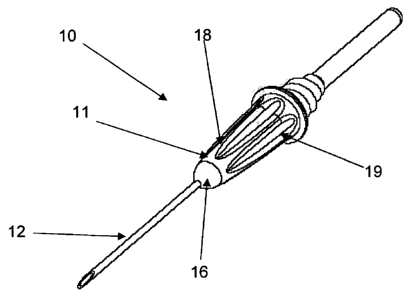
In Figure 1 there is shown a perspective view of a device 10 for drawing fluid
from a
lumen according to an embodiment of the present invention. The device 10
comprises
a body 11, a first needle portion 12 and a second needle portion (obscured)
that is
covered by a flexible sheath 13. During transportation and storage the first
needle
portion 11 is covered by a first needle cap 14 while the second needle portion
(obscured) is covered by a second needle cap 15.
The first needle portion 12 is in the form of a tubular puncture needle
adapted to
puncture a patient's skin (not shown) in order to draw fluid (such as blood)
through
the device 10.
The device 10 is provided with retaining means 16 in a forward portion of the
body
11, the retaining means 16 being adapted to retain the first needle portion 12
in place
within the body 11. The rear portion of the body 11 is provided with a screw-
threaded
portion 17 adapted to facilitate connecting the device 10 to a complementary
screw-
threaded portion of a medical device (not shown). In use, when the device 10
is
brought into communication with a medical device (not shown), the second
needle
portion (obscured) is held against a seal (such as a membrane, bung, plug or
the like)
in the medical device and a force is applied to the device 10 such that the
second
2 0 needle portion (obscured) punctures the flexible sheath 13 from within
and
simultaneously punctures the seal of the medical device. In this way, fluid
may be
retained within the device 10 until such time as the device 10 is in
communication
with a medical device (not shown).
2 5 The body 11 includes an observation portion 18 fabricated from a
transparent or semi-
transparent material, and through which the fluid "flash" inside the body 11
may be
observed. The observation portion 18 is provided with a plurality of recesses
19 in the
form of channels or grooves that are adapted to refract, multiply and/or
enhance the
image of the fluid within the body 11.
In Figure 2 there is shown a side view of a device 10 for drawing fluid from a
lumen.
In this Figure, the device 10 is shown in its storage or transportation
condition,
wherein the first and second needle portions (obscured) are covered by the
first needle
CA 02778394 2012-04-20
WO 2011/047413 PCT/AU2010/001334
9
cap 14 and the second needle cap 15 respectively. In this way, the device 10
may be
stored and transported without damaging the needle portions and maintaining
the
sterility of the device 10.
In Figure 3 there is shown a side view of a device 10 for drawing fluid from a
lumen.
In this Figure the second needle cap (not shown) has been removed to expose
the
flexible sheath 13 covering the second needle portion (obscured).
Figure 4 illustrates a cross-sectional view of the device 10 for drawing fluid
from a
lumen illustrated in Figure 3 through section 3-3. In this Figure it may be
seen that
the first needle cap 14 is provided with a plurality of projections 21
thereon. The
projections 21 are adapted to be located in the recesses 19 in the body 11.
Preferably,
the projections 21 engage with the recesses 19 in a frictional engagement so
as to
prevent the needle cap 14 from falling off or being knocked off inadvertently
during
storage or transportation (or the device 10 slipping out of the needle cap 14
when
being prepared for use).
In addition, the projections 21 prevent the needle cap 14 from spinning on the
device
10 when the device 10 is being connected to a medical device (not shown), for
2 0 instance in a screw-threaded engagement. Thus, the projections 21
effectively transfer
rotational torque to the device 10 so that the device 10 may be securely
attached to a
medical device (not shown).
In this Figure, it may be seen that the first needle portion 12 extends
through the body
2 5 11. The first needle portion 12 is substantially tubular such that
fluid withdrawn from
a lumen passes through the interior of the first needle portion 12. In
addition, a
chamber portion 22 in the form of an annular ring surrounding the first needle
portion
12 (and substantially co-axial therewith) may be seen.
3 0 In Figure 5 a perspective view of a device 10 for drawing fluid from a
lumen is
shown. In this Figure, the tubular nature of the first needle section 12 may
be seen,
along with the retaining means 16 in a forward portion of the body 11.
CA 02778394 2012-04-20
WO 2011/047413 PCT/AU2010/001334
It may also be seen that the recesses 19 in the observation portion 18
extending
substantially co-axially with the first needle portion 12. The recesses 19 are
substantially concave so that even a small amount of blood flash will be
refracted and
multiplied so that a user will immediately be aware that the first needle
portion 12 is
5 correctly positioned.
Turning now to Figure 6, there is shown a sectional perspective view of a
device 10
for drawing fluid from a lumen. In particular, the rear portion of the device
10 is
shown. In this Figure the second needle portion 23 may be seen extending
through the
10 rear portion of the device 10. The second needle portion 23 is
surrounded by (and
substantially co-axial with) the chamber portion 22 along a portion of the
length of the
second needle portion 23.
The second needle portion 23 is covered along a portion of its length by the
flexible
sheath 13 that prevents fluid in the second needle portion 23 from being lost
or
contaminated or from coming into contact with a user. When the second needle
portion 23 is brought into contact with a medical device (not shown), the
second
needle portion 23 simultaneously punctures both the seal (or bung etc) of the
medical
device (not shown) and the flexible sheath 13, allowing fluid to flow from the
second
2 0 needle portion 23 into the medical device.
In addition, the flexible sheath 13 is resealable, such that, if multiple
medical devices
(such as blood collection tubes) must be connected to the device 10 in a
sequential
manner, the sheath 13 will cover the second needle portion 23 once a first
medical
2 5 device is removed and before a second medical device is connected. In
this way, the
second needle portion 23 is covered to prevent accidental injury or fluid flow
through
the needle while no medical device is attached.
Figure 7 illustrates a cross-sectional view of a device 10 for drawing fluid
from a
3 0 lumen. In this Figure it may be seen that the first needle portion 12
and the second
needle portion 23 are actually opposing ends of a single needle. The needle is
provided with an aperture 24 that is aligned with the chamber portion 22 of
the body
11 such that some of the fluid flowing through the needle enters the chamber
portion
CA 02778394 2012-04-20
WO 2011/047413 PCT/AU2010/001334
11
22 through the aperture 24. The fluid entering the chamber portion 22 may be
observed through the observation portion 18 of the body 11. The plurality of
recesses
19 in the observation portion 18 ensure that, even if only a small amount of
fluid
enters the chamber portion 22, the image of the fluid will be multiplied or
enhanced
such that a user will quickly and easily be able to identify the presence of
the fluid.
Thus, the user will immediately know that the first needle portion 12 is
correctly
positioned in the lumen.
The forward end of the body 11 is provided with a retaining means 16 which
engages
1.0 with the body 11 and retains the first needle portion 12 therein.
The rear end of the body comprises a screw-threaded portion 17 for allowing
the
device 10 to be brought into screw-threaded engagement with a suitable medical
device (not shown). The flexible sheath 13 may also be seen engaged with and
1 5 retained on the rear portion of the body 11.
In Figure 8 there is shown an exploded perspective view of a device 10 for
drawing
fluid from a lumen. All of the component parts of the device 10 may be clearly
seen
in this Figure.
Firstly it may be seen that the first needle portion 12 and the second needle
portion 23
comprise opposing ends of a single needle, the needle having an aperture 24
therein
through which some of the fluid passing through the needle may exit into the
chamber
portion (obscured).
The needle passes through and is retained in a body 11, the body 11 having an
observation portion 18 fabricated from a transparent or semi-transparent
material to
allow inspection of the interior of the body 11. The observation portion 18 is
provided with a plurality of recesses 19 that enhance, refract and/or multiply
the
3 0 image of the fluid in the chamber portion (obscured) making
identification of the fluid
(such as a blood flash) fast and easy.
The rear end of the body has a flexible sheath 13 attached thereto, the
flexible sheath
CA 02778394 2012-04-20
WO 2011/047413 PCT/AU2010/001334
= 12
13 covering the second needle portion 23 to prevent leakage or contamination
of the
fluid in the needle, and to prevent the user from coming into contact with the
fluid in
the needle.
The forward end of the body 11 is provided with a retaining means 16 which
engages
with the body and retains the first needle portion 12 therein.
Finally, the device 10 is provided with a first needle cap 14 and a second
needle cap
that protect the first needle portion 12 and the second needle portion 23
10 respectively from damage and contamination during storage and
transportation of the
device 10.
It will be understood that the present invention provides a number of
important
advantages over the prior art. Firstly, the presence of the enhancement means
allows a
15 user to quickly and easily determine whether the first needle portion is
correctly
positioned, making the process of drawing the fluid faster and less
uncomfortable for
the patient. In addition, the ability to rapidly determine the correct
location of the first
needle portion reduces the likelihood of injuring the patient due to incorrect
positioning.
Furthermore, by providing a single needle, rather than first and second
needles as in
many prior art devices, the number of parts required to construct the device
is reduced,
as well as the cost and ease of construction. In addition, providing a single
needle
rather than two separate needles reduces the likelihood that a needle will
come loose
2 5 and fall out of the device, making the device unusable.
Those skilled in the art will appreciate that the present invention may be
susceptible to
variations and modifications other than those specifically described. It will
be
understood that the present invention encompasses all such variations and
3 0 modifications that fall within its spirit and scope.