Note: Descriptions are shown in the official language in which they were submitted.
I I
CA 2778953 2017-04-20
COMPOSITIONS, KITS, AND METHODS FOR THE DIAGNOSIS, PROGNOSIS,
MONITORING, TREATMENT AND MODULATION OF POST-TRANSPLANT
LYMPHOPROLIFERATIVE DISORDERS AND HYPDXIA ASSOCIATED
ANGIOGENESIS DISORDERS USING GALECTIN-1
10
Background of the Invention
Post-transplant lymphoproliferative disorders (PTLD) are potentially fatal
conditions associated with immunocompromised solid organ and stem cell
transplantation
that can have 70-80% mortality (Gottschalk et al. (2005) Annu. Rev. Med. 56,
29-44; Paya
etal. (1999) Transplantation 68, 1517-1525). PTLD is often associated with
viral
infection, such that latent viral infection of the transplanted material can
cause
complications in the transplant subject. For example, Epstein-Barr virus (EBV)-
associated
PTLD derives from herpes virus exposure that establishes latent infection in a
majority of
healthy adults. Proliferation of EBV-infected B cells in PTLD is maintained by
expression
of EBV latent genes, such as latent membrane protein 1 (LMP1) and LMP2A, viral
immune
evasion strategies, and impaired host immune surveillance. The incidence of
PTLD varies
according to the organ transplanted, as well as the intensity and duration of
immunosuppression. In renal transplant recipients PTLD occurs in 1-2% of
patients, but
the incidence is as high as 20% in small bowel transplant and 1%-10% in lung,
heart, liver,
and kidney transplant recipients (Gottschalk et al. (2005) Annu. Rev. Med. 56,
29-44; Paya
etal. (1999) Transplantation 68, 1517-1525). Children and transplant
recipients without
previously established anti-EBV immunity are among those at greatest risk for
development
of a PTLD. There is no accepted standard of therapy for PTLD, and the
progression of the
disease in patients is often not responsive to currently available therapies.
Management of
early PTLD lesions is currently based on reduction or withdrawal of
immunosuppression
which increases the risk of graft rejection.
In addition, cancer cells adapt to low oxygen tension by promoting the
expression of
genes associated with anaerobic metabolism, invasion and angiogenesis (Pugh
etal. (2003)
1
09-78A51 90M04-25
WO 2011/060272 PCT/US2010/056547
Nat Med 9, 677-684; Fraisl et al. (2009) Dev Cell 16, 167-179). The concerted
action of
hypoxia-regulated pathways allows tumor cells to sprout new vessels, co-opt
host vessels
and/or recruit angio-competent bone marrow-derived cells to generate
functionally
abnormal tumor vasculaturcs (Ferrara et at. (2005) Nature 438, 967-974). In
spite of the
well-established roles of hypoxia-inducible factor (HIF)-lcc and vascular
endothelial
growth factor (VEGF), increasing evidence suggests the contribution of
alternative 'non-
canonical' pathways to hypoxia-driven neovascularization (Ferrara, N. (2010)
C.,vtokine
Growth Factor Rev 21, 21-26). This proposition is firmly grounded on emerging
preclinical and clinical data demonstrating 'evasive resistance' or 'intrinsic
refractoriness'
to VEGF-targeted therapies, which fail to produce enduring clinical benefits
(Ferrara, N.
(2010) Cytokine Growth Factor Rev 21, 21-26; Ebos et al. (2009) Cancer Cell
15, 232-239;
Paez-Ribes et at. (2009) Cancer Cell 15, 220-231).
The mechanisms underlying 'evasive resistance' involve revascularization as a
result of the delivery of alternative pro-angiogenic signals (Bcrgers et al.
(2008) Nat Rev
Cancer 8, 592-603) and/or mobilization of bone marrow-derived inflammatory
cells, which
together with endothelial and pericyte progenitors, are recruited to the tumor
vasculature
(Shojaei et at. (2007) Nat Biotechnol 25, 911-920; Bergers et at. (2008) Nat
Rev Cancer 8,
592-603). Future anti-angiogenic therapies might capitalize on an improved
understanding
of these compensatory pathways, as well as the elucidation of the molecular
underpinnings
of blood vessel normalization and the identification of hallmark signatures
which
distinguish healthy from tumor-associated endothelium (Jain, R. K. (2005)
Science 307, 58-
62). Although substantial changes in the endothelial cell (EC) surface
glycome' were
apparent under different culture conditions (Garcia-Vallejo et at. (2006)J
Cell Physiol 206,
203-210; Willhauck-Fleckenstein et at. (2010) Angiogenesis 13, 25-42),
suggesting a role
for glycan structures in differentially regulating angiogenesis in hypoxic
versus normoxic
and in neoplastic versus healthy tissues, the specific glycan structures,
mediating
molecules, and mechanisms were not known prior to the results described
herein.
Programmed remodeling of cell surface glycans can control cellular processes
by
displaying or masking ligands for endogenous lectins (Paulson et at. (2006)
Nat Chem Biol
2, 238-248; van Kooyk et al. (2008) Nat Ininzunol 9, 593-601). Recent efforts
involving
genetic manipulation of .7\T- and 0-glycosylation pathways have revealed
essential roles for
multivalent lectin-glycan lattices in the control of receptor signaling
(Ohtsubo, et al. (2006)
Cell 126, 855-867; Dennis et at. (2009) Cell 139, 1229-1241; Dam et at. (2010)
2
CA 077789532012-04-25
WO 2011/060272 PCT/US2010/056547
Gl,vcobiology 20, 1061-1064). Regulated glycosylation can control sprouting
angiogenesis
by modulating binding of Notch receptor to its ligands Delta-like 4 (D114) or
Jaggedl
(Benedito etal. (2009) Cell 137, 1124-1135), fine-tuning neuropilin-1 (NRP-1)
signaling
(Shintani etal.. (2006) EMBO J25, 3045-3055) and facilitating CD31-mediated
homophylic interactions (Kitazume et al. (2010)J Biol Chem 285, 6515-6521).
Yet,
whether differential glycosylation enables the formation of discrete lectin-
glycan lattices
and signaling clusters that are functionally relevant to angiogenesis remains
largely
unexplored.
In view of the above, it is clear that there remains a need in the art for
compositions
and methods to specifically boost host anti-viral (e.g., anti-EBV) immune
responses, as
well as inhibiting hypoxia associated angiogenesis in a number of disorders.
Summary of the Invention
The present invention relates in general to a role of galectin-1 (Gall) in
diagnosing,
prognosing, monitoring, treating and/or modulating PTLD, including EBV-
associated
PTLD and/or hypoxia associated angiogenesis disorders.
The present inventors have determined a vascular regulatory circuit involving
Galectin-1 (Gall), a member of a highly-conserved family of animal lectins, is
expressed
and secreted by a variety of tumors where it contributes to malignant
transformation and
metastasis (Paez-Ribes et al. (2009) Cancer Cell 15, 220-231; Liu et al.
(2005) Nature Rev
Cancer 5, 29-41), based on the differential glycosylation of ECs that promotes
the
formation of lectin-glycan lattices. These interactions couple tumor hypoxia
to VEGFR2-
mediated neovascularization through mechanisms that are independent of H1F-la
and
VEGF. The `glycosylation signature' of ECs can be selectively altered by
tolerogenic,
inflammatory, proliferative and hypoxic stimuli, which can either enable or
hinder
formation of these lattices. Targeted disruption of Gall -glycan interactions,
through Gall
blockade or prevention of N-glycan branching, attenuated hypoxia-driven
angiogenesis,
while promoting extensive remodeling of vascular networks and increased influx
and
expansion of immune effector cells into the tumor parenchyma. These results
underscore
novel opportunities for targeting aberrant vascular networks, while
simultaneously
potentiating T cell-mediated antitumor immunity.
The present invention is also based, in part, on the identification of novel
anti-Gall
monoclonal antibodies. Accordingly, in one aspect, the present invention
features a
3
CA 077789532012-04-25
WO 2011/060272 PCT/US2010/056547
monoclonal antibody, or antigen-binding fragment thereof, wherein the
monoclonal
antibody comprises a heavy chain sequence with at least about 95% identity to
a heavy
chain sequence selected from the group consisting of the sequences listed in
Table 1 or a
light chain sequence with at least about 95% identity to a light chain
sequence selected
from the group consisting of the sequences listed in Table 1. In one
embodiment, the
monoclonal antibody comprises a heavy chain CDR sequence with at least about
95%
identity to a heavy chain CDR sequence selected from the group consisting of
the
sequences listed in Table 1 or a light chain CDR sequence with at least about
95% identity
to a light chain CDR sequence selected from the group consisting of the
sequences listed in
Table 1. In another embodiment, the monoclonal antibody, or antigen-binding
fragment
thereof comprises a heavy chain sequence selected from the group consisting of
the
sequences listed in Table 1 or a light chain sequence selected from the group
consisting of
the sequences listed in Table 1. In still another embodiment, the monoclonal
antibody, or
antigen-binding fragment thereof comprises a heavy chain CDR sequence selected
from the
group consisting of the sequences listed in Table 1 or a light chain CDR
sequence selected
from the group consisting of the sequences listed in Table 1. In yet another
embodiment,
the monoclonal antibody, or antigen-binding fragment thereof is chimeric,
humanized,
composite, or human. In another embodiment, the monoclonal antibody, or
antigen-binding
fragment thereof, is detectably labeled, comprises an effector domain,
comprises an Fe
domain, is a single-chain antibody, or is a Fab fragment. In still another
embodiment, the
monoclonal antibody, or antigen-binding fragment thereof, inhibits the binding
of
commercial antibody to Gall. In yet another embodiment, the monoclonal
antibody, or
antigen-binding fragment thereof, reduces or inhibits at least one Gall
activity (e.g.,
binding to beta-galacostides) relative to the absence of the monoclonal
antibody or antigen-
.. binding fragment thereof selected from the group consisting. Host cells
expressing the
described monoclonal antibodies, or antigen-binding fragment thereof, are also
contemplated.
In another aspect, the present invention features isolated nucleic acid
molecules that
hybridize, under stringent conditions, with the complement of a nucleic acid
encoding a
polypeptide selected from the group consisting of the sequences listed in
Table 1, or
sequences with at least about 95% homology to a nucleic acid encoding a
polypeptide
selected from the group consisting of the sequences listed in Table 1. In one
embodiment,
4
CA 077789532012-04-25
WO 2011/060272 PCT/US2010/056547
the isolatd nucleic acid is comprised within a vector. In another embodiment,
a host cell
comprises
In still another aspect, the present invention features a device or kit
comprising at
least one monoclonal antibody or antigen-binding fragment thereof of the
present invention,
said device or kit optionally comprising a label to detect the at least one
monoclonal
antibody or antigen-binding fragment thereof of the present invention, or a
complex
comprising the monoclonal antibody or antigen-binding fragment thereof of the
present
invention.
In yet another aspect, the present invention features a pharmaceutical
composition
comprising the antibody or antigen-binding fragment thereof of the present
invention, in a
pharmaceutically acceptable carrier.
In another aspect, the present invention features a method of detecting the
presence
or level of a Gall polypeptide said method comprising obtaining a sample and
detecting
said polypeptide in a sample by use of at least one monoclonal antibody or
antigen-binding
fragment thereof of the present invention. In one embodiment, the method
utilizes at least
one monoclonal antibody or antigen-binding fragment thereof of the present
invention to
form a complex with a Gall polypeptide and the complex is detected in the form
of an
enzyme linked immunosorbent assay (ELISA), radioimmune assay (MA), or
immunochemically.
In another aspect, the present invention features a method for monitoring the
progression of a disease in a subject, the method comprising detecting in a
subject sample
at a first point in time the level of expression of Gall using at least one
monoclonal
antibody or antigen-binding fragment thereof of the present invention;
repeating the
previous step at a subsequent point in time; and comparing the level of
expression of said
Gall detected in steps a) and b) to monitor the progression of the disease in
the subject. In
one embodiment, the subject has undergone treatment to ameliorate the disease
between the
first point in time and the subsequent point in time.
In still another aspect, the present invention features a method for
predicting the
clinical outcome of a subject afflicted with a disease, the method comprising
determining
the level of expression of Gall in a patient sample using at least one
monoclonal antibody
or antigen-binding fragment thereof of the present invention; determining the
level of
expression of Gall in a sample from a control subject having a good clinical
outcome using
at least one monoclonal antibody or antigen-binding fragment thereof of the
present
5
CA 077789532012-04-25
WO 2011/060272 PCT/US2010/056547
invention; and comparing the level of expression of Gall in the patient sample
and in the
sample from the control subject; wherein a significantly higher level of
expression in the
patient sample as compared to the expression level in the sample from the
control subject is
an indication that the patient has a poor clinical outcome.
In yet another aspect, the present invention features a method of assessing
the
efficacy of a therapy for a disease in a subject, the method comprising
comparing the level
of expression of Gall using at least one monoclonal antibody or antigen-
binding fragment
thereof of the present invention, in a first sample obtained from the subject
prior to
providing at least a portion of the therapy to the subject, and the level of
expression of Gall
in a second sample obtained from the subject following provision of the
portion of the
therapy, wherein a significantly lower level of expression of Gall in the
second sample,
relative to the first sample, is an indication that the therapy is efficacious
for inhibiting the
disease in the subject.
In another aspect, the present invention features a method for treating a
subject
.. afflicted with a disease comprising administering at least one monoclonal
antibody or
antigen-binding fragment thereof of the present invention, such that the
subject afflicted
with the disease is treated.
In another aspect, the present invention features a method for monitoring the
progression of a viral-associated PTLD or hypoxia associated angiogenesis
disorder in a
.. subject, the method comprising detecting in a subject sample at a first
point in time the
level of expression of Gall; repeating the previous step at a subsequent point
in time; and
comparing the level of expression of said Gall detected at various time points
to monitor
the progression of the viral-associated PTLD or hypoxia associated
angiogenesis disorder
in the subject. In one embodiment, the subject has undergone treatment to
ameliorate the
viral-associated PTLD or hypoxia associated angiogenesis disorder between the
first point
in time and the subsequent point in time.
In yet another aspect, the present invention features a method of assessing
the
efficacy of a test compound for inhibiting a viral-associated PTLD in a
subject, the method
comprising comparing the level of expression of Gall in a first sample
obtained from the
subject and exposed to the test compound; and the level of expression of Gall
in a second
sample obtained from the subject, wherein the second sample is not exposed to
the test
compound, and a significantly lower level of expression of Gall, relative to
the second
sample, is an indication that the test compound is efficacious for inhibiting
the viral-
6
CA 077789532012-04-25
WO 2011/060272 PCT/US2010/056547
associated PTLD in the subject. In one embodiment, the first and second
samples are
portions of a single sample obtained from the subject or portions of pooled
samples
obtained from the subject.
In another aspect, the present invention features a method for predicting the
clinical
outcome of a subject afflicted with a viral-associated PTLD or hypoxia
associated
angiogenesis disorder patient, the method comprising determining the level of
expression of
Gall in a patient sample; determining the level of expression of Gall in a
sample from a
control subject having a good clinical outcome; and comparing the level of
expression of
Gall in the patient sample and in the sample from the control subject; wherein
a
significantly higher level of expression in the patient sample as compared to
the expression
level in the sample from the control subject is an indication that the patient
has a poor
clinical outcome.
In another aspect, the present invention features a method of assessing the
efficacy
of a therapy for a viral-associated PTLD or hypoxia associated angiogenesis
disorder in a
subject, the method comprising comparing the level of expression of Gall in a
first sample
obtained from the subject prior to providing at least a portion of the therapy
to the subject,
and the level of expression of Gall in a second sample obtained from the
subject following
provision of the portion of the therapy, wherein a significantly lower level
of expression of
Gall in the second sample, relative to the first sample, is an indication that
the therapy is
efficacious for inhibiting the viral-associated PTLD or hypoxia associated
angiogenesis
disorder in the subject.
In some embodiments of the methods of the present invention, a sample
comprises
cells obtained from a subject. In another embodiment, cells are in a fluid
selected from the
group consisting of whole blood fluid, serum fluid, plasma fluid, interstitial
fluid,
cerebrospinal fluid, lymph fluid, saliva, stool, and urine. In still another
embodiment, the
level of Gall expression is assessed using a reagent which specifically binds
with a Gall
protein, polypeptide or protein fragment thereof (e.g., an antibody, an
antibody derivative,
or an antibody fragment). In yet another embodiment, the level of Gall
expression is
assessed by detecting the presence in the sample of a transcribed
polynucleotide encoded
by a Gall polynucleotide (e.g., mRNA or cDNA) or a portion of said transcribed
polynucleotide. In another embodiment, the step of detecting further comprises
amplifying
the transcribed polynucleotide. In still another embodiment, the level of Gall
expression is
assessed by detecting the presence in the sample of a transcribed
polynucleotide which
7
CA 077789532012-04-25
WO 2011/060272 PCT/US2010/056547
anneals with a Gall polynucicotide or anneals with a portion of a Gall
polynucicotidc,
under stringent hybridization conditions. In yet another embodiment, a
significant increase
comprises an at least two fold or at least five fold increase between the
level of expression
of Gall in the subject sample relative to the normal level of expression of
Gall in the
sample from the control subject.
In another aspect, the present invention features a method for modulating an
immune response in a subject afflicted with a viral-associated PTLD or hypoxia
associated
angiogenesis disorder comprising contacting an immune cell with an agent that
modulates
the interaction between Gall or a fragment thereof and its natural binding
partner(s) to
thereby modulate the immune response. In one embodiment, the immune response
is
upregulated. In another embodiment, the immune response is downregulated. In
still
another embodiment, signaling via the Gall binding partner is inhibited using
an agent
selected from the group consisting of: a blocking antibody or an antigen
binding fragment
thereof that recognizes Gall, a blocking antibody or an antigen binding
fragment thereof
that recognizes the Gall binding partner(s) or a fragment thereof, natural
ligands, small
molecules, aptamers, peptides, peptidomimetics, glycan-related compounds,
glycomimetics,
and RNA interference molecules. In yet another embodiment, the immune cell is
further
contacted (e.g., in vivo or in vitro) with an additional agent that
uprcgulatcs an immune
response.
In still another aspect, the present invention features a method for treating
a subject
afflicted with a viral-associated PTLD or hypoxia associated angiogenesis
disorder
comprising administering an agent that inhibits the interaction between Gall
and its natural
binding partner(s) on cells of the subject such that the subject afflicted
with the viral-
associated PTLD is treated. In one embodiment, the agent is selected from the
group
consisting of: a blocking antibody or an antigen binding fragment thereof that
recognizes
Gall, a blocking antibody or an antigen binding fragment thereof that
recognizes the Gall
binding partner(s) or a fragment thereof, natural ligands, small molecules,
aptamers,
peptides, peptidomimetics, glycan-related compounds, glycomimetics, and RNA
interference molecules. In another embodiment, a second agent that upregulates
an immune
response, downregulates hypoxia associated angiogenesis (e.g., VEGF-targeted
therapeutic
such as an anti-VEGF antibody), or combination thereof, is administered to the
subject.
In yet another aspect, the present invention features a method for modulating
angiogenesis in a hypoxia associated angiogenesis disorder comprising
contacting a cell
8
CA 077789532012-04-25
WO 2011/060272
PCT/US2010/056547
exhibiting hypoxia associated angiogenesis with an agent that modulates the
interaction
between Gall or a fragment thereof and its natural binding partner(s) to
thereby modulate
angiogenesis. In one embodiment, hypoxia assocaited angiogenesis is
downregulated. In
another embodiment, downregulation of hypoxia associated angiogenesis is
determined by
at least one effect selected from the group consisting of: reduction in vessel
diameter,
reduction in vessel distribution, reduction in tortuous vessels, increase in
pericyte coverage,
increase in the fraction of pericytes that are mature, and reduction in
pimonidazole adduct
formation. In still another embodiment, hypoxia associated angiogenesis is
modulated
using an agent selected from the group consisting of: a blocking antibody or
an antigen
binding fragment thereof that recognizes Gall, a blocking antibody or an
antigen binding
fragment thereof that recognizes a Gall binding partner(s) or a fragment
thereof, natural
ligands, small molecules, aptamers, peptides, peptidomimetics, glycan-related
compounds,
glycomimetics, and RNA interference molecules. In yet another embodiment, the
method
further comprises contacting the cell (e.g., in vivo or in vitro) exhibiting
hypoxia associated
angiogenesis with an additional agent that downregulates hypoxia associate
angiogenesis.
In another aspect, the present invention features an isolated complex
comprising a
Gall polypeptide and a VEGFR2 polypeptide. In one embodiment, the Gall
polypeptide is
a polypeptide describe herein or fragment thereof that is capable of binding
to a VEGFR2
polypeptide and the VEGR2 polypeptide described herein or fragment thereof
that is
capable of binding to a Gall polypeptide. In another embodiment, at least one
polypeptide
or fragment is a fusion protein. In still another embodiment, at least one
polypeptide or
fragment is labeled. In yet another embodiment, the complex is generated
within a host
cell. In another embodiment, the Gall polypeptide or fragment thereof and said
VEGFR2
polypeptide or fragment thereof are covalently
In still another aspect, the present invention features an isolated antibody
of the
present invention has the ability to disrupt a complex comprising a Gall
polypeptide and a
VEGFR2 polypeptide.
In yet another aspect, the present invention features a method for identifying
a
compound that modulates a GallNEGFR2 complex comprising: a) contacting the
complex
with a test compound; and b) assaying the amount or activity of the complex,
wherein a
change in the amount or activity of the complex in the rpesence of the test
compound as
compared to the amount or activity of the complex in the absence of the test
compound is
indicative of a compound that modulates a GallNEGFR2 complex.
9
CA 077789532012-04-25
WO 2011/060272 PCT/US2010/056547
In another aspect, the present invention features a hybridoma, 14-19 8F4F8G7,
deposited under accession number PTA-10535
For any method described herein, the relevant condition, disease, or disorder
can be
a viral-associated PTLD, cancer, and/or a hypoxia associated angiogenesis
disorder. In
addition, the level of expression of a marker, such as Gall to be analyzed,
can be
determined by the amount, structure, subcellular localization, and/or activity
of the marker,
as described further herein. Moreover, the progress, outcome, or efficacy of
any method
describe herein can be measured by at least one criteria selected from the
group consisting
of survival until mortality, pathological complete response, clinical complete
remission,
clinical partial remission, clinical stable disease, recurrence-free survival,
metastasis free
survival, and disease free survival, according to, but not limited by,
exemplary
embodiments described further herein. Also, the Gall binding partner can be
VEGFR2 for
any method, composition, and/or complex of the present invention.
Brief Description of the Drawings
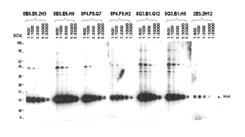
Figure 1 shows serial dilution-based reactivity data for anti-human Gall
monoclonal antibodies assayed against endogenous Gall from a Hodgkin lymphoma
cell
line.
Figure 2 shows cross-reactivity data for anti-human Gall monoclonal antibodies
assayed against endogenous cynomologous monkey and mouse Gall.
Figure 3 shows a schematic diagram of GST-tagged human Gall (hGall) fragments
utilized in epitope mapping analyses of the anti-human Gall monoclonal
antibodies.
Figure 4 shows epitope mapping data for anti-human Gall monoclonal antibodies.
Figure 5 shows Gall transcript abundance in EBV-transformed lymphoblastoid
cell
lines.Gall transcript abundance in HL lines, LCLs, normal B-cells and
additional B-cell
neoplasmswas assessed using publically available gene expression profiles
(Kuppers R.
(2009) Nat Rev Cancer 9:15-27). Color scale at the bottom indicates relative
expression
SEM. Red connotes high-level expression; blue indicates low-level expression.
Figure 6 shows specificity of the anti-Gall monoclonal antibody, 8F4F8G7, for
endogenous Gall. The newly developed Gall mAb specifically detected endogenous
Gall
from the cHL cell line, L428, on immunoblots.
Figures 7A-7B shows Gal 1 expression in EBV-transformed LCLs and EBV+
primary PTLDs. Figure 7A shows Gall expression in a cHL cell line (L428), a
series of
CA 077789532012-04-25
WO 2011/060272 PCT/US2010/056547
EBV-transformed LCLs (NOR-, RIC-, STA-, FOL-, LOV-, RIV-, WOL-, FW-, VS-, MA-,
SC-, DS-, AND DW-LCL), and a DLBCL cell line (SU-DHL6). Figure 7B shows Gall
immunohistochemical staining of three representative primary EBV+ PTLDs
(panels a, b,
and c) and a DLBCL (panel d). The recently developed murine aGallmAb, 8F4F8G7,
was
used at 1:20,000 in immunoblots in Figure 7A and 1:40,000 in IHC in Figure 7B.
Original
magnifications: x1000.
Figure 8 shows Gall expression in primary post-transplant lymphoproliferative
disorders (PTLDs) and DLBCLs. Gall immunohistochemistry (IHC) was performed
with
the previously described rabbit anti-Gall polyclonal antiserum (Juszczynski
etal. (2007)
Proc Nat! Acad Sci US A 104:13134-9). Representative primary EBV+ PTLDs
(panels a,
b, and c) and DLBCLs (panels d, e, and 0 are shown. Original magnifications:
x1000.
Figures 9A-9E show AP-1 dependent Gall expression in EBV-transformed LCLs
and primary PTLDs. Figure 9A shows total phospho-cJun and JunB expression in a
cHL
cell line, L428, and two EBV-transformed LCLs, RIC and NOR -actin was used as
a
loading control. Figure 9B shows results of ChIP-PCR analysis of cJun and JunB
binding
to Gall enhancer regions in the cHL cell line, L428, and two LCLs, NOR and
RIC. Results
are representative of triplicate experiments. Figure 9C shows results of
densitometric
analyses of ChIP-PCR data from Figure 9B. Figure 9D shows Gall promoter and
enhancer-driven luciferase activity in LCLs. NOR cells were cotransfected with
300ng of
the pGL3-Gall -promoter constructs (without or with the wild-type or mutant AP-
1
dependent Gall enhancer) and 100 ng of the control reporter plasmid, pRL-TK,
and
evaluated for relative luciferase activity as described (Juszczynski et al.
(2007) Proc Nall
Acad Sci US A 104:13134-9). Figure 9E shows IHC analysis of JunB (panels a,c,
and e)
and phospho-cJun (panels b,d, and f) in 3 primary PTLDs. The PTLDs had
uniformly high
nuclear staining of JunB and positive phospho-cJun staining of variable
intensity.
Figures 10A-10B show Gall transcript abundance in LMP1-expressing CD10+
germinal center B cells.Gall transcript abundance in normal CD10 germinal
center B-cells
with and without LMP1 transduction was assessed using publically available
gene
expression profiles (Vockerodt etal. (2008)J Pathol (2008) 216:83-92). Gall
induction is
shown on a heat map, with red indicating high expression and blue indicating
low
expression in Figure 10A. Figure 10B shows that Gall was 2 fold more abundant
in LMP-
transduced germinal center B cells (p <.002).
11
CA 077789532012-04-25
WO 2011/060272 PCT/US2010/056547
Figures 11A-11C show induction of Gall expression by LMP1 and 2A. Figure
11A shows LMP1- and LMP2A-enhanced Gall promoter-driven luciferase activity.
293T
cells were co-transfected with the pGL3-LGALS1 promoter (Juszczynski et al.
(2007) Proc
Nat! Acad Sci US A 104:13134-9), control reporter plasmid pRL-PGK and pFLAG-
CMV2
empty vector or expression vector LMP1-FLAG or LMP2A-FLAG or LMP1-FLAG plus
LMP2A-FLAG and evaluated for relative luciferase activity. Figure 11B shows
RNAi-
mediated down-regulation of LMP2A in EBV-transformed LCL. NOR. 13¨actin was
used
as a loading control. Figure 11C shows chemical inhibition of PI3K activity
(25 uM
Ly294002) and associated change in Gall expression in EBV-transformed LCLs.
Figure 12 shows regulatory element analysis of the Gall promoter. Analysis of
transcription factor binding motifs and modules within the Gall promoter
region revealed a
single NFKB binding site, and a NFAT/NF-Y module. The Gall promoter region
included
in the luciferase constructs (Figures 11A-11C) is shown in blue relative to
the transcription
start site (TSS).
Figures 13A-13B show the anti-Gall mAb 8F4F8G7 inhibits rGall-induced
apoptosis of in vitro activated T cells. Anti-CD3/CD28 activated human T cells
were
treated with 10 jiM of rGall alone or 10 ,u,A4 rGall pre-incubated with 5 p.M
of anti-Gall
mAb (8F4F8G7) or an isotype-matched IgG2b control and evaluated thereafter
with a flow
cytometric apoptosis assay (Annexin V-FITC and PI staining). The percentage of
cells in
each quadrant is indicated in Figure 13A. Figure 13B shows a histogram
summarizing the
percentage of annexin V+ cells in the absence of rGal or the presence of rGal
alone or
rGall pre-incubated with the aGall mAb or isotype control.
Figures 14A-14B show that Gall neutralizing mAb, 8F4F8G7, inhibits Gall-
mediated apoptosis of EBV-specific CTLs. Figure 14A shows results of EBV-
specific
CTLs treated with rGall alone or rGall pre-incubated with aGall mAb or isotype
control.
The percentage of viable CD8+ CTLs (7-AAD negative) is shown on the top of the
gate.
Figure 14B shows a histogram summarizing the percentage of viable EBV-specific
CD8+
CTLs following the indicated treatments.
Figures 15A-15B show results of aGal mAb-mediated inhibition of rGall-mediated
apoptosis of EBV-specific CTLs generated from a second independent donor.
Figure 15A
shows results of EBV-specific CTLs treated with rGall alone or rGall pre-
incubated with
anti-Gall mAb or isotype control IgG2b as in Figures 14A-14B. The percentage
of viable
CD8+ CTLs (7-AAD negative) is shown on the top of the gate. Figure 15B shows a
12
CA 077789532012-04-25
WO 2011/060272 PCT/US2010/056547
histogram summarizing the percentage of viable CD8+ CTLs following the
indicated
treatments.
Figures 16A-16S show that differential glycosylation of endothelial cells
(ECs)
controls the formation of lectin-glycan lattices. Figure 16A shows the glycan
repertoire of
.. HUVEC under resting conditions (2% FCS) detected with biotinylated L-PHA,
LEL, SNA,
MAL II, PNA and HPA (filled histograms) or with PE¨conjugated stravidin alone
(open
histograms). Data are representative of eight independent experiments. Figure
16B shows
the glycan repertoire of HUVEC under resting, proliferative (bFGF),
tolerogenic (IL-10
and/or TGF-131) or pro-inflammatory (TNF, IFN-y and/or IL-17) conditions. rMFI
(relative
mean fluorescence intensity) = (MFI with lectin ¨ MFI without lectin) / MFI
without lectin.
Data are presented as the ratio relative to resting conditions (dotted line;
value=1) and are
the mean SEM of four independent experiments. Figure 16C shows binding
results of
488-Gall to HUVEC with or without lactose or sucrose, swainsonine or benzyl-a-
GaINAc.
Data are the mean SEM of three independent experiments. Figure 16D shows
bnding
results of 488-Gall to HUVEC transfected with GnT5 or GCNT1 siRNA. Cells
without
siRNA or transfected with scrambled (src) siRNA were used as controls. Data
are the mean
SEM of at least three independent experiments. Figure 16E shows binding
results of
488-Gall to HUVEC exposed to tolerogenic, proliferative or inflammatory
stimuli. Data
are presented as the rMFI ratio relative to resting ECs (dotted line; value=1)
and are the
mean + SEM of four independent experiments. * P < 0.05, ** P < 0.01 versus
control.
Figures 16F-1611 show results of [3H]thymidine incorporation (Figure 16F),
migration
(Figure 16G) and tube formation (Figure 16H) of ECs transfected or not with
GnT5,
GCNT1 or scr siRNA and treated or not with Gall (liuM) and/or VEGF (20 ng/ml)
with or
without lactose. t P < 0.05 vs Gall; *P < 0.05 ** P < 0.01 versus control.
Data are the
.. mean SEM of at least five independent experiments. Figure 161 shows tube
formation
induced by Gall or VEGF in HUVEC transfected with GnT5, GCNT1 or scr siRNA. *
P <
0.05 versus scr siRNA. Data are the mean SEM of three independent
experiments.
Figure 16J shows in vivo vascularization of Matrigel sponges containing Gall
with or
without lactose and the right panel in particular shows quantification of
hemoglobin
content. Data are representative of two independent experiments. Figure 16K
shows
schematic representation of N- and 0-glycan biosynthesis, including relevant
glycosyltransferases, such as a2-6 sialyltransferase 1 (ST6Gall),
N- acetylglucosaminyltransferase 5 (GnT5), a2-3 sialyltransferase 1 (ST3Gall)
and core 2
13
CA 077789532012-04-25
WO 2011/060272 PCT/US2010/056547
N-acetylglucosaminyltransferase 1 (GCNT1), the coordinated actions of which
lead to the
generation or masking of common glycosylated ligands for galectins (N-
acetyllactosamine;
LacNAc) or poly-LacNAc residues in complex N-glycans or core 2 0-glycans) at
the top,
whereas the bottom shows schematic representation of lectin-binding sites in N-
and 0-
.. glycans. Specific residues recognized by MAL II, LEL, SNA and L-PHA on
complex N-
glycans and by HPA, PNA and LEL on 0-glycans are indicated (green). The common
glycosylated ligand for Gall (LacNAc) is also indicated (purple). Figure 16L
shows
binding results of biotinylated L-PHA to HUVEC transfected with GnT5 (filled
histogram)
or with scrambled (scr) (open black histogram) siRNA. Cells stained with PE-
conjugated
Stravidin alone were used as negative control (open grey histogram). Data are
representative of four independent experiments. Figure 16M shows results of
qRT-PCR
analysis of GnT5 mRNA, whereas Figure 16N shows that for GCNT1 mRNA of HUVEC
transfected with different concentrations of specific siRNA relative to RN18S1
mRNA
(AU: arbitrary units). **P < 0.01 versus control. Data are the mean SEM of
four
independent experiments. Figures 160-16Q show dose-dependent proliferation
(Figure
160), migration (Figure 16P) and tube formation (Figure 16Q) of HUVEC
incubated with
or without different concentrations of Gall, VEGF (20 ng/m1) or both. Gall
effects were
completely prevented by co-incubation with 30 mM lactose. * P < 0.05 and ** P
< 0.01,
versus control; t P < 0.05 vs Gall (luM). Data are the mean SEM of five
experiments.
Figure 16R shows light microscopy images of capillary tube formation (upper
panels) and
migration (lower panels) of HUVEC incubated with Gall in the presence or
absence of
lactose. VEGF was used as positive control. Images representative of five
independent
experiments are shown. Figure 16S shows dose-dependent invasion of HUVEC in
the
presence or absence of different concentrations of Gall or VEGF (20 ng/ml).
Results are
plotted as invasion index calculated as the number of fluorescent invasive
cells relative to
control. * P < 0.05 and ** P < 0.01. Data are the mean SEM of five
experiments.
Figures 17A-17R show the galectin-1 co-opts VEGFR2 signaling pathways through
the formation of lectin-glycan lattices on highly branched complex N-glycans.
Figure 17A
shows results of a phospho-RTK signaling array of HUVEC exposed to medium
(control),
.. VEGF or Gall, wherein in the left panel, arrows indicate proteins with
increased
phosphorylation intensity. Data are representative of three independent
experiments. By
contrast, the right panel shows quantification of pixel intensity. * P < 0.05,
** P < 0.01
versus control. Data are the mean SEM of three independent experiments.
Figure 17B
14
CA 077789532012-04-25
WO 2011/060272 PCT/US2010/056547
shows immunoblot results of VEGFR2, Akt and Erk1/2 phosphorylation in HUVEC
treated
with different concentrations of Gall. Data are representative of six
independent
experiments. Figures 17C-17E shows Gall-induced proliferation (Figure 17C),
migration
(Figure 17D) and tube formation (Figure 17E) on HUVEC pre-incubated with
pharmacological inhibitors of PI(3)K/Akt (LY294002), Erk1/2 (U0126), JAK2-
STAT3
(AG490), Jnk/SAP (SP600125), p38 (SB202190) or NF-KB (BAY11-7082). ** P < 0.01
versus Gall. Data are the mean SEM of five independent experiments. Figure
17F
shows immunoblot analysis of VEGFR2, Akt and Erk1/2 phosphorylation induced by
Gall
or VEGF in HUVEC transfected with VEGFR2 or GnT5 siRNA. Data are
representative of
three independent experiments. Figure 17G shows co-immunoprecipitation results
followed by immunoblot analysis of HUVEC lysates, wherein the left panel shows
results
from cells treated with or without Gall and the right panel shows rseults from
cells
transfected or not with GnT5 or GCNT1 siRNA or exposed to PNGase F and treated
with
Gall. Input, whole cell lysate; TB, immunoblot; IP, immunoprecipitation. Data
are
representative of three independent experiments. Figure 1711 shows laser
confocal
microscopy results of HUVEC transfected or not with GnT5 siRNA and treated
with Gall
or buffer control stained for VEGFR2 (red) or for nuclei (DAPI; blue). Images
are
representative of four independent experiments are shown. Figure 171 shows
tube
formation results of HUVEC transfected or not with VEGFR2, NRP-1, VEGF or scr
siRNA
treated or not with Gall. * P < 0.05 versus Gall. Data are representative of
three
independent experiments. Figure 17J shows tube formation results of HUVEC pre-
treated
with lactose or blocking antibodies to VEGFR1, VEGFR2, VEGFR3 or VEGF. * P <
0.05
versus Gall. Data are representative of three independent experiments. Figure
17K shows
fold increase results in the phosphorylation status of a panel of growth
factor receptor
tyrosine kinases (RTKs) and signaling nodes as determined by phospho-RTK
signaling
array upon exposure of HUVEC to Gall or VEGF. The relative signal intensity of
each
spot, quantified as pixel intensity is represented relative to control
intensity (value=1,
dotted line). * P < 0.05; ** P < 0.01 versus control. Data are the mean SEM
of three
independent experiments. Figure 17L shows immunoblot analysis results of
VEGFR2 and
Figure 17M shows immunoblot analysis results of NRP-1 in HUVEC transfected
with
specific siRNA (100 nM). Data are representative of three independent
experiments.
Figure 17N shows co-immunoprecipitation results followed by immunoblot
analysis of cell
lysates derived from HUVEC cultured with or without Gall. Input, whole cell
lysate; TB,
CA 077789532012-04-25
WO 2011/060272
PCT/US2010/056547
immunoblot; IP, immunoprecipitation. Data arc representative of three
independent
experiments. Figure 20 shows ELISA results of VEGF secretion by HUVEC after
specific
siRNA-mediated silencing. nd, not-detected. Data are the mean SEM of four
experiments. Figures 17P-17Q show migration results of HUVEC induced by Gall
or
VEGF in transwells. Cells were transfected with 100 nM siRNA specific for
VEGFR2,
NRP-1 or VEGF (Figure 17P), or were incubated with specific blocking
antibodies to
VEGFR2 or VEGF (Figure 17Q). * P < 0.05, ** P< 0.01 versus medium, Gall or
VEGF
alone. Data are representative of three independent experiments. Figure 17R
shows
ELISA results of VEGF secretion by HUVEC incubated with different
concentrations of
Gall with or without lactose. Hypoxia was used as positive control of VEGF
secretion.
Data are the mean SEM of six independent experiments.
Figures 18A-18U show the galectin-l-glycan lattices link tumor hypoxia to
VEGFR2-mediated angiogenesis. Figure 18A shows the glycan repertoire on HUVEC
incubated in hypoxia (black filled histograms) or normoxia (grey filled
histograms),
detected with biotinylated L-PHA, LEL, SNA, MAL II or PNA, or with
PE¨conjugated
stravidin alone (open histograms). Data are representative of five independent
experiments.
Figure 18B shows binding results of 488-Gall to HUVEC exposed to hypoxia or
normoxia. ** P < 0.01. Data are the mean SEM of five independent
experiments.
Figures 18C-18F show expression of Gall in KS cells transfected with or
without HIF-lcc
siRNA or a super-repressor form of IKB-a (IKB-a-SR) and incubated under
hypoxia or
normoxia. Figure 18C shows promoter activity and data are the mean SEM of
five
independent experiments. Figure 18D shows ciRT-PCR results of Gall mRNA
relative to
RN1851. AU, arbitrary units. **P < 0.01. Data are the mean SEM of three
independent
experiments. Figure 18E shows immunoblot results of Gall, IKB-a and HIF-la.
Data are
representative of four experiments. Figure 18F shows ELISA results of Gall
secretion.
**P < 0.01. Data are the mean SEM of three independent experiments. Figure
18G
shows ELISA results of Gall secretion by KS cells cultured in hypoxia or
normoxia in the
presence or absence of N-acetyl-cysteine (NAC; 0.5 mM). Figure 18H shows ELISA
results of Gall secretion by KS cells exposed to H202 (0.5 mM) in the presence
or absence
of BAY 11-7082. Data are the mean SEM of three independent experiments.
Figure 181
shows immunoperoxidase staining results of Gall in non-hypoxic and hypoxic
areas of KS
xcnografts in the upper panels, whereas the lower panels show
immunofluorcscence of
Gall and Hypoxyprobe-1 staining. Images are representative of three
independent
16
CA 077789532012-04-25
WO 2011/060272 PCT/US2010/056547
experiments. Figure 18J show tube formation results by HUVEC incubated with
conditioned medium (CM) from normoxic or hypoxic KS cells transfected or not
with scr
or VEGF siRNA and/or Gall shRNA. ** P < 0.01. Data are the mean SEM of four
independent experiments. Figure 18K shows hemoglobin content results of
Matrigel plugs
containing CM of KS cells transfected or not with Gall or scr shRNA, cultured
under
hypoxic or normoxic conditions and inoculated into wild-type or Lgals1-/-
mice. ** P <
0.01. Data are the mean SEM of four independent experiments. Figure 18L
shows tube
formation results by HUVEC transfected with GnT5, GCNT1 or scr siRNA incubated
with
CM from normoxic or hypoxic KS cells. ** P < 0.01. Data are the mean SEM of
four
independent experiments. Figure 18M shows immunoblot analysis of Gall
expression
induced by hypoxia in human and mouse melanoma (A375 and B16-F0), mouse breast
carcinoma (4T1) and human prostate carcinoma (LNCaP) cell lines. Right panel,
quantification of band intensity relative to that of actin. Data are
representative of three
independent experiments. Figure 18N shows secretion of Gall by KS cells
cultured under
hypoxic or normoxic conditions in the presence or absence of HIF-la or NF-KB
inhibitor.
** P <0.01. Data are the mean SEM of three independent experiments. Figure
180
shows expression results of Gall upon treatment of KS cells with CoC12
(chemical activator
of HIF-1a) evaluated by immunoblot (left panel) or promoter activity (right
panel) assays.
Modulation of pGL3-Gall-Luciferase activity relative to renilla expression is
shown. ** P
<0.01. Data are the mean + SEM of three independent experiments. Figure 18P
shows
putative NF-KB consensus sequences revealed by in silica analysis
(MatInspector Software)
of the regulatory sequences of human LGALS1 gene. A fragment ranging from 2400
bp
upstream to 2500 bp downstream from the start site (+1) of LGALS1 coding
sequence was
analyzed. A relevant NF-KB consensus sequence (# 3) located at the promoter
sequence
341 bp upstream of the start site is highlighted. A schematic representation
of the LGALS1
gene fragment indicating the eight putative NF-KB consensus sequences is
shown. A
schematic representation of pGL3-Gall-Luc, used in luciferase assays, which
consists of
LGALS1 promoter region (-473 to +67, encompassing NF-KB consensus sequence #
3)
ligated into the pGL3 promoterless reporter vector is shown. Figure 18Q shows
ELISA
results of Gall secretion and immunoblot analysis of Gall and IKB-a expression
(inset) by
KS cells cultured in hypoxia or normoxia in the presence or absence of
increasing
concentrations of the ROS scavenger NAC. * P < 0.05; ** P < 0.01 versus
control. Data
17
CA 077789532012-04-25
WO 2011/060272 PCT/US2010/056547
are the mean SEM. of three independent experiments. Figure 18R shows ELISA
results
of Gall secretion by KS cells cultured with increasing concentrations of H202.
Data are the
mean SEM of three independent experiments. Figure 18S shows immunoblot
results of
KS cells expressing shRNA constructs that target different sequences of human
Gall
mRNA (sh-Ga11.1, sh-Ga11.2 and sh-Gall -3) or scrambled shRNA (sh-scr)
cultured under
normoxic (upper panel) or hypoxic (lower panel) conditions. rGall, recombinant
Gall.
The lower right panel shows laser confocal microscopy results of sh-Ga1-1.2 KS
cells co-
infected with GFP-encoding vector fixed and stained with anti-Gall antibody
(red). Data
are representative of five independent experiments. Figures 18T-18U show EL1SA
results
of VEGF (Figure 18T) or Gall (Figure 18U) secretion by sh-scr or sh-Ga11.2 KS
cells
transfected with 100 nM siRNA specific for VEGF (VEGF siRNA) or scr siRNA
incubated
under normoxic or hypoxic conditions. Data are the mean SEM of four
independent
experiments.
Figures 19A-19N show that targeting galectin-l-glycan lattices in vivo
prevents
tumor growth and angiogenesis. Figures 19A-19C show results of nude mice
inoculated
with KS clones (5 x 10⁶ cells) expressing Gall shRNA (sh-Ga11.1 and sh-
Gall.2), control
KS cells expressing scr shRNA (sh-scr) or wild-type KS cells (KS wt). * P <
0.05, ** P <
0.01 versus sh-scr. Data are the mean SEM of four independent experiments
with five
animals per group. Figure 19A shows results of tumor growth. Figure 19B show
results
of flow cytometry of tumor-associated CD34+ ECs. Dot plots are representative
of four
independent experiments. Figure 19C shows results of tumor hemoglobin content.
Figure
19D shows Gall transcript profiles of mouse mECK36 KS tumors compared to
normal skin
in the left panel, whereas the right panel shows laser confocal microscopy of
mECK36
stained for Gall and LANA. Figure 19E shows a Gall transcript profile of human
KS
compared to normal skin. Figure 19F shows representative images of human
benign
vascular lesions (n=26) and primary KS tumors (n=15) stained with H&E or with
anti-Gall
antibody, wherein quantification of Gall expression is shown to the right. **
P < 0.01.
Figure 19G shows results of in vitro cell growth of KS clones expressing Gall
shRNA (sh-
Ga11.1 and sh.Ga11.2), scr shRNA (sh-scr) or wild-type KS cells (KS wt). Data
are the
mean SEM of four independent experiments. Figure 19H shows flow cytometry
results
of tumor-associated CD34 ECs of nude mice inoculated with KS clones. Data are
the mean
SEM of three independent experiments. ** P < 0.01 versus sh-scr. Figure 191
shows
immunoblot results of KS clones generated by limited dilution of antisense
transfectants
18
CA 077789532012-04-25
WO 2011/060272 PCT/US2010/056547
(As-Ga11.1, As-Gall .2 and As-Ga11.3) or wild type KS cells (KS wt). Data arc
representative of three experiments. Figure 19J shows in vitro cell growth
results of KS wt
cells, control KS cells transfected with vector alone (As-control) and Gall
knockdown KS
clones. Data are the mean SEM of three independent experiments. Figures 19K-
19M
show results of nude mice inoculated with As-Ga11.1, As-Gall .2, As-Ga11.3, As-
control or
wt KS cells. Figure 19K shows kinetics of tumor growth. P< 0.05. Data are the
mean
SEM of three independent experiments with three animals per group. Figure 19L
shows
quantitative analysis of tumor microvessel density. * P< 0.05. Data are the
mean SEM
of three independent experiments with three animals per group. Figure 19M
shows qRT-
PCR results of Gall mRNA in mECK36 KS tumors and normal skin. ** P < 0.01.
Figure
19N shows representative images of human benign vascular lesions (n=26) and
primary KS
tumors (n=15) stained with H&E or with anti-Gall antibody.
Figures 20A-20J show that targeted disruption of galectin- 1 -glycan lattices
in vivo
targets both vascular and immune compartments. Figures 20A-20F show results
from B6
mice inoculated with B16 clones (2 x 10⁵ cells) expressing Gall shRNA (sh-
Ga11.1 and sh-
Ga11.2), sh-scr or wild-type B16 cells (B16 wt). For Figures 20A-20C, * P<
0.05, ** P <
0.01 versus sh-scr, whereas for Figures 20D-20F, ** P< 0.01 versus sh-scr.
Data are the
mean SEM of three independent experiments. Figure 5A shows the kinetics of
tumor
growth. Figure 20B shows the results of flow cytometry of tumor-associated
CD34- ECs.
Figure 20C shows tumor hemoglobin content. Figure 20D shows proliferation and
Figure
20E shows secretion of IFN-y and IL-17 by TDLN cells from mice receiving B16
knockdown clones or control transfectants after ex vivo restimulation with B16
cells. nd,
not detected. Figure 20F shows flow cytometry results of CD4+CD25+FoxP3+ Tieg
cells in
TDLN from mice receiving knockdown clones or control transfectants. Figure 20G
shows
confocal microscopy results of lectin staining (green) and CD31 ECs (red) in
B16 tumors
and normal skin, wherein the left panel shows quantification of fluorescence
intensity (10
fields per tumor, 200X). Mean represents the ratio of green versus red
fluorescence.
Figure 2011 shows IHC of biopsies (n=19) from patients with primary melanoma
stained
with anti-Gall or anti-CD31 antibodies, wherein representative images are
shown and the
right panel shows the correlation between Gall expression and microvascular
density
(MVD). Figure 201 shows immunoblot results of B16 clones expressing Gall shRNA
(sh-
Ga11.1 and sh-Ga11.2), control B16 cells expressing scr shRNA (sh-scr) or wild-
type B16
cells (B16 wt). Data are representative of three experiments. Figure 20J shows
in vitro
19
CA 077789532012-04-25
WO 2011/060272 PCT/US2010/056547
cell growth of B16 clones expressing Gall shRNA (sh-Ga11.1 and sh.Ga11.2), scr
shRNA
(sh-scr) or wild-type B16 cells (B16 wt). Data are the mean SEM of three
independent
experiments.
Figures 21A-21K show that mAb-mediated galectin-1 blockade modulates vascular
biology and attenuates abnormal angiogenesis in vivo. Figure 21A shows results
of
binding of 488-Gall to HUVEC in the presence or absence of 8F4F8G7 mAb (0.5
M),
isotype control (Iso) or lactose. The filled histogram shows non-specific
binding
determined with unlabeled Gall. Data are representative of four independent
experiments.
Figures 21B-21D show the functional activity of 8F4F8G7 mAb in vitro (** P <
0.01
versus isotype. Data are the mean SEM for Figures 6B-6D or are
representative of three
independent experiments for Figure 6E.) Figure 21B shows proliferation, Figure
21C
shows migration, and Figure 21D shows tube formation of HUVEC incubated with
Gall or
VEGF in the presence or absence of 8F4F8G7 mAb or isotype control. Figure 21E
shows
immunoblot results of VEGFR2 phosphorylation induced by Gall in HUVEC
incubated
with 8F4F8G7 mAb or isotype control or in HUVEC transfected with GnT5 siRNA.
Figures 21F-21H show the results of nude mice inoculated with wild-type KS
treated in
vivo with 8F4F8G7 mAb (7.5 mg/kg) or isotype control every three days (* P <
0.05 versus
isotype. Data are the mean SEM of four independent experiments with five
animals per
group). Figure 21F shows the kinetics of tumor growth. Figure 21G shows the
results of
flow cytometry of tumor-associated CD34- ECs. Figure 21H shows tumor
hemoglobin
content. Figure 211 shows binding results of fluorescently-labeled (488)-Gall
to HUVEC
in the presence or absence of 8F4F8G7, 8B5E6H9 or 2E52H12 anti-Gall mAb (all
used at
0.5 M). * P < 0.05 versus control. Data are the mean SEM of three
independent
experiments. Figure 21J shows binding results of 488-Gal3 (20 g/ml, left
panel) and 488-
Gal8 (20 g/ml, right panel) to HUVEC in the presence or absence of 8F4F8G7
mAb (0.5
M). Filled histogram show non-specific binding determined with unlabeled
galectins.
Data are representative of three independent experiments. Figure 21K shows
tumor
growth results in nude mice inoculated with wild-type KS cells and treated in
vivo every
three days with different doses of 8F4F8G7 mAb or with isotype control. Data
are the
mean + SEM of three independent experiments. * P < 0.05 versus isotype
control.
Figures 22A-22R show that therapeutic administration of a neutralizing anti-
galectin-1 mAb promotes vascular remodeling and tumor-specific immunity.
Figures 22A-
22J show the results of B6 mice inoculated with 2 x 10⁵ wild type B16 cells
treated in vivo
CA 077789532012-04-25
WO 2011/060272 PCT/US2010/056547
with 8F4F8G7 mAb (7.5 mg/kg) or with isotypc control every three days. Figure
22A
shows kinetics of tumor growth. * P < 0.05, ** P < 0.01 versus B16. Data are
the mean
SEM of four independent experiments with six animals per group. Figure 22B
shows
confocal microscopy results of lectin (GLS-1134)-perfused vessels in sized-
matched tumors.
Figure 22C shows quantification of vessel diameters (10 fields per tumor,
200X). Figure
22D shows confocal microscopy results of lectin-perfused vessels (green)
labeled with anti-
aSMA antibody (red). Arrows indicate vessel-associated pericytes, wherein the
right panel
shows the percentage of tumor vessels with pericyte coverage (10 fields per
tumor, 200X).
Figure 22E shows confocal microscopy results of tumors stained with anti-
desmin (red,
upper panels) or anti-RGS5 (red, lower panels). ECs were stained with anti-
CD31 (green)
and quantification of vessels covered by pericytes expressing RGS5, desmin,
aSMA and
PDGFRI3 is shown to the right. For Figures 22C-22E, **P < 0.01 versus isotype
control
and data are the mean SEM of three independent experiments with four animals
per
group. Figure 22F shows confocal microscopy results of B16 sized-matched
tumors
immunostained with Hypoxyprobe-1. Figures 22G shows proliferation results and
Figure
22H shows secretion results of 1FN-y and 1L-17 (Figure 22H) by TDLN cells from
mice
treated with 8F4F8G7 mAb or isotype control in response to ex vivo
restimulation with B16
cells, wherein **P < 0.01 versus isotype control and data are the mean SEM
of four
independent experiments with four mice per group for both figures. Figure 221
shows flow
cytometry results of CD25 'FoxP3 TDLN cells from mice given 8F4F8G7 mAb or
isotype
control. Dot plots are representative of four independent experiments. Figure
7J shows
confocal microscopy results of tumor infiltrating-CD8 T cells in the left
panel, whereas the
right panel shows flow cytometry results of IFN-y-expressing tumor
infiltrating-CD8+ T
cells. Data are the mean SEM of three independent experiments with four mice
per
group. Figure 22K shows results of spleen T cells purified from B16 tumor-
bearing mice,
stained with CFSE and transferred (5 x 10⁶) to mice with established syngeneic
tumors
treated with 8F4F8G7 mAb or with isotype control. Representative dot plots of
CFSE T
cells reaching tumors and spleen of recipient mice are shown. The number at
the top right
of the figure indicates positive events. Figure 22L shows the number of
fluorescently-
labeled beads (relative to 1 x 10⁵ events) reaching tumors and spleen of mice
given
8F4F8G7 mAb or isotype control 15 min after inoculation. Data are the mean
SEM of
two independent experiments with four animals per group. **P < 0.01 versus
isotype
control. Figure 22M shows tumor growth results in B6-Ragl-/- immunodeficient
mice
21
CA 077789532012-04-25
WO 2011/060272 PCT/US2010/056547
inoculated with 2 x i05 wild type B16 cells treated in vivo with 8F4F8G7 mAb
(7.5 mg/kg)
or with isotype control every three days. * P < 0.05 versus isotype control.
Data are the
mean SEM of two independent experiments with four animals per group. Figure
22N-
22Q show results of immunocompetent B6 mice inoculated with 2 x 10⁵wild-type
B16
cells treated in vivo with 8F4F8G7 mAb (7.5 mg/kg) or with isotype control
every three
days. Figure 22N shows flow cytometry results of tumor-associated CD34 ECs. P
= N.S.
at day 20 after tumor inoculation. Figure 220 shows laser confocal microscopy
results of
tumors immunostained with anti-Rgs5 (red) or anti-desmin (red). ECs were
stained with
anti-CD31 (green). Figure 22P shows flow cytometry results of IFN-y-, IL-17-
and IL-10-
producing CD4+ T cells in TDLN from mice treated with 8F4F8G7 mAb or isotype
control
in response to ex vivo restimulation with B16 cells. Numbers in the top right
quadrants
indicate percentage of double positive cells. Data are representative of three
independent
experiments with four mice per group. Figure 22Q shows flow cytometry results
of FoxP3
expression within CD4 'CD25 cells in TDLN of B16 tumors from mice treated with
8F4F8G7 mAb or isotype control. Data are the mean SEM of three independent
experiments. Figure 22R shows results of spleen T cells isolated from B16
tumor-bearing
mice and stained with CFSE inoculated (5 x 10⁶) in mice with established
syngeneic tumors
treated with the 8F4F8G7 mAb or with isotype control. The number of CFSE+
cells/ 0.1
cm- in tumors and spleen of recipient mice is shown.
Detailed Description of the Invention
The present invention is based, in part, on the discovery that galectin-1
(Gall) is
overexpressed by viral-associated post-transplantation lymphoblastoid cells
and that the
Gall overexpression by such cells is directly implicated in the development
and
maintenance of a tolerogenic and immunosuppressive microenvironment, leading
to an
ineffective host anti-proliferative immune response. The present invention is
further based,
in part, on the discoery that hypoxia promotes upregulation of Gall resulting
in
angiogenesis such that targeted disruption of Gall -glycan lattices attenuates
hypoxia
associated angiogenesis, while promoting pericyte maturation and vacular
remoding. Thus,
agents such as natural ligands, derivatives of natural ligands, small
molecules, RNA
interference, aptamer, peptides, peptidomimetics, glycan-related compounds,
glycomimetics, and antibodies that specifically bind to the Gall gene or gene
products, or
fragments thereof, can be utilized for the diagnosis, prognosis, monitoring
and/or treatment
22
CA 077789532012-04-25
WO 2011/060272 PCT/US2010/056547
of viral-associated PTLD, e.g., EBV-associated PTLD, and/or hypoxia associated
angiogenesis disorders. In addition, such agents can be utilized to modulate,
e.g., increase,
immune surveillance in viral-associated PTLD, e.g., EBV-associated PTLD and/or
downregulate hypoxia associated angiogenesis. Moreover, agents such as Gall
gene
sequences, Gall gene products, anti-Gall RNA interference molecules, anti-Gall
antibodies (i.e., antibodies that specifically bind to Gall gene products or
fragments
thereof), or fragments thereof, can be utilized to restore immune surveillance
and
neutralization of viral-associated PTLD, e.g., EBV-associated PTLD, and/or
downregulate
hypoxia associated angiogenesis.
Thus, it has been discovered that a higher than normal level of expression of
Gall
correlates with the presence of a viral-associated PTLD, e.g., EBV-associated
PTLD,
and/or hypoxia associated angiogenesis disorders in a subject. Gall
polypeptides and
fragments thereof, e.g., biologically active or antigenic fragments thereof,
are provided, as
reagents or targets in assays applicable to treatment and/or diagnosis of
viral-associated
PTLD, e.g., EBV-associated PTLD, and/or hypoxia associated angiogenesis
disorders. In
particular, the methods and compositions of the present invention relate to
detection and/or
modulation of expression and/or activity of a Gall gene or fragment thereof,
e.g.,
biologically active fragments thereof, as well as to the detection and/or
modulation of
expression and/or activity of gene products or fragments thereof encoded by
the Gall gene,
.. e.g., biologically active fragments thereof. The methods of the present
invention can utilize
the Gall gene sequence or fragments thereof, as well as gene products of the
Gall gene
and/or modulators thereof or fragments thereof, e.g., antibodies which
specifically bind to
such Gall gene products. The present invention further features methods for
detecting the
presence, absence, stage, and other characteristics of viral-associated PTLD,
e.g., EBV-
associated PTLD, and/or hypoxia associated angiogenesis disorders in a sample
that are
relevant to prevention, diagnosis, characterization, and therapy in a patient.
In addition, the
present invention also features compositions of matter, including antibodies
(e.g.,
antibodies which specifically bind to any one of the polypeptides described
herein) as well
as fusion polypeptides, including all or a fragment of a polypeptide described
herein.
Moreover, the present invention features compositions useful for the reduction
of Gall
nucleic acids (e.g., Gall mRNA or hnRNA or fragments thereof), including RNA
interference compositions, directed against Gall nucleic acids or fragments
thereof.
23
CA 077789532012-04-25
WO 2011/060272 PCT/US2010/056547
I. Definitions
The articles "a" and "an" are used herein to refer to one or to more than one
(i.e. to
at least one) of the grammatical object of the article. By way of example, "an
element"
means one element or more than one element.
The term "altered amount" of a marker of a marker refers to increased or
decreased
copy number of a marker and/or increased or decreased nucleic acid level of a
particular
marker gene or genes in a sample, as compared to that of the marker in a
control sample.
The term "altered amount" of a marker also includes an increased or decreased
protein level
of a marker in a sample, as compared to the protein level of the marker in a
normal, control
.. sample.
The term "altered activity" of a marker refers to an activity of a marker
which is
increased or decreased in a disease state, e.g., in a biological sample, as
compared to the
activity of the marker in a normal, control sample. Altered activity of a
marker may be the
result of, for example, altered expression of the marker, altered protein
level of the marker,
.. altered structure of the marker, or, e.g., an altered interaction with
other proteins involved
in the same or different pathway as the marker, or altered interaction with
transcriptional
activators or inhibitors.
The term "altered structure" of a marker refers to the presence of mutations
or
allelic variants within the marker gene or maker protein, e.g., mutations
which affect
expression or activity of the marker, as compared to the normal or wild-type
gene or
protein. For example, mutations include, but are not limited to substitutions,
deletions, or
addition mutations. Mutations may be present in the coding or non-coding
region of the
marker.
The term "altered subcellular localization" of a marker refers to the
mislocalization
.. of the marker within a cell relative to the normal localization within the
cell e.g., within a
healthy and/or wild-type cell. An indication of normal localization of the
marker can be
determined through an analysis of subcellular localization motifs known in the
field that are
harbored by marker polypeptides or, for example, through cellular analyses
such as
internalization of normally extracellular mature functional Gall.
The term "angiogenesis" or "neovascularization" refers to the process by which
new
blood vessels develop from pre-existing vessels [Varner et al. (1999)
Angiogen. 3(1):53-60;
Mousa et al. (2000) Angiogen. Stim. & Inhib. 35-42; 44. Kim et al. (2000)
Amer. J. Path.
156:1345-1362; Kim et al. (2000) J. Biol. Chem. 275:33920-33928; Kumar et al.
(2000)
24
CA 077789532012-04-25
WO 2011/060272 PCT/US2010/056547
Angiogcnesis: From Molecular to Integrative Pharm. 169-180]. Endothelial cells
from pre-
existing blood vessels or from circulating endothelial stem cells [Takahashi
et al. (1995)
Nat. Med. 5:434-438; Isner et al. (1999) J. Clin. Invest. 103:1231-1236]
become activated
to migrate, proliferate, and differentiate into structures with lumens,
forming new blood
vessels, in response to growth factor or hormonal cues, or hypoxic or ischemic
conditions.
During ischemia, such as occurs in cancer, the need to increase oxygenation
and delivery of
nutrients apparently induces the secretion of angiogenic factors by the
affected tissue; these
factors stimulate new blood vessel formation. Several additional terms are
related to
angiogenesis.
For example, the term "tissue exhibiting angiogenesis" referes to a tissue in
which
new blood vessels are developing from pre-existing blood vessels.
As used herein, the term "inhibiting angiogenesis," "diminishing
angiogenesis,"
"reducing angiogenesis," and grammatical equivalents thereof refer to reducing
the level of
angiogenesis in a tissue to a quantity which is at least 10%, 15%, 20%, 25%,
30%, 35%,
40%, 45%, 50%, 55%, 60%, 65%, 70%, 75%, 80%, 85%, 90%, 95%, 99% or less than
the
quantity in a corresponding control tissue, and most preferably is at the same
level which is
observed in a control tissue. A reduced level of angiogenesis need not,
although it may,
mean an absolute absence of angiogenesis. The invention does not require, and
is not
limited to, methods that wholly eliminate angiogenesis. The level of
angiogenesis may be
.. determined using methods well known in the art, including, without
limitation, counting the
number of blood vessels and/or the number of blood vessel branch points, as
discussed
herein and in the examples. An alternative in vitro assay contemplated
includes the tubular
cord formation assay that shows growth of new blood vessels at the cellular
level [D. S.
Grant et al., Cell, 58: 933-943 (1989)]. Art-accepted in vivo assays are also
known, and
involve the use of various test animals such as chickens, rats, mice, rabbits
and the like.
These in vivo assays include the chicken chorioallantoic membrane (CAM) assay,
which is
suitable for showing anti-angiogenic activity in both normal and neoplastic
tissues [D. H.
Ausprunk, Amer. J. Path., 79, No. 3: 597-610 (1975) and L. Ossonowski and E.
Reich,
Cancer Res., 30: 2300-2309 (1980)]. Other in vivo assays include the mouse
metastasis
assay, which shows the ability of a compound to reduce the rate of growth of
transplanted
tumors in certain mice, or to inhibit the formation of tumors or preneoplastic
cells in mice
which are predisposed to cancer or which express chemically-induced cancer [M.
J.
Humphries et al., Science, 233: 467-470 (1986) and M. J. Humphries et al., J.
Clin. Invest.,
CA 077789532012-04-25
WO 2011/060272 PCT/US2010/056547
81: 782-790 (1988)]. Moreover, in some embodiments, angiogenesis can be
measured
according to such atributes as pericyte maturation and vascular remodeling as
described
further herein.
As used herein, the term "hypoxia associated angiogenesis" or "hypoxia-induced
angiogenesis" refers generally to the process of pathological angiogenesis in
non-neoplastic
disease states and is typically, although not necessarily, accompanied by a
transition to a
neoplastic state. Hypoxia-induced transcription factors (HIFs) induce the
expression of
angiogeneic factors including HIF-lzlpha, VEGF, nitric oxide synthase, PDFG,
Ang2, and
others. As a result, hypoxia associated angiogenesis encompasses a well-known
set of
pathological conditions characterized by such a process Pugh et al. (2003) Nat
Med 9, 677-
684; Fraisl et al. (2009) Dev Cell /6, 167-179;Ferrara et al. (2005) Nature
438, 967-974;
Ferrara, N. (2010) Cytokine Growth Factor Rev 21, 21-26]. In some embodiments,
the set
of hypoxia associate angiogenesis pathologies includes, but is not limited to,
neoplasms and
cancers, age-related macular degeneration, diabetes retinopathy,
atherosclerosis, chronic
obstructive lung disease, and psoriasis.
The term "organized vasculature" means substantially branched blood vessels,
or
blood vessels with a normal or increased degree of branching, so as to promote
blood
supply to surrounding tissue. The term "disorganized vasculaturc" means
substantially
unbranched blood vessels, or blood vessels with a reduced degree of branching,
so as to
impair blood supply to surrounding tissue.
Unless otherwise specified here within, the terms "antibody" and "antibodies"
broadly encompass naturally-occurring forms of antibodies (e.g. IgG, IgA, IgM,
IgE) and
recombinant antibodies such as single-chain antibodies, chimeric and humanized
antibodies
and multi-specific antibodies, as well as fragments and derivatives of all of
the foregoing,
which fragments and derivatives have at least an antigenic binding site.
Antibody
derivatives may comprise a protein or chemical moiety conjugated to an
antibody. An
"antibody" refers to a glycoprotein comprising at least two heavy (H) chains
and two light
(L) chains inter-connected by disulfide bonds, or an antigen binding portion
thereof. Each
heavy chain is comprised of a heavy chain variable region (abbreviated herein
as VH) and a
heavy chain constant region. The heavy chain constant region is comprised of
three
domains, CH1, CH2 and CH3. Each light chain is comprised of a light chain
variable
region (abbreviated herein as VL) and a light chain constant region. The light
chain
constant region is comprised of one domain, CL. The VH and VL regions can be
further
26
CA 077789532012-04-25
WO 2011/060272 PCT/US2010/056547
subdivided into regions of hypervariability, termed complementarity
determining regions
(CDR), interspersed with regions that are more conserved, termed framework
regions (FR).
Each VH and VL is composed of three CDRs and four FRs, arranged from amino-
terminus
to carboxy-terminus in the following order: FR1, CDR1, FR2, CDR2, FR3, CDR3,
FR4.
The variable regions of the heavy and light chains contain a binding domain
that interacts
with an antigen. "Inactivating antibodies" refers to antibodies that do not
induce the
complement system.
The term "antibody" as used herein also includes an "antigen-binding portion"
of an
antibody (or simply "antibody portion"). The term "antigen-binding portion",
as used
herein, refers to one or more fragments of an antibody that retain the ability
to specifically
bind to an antigen (e.g., Gall polypeptide or fragment thereof). It has been
shown that the
antigen-binding function of an antibody can be performed by fragments of a
full-length
antibody. Examples of binding fragments encompassed within the term "antigen-
binding
portion" of an antibody include (i) a Fab fragment, a monovalent fragment
consisting of the
VL, VH, CL and CH1 domains; (ii) a F(ab')2 fragment, a bivalent fragment
comprising two
Fab fragments linked by a disulfide bridge at the hinge region; (iii) a Fd
fragment
consisting of the VH and CH1 domains; (iv) a Fv fragment consisting of the VL
and VH
domains of a single arm of an antibody, (v) a dAb fragment (Ward et al.,
(1989) Nature
341:544-546), which consists of a VH domain; and (vi) an isolated
complementarity
determining region (CDR). Furthermore, although the two domains of the Fv
fragment, VL
and VH, are coded for by separate genes, they can be joined, using recombinant
methods,
by a synthetic linker that enables them to be made as a single protein chain
in which the VL
and VH regions pair to form monovalent polypeptides (known as single chain Fv
(scFv);
see e.g., Bird et al. (1988) Science 242:423-426; and Huston et al. (1988)
Proc. Natl. Acad.
Sci. USA 85:5879-5883; and Osbourn et al. 1998, Nature Biotechnology 16: 778).
Such
single chain antibodies are also intended to be encompassed within the term
"antigen-
binding portion" of an antibody. Any VH and VL sequences of specific scFv can
be linked
to human immunoglobulin constant region cDNA or genomic sequences, in order to
generate expression vectors encoding complete IgG polypeptides or other
isotypes. VH and
VL can also be used in the generation of Fab , Fv or other fragments of
immunoglobulins
using either protein chemistry or recombinant DNA technology. Other forms of
single
chain antibodies, such as diabodies are also encompassed. Diabodies are
bivalent,
bispecific antibodies in which VH and VL domains are expressed on a single
polypeptide
27
CA 077789532012-04-25
WO 2011/060272 PCT/US2010/056547
chain, but using a linker that is too short to allow for pairing between the
two domains on
the same chain, thereby forcing the domains to pair with complementary domains
of
another chain and creating two antigen binding sites (see e.g., Holliger, P.,
etal. (1993)
Proc. Natl. Acad. Sci. USA 90:6444-6448; Poljak, R. J., etal. (1994) Structure
2:1121-
1123).
Still further, an antibody or antigen-binding portion thereof may be part of
larger
immunoadhesion polypeptides, formed by covalent or noncovalent association of
the
antibody or antibody portion with one or more other proteins or peptides.
Examples of
such immunoadhesion polypeptides include use of the streptavidin core region
to make a
tetrameric scFv polypeptide (Kipriyanov, S.M., etal. (1995) Human Antibodies
and
Hybridomas 6:93-101) and use of a cysteine residue, a marker peptide and a C-
terminal
polyhistidine tag to make bivalent and biotinylated scFv polypeptides
(Kipriyanov, S.M., et
al. (1994) Mol. Immunol. 31:1047-1058). Antibody portions, such as Fab and
F(ab')2
fragments, can be prepared from whole antibodies using conventional
techniques, such as
papain or pepsin digestion, respectively, of whole antibodies. Moreover,
antibodies,
antibody portions and immunoadhesion polypeptides can be obtained using
standard
recombinant DNA techniques, as described herein.
Antibodies may be polyclonal or monoclonal; xenogeneic, allogeneic, or
syngeneic;
or modified forms thereof (e.g., humanized, chimeric, etc.). Antibodies may
also be fully
human. In one embodiment, antibodies of the present invention bind
specifically or
substantially specifically to Gall polypeptides or fragments thereof. The
terms
"monoclonal antibodies" and "monoclonal antibody composition", as used herein,
refer to a
population of antibody polypeptides that contain only one species of an
antigen binding site
capable of immunoreacting with a particular epitope of an antigen, whereas the
term
"polyclonal antibodies" and "polyclonal antibody composition" refer to a
population of
antibody polypeptides that contain multiple species of antigen binding sites
capable of
interacting with a particular antigen. A monoclonal antibody composition
typically
displays a single binding affinity for a particular antigen with which it
immunoreacts.
The term "body fluid" refers to fluids that are excreted or secreted from the
body as
well as fluids that are normally not (e.g. amniotic fluid, aqueous humor,
bile, blood and
blood plasma, cerebrospinal fluid, cerumen and earwax, cowper's fluid or pre-
ejaculatory
fluid, chyle, chyme, stool, female ejaculate, interstitial fluid,
intracellular fluid, lymph,
28
CA 077789532012-04-25
WO 2011/060272 PCT/US2010/056547
menses, breast milk, mucus, pleural fluid, pus, saliva, sebum, semen, scrum,
sweat,
synovial fluid, tears, urine, vaginal lubrication, vitreous humor, vomit).
The terms "cancer" or "tumor" or "hyperproliferative disorder" refer to the
presence
of cells possessing characteristics typical of cancer-causing cells, such as
uncontrolled
proliferation, immortality, metastatic potential, rapid growth and
proliferation rate, and
certain characteristic morphological features. Cancer cells are often in the
form of a tumor,
but such cells may exist alone within an animal, or may be a non-tumorigenic
cancer cell,
such as a leukemia cell. Cancers include, but are not limited to, B cell
cancer, e.g., multiple
myeloma, Waldenstrom's macroglobulinemia, the heavy chain diseases, such as,
for
example, alpha chain disease, gamma chain disease, and mu chain disease,
benign
monoclonal gammopathy, and immunocytic amyloidosis, melanomas, breast cancer,
lung
cancer, bronchus cancer, colorectal cancer, prostate cancer, pancreatic
cancer, stomach
cancer, ovarian cancer, urinary bladder cancer, brain or central nervous
system cancer,
peripheral nervous system cancer, esophageal cancer, cervical cancer, uterine
or
endometrial cancer, cancer of the oral cavity or pharynx, liver cancer, kidney
cancer,
testicular cancer, biliary tract cancer, small bowel or appendix cancer,
salivary gland
cancer, thyroid gland cancer, adrenal gland cancer, osteosarcoma,
chondrosarcoma, cancer
of hematological tissues, and the like.
The terms "CDR", and its plural "CDRs", refer to a complementarity determining
region (CDR) of which three make up the binding character of a light chain
variable region
(CDRL1, CDRL2 and CDRL3) and three make up the binding character of a heavy
chain
variable region (CDRH1, CDRH2 and CDRH3). CDRs contribute to the functional
activity
of an antibody molecule and are separated by amino acid sequences that
comprise
scaffolding or framework regions. The exact definitional CDR boundaries and
lengths are
subject to different classification and numbering systems. CDRs may therefore
be referred
to by Kabat, Chothia, contact or any other boundary definitions. Despite
differing
boundaries, each of these systems has some degree of overlap in what
constitutes the so
called "hypervariable regions" within the variable sequences. CDR definitions
according to
these systems may therefore differ in length and boundary areas with respect
to the adjacent
framework region. See for example Kabat, Chothia, and/or MacCallum et al.,
(Kabat et al.,
in "Sequences of Proteins of Immunological Interest," 51h Edition, U.S.
Department of
Health and Human Services, 1992; Chothia et al. (1987) J. Mol. Biol. 196, 901;
and
29
I I
CA 2778953 2017-04-20
MacCallum etal., J. Mol. Biol. (1996) 262, 732.
As used herein, the term "classifying" includes "to associate" or "to
categorize" a
sample with a disease state. In certain instances, "classifying" is based on
statistical
evidence, empirical evidence, or both. In certain embodiments, the methods and
systems of
classifying use of a so-called training set of samples having known disease
states. Once
established, the training data set serves as a basis, model, or template
against which the
features of an unknown sample are compared, in order to classify the unknown
disease state
of the sample. In certain instances, classifying the sample is akin to
diagnosing the disease
state of the sample. In certain other instances, classifying the sample is
akin to
differentiating the disease state of the sample from another disease state.
As used herein, the term "coding region" refers to regions of a nucleotide
sequence
comprising codons which are translated into amino acid residues, whereas the
term
"noncoding region" refers to regions of a nucleotide sequence that are not
translated into
amino acids (e.g., 5' and 3' untranslated regions).
"Complementary" refers to the broad concept of sequence complementarity
between
regions of two nucleic acid strands or between two regions of the same nucleic
acid strand.
It is known that an adenine residue of a first nucleic acid region is capable
of forming
specific hydrogen bonds ("base pairing") with a residue of a second nucleic
acid region
which is antiparallel to the first region if the residue is thymine or uracil.
Similarly, it is
known that a cytosine residue of a first nucleic acid strand is capable of
base pairing with a
residue of a second nucleic acid strand which is antiparallel to the first
strand if the residue
is guanine. A first region of a nucleic acid is complementary to a second
region of the same
or a different nucleic acid if, when the two regions are arranged in an
antiparallel fashion, at
least one nucleotide residue of the first region is capable of base pairing
with a residue of
the second region. In one embodiment, the first region comprises a first
portion and the
second region comprises a second portion, whereby, when the first and second
portions are
arranged in an antiparallel fashion, at least about 50%, and preferably at
least about 75%, at
least about 90%, or at least about 95% of the nucleotide residues of the first
portion are
capable of base pairing with nucleotide residues in the second portion. In
another
embodiment, all nucleotide residues of the first portion are capable of base
pairing with
nucleotide residues in the second portion.
CA 077789532012-04-25
WO 2011/060272 PCT/US2010/056547
As used herein, the term "composite antibody" refers to an antibody which has
variable
regions comprising germline or non-germline immunoglobulin sequences from two
or more
unrelated variable regions. Additionally, the term "composite, human antibody"
refers to
an antibody which has constant regions derived from human germline or non-
germline
immunoglobulin sequences and variable regions comprising human germline or non-
germline sequences from two or more unrelated human variable regions. A
composite,
human antibody is useful as an effective component in a therapeutic agent
according to the
present invention since the antigenicity of the composite, human antibody in
the human
body is lowered.
.. As used herein, the term "Fe region" is used to define a C-terminal region
of an
immunoglobulin heavy chain, including native-sequence Fe regions and variant
Fe regions.
Although the boundaries of the Fe region of an immunoglobulin heavy chain
might vary,
the human IgG heavy-chain Fe region is usually defined to stretch from an
amino acid
residue at position Cys226, or from Pro230, to the carboxyl-terminus thereof.
Suitable
native-sequence Fe regions for use in the antibodies of the present invention
include human
IgGl, IgG2 (IgG2A, IgG2B), IgG3 and IgG4.
As used herein, "Fe receptor" or "FcR" describes a receptor that binds to the
Fe
region of an antibody. The preferred FcR is a native sequence human FcR.
Moreover, a
preferred FcR is one which binds an IgG antibody (a gamma receptor) and
includes
.. receptors of the Fe RI, Fe Rh, and Fe Rill subclasses, including allelic
variants and
alternatively spliced forms of these receptors, Fe Rh l receptors include Fe
RITA (an
"activating receptor") and Fe RIIB (an "inhibiting receptor"), which have
similar amino
acid sequences that differ primarily in the cytoplasmic domains thereof.
Activating
receptor Fe RHA contains an immunoreceptor tyrosine-based activation motif
(ITAM) in
its cytoplasmic domain. Inhibiting receptor Fe RIIB contains an immunoreceptor
tyrosine-based inhibition motif (ITIM) in its cytoplasmic domain (see M.
Daeron, Amyl.
Rev. Immunol. 15:203-234 (1997). FcRs are reviewed in Ravetch and Kinet, Annu.
Rev.
Immunol. 9: 457-92 (1991); Capel et al., Immunomethods 4: 25-34 (1994); and de
Haas et
al., J. Lab. Clin. Med. 126: 330-41 (1995). Other FcRs, including those to be
identified in
the future, are encompassed by the term "FcR" herein.
A molecule is "fixed" or "affixed" to a substrate if it is covalently or non-
covalently
associated with the substrate such the substrate can be rinsed with a fluid
(e.g. standard
31
CA 077789532012-04-25
WO 2011/060272 PCT/US2010/056547
saline citrate, pH 7.4) without a substantial fraction of the molecule
dissociating from the
substrate.
As used herein, "Framework" or "FR" residues are those variable-domain
residues other
than the HVR residues as herein defined.
.. As used herein, the term "heterologous antibody" is defined in relation to
the transgenic
non-human organism producing such an antibody. This term refers to an antibody
having
an amino acid sequence or an encoding nucleic acid sequence corresponding to
that found
in an organism not consisting of the transgenic non-human animal, and
generally from a
species other than that of the transgenic non-human animal.
"Homologous" as used herein, refers to nucleotide sequence similarity between
two
regions of the same nucleic acid strand or between regions of two different
nucleic acid
strands. When a nucleotide residue position in both regions is occupied by the
same
nucleotide residue, then the regions are homologous at that position. A first
region is
homologous to a second region if at least one nucleotide residue position of
each region is
.. occupied by the same residue. Homology between two regions is expressed in
terms of the
proportion of nucleotide residue positions of the two regions that are
occupied by the same
nucleotide residue. By way of example, a region having the nucleotide sequence
5'-
ATTGCC-3' and a region having the nucleotide sequence 5'-TATGGC-3' share 50%
homology. Preferably, the first region comprises a first portion and the
second region
.. comprises a second portion, whereby, at least about 50%, and preferably at
least about
75%, at least about 90%, or at least about 95% of the nucleotide residue
positions of each
of the portions are occupied by the same nucleotide residue. More preferably,
all
nucleotide residue positions of each of the portions are occupied by the same
nucleotide
residue.
As used herein, the term "host cell" is intended to refer to a cell into which
a nucleic
acid of the present invention, such as a recombinant expression vector of the
present
invention, has been introduced. The terms "host cell" and "recombinant host
cell" are used
interchangeably herein. It should be understood that such terms refer not only
to the
particular subject cell but to the progeny or potential progeny of such a
cell. Because
certain modifications may occur in succeeding generations due to either
mutation or
environmental influences, such progeny may not, in fact, be identical to the
parent cell, but
are still included within the scope of the term as used herein.
32
CA 077789532012-04-25
WO 2011/060272 PCT/US2010/056547
The term "humanized antibody", as used herein, is intended to include
antibodies
made by a non-human cell having variable and constant regions which have been
altered to
more closely resemble antibodies that would be made by a human cell. For
example, by
altering the non-human antibody amino acid sequence to incorporate amino acids
found in
human germline immunoglobulin sequences. The humanized antibodies of the
present
invention may include amino acid residues not encoded by human germline
immunoglobulin sequences (e.g., mutations introduced by random or site-
specific
mutagenesis in vitro or by somatic mutation in vivo), for example in the CDRs.
The term
"humanized antibody", as used herein, also includes antibodies in which CDR
sequences
derived from the germline of another mammalian species, such as a mouse, have
been
grafted onto human framework sequences.
As used herein, the term "hypervariable region," "HVR," or "HV," refers to the
regions of an antibody-variable domain that are hypervariable in sequence
and/or form
structurally defined loops. Generally, antibodies comprise six HVRs; three in
the VH (H1,
H2, H3), and three in the VL (L1, L2, L3). In native antibodies, H3 and L3
display the
most diversity of the six HVRs, and H3 in particular is believed to play a
unique role in
conferring fine specificity to antibodies. See, e.g.,Xu et al. (2000) Immunity
13, 37-45;
Johnson and Wu in Methods in Molecular Biology 248, 1-25 (Lo, ed., Human
Press,
Totowa, NJ, 2003)). Indeed, naturally occurring camelid antibodies consisting
of a heavy
chain only are functional and stable in the absence of light chain (see, e.g.,
Hamers-
Casterman et al. (1993) Nature 363:446-448 (1993) and Sheriff et al. (1996)
Nature Struct.
Biol. 3, 733-736).
As used herein, the term "immune cell" refers to cells that play a role in the
immune
response. Immune cells are of hematopoietic origin, and include lymphocytes,
such as B
cells and T cells; natural killer cells; myeloid cells, such as monocytes,
macrophages,
eosinophils, mast cells, basophils, and granulocytes.
As used herein, the term "immune disorder" includes immune diseases,
conditions,
and predispositions to, including, but not limited to, Hodgkin lymphoma
(including, e.g.,
lymphocyte-rich classical Hodgkin lymphoma, mixed cellularity classical
Hodgkin
lymphoma, lymphocyte-depleted classical Hodgkin lymphoma, nodular sclerosis
classical
Hodgkin lymphoma, anaplastic large cell lymphoma, or MLL+ pre B-cell ALL),
cancer,
chronic inflammatory disease and disorders (including, e.g., Crohn's disease,
inflammatory
bowel disease, reactive arthritis, and Lyme disease), insulin-dependent
diabetes, organ
33
CA 077789532012-04-25
WO 2011/060272 PCT/US2010/056547
specific autoimmunity (including, e.g., multiple sclerosis, Hashimoto's
thyroiditis,
autoimmune uveitis, and Grave's disease), contact dermatitis, psoriasis, graft
rejection, graft
versus host disease, sarcoidosis, atopic conditions (including, e.g., asthma
and allergy
including, but not limited to, allergic rhinitis and gastrointestinal
allergies such as food
allergies), eosinophilia, conjunctivitis, glomerular nephritis, systemic lupus
erythematosus,
scleroderma, certain pathogen susceptibilities such as helminthic (including,
e.g.,
leishmaniasis) and certain viral infections (including, e.g., HIV and
bacterial infections
such as tuberculosis and lepromatous leprosy).
As used herein, the term "immune response" includes T cell mediated and/or B
cell
mediated immune responses. Exemplary immune responses include T cell
responses, e.g.,
cytokine production, and cellular cytotoxicity. In addition, the term immune
response
includes immune responses that are indirectly effected by T cell activation,
e.g., antibody
production (humoral responses) and activation of cytokine responsive cells,
e.g.,
macrophages.
As used herein, the term "inhibit" includes the decrease, limitation, or
blockage, of,
for example a particular action, function, or interaction.
As used herein, the term "interaction", when referring to an interaction
between two
molecules, refers to the physical contact (e.g., binding) of the molecules
with one another.
Generally, such an interaction results in an activity (which produces a
biological effect) of
.. one or both of said molecules. The activity may be a direct activity of one
or both of the
molecules, (e.g., signal transduction). Alternatively, one or both molecules
in the
interaction may be prevented from binding their ligand, and thus be held
inactive with
respect to ligand binding activity (e.g., binding its ligand and triggering or
inhibiting an
immune response). To inhibit such an interaction results in the disruption of
the activity of
one or more molecules involved in the interaction. To enhance such an
interaction is to
prolong or increase the likelihood of said physical contact, and prolong or
increase the
likelihood of said activity.
As used herein, an "antisense" nucleic acid polypeptide comprises a nucleotide
sequence which is complementary to a "sense" nucleic acid encoding a protein,
e.g.,
complementary to the coding strand of a double-stranded cDNA polypeptide,
complementary to an mRNA sequence or complementary to the coding strand of a
gene.
Accordingly, an antisense nucleic acid polypeptide can hydrogen bond to a
sense nucleic
acid polypeptide.
34
CA 077789532012-04-25
WO 2011/060272 PCT/US2010/056547
As used herein, the term an "isolated antibody" is intended to refer to an
antibody
which is substantially free of other antibodies having different antigenic
specificities (e.g.,
an isolated antibody that specifically binds to human Gall and is
substantially free of
antibodies that do not bind to Gall). An isolated antibody that specifically
binds to an
epitope of human Gall may, however, have cross-reactivity to other Gall
proteins,
respectively, from different species. However, in some embodiments, the
antibody
maintains higher affinity and selectivity for human Gall. In addition, an
isolated antibody
is typically substantially free of other cellular material and/or chemicals.
In one
embodiment of the present invention, a combination of "isolated" monoclonal
antibodies
having different specificities to human Gall are combined in a well defined
composition.
As used herein, an "isolated protein" refers to a protein that is
substantially free of
other proteins, cellular material, separation medium, and culture medium when
isolated
from cells or produced by recombinant DNA techniques, or chemical precursors
or other
chemicals when chemically synthesized. An "isolated" or "purified" protein or
biologically
active portion thereof is substantially free of cellular material or other
contaminating
proteins from the cell or tissue source from which the antibody, polypeptide,
peptide or
fusion protein is derived, or substantially free from chemical precursors or
other chemicals
when chemically synthesized. The language "substantially free of cellular
material"
includes preparations of Gall polypeptide or fragment thereof, in which the
protein is
separated from cellular components of the cells from which it is isolated or
recombinantly
produced. In one embodiment, the language "substantially free of cellular
material"
includes preparations of Gall protein or fragment thereof, having less than
about 30% (by
dry weight) of non-Gall protein (also referred to herein as a "contaminating
protein"), more
preferably less than about 20% of non-Gall protein, still more preferably less
than about
10% of non-Gall protein, and most preferably less than about 5% non-Gall
protein. When
antibody, polypeptide, peptide or fusion protein or fragment thereof, e.g., a
biologically
active fragment thereof, is recombinantly produced, it is also preferably
substantially free
of culture medium, i.e., culture medium represents less than about 20%, more
preferably
less than about 10%, and most preferably less than about 5% of the volume of
the protein
preparation.
As used herein, the term "isotype" refers to the antibody class (e.g., IgM or
IgG1)
that is encoded by heavy chain constant region genes.
CA 077789532012-04-25
WO 2011/060272 PCT/US2010/056547
As used herein, the term "KD" is intended to refer to the dissociation
equilibrium constant
of a particular antibody-antigen interaction. The binding affinity of
antibodies of the
disclosed invention may be measured or determined by standard antibody-antigen
assays,
for example, competitive assays, saturation assays, or standard immunoassays
such as
ELISA or RIA.
As used herein, a "kit" is any manufacture (e.g. a package or container)
comprising
at least one reagent, e.g. a probe, for specifically detecting or modulating
the expression of
a marker of the present invention. The kit may be promoted, distributed, or
sold as a unit
for performing the methods of the present invention.
As used herein, the term "monoclonal antibody", refers to an antibody which
displays a single binding specificity and affinity for a particular epitope.
Accordingly, the
term "human monoclonal antibody" refers to an antibody which displays a single
binding
specificity and which has variable and constant regions derived from human
germline or
non-germline immunoglobulin sequences. In one embodiment, human monoclonal
antibodies are produced by a hybridoma which includes a B cell obtained from a
transgenic
non-human animal, e.g., a transgenic mouse, having a genome comprising a human
heavy
chain transgene and a light chain transgene fused to an immortalized cell.
A "post-transplantation lymphoproliferative disorder", "PTLD", and/or "viral-
associated PTLD" each refers to a disorder in which lymphocytes, which are
white blood
cells produced in the lymphatic tissue (e.g., lymph nodes, spleen, and/or
thymus), are over-
produced or act abnormally and are caused by or correlated with a virus.
Lymphoid cells
include thymus derived lymphocytes (T cells); bone marrow-derived lymphocytes
(B cells),
and natural killer (NK cells), for example. Lymphocytes progress through a
number of
different stages, including proliferation, activation, and maturation, and
lymphoma or
aberrant proliferation can develop at each stage. Disorders may be malignant
neoplasms
(and may be classified as aggressive or indolent, or as low, intermediate or
high-grade),
including those associated with IFN-.gamma., or the disorders may involve non-
malignant
aberrant expansion of lymphoid cells. LPDs include any monoclonal or
polyclonal LPD
that is not resolving without treatment and/or that involves excessive
cellular proliferation,
such as an expanding, monoclonal, polyclonal or oligoclonal, lymphoid
neoplasm. Cellular
proliferation may be more rapid than normal and may continue after the stimuli
that
initiated the new growth cease. A neoplasm will show partial or complete lack
of structural
36
CA 077789532012-04-25
WO 2011/060272 PCT/US2010/056547
organization and functional coordination with the normal tissue, and may form
a distinct
mass of tissue that may be either benign (benign tumor) or malignant (cancer).
Such viral-associated PTLD may be caused by or associated with, e.g., Epstein-
Barr
virus (EBV), a herpes virus, HHV-8, cytomegalovirus, C-type retrovirus, human
T-
lymphotropic virus type 1 (C-type retrovirus), and/or human immunodeficiency
virus (HIV,
HIV-1, HIV-2). HIV- and/or AIDS-associated cancers include HIV-associated
LPDs, such
as Karposi sarcoma, non-Hodgkin's lymphoma, central nervous system (CNS)
lymphoma,
adult T-cell leukemia/lymphoma (HTLV-1+), and AIDS-associated lymphoma. Immune
deficiency such as in AIDS patients, organ transplant recipients, and genetic
immune
disorders may allow latent EBV to reactivate, causing proliferation of
abnormal
lymphocytes and the potential to develop an EBV-associated LPD, for example.
Methods
to detect the presence of virus or viral infection in an aberrant cell, such
as a cell involved
in a PTLD, are known in the art. Viral nucleic acids or polypeptides may be
detected in a
cell, tissue, or organism such as an aberrant cell, for example. Also, methods
to detect
immune response specific for a virus are known. A delayed type-
hypersensitivity (DTH)
assay, such as a trans vivo DTH assay may be used to detect regulatory T
cells, for
example. In such an assay, human or other mammalian peripheral blood
mononuclear cells
(PBMC) may be mixed with a carrier control with and without viral antigen, for
example,
and injected into a heterologous naive recipient, such as the pinnae or
footpad of naive
mice. If the donor of the PBMC had previously been sensitized to the challenge
antigen,
DTH-like swelling responses are observed.
A "marker" is a gene whose altered level of expression in a tissue or cell
from its
expression level in normal or healthy tissue or cell is associated with a
disease state, such as
cancer. A "marker nucleic acid" is a nucleic acid (e.g., mRNA, cDNA) encoded
by or
corresponding to a marker of the present invention. Such marker nucleic acids
include
DNA (e.g., cDNA) comprising the entire or a partial sequence of any of the
nucleic acid
sequences set forth in the Sequence Listing or the complement of such a
sequence. The
marker nucleic acids also include RNA comprising the entire or a partial
sequence of any of
the nucleic acid sequences set forth in the Sequence Listing or the complement
of such a
sequence, wherein all thymidine residues are replaced with uridine residues. A
"marker
protein" is a protein encoded by or corresponding to a marker of the present
invention. A
marker protein comprises the entire or a partial sequence of any of the
sequences set forth
in the Sequence Listing. The terms "protein" and "polypeptide" are used
interchangeably.
37
CA 077789532012-04-25
WO 2011/060272 PCT/US2010/056547
As used herein, the term "modulate" includes up-regulation and down-
regulation,
e.g., enhancing or inhibiting a response.
The "normal" level of expression of a marker is the level of expression of the
marker in cells of a subject, e.g., a human patient, not afflicted with a
viral-associated
PTLD. An "over-expression" or "significantly higher level of expression" of a
marker
refers to an expression level in a test sample that is greater than the
standard error of the
assay employed to assess expression, and is preferably at least twice, and
more preferably
three, four, five or ten times the expression level of the marker in a control
sample (e.g.,
sample from a healthy subjects not having the marker associated disease) and
preferably,
the average expression level of the marker in several control samples. A
"significantly
lower level of expression" of a marker refers to an expression level in a test
sample that is
at least twice, and more preferably three, four, five or ten times lower than
the expression
level of the marker in a control sample (e.g., sample from a healthy subject
not having the
marker associated disease) and preferably, the average expression level of the
marker in
several control samples.
As used herein, the term "nucleic acid molecule" is intended to include DNA
molecules and RNA molecules. A nucleic acid molecule may be single-stranded or
double-
stranded, but preferably is double-stranded DNA. As used herein, the term
"isolated
nucleic acid molecule" in reference to nucleic acids encoding antibodies or
antibody
portions (e.g., VIL VL, CDR3) that bind to Gall, is intended to refer to a
nucleic acid
molecule in which the nucleotide sequences encoding the antibody or antibody
portion are
free of other nucleotide sequences encoding antibodies or antibody portions
that bind
antigens other than Gall, which other sequences may naturally flank the
nucleic acid in
human genomic DNA.
A nucleic acid is "operably linked" when it is placed into a functional
relationship
with another nucleic acid sequence. For instance, a promoter or enhancer is
operably
linked to a coding sequence if it affects the transcription of the sequence.
With respect to
transcription regulatory sequences, operably linked means that the DNA
sequences being
linked are contiguous and, where necessary to join two protein coding regions,
contiguous
and in reading frame. For switch sequences, operably linked indicates that the
sequences
are capable of effecting switch recombination.
An "over-expression" or "significantly higher level of expression" of a marker
refers
to an expression level in a test sample that is greater than the standard
error of the assay
38
CA 077789532012-04-25
WO 2011/060272 PCT/US2010/056547
employed to assess expression, and is preferably at least twice, and more
preferably 2.1,
2.2, 2.3, 2.4, 2.5, 2.6, 2.7, 2.8, 2.9, 3, 3.5, 4, 4.5, 5, 5.5, 6, 6.5, 7,
7.5, 8, 8.5, 9, 9.5, 10, 10.5,
11, 12, 13, 14, 15, 16, 17, 18, 19, 20 times or more higher than the
expression activity or
level of the marker in a control sample (e.g., sample from a healthy subject
not having the
marker associated disease) and preferably, the average expression level of the
marker in
several control samples. A "significantly lower level of expression" of a
marker refers to
an expression level in a test sample that is at least twice, and more
preferably 2.1, 2.2, 2.3,
2.4, 2.5, 2.6, 2.7, 2.8, 2.9, 3, 3.5, 4, 4.5, 5, 5.5, 6, 6.5, 7, 7.5, 8, 8.5,
9, 9.5, 10, 10.5, 11, 12,
13, 14, 15, 16, 17, 18, 19, 20 times or more lower than the expression level
of the marker in
a control sample (e.g., sample from a healthy subject not having the marker
associated
disease) and preferably, the average expression level of the marker in several
control
samples.
The terms "polypeptide fragment" or "fragment", when used in reference to a
reference polypeptide, refers to a polypeptide in which amino acid residues
are deleted as
compared to the reference polypeptide itself, but where the remaining amino
acid sequence
is usually identical to the corresponding positions in the reference
polypeptide. Such
deletions may occur at the amino-terminus, internally, or at the carboxy-
terminus of the
reference polypeptide, or alternatively both. Fragments typically are at least
5, 6, 8 or 10
amino acids long, at least 14 amino acids long, at least 20, 30, 40 or 50
amino acids long, at
least 75 amino acids long, or at least 100, 150, 200, 300, 500 or more amino
acids long.
They can be, for example, at least and/or including 10, 15, 20, 25, 30, 35,
40, 45, 50, 55, 60,
65, 70, 75, 80, 85, 90, 95, 100, 120, 140, 160, 180, 200, 220, 240, 260, 280,
300, 320, 340,
360, 380, 400, 420, 440, 460, 480, 500, 520, 540, 560, 580, 600, 620, 640,
660, 680, 700,
720, 740, 760, 780, 800, 820, 840, 860, 880, 900, 920, 940, 960, 980, 1000,
1020, 1040,
1060, 1080, 1100, 1120, 1140, 1160, 1180, 1200, 1220, 1240, 1260, 1280, 1300,
1320,
1340 or more long so long as they are less than the length of the full-length
polypeptide.
Alternatively, they can be no longer than and/or excluding such a range so
long as they are
less than the length of the full-length polypeptide. The VEGFR2-GAL1
intereaction
involves N-glycosylation sites as it is prevented by treatment with
swainsonine or siRNA-
mediated silencing of GnT5 glycosyltransferasem, which is responsible for
generating
complex N-glycans. A fragment can retain one or more of the biological
activities of the
reference polypeptide. In various embodiments, a fragment may comprise an
enzymatic
activity and/or an interaction site of the reference polypeptide. In another
embodiment, a
39
CA 077789532012-04-25
WO 2011/060272 PCT/US2010/056547
fragment may have immunogenic properties. In an exemplary embodiment the
fragment
comprises a binding domain. In one exemplary embodiment a Gall fragment is
able to
form a complex with a VEGFR2 polypeptide, or a fragment thereof. In another
embodiment a VEGFR2 fragment is able to form a complex with a Gall
polypeptide, or a
fragment thereof
The term "probe" refers to any molecule which is capable of selectively
binding to a
specifically intended target molecule, for example, a nucleotide transcript or
protein
encoded by or corresponding to a marker. Probes can be either synthesized by
one skilled
in the art, or derived from appropriate biological preparations. For purposes
of detection of
the target molecule, probes may be specifically designed to be labeled, as
described herein.
Examples of molecules that can be utilized as probes include, but are not
limited to, RNA,
DNA, proteins, antibodies, and organic molecules.
As used herein, the term "rearranged" refers to a configuration of a heavy
chain or light
chain immunoglobulin locus wherein a V segment is positioned immediately
adjacent to a
D-J or J segment in a conformation encoding essentially a complete VH and VL
domain,
respectively. A rearranged immunoglobulin gene locus can be identified by
comparison to
germline DNA; a rearranged locus will have at least one recombined
heptamer/nonamer
homology element.
As used herein, the term "recombinant host cell" (or simply "host cell"), is
intended
to refer to a cell into which a recombinant expression vector has been
introduced. It should
be understood that such terms are intended to refer not only to the particular
subject cell but
to the progeny of such a cell. Because certain modifications may occur in
succeeding
generations due to either mutation or environmental influences, such progeny
may not, in
fact, be identical to the parent cell, but are still included within the scope
of the term "host
cell" as used herein.
As used herein, the term "recombinant human antibody" includes all human
antibodies that are prepared, expressed, created or isolated by recombinant
means, such as
(a) antibodies isolated from an animal (e.g., a mouse) that is transgenic or
transchromosomal for human immunoglobulin genes or a hybridoma prepared
therefrom
(described further below), (b) antibodies isolated from a host cell
transformed to express
the antibody, e.g., from a transfectoma, (c) antibodies isolated from a
recombinant,
combinatorial human antibody library, and (d) antibodies prepared, expressed,
created or
isolated by any other means that involve splicing of human immunoglobulin gene
CA 077789532012-04-25
WO 2011/060272 PCT/US2010/056547
sequences to other DNA sequences. Such recombinant human antibodies have
variable and
constant regions derived from human germline and/or non-germline
immunoglobulin
sequences. In certain embodiments, however, such recombinant human antibodies
can be
subjected to in vitro mutagenesis (or, when an animal transgenic for human Ig
sequences is
used, in vivo somatic mutagenesis) and thus the amino acid sequences of the
VII and VL
regions of the recombinant antibodies are sequences that, while derived from
and related to
human germline VH and VL sequences, may not naturally exist within the human
antibody
germline repertoire in vivo.
The present invention "response" is generallyrelated to for example,
determining the
effects on progression, efficacy, or outcome of a clinical intervention. In
some
embodiments, responses relate directly to a change in tumor mass and/or volume
after
initiation of clinical intervention (e.g., administration of an anti-Gall
monoclonal
antibody). For example, hyperproliferative disorder responses may be assessed
according
to the size of a tumor after systemic intervention compared to the initial
size and
.. dimensions as measured by CT, PET, mammogram, ultrasound or palpation.
Response
may also be assessed by caliper measurement or pathological examination of the
tumor
after biopsy or surgical resection. Response may be recorded in a quantitative
fashion like
percentage change in tumor volume or in a qualitative fashion like
"pathological complete
response" (pCR), "clinical complete remission" (cCR), "clinical partial
remission" (cPR),
"clinical stable disease" (cSD), "clinical progressive disease" (cPD) or other
qualitative
criteria. Assessment may be done early after the onset of the clinical
intervention, e.g.,
after a few hours, days, weeks or preferably after a few months. A typical
endpoint for
response assessment is upon termination of the clinical intervention or upon
surgical
removal of residual tumor cells and/or the tumor bed.
As used herein, the term "specific binding" refers to antibody binding to a
predetermined antigen. Typically, the antibody binds with an affinity (KD) of
approximately
less than 10-7 M, such as approximately less than 10-8 M, 10-9 M or 10-10 M or
even lower
when determined by surface plasmon resonance (SPR) technology in a BIACORE
assay
instrument using human FAS and/or USP2a as the analyte and the antibody as the
ligand,
and binds to the predetermined antigen with an affinity that is at least 1.1-,
1.2-, 1.3-, 1.4-,
1.5-, 1.6-, 1.7-, 1.8-, 1.9-, 2.0-, 2.5-, 3.0-, 3.5-, 4.0-, 4.5-, 5.0-, 6.0-,
7.0-, 8.0-, 9.0-, or 10.0-
fold or greater than its affinity for binding to a non-specific antigen (e.g.,
BSA, casein)
other than the predetermined antigen or a closely-related antigen. The phrases
"an antibody
41
CA 077789532012-04-25
WO 2011/060272 PCT/US2010/056547
recognizing an antigen" and "an antibody specific for an antigen" are used
interchangeably
herein with the term "an antibody which binds specifically to an antigen."
As used herein, "subject" refers to any healthy animal, mammal or human, or
any
animal, mammal or human afflicted with a viral-associated PTLD, e.g., EBV-
associated
PTLD. The term "subject" is interchangeable with "patient". The term "non-
human
animal" includes all vertebrates, e.g., mammals and non-mammals, such as non-
human
primates, sheep, dog, cow, chickens, amphibians, reptiles, etc.
The language "substantially free of chemical precursors or other chemicals"
includes preparations of antibody, polypeptide, peptide or fusion protein in
which the
protein is separated from chemical precursors or other chemicals which are
involved in the
synthesis of the protein. In one embodiment, the language "substantially free
of chemical
precursors or other chemicals" includes preparations of antibody, polypeptide,
peptide or
fusion protein having less than about 30% (by dry weight) of chemical
precursors or non-
antibody, polypeptide, peptide or fusion protein chemicals, more preferably
less than about
20% chemical precursors or non-antibody, polypeptide, peptide or fusion
protein chemicals,
still more preferably less than about 10% chemical precursors or non-antibody,
polypeptide, peptide or fusion protein chemicals, and most preferably less
than about 5%
chemical precursors or non- antibody, polypeptide, peptide or fusion protein
chemicals.
As used herein, the term "survival" includes all of the following: survival
until
mortality, also known as overall survival (wherein said mortality may be
either irrespective
of cause or tumor related); "recurrence-free survival" (wherein the term
recurrence shall
include both localized and distant recurrence); metastasis free survival;
disease free
survival (wherein the term disease shall include cancer and diseases
associated therewith).
The length of said survival may be calculated by reference to a defined start
point (e.g. time
of diagnosis or start of treatment) and end point (e.g. death, recurrence or
metastasis). In
addition, criteria for efficacy of treatment can be expanded to include
response to
chemotherapy, probability of survival, probability of metastasis within a
given time period,
and probability of tumor recurrence.
A "transcribed polynucleotide" or "nucleotide transcript" is a polynucleotide
(e.g. an
mRNA, hnRNA, a cDNA, or an analog of such RNA or cDNA) which is complementary
to
or homologous with all or a portion of a mature mRNA made by transcription of
a marker
of the present invention and normal post-transcriptional processing (e.g.
splicing), if any, of
the RNA transcript, and reverse transcription of the RNA transcript.
42
CA 077789532012-04-25
WO 2011/060272
PCT/US2010/056547
As used herein, the term "T cell" includes CD4+ T cells and CD8+ T cells. The
term T cell also includes both T helper 1 type T cells and T helper 2 type T
cells. The term
"antigen presenting cell" includes professional antigen presenting cells
(e.g., B
lymphocytes, monocytes, dendritic cells, Langerhans cells) as well as other
antigen
presenting cells (e.g., keratinocytes, endothelial cells, astrocytes,
fibroblasts,
oligodendrocytes).
As used herein, the term "unrearranged" or "germline configuration" in
reference to
a V segment refers to the configuration wherein the V segment is not
recombined so as to
be immediately adjacent to a D or J segment.
As used herein, the term "vector" refers to a nucleic acid capable of
transporting
another nucleic acid to which it has been linked. One type of vector is a
"plasmid", which
refers to a circular double stranded DNA loop into which additional DNA
segments may be
ligated. Another type of vector is a viral vector, wherein additional DNA
segments may be
ligated into the viral genome. Certain vectors are capable of autonomous
replication in a
host cell into which they are introduced (e.g., bacterial vectors having a
bacterial origin of
replication and episomal mammalian vectors). Other vectors (e.g., non-episomal
mammalian vectors) are integrated into the genome of a host cell upon
introduction into the
host cell, and thereby are replicated along with the host genome. Moreover,
certain vectors
are capable of directing the expression of genes to which they are operatively
linked. Such
vectors are referred to herein as "recombinant expression vectors" or simply
"expression
vectors". In general, expression vectors of utility in recombinant DNA
techniques are often
in the form of plasmids. In the present specification, "plasmid" and "vector"
may be used
interchangeably as the plasmid is the most commonly used form of vector.
However, the
invention is intended to include such other forms of expression vectors, such
as viral
vectors (e.g., replication defective retroviruses, adenoviruses and adeno-
associated viruses),
which serve equivalent functions.
There is a known and definite correspondence between the amino acid sequence
of a
particular protein and the nucleotide sequences that can code for the protein,
as defined by
the genetic code (shown below). Likewise, there is a known and definite
correspondence
.. between the nucleotide sequence of a particular nucleic acid and the amino
acid sequence
encoded by that nucleic acid, as defined by the genetic code.
43
CA 077789532012-04-25
WO 2011/060272 PCT/US2010/056547
GENETIC CODE
Alanine (Ala, A) GCA, GCC, GCG, GCT
Arginine (Arg, R) AGA, ACG, CGA, CGC, CGG, CGT
Asparagine (Asn, N) AAC, AAT
Aspartic acid (Asp, D) GAC, GAT
Cysteine (Cys, C) TGC, TGT
Glutamic acid (Glu, E) GAA, GAG
Glutamine (Gln, Q) CAA, CAG
Glycine (Gly, G) GGA, GGC, GGG, GGT
Histidine (His, H) CAC, CAT
Isoleucine (Ile, I) ATA, ATC, ATT
Leucine (Leu, L) CTA, CTC, CTG, CTT, TTA, TTG
Lysine (Lys, K) AAA, AAG
Methionine (Met, M) ATG
Phenylalanine (Phe, F) TTC, TTT
Proline (Pro, P) CCA, CCC, CCG, CCT
Serine (Ser, S) AGC, AGT, TCA, TCC, TCG, TCT
Threonine (Thr, T) ACA, ACC, ACG, ACT
Tryptophan (Trp, W) TGG
Tyrosine (Tyr, Y) TAC, TAT
Valine (Val, V) GTA, GTC, GTG, GTT
Termination signal (end) TAA, TAG, TGA
An important and well known feature of the genetic code is its redundancy,
whereby, for most of the amino acids used to make proteins, more than one
coding
nucleotide triplet may be employed (illustrated above). Therefore, a number of
different
nucleotide sequences may code for a given amino acid sequence. Such nucleotide
sequences are considered functionally equivalent since they result in the
production of the
same amino acid sequence in all organisms (although certain organisms may
translate some
sequences more efficiently than they do others). Moreover, occasionally, a
methylated
variant of a purine or pyrimidine may be found in a given nucleotide sequence.
Such
methylations do not affect the coding relationship between the trinucleotide
codon and the
corresponding amino acid.
44
CA 077789532012-04-25
WO 2011/060272 PCT/US2010/056547
For nucleic acids, the term "substantial homology" indicates that two nucleic
acids,
or designated sequences thereof, when optimally aligned and compared, are
identical, with
appropriate nucleotide insertions or deletions, in at least about 80% of the
nucleotides,
usually at least about 81%, 82%, 83%, 84%, 85%, 86%, 87%, 88%, 89%, 90%, 91%,
92%,
93%, 94%, 95%, 96%, or more of the nucleotides, and more preferably at least
about 97%,
98%, 99% or more of the nucleotides. Alternatively, substantial homology
exists when the
segments will hybridize under selective hybridization conditions, to the
complement of the
strand.
The percent identity between two sequences is a function of the number of
identical
positions shared by the sequences (i.e.,% identity= # of identical
positions/total # of
positions x 100), taking into account the number of gaps, and the length of
each gap, which
need to be introduced for optimal alignment of the two sequences. The
comparison of
sequences and determination of percent identity between two sequences can be
accomplished using a mathematical algorithm, as described in the non-limiting
examples
below.
The percent identity between two nucleotide sequences can be determined using
the
GAP program in the GCG software package (available on the world wide web at
the GCG
company website), using a NWSgapdna.CMP matrix and a gap weight of 40, 50, 60,
70, or
80 and a length weight of 1, 2, 3, 4, 5, or 6. The percent identity between
two nucleotide or
amino acid sequences can also be determined using the algorithm of E. Meyers
and W.
Miller (CABIOS, 4:1117 (1989)) which has been incorporated into the ALIGN
program
(version 2.0), using a PAM120 weight residue table, a gap length penalty of 12
and a gap
penalty of 4. In addition, the percent identity between two amino acid
sequences can be
determined using the Needleman and Wunsch (J. Mol. Biol. (48):444 453 (1970))
algorithm
which has been incorporated into the GAP program in the GCG software package
(available on the world wide web at the GCG company website), using either a
Blosum 62
matrix or a PAM250 matrix, and a gap weight of 16, 14, 12, 10, 8, 6, or 4 and
a length
weight of 1, 2, 3,4, 5, or 6.
The nucleic acid and protein sequences of the present invention can further be
used
as a "query sequence" to perform a search against public databases to, for
example, identify
related sequences. Such searches can be performed using the NBLAST and XBLAST
programs (version 2.0) of Altschul, et al. (1990) J. Mol. Biol. 215:403 10.
BLAST
nucleotide searches can be performed with the NBLAST program, score=100,
CA 077789532012-04-25
WO 2011/060272 PCT/US2010/056547
wordlength=12 to obtain nucleotide sequences homologous to the nucleic acid
molecules of
the present invention. BLAST protein searches can be performed with the XBLAST
program, score=50, wordlength=3 to obtain amino acid sequences homologous to
the
protein molecules of the present invention. To obtain gapped alignments for
comparison
purposes, Gapped BLAST can be utilized as described in Altschul et at., (1997)
Nucleic
Acids Res. 25(17):3389 3402. When utilizing BLAST and Gapped BLAST programs,
the
default parameters of the respective programs (e.g., )(BLAST and NBLAST) can
be used
(available on the world wide web at the NCBI website).
The nucleic acids may be present in whole cells, in a cell lysate, or in a
partially
.. purified or substantially pure form. A nucleic acid is "isolated" or
"rendered substantially
pure" when purified away from other cellular components or other contaminants,
e.g., other
cellular nucleic acids or proteins, by standard techniques, including
alkaline/SDS treatment,
CsC1 banding, column chromatography, agarose gel electrophoresis and others
well known
in the art (see, F. Ausubel, et al., ed. Current Protocols in Molecular
Biology, Greene
Publishing and Wiley Interscience, New York (1987)).
The nucleic acid compositions of the present invention, while often in a
native
sequence (except for modified restriction sites and the like), from either
cDNA, genomic or
mixtures thereof may be mutated, in accordance with standard techniques to
provide gene
sequences. For coding sequences, these mutations, may affect amino acid
sequence as
desired. In particular, DNA sequences substantially homologous to or derived
from native
V, D, J, constant, switches and other such sequences described herein are
contemplated
(where "derived" indicates that a sequence is identical or modified from
another sequence).
In view of the foregoing, the nucleotide sequence of a DNA or RNA coding for a
fusion protein or polypeptide of the present invention (or any portion
thereof) can be used
to derive the fusion protein or polypeptide amino acid sequence, using the
genetic code to
translate the DNA or RNA into an amino acid sequence. Likewise, for fusion
protein or
polypeptide amino acid sequence, corresponding nucleotide sequences that can
encode the
fusion protein or polypeptide can be deduced from the genetic code (which,
because of its
redundancy, will produce multiple nucleic acid sequences for any given amino
acid
sequence). Thus, description and/or disclosure herein of a nucleotide sequence
which
encodes a fusion protein or polypeptide should be considered to also include
description
and/or disclosure of the amino acid sequence encoded by the nucleotide
sequence.
Similarly, description and/or disclosure of a fusion protein or polypeptide
amino acid
46
I I
CA 2778953 2017-04-20
sequence herein should be considered to also include description and/or
disclosure of all
possible nucleotide sequences that can encode the amino acid sequence.
II. Description
The present invention relates, in part, to compositions, kits, and methods for
the
diagnosis, prognosis, monitoring, and modulation of viral-associated PTLD
and/or hypoxia
associated angiogenesis of a gene referred to herein as the galectin-1 (Gall)
gene or a
fragment thereof. In particular, the methods and compositions of the present
invention
relate to detection and/or modulation of expression and/or activity of Gall or
a fragment
thereof, e.g., a biologically active fragment thereof, as well as to the
detection and/or
modulation of expression and/or activity of gene products encoded by the Gall
gene (i.e., a
"Gall gene product") or fragments thereof, e.g., biologically active fragments
thereof. The
present invention can utilize the Gall gene sequence or fragments thereof, as
well as gene
products of the Gall gene and/or modulators thereof, e.g., antibodies which
specifically
bind to such Gall gene products, or fragments thereof.
Sequences, structures, domains, biophysical characteristics, and functions of
Gall
gene and gene products have been described in the art. See, for example,
Rabinovich et al.
(2002) Trends Irnmunol 23:313-320; Liu and Rabinovich (2005) Nature Reviews
Cancer
5:29-41; Rubinstein et al. (2004) Cancer Cell 5:241-251; Le et al. (2005)J
Clin Oncol
23:8932-8941; Vasta et al. (2004) Curr Opin Struct Biol 14:617-630; Toscano et
al. (2007)
Cyt Growth Fact Rev 18:57-71; Camby et al. (2006) Glycobiol 16:137R-157R,
Gall gene and gene products from many species are known and include, for
example,
chimpanzee Gall (NCBI Accession XM_001162066), rat Gall (NCBI Accession
NM 019904), mouse Gall (NM_008495), and chicken Gall (NM_205495). Human Gall
sequences include those listed below.
Gall coding nucleic acid sequence:
ATGGCTTGTG GTCTGGTCGC CAGCAACCTG AATCTCAAAC CTGGAGAGTG
CC1TCGAGTG CGAGGCGAGG TGGCTCCTGA CGCTAAGAGC TTCGTGCTGA
ACCTGGGCAA AGACAGCAAC AACCTGTGCC TGCACTTCAA CCCTCGCTTC
AACGCCCACG GCGACGCCAA CACCATCGTG TGCAACAGCA AGGACGGCGG
GGCCTGGGGG ACCGAGCAGC GGGAGGCTGT CTTTCCCTTC CAGCCTGGAA
GTGTTGCAGA GGTGTGCATC ACCTTCGACC AGGCCAACCT GACCGTCAAG
47
I I
CA 2778953 2017-04-20
CTGCCAGATG GATACGAATT CAAGTTCCCC AACCGCCTCA ACCTGGAGGC
CATCAACTAC ATGGCAGCTG ACGGTGACTT CAAGATCAAA TGTGTGGCCT
TTGACTGA
Gall protein sequence:
MACGLVASNL NLKPGECLRV RGEVAPDAKS FVLNLGKDSN NLCLHFNPRF
NAHGDANTIV CNSKDGGAWG TEQREAVFPF QPGSVAEVCI TFDQANLTVK
LPDGYEFKFP NRLNLEAINY MAADGDFKIK CVAFD
Similarly, sequences, structures, domains, biophysical characteristics, and
functions
of VEGFR2 gene and gene products, and glycosylated forms thereof, have been
described
in the art. See, for example, Terman et al. (1992) Biochenz. Biophys. Res.
Commun.
187:1579-1586; Witte et al. (1998) Cancer Metastasis 17:155-161; Ortega etal.
(1999)
Front. Biosci. 4:D141-D152; Shibuya (2002) Biol. Chem. 383:1573-1579; Olsson
et al.
(2006) Nat. Rev. Mol. Cell. Biol. 7:359-371; and Shibuya (2006) J. Biochem.
Mol. Biol.
39:469-478. VEGFR2 gene and gene products from many species are known and
include, for
example, chimpanzee VEGFR2 (NCBI Accession Xlvl_517284.2 and XP_517284.2 ),
dog
VEGFR2 (NCBI Accession XM_539273.2 and XP_539273.2 ), cow VEGFR2 (NCBI
Accession XM_611785.3 and XP 611785.3 ), mouse VEGFR2 (NCBI Accession
NM_010612.2 and NP_ 034742.2) and chicken Gall (NM 001004368.1 and
NP_001004368.1). Human VEGFR2 sequences include those listed below. In
addition,
glycosylated forms of VEGFR2 all known in the art as described, for example,
by Zhang et al.
(2010) Cell Death Differ. 17:499.
VEGFR2 coding nucleic acid sequence (NM_002253.2):
1 atgcagagca aggtgctgct ggccgtcgcc ctgtggctct gcgtggagac
ccgggccgcc
61 tctgtgggtt tgcctagtgt ttctcttgat ctgcccaggc tcagcataca
aaaagacata
121 cttacaatta aggctaatac aactcttcaa attacttgca ggggacagag
ggacttggac
181 tggctttggc ccaataatca gagtggcagt gagcaaaggg tggaggtgac
tgagtgcagc
241 gatggcctct tctgtaagac actcacaatt ccaaaagtga tcggaaatga
cactggagcc
301 tacaagtgct tctaccggga aactgacttg gcctcggtca tttatqtcta
tgttcaagat
361 tacagatctc catttattgc ttctgttagt gaccaacatg gagtcgtgta
cattactqag
421 aacadaaaca aaactgtggt gattccatgt ctcgggtcca tttcaaatct
caacgtgtca
48
CA 077789532012-04-25
WO 2011/060272
PCT/US2010/056547
481 ctttgtgcaa gatacccaga aaagagattt gttcctgatg gtaacagaat
ttcctgggac
541 agcaagaagg gctttactat tcccagctac atgatcagct atgctggcat
ggtottctgt
601 gaagcaaaaa ttaatgatga aagttaccag tctattatgt acatagttgt
cgttgtaggg
661 tataggattt atgatgtggt tctgagtccg tctcatggaa ttgaactatc
tgttggagaa
721 aagcttgtct taaattgtac agcaagaact gaactaaatg tggggattga
cttcaactgg
781 gaataccctt cttcgaagca tcagcataag aaacttgtaa accgagacct
aaaaacccag
841 tctgggagtg agatgaagaa atttttgagc accttaacta tagatggtgt
aacccggagt
901 gaccaaggat tgtacacctg tgcagcatcc agtgggctga tgaccaagaa
gaacagcaca
961 tttgtcaggg tccatgaaaa accttttgtt gcttttggaa gtggcatgga
atctctggtg
1021 gaagccacgg tgggggagcg tgtcagaatc cctgcgaagt accttggtta
cccaccccca
1081 gaaataaaat ggtataaaaa tggaataccc cttgagtcca atcacacaat
taaagcgggg
1141 catgtactga cgattatgga agtgagtgaa agagacacag gaaattacac
tgtcatcctt
1201 accaatccca tttcaaagga gaagcagagc catgtggtct ctctggttgt
gtatgtccca
1261 ccccagattg gtgagaaatc tctaatctct cctgtggatt cctaccagta
cggcaccact
1321 caaacgctga catgtacggt ctatgccatt cctcccccgc atcacatcca
ctggtattgg
1381 cagttggagg aagagtgcgc caacgagccc agccaagctg tctcagtgac
aaacccatac
1441 ccttgtgaag aatggagaag tgtggaggac ttccagggag gaaataaaat
tgaagttaat
1501 aaaaatcaat ttgctctaat tgaaggaaaa aacaaaactg taagtaccct
tgttatccaa
1561 gcggcaaatg tgtcagcttt gtacaaatgt gaagcggtca acaaagtcgg
gagaggagag
1621 agggtgatct cottccacgt gaccaggggt cctgaaatta ctttgcaacc
tgacatgcag
1681 cccactgagc aggagagcgt gtctttgtgg tgcactgcag acagatctac
gtttgagaac
1741 ctcacatggt acaagcttgg cccacagcct ctgccaatcc atgtgggaga
gttgcccaca
1801 cctgtttgca agaacttgga tactctttgg aaattgaatg ccaccatgtt
ctctaatagc
1861 acaaatgaca ttttgatcat ggagcttaag aatgcatcct tgcaggacca
aggagactat
1921 gtotgccttg ctcaagacag gaagaccaag aaaagacatt gcgtggtcag
gcagctcaca
1981 gtcctagagc gtgtggcacc cacgatcaca ggaaacctgg agaatcagac
gacaagtatt
2041 ggggaaagca tcgaagtctc atgcacggca tctgggaatc cocctccaca
gatcatgtgg
2101 tttaaagata atgagaccct tgtagaagac tcaggcattg tattgaagga
tgggaaccgg
2161 aacctcacta tccgcagagt gaggaaggag gacgaaggcc tctacacctg
ccaggcatgc
2221 agtgttcttg gctgtgcaaa agtggaggca tttttcataa tagaaggtgc
ccaggaaaag
2281 acgaacttgg aaatcattat tctagtaggc acggcggtga ttgccatgtt
cttctggcta
2341 cttottgtca tcatcctacg gaccgttaag cgggccaatg gaggggaact
gaagacaggc
49
CA 077789532012-04-25
WO 2011/060272
PCT/US2010/056547
2401 tacttgtcca tcgtcatgga tccagatgaa ctcccattgg atgaacattg
tgaacgactg
2461 ccttatgatg ccagcaaatg ggaattcccc agagaccggc tgaagctagg
taagcctctt
2521 ggccgtggtg cctttggcca agtgattgaa gcagatgcct ttggaattga
caagacagca
2581 acttgcagga cagtagcagt caaaatgttg aaagaaggag caacacacag
tgagcatcga
2641 gctctcatgt ctgaactcaa gatcctcatt catattggtc accatctcaa
tgtggtcaac
2701 cttctaggtg cctgtaccaa gccaggaggg ccactcatgg tgattgtgga
attctgcaaa
2761 tttggaaacc tgtccactta cctgaggagc aagagaaatg aatttgtccc
ctacaagacc
2821 aaaggggcac gattccgtca agggaaagac tacgttggag caatccctgt
ggatctgaaa
2881 cggcgcttgg acagcatcac cagtagccag agctcagcca gctctggatt
tgtggaggag
2941 aagtccctca gtgatgtaga agaagaggaa gctcctgaag atctgtataa
ggacttcctg
3001 accttggagc atctcatctg ttacagcttc caagtggcta agggcatgga
gttcttggca
3061 tcgcgaaagt gtatccacag ggacctggcg gcacgaaata tcctcttatc
ggagaagaac
3121 gtggttaaaa tctgtgactt tggcttggcc cgggatattt ataaagatcc
agattatgtc
3181 agaaaaggag atgctcgcct ccctttgaaa tggatggccc cagaaacaat
ttttgacaga
3241 gtgtacacaa tccagagtga cgtctggtct tttggtgttt tgctgtggga
aatattttcc
3301 ttaggtgctt ctccatatcc tggggtaaag attgatgaag aattttgtag
gcgattgaaa
3361 gaaggaacta gaatgagggc ccctgattat actacaccag aaatgtacca
gaccatgctg
3421 gactgctggc acggggagcc cagtcagaga cccacgtttt cagagttggt
ggaacatttg
3481 ggaaatctct tgcaagctaa tgctcagcag gatggcaaag actacattgt
tcttccgata
3541 tcagagactt tgagcatgga agaggattct ggactctctc tgcctacctc
acctgtttcc
3601 tgtatggagg aggaggaagt atgtgacccc aaattccatt atgacaacac
agcaggaatc
3661 agtcagtatc tgcagaacag taagcgaaag agccggcctg tgagtgtaaa
aacatttgaa
3721 gatatcccgt tagaagaacc agaagtaaaa gtaatcccag atgacaacca
gacggacagt
3781 ggtatggttc ttgcctcaga agagctgaaa actttggaag acagaaccaa
attatctcca
3841 tcttttggtg gaatggtgcc cagcaaaagc agggagtctg tggcatctga
aggctcaaac
3901 cagacaagcg gctaccagtc cggatatcac tccgatgaca cagacaccac
cgtgtactcc
3961 agtgaggaag cagaactttt aaagctgata gagattggag tgcaaaccgg
tagcacagcc
4021 cagattctcc agcctgactc ggggaccaca ctgagctctc ctcctgttta a
VEGFR2 protein sequence (NP_002244.1):
1 mqskv1lava lwicvetraa svglpsysld 1prlsigkdi 1tikanttlq
itcrgqrdld
61 wlwpnnqsgs eqrvevtecs dglfcktlti pkvigndtga ykofyretd1
asviyvyvqd
121 yrspfiasys dqhgvvyite nknktvvipc lgsisnlnvs lcarypekrf
vpdgnriswd
CA 077789532012-04-25
WO 2011/060272 PCT/US2010/056547
181 skkgftipsy misyagmvfc eakindesyq slmyivvvvg yriydvvlsp
shglelsvge
241 klvinctart elnvgidfnw eypsskhqhk klvnrdlktq sgsemkkfls
tltldgvtrs
301 dqglytcaas sg1mtkknst fvrvhekpfv afgsgmes1v eatvgervri
pakylgyppp
361 eikwyknglp lesnhtikag hvltimevse rdtgnytvil tnplskekqs
hvvslvvyvp
421 pqigekslls pvdsyqygtt qtitctvyai ppphhihwyw qleeecanep
sqaysvtnpy
481 pceewrsved fqggnkievn knqfaliegk nktvstiviq aanvsalykc
eavnkvgrge
541 rvisfhvtrg peit1qpdmq pteqesys1w ctadrstfen 1twyk1gpqp
1pihvgelpt
601 pvcknldtlw klnatmfsns tndilimelk naslqdqgdy vclaqdrktk
krhcvvrqlt
661 vlervaptlt gnlenqttsi gesievscta sgnpppqimw fkdnetived
sgivlkdgnr
721 nitirrvrke deglytcqac svlgcakvea ffiiegaqek tnleiiilvg
tavlamffwl
781 llviilrtvk ranggelktg ylsivmdpde 1pldehcerl pydaskwefp
rdrlklgkpl
841 grgafgqvle adafgidkta tortvavkm1 kegathsehr almselkili
highh1nvvn
901 llgactkpgg plmvivefck fgnlstylrs krnefvpykt kgarfrqgkd
yvgaipvd1k
961 rrldsitssq ssassgfvee kslsdveeee apedlykdfl tlehlicysf
qvakgmefla
1021 srkcihrdla arnillsekn vvkicdfgla rdiykdpdyv rkgdarlplk
wmapetifdr
1081 vytiqsdvws fgv11weifs lgaspypgvk ideeforr1k egtrmrapdy
ttpemyqtml
1141 dcwhgepsqr ptfselvehl gnllqanaqq dgkdyivlpi setlsmeeds
glsiptspvs
1201 cmeeeevcdp kfhydntagi sqylqnskrk srpvsvktfe dipleepevk
vipddnqtds
1261 gmvlaseelk tledrtklsp sfggmvpsks resvasegsn qtsgyqsgyh
sddtdttvys
1321 seeaellkli eigvqtgsta qi1qpdsgtt 1ssppv
The present invention is based, in part, on the discovery that Gall is
overexpressed
by viral-associated post-transplantation lymphoblastoid cells and that the
Gall
overexpression by such cells is directly implicated in the development and
maintenance of a
tolerogenic and immunosuppressive microenvironment, leading to an ineffective
host anti-
proliferative immune response. The present invention is further based, in
part, on the
discovery that hypoxia promotes upregulation of Gall, which results in
angiogenesis
mediated by VEGFR2 signaling and whose targeted disruption downregulates
hypoxia-
driven angiogenesis, while promoint pericyte maturation and vascular
mremodeling, Thus,
agents such as natural ligands, derivatives of natural ligands, and small
molecules, RNA
interference, aptamer, peptides, peptidomimetics, glycan-related compounds,
glycomimetics, and antibodies that specifically bind to the Gall gene or gene
products or
fragments thereof can be utilized to modulate (e.g., increase) immune
surveillance in viral-
51
CA 077789532012-04-25
WO 2011/060272 PCT/US2010/056547
associated PTLD, e.g., EBV-associated PTLD, and/or hypoxia associated
angiogenesis
disorders. Additionally, agents such as Gall gene sequences, Gall gene
products, anti-
Gall RNA interference molecules, anti-Gall antibodies (i.e., antibodies that
specifically
bind to Gall gene products), or fragments thereof, can be utilized to reduce
the level of
.. TH2 cell activity and/or increase the level of TH1 cell activity to restore
immune
surveillance in viral-associated PTLD, e.g., EBV-associated PTLD, and/or
downregulate
hypoxia associated angiogenesis associated.
The Gall gene is also expressed in other cells known in the art. See, for
example,
Gottschalk et al. (2005) Anna. Rev. Med. 56, 29-44; Nalesnik et al. (1999)
Clin. Transplant.
.. 13, 39-44; Toscano et al. (2007) Nat. lintnanol. 8, 825-834; llarregui et
al. (2009) Nat.
hatnunol. 10, 981-991; Re et al. (2005)1 Clin. Oncol. 23, 6379-6386; Marshall
et al.
(2004) Blood 103, 1755-1762; Gandhi et al. (2006) Blood 108, 2280-2289;
Juszczynski et
al. (2007) Proc. Natl. Acad. Sci. U.S.A. 104, 13134-13139; Rodig et al. (2008)
Clin. Cancer
Res. 14, 3338-3344; Rabinovich et al. (2002) Trends Immanol 23:313-320; Liu
and
.. Rabinovich (2005) Nature Reviews Cancer 5:29-41; Rubinstein etal. (2004)
Cancer Cell
5:241-251; Le etal. (2005) J Clin Oncol 23:8932-8941; Vasta etal. (2004) Carr
Opin
Struct Biol 14:617-630; Toscano et al. (2007) Cyt Growth Fact Rev 18:57-71;
Camby et al.
(2006) Glycobiol 16:137R-157R, each of which is incorporated herein, by
reference, in its
entirety. Thus, the above-described compositions (e.g., natural ligands,
derivatives of
natural ligands, and small molecules, RNA interference, aptamer, peptides,
peptidomimetics, glycan-related compounds, glycomimetics, antibodies that
specifically
bind to the Gall gene or gene products, or fragments thereof) can also be
utilized to
modulate immune responses in these immune-related cells.
111. Agents that Modulate Immune Cell Activation
The agents of the invention can modulate, e.g., up or down regulate,
expression
and/or activity of gene products or fragments thereof encoded by the Gall gene
or fragment
thereof and, thereby, modulate, e.g., up or downregulate, an immune response.
The
interaction between a Gall polypeptide or a fragment thereof and its natural
binding
partner(s) or a fragment(s) thereof in the context of cHL results in a
tolerogenic and/or
immunosuppressive microenvironment. Thus, in one embodiment, agents which
block the
interactions between a Gall polypeptide or a fragment thereof and its natural
binding
partner(s) or a fragment(s) thereof can enhance an immune response (e.g.,
restore immune
52
CA 077789532012-04-25
WO 2011/060272 PCT/US2010/056547
surveillance in viral-associated PTLD, e.g., EBV-associated PTLD) , and/or
downregulate
hypoxia associated angiogenesis. In another embodiment, agents that increase
the
interactions between a Gall polypeptide or a fragment thereof and its natural
binding
partner(s) or a fragment(s) thereof can decrease an immune response (e.g.,
immunosuppression). Exemplary agents for modulating a Gall-mediated immune
response
include antibodies against Gall which inhibit the interactions between a Gall
polypeptide
or a fragment thereof and its natural binding partner(s) or a fragment(s)
thereof; small
molecules, peptides, peptidomimetics, glycan-related compounds, glycomimetics,
natural
ligands, and derivatives of natural ligands, which inhibit the interactions
between a Gall
polypeptide or a fragment thereof and its natural binding partner(s) or a
fragment(s) thereoff,
and RNA interference, antisense, and nucleic acid aptamers that reduce Gall
nucleic acids
or Gall expression products or fragments thereof.
1. Isolated Nucleic Acid Molecules
One aspect of the present invention pertains to isolated nucleic acid
molecules that
encode polypeptides of the present invention (e.g., including the sequences in
Table 1) or
biologically active portions thereof, as well as nucleic acid fragments
sufficient for use as
hybridization probes to identify nucleic acid molecules encoding these
polypeptides and
fragments for use as PCR primers for the amplification or mutation of the
nucleic acid
molecules. As used herein, the term "nucleic acid molecule" is intended to
include DNA
molecules (e.g., cDNA or genomic DNA) and RNA molecules (e.g., mRNA) and
analogs of
the DNA or RNA generated using nucleotide analogs. The nucleic acid molecule
can be
single-stranded or double-stranded, but preferably is double-stranded DNA.
The term "isolated nucleic acid molecule" includes nucleic acid molecules
which
are separated from other nucleic acid molecules which are present in the
natural source of
the nucleic acid. For example, with regards to genomic DNA, the term
"isolated" includes
nucleic acid molecules which are separated from the chromosome with which the
genomic
DNA is naturally associated. In some embodiments an "isolated" nucleic acid
molecule is
free of sequences which naturally flank the nucleic acid (i.e., sequences
located at the 5' and
3' ends of the nucleic acid molecule) in the genomic DNA of the organism from
which the
nucleic acid is derived. For example, an "isolated" nucleic acid molecule,
such as a cDNA
molecule, can be substantially free of other cellular material, or culture
medium, when
produced by recombinant techniques, or substantially free of chemical
precursors or other
53
CA 077789532012-04-25
WO 2011/060272 PCT/US2010/056547
chemicals when chemically synthesized.
A nucleic acid molecule of the present invention (e.g., including the
sequences in
Table 1), or a portion thereof, can be isolated using standard molecular
biology techniques
and the sequence information provided herein. For example, a nucleic acid
molecule
encompassing all or a portion of sequences shown in Table 1 can be isolated by
the
polymerase chain reaction (PCR) using synthetic oligonucleotide primers
designed based
upon the sequences shown in Table 1.
A nucleic acid molecule of the present invention can be amplified using cDNA,
mRNA or, alternatively, genomic DNA as a template and appropriate
oligonucleotide
primers according to standard PCR amplification techniques. The nucleic acid
molecule so
amplified can be cloned into an appropriate vector and characterized by DNA
sequence
analysis. Furthermore, oligonucleotides corresponding to nucleic acid
sequences of the
present invention can be prepared by standard synthetic techniques, e.g.,
using an
automated DNA synthesizer.
In another embodiment, an isolated nucleic acid molecule of the present
invention
comprises a nucleic acid molecule which is a complement of a nucleic acid
molecule of the
present invention (e.g., including the sequences in Table 1), or a portion
thereof. A nucleic
acid molecule which is complementary to a nucleic acid molecule of the present
invention
(e.g., including the sequences in Table 1), or a portion thereof, is one which
is sufficiently
complementary to the nucleotide sequence shown in Table 1, such that it can
hybridize to
the respective nucleotide sequence shown in Table 1, thereby forming a stable
duplex.
In still another embodiment, an isolated nucleic acid molecule of the present
invention comprises a nucleotide sequence which is at least about 70%, 75%,
80%, 85%,
90%, 91%, 92%, 93%, 94%, 95%, 96%, 97%, 98%, 99% or more identical to the
entire
length of the nucleotide sequence shown in Table 1, or a portion of any of
these nucleotide
sequences.
Moreover, the nucleic acid molecule of the present invention can comprise only
a
portion of a nucleic acid molecule of the present invention (e.g., including
the sequences in
Table 1), or a portion thereof, for example, a fragment which can be used as a
probe or
primer or a fragment which encodes a portion of a polypeptide of the present
invention,
e.g., those in Table 1. The probe/primer typically comprises substantially
purified
oligonucleotide. The oligonucleotide typically comprises a region of
nucleotide sequence
that hybridizes under stringent conditions to at least about 12 or 15,
preferably about 20 or
54
CA 077789532012-04-25
WO 2011/060272 PCT/US2010/056547
25, more preferably about 30, 35, 40, 45, 50, 55, 60, 65, or 75 consecutive
nucleotides of a
nucleic acid molecule of the present invention (e.g., including the sequences
in Table 1); of
an anti-sense sequence of a nucleic acid molecule of the present invention
(e.g., including
the sequences in Table 1); or of a mutant of a nucleic acid molecule of the
present invention
(e.g., including the sequences in Table 1).
Probes based on a nucleic acid molecule of the present invention (e.g.,
including the
sequences in Table 1) can be used to detect transcripts or genomic sequences
encoding the
same or homologous polypeptides. In one embodiment, the probe further
comprises a label
group attached thereto, e.g., the label group can be a radioisotope, a
fluorescent compound,
.. an enzyme, or an enzyme co-factor.
A nucleic acid fragment encoding a "biologically active portion of a
polypeptide of
the present invention" can be prepared by isolating a portion of the
nucleotide sequence of a
nucleic acid molecule of the present invention (e.g., including the sequences
in Table 1)
which encodes a polypeptide having a biological activity of a polypeptide of
the present
invention (e.g., the ability to bind to its antigenic target, such as human
Gall), expressing
the encoded portion of the polypeptide of the present invention (e.g., by
recombinant
expression in vitro) and assessing the activity of the encoded portion of the
polypeptide of
the present invention.
In other embodiments, a nucleic acid fragment encoding a "peptide epitope of
the
present invention" can be prepared by isolating a portion of the nucleotide
sequence of a
nucleic acid molecule of the present invention (e.g., including the sequences
in Table 1)
which encodes a polypeptide for which antibodies raised against the
polypeptide are
specific (e.g., a human Gall peptide epitopes shown in Table 1).
The invention further encompasses nucleic acid molecules that differ from
nucleotide sequence(s) shown in Table 1 due to degeneracy of the genetic code
and thus
encode the same polypeptides as those encoded by the respective nucleotide
sequence
shown in Table 1. In another embodiment, an isolated nucleic acid molecule of
the present
invention has a nucleotide sequence encoding a polypeptide of the present
invention (e.g.,
including the sequences in Table 1).
Nucleic acid molecules corresponding to homologues of a nucleic acid molecule
of
the present invention (e.g., including the sequences in Table 1) can be
isolated based on
their homology to the nucleic acids disclosed herein using the cDNAs disclosed
herein, or a
portion thereof, as a hybridization probe according to standard hybridization
techniques
CA 077789532012-04-25
WO 2011/060272 PCT/US2010/056547
under stringent hybridization conditions.
Accordingly, in another embodiment, an isolated nucleic acid molecule of the
present invention is at least 15, 20, 25, 30 or more nucleotides in length and
hybridizes
under stringent conditions to the nucleic acid molecule comprising a nucleic
acid molecule
of the present invention (e.g., including the sequences in Table 1).
As used herein, the term "hybridizes under stringent conditions" is intended
to
describe conditions for hybridization and washing under which nucleotide
sequences that
are significantly identical or homologous to each other remain hybridized to
each other.
Preferably, the conditions are such that sequences at least about 70%, more
preferably at
least about 80%, even more preferably at least about 85% or 90% identical to
each other
remain hybridized to each other. Such stringent conditions are known to those
skilled in
the art and can be found in Current Protocols in Molecular Biology, Ausubel et
al., eds.,
John Wiley & Sons, Inc. (1995), sections 2, 4 and 6. Additional stringent
conditions can be
found in Molecular Cloning: A Laboratory Manual, Sambrook et al., Cold Spring
Harbor
Press, Cold Spring Harbor, N.Y. (1989), chapters 7, 9 and 11. A non-limiting
example of
stringent hybridization conditions includes hybridization in 4x or 6x sodium
chloride/sodium citrate (SSC), at about 65-70 C (or hybridization in 4x SSC
plus 50%
formamide at about 42-50 C) followed by one or more washes in lx SSC, at about
65-
70 C. A further non-limiting example of stringent hybridization conditions
includes
hybridization at 6x SSC at 45 C, followed by one or more washes in 0.2x SSC,
0.1% SDS
at 65 C. A non-limiting example of highly stringent hybridization conditions
includes
hybridization in lx SSC, at about 65-70 C (or hybridization in lx SSC plus 50%
formamide at about 42-50 C) followed by one or more washes in 0.3x SSC, at
about 65-
70 C. A non-limiting example of reduced stringency hybridization conditions
includes
hybridization in 4x or 6x SSC, at about 50-60 C (or alternatively
hybridization in 6x SSC
plus 50% formamide at about 40-45 C) followed by one or more washes in 2x, at
about 50-
60 C. Ranges intermediate to the above-recited values, e.g., at 65-70 C or at
42-50 C are
also intended to be encompassed by the present invention. SSPE (lx SSPE is
0.15M NaC1,
10mM NaH2PO4, and 1.25 mM EDTA, pH 7.4) can be substituted for SSC (lx SSC is
0.15M NaC1 and 15mM sodium citrate) in the hybridization and wash buffers;
washes are
performed for 15 minutes each after hybridization is complete. The
hybridization
temperature for hybrids anticipated to be less than 50 base pairs in length
should be 5-10 C
56
CA 077789532012-04-25
WO 2011/060272 PCT/US2010/056547
less than the melting temperature (Tin) of the hybrid, where Tin is determined
according to
the following equations. For hybrids less than 18 base pairs in length, Tn, (
C) =2(# of A +
T bases)+4(# of G +C bases). For hybrids between 18 and 49 base pairs in
length, Tin ( C)
=81.5+16.6(logio[Na+1)+0.41(%G+C)-(600/N), where N is the number of bases in
the
hybrid, and [Nat] is the concentration of sodium ions in the hybridization
buffer ([Na] for
lx SSC=0.165 M). It will also be recognized by the skilled practitioner that
additional
reagents may be added to hybridization and/or wash buffers to decrease non-
specific
hybridization of nucleic acid molecules to membranes, for example,
nitrocellulose or nylon
membranes, including but not limited to blocking agents (e.g., BSA or salmon
or herring
sperm carrier DNA), detergents (e.g., SDS), chelating agents (e.g., EDTA),
Ficoll, PVP and
the like. When using nylon membranes, in particular, an additional non-
limiting example
of stringent hybridization conditions is hybridization in 0.25-0.5M NaH2PO4,
7% SDS at
about 65 C, followed by one or more washes at 0.02M NaH2PO4, 1% SDS at 65 C,
see
e.g., Church and Gilbert (1984) Proc. Natl. Acad. Sci. USA 81:1991-1995 (or
alternatively
0.2x SSC, 1% SDS).
The skilled artisan will further appreciate that changes can be introduced by
mutation into a nucleic acid molecule of the present invention (e.g.,
including the sequences
in Table 1), thereby leading to changes in the amino acid sequence of the
encoded
polypeptides of the present invention, without altering the functional ability
of the
polypeptides. For example, nucleotide substitutions leading to amino acid
substitutions at
"non-essential" amino acid residues can be made in a nucleic acid molecule of
the present
invention (e.g., including the sequences in Table 1). A "non-essential" amino
acid residue
is a residue that can be altered from a nucleic acid molecule of the present
invention (e.g.,
including the sequences in Table 1) without altering the biological activity,
whereas an
"essential" amino acid residue is required for biological activity. For
example, amino acid
residues that are conserved among the polypeptides of the present invention,
e.g., those
required for binding of the polypeptides to its target antigen, are predicted
to be particularly
unamenable to alteration.
Accordingly, another aspect of the present invention pertains to nucleic acid
molecules encoding polypeptides of the present invention (e.g., including the
sequences in
Table 1) that contain changes in amino acid residues that are not essential
for activity.
Such polypeptides differ in amino acid sequence from the sequences in Table 1,
or portions
thereof, yet retain biological activity. In one embodiment, the isolated
nucleic acid
57
CA 077789532012-04-25
WO 2011/060272 PCT/US2010/056547
molecule comprises a nucleotide sequence encoding a polypeptide, wherein the
polypeptide
comprises an amino acid sequence at least about 71%, 75%, 80%, 85%, 90%, 91%,
92%,
93%, 94%, 95%, 96%, 97%, 98%, 99% or more identical to the sequences in Table
1, or
portions thereof
An isolated nucleic acid molecule encoding a polypeptide identical to the
polypeptides of the sequences in Table 1, or portions thereof, can be created
by introducing
one or more nucleotide substitutions, additions or deletions into the
nucleotide sequence of
the sequences in Table 1, or portions thereof, such that one or more amino
acid
substitutions, additions or deletions are introduced into the encoded
polypeptide. Mutations
can be introduced into nucleic acid molecules of the present invention (e.g.,
including the
sequences in Table 1) by standard techniques, such as site-directed
mutagenesis and PCR-
mediated mutagenesis. In one embodiment, conservative amino acid substitutions
are made
at one or more predicted non-essential amino acid residues. A "conservative
amino acid
substitution" is one in which the amino acid residue is replaced with an amino
acid residue
having a similar side chain. Families of amino acid residues having similar
side chains
have been defined in the art. These families include amino acids with basic
side chains
(e.g., lysine, arginine, histidine), acidic side chains (e.g., aspartic acid,
glutamic acid),
uncharged polar side chains (e.g., asparaginc, glutamine, scrine, threonine,
tyrosine,
cysteine), nonpolar side chains (e.g., glycine, alanine, valine, leucine,
isoleucine, proline,
phenylalanine, methionine, tryptophan), beta-branched side chains (e.g.,
threonine, valine,
isoleucine) and aromatic side chains (e.g., tyrosine, phenylalanine,
tryptophan, histidine).
Thus, a predicted nonessential amino acid residue in a polypeptide of the
present invention
(e.g., including the sequences in Table 1) can be replaced with another amino
acid residue
from the same side chain family. Alternatively, in another embodiment,
mutations can be
introduced randomly along all or part of a nucleic acid molecule(s) of the
present invention
(e.g., including the sequences in Table 1), such as by saturation mutagenesis,
and the
resultant mutants can be screened for biological activity to identify mutants
that retain
activity. Following mutagenesis of a nucleic acid molecule of the present
invention (e.g.,
including the sequences in Table 1), the encoded polypeptide can be expressed
recombinantly and the activity of the polypeptide can be determined.
In one embodiment, a mutant polypeptide of the present invention can be
assayed
for the ability to bind to and/or modulate the activity of Gall.
Yet another aspect of the present invention pertains to isolated nucleic acid
58
CA 077789532012-04-25
WO 2011/060272 PCT/US2010/056547
molecules encoding fusion proteins. Such nucleic acid molecules, comprising at
least a
first nucleotide sequence encoding a polypeptide of the present invention
(e.g., including
the sequences in Table 1) operatively linked to a second nucleotide sequence
encoding a
polypeptide of the present invention (e.g., including the sequences in Table
1) can be
prepared by standard recombinant DNA techniques.
The expression characteristics of a nucleic acid molecules of the present
invention
(e.g., including the sequences in Table 1) within a cell line or microorganism
may be
modified by inserting a heterologous DNA regulatory element into the genome of
a stable
cell line or cloned microorganism such that the inserted regulatory element is
operatively
linked with a nucleic acid molecule of the present invention (e.g., including
the sequences
in Table 1). For example, a heterologous regulatory element may be inserted
into a stable
cell line or cloned microorganism, such that it is operatively linked with a
nucleic acid
molecule of the present invention (e.g., including the sequences in Table 1),
using
techniques, such as targeted homologous recombination, which are well known to
those of
skill in the art, and described, e.g., in Chappel, U.S. Pat. No. 5,272,071;
PCT publication
No. WO 91/06667, published May 16, 1991.
2. Isolated Polypeptide Molecules
Another aspect of the present invention pertains to isolated polypeptides of
the
present invention (e.g., including the sequences in Table 1) and biologically
active portions
thereof. In one embodiment, polypeptides of the present invention (e.g.,
including the
sequences in Table 1), and biologically active portions thereof can be
isolated from cells or
tissue sources by an appropriate purification scheme using standard protein
purification
techniques. In another embodiment, polypeptides of the present invention
(e.g., including
the sequences in Table 1), and biologically active portions thereof are
produced by
recombinant DNA techniques. Alternatively, polypeptides of the present
invention (e.g.,
including the sequences in Table 1), and biologically active portions thereof
can be
chemically synthesized using standard peptide synthesis techniques.
As used herein, a "biologically active portion" of polypeptide(s) of the
present invention (e.g., including the sequences in Table 1) include
polypeptides which
participate in an interaction between Gall and a non-Gall molecule.
Biologically active
portions of a polypeptide(s) of the present invention (e.g., including the
sequences in Table
1) include peptides comprising amino acid sequences sufficiently identical to
or derived
59
CA 077789532012-04-25
WO 2011/060272 PCT/US2010/056547
from the amino acid sequence of polypeptide(s) of the present invention (e.g.,
including the
sequences in Table 1), which include fewer amino acids than the respective,
full length
polypeptide(s) of the present invention (e.g., including the sequences in
Table 1), and
exhibit at least one activity of the respective polypeptide(s) of the present
invention (e.g.,
including the sequences in Table 1). In one embodiment, biologically active
portions
comprise a domain or motif with the ability to specifically bind Gall
according to the
antigen, respectively, to which it was raised or designed to bind.
In another embodiment, polypeptide(s) of the present invention (e.g.,
including the
sequences in Table 1) has an amino acid sequence shown in Table 1. In other
embodiments, the polypeptide is substantially identical to polypeptide(s)
shown in Table 1,
and retains the functional activity of the respective polypeptide(s) shown in
Table 1, yet
differs in amino acid sequence due to mutagenesis, as described in detail
herein.
Accordingly, in another embodiment, a polypeptide(s) of the present invention
is a
polypeptide which comprises an amino acid sequence at least about 71%, 75%,
80%, 85%,
90%, 91%, 92%, 93%, 94%, 95%, 96% 97%, 98%, 99%, 99.5%, or 99.9% or more
identical to a polypeptide(s) shown in Table 1.
To determine the percent identity of two amino acid sequences or of two
nucleic
acid sequences, the sequences are aligned for optimal comparison purposes
(e.g., gaps can
be introduced in one or both of a first and a second amino acid or nucleic
acid sequence for
optimal alignment and non-identical sequences can be disregarded for
comparison
purposes). In one embodiment, the length of a reference sequence aligned for
comparison
purposes is at least 30%, preferably at least 40%, more preferably at least
50%, even more
preferably at least 60%, and even more preferably at least 70%, 80%, 90%, 95%,
96%,
97%, 98%, 99%, 99.5%, or 99.9% of the length of the reference sequence. The
amino acid
residues or nucleotides at corresponding amino acid positions or nucleotide
positions are
then compared. When a position in the first sequence is occupied by the same
amino acid
residue or nucleotide as the corresponding position in the second sequence,
then the
molecules are identical at that position (as used herein amino acid or nucleic
acid "identity"
is equivalent to amino acid or nucleic acid "homology"). The percent identity
between the
two sequences is a function of the number of identical positions shared by the
sequences,
taking into account the number of gaps, and the length of each gap, which need
to be
introduced for optimal alignment of the two sequences.
The invention also provides chimeric or fusion proteins. As used herein, a
CA 077789532012-04-25
WO 2011/060272 PCT/US2010/056547
"chimeric protein" or "fusion protein" comprises a polypeptide(s) of the
present invention
(e.g., including the sequences in Table 1) operatively linked to a polypeptide
not of the
present invention. A "polypeptide(s) of the present invention" refers to a
polypeptide
having an amino acid sequence corresponding to a polypeptide shown in Table 1,
whereas a
"polypeptide not of the present invention " refers to a polypeptide not having
an amino acid
sequence corresponding to a polypeptide which is not substantially homologous
to a
polypeptide shown in Table 1, e.g., a polypeptide which is different from a
polypeptide
shown in Table 1 and which is derived from the same or a different organism.
Within the
fusion protein, the term "operatively linked" is intended to indicate that the
polypeptide(s)
of the present invention and the polypeptide(s) not of the present invention
are fused in-
frame to each other. The polypeptide(s) not of the present invention can be
fused to the N-
terminus or C-terminus of the polypeptide(s) of the present invention and
corresponds to a
moiety that alters the solubility, binding affinity, stability, or valency of
the polypeptide(s)
of the present invention. In a preferred embodiment, the fusion protein
comprises at least
one biologically active portion of a Gall molecule, e.g., the carbohydrate
recognition
domain (CRD).
A chimeric or fusion polypeptide(s) of the present invention (e.g., including
the
sequences in Table 1) can be produced by standard recombinant DNA techniques.
For
example, DNA fragments coding for the different polypeptide sequences are
ligated
together in-frame in accordance with conventional techniques, for example by
employing
blunt-ended or stagger-ended termini for ligation, restriction enzyme
digestion to provide
for appropriate termini, filling-in of cohesive ends as appropriate, alkaline
phosphatase
treatment to avoid undesirable joining, and enzymatic ligation. In another
embodiment, the
fusion gene can be synthesized by conventional techniques including automated
DNA
synthesizers. Alternatively, PCR amplification of gene fragments can be
carried out using
anchor primers which give rise to complementary overhangs between two
consecutive gene
fragments which can subsequently be annealed and reamplified to generate a
chimeric gene
sequence (see, for example, Current Protocols in Molecular Biology, Ausubel et
at., eds.,
John Wiley & Sons: 1992). Moreover, many expression vectors are commercially
available
that already encode a fusion moiety (e.g., a GST polypeptide).
In one embodiment, the second peptide may optionally correspond to a moiety
that
alters the solubility, affinity, stability or valency of the first peptide,
for example, an
immunoglobulin constant region. Preferably, the first peptide consists of a
portion of Gall
61
I I
CA 2778953 2017-04-20
=
that comprises at least one biologically active portion of a Gall molecule,
e.g., the
carbohydrate recognition domain (CRD). In another preferred embodiment, the
first
peptide consists of a portion of a biologically active molecule (e.g. the
extracellular portion
of the polypeptide or the ligand binding portion). The second peptide can
include an
immunoglobulin constant region, for example, a human Cyl domain or C74 domain
(e.g.,
the hinge, CH2 and CH3 regions of human IgCyl, or human IgCy4, see e.g., Capon
et al.
US patent 5,116,964; 5,580,756; 5,844,095 and the like).
Such constant regions may retain regions which mediate effector function (e.g.
Fc receptor
binding) or may be altered to reduce effector function. A resulting fusion
protein may have
altered solubility, binding affinity, stability and/or valency (i.e., the
number of binding sites
available per polypeptide) as compared to the independently expressed first
peptide, and
may increase the efficiency of protein purification. Fusion proteins and
peptides produced
by recombinant techniques can be secreted and isolated from a mixture of cells
and medium
containing the protein or peptide. Alternatively, the protein or peptide can
be retained
cytoplasmically and the cells harvested, lysed and the protein isolated. A
cell culture
typically includes host cells, media and other byproducts. Suitable media for
cell culture
are well known in the art. Protein and peptides can be isolated from cell
culture media, host
cells, or both using techniques known in the art for purifying proteins and
peptides.
Techniques for transfecting host cells and purifying proteins and peptides are
known in the
art.
In another embodiment, the fusion protein is a GST fusion protein with a
polypeptide(s) of the present invention. Such fusion proteins can facilitate
the purification
of recombinant polypeptides of the present invention. In another embodiment,
the fusion
protein contains a heterologous signal sequence at its N-terminus. In yet
another
embodiment, the fusion protein contains a cytotoxic moiety (e.g., toxin). In
certain host
cells (e.g., mammalian host cells), expression and/or secretion of
polypeptide(s) of the
present invention can be increased through use of a heterologous signal
sequence.
The fusion proteins of the present invention can be used as immunogens to
produce
antibodies in a subject. Such antibodies may be used to purify the respective
natural
polypeptides from which the fusion proteins were generated, or in screening
assays to
identify polypeptides which inhibit the interactions between a Gall
polypeptide or a
fragment thereof and its natural binding partner(s) or a fragment(s) thereof.
The amino acid sequences of polypeptide(s) of the present invention (e.g.,
including
62
11
I I
CA 2778953 2017-04-20
the sequences in Table 1) identified herein will enable those of skill in the
art to produce
polypeptides corresponding to polypeptide(s) of the present invention (e.g.,
including the
sequences in Table 1). Such polypeptides can be produced in prokaryotic or
eukaryotic
host cells by expression of polynucleotides encoding a polypeptide(s) of the
present
invention (e.g., including the sequences in Table 1). Alternatively, such
peptides can be
synthesized by chemical methods. Methods for expression of heterologous
polypeptides in
recombinant hosts, chemical synthesis of polypeptides, and in vitro
translation are well
known in the art and are described further in Maniatis et al., Molecular
Cloning: A
Laboratory Manual (1989), 2nd Ed., Cold Spring Harbor, N. Y.; Berger and
Kimmel,
Methods in Enzymology, Volume 152, Guide to Molecular Cloning Techniques
(1987),
Academic Press, Inc., San Diego, Calif.; Merrifield, J. (1969) J. Am. Chem.
Soc. 91:501;
Chaiken I. M. (1981) CRC Crit. Rev. Biochem. 11:255; Kaiser et al. (1989)
Science
243:187; Merrifield, B. (1986) Science 232:342; Kent, S. B. H. (1988) Annu.
Rev.
Biochem. 57:957; and Offord, R. E. (1980) Semisynthetic Proteins, Wiley
Publishing).
In another aspect of this invention, peptides or peptide mimetics can be used
to
antagonize or promote the interactions between a Gall polypeptide or a
fragment thereof
and its natural binding partner(s) or a fragment(s) thereof. In one
embodiment, variants of
Gall which function as a modulating agent for the respective full length
protein, can be
identified by screening combinatorial libraries of mutants, e.g., truncation
mutants, for
antagonist activity. In one embodiment, a variegated library of Gall variants
is generated
by combinatorial mutagenesis at the nucleic acid level and is encoded by a
variegated gene
library. A variegated library of Gall variants can be produced, for instance,
by
enzymatically ligating a mixture of synthetic oligonucleotides into gene
sequences such
that a degenerate set of potential polypeptide sequences is expressible as
individual
polypeptides containing the set of polypeptide sequences therein. There are a
variety of
methods which can be used to produce libraries of polypeptide variants from a
degenerate
oligonucleotide sequence. Chemical synthesis of a degenerate gene sequence can
be
performed in an automatic DNA synthesizer, and the synthetic gene then ligated
into an
appropriate expression vector. Use of a degenerate set of genes allows for the
provision, in
one mixture, of all of the sequences encoding the desired set of potential
polypeptide
sequences. Methods for synthesizing degenerate oligonucleotides are known in
the art (see,
63
CA 077789532012-04-25
WO 2011/060272 PCT/US2010/056547
e.g., Narang, S. A. (1983) Tetrahedron 39:3; Itakura et al. (1984) Annu. Rev.
Biochem.
53:323; Itakura et al. (1984) Science 198:1056; Ike et al. (1983) Nucleic Acid
Res. 11:477.
In addition, libraries of fragments of a polypeptide coding sequence can be
used to
generate a variegated population of polypeptide fragments for screening and
subsequent
.. selection of variants of a given polypeptide. In one embodiment, a library
of coding
sequence fragments can be generated by treating a double stranded PCR fragment
of a
polypeptide coding sequence with a nuclease under conditions wherein nicking
occurs only
about once per polypeptide, denaturing the double stranded DNA, renaturing the
DNA to
form double stranded DNA which can include sense/antisense pairs from
different nicked
products, removing single stranded portions from reformed duplexes by
treatment with Si
nuclease, and ligating the resulting fragment library into an expression
vector. By this
method, an expression library can be derived which encodes N-terminal, C-
terminal and
internal fragments of various sizes of the polypeptide.
Several techniques are known in the art for screening gene products of
combinatorial libraries made by point mutations or truncation, and for
screening cDNA
libraries for gene products having a selected property. Such techniques are
adaptable for
rapid screening of the gene libraries generated by the combinatorial
mutagenesis of
polypeptides. The most widely used techniques, which arc amenable to high
through-put
analysis, for screening large gene libraries typically include cloning the
gene library into
replicable expression vectors, transforming appropriate cells with the
resulting library of
vectors, and expressing the combinatorial genes under conditions in which
detection of a
desired activity facilitates isolation of the vector encoding the gene whose
product was
detected. Recursive ensemble mutagenesis (REM), a technique which enhances the
frequency of functional mutants in the libraries, can be used in combination
with the
screening assays to identify variants of Gall (Arkin and Youvan (1992) Proc.
Natl. Acad.
Sci. USA 89:7811-7815; Delagrave et al. (1993) Protein Eng. 6(3):327-331). In
one
embodiment, cell based assays can be exploited to analyze a variegated
polypeptide library.
For example, a library of expression vectors can be transfected into a cell
line which
ordinarily synthesizes Gall. The transfected cells are then cultured such that
the full length
.. polypeptide and a particular mutant polypeptide are produced and the effect
of expression
of the mutant on the full length polypeptide activity in cell supernatants can
be detected,
e.g., by any of a number of functional assays. Plasmid DNA can then be
recovered from
64
I I
CA 2778953 2017-04-20
the cells which score for inhibition, or alternatively, potentiation of full
length polypeptide
activity, and the individual clones further characterized.
Systematic substitution of one or more amino acids of a polypeptide amino acid
sequence with a D-amino acid of the same type (e.g., D-lysine in place of L-
lysine) can be
used to generate more stable peptides. In addition, constrained peptides
comprising a
polypeptide amino acid sequence of interest or a substantially identical
sequence variation
can be generated by methods known in the art (Rizo and Gierasch (1992) Annu.
Rev.
Biochem. 61:387); for example, by adding internal cysteine residues capable of
forming
intramolecular disulfide bridges which cyclize the peptide.
The amino acid sequences disclosed herein will enable those of skill in the
art to
produce polypeptides corresponding peptide sequences and sequence variants
thereof,
Such polypeptides can be produced in prokaryotic or eukaryotic host cells by
expression of
polynucleotides encoding the peptide sequence, frequently as part of a larger
polypeptide.
Alternatively, such peptides can be synthesized by chemical methods. Methods
for
expression of heterologous proteins in recombinant hosts, chemical synthesis
of
polypcptides, and in vitro translation are well known in the art and are
described further in
Maniatis et al. Molecular Cloning: A Laboratory Manual (1989), 2nd Ed., Cold
Spring
Harbor, N.Y.; Berger and Kimmel, Methods in Enzymology, Volume 152, Guide to
Molecular Cloning Techniques (1987), Academic Press, Inc., San Diego, Calif.;
Merrifield,
J. (1969)J. Am. Chem. Soc. 91:501; Chaiken I. M. (1981) CRC Crit. Rev.
Biochem. 11:
255; Kaiser et al. (1989) Science 243:187; Merrifield, B. (1986) Science
232:342; Kent, S.
B. H. (1988) Annu. Rev. Biochem. 57:957; and Offord, R. E. (1980)
Semisynthetic Proteins,
Wiley Publishing).
In another aspect of the present invention, peptides are provided in which the
peptides have an amino acid sequence identical or similar to the Gall binding
site of its
natural binding partner(s) or a fragment(s) thereof. In one embodiment, the
peptide
competes with a Gall polypeptide or a fragment thereof for binding its natural
binding
partner(s) or a fragment(s) thereof. hi a preferred embodiment, the peptide
carries
carbohydrate moieties recognized by a Gall polypeptide or a fragment thereof
and said
peptide competes with the Gall polypeptide or a fragment thereof for binding
the Gall
natural binding partner(s) or a fragment(s) thereof.
11
I I
CA 2778953 2017-04-20
Peptides can be produced, typically by direct chemical synthesis, and used
e.g., as
antagonists of the interactions between a Gall polypeptide or a fragment
thereof and its
natural binding partner(s) or a fragment(s) thereof. Peptides can be produced
as modified
peptides, with nonpeptide moieties attached by covalent linkage to the N-
terminus and/or
C-terminus. In certain preferred embodiments, either the carboxy-terminus or
the amino-
terminus, or both, are chemically modified. The most common modifications of
the
terminal amino and carboxyl groups are acetylation and amidation,
respectively. Amino-
terminal modifications such as acylation (e.g., acetylation) or alkylation
(e.g., methylation)
and carboxy-terminal-modifications such as amidation, as well as other
terminal
modifications, including cyclization, can be incorporated into various
embodiments of the
present invention. Certain amino-terminal and/or carboxy-terminal
modifications and/or
peptide extensions to the core sequence can provide advantageous physical,
chemical,
biochemical, and pharmacological properties, such as: enhanced stability,
increased
potency and/or efficacy, resistance to serum proteases, desirable
pharmacokinetic
properties, and others. Peptides disclosed herein can be used therapeutically
to treat
disease, e.g., by altering costimulation in a patient.
Peptidomimetics (Fauchere, J. (1986) Adv. Drug Res. 15:29; Veber and
Freidinger (1985) TINS p.392; and Evans et al. (1987) 1 Med. Chem. 30:1229)
are
usually developed with the aid of computerized molecular modeling. Peptide
mimetics that are structurally similar to therapeutically useful peptides
can be used to produce an equivalent therapeutic or prophylactic effect.
Generally,
peptidomimetics are structurally similar to a paradigm polypeptide (i.e., a
polypeptide that
has a biological or pharmacological activity), such as a human Gall
polypeptide or a
fragment thereof, but have one or more peptide linkages optionally replaced by
a linkage
selected from the group consisting of: -CH2NH-, -CH2S-, -CH2-CH2-, -CH=CH-
(cis and
trans), -COCH2-, -CH(OH)CH2-, and -CH2S0-, by methods known in the art and
further
described in the following references: Spatola, A. F. in "Chemistry and
Biochemistry of
Amino Acids, Peptides, and Proteins" Weinstein, B., ed., Marcel Dekker, New
York, p. 267
(1983); Spatola, A. F., Vega Data (March 1983), Vol. 1, Issue 3, "Peptide
Backbone
Modifications" (general review); Morley, J. S. (1980) Trends Pharm. Sci. pp.
463-468
(general review); Hudson, D. etal. (1979) Int. I Pept. Prot. Res. 14:177-185 (-
CH2NH-,
CH2CH2-); Spatola, A. F. etal. (1986) Life Sci. 38:1243-1249 (-CH2-S); Hann,
M. M.
(1982)1 Chem. Soc. Perkin Trans. 1. 307-314 (-CH-CH-, cis and trans);
Almquist, R. G. et
66
I I
CA 2778953 2017-04-20
al. (190) J. Med. Chem. 23:1392-1398 (-COCH2-); Jennings-White, C. et al.
(1982)
Tetrahedron Lett. 23:2533 (-COCH2-); Szelke, M. et al. European Appin. EP
45665 (1982)
CA: 97:39405 (1982)(-CH(OH)CH2-); Holladay, M. W. etal. (1983) Tetrahedron
Lett.
(1983) 24:4401-4404 (-C(OH)CH2-); and Hruby, V. J. (1982) Life Sri. (1982)
31:189-199
(-CH2-S-). In one embodiment, the non-peptide linkage is -CH2NH-. Such peptide
mimetics may have significant advantages over polypeptide embodiments,
including,
for example: more economical production, greater chemical stability, enhanced
pharmacological properties (half-life, absorption, potency,
efficacy, etc.), altered specificity (e.g., a broad-spectrum of biological
activities), reduced
antigenicity, and others. Labeling of peptidomimetics usually involves
covalent attachment
of one or more labels, directly or through a spacer (e.g., an amide group), to
non-interfering
position(s) on the peptidomimetic that are predicted by quantitative structure-
activity data
and/or molecular modeling. Such non-interfering positions generally are
positions that do
not form direct contacts with the macropolypeptides(s) to which the
peptidomimetic binds
to produce the therapeutic effect. Derivitization (e.g., labeling) of
peptidomimetics should
not substantially interfere with the desired biological or pharmacological
activity of the
peptidomimetic.
Similarly, glycan-related compounds and/or glycomimetics can be used according
to the methods of the present invention and according to well known methods in
the art
(see, e.g., U.S. Pat. Pub. 20080200406, 20080112955, and 2004092015). For
example,
glycan-related compounds or glycomimetic analogs of proteins or peptides
described herein
can be used to modulate immune responses and/or hypoxia associated
angiogenesis. The
terms related to any glycosidic structure, disaccharide, trisaccharide,
tetrasaccharide,
pentasaccharide or higher order saccharide structure, branched or linear,
substituted or
unsubstituted by other chemical groups. In some embodiments, proteins,
peptides, and
antibodies may be glycosylated such that the glycosidic structure are
recognized by
glycosidic and/or glycoylated protein antibodies.
For example, the glycan can be a glycoaminoacid, a glycopeptide, a glycolipid,
a
glycoaminoglycan (GAG), a glycoprotein, a whole cell, a cellular component, a
glycoconjugate, a glycomimetic, a glycophospholipid anchor (GPI), glycosyl
phosphatidylinositol (GPI)-linked glycoconjugates, bacterial
lipopolysaccharides and
endotoxins. The glycans can also include N-glycans, 0-glycans, glycolipids and
glycoproteins. The glycans can also include 2 or more sugar units. Any type of
sugar unit
67
CA 077789532012-04-25
WO 2011/060272 PCT/US2010/056547
can be present in the glycans of the invention, including, for example,
allosc, altrosc,
arabinose, glucose, galactose, gulose, fucose, fructose, idose, lyxose,
mannose, ribose,
talose, xylose, or other sugar units. The figures provided herein list other
examples of
sugar units that can be used in the glycans of the invention. Such sugar units
can have a
variety of modifications and substituents. Some examples of the types of
modifications and
substituents contemplated are provided in the figures herein. For example,
sugar units can
have a variety of substituents in place of the hydroxy (--OH), carboxylate
(¨COO), and
methylenehydroxy (--CH2--OH) substituents. Thus, lower alkyl moieties can
replace any of
the hydrogen atoms from the hydroxy (--OH), carboxylic acid (--COOH) and
methylenehydroxy (--CH2--OH) substituents of the sugar units in the glycans of
the
invention. For example, amino acetyl (--NH¨00--CH3) can replace any of the
hydroxy or
hydrogen atoms from the hydroxy (--OH), carboxylic acid (--COOH) and
methylenehydroxy (--CH2--OH) substituents of the sugar units in the glycans of
the
invention. N-acetylneuraminic acid can replace any of the hydrogen atoms from
the
hydroxy (--OH), carboxylic acid (--COOH) and methylenehydroxy (--CH2--OH)
substituents of the sugar units in the glycans of the invention. Sialic acid
can replace any of
the hydrogen atoms from the hydroxy (--OH), carboxylic acid (--COOH) and
methylenehydroxy (--CH2--OH) substituents of the sugar units in the glycans of
the
invention. Amino or lower alkyl amino groups can replace any of the OH groups
on the
hydroxy (--OH), carboxylic acid (--COOH) and methylenehydroxy (--CH2--OH)
substituents of the sugar units in the glycans of the invention. Sulfate (--
SO4) or phosphate
(--PO4-) can replace any of the OH groups on the hydroxy (--OH), carboxylic
acid (--
COOH) and methylenehydroxy (--CH2--OH) substituents of the sugar units in the
glycans
of the invention. Hence, substituents that can be present instead of, or in
addition to, the
substituents typically present on the sugar units include N-acetyl, N-
acetylneuraminic acid,
oxy (0), sialic acid, sulfate (--SO4), phosphate (--PO4), lower alkoxy, lower
alkanoyloxy,
lower acyl, and/or lower alkanoylaminoalkyl.
The following definitions are used, unless otherwise described: Alkyl, alkoxy,
alkenyl, alkynyl, etc. denote both straight and branched groups; but reference
to an
individual radical such as "propyl" embraces only the straight chain radical,
when a
branched chain isomer such as "isopropyl" has been specifically referred to.
Halo is fluor ,
chloro, bromo, or iodo.
Specifically, lower alkyl refers to (Ci-C6)alkyl, which can be methyl, ethyl,
propyl,
68
CA 077789532012-04-25
WO 2011/060272 PCT/US2010/056547
isopropyl, butyl, iso-butyl, sec-butyl, pentyl, 3-pentyl, or hexyl; (C3-
C6)cycloalkyl can be
cyclopropyl, cyclobutyl, cyclopentyl, or cyclohexyl; (C3-C6)cycloalkyl(Ci-
C6)alkyl can be
cyclopropylmethyl, cyclobutylmethyl, cyclopentylmethyl, cyclohexylmethyl, 2-
cyclopropylethyl, 2-cyclobutylethyl, 2-cyclopentylethyl, or 2-cyclohexylethyl;
(C1-
.. C6)alkoxy can be methoxy, ethoxy, propoxy, isopropoxy, butoxy, iso-butoxy,
sec-butoxy,
pentoxy, 3-pentoxy, or hexyloxy.
It will be appreciated by those skilled in the art that the glycans of the
invention
having one or more chiral centers may exist in and be isolated in optically
active and
racemic forms. Some compounds may exhibit polymorphism. It is to be understood
that the
present invention encompasses any racemic, optically-active, polymorphic, or
stereoisomeric form, or mixtures thereof, of a glycan of the invention, it
being well known
in the art how to prepare optically active forms (for example, by resolution
of the racemic
form by recrystallization techniques, by synthesis from optically-active
starting materials,
by chiral synthesis, or by chromatographic separation using a chiral
stationary phase).
3. Anti-Gall Antibodies
Without being bound by theory, and offered to improve the understanding of the
disclosed invention, it is believed that the antibodies of the present
invention are unique
relative to known Gall binding antibodies within at least one of the CDRs
(complementarity determining regions) which participate in binding to the Gall
polypeptide. This belief is based in part on the well known structural
arrangement of
elements, including the CDR containing hypervariable regions, of an antibody's
structure.
Antibodies of the present invention may also differ from known Gall binding
antibodies at
more than one CDR and/or at more than one amino acid position within one or
more CDR.
These differences may provide the antibodies of the disclosed invention with
the
characteristic of binding to a different epitope than previous antibodies
against Gall so as,
for example, to be specific to human Gall (i.e., not cross-reactive with Gall
molecules in
other species). Accordingly, the anti-human GAL1 antibodies of the present
invention
recognize human GAL1 with higher specificity and sensitivity relative to known
GAL1
antibodies. Such antibodies are suitable for, among other uses, Western
blotting (or
immunoblotting), immunohistochemistry (IHC), detection of denatured or fixed
forms of
Gall, ELISA assays, and RIA assays.
69
CA 077789532012-04-25
WO 2011/060272 PCT/US2010/056547
The antibodies of the present invention and antigen-binding fragments thereof
may
also inhibit Gall activity and so act as Gall inhibitors. Such antibodies, and
fragments,
may be used to both detect the presence of Gall and to inhibit Gall activity
without the
need for introduction of an additional Gall inhibitor. Alternatively, a Gall
inhibitory
antibody or antigen-binding fragment thereof may be used in combination with
another
Gall inhibitor, such as in a composition for inhibiting Gall activity or as
administered,
separately or in combination, to a subject as part of a method to inhibit Gall
activity.
Monoclonal antibodies of the present invention can be produced using a variety
of
known techniques, such as the standard somatic cell hybridization technique
described by
Kohler and Milstein, Nature 256: 495 (1975). Although somatic cell
hybridization
procedures are preferred, in principle, other techniques for producing
monoclonal
antibodies also can be employed, e.g., viral or oncogenic transformation of B
lymphocytes,
phage display technique using libraries of human antibody genes.
One method for generating hybridomas which produce monoclonal antibodies of
the
present invention is the murine system. Hybridoma production in the mouse is
well known
in the art, including immunization protocols and techniques for isolating and
fusing
immunized splenocytes.
Polyclonal antibodies can be prepared as described above by immunizing a
suitable
subject with a polypeptide immunogen. An antigenic peptide of Gall comprises
at least 8
amino acid residues and encompasses an epitope present in the respective full
length
molecule such that an antibody raised against the peptide forms a specific
immune complex
with the respective full length molecule. Preferably, the antigenic peptide
comprises at
least 10 amino acid residues. Preferred epitopes encompassed by the antigenic
peptides are
regions of Gall that mediate ligand specific carbohydrate binding, e.g., the
Gall
carbohydrate recognition domain, amino acids 30 to 90 of human Gall, and amino
acids 62
to 86 of human Gall. In one embodiment such epitopes can be specific for a
given
polypeptide molecule from one species, such as mouse or human (i.e., an
antigenic peptide
that spans a region of the polypeptide molecule that is not conserved across
species is used
as immunogen; such non conserved residues can be determined using an alignment
such as
that provided herein). The polypeptide antibody titer in the immunized subject
can be
monitored over time by standard techniques, such as with an enzyme linked
immunosorbent
assay (ELISA) using immobilized polypeptide. If desired, the antibody directed
against the
antigen can be isolated from the mammal (e.g., from the blood) and further
purified by well
CA 077789532012-04-25
WO 2011/060272 PCT/US2010/056547
known techniques, such as protein A chromatography to obtain the IgG fraction.
At an
appropriate time after immunization, e.g., when the antibody titers are
highest, antibody-
producing cells can be obtained from the subject and used to prepare
monoclonal antibodies
by standard techniques, such as the hybridoma technique originally described
by Kohler
and Milstein (1975) Nature 256:495-497) (see also Brown etal. (1981) J.
Immunol.
127:539-46; Brown etal. (1980) J. Biol. Chem. 255:4980-83; Yeh et al. (1976)
Proc. Natl.
Acad. Sc!. 76:2927-31; and Yeh etal. (1982) Int. .I. Cancer 29:269-75), the
more recent
human B cell hybridoma technique (Kozbor etal. (1983) Immunol. Today 4:72),
the EBV-
hybridoma technique (Cole etal. (1985) Monoclonal Antibodies and Cancer
Therapy, Alan
R. Liss, Inc., pp. 77-96) or trioma techniques. The technology for producing
monoclonal
antibody hybridomas is well known (see generally Kenneth, R. H. in Monoclonal
Antibodies: A New Dimension In Biological Analyses, Plenum Publishing Corp.,
New
York, New York (1980); Lerner, E. A. (1981) Yale J. Biol. Med. 54:387-402;
Gefter, M. L.
et al. (1977) Somatic Cell Genet. 3:231-36). Briefly, an immortal cell line
(typically a
myeloma) is fused to lymphocytes (typically splenocytes) from a mammal
immunized with
an immunogen as described above, and the culture supernatants of the resulting
hybridoma
cells are screened to identify a hybridoma producing a monoclonal antibody
that binds to
the polypeptide antigen, preferably specifically.
Any of the many well known protocols used for fusing lymphocytes and
immortalized cell lines can be applied for the purpose of generating an anti-
Gall
monoclonal antibody (see, e.g., Galfre, G. etal. (1977) Nature 266:55052;
Gefter et al.
(1977) supra; Len-ier (1981) supra; Kenneth (1980) supra). Moreover, the
ordinary skilled
worker will appreciate that there are many variations of such methods which
also would be
useful. Typically, the immortal cell line (e.g., a myeloma cell line) is
derived from the
same mammalian species as the lymphocytes. For example, murine hybridomas can
be
made by fusing lymphocytes from a mouse immunized with an immunogenic
preparation of
the present invention with an immortalized mouse cell line. Preferred immortal
cell lines
are mouse myeloma cell lines that are sensitive to culture medium containing
hypoxanthine,
aminopterin and thymidine ("HAT medium"). Any of a number of myeloma cell
lines can
be used as a fusion partner according to standard techniques, e.g., the P3-
NS1/1-Ag4-1, P3-
x63-Ag8.653 or Sp2/0-Ag14 myeloma lines. These myeloma lines are available
from the
American Type Culture Collection (ATCC), Rockville, Md. Typically, HAT-
sensitive
mouse myeloma cells are fused to mouse splenocytes using polyethylene glycol
("PEG").
71
CA 077789532012-04-25
WO 2011/060272 PCT/US2010/056547
Hybridoma cells resulting from the fusion are then selected using HAT medium,
which kills
unfused and unproductively fused myeloma cells (unfused splenocytes die after
several
days because they are not transformed). Hybridoma cells producing a monoclonal
antibody
of the present invention are detected by screening the hybridoma culture
supernatants for
antibodies that bind a given polypeptide, e.g., using a standard ELISA assay.
As an alternative to preparing monoclonal antibody-secreting hybridomas, a
monoclonal specific for one of the above described polypeptides can be
identified and
isolated by screening a recombinant combinatorial immunoglobulin library
(e.g., an
antibody phage display library) with the appropriate polypeptide to thereby
isolate
immunoglobulin library members that bind the polypeptide. Kits for generating
and
screening phage display libraries are commercially available (e.g., the
Pharmacia
Recombinant Phage Antibody System, Catalog No. 27-9400-01; and the Stratagene
SurfZAPTM Phage Display Kit, Catalog No. 240612). Additionally, examples of
methods
and reagents particularly amenable for use in generating and screening an
antibody display
library can be found in, for example, Ladner et al. U.S. Patent No. 5,223,409;
Kang et al.
International Publication No. WO 92/18619; Dower etal. International
Publication No.
WO 91/17271; Winter etal. International Publication WO 92/20791; Markland
etal.
International Publication No. WO 92/15679; Brcitling et al. International
Publication WO
93/01288; McCafferty etal. International Publication No. WO 92/01047; Garrard
etal.
International Publication No. WO 92/09690; Ladner et al. International
Publication No.
WO 90/02809; Fuchs etal. (1991) Biotechnology (NY) 9:1369-1372; Hay etal.
(1992)
Hum. Antibod. Hybridomas 3:81-85; Huse etal. (1989) Science 246:1275-1281;
Griffiths et
al. (1993) EMBO J. 12:725-734; Hawkins et al. (1992)J. (Viol. Biol. 226:889-
896; Clarkson
et al. (1991) Nature 352:624-628; Gram etal. (1992) Proc. Natl. Acad. Sci. USA
89:3576-
3580; Garrard etal. (1991) Biotechnology (NY) 9:1373-1377; Hoogenboom etal.
(1991)
Nucleic Acids Res. 19:4133-4137; Barbas etal. (1991) Proc. Natl. Acad. Sci.
USA 88:7978-
7982; and McCafferty etal. (1990) Nature 348:552-554.
Additionally, recombinant anti-Gall antibodies, such as chimeric, composite,
and
humanized monoclonal antibodies, which can be made using standard recombinant
DNA
techniques, can be generated. Such chimeric, composite, and humanized
monoclonal
antibodies can be produced by recombinant DNA techniques known in the art, for
example
using methods described in Robinson et al. International Patent Publication
PCT/U586/02269; Akira etal. European Patent Application 184,187; Taniguchi, M.
72
CA 077789532012-04-25
WO 2011/060272 PCT/US2010/056547
European Patent Application 171,496; Morrison et al. European Patent
Application
173,494; Neuberger etal. PCT Application WO 86/01533; Cabilly et al.0 U.S.
Patent No.
4,816,567; Cabilly et al. European Patent Application 125,023; Better etal.
(1988) Science
240:1041-1043; Liu etal. (1987) Proc. Natl. Acad. Sci. USA 84:3439-3443; Liu
etal.
(1987) J. Immunol. 139:3521-3526; Sun etal. (1987) Proc. Natl. Acad. Sci.
84:214-218;
Nishimura etal. (1987) Cancer Res. 47:999-1005; Wood etal. (1985) Nature
314:446-449;
and Shaw etal. (1988) J. Natl. Cancer Inst. 80:1553-1559); Morrison, S. L.
(1985) Science
229:1202-1207; Oi etal. (1986) Biotechniques 4:214; Winter U.S. Patent
5,225,539; Jones
et al. (1986) Nature 321:552-525; Verhoeyan etal. (1988) Science 239:1534; and
Beidler
etal. (1988)J. Immunol. 141:4053-4060.
In addition, humanized antibodies can be made according to standard protocols
such
as those disclosed in US Patent 5,565,332. In another embodiment, antibody
chains or
specific binding pair members can be produced by recombination between vectors
comprising nucleic acid molecules encoding a fusion of a polypeptide chain of
a specific
binding pair member and a component of a replicable generic display package
and vectors
containing nucleic acid molecules encoding a second polypeptide chain of a
single binding
pair member using techniques known in the art, e.g., as described in US
Patents 5,565,332,
5,871,907, or 5,733,743. The use of intracellular antibodies to inhibit
protein function in a
cell is also known in the art (see e.g., Carlson, J. R. (1988) Mot. Cell.
Biol. 8:2638-2646;
Biocca, S. etal. (1990) EMBO J. 9:101-108; Werge, T. M. et al. (1990) FEBS
Lett.
274:193-198; Carlson, J. R. (1993) Proc. Natl. Acad. Sci. USA 90:7427-7428;
Marasco, W.
A. et al. (1993) Proc. Natl. Acad. Sci. USA 90:7889-7893; Biocca, S. et al.
(1994)
Biotechnology (NY) 12:396-399; Chen, S-Y. etal. (1994) Hum. Gene Ther. 5:595-
601;
Duan, L etal. (1994) Proc. Natl. Acad. Sci. USA 91:5075-5079; Chen, S-Y. etal.
(1994)
__ Proc. Natl. Acad. Sci. USA 91:5932-5936; Beerli, R. R. etal. (1994) J.
Biol. Chem.
269:23931-23936; Beerli, R. R. etal. (1994) Biochein. Biophys. Res. Commun.
204:666-
672; Mhashilkar, A. M. etal. (1995) EMBO J. 14:1542-1551; Richardson, J. H.
etal.
(1995) Proc. Natl. Acad. Sci. USA 92:3137-3141; PCT Publication No. WO
94/02610 by
Marasco etal.; and PCT Publication No. WO 95/03832 by Duan et al.).
In another embodiment, human monoclonal antibodies directed against Gall can
be
generated using transgenic or transchromosomal mice carrying parts of the
human immune
system rather than the mouse system. In one embodiment, transgenic mice,
referred to
herein as "HuMAb mice" which contain a human immunoglobulin gene miniloci that
73
CA 077789532012-04-25
WO 2011/060272 PCT/US2010/056547
encodes unrearranged human heavy (I, and y) and K light chain immunoglobulin
sequences,
together with targeted mutations that inactivate the endogenous 11 and lc
chain loci
(Lonberg, N. et al. (1994) Nature 368(6474): 856 859). Accordingly, the mice
exhibit
reduced expression of mouse IgM or K, and in response to immunization, the
introduced
human heavy and light chain transgenes undergo class switching and somatic
mutation to
generate high affinity human IgGic monoclonal antibodies (Lonberg, N. et al.
(1994), supra;
reviewed in Lonberg, N. (1994) Handbook of Experimental Pharmacology 113:49
101;
Lonberg, N. and Huszar, D. (1995) Intern. Rev. Immunol. Vol. 13: 65 93, and
Harding, F.
and Lonberg, N. (1995) Ann. N. Y Acad. Sci 764:536 546). The preparation of
HuMAb
mice is described in Taylor, L. et al. (1992) Nucleic Acids Research 20:6287
6295; Chen, J.
etal. (1993) International Immunology 5: 647 656; Tuaillon etal. (1993) Proc.
Natl. Acad.
Sci USA 90:3720 3724; Choi etal. (1993) Nature Genetics 4:117 123; Chen, J. et
al.
(1993) EMBO J. 12: 821 830; Tuaillon etal. (1994) J. Immunol. 152:2912 2920;
Lonberg
et al., (1994) Nature 368(6474): 856 859; Lonberg, N. (1994) Handbook of
Experimental
Pharmacology 113:49 101; Taylor, L. etal. (1994) International Immunology 6:
579 591;
Lonberg, N. and Huszar, D. (1995) Intern. Rev. Immunol. Vol. 13: 65 93;
Harding, F. and
Lonberg, N. (1995) Ann. N.Y. Acad. Sci 764:536 546; Fishwild, D. et al. (1996)
Nature
Biotechnology 14: 845 851. See further, U.S. Patent Nos. 5,545,806; 5,569,825;
5,625,126;
5,633,425; 5,789,650; 5,877,397; 5,661,016; 5,814,318; 5,874,299; and
5,770,429; all to
Lonberg and Kay, and GenPharm International; U.S. Patent No. 5,545,807 to
Surani etal.;
International Publication Nos. WO 98/24884, published on Jun. 11, 1998; WO
94/25585,
published Nov. 10, 1994; WO 93/1227, published Jun. 24, 1993; WO 92/22645,
published
Dec. 23, 1992; WO 92/03918, published Mar. 19, 1992.
In another embodiment, an antibody for use in the invention is a bispecific
antibody.
A bispecific antibody has binding sites for two different antigens within a
single antibody
polypeptide. Antigen binding may be simultaneous or sequential. Triomas and
hybrid
hybridomas are two examples of cell lines that can secrete bispecific
antibodies. Examples
of bispecific antibodies produced by a hybrid hybridoma or a trioma are
disclosed in U.S.
Pat. 4,474,893. Bispecific antibodies have been constructed by chemical means
(Staerz et
al. (1985) Nature 314:628, and Perez etal. (1985) Nature 316:354) and
hybridoma
technology (Staerz and Bevan (1986) Proc. Natl. Acad. Sci. USA, 83:1453, and
Staerz and
Bevan (1986) immunol. Today 7:241). Bispecific antibodies are also described
in U.S.
Patent 5,959,084. Fragments of bispecific antibodies are described in US
Patent 5,798,229.
74
CA 077789532012-04-25
WO 2011/060272 PCT/US2010/056547
Bispecific agents can also be generated by making heterohybridomas by fusing
hybridomas or other cells making different antibodies, followed by
identification of clones
producing and co-assembling both antibodies. They can also be generated by
chemical or
genetic conjugation of complete immunoglobulin chains or portions thereof such
as Fab
and Fv sequences. The antibody component can bind to Gall. In one embodiment,
the
bispecific antibody could specifically bind to both Gall and a non-Gall
molecule.
Yet another aspect of the present invention pertains to anti-Gall polypeptide
antibodies that are obtainable by a process comprising, immunizing an animal
with an
immunogenic Gall polypeptide or an immunogenic portion thereof (e.g., Gall
polypeptides
shown in Table 1), and then isolating from the animal antibodies that
specifically bind to
the polypeptide.
In still another aspect of the present invention, partial or known antibody
sequences
can be used to generate and/or express new antibodies. Antibodies interact
with target
antigens predominantly through amino acid residues that are located in the six
heavy and
light chain complementarity determining regions (CDRs). For this reason, the
amino acid
sequences within CDRs are more diverse between individual antibodies than
sequences
outside of CDRs. Because CDR sequences are responsible for most antibody-
antigen
interactions, it is possible to express recombinant antibodies that mimic the
properties of
specific naturally occurring antibodies by constructing expression vectors
that include CDR
sequences from the specific naturally occurring antibody grafted onto
framework sequences
from a different antibody with different properties (see, e.g., Riechmann, L.
etal., 1998,
Nature 332:323 327; Jones, P. etal., 1986, Nature 321:522 525; and Queen, C.
etal., 1989,
Proc. Natl. Acad. See. U.S.A. 86:10029 10033). Such framework sequences can be
obtained from public DNA databases that include germline or non-germline
antibody gene
sequences. These germline sequences will differ from mature antibody gene
sequences
because they will not include completely assembled variable genes, which are
formed by
V(D)J joining during B cell maturation. Germline gene sequences will also
differ from the
sequences of a high affinity secondary repertoire antibody at individual
evenly across the
variable region. For example, somatic mutations are relatively infrequent in
the amino-
terminal portion of framework region. For example, somatic mutations are
relatively
infrequent in the amino terminal portion of framework region 1 and in the
carboxy-terminal
portion of framework region 4. Furthermore, many somatic mutations do not
significantly
alter the binding properties of the antibody. For this reason, it is not
necessary to obtain the
CA 077789532012-04-25
WO 2011/060272 PCT/US2010/056547
entire DNA sequence of a particular antibody in order to recreate an intact
recombinant
antibody having binding properties similar to those of the original antibody.
Partial heavy
and light chain sequence spanning the CDR regions is typically sufficient for
this purpose.
The partial sequence is used to determine which germline and/or non-germline
variable and
.. joining gene segments contributed to the recombined antibody variable
genes. The
germline and/or non-germline sequence is then used to fill in missing portions
of the
variable regions. Heavy and light chain leader sequences are cleaved during
protein
maturation and do not contribute to the properties of the final antibody. To
add missing
sequences, cloned cDNA sequences can be combined with synthetic
oligonucleotides by
.. ligation or PCR amplification. Alternatively, the entire variable region
can be synthesized
as a set of short, overlapping, oligonucleotides and combined by PCR
amplification to
create an entirely synthetic variable region clone. This process has certain
advantages such
as elimination or inclusion or particular restriction sites, or optimization
of particular
codons. The process can also be used to screen libraries of particular
immunoglobulin
.. encoding sequences in one species (e.g., human) to design cognate
immunoglobulin
encoding sequences from known antibody sequence in another species (e.g.,
mouse).
The nucleotide sequences of heavy and light chain transcripts from a hybridoma
are
used to design an overlapping set of synthetic oligonucleotides to create
synthetic V
sequences with identical amino acid coding capacities as the natural
sequences. The
.. synthetic heavy and kappa chain sequences can differ from the natural
sequences in three
ways: strings of repeated nucleotide bases are interrupted to facilitate
oligonucleotide
synthesis and PCR amplification; optimal translation initiation sites are
incorporated
according to Kozak's rules (Kozak, 1991, J. Biol. Chem. 266L19867019870); and,
HindIII
sites are engineered upstream of the translation initiation sites.
For both the heavy and light chain variable regions, the optimized coding, and
corresponding non-coding, strand sequences are broken down into 30-50
nucleotide
approximately the midpoint of the corresponding non-coding oligonucleotide.
Thus, for
each chain, the oligonucleotides can be assembled into overlapping double
stranded sets
that span segments of 150-400 nucleotides. The pools are then used as
templates to
produce PCR amplification products of 150-400 nucleotides. Typically, a single
variable
region oligonucleotide set will be broken down into two pools which are
separately
amplified to generate two overlapping PCR products. These overlapping products
are then
combined by PCR amplification to form the complete variable region. It may
also be
76
CA 077789532012-04-25
WO 2011/060272 PCT/US2010/056547
desirable to include an overlapping fragment of the heavy or light chain
constant region in
the PCR amplification to generate fragments that can easily be cloned into the
expression
vector constructs.
The reconstructed heavy and light chain variable regions are then combined
with
cloned promoter, leader sequence, translation initiation, leader sequence,
constant region, 3'
untranslated, polyadenylation, and transcription termination, sequences to
form expression
vector constructs. The heavy and light chain expression constructs can be
combined into a
single vector, co-transfected, serially transfected, or separately transfected
into host cells
which are then fused to form a host cell expressing both chains.
Plasmids for this use are known in the art. Fully human and chimeric
antibodies of
the present invention also include IgG2, TgG3, IgE, IgA, TgM, and IgD
antibodies. Similar
plasmids can be constructed for expression of other heavy chain isotypes, or
for expression
of antibodies comprising lambda light chains.
Thus, in another aspect of the present invention, the structural features of
known,
non-human or human antibodies (e.g., a mouse anti-human Gall antibody) can be
used to
create structurally related human anti-human Gall antibodies that retain at
least one
functional property of the antibodies of the present invention, such as
binding to Gall.
Another functional property includes inhibiting binding of the original known,
non-human
or human antibodies in a competition ELISA assay. In addition, one or more CDR
or
variable regions of the present invention (e.g., including the sequences of
Table 1, or
portions thereof) can be combined recombinantly with known human framework
regions
and CDRs to create additional, recombinantly-engineered, human anti-Gall
antibodies of
the present invention.
Since it is well known in the art that antibody heavy and light chain CDR3
domains
play a particularly important role in the binding specificity/affinity of an
antibody for an
antigen, the recombinant antibodies of the present invention prepared as set
forth above
preferably comprise the heavy and light chain CDR3s of variable regions of the
present
invention (e.g., including the sequences of Table 1, or portions thereof). The
antibodies
further can comprise the CDR2s of variable regions of the present invention
(e.g., including
the sequences of Table 1, or portions thereof). The antibodies further can
comprise the
CDR1s of variable regions of the present invention (e.g., including the
sequences of Table
1, or portions thereof). In other embodiments, the antibodies can comprise any
combinations of the CDRs.
77
CA 077789532012-04-25
WO 2011/060272
PCT/US2010/056547
The CDR1, 2, and/or 3 regions of the engineered antibodies described above can
comprise the exact amino acid sequence(s) as those of variable regions of the
present
invention (e.g., including the sequences of Table 1, or portions thereof)
disclosed herein.
However, the ordinarily skilled artisan will appreciate that some deviation
from the exact
.. CDR sequences may be possible while still retaining the ability of the
antibody to bind
Gall effectively (e.g., conservative sequence modifications). Accordingly, in
another
embodiment, the engineered antibody may be composed of one or more CDRs that
are, for
example, 50%, 60%, 70%, 80%, 85%, 90%, 91%, 92%, 93%, 94%, 95%, 96%, 97%, 98%,
99%, or 99.5% identical to one or more CDRs of the present invention (e.g.,
including the
sequences of Table 1, or portions thereof).
In another aspect, the present invention features anti-Gall antibodies
conjugated to
a therapeutic moiety, such as a cytotoxin, a drug, and/or a radioisotope. When
conjugated
to a cytotoxin, these antibody conjugates are referred to as "immunotoxins." A
cytotoxin or
cytotoxic agent includes any agent that is detrimental to (e.g., kills) cells.
Examples
include taxol, cytochalasin B, gramicidin D, ethidium bromide, emetine,
mitomycin,
etoposide, tenoposide, vincristine, vinblastine, colchicin, doxorubicin,
daunorubicin,
dihydroxy anthracin dione, mitoxantrone, mithramycin, actinomycin D, 1-
dehydrotestosterone, glucocorticoids, procaine, tetracainc, lidocainc,
propranolol, and
puromycin and analogs or homologs thereof. Therapeutic agents include, but are
not
limited to, antimetabolites (e.g., methotrexate, 6-mercaptopurine, 6-
thioguanine, cytarabine,
5-fluorouracil decarbazine), alkylating agents (e.g., mechlorethamine, thioepa
chlorambucil, melphalan, carmustine (BSNU) and lomustine (CCNU),
cyclothosphamide,
busulfan, dibromomannitol, streptozotocin, mitomycin C, and cis-
dichlorodiamine platinum
(II) (DDP) cisplatin), anthracyclines (e.g., daunorubicin (formerly
daunomycin) and
doxorubicin), antibiotics (e.g., dactinomycin (formerly actinomycin),
bleomycin,
mithramycin, and anthramycin (AMC)), and anti-mitotic agents (e.g.,
vincristine and
vinblastine). An antibody of the present invention can be conjugated to a
radioisotope, e.g.,
radioactive iodine, to generate cytotoxic radiopharmaceuticals for treating a
related
disorder, such as a cancer.
Conjugated anti-Gall antibodies can be used diagnostically or prognostically
to
monitor polypeptide levels in tissue as part of a clinical testing procedure,
e.g., to, for
example, determine the efficacy of a given treatment regimen. Detection can be
facilitated
by coupling (i e., physically linking) the antibody to a detectable substance.
Examples of
78
CA 077789532012-04-25
WO 2011/060272 PCT/US2010/056547
detectable substances include various enzymes, prosthetic groups, fluorescent
materials,
luminescent materials, bioluminescent materials, and radioactive materials.
Examples of
suitable enzymes include horseradish peroxidase, alkaline phosphatase, P-
galactosidase, or
acetylcholinesterase; examples of suitable prosthetic group complexes include
streptavidinlbiotin and avidin/biotin; examples of suitable fluorescent
materials include
umbelliferone, fluorescein, fluorescein isothiocyanate, rhodamine,
dichlorotriazinylamine
fluorescein, dansyl chloride or phycoerythrin; an example of a luminescent
material
includes luminol; examples of bioluminescent materials include luciferase,
luciferin, and
aequorin, and examples of suitable radioactive material include 1251, 1311-,
35S or 3H.
The antibody conjugates of the present invention can be used to modify a given
biological response. The therapeutic moiety is not to be construed as limited
to classical
chemical therapeutic agents. For example, the drug moiety may be a protein or
polypeptide
possessing a desired biological activity. Such proteins may include, for
example, an
enzymatically active toxin, or active fragment thereof, such as abrin, ricin
A, pseudomonas
exotoxin, or diphtheria toxin; a protein such as tumor necrosis factor or
interferon-.gamma.;
or, biological response modifiers such as, for example, lymphokines,
interleukin-1 ("IL-1"),
interleukin-2 ("IL-2"), interleukin-6 ("IL-6"), granulocyte macrophage colony
stimulating
factor ("GM-CSF"), granulocyte colony stimulating factor ("G-CSF"), or other
cytokincs or
growth factors.
Techniques for conjugating such therapeutic moiety to antibodies are well
known,
see, e.g., Amon etal., "Monoclonal Antibodies For Immunotargeting Of Drugs In
Cancer
Therapy", in Monoclonal Antibodies And Cancer Therapy, Reisfeld et al. (eds.),
pp. 243 56
(Alan R. Liss, Inc. 1985); Hellstrom et al., "Antibodies For Drug Delivery",
in Controlled
Drug Delivery (2nd Ed.), Robinson etal. (eds.), pp. 623 53 (Marcel Dekker,
Inc. 1987);
Thorpe, "Antibody Carriers Of Cytotoxic Agents In Cancer Therapy: A Review",
in
Monoclonal Antibodies '84: Biological And Clinical Applications, Pinchera et
al. (eds.), pp.
475 506 (1985); "Analysis, Results, And Future Prospective Of The Therapeutic
Use Of
Radiolabeled Antibody In Cancer Therapy", in Monoclonal Antibodies For Cancer
Detection And Therapy, Baldwin et al. (eds.), pp. 303 16 (Academic Press
1985), and
Thorpe et al., "The Preparation And Cytotoxic Properties Of Antibody-Toxin
Conjugates",
Immunol. Rev., 62:11958 (1982).
79
CA 077789532012-04-25
WO 2011/060272 PCT/US2010/056547
4. Recombinant Expression Vectors and Host Cells
Another aspect of the present invention pertains to vectors, preferably
expression
vectors, containing a nucleic acid molecule encoding a polypeptide of the
present invention
(e.g., including the sequences of Table 1, or portions thereof). As used
herein, the term
"vector" refers to a nucleic acid molecule capable of transporting another
nucleic acid to
which it has been linked. One type of vector is a "plasmid", which refers to a
circular
double stranded DNA loop into which additional DNA segments can be ligated.
Another
type of vector is a viral vector, wherein additional DNA segments can be
ligated into the
viral genome. Certain vectors are capable of autonomous replication in a host
cell into
which they are introduced (e.g., bacterial vectors having a bacterial origin
of replication and
episomal mammalian vectors). Other vectors (e.g., non-episomal mammalian
vectors) are
integrated into the genome of a host cell upon introduction into the host
cell, and thereby
are replicated along with the host genome. Moreover, certain vectors are
capable of
directing the expression of genes to which they are operatively linked. Such
vectors are
referred to herein as "expression vectors". In general, expression vectors of
utility in
recombinant DNA techniques are often in the form of plasmids. In the present
specification,
"plasmid" and "vector" can be used interchangeably as the plasmid is the most
commonly
used form of vector. However, the invention is intended to include such other
forms of
expression vectors, such as viral vectors (e.g., replication defective
retroviruses,
adenoviruses and adeno-associated viruses), which serve equivalent functions.
The recombinant expression vectors of the present invention comprise a nucleic
acid
of the present invention in a form suitable for expression of the nucleic acid
in a host cell,
which means that the recombinant expression vectors include one or more
regulatory
sequences, selected on the basis of the host cells to be used for expression,
which is
operatively linked to the nucleic acid sequence to be expressed. Within a
recombinant
expression vector, "operably linked" is intended to mean that the nucleotide
sequence of
interest is linked to the regulatory sequence(s) in a manner which allows for
expression of
the nucleotide sequence (e.g., in an in vitro transcription/translation system
or in a host cell
when the vector is introduced into the host cell). The term "regulatory
sequence" is
intended to include promoters, enhancers and other expression control elements
(e.g.,
polyadenylation signals). Such regulatory sequences are described, for
example, in
Goeddel (1990) Methods Enzymol. 185:3-7. Regulatory sequences include those
which
direct constitutive expression of a nucleotide sequence in many types of host
cells and those
CA 077789532012-04-25
WO 2011/060272 PCT/US2010/056547
which direct expression of the nucleotide sequence only in certain host cells
(e.g., tissue-
specific regulatory sequences). It will be appreciated by those skilled in the
art that the
design of the expression vector can depend on such factors as the choice of
the host cell to
be transformed, the level of expression of protein desired, and the like. The
expression
vectors of the present invention can be introduced into host cells to thereby
produce
proteins or peptides, including fusion proteins or peptides, encoded by
nucleic acids as
described herein.
The recombinant expression vectors of the present invention can be designed
for
expression of polypeptides of the present invention (e.g., including the
sequences of Table
1, or portions thereof) in prokaryotic or eukaryotic cells. For example, the
polypeptides can
be expressed in bacterial cells such as E. coli, insect cells (using
baculovirus expression
vectors), yeast cells, or mammalian cells. Suitable host cells are discussed
further in
Goeddel (1990) supra. Alternatively, the recombinant expression vector can be
transcribed
and translated in vitro, for example using T7 promoter regulatory sequences
and T7
polymerase.
Expression of polypeptides in prokaryotes is most often carried out in E. coli
with
vectors containing constitutive or inducible promoters directing the
expression of either
fusion or non-fusion proteins. Fusion vectors add a number of amino acids to a
polypeptide
encoded therein, usually to the amino terminus of the recombinant polypeptide.
Such
fusion vectors typically serve three purposes: 1) to increase expression of
recombinant
polypeptide; 2) to increase the solubility of the recombinant polypeptide; and
3) to aid in
the purification of the recombinant polypeptide by acting as a ligand in
affinity purification.
Often, in fusion expression vectors, a proteolytic cleavage site is introduced
at the junction
of the fusion moiety and the recombinant polypeptide to enable separation of
the
recombinant polypeptide from the fusion moiety subsequent to purification of
the fusion
protein. Such enzymes, and their cognate recognition sequences, include Factor
Xa,
thrombin and enterokinase. Typical fusion expression vectors include pGEX
(Pharmacia
Biotech Inc; Smith, D. B. and Johnson, K. S. (1988) Gene 67:31-40), pMAL (New
England
Biolabs, Beverly, Mass.) and pRIT5 (Pharmacia, Piscataway, NJ) which fuse
glutathione 5-
transferase (GST), maltose E binding protein, or protein A, respectively, to
the target
recombinant polypeptide.
Examples of suitable inducible non-fusion E. coli expression vectors include
pTrc
(Amann et at. (1988) Gene 69:301-315) and pET 1 Id (Studier et at. (1990)
Methods
81
CA 077789532012-04-25
WO 2011/060272 PCT/US2010/056547
Enzymol. 185:60-89). Target gene expression from the pTrc vector relies on
host RNA
polymerase transcription from a hybrid trp-lac fusion promoter. Target gene
expression
from the pET 11 d vector relies on transcription from a T7 gn10-lac fusion
promoter
mediated by a coexpressed viral RNA polymerase (T7 gni). This viral polymerase
is
supplied by host strains BL21(DE3) or HMS174(DE3) from a resident prophage
harboring
a T7 gni gene under the transcriptional control of the lacUV 5 promoter.
One strategy to maximize recombinant polypeptide expression in E. coil is to
express the polypeptide in host bacteria with impaired capacity to
proteolytically cleave the
recombinant polypeptide (Gottesman, S. (1990) Methods Enzymol. 185:119-128).
Another
strategy is to alter the nucleic acid sequence of the nucleic acid to be
inserted into an
expression vector so that the individual codons for each amino acid are those
preferentially
utilized in E. coli (Wada etal. (1992) Nucleic Acids Res. 20:2111-2118). Such
alteration
of nucleic acid sequences of the present invention can be carried out by
standard DNA
synthesis techniques.
In another embodiment, the expression vector is a yeast expression vector.
Examples of vectors for expression in yeast S. cerevisiae include pYepSecl
(Baldari et al.
(1987) EMBO J. 6:229-234), pMFa (Kurjan and Herskowitz (1982) Cell 30:933-
943),
pJRY88 (Schultz etal. (1987) Gene 54:113-123), pYES2 (Invitrogen Corporation,
San
Diego, Calif.), and picZ (Invitrogen Corp, San Diego, Calif.).
Alternatively, polypeptides of the present invention (e.g., including the
sequences of
Table 1, or portions thereof) can be expressed in insect cells using
baculovirus expression
vectors. Baculovirus vectors available for expression of polypeptides in
cultured insect
cells (e.g., Sf 9 cells) include the pAc 'series (Smith etal. (1983) Mol. Cell
Biol. 3:2156-
2165) and the pVL series (Lucklow and Summers (1989) Virology 170:31-39).
In yet another embodiment, a nucleic acid of the present invention (e.g.,
including
the sequences of Table 1, or portions thereof) is expressed in mammalian cells
using a
mammalian expression vector. Examples of mammalian expression vectors include
pCDM8 (Seed, B. (1987) Nature 329:840) and pMT2PC (Kaufman etal. (1987) EMBO
J.
6:187-195). When used in mammalian cells, the expression vector's control
functions are
often provided by viral regulatory elements. For example, commonly used
promoters are
derived from polyoma, Adenovirus 2, cytomegalovirus and Simian Virus 40. For
other
suitable expression systems for both prokaryotic and eukaryotic cells see
chapters 16 and
17 of Sambrook, J. et al., Molecular Cloning: A Laboratory Manual. 2nd ed.,
Cold Spring
82
CA 077789532012-04-25
WO 2011/060272 PCT/US2010/056547
Harbor Laboratory, Cold Spring Harbor Laboratory Press, Cold Spring Harbor,
N.Y., 1989.
In another embodiment, the recombinant mammalian expression vector is capable
of
directing expression of the nucleic acid preferentially in a particular cell
type (e.g., tissue-
specific regulatory elements are used to express the nucleic acid). Tissue-
specific
regulatory elements are known in the art. Non-limiting examples of suitable
tissue-specific
promoters include the albumin promoter (liver-specific; Pinkert et al. (1987)
Genes Dev.
1:268-277), lymphoid-specific promoters (Calame and Eaton (1988) Adv. Immunol.
43:235-275), particular promoters of T cell receptors (Winoto and Baltimore
(1989) EMBO
J. 8:729-733) and immunoglobulins (Banerji et al. (1983) Cell 33:729-740;
Queen and
Baltimore (1983) Cell 33:741-748), neuron-specific promoters (e.g., the
neurofilament
promoter; Byrne and Ruddle (1989) Proc. Natl. Acad. Sci. USA 86:5473-5477),
pancreas-
specific promoters (Edlund et al. (1985) Science 230:912-916), and mammary
gland-
specific promoters (e.g., milk whey promoter; U.S. Pat. No. 4,873,316 and
European
Application Publication No. 264,166). Developmentally-regulated promoters are
also
encompassed, for example by the murine hox promoters (Kessel and Gruss (1990)
Science
249:374-379) and the .alpha.-fetoprotein promoter (Campes and Tilghman (1989)
Genes
Dev. 3:537-546).
Another aspect of the present invention pertains to host cells into which a
nucleic
acid molecule of the present invention (e.g., Table 1) is introduced within a
recombinant
expression vector or a nucleic acid molecule containing sequences which allow
it to
homologously recombine into a specific site of the host cell's genome. The
terms "host
cell" and "recombinant host cell" are used interchangeably herein. It is
understood that
such terms refer not only to the particular subject cell but to the progeny or
potential
progeny of such a cell. Because certain modifications may occur in succeeding
generations
due to either mutation or environmental influences, such progeny may not, in
fact, be
identical to the parent cell, but are still included within the scope of the
term as used herein.
A host cell can be any prokaryotic or eukaryotic cell. For example, a
polypeptide of
the present invention (e.g., including the sequences of Table 1, or portions
thereof) can be
expressed in bacterial cells such as E. coli, insect cells, yeast or mammalian
cells (such as
Chinese hamster ovary cells (CHO) or COS cells). Other suitable host cells are
known to
those skilled in the art.
Vector DNA can be introduced into prokaryotic or eukaryotic cells via
conventional
transformation or transfection techniques. As used herein, the terms
"transformation" and
83
CA 077789532012-04-25
WO 2011/060272 PCT/US2010/056547
"transfection" are intended to refer to a variety of art-recognized techniques
for introducing
foreign nucleic acid (e.g., DNA) into a host cell, including calcium phosphate
or calcium
chloride co-precipitation, DEAE-dextran-mediated transfection, lipofection, or
electroporation. Suitable methods for transforming or transfecting host cells
can be found in
Sambrook etal. (Molecular Cloning: A Laboratory Manual. 2nd, ed., Cold Spring
Harbor
Laboratory, Cold Spring Harbor Laboratory Press, Cold Spring Harbor, N.Y.,
1989), and
other laboratory manuals.
For stable transfection of mammalian cells, it is known that, depending upon
the
expression vector and transfection technique used, only a small fraction of
cells may
integrate the foreign DNA into their genome. In order to identify and select
these
integrants, a gene that encodes a selectable marker (e.g., resistance to
antibiotics) is
generally introduced into the host cells along with the gene of interest.
Preferred selectable
markers include those which confer resistance to drugs, such as G418,
hygromycin and
methotrexate. Nucleic acid encoding a selectable marker can be introduced into
a host cell
on the same vector as that encoding a Gall polypeptide or anti-Gall antibody
polypeptide
or can be introduced on a separate vector. Cells stably transfected with the
introduced
nucleic acid can be identified by drug selection (e.g., cells that have
incorporated the
selectable marker gene will survive, while the other cells die).
A host cell of the present invention, such as a prokaryotic or eukaryotic host
cell in
culture, can be used to produce (i.e., express) a polypeptide of the present
invention (e.g.,
including the sequences of Table 1, or portions thereof). Accordingly, the
invention further
provides methods for producing a polypeptide of the present invention (e.g.,
including the
sequences of Table 1, or portions thereof) using the host cells of the present
invention. In
one embodiment, the method comprises culturing the host cell of the present
invention (into
which a recombinant expression vector encoding a polypeptide of the present
invention
(e.g., including the sequences of Table 1, or portions thereof) has been
introduced) in a
suitable medium such that a polypeptide of the present invention (e.g.,
including the
sequences of Table 1, or portions thereof) is produced. In another embodiment,
the method
further comprises isolating a polypeptide of the present invention (e.g.,
including the
sequences of Table 1, or portions thereof) from the medium or the host cell.
The host cells of the present invention can also be used to produce non-human
transgenic animals, as described below.
84
CA 077789532012-04-25
WO 2011/060272 PCT/US2010/056547
5. Other Agents
Also encompassed by the present invention are small molecules which can
modulate
(either enhance or inhibit) interactions, e.g., the interactions between a
Gall polypeptide or
a fragment thereof and its natural binding partner(s) or a fragment(s) thereof
The small
molecules of the present invention can be obtained using any of the numerous
approaches
in combinatorial library methods known in the art, including: spatially
addressable parallel
solid phase or solution phase libraries; synthetic library methods requiring
deconvolution;
the 'one-bead one-compound' library method; and synthetic library methods
using affinity
chromatography selection. (Lam, K. S. (1997) Anticancer Drug Des. 12:145).
Examples of methods for the synthesis of molecular libraries can be found in
the art,
for example in: DeWitt et al. (1993) Proc. Natl. Acad. Sci. USA 90:6909; Erb
et al. (1994)
Proc. Natl. Acad. Sci. USA 91:11422; Zuckermann et al . (1994)1 Med. Chem.
37:2678;
Cho et al. (1993) Science 261:1303; Carrell etal. (1994) Angew. Chem. Int. Ed.
Engl.
33:2059; Carell etal. (1994) Angew. Chem. Int. Ed. Engl. 33:2061; and in
Gallop etal.
(1994)J. Med. Chem. 37:1233.
Libraries of compounds can be presented in solution (e.g., Houghten (1992)
Biotechniques 13:412-421), or on beads (Lam (1991) Nature 354:82-84), chips
(Fodor
(1993) Nature 364:555-556), bacteria (Ladner USP 5,223,409), spores (Ladner
USP '409),
plasmids (Cull etal. (1992) Proc. Natl. Acad. Sci. USA 89:1865-1869) or on
phage (Scott
and Smith (1990) Science 249:386-390); (Devlin (1990) Science 249:404-406);
(Cwirla et
al. (1990) Proc. Natl. Acad. Sci. USA 87:6378-6382); (Felici (1991)1 Mol.
Biol. 222:301-
310); (Ladner supra.). Compounds can be screened in cell based or non-cell
based assays.
Compounds can be screened in pools (e.g. multiple compounds in each testing
sample) or
as individual compounds. In one embodiment, the small molecule binds to the
binding site
involved in interactions between a Gall polypeptide or a fragment thereof and
its natural
binding partner(s) or a fragment(s) thereof
Also provided herein are compositions comprising one or more nucleic acids
comprising or capable of expressing at least 1, 2, 3, 4, 5, 10, 20 or more
small nucleic acids
or antisense oligonucleotides or derivatives thereof, wherein said small
nucleic acids or
antisense oligonucleotides or derivatives thereof in a cell specifically
hybridize (e.g., bind)
under cellular conditions, with cellular nucleic acids (e.g., Gall mRNA or a
fragment
thereof). In one embodiment, expression of the small nucleic acids or
antisense
oligonucleotides or derivatives thereof in a cell can enhance or upregulate
one or more
CA 077789532012-04-25
WO 2011/060272 PCT/US2010/056547
biological activities associated with the corresponding wild-type, naturally
occurring, or
synthetic small nucleic acids. In another embodiment, expression of the small
nucleic acids
or antisense oligonucleotides or derivatives thereof in a cell can inhibit
expression or
biological activity of cellular nucleic acids and/or proteins, e.g., by
inhibiting transcription,
translation and/or small nucleic acid processing of, for example, the Gall
gene or gene
products or fragment(s) thereof In one embodiment, the small nucleic acids or
antisense
oligonucleotides or derivatives thereof are small RNAs (e.g., microRNAs) or
complements
of small RNAs. In another embodiment, the small nucleic acids or antisense
oligonucleotides or derivatives thereof can be single or double stranded and
are at least six
nucleotides in length and are less than about 1000, 900, 800, 700, 600, 500,
400, 300, 200,
100, 50, 40, 30, 25, 24, 23, 22, 21,20, 19, 18, 17, 16, 15, or 10 nucleotides
in length. In
another embodiment, a composition may comprise a library of nucleic acids
comprising or
capable of expressing small nucleic acids or antisense oligonucleotides or
derivatives
thereof, or pools of said small nucleic acids or antisense oligonucleotides or
derivatives
thereof A pool of nucleic acids may comprise about 2-5, 5-10, 10-20, 10-30 or
more
nucleic acids comprising or capable of expressing small nucleic acids or
antisense
oligonucleotides or derivatives thereof.
In one embodiment, binding may be by conventional base pair complementarity,
or,
for example, in the case of binding to DNA duplexes, through specific
interactions in the
major groove of the double helix. In general, "antisense" refers to the range
of techniques
generally employed in the art, and includes any process that relies on
specific binding to
oligonucleotide sequences.
Small nucleic acid and/or antisense constructs of the methods and compositions
presented herein can be delivered, for example, as an expression plasmid
which, when
transcribed in the cell, produces RNA which is complementary to at least a
unique portion
of cellular nucleic acids (e.g., small RNAs, mRNA, and/or genomic DNA).
Alternatively,
small nucleic acids and/or antisense constructs are oligonucleotide probes
that are
generated ex vivo and which, when introduced into the cell, results in
hybridization with
cellular nucleic acids (e.g., Gall mRNA or a fragment thereof). Such
oligonucleotide
probes are preferably modified oligonucleotides that are resistant to
endogenous nucleases,
e.g., exonucleases and/or endonucleases, and are therefore stable in vivo.
Exemplary
nucleic acid molecules for use as small nucleic acids and/or antisense
oligonucleotides are
phosphoramidate, phosphothioate and methylphosphonate analogs of DNA (see also
U.S.
86
CA 077789532012-04-25
WO 2011/060272
PCT/US2010/056547
Patents 5,176,996; 5,264,564; and 5,256,775). Additionally, general approaches
to
constructing oligomers useful in antisense therapy have been reviewed, for
example, by
Van der Krol et al. (1988) BioTechniques 6:958-976; and Stein et al. (1988)
Cancer Res
48:2659-2668.
Antisense approaches may involve the design of oligonucleotides (either DNA or
RNA) that are complementary to cellular nucleic acids (e.g., Gall mRNA or a
fragment
thereof). Absolute complementarity is not required. In the case of double-
stranded
antisense nucleic acids, a single strand of the duplex DNA may thus be tested,
or triplex
formation may be assayed. The ability to hybridize will depend on both the
degree of
complementarity and the length of the antisense nucleic acid. Generally, the
longer the
hybridizing nucleic acid, the more base mismatches with a nucleic acid (e.g.,
RNA) it may
contain and still form a stable duplex (or triplex, as the case may be). One
skilled in the art
can ascertain a tolerable degree of mismatch by use of standard procedures to
determine the
melting point of the hybridized complex.
Oligonucleotides that are complementary to the 5' end of the mRNA, e.g., the
5'
untranslated sequence up to and including the AUG initiation codon, should
work most
efficiently at inhibiting translation. However, sequences complementary to the
3'
untranslated sequences of mRNAs have recently been shown to be effective at
inhibiting
translation of mRNAs as well. (Wagner, R. (1994) Nature 372:333). Therefore,
oligonucleotides complementary to either the 5' or 3' untranslated, non-coding
regions of
genes could be used in an antisense approach to inhibit translation of
endogenous mRNAs.
Oligonucleotides complementary to the 5' untranslated region of the mRNA may
include
the complement of the AUG start codon. Antisense oligonucleotides
complementary to
mRNA coding regions are less efficient inhibitors of translation but could
also be used in
accordance with the methods and compositions presented herein. Whether
designed to
hybridize to the 5', 3' or coding region of cellular mRNAs, small nucleic
acids and/or
antisense nucleic acids should be at least six nucleotides in length, and can
be less than
about 1000, 900, 800, 700, 600, 500, 400, 300, 200, 100, 50, 40, 30, 25, 24,
23, 22, 21,20,
19, 18, 17, 16, 15, or 10 nucleotides in length.
Regardless of the choice of target sequence, in vitro studies may be performed
to
quantitate the ability of the antisense oligonucleotide to inhibit gene
expression. In one
embodiment these studies utilize controls that distinguish between antisense
gene inhibition
and nonspecific biological effects of oligonucleotides. In another embodiment
these studies
87
CA 077789532012-04-25
WO 2011/060272 PCT/US2010/056547
compare levels of the target nucleic acid or protein with that of an internal
control nucleic
acid or protein. Additionally, it is envisioned that results obtained using
the antisense
oligonucleotide are compared with those obtained using a control
oligonucleotide. It is
preferred that the control oligonucleotide is of approximately the same length
as the test
oligonucleotide and that the nucleotide sequence of the oligonucleotide
differs from the
antisense sequence no more than is necessary to prevent specific hybridization
to the target
sequence.
Small nucleic acids and/or antisense oligonucleotides can be DNA or RNA or
chimeric mixtures or derivatives or modified versions thereof, single-stranded
or double-
.. stranded. Small nucleic acids and/or antisense oligonucleotides can be
modified at the base
moiety, sugar moiety, or phosphate backbone, for example, to improve stability
of the
molecule, hybridization, etc., and may include other appended groups such as
peptides
(e.g., for targeting host cell receptors), or agents facilitating transport
across the cell
membrane (see, e.g., Letsinger et al. (1989) Proc. Natl. Acad. Sci. U.S.A.
86:6553-6556;
Lemaitre et al. (1987) Proc. Natl. Acad. Sci. 84:648-652; PCT Publication No.
W088/09810, published December 15, 1988) or the blood-brain barrier (see,
e.g., PCT
Publication No. W089/10134, published April 25, 1988), hybridization-triggered
cleavage
agents. (See, e.g., Krol et al. (1988) BioTechniques 6:958-976) or
intercalating agents.
(See, e.g., Zon (1988), Pharm. Res. 5:539-549). To this end, small nucleic
acids and/or
antisense oligonucleotides may be conjugated to another molecule, e.g., a
peptide,
hybridization triggered cross-linking agent, transport agent, hybridization-
triggered
cleavage agent, etc.
Small nucleic acids and/or antisense oligonucleotides may comprise at least
one
modified base moiety which is selected from the group including but not
limited to 5-
fluorouracil, 5-bromouracil, 5-chlorouracil, 5-iodouracil, hypoxanthine,
xantine, 4-
acetylcytosine, 5-(carboxyhydroxytiethyl) uracil, 5-carboxymethylaminomethy1-2-
thiouridine, 5-carboxymethylaminomethyluracil, dihydrouracil, beta-D-
galactosylqueosine,
inosine, N6-isopentenyladenine, 1-methylguanine, 1-methylinosine, 2,2-
dimethylguanine,
2-methyladenine, 2-methylguanine, 3-methylcytosine, 5-methylcytosine, N6-
adenine,
.. 7-methylguanine, 5-methylaminomethyluracil, 5-methoxyaminomethy1-2-
thiouracil, beta-
D-mannosylqueosine, 5'-methoxycarboxymethyluracil, 5-methoxyuracil, 2-
methylthio-N6-
isopentenyladenine, uracil-5-oxyacetic acid (v), wybutoxosine, pseudouracil,
queosine,
2-thiocytosine, 5-methyl-2-thiouracil, 2-thiouracil, 4-thiouracil, 5-
methyluracil, uracil-5-
88
CA 077789532012-04-25
WO 2011/060272
PCT/US2010/056547
oxyacetic acid methylester, uracil-5-oxyacetic acid (v), 5-methy1-2-
thiouracil, 3-(3-amino-
3-N-2-carboxypropyl) uracil, (acp3)w, and 2,6-diaminopurine. Small nucleic
acids and/or
antisense oligonucleotides may also comprise at least one modified sugar
moiety selected
from the group including but not limited to arabinose, 2-fluoroarabinose,
xylulose, and
.. hexose.
Small nucleic acids and/or antisense oligonucleotides can also contain a
neutral
peptide-like backbone. Such molecules are termed peptide nucleic acid (PNA)-
oligomers
and are described, e.g., in Perry-O'Keefe et al. (1996) Proc. Natl. Acad. Sci.
U.S.A.
93:14670 and in Eglom et al. (1993) Nature 365:566. One advantage of PNA
oligomers is
their capability to bind to complementary DNA essentially independently from
the ionic
strength of the medium due to the neutral backbone of the DNA. In yet another
embodiment, small nucleic acids and/or antisense oligonucleotides comprises at
least one
modified phosphate backbone selected from the group consisting of a
phosphorothioate, a
phosphorodithioate, a phosphoramidothioate, a phosphoramidate, a
phosphordiamidate, a
methylphosphonate, an alkyl phosphotriester, and a formacetal or analog
thereof.
In a further embodiment, small nucleic acids and/or antisense oligonucleotides
are
a-anomeric oligonucleotides. An a-anomeric oligonucleotide forms specific
double-
stranded hybrids with complementary RNA in which, contrary to the usual b-
units, the
strands run parallel to each other (Gautier et al. (1987) Nucl. Acids Res.
15:6625-6641).
The oligonucleotide is a 2'-0-methylribonucleotide (Tnoue et al. (1987) Nucl.
Acids Res.
15:6131-6148), or a chimeric RNA-DNA analogue (Inoue et al. (1987) FEBS Lett.
215:327-330).
Small nucleic acids and/or antisense oligonucleotides of the methods and
compositions presented herein may be synthesized by standard methods known in
the art,
e.g., by use of an automated DNA synthesizer (such as are commercially
available from
Biosearch, Applied Biosystems, etc.). As examples, phosphorothioate
oligonucleotides
may be synthesized by the method of Stein et al. (1988) Nucl. Acids Res.
16:3209,
methylphosphonate oligonucleotides can be prepared by use of controlled pore
glass
polymer supports (Sarin et al. (1988) Proc. Natl. Acad. Sci. U.S.A. 85:7448-
7451), etc.
Small nucleic acids and/or antisense oligonucleotides can be delivered to
cells in
vivo. A number of methods have been developed for delivering small nucleic
acids and/or
antisense oligonucleotides DNA or RNA to cells; e.g., antisense molecules can
be injected
directly into the tissue site, or modified antisense molecules, designed to
target the desired
89
CA 077789532012-04-25
WO 2011/060272 PCT/US2010/056547
cells (e.g., antisense linked to peptides or antibodies that specifically bind
receptors or
antigens expressed on the target cell surface) can be administered
systematically.
In one embodiment, small nucleic acids and/or antisense oligonucleotides may
comprise or be generated from double stranded small interfering RNAs (siRNAs),
in which
sequences fully complementary to cellular nucleic acids (e.g. mRNAs) sequences
mediate
degradation or in which sequences incompletely complementary to cellular
nucleic acids
(e.g., mRNAs) mediate translational repression when expressed within cells. In
another
embodiment, double stranded siRNAs can be processed into single stranded
antisense
RNAs that bind single stranded cellular RNAs (e.g., microRNAs) and inhibit
their
expression. RNA interference (RNAi) is the process of sequence-specific, post-
transcriptional gene silencing in animals and plants, initiated by double-
stranded RNA
(dsRNA) that is homologous in sequence to the silenced gene. In vivo, long
dsRNA is
cleaved by ribonuclease III to generate 21- and 22-nucleotide siRNAs. It has
been shown
that 21-nucleotide siRNA duplexes specifically suppress expression of
endogenous and
heterologous genes in different mammalian cell lines, including human
embryonic kidney
(293) and HeLa cells (Elbashir et al. (2001) Nature 411:494-498). Accordingly,
translation
of a gene in a cell can be inhibited by contacting the cell with short double
stranded RNAs
having a length of about 15 to 30 nucleotides or of about 18 to 21 nucleotides
or of about
19 to 21 nucleotides. Alternatively, a vector encoding for such siRNAs or
short hairpin
RNAs (shRNAs) that are metabolized into siRNAs can be introduced into a target
cell (see,
e.g., McManus etal. (2002) RNA 8:842; Xia etal. (2002) Nature Biotechnology
20:1006;
and Brummelkamp et al. (2002) Science 296:550). Vectors that can be used are
commercially available, e.g., from OligoEngine under the name pSuper RNAi
Systemmt.
An exemplary Gall shRNA target sequence is GCTGCCAGATGGATACGAA.
Ribozyme molecules designed to catalytically cleave cellular mRNA transcripts
can
also be used to prevent translation of cellular mRNAs (e.g., Gall mRNA or a
fragment
thereof) and expression of cellular polypeptides, or both (See, e.g., PCT
International
Publication W090/11364, published October 4, 1990; Sarver et al. (1990)
Science
247:1222-1225 and U.S. Patent No. 5,093,246). While ribozymes that cleave mRNA
at site
specific recognition sequences can be used to destroy cellular mRNAs, the use
of
hammerhead ribozymes is preferred. Hammerhead ribozymes cleave mRNAs at
locations
dictated by flanking regions that form complementary base pairs with the
target mRNA.
The sole requirement is that the target mRNA have the following sequence of
two bases:
CA 077789532012-04-25
WO 2011/060272 PCT/US2010/056547
5'-UG-3'. The construction and production of hammerhead ribozymes is well
known in the
art and is described more fully in Haseloff and Gerlach (1988) Nature 334:585-
591. The
ribozyme may be engineered so that the cleavage recognition site is located
near the 5' end
of cellular mRNAs; i.e., to increase efficiency and minimize the intracellular
accumulation
of non-functional mRNA transcripts.
The ribozymes of the methods and compositions presented herein also include
RNA
endoribonucleases (hereinafter "Cech-type ribozymes") such as the one which
occurs
naturally in Tetrahymena thermophila (known as the IVS, or L-19 IVS RNA) and
which
has been extensively described by Thomas Cech and collaborators (Zaug, et al.
(1984)
Science 224:574-578; Zaug, et al. (1986) Science 231:470-475; Zaug, et al.
(1986) Nature
324:429-433; published International patent application No. W088/04300 by
University
Patents Inc.; Been, et al. (1986) Cell 47:207-216). The Cech-type ribozymes
have an eight
base pair active site which hybridizes to a target RNA sequence whereafter
cleavage of the
target RNA takes place. The methods and compositions presented herein
encompasses
those Cech-type ribozymes which target eight base-pair active site sequences
that are
present in cellular genes.
As in the antisense approach, the ribozymes can be composed of modified
oligonucleotides (e.g., for improved stability, targeting, etc.) and should be
delivered to
cells which express Gall genes or a fragment thereof in vivo. A preferred
method of
delivery involves using a DNA construct "encoding" the ribozyme under the
control of a
strong constitutive pol III or pol II promoter, so that transfected cells will
produce sufficient
quantities of the ribozyme to destroy endogenous cellular messages and inhibit
translation.
Because ribozymes unlike antisense molecules, are catalytic, a lower
intracellular
concentration is required for efficiency.
Nucleic acid molecules to be used in triple helix formation for the inhibition
of
transcription of cellular genes (e.g., the Gall gene or a fragment thereof)
are preferably
single stranded and composed of deoxyribonucleotides. The base composition of
these
oligonucleotides should promote triple helix formation via Hoogsteen base
pairing rules,
which generally require sizable stretches of either purines or pyrimidines to
be present on
one strand of a duplex. Nucleotide sequences may be pyrimidine-based, which
will result
in TAT and CGC triplets across the three associated strands of the resulting
triple helix.
The pyrimidine-rich molecules provide base complementarity to a purine-rich
region of a
single strand of the duplex in a parallel orientation to that strand. In
addition, nucleic acid
91
CA 077789532012-04-25
WO 2011/060272 PCT/US2010/056547
molecules may be chosen that are purinc-rich, for example, containing a
stretch of G
residues. These molecules will form a triple helix with a DNA duplex that is
rich in GC
pairs, in which the majority of the purine residues are located on a single
strand of the
targeted duplex, resulting in CGC triplets across the three strands in the
triplex.
Alternatively, the potential sequences that can be targeted for triple helix
formation
may be increased by creating a so called "switchback" nucleic acid molecule.
Switchback
molecules are synthesized in an alternating 5.-3', 3'-5' manner, such that
they base pair
with first one strand of a duplex and then the other, eliminating the
necessity for a sizable
stretch of either purines or pyrimidines to be present on one strand of a
duplex.
Small nucleic acids, antisense oligonucleotides, ribozymes, and triple helix
molecules of the methods and compositions presented herein may be prepared by
any
method known in the art for the synthesis of DNA and RNA molecules. These
include
techniques for chemically synthesizing oligodeoxyribonucleotides and
oligoribonucleofides
well known in the art such as for example solid phase phosphoramidite chemical
synthesis.
Alternatively, RNA molecules may be generated by in vitro and in vivo
transcription of
DNA sequences encoding the antisense RNA molecule. Such DNA sequences may be
incorporated into a wide variety of vectors which incorporate suitable RNA
polymerase
promoters such as the T7 or SP6 polymcrase promoters. Alternatively, antisense
cDNA
constructs that synthesize antisense RNA constitutively or inducibly,
depending on the
promoter used, can be introduced stably into cell lines.
Moreover, various well-known modifications to nucleic acid molecules may be
introduced as a means of increasing intracellular stability and half-life.
Possible
modifications include but are not limited to the addition of flanking
sequences of
ribonucleotides or deoxyribonucleotides to the 5' and/or 3' ends of the
molecule or the use
of phosphorothioate or 2' 0-methyl rather than phosphodiesterase linkages
within the
oligodeoxyribonucleotide backbone. One of skill in the art will readily
understand that
regulatable proteins, inhibitory mutants, small nucleic acids, and antisense
oligonucleotides
can be further linked to another peptide or polypeptide (e.g., a heterologous
peptide), e.g.,
that serves as a means of protein detection. Non-limiting examples of label
peptide or
polypeptide moieties useful for detection in the invention include, without
limitation,
suitable enzymes such as horseradish peroxidase, alkaline phosphatase, beta-
galactosidase,
or acetylcholinesterase; epitope tags, such as FLAG, MYC, HA, or HIS tags;
fluorophores
such as green fluorescent protein; dyes; radioisotopes; digoxygenin; biotin;
antibodies;
92
CA 077789532012-04-25
WO 2011/060272 PCT/US2010/056547
polymers; as well as others known in the art, for example, in Principles of
Fluorescence
Spectroscopy, Joseph R. Lakowicz (Editor), Plenum Pub Corp, 2nd edition (July
1999).
The modulatory agents described herein (e.g. antibodies, small molecules,
peptides,
fusion proteins, or small nucleic acids) can be incorporated into
pharmaceutical
compositions and administered to a subject in vivo. The compositions may
contain a single
such molecule or agent or any combination of modulatory agents described
herein.
IV. Methods of Selecting Agents that Modulate Immune Cell Activation and/or
Hypoxia
Associated Angiogenesis
Another aspect of the present invention relates to methods of selecting agents
(e.g.,
antibodies, fusion proteins, peptides, small molecules, or small nucleic
acids) which
modulate an immune response by modulating the interactions between a Gall
polypeptide
or a fragment thereof and its natural binding partner(s) or a fragment(s)
thereof Such
methods utilize screening assays, including cell based and non-cell based
assays.
In one embodiment, the invention relates to assays for screening candidate or
test
compounds which bind to, or modulate the activity of, a Gall polypeptide or a
fragment
thereof, e.g., modulate the ability of a Gall polypeptide or a fragment
thereof to interact
with, e.g., bind to, its natural binding partner(s) or a fragment(s) thereof.
In one
embodiment, a method for identifying an agent to modulate an immune response
and/or
hypoxia associated angiogenesis entails determining the ability of the agent
to modulate,
e.g., enhance or inhibit, the interactions between a Gall polypeptide or a
fragment thereof
and its natural binding partner(s) or a fragment(s) thereof. Such agents
include, without
limitation, antibodies, proteins, fusion proteins and small molecules.
In one embodiment, a method for identifying an agent which enhances an immune
response entails determining the ability of the candidate agent to inhibit the
interactions
between a Gall polypeptide or a fragment thereof and its natural binding
partner(s) or a
fragment(s) thereof In another embodiment, a method for identifying an agent
to decrease
an immune response entails determining the ability of a candidate agent to
enhance the
interactions between a Gall polypeptide or a fragment thereof and its natural
binding
partner(s) or a fragment(s) thereof In still another embodiment, a method for
identifying
an agent which decreases hypoxia associated angiogenesis entails determining
he ability of
the candidate agent ot in hibit the interactions between a Gall polypeptide or
a fragment
thereof and its natural binding partner(s) or a fragment(s) thereof.
93
CA 077789532012-04-25
WO 2011/060272 PCT/US2010/056547
In one embodiment, an assay is a cell-based assay, comprising contacting a
cell
expressing a Gall polypeptide or a fragment thereof, with a test compound and
determining
the ability of the test compound to modulate (e.g. stimulate or inhibit) the
binding between
a Gall polypeptide or a fragment thereof and its natural binding partner(s) or
a fragment(s)
thereof. Determining the ability of a Gall polypeptide or a fragment thereof
to bind to, or
interact with, a binding partner or a fragment thereof, can be accomplished,
e.g., by
measuring direct binding or by measuring a parameter of immune cell activation
and/or
hypoxia associated angiogenesis..
For example, in a direct binding assay, a Gall polypeptide, a Gall binding
partner(s) , or a fragment(s) thereof, can be coupled with a radioisotope or
enzymatic label
such that binding of the Gall polypeptide or a fragment thereof to its natural
binding
partner(s) or a fragment(s) thereof can be determined by detecting the labeled
molecule in a
complex. For example, a Gall polypeptide, a Gall binding partner(s), or a
fragment(s)
thereof, can be labeled with 1251, 35S, 14C, or 31-1, either directly or
indirectly, and the
radioisotope detected by direct counting of radioemmission or by scintillation
counting.
Alternatively, a Gall polypeptide, a Gall binding partner(s), or a fragment(s)
thereof, can
be enzymatically labeled with, for example, horseradish peroxidase, alkaline
phosphatase,
or luciferase, and the enzymatic label detected by determination of conversion
of an
appropriate substrate to product.
It is also within the scope of this invention to determine the ability of a
compound to
modulate the interactions between a Gall polypeptide or a fragment thereof and
its natural
binding partner(s) or a fragment(s) thereof, without the labeling of any of
the interactants.
For example, a microphysiometer can be used to detect the interactions between
a Gall
polypeptide or a fragment thereof and its natural binding partner(s) or a
fragment(s) thereof
without the labeling of either a Gall polypeptide or a fragment thereof and
its natural
binding partner(s) or a fragment(s) thereof (McConnell, H. M. et al. (1992)
Science
257:1906-1912). As used herein, a "microphysiometer" (e.g., Cytosensor) is an
analytical
instrument that measures the rate at which a cell acidifies its environment
using a light-
addressable potentiometric sensor (LAPS). Changes in this acidification rate
can be used as
an indicator of the interaction between compound and receptor.
In a preferred embodiment, determining the ability of the blocking agents
(e.g.
antibodies, fusion proteins, peptides, or small molecules) to antagonize the
interaction
between a given set of polypeptides can be accomplished by determining the
activity of one
94
CA 077789532012-04-25
WO 2011/060272 PCT/US2010/056547
or more members of the set of interacting molecules. For example, the activity
of Gall can
be determined by detecting induction of a cellular second messenger (e.g., H-
Ras),
detecting catalytic/enzymatic activity of an appropriate substrate, detecting
the induction of
a reporter gene (comprising a target-responsive regulatory element operatively
linked to a
nucleic acid encoding a detectable marker, e.g., chloramphenicol acetyl
transferase), or
detecting a cellular response regulated by a Gall polypeptide or a fragment
thereof.
Determining the ability of the blocking agent to bind to or interact with said
polypeptide
can be accomplished by measuring the ability of an agent to modulate immune
responses,
for example, by detecting changes in type and amount of cytokine secretion,
changes in
apoptosis or proliferation, changes in gene expression or activity associated
with cellular
identity, or by interfering with the ability of said polypeptide to bind to
antibodies that
recognize a portion thereof
Agents that block or inhibit interactions between a Gall polypeptide or a
fragment
thereof and its natural binding partner(s) or a fragment(s) thereof (e.g.,
blocking antibodies
to a Gall polypeptide or a fragment thereof) can be identified by their
ability to inhibit
immune cell proliferation, and/or effector function, induce apoptosis, or to
induce anergy
when added to an in vitro assay. For example, cells can be cultured in the
presence of an
agent that stimulates signal transduction via an activating receptor. A number
of
recognized readouts of cell activation can be employed to measure, cell
proliferation,
apoptosis, or effector function (e.g., antibody production, cytokine
production,
phagocytosis) in the presence of the activating agent. The ability of a test
agent to block
this activation can be readily determined by measuring the ability of the
agent to effect a
decrease in proliferation, increase apoptosis, or effector function being
measured, using
techniques known in the art.
A number of art-recognized methods are further known to determine whether a
candidate agent can reduce hypoxia associated angiogenesis. For example,
endothelial cell
adhesion and migration are known to regulate endothelial cell survival,
proliferation, and
motility during new blood vessel growth in normal and pathologic conditions
that involve
angiogenesis. The term "endothelial cell adhesion" as used herein refers to
the adhesion of
an endothelial cell to one or more components of the extracellular matrix
(e.g., fibronectin,
collagens I-XVIII, laminin, vitronectin, fibrinogen, osteopontin, Del 1,
tenascin, von
Willebrands's factor, etc.), to a ligand which is expressed on the cell
surface (e.g., VCAM,
ICAM, LI-CAM, VE-cadherin, integrin a2, integrin a3, etc.) and/or to another
cell (e.g.,
CA 077789532012-04-25
WO 2011/060272 PCT/US2010/056547
another endothelial cell, to a fibroblast cell, stromal cell, tumor cell,
etc.) The terms
"inhibiting endothelial cell adhesion" and "reducing endothelial cell
adhesion" refer to
reducing the level of adhesion of an endothelial cell to one or more
components of the
extracellular matrix (e.g., fibronectin, collagens I-XVIII, laminin,
vitronectin, fibrinogen,
osteopontin, Del 1, tenascin, von Willebrands's factor, etc.), and/or to
another cell (e.g.,
another endothelial cell, fibroblast cell, stromal cell, tumor cell, etc.) to
a quantity which is
preferably 10% less than, more preferably 50% less than, yet more preferably
75% than,
even more preferably 90% less than, the quantity in a corresponding control
endothelial
cell, and most preferably is at the same level which is observed in a control
endothelial cell.
A reduced level of endothelial cell adhesion need not, although it may, mean
an absolute
absence of cell adhesion. The invention does not require, and is not limited
to, methods that
wholly eliminate cell adhesion. The level of endothelial cells adhesion may be
determined
using methods well known in the art. The term "endothelial cell migration" as
used herein
refers to the translocation of an endothelial cell across one or more
components of the
extracellular matrix (e.g., fibronectin, collagens I-XVIII, laminin,
vitronectin, fibrinogen,
osteopontin, Del 1, tenascin, von Willebrands's factor, etc.), or along the
surface of another
cell (e.g., another endothelial cell, fibroblast cell, stromal cell, tumor
cell, etc.).
The terms "inhibiting endothelial cell migration" and "reducing endothelial
cell
migration" refer to reducing the level of migration of an endothelial cell to
a quantity which
is preferably 10% less than, more preferably 50% less than, yet more
preferably 75% less
than, and even more preferably 90% less than, the quantity in a corresponding
control
endothelial cells, and most preferably is at the same level which is observed
in a control
endothelial cell. A reduced level of endothelial cell migration need not,
although it may,
mean an absolute absence of cell migration. The invention does not require,
and is not
limited to, methods that wholly eliminate cell migration. The level of
endothelial cells
migration may be determined using methods well known in the art, such as time
lapse video
microscopy, scratch type wound assay.
For hypoxia associated angiogenesis involving ischemia, several art-recognized
models for studying ischemia are known. These include, but are not limited to,
experimentally induced rat hindlimb ischemia (see, e.g., Takeshita, S. et al.,
Circulation
(1998) 98: 1261-63; and Takeshita, S. et al. (1994) Circulation 90(145; part
II):228-234), a
partially ischemic hindlimb rabbit model (see, e.g., Hopkins, S. et al., J.
Vase. Surg. (1998)
27: 886-894), and a chronic porcine myocardial ischemia model (see, e.g.,
Harada, K. et al.,
96
CA 077789532012-04-25
WO 2011/060272 PCT/US2010/056547
Am. J. Physiol. (1996) 270: 886-94; and Hariawala, M. et al., 1996, J. Surg.
Res. 63: 77-
82). Another assay includes a rabbit model of hindlimb ischemia (see, e.g.,
Takeshita, S. et
al., 1994, Circulation 90(#5; part 14228-234).
In yet another embodiment, an assay of the present invention is a cell-free
assay in
which a Gall polypeptide or a fragment thereof, e.g. a biologically active
fragment thereof,
is contacted with a test compound, and the ability of the test compound to
bind to the
polypeptide, or biologically active portion thereof, is determined. Binding of
the test
compound to a Gall polypeptide or a fragment thereof, can be determined either
directly or
indirectly as described above. In a preferred embodiment, the assay includes
contacting the
Gall polypeptide or fragment thereof, with a Gall natural binding partner(s)
or fragment(s)
thereof, to form an assay mixture, contacting the assay mixture with a test
compound, and
determining the ability of the test compound to interact with the polypeptide
in the assay
mixture, wherein determining the ability of the test compound to interact with
the
polypeptide comprises determining the ability of the test compound to
preferentially bind to
the polypeptide or fragment thereof, as compared to the binding partner.
For example, a Gall polypeptide or a fragment thereof and its natural binding
partner(s) or a fragment(s) thereof can be used to form an assay mixture and
the ability of a
polypeptide to block this interaction can be tested by determining the ability
of a Gall
polypeptide or a fragment thereof to bind to the Gall natural binding
partner(s) or a
fragment(s) thereof, by one of the methods described above for determining
direct binding.
Determining the ability of a Gall polypeptide or a fragment thereof and its
natural binding
partner(s) or a fragment(s) thereof can also be accomplished using a
technology such as
real-time Biomolecular Interaction Analysis (BIA) (Sjolander, S. and
Urbaniczky, C.
(1991) Ana/. Chem. 63:2338-2345 and Szabo et al. (1995) CWT. Opin. Struct.
Biol. 5:699-
705). As used herein, "BIA" is a technology for studying biospecific
interactions in real
time, without labeling any of the interactants (e.g., BIAcore). Changes in the
optical
phenomenon of surface plasmon resonance (SPR) can be used as an indication of
real-time
reactions between biological polypeptides. A Gall polypeptide or a fragment
thereof can
be immobilized on a BIAcore chip and multiple agents, e.g., blocking
antibodies, fusion
proteins, peptides, or small molecules, can be tested for binding to the
immobilized Gall
polypeptide or fragment thereof. An example of using the BIA technology is
described by
Fitz et al. (1997) Oncogene 15:613.
97
CA 077789532012-04-25
WO 2011/060272 PCT/US2010/056547
The cell-free assays of the present invention are amenable to use of both
soluble
and/or membrane-bound forms of proteins (e.g., Gall polypeptides, Gall binding
partner(s)
polypeptides, and fragments thereof). In the case of cell-free assays in which
a membrane-
bound form protein is used (e.g., a cell surface Gall polypeptide or a
fragment thereof or
Gall natural binding partner(s) or a fragment(s) thereof) it may be desirable
to utilize a
solubilizing agent such that the membrane-bound form of the protein is
maintained in
solution. Examples of such solubilizing agents include non-ionic detergents
such as n-
octylglucoside, n-dodecylglucoside, n-dodecylmaltoside, octanoyl-N-
methylglucamide,
decanoyl-N-methylglucamide, Triton X-100, Triton X-114, Thesit ,
Isotridecypoly(ethylene glycol ether)n, 3-[(3-cholamidopropyl)dimethylamminio]-
1-
propane sulfonate (CHAPS), 3-[(3-cholami dopropyl)dimethylamminio]-2-hydroxy-1-
propane sulfonate (CHAPSO), or N-dodecy1=N,N-dimethy1-3-ammonio-1-propane
sulfonate.
In one or more embodiments of the above described assay methods, it may be
desirable to immobilize either the Gall polypeptide, the Gall natural binding
partner(s)
polypeptide, or fragments thereof, to facilitate separation of complexed from
uncomplexed
forms of one or both of the proteins, as well as to accommodate automation of
the assay.
Binding of a test compound to a Gall polypeptide, a Gall natural binding
partner(s)
polypeptide, or fragments thereof, can be accomplished in any vessel suitable
for
containing the reactants. Examples of such vessels include microtiter plates,
test tubes, and
micro-centrifuge tubes. In one embodiment, a fusion protein can be provided
which adds a
domain that allows one or both of the proteins to be bound to a matrix. For
example,
glutathione-S-transferase/Gall or glutathione-S-transferase/Gall natural
binding partner(s)
fusion proteins, can be adsorbed onto glutathione Sepharose0 beads (Sigma
Chemical, St.
Louis, MO) or glutathione derivatized microtiter plates, which are then
combined with the
test compound, and the mixture incubated under conditions conducive to complex
formation (e.g., at physiological conditions for salt and pH). Following
incubation, the
beads or microtiter plate wells are washed to remove any unbound components,
the matrix
immobilized in the case of beads, complex determined either directly or
indirectly, for
example, as described above. Alternatively, the complexes can be dissociated
from the
matrix, and the level of Gall binding or activity determined using standard
techniques.
In an alternative embodiment, determining the ability of the test compound to
modulate the activity of a Gall or Gall natural binding partner(s) can be
accomplished by
98
CA 077789532012-04-25
WO 2011/060272 PCT/US2010/056547
determining the ability of the test compound to modulate the expression or
activity of a
gene, e.g., nucleic acid, or gene product, e.g., polypeptide, that functions
downstream of
Gall or a Gall natural binding partner(s), e.g., a polypeptide that functions
downstream of
the Gall natural binding partner(s). For example, levels of second messengers
can be
determined, the activity of the interactor polypeptide on an appropriate
target can be
determined, or the binding of the interactor to an appropriate target can be
determined as
previously described.
In another embodiment, modulators of Gall expression are identified in a
method
wherein a cell is contacted with a candidate compound and the expression of
Gall mRNA
or polypeptide or fragments thereof in the cell is determined. The level of
expression of
Gall mRNA or polypeptide or fragments thereof in the presence of the candidate
compound is compared to the level of expression of Gall mRNA or polypeptide or
fragments thereof in the absence of the candidate compound. The candidate
compound can
then be identified as a modulator of Gall expression based on this comparison.
For
example, when expression of Gall mRNA or polypeptide or fragments thereof is
greater
(statistically significantly greater) in the presence of the candidate
compound than in its
absence, the candidate compound is identified as a stimulator of Gall
expression.
Alternatively, when expression of Gall mRNA or polypeptide or fragments
thereof is
reduced (statistically significantly less) in the presence of the candidate
compound rather
than in its absence, the candidate compound is identified as an inhibitor of
Gall expression.
The expression level of Gall mRNA or polypeptide or fragments thereof in the
cells can be
determined by methods described herein for detecting Gall mRNA or polypeptide
or
fragments thereof
In yet another aspect of the present invention, Gall polypeptides or fragments
thereof can be used as "bait proteins" in a two-hybrid assay or three-hybrid
assay (see, e.g.,
U.S. Pat. No. 5,283,317; Zervos etal. (1993) Cell 72:223-232; Madura et al.
(1993) J. Biol.
Chem. 268:12046-12054; Bartel et al. (1993) Biotechniques 14:920-924; lwabuchi
et al.
(1993) Oncogene 8:1693-1696; and Brent W094/10300), to identify other
polypeptides
which bind to or interact with Gall or fragments thereof ("Gall-binding
proteins", "Gall
binding partners", or "Gall -bp") and are involved in Gall activity. Such Gall-
binding
proteins are also likely to be involved in the propagation of signals by the
Gall
polypeptides or Gall natural binding partner(s) as, for example, downstream
elements of a
99
CA 077789532012-04-25
WO 2011/060272
PCT/US2010/056547
Gall-mediated signaling pathway. Alternatively, such Gall-binding polypeptides
may be
Gall inhibitors.
The two-hybrid system is based on the modular nature of most transcription
factors,
which consist of separable DNA-binding and activation domains. Briefly, the
assay utilizes
two different DNA constructs. In one construct, the gene that codes for a Gall
polypeptide
is fused to a gene encoding the DNA binding domain of a known transcription
factor (e.g.,
GAL-4). In the other construct, a DNA sequence, from a library of DNA
sequences, that
encodes an unidentified polypeptide ("prey" or "sample") is fused to a gene
that codes for
the activation domain of the known transcription factor. If the "bait" and the
"prey"
polypeptides are able to interact, in vivo, forming a Gall-dependent complex,
the DNA-
binding and activation domains of the transcription factor are brought into
close proximity.
This proximity allows transcription of a reporter gene (e.g., LacZ) which is
operably linked
to a transcriptional regulatory site responsive to the transcription factor.
Expression of the
reporter gene can be detected and cell colonies containing the functional
transcription factor
can be isolated and used to obtain the cloned gene which encodes the
polypeptide which
interacts with the Gall polypeptide.
In another aspect, the invention pertains to a combination of two or more of
the
assays described herein. For example, a modulating agent can be identified
using a cell-
based or a cell-free assay, and the ability of the agent to modulate the
activity of a Gall
polypeptide or a fragment thereof can be confirmed in vivo, e.g., in an animal
such as an
animal model for cellular transformation and/or tumorigenesis.
This invention further pertains to novel agents identified by the above-
described
screening assays. Accordingly, it is within the scope of this invention to
further use an
agent identified as described herein in an appropriate animal model. For
example, an agent
identified as described herein can be used in an animal model to determine the
efficacy,
toxicity, or side effects of treatment with such an agent. Alternatively, an
agent identified
as described herein can be used in an animal model to determine the mechanism
of action
of such an agent. Furthermore, this invention pertains to uses of novel agents
identified by
the above-described screening assays for treatments as described herein.
V. Pharmaceutical Compositions
Gall modulating agents (e.g., agents that inhibit or promote the interactions
between a Gall polypeptide or a fragment thereof and its natural binding
partner(s) or a
100
CA 077789532012-04-25
WO 2011/060272 PCT/US2010/056547
fragment thereof, including, e.g., blocking antibodies, peptides, fusion
proteins, or small
molecules) can be incorporated into pharmaceutical compositions suitable for
administration to a subject. Such compositions typically comprise the
antibody, peptide,
fusion protein or small molecule and a pharmaceutically acceptable carrier. As
used herein
the language "pharmaceutically acceptable carrier" is intended to include any
and all
solvents, dispersion media, coatings, antibacterial and antifungal agents,
isotonic and
absorption delaying agents, and the like, compatible with pharmaceutical
administration.
The use of such media and agents for pharmaceutically active substances is
well known in
the art. Except insofar as any conventional media or agent is incompatible
with the active
compound, use thereof in the compositions is contemplated. Supplementary
active
compounds can also be incorporated into the compositions.
A pharmaceutical composition of the present invention is formulated to be
compatible with its intended route of administration. Examples of routes of
administration
include parenteral, e.g., intravenous, intradermal, subcutaneous, oral (e.g.,
inhalation),
transdermal (topical), transmucosal, and rectal administration. Solutions or
suspensions
used for parenteral, intradermal, or subcutaneous application can include the
following
components: a sterile diluent such as water for injection, saline solution,
fixed oils,
polyethylene glycols, glycerin, propylene glycol or other synthetic solvents;
antibacterial
agents such as benzyl alcohol or methyl parabens; antioxidants such as
ascorbic acid or
sodium bi sulfite; chelating agents such as ethyl enedi aminetetraaceti c
acid; buffers such as
acetates, citrates or phosphates and agents for the adjustment of tonicity
such as sodium
chloride or dextrose. pH can be adjusted with acids or bases, such as
hydrochloric acid or
sodium hydroxide. The parenteral preparation can be enclosed in ampules,
disposable
syringes or multiple dose vials made of glass or plastic.
Pharmaceutical compositions suitable for injectable use include sterile
aqueous
solutions (where water soluble) or dispersions and sterile powders for the
extemporaneous
preparation of sterile injectable solutions or dispersion. For intravenous
administration,
suitable carriers include physiological saline, bacteriostatic water,
Cremophor ELTM
(BASF, Parsippany, NJ) or phosphate buffered saline (PBS). In all cases, the
composition
should be sterile and should be fluid to the extent that easy syringeability
exists. It must be
stable under the conditions of manufacture and storage and should be preserved
against the
contaminating action of microorganisms such as bacteria and fungi. The carrier
can be a
solvent or dispersion medium containing, for example, water, ethanol, polyol
(for example,
101
CA 077789532012-04-25
WO 2011/060272 PCT/US2010/056547
glycerol, propylene glycol, and liquid polyethylene glycol, and the like), and
suitable
mixtures thereof. The proper fluidity can be maintained, for example, by the
use of a
coating such as lecithin, by the maintenance of the required particle size in
the case of
dispersion and by the use of surfactants. Prevention of the action of
microorganisms can be
achieved by various antibacterial and antifungal agents, for example,
parabens,
chlorobutanol, phenol, ascorbic acid, thimerosal, and the like. In many cases,
it is
preferable to include isotonic agents, for example, sugars, polyalcohols such
as manitol,
sorbitol, sodium chloride in the composition. Prolonged absorption of the
injectable
compositions can be brought about by including in the composition an agent
which delays
absorption, for example, aluminum monostearate and gelatin.
Sterile injectable solutions can be prepared by incorporating the active
compound
(e.g., blocking antibodies, peptides, fusion proteins, or small molecules that
inhibit the
interactions between a Gall polypeptide or a fragment thereof and its natural
binding
partner(s) or a fragment(s) thereof) in the required amount in an appropriate
solvent with
one or a combination of ingredients enumerated above, as required, followed by
filtered
sterilization. Generally, dispersions are prepared by incorporating the active
compound
into a sterile vehicle which contains a basic dispersion medium and the
required other
ingredients from those enumerated above. In the case of sterile powders for
the preparation
of sterile injectable solutions, the preferred methods of preparation are
vacuum drying and
freeze-drying which yields a powder of the active ingredient plus any
additional desired
ingredient from a previously sterile-filtered solution thereof.
Oral compositions generally include an inert diluent or an edible carrier.
They can
be enclosed in gelatin capsules or compressed into tablets. For the purpose of
oral
therapeutic administration, the active compound can be incorporated with
excipients and
used in the form of tablets, troches, or capsules. Oral compositions can also
be prepared
using a fluid carrier for use as a mouthwash, wherein the compound in the
fluid carrier is
applied orally and swished and expectorated or swallowed. Pharmaceutically
compatible
binding agents, and/or adjuvant materials can be included as part of the
composition. The
tablets, pills, capsules, troches and the like can contain any of the
following ingredients, or
compounds of a similar nature: a binder such as microcrystalline cellulose,
gum tragacanth
or gelatin; an excipient such as starch or lactose, a disintegrating agent
such as alginic acid,
Primogel, or corn starch; a lubricant such as magnesium stearate or Sterotes;
a glidant such
102
CA 077789532012-04-25
WO 2011/060272 PCT/US2010/056547
as colloidal silicon dioxide; a sweetening agent such as sucrose or saccharin;
or a flavoring
agent such as peppermint, methyl salicylate, or orange flavoring.
For administration by inhalation, the compounds are delivered in the form of
an
aerosol spray from pressured container or dispenser which contains a suitable
propellant,
e.g., a gas such as carbon dioxide, or a nebulizer.
Systemic administration can also be by transmucosal or transdermal means. For
transmucosal or transdermal administration, penetrants appropriate to the
barrier to be
permeated are used in the formulation. Such penetrants are generally known in
the art, and
include, for example, for transmucosal administration, detergents, bile salts,
and fusidic
acid derivatives. Transmucosal administration can be accomplished through the
use of
nasal sprays or suppositories. For transdermal administration, the active
compounds are
formulated into ointments, salves, gels, or creams as generally known in the
art.
The compounds can also be prepared in the form of suppositories (e.g., with
conventional suppository bases such as cocoa butter and other glycerides) or
retention
enemas for rectal delivery.
In one embodiment, modulatory agents are prepared with carriers that will
protect
the compound against rapid elimination from the body, such as a controlled
release
formulation, including implants and microencapsulated delivery systems.
Biodegradable,
biocompatible polymers can be used, such as ethylene vinyl acetate,
polyanhydrides,
polyglycolic acid, collagen, polyorthoesters, and polylactic acid. Methods for
preparation
of such formulations should be apparent to those skilled in the art. The
materials can also
be obtained commercially from Alza Corporation and Nova Pharmaceuticals, Inc.
Liposomal suspensions (including liposomes targeted to infected cells with
monoclonal
antibodies to viral antigens) can also be used as pharmaceutically acceptable
carriers.
These can be prepared according to methods known to those skilled in the art,
for example,
as described in U.S. Patent No. 4,522,811.
It is especially advantageous to formulate oral or parenteral compositions in
dosage
unit form for ease of administration and uniformity of dosage. Dosage unit
form as used
herein refers to physically discrete units suited as unitary dosages for the
subject to be
treated; each unit containing a predetermined quantity of active compound
calculated to
produce the desired therapeutic effect in association with the required
pharmaceutical
carrier. The specification for the dosage unit forms of the present invention
are dictated by,
and directly dependent on, the unique characteristics of the active compound,
the particular
103
CA 077789532012-04-25
WO 2011/060272 PCT/US2010/056547
therapeutic effect to be achieved, and the limitations inherent in the art of
compounding
such an active compound for the treatment of individuals.
Toxicity and therapeutic efficacy of such compounds can be determined by
standard
pharmaceutical procedures in cell cultures or experimental animals, e.g., for
determining
the LD50 (the dose lethal to 50% of the population) and the ED50 (the dose
therapeutically
effective in 50% of the population). The dose ratio between toxic and
therapeutic effects is
the therapeutic index and it can be expressed as the ratio LD50/ED50.
Compounds which
exhibit large therapeutic indices are preferred. While compounds that exhibit
toxic side
effects can be used, care should be taken to design a delivery system that
targets such
compounds to the site of affected tissue in order to minimize potential damage
to
uninfected cells and, thereby, reduce side effects.
The data obtained from the cell culture assays and animal studies can be used
in
formulating a range of dosage for use in humans. The dosage of such compounds
lies
preferably within a range of circulating concentrations that include the ED50
with little or
no toxicity. The dosage may vary within this range depending upon the dosage
form
employed and the route of administration utilized. For any compound used in
the method
of the present invention, the therapeutically effective dose can be estimated
initially from
cell culture assays. A dose can be formulated in animal models to achieve a
circulating
plasma concentration range that includes the IC50 (i.e., the concentration of
the test
.. compound which achieves a half-maximal inhibition of symptoms) as
determined in cell
culture. Such information can be used to more accurately determine useful
doses in
humans. Levels in plasma can be measured, for example, by high performance
liquid
chromatography.
As defined herein, a therapeutically effective amount of protein or
polypeptide (i. e.,
an effective dosage) ranges from about 0.001 to 30 mg/kg body weight,
preferably about
0.01 to 25 mg/kg body weight, more preferably about 0.1 to 20 mg/kg body
weight, and
even more preferably about 1 to 10 mg/kg, 2 to 9 mg,/kg, 3 to 8 mg/kg, 4 to 7
mg/kg, or 5 to
6 mg/kg body weight. The skilled artisan will appreciate that certain factors
may influence
the dosage required to effectively treat a subject, including but not limited
to the severity of
the disease or disorder, previous treatments, the general health and/or age of
the subject,
and other diseases present. Moreover, treatment of a subject with a
therapeutically
effective amount of a protein, polypeptide, or antibody can include a single
treatment or,
preferably, can include a series of treatments.
104
CA 077789532012-04-25
WO 2011/060272 PCT/US2010/056547
In a preferred example, a subject is treated with antibody, protein, or
polypeptide in
the range of between about 0.1 to 20 mg/kg body weight, one time per week for
between
about 1 to 10 weeks, preferably between 2 to 8 weeks, more preferably between
about 3 to
7 weeks, and even more preferably for about 4, 5, or 6 weeks. It will also be
appreciated
that the effective dosage of antibody, protein, or polypeptide used for
treatment may
increase or decrease over the course of a particular treatment. Changes in
dosage may result
and become apparent from the results of diagnostic assays as described herein.
The present invention encompasses agents which modulate expression or activity
of
Gall nucleic acid, polypeptide, or fragments thereof. An agent may, for
example, be a
small molecule. For example, such small molecules include, but are not limited
to,
peptides, peptidomimetics, amino acids, amino acid analogs, polynucleotides,
polynucleotide analogs, nucleotides, nucleotide analogs, organic or inorganic
compounds
(i.e., including heterorganic and organometallic compounds) having a molecular
weight less
than about 10,000 grams per mole, organic or inorganic compounds having a
molecular
weight less than about 5,000 grams per mole, organic or inorganic compounds
having a
molecular weight less than about 1,000 grams per mole, organic or inorganic
compounds
having a molecular weight less than about 500 grams per mole, and salts,
esters, and other
pharmaceutically acceptable forms of such compounds. It is understood that
appropriate
doses of small molecule agents depends upon a number of factors within the
scope of
knowledge of the ordinarily skilled physician, veterinarian, or researcher.
The dose(s) of
the small molecule will vary, for example, depending upon the identity, size,
and condition
of the subject or sample being treated, further depending upon the route by
which the
composition is to be administered, if applicable, and the effect which the
practitioner
desires the small molecule to have upon the nucleic acid or polypeptide of the
present
invention.
Exemplary doses include milligram or microgram amounts of the small molecule
per kilogram of subject or sample weight (e.g., about 1 microgram per kilogram
to about
500 milligrams per kilogram, about 100 micrograms per kilogram to about 5
milligrams per
kilogram, or about 1 microgram per kilogram to about 50 micrograms per
kilogram). It is
furthermore understood that appropriate doses of a small molecule depend upon
the
potency of the small molecule with respect to the expression or activity to be
modulated.
Such appropriate doses may be determined using the assays described herein.
When one or
more of these small molecules is to be administered to an animal (e.g., a
human) in order to
105
CA 077789532012-04-25
WO 2011/060272 PCT/US2010/056547
modulate expression or activity of a polypeptide or nucleic acid of the
present invention, a
physician, veterinarian, or researcher may, for example, prescribe a
relatively low dose at
first, subsequently increasing the dose until an appropriate response is
obtained. In
addition, it is understood that the specific dose level for any particular
animal subject will
depend upon a variety of factors including the activity of the specific
compound employed,
the age, body weight, general health, gender, and diet of the subject, the
time of
administration, the route of administration, the rate of excretion, any drug
combination, and
the degree of expression or activity to be modulated.
Further, an antibody (or fragment thereof) may be conjugated to a therapeutic
moiety such as a cytotoxin, a therapeutic agent or a radioactive metal ion. A
cytotoxin or
cytotoxic agent includes any agent that is detrimental to cells. Examples
include taxol,
cytochalasin B, gramicidin D, ethidium bromide, emetine, mitomycin, etoposide,
tenoposide, vincristine, vinblastine, colchicin, doxorubicin, daunorubicin,
dihydroxy
anthracin dione, mitoxantrone, mithramycin, actinomycin D, 1-
dehydrotestosterone,
glucocorticoids, procaine, tetracaine, lidocaine, propranolol, and puromycin
and analogs or
homologs thereof Therapeutic agents include, but are not limited to,
antimetabolites (e.g.,
methotrexate, 6-mercaptopurine, 6-thioguanine, cytarabine, 5-fluorouracil
decarbazine),
alkylating agents (e.g., mechlorethamine, thiocpa chlorambucil, mclphalan,
carmustinc
(BSNU) and lomustine (CCNU), cyclothosphamide, busulfan, dibromomannitol,
streptozotocin, mitomycin C, and cis-dichlorodiamine platinum (II) (DDP)
cisplatin),
anthracyclines (e.g., daunorubicin (formerly daunomycin) and doxorubicin),
antibiotics
(e.g., dactinomycin (formerly actinomycin), bleomycin, mithramycin, and
anthramycin
(AMC)), and anti-mitotic agents (e.g., vincristine and vinblastine).
The conjugates of the present invention can be used for modifying a given
biological response, the drug moiety is not to be construed as limited to
classical chemical
therapeutic agents. For example, the drug moiety may be a protein or
polypeptide
possessing a desired biological activity. Such polypeptides may include, for
example, a
toxin such as abrin, ricin A, pseudomonas exotoxin, or diphtheria toxin; a
protein such as
tumor necrosis factor, alpha-interferon, beta-interferon, nerve growth factor,
platelet
derived growth factor, tissue plasminogen activator; or biological response
modifiers such
as, for example, lymphokines, interleukin-1 ("IL-1"), interleukin-2 ("IL-2"),
interleukin-6
("IL-6"), granulocyte macrophage colony stimulating factor ("GM-CSF"),
granulocyte
colony stimulating factor ("G-CSF"), or other growth factors.
106
CA 077789532012-04-25
WO 2011/060272 PCT/US2010/056547
Techniques for conjugating such therapeutic moiety to antibodies are well
known,
see, e.g., Amon et al., "Monoclonal Antibodies For Immunotargeting Of Drugs In
Cancer
Therapy", in Monoclonal Antibodies And Cancer Therapy, Reisfeld et al. (eds.),
pp. 243-56
(Alan R. Liss, Inc. 1985); Hellstrom et al., "Antibodies For Drug Delivery",
in Controlled
.. Drug Delivery (2nd Ed.), Robinson et al. (eds.), pp. 623-53 (Marcel Dekker,
Inc. 1987);
Thorpe, "Antibody Carriers Of Cytotoxic Agents In Cancer Therapy: A Review",
in
Monoclonal Antibodies '84: Biological And Clinical Applications, Pinchera et
al. (eds.), pp.
475-506 (1985); "Analysis, Results, And Future Prospective Of The Therapeutic
Use Of
Radiolabeled Antibody In Cancer Therapy", in Monoclonal Antibodies For Cancer
Detection And Therapy, Baldwin et al. (eds.), pp. 303-16 (Academic Press
1985); and
Thorpe et al. "The Preparation And Cytotoxic Properties Of Antibody-Toxin
Conjugates",
Immunol. Rev. 62:119-58 (1982). Alternatively, an antibody can be conjugated
to a second
antibody to form an antibody heteroconjugate as described by Segal in U.S.
Pat. No.
4,676,980.
The above described modulating agents may be administered it the form of
expressible nucleic acids which encode said agents. Such nucleic acids and
compositions
in which they are contained, are also encompassed by the present invention.
For instance,
the nucleic acid molecules of the present invention can be inserted into
vectors and used as
gene therapy vectors. Gene therapy vectors can be delivered to a subject by,
for example,
intravenous injection, local administration (see U.S. Patent 5,328,470) or by
stereotactic
injection (see e.g., Chen et al. (1994) Proc. Natl. Acad. Sci. USA 91:3054-
3057). The
pharmaceutical preparation of the gene therapy vector can include the gene
therapy vector
in an acceptable diluent, or can comprise a slow release matrix in which the
gene delivery
vehicle is imbedded. Alternatively, where the complete gene delivery vector
can be
produced intact from recombinant cells, e.g., retroviral vectors, the
pharmaceutical
preparation can include one or more cells which produce the gene delivery
system.
The pharmaceutical compositions can be included in a container, pack, or
dispenser
together with instructions for administration.
VI. Uses and Methods of the Invention
The Gall molecules, e.g., the Gall nucleic acid molecules, polypeptides,
polypeptide homologues, antibodies, and fragments thereof, described herein
can be used in
one or more of the following methods: a) screening assays; b) predictive
medicine (e.g.,
107
I .. I
CA 2778953 2017-04-20
=
diagnostic assays, prognostic assays, and monitoring clinical trials); and c)
methods of
treatment (e.g., therapeutic and prophylactic, e.g., by up- or down-modulating
the immune
response and/or downregulating hypoxia associated angio genesis). As described
herein, a
Gall polypeptide or fragment thereof of the present invention has one or more
of the
following activities: 1) binds to and/or modulates the activity of its natural
binding
partner(s), 2) modulates intra- or intercellular signaling, 3) modulates
activation and/or
proliferation of lymphocytes, 4) modulates the immune response of an organism,
e.g., a
mammalian organism, such as a mouse or human, and 5) modulates hypoxia
associated
angiogenesis. See, for example, Toscano et al. (2007) Cyt Growth Fact Rev
18:57-71;
Camby et al. (2006) Glycobiol 16:137R-157R.
The isolated nucleic acid molecules of the present invention can be used, for
example, to express a Gall polypeptide or a fragment thereof (e.g., via a
recombinant
expression vector in a host cell in gene therapy applications), to detect Gall
mRNA or a
fragment thereof (e.g., in a biological sample) or a genetic alteration in a
Gall gene, and to
modulate Gall activity, as described further below. The Gall polypeptides or
fragments
thereof can be used to treat viral-associated PTLD, e.g., EBV-associated PTLD,
and/or
hypoxia associated angiogenesis disorders,.
In addition, the Gall polypeptides or fragments thereof can be used to screen
for
naturally occurring Gall binding partner(s), to screen for drugs or compounds
which
modulate Gall activity, as well as to treat hypoxia associated angiogenesis
disorders and/or
viral-associated PTLD, e.g., EBV-associated PTLD, characterized by
insufficient or
excessive production of Gall polypeptide or a fragment thereof or production
of Gall
polypeptide forms which have decreased, aberrant or unwanted activity compared
to Gall
wild-type polypeptides or fragments thereof (e.g., viral-associated PTLD,
e.g., EBV-
associated PTLD). Moreover, the anti-Gall antibodies or fragments thereof of
the present
invention can be used to detect and isolate Gall polypeptides or fragments
thereof, regulate
the bioavailability of Gall polypeptides or fragments thereof, and modulate
Gall activity,
e.g., by modulating the interaction between a Gall polypeptide or a fragment
thereof and its
natural binding partner(s) or a fragment(s) thereof.
108
CA 077789532012-04-25
WO 2011/060272 PCT/US2010/056547
A. Screening Assays
In one aspect, the invention relates to a method for preventing in a subject,
a disease
or condition associated with an unwanted or less than desirable immune
response. Subjects
at risk for a disease that would benefit from treatment with the claimed
agents and/or
.. methods can be identified, for example, by any of a combination of
diagnostic or prognostic
assays known in the art and described herein (see, for example, agents and
assays described
in IV. Methods of Selecting Agents that Modulate Immune Cell Activation).
B. Predictive Medicine
The present invention also pertains to the field of predictive medicine in
which
diagnostic assays, prognostic assays, and monitoring clinical trials are used
for prognostic
(predictive) purposes to thereby treat an individual prophylactically.
Accordingly, one
aspect of the present invention relates to diagnostic assays for determining
Gall
polypeptide and/or nucleic acid expression as well as Gall activity, in the
context of a
biological sample (e.g., blood, serum, cells, or tissue) to thereby determine
whether an
individual is afflicted with a hypoxia associated angiogenesis disorder and/or
a viral-
associated PTLD, e.g., EBV-associated PTLD, or is at risk of developing a
hypoxia
associated angiogenesis disorder and/or a viral-associated PTLD, e.g., EBV-
associated
PTLD, associated with aberrant or unwanted Gall expression or activity. The
invention
also provides for prognostic (or predictive) assays for determining whether an
individual is
at risk of developing a hypoxia associated angiogenesis disorder and/or a
viral-associated
PTLD, e.g., EBV-associated PTLD, associated with Gall polypeptide, nucleic
acid
expression or activity. For example, mutations in a Gall gene can be assayed
in a
biological sample.
Such assays can be used for prognostic or predictive purpose to thereby
prophylactically treat an individual prior to the onset of a hypoxia
associated angiogenesis
disorder and/or a viral-associated PTLD, e.g., EBV-associated PTLD,
characterized by or
associated with Gall polypeptide, nucleic acid expression or activity.
Another aspect of the present invention pertains to monitoring the influence
of
agents (e.g., drugs, compounds) on the expression or activity of Gall in
clinical trials.
These and other agents are described in further detail in the following
sections.
109
CA 077789532012-04-25
WO 2011/060272 PCT/US2010/056547
1. Diagnostic Assays
The present invention provides, in part, methods, systems, and code for
accurately
classifying whether a biological sample is associated with a hypoxia
associated
angiogenesis disorder and/or a viral-associated PTLD, e.g., EBV-associated
PTLD,
associated with aberrant expression or activity of by Gall. In some
embodiments, the
present invention is useful for classifying a sample (e.g., from a subject) as
associated with
or at risk for a hypoxia associated angiogenesis disorder and/or a viral-
associated PTLD,
e.g., EBV-associated PTLD, mediated by Gall (known as a GAL1 sample and/or
Gall
sample) using a statistical algorithm and/or empirical data (e.g., the
presence or level of an
Gall).
An exemplary method for detecting the level of expression or activity of Gall
or
fragments thereof, and thus useful for classifying whether a sample is
associated with a
disease or disorder mediated by Gall or a clinical subtype thereof involves
obtaining a
biological sample from a test subject and contacting the biological sample
with an antibody
or antigen-binding fragment thereof of the present invention capable of
detecting Gall such
that the level of expression or activity of Gall is detected in the biological
sample. In some
embodiments, at least one antibody or antigen-binding fragment thereof is
used, wherein
two, three, four, five, six, seven, eight, nine, ten, or more such antibodies
or antibody
fragments can be used in combination (e.g., in sandwich ELISAs) or in serial.
In certain
instances, the statistical algorithm is a single learning statistical
classifier system. For
example, a single learning statistical classifier system can be used to
classify a sample as a
GAL1 sample based upon a prediction or probability value and the presence or
level of
Gall. The use of a single learning statistical classifier system typically
classifies the
sample as a Gall sample (e.g., ulcerative colitis) sample with a sensitivity,
specificity,
.. positive predictive value, negative predictive value, and/or overall
accuracy of at least
about 75%, 76%, 77%, 78%, 79%, 80%, 81%, 82%, 83%, 84%, 85%, 86%, 87%, 88%,
89%, 90%, 91%, 92%, 93%, 94%, 95%, 96%, 97%, 98%, or 99%.
Other suitable statistical algorithms are well known to those of skill in the
art. For
example, learning statistical classifier systems include a machine learning
algorithmic
technique capable of adapting to complex data sets (e.g., panel of markers of
interest) and
making decisions based upon such data sets. In some embodiments, a single
learning
statistical classifier system such as a classification tree (e.g., random
forest) is used. In
other embodiments, a combination of 2, 3, 4, 5, 6, 7, 8, 9, 10, or more
learning statistical
110
CA 077789532012-04-25
WO 2011/060272 PCT/US2010/056547
classifier systems are used, preferably in tandem. Examples of learning
statistical classifier
systems include, but are not limited to, those using inductive learning (e.g.,
decision/classification trees such as random forests, classification and
regression trees
(C&RT), boosted trees, etc.), Probably Approximately Correct (PAC) learning,
connectionist learning (e.g., neural networks (NN), artificial neural networks
(ANN), neuro
fuzzy networks (NFN), network structures, perceptrons such as multi-layer
perceptrons,
multi-layer feed-forward networks, applications of neural networks, Bayesian
learning in
belief networks, etc.), reinforcement learning (e.g., passive learning in a
known
environment such as naive learning, adaptive dynamic learning, and temporal
difference
learning, passive learning in an unknown environment, active learning in an
unknown
environment, learning action-value functions, applications of reinforcement
learning, etc.),
and genetic algorithms and evolutionary programming. Other learning
statistical classifier
systems include support vector machines (e.g., Kernel methods), multivariate
adaptive
regression splines (MARS), Levenberg-Marquardt algorithms, Gauss-Newton
algorithms,
mixtures of Gaussians, gradient descent algorithms, and learning vector
quantization
(LVQ). In certain embodiments, the method of the present invention further
comprises
sending the Gall sample classification results to a clinician, e.g., a
gastroenterologist or a
general practitioner.
In another embodiment, the method of the present invention further provides a
diagnosis in the form of a probability that the individual has a condition or
disorder
associated with aberrant expression or activity of Gall. For example, the
individual can
have about a 0%, 5%, 10%, 15%, 20%, 25%, 30%, 35%, 40%, 45%, 50%, 55%, 60%,
65%,
70%, 75%, 80%, 85%, 90%, 95%, or greater probability of having the condition
or
disorder. In yet another embodiment, the method of the present invention
further provides a
prognosis of the condition or disorder in the individual. In some instances,
the method of
classifying a sample as a Gall sample is further based on the symptoms (e.g.,
clinical
factors) of the individual from which the sample is obtained. The symptoms or
group of
symptoms can be, for example, diarrhea, abdominal pain, cramping, fever,
anemia, weight
loss, anxiety, depression, and combinations thereof. In some embodiments, the
diagnosis of
an individual as having a condition or disorder associated with aberrant
expression or
activity of Gall is followed by administering to the individual a
therapeutically effective
amount of a drug useful for treating one or more symptoms associated with the
condition or
disorder (e.g., chemotherapeutic agents).
111
CA 077789532012-04-25
WO 2011/060272 PCT/US2010/056547
In one embodiment, the methods further involve obtaining a control biological
sample (e.g., biological sample from a subject who does not have a condition
or disorder
mediated by Gall), a biological sample from the subject during remission or
before
developing a condition or disorder mediated by Gall, or a biological sample
from the
subject during treatment for developing a condition or disorder mediated by
Gall.
An exemplary method for detecting the presence or absence of Gall polypeptide
or
nucleic acid or fragments thereof in a biological sample involves obtaining a
biological
sample from a test subject and contacting the biological sample with a
compound or an
agent capable of detecting Gall polypeptide or nucleic acid that encodes Gall
polypeptide
.. (e.g., mRNA or genomic DNA) or fragments thereof such that the presence of
Gall
polypeptide or nucleic acid or fragments thereof is detected in the biological
sample. A
preferred agent for detecting Gall mRNA, genomic DNA, or fragments thereof is
a labeled
nucleic acid probe capable of hybridizing to Gall mRNA, genomic DNA, or
fragments
thereof. The nucleic acid probe can be, for example, full length Gall nucleic
acid, or a
portion thereof, such as an oligonucleotide of at least 15, 30, 50, 100, 250
or 500
nucleotides in length and sufficient to specifically hybridize under stringent
conditions to
Gall mRNA or genomic DNA. Other suitable probes for use in the diagnostic
assays of the
present invention are described herein.
A preferred agent for detecting a Gall polypeptide or a fragment thereof is an
antibody capable of binding to a Gall polypeptide, preferably an antibody with
a detectable
label. Antibodies can be polyclonal, or more preferably, monoclonal. An intact
antibody,
or a fragment thereof (e.g., Fab or F(ab')2) can be used. The term "labeled",
with regard to
the probe or antibody, is intended to encompass direct labeling of the probe
or antibody by
coupling (i.e., physically linking) a detectable substance to the probe or
antibody, as well as
indirect labeling of the probe or antibody by reactivity with another reagent
that is directly
labeled. Examples of indirect labeling include detection of a primary antibody
using a
fluorescently labeled secondary antibody and end-labeling of a DNA probe with
biotin such
that it can be detected with fluorescently labeled streptavidin. The term
"biological
sample" is intended to include tissues, cells, and biological fluids isolated
from a subject, as
well as tissues, cells, and fluids present within a subject. That is, the
detection method of
the present invention can be used to detect Gall mRNA, polypeptide, genomic
DNA, or
fragments thereof, in a biological sample in vitro as well as in vivo. For
example, in vitro
techniques for detection of Gall mRNA or a fragment thereof include Northern
112
CA 077789532012-04-25
WO 2011/060272
PCT/US2010/056547
hybridizations and in situ hybridizations. In vitro techniques for detection
of Gall
polypeptide include enzyme linked immunosorbent assays (ELISAs), Western
blots,
immunoprecipitations and immunofluorescence. In vitro techniques for detection
of Gall
genomic DNA or a fragment thereof include Southern hybridizations.
Furthermore, in vivo
techniques for detection of a Gall polypeptide or a fragment thereof include
introducing
into a subject a labeled anti-Gall antibody. For example, the antibody can be
labeled with
a radioactive marker whose presence and location in a subject can be detected
by standard
imaging techniques.
In one embodiment, the biological sample contains polypeptide molecules from
the
test subject. Alternatively, the biological sample can contain mRNA molecules
from the
test subject or genomic DNA molecules from the test subject. A preferred
biological
sample is a serum sample isolated by conventional means from a subject.
In another embodiment, the methods further involve obtaining a control
biological
sample from a control subject, contacting the control sample with a compound
or agent
capable of detecting Gall polypeptide, mRNA, genomic DNA, or fragments
thereof, such
that the presence of Gall polypeptide, mRNA, genomic DNA, or fragments
thereof, is
detected in the biological sample, and comparing the presence of Gall
polypeptide, mRNA,
gcnomic DNA, or fragments thereof, in the control sample with the presence of
Gall
polypeptide, mRNA, genomic DNA, or fragments thereof in the test sample.
In still other embodiments, the antibodies can be associated with a component
or
device for the use of the antibodies in an ELISA or RIA. Non-limiting examples
include
antibodies immobilized on solid surfaces for use in these assays (e.g., linked
and/or
conjugated to a detectable label based on light or radiation emission as
described above).
In other embodiments, the antibodies are associated with a device or strip for
detection of
Gall by use of an immunochromatographic or immunochemical assay such as in a
"sandwich" or competitive assay. Additional examples of such devices or strips
are those
designed for home testing or rapid point of care testing. Further examples
include those that
are designed for the simultaneous analysis of multiple analytes in a single
sample. For
example, an unlabeled antibody of the invention may be applied to a "capture"
Gall
polypeptides in a biological sample and the captured (or immobilized) Gall
polypeptides
may be bound to a labeled form of an anti-Gall antibody of the invention for
detection.
Other standard embodiments of immunoassays are well known the skilled artisan,
including
113
CA 077789532012-04-25
WO 2011/060272 PCT/US2010/056547
assays based on, for example, immunodiffusion, immunoelectrophoresis,
immunohistopathology, immunohistochemistry, and histopathology.
The invention also encompasses kits for detecting the presence of a Gall
nucleic
acid, polypeptide, or fragments thereof, in a biological sample. For example,
the kit can
comprise a labeled compound or agent capable of detecting a Gall nucleic acid,
polypeptide, or fragments thereof in a biological sample; means for
determining the amount
of the Gall nucleic acid, polypeptide, or fragments thereof in the sample; and
means for
comparing the amount of the Gall nucleic acid, polypeptide, or fragments
thereof in the
sample with a standard. The compound or agent can be packaged in a suitable
container.
The kit can further comprise instructions for using the kit to detect the Gall
nucleic acid,
polypeptide, or fragments thereof.
2. Prognostic Assays
The diagnostic methods described herein can furthermore be utilized to
identify
subjects having or at risk of developing a hypoxia associated angiogenesis
disorder and/or a
viral-associated PTLD, e.g., EBV-associated PTLD, associated with aberrant or
unwanted
Gall expression or activity. As used herein, the term "aberrant" includes a
Gall expression
or activity which deviates from the wild type Gall expression or activity.
Aberrant
expression or activity includes increased or decreased expression or activity,
as well as
expression or activity which does not follow the wild type developmental
pattern of
expression or the subcellular pattern of expression. For example, aberrant
Gall expression
or activity is intended to include the cases in which a mutation in the Gall
gene or
regulatory sequence thereof causes the Gall gene to be under-expressed or over-
expressed
and situations in which such mutations result in a non-functional Gall
polypeptide or a
polypeptide which does not function in a wild-type fashion, e.g., a
polypeptide which does
not interact with a Gall binding partner(s) or one which interacts with a non-
Gall binding
partner(s). As used herein, the term "unwanted" includes an unwanted
phenomenon
involved in a biological response such as immune cell activation. For example,
the term
unwanted includes a Gall expression or activity which is undesirable in a
subject.
The assays described herein, such as the preceding diagnostic assays or the
following assays, can be utilized to identify a subject having or at risk of
developing a
hypoxia associated angiogenesis disorder and/or a viral-associated PTLD, e.g.,
EBV-
associated PTLD, associated with a misregulation in Gall polypeptide activity
or nucleic
114
CA 077789532012-04-25
WO 2011/060272 PCT/US2010/056547
acid expression. Thus, the present invention provides a method for identifying
a hypoxia
associated angiogenesis disorder and/or a viral-associated PTLD, e.g., EBV-
associated
PTLD, associated with aberrant or unwanted Gall expression or activity in
which a test
sample is obtained from a subject and Gall polypeptide or nucleic acid (e.g.,
mRNA or
genomic DNA) is detected, wherein the presence of Gall polypeptide or nucleic
acid is
diagnostic for a subject having or at risk of developing a hypoxia associated
angiogenesis
disorder and/or a viral-associated PTLD, e.g., EBV-associated PTLD, associated
with
aberrant or unwanted Gall expression or activity. As used herein, a "test
sample" refers to
a biological sample obtained from a subject of interest. For example, a test
sample can be a
biological fluid (e.g., cerebrospinal fluid or serum), cell sample, or tissue.
Furthermore, the prognostic assays described herein can be used to determine
whether a subject can be administered an agent (e.g., an agonist, antagonist,
peptidomimetic, polypeptide, peptide, nucleic acid, small molecule, or other
drug
candidate) to treat a hypoxia associated angiogenesis disorder and/or a viral-
associated
.. PTLD, e.g., EBV-associated PTLD, associated with aberrant or unwanted Gall
expression
or activity. For example, such methods can be used to determine whether a
subject can be
effectively treated with an agent for a hypoxia associated angiogenesis
disorder and/or a
viral-associated PTLD, e.g., EBV-associated PTLD. Thus, the present invention
provides
methods for determining whether a subject can be effectively treated with an
agent for a
hypoxia associated angiogenesis disorder and/or a viral-associated PTLD, e.g.,
EBV-
associated PTLD, associated with aberrant or unwanted Gall expression or
activity in
which a test sample is obtained and Gall polypeptide or nucleic acid
expression or activity
is detected (e.g., wherein the abundance of Gall polypeptide or nucleic acid
expression or
activity is diagnostic for a subject that can be administered the agent to
treat a hypoxia
associated angiogenesis disorder and/or a viral-associated PTLD, e.g., EBV-
associated
PTLD, associated with aberrant or unwanted Gall expression or activity).
The methods of the present invention can also be used to detect genetic
alterations
in a Gall gene, thereby determining if a subject with the altered gene is at
risk for a
hypoxia associated angiogenesis disorder and/or a viral-associated PTLD, e.g.,
EBV-
associated PTLD, characterized by misregulation in Gall polypeptide activity
or nucleic
acid expression. In preferred embodiments, the methods include detecting, in a
sample of
cells from the subject, the presence or absence of a genetic alteration
characterized by at
least one alteration affecting the integrity of a gene encoding a Gall
polypeptide, or the
115
CA 077789532012-04-25
WO 2011/060272 PCT/US2010/056547
mis-expression of the Gall gene. For example, such genetic alterations can be
detected by
ascertaining the existence of at least one of 1) a deletion of one or more
nucleotides from a
Gall gene, 2) an addition of one or more nucleotides to a Gall gene, 3) a
substitution of
one or more nucleotides of a Gall gene, 4) a chromosomal rearrangement of a
Gall gene,
5) an alteration in the level of a messenger RNA transcript of a Gall gene, 6)
aberrant
modification of a Gall gene, such as of the methylation pattern of the genomic
DNA, 7) the
presence of a non-wild type splicing pattern of a messenger RNA transcript of
a Gall gene,
8) a non-wild type level of a Gall polypeptide, 9) allelic loss of a Gall
gene, and 10)
inappropriate post-translational modification of a Gall polypeptide. As
described herein,
there are a large number of assays known in the art which can be used for
detecting
alterations in a Gall gene. A preferred biological sample is a tissue or serum
sample
isolated by conventional means from a subject.
In certain embodiments, detection of the alteration involves the use of a
probe/primer in a polymerase chain reaction (PCR) (see, e.g., U.S. Pat. Nos.
4,683,195 and
4,683,202), such as anchor PCR or RACE PCR, or, alternatively, in a ligation
chain
reaction (LCR) (see, e.g., Landegran et al. (1988) Science 241:1077-1080; and
Nakazawa et
al. (1994) Proc. Natl. Acad. Sci. USA 91:360-364), the latter of which can be
particularly
useful for detecting point mutations in a Gall gene (see Abravaya et al.
(1995) Nucleic
Acids Res. 23:675-682). This method can include the steps of collecting a
sample of cells
from a subject, isolating nucleic acid (e.g., genomic, mRNA or both) from the
cells of the
sample, contacting the nucleic acid sample with one or more primers which
specifically
hybridize to a Gall gene under conditions such that hybridization and
amplification of the
Gall gene (if present) occurs, and detecting the presence or absence of an
amplification
product, or detecting the size of the amplification product and comparing the
length to a
control sample. It is anticipated that PCR and/or LCR may be desirable to use
as a
preliminary amplification step in conjunction with any of the techniques used
for detecting
mutations described herein.
Alternative amplification methods include: self sustained sequence replication
(Guatelli, J. C. et al. (1990) Proc. Natl. Acad. Sci. USA 87:1874-1878),
transcriptional
amplification system (Kwoh, D. Y. et al. (1989) Proc. Natl. Acad. Sci. USA
86:1173-1177),
Q-Beta Replicase (Lizardi, P.M. et al. (1988) Bio-Technology 6:1197), or any
other
nucleic acid amplification method, followed by the detection of the amplified
molecules
using techniques well known to those of skill in the art. These detection
schemes are
116
CA 077789532012-04-25
WO 2011/060272 PCT/US2010/056547
especially useful for the detection of nucleic acid molecules if such
molecules are present in
very low numbers.
In an alternative embodiment, mutations in a Gall gene from a sample cell can
be
identified by alterations in restriction enzyme cleavage patterns. For
example, sample and
control DNA is isolated, amplified (optionally), digested with one or more
restriction
endonucleases, and fragment length sizes are determined by gel electrophoresis
and
compared. Differences in fragment length sizes between sample and control DNA
indicates
mutations in the sample DNA. Moreover, the use of sequence specific ribozymes
(see, for
example, U.S. Pat. No. 5,498,531) can be used to score for the presence of
specific
mutations by development or loss of a ribozyme cleavage site.
In other embodiments, genetic mutations in Gall can be identified by
hybridizing a
sample and control nucleic acids, e.g., DNA or RNA, to high density arrays
containing
hundreds or thousands of oligonucleotide probes (Cronin, M. T. et al. (1996)
Hum. Mutat.
7:244-255; Kozal, M. J. et al. (1996) Nat. Med. 2:753-759). For example,
genetic
mutations in Gall can be identified in two dimensional arrays containing light-
generated
DNA probes as described in Cronin et al. (1996) supra. Briefly, a first
hybridization array
of probes can be used to scan through long stretches of DNA in a sample and
control to
identify base changes between the sequences by making linear arrays of
sequential,
overlapping probes. This step allows the identification of point mutations.
This step is
followed by a second hybridization array that allows the characterization of
specific
mutations by using smaller, specialized probe arrays complementary to all
variants or
mutations detected. Each mutation array is composed of parallel probe sets,
one
complementary to the wild-type gene and the other complementary to the mutant
gene.
In yet another embodiment, any of a variety of sequencing reactions known in
the
art can be used to directly sequence the Gall gene and detect mutations by
comparing the
sequence of the sample Gall with the corresponding wild-type (control)
sequence.
Examples of sequencing reactions include those based on techniques developed
by Maxam
and Gilbert (1977) Proc. Natl. Acad. Sci. USA 74:560 or Sanger (1977) Proc.
Natl. Acad
Sci. USA 74:5463. It is also contemplated that any of a variety of automated
sequencing
procedures can be utilized when performing the diagnostic assays (Naeve, C. W.
(1995)
Biotechniques 19:448-53), including sequencing by mass spectrometry (see,
e.g., PCT
International Publication No. WO 94/16101; Cohen et al. (1996) Adv.
Chromatogr. 36:127-
162; and Griffin et al. (1993) Appl. Biochem. Biotechnol. 38:147-159).
117
CA 077789532012-04-25
WO 2011/060272 PCT/US2010/056547
Other methods for detecting mutations in the Gall gene include methods in
which
protection from cleavage agents is used to detect mismatched bases in RNA/RNA
or
RNA/DNA heteroduplexes (Myers et al. (1985) Science 230:1242). In general, the
art
technique of "mismatch cleavage" starts by providing heteroduplexes formed by
hybridizing (labeled) RNA or DNA containing the wild-type Gall sequence with
potentially mutant RNA or DNA obtained from a tissue sample. The double-
stranded
duplexes are treated with an agent which cleaves single-stranded regions of
the duplex such
as which will exist due to basepair mismatches between the control and sample
strands.
For instance, RNA/DNA duplexes can be treated with RNase and DNA/DNA hybrids
treated with SI nuclease to enzymatically digest the mismatched regions. In
other
embodiments, either DNA/DNA or RNA/DNA duplexes can be treated with
hydroxylamine
or osmium tetroxide and with piperidine in order to digest mismatched regions.
After
digestion of the mismatched regions, the resulting material is then separated
by size on
denaturing polyacrylamide gels to determine the site of mutation. See, for
example, Cotton
et al. (1988) Proc. Natl. Acad. Sci. USA 85:4397 and Saleeba et al. (1992)
Methods
Enzymol. 217:286-295. In a preferred embodiment, the control DNA or RNA can be
labeled for detection.
In still another embodiment, the mismatch cleavage reaction employs one or
more
proteins that recognize mismatched base pairs in double-stranded DNA (so
called "DNA
mismatch repair" enzymes) in defined systems for detecting and mapping point
mutations
in Gall cDNAs obtained from samples of cells. For example, the mutY enzyme of
E. coli
cleaves A at G/A mismatches and the thymidine DNA glycosylase from HeLa cells
cleaves
T at G/T mismatches (Hsu et al. (1994) Carcinogenesis 15:1657-1662). According
to an
exemplary embodiment, a probe based on a Gall sequence, e.g., a wild-type Gall
sequence,
is hybridized to a cDNA or other DNA product from a test cell(s). The duplex
is treated
with a DNA mismatch repair enzyme, and the cleavage products, if any, can be
detected
from electrophoresis protocols or the like. See, for example, U.S. Pat. No.
5,459,039.
In other embodiments, alterations in electrophoretic mobility may be used to
identify mutations in Gall genes. For example, single strand conformation
polymorphism
(SSCP) may be used to detect differences in electrophoretic mobility between
mutant and
wild type nucleic acids (Orita et al. (1989) Proc Natl. Acad. Sci USA 86:2766;
see also
Cotton (1993) Mutat. Res. 285:125-144 and Hayashi (1992) Genet. Anal. Tech.
Appl. 9:73-
79). Single-stranded DNA fragments of sample and control Gall nucleic acids
will be
118
CA 077789532012-04-25
WO 2011/060272 PCT/US2010/056547
denatured and allowed to renature. The secondary structure of single-stranded
nucleic
acids varies according to sequence, the resulting alteration in
electrophoretic mobility
enables the detection of even a single base change. The DNA fragments may be
labeled or
detected with labeled probes. The sensitivity of the assay may be enhanced by
using RNA
(rather than DNA), in which the secondary structure is more sensitive to a
change in
sequence. In a preferred embodiment, the subject method utilizes heteroduplex
analysis to
separate double stranded heteroduplex molecules on the basis of changes in
electrophoretic
mobility (Keen et al. (1991) Trends Genet. 7:5).
In yet another embodiment the movement of mutant or wild-type fragments in
polyacrylamide gels containing a gradient of denaturant is assayed using
denaturing
gradient gel electrophoresis (DGGE) (Myers et al. (1985) Nature 313:495). When
DGGE
is used as the method of analysis, DNA will be modified to ensure that it does
not
completely denature, for example by adding a GC clamp of approximately 40 bp
of high-
melting GC-rich DNA by PCR. In a further embodiment, a temperature gradient is
used in
place of a denaturing gradient to identify differences in the mobility of
control and sample
DNA (Rosenbaum and Reissner (1987) Biophys. Chem. 265:12753).
Examples of other techniques for detecting point mutations include, but are
not
limited to, selective oligonucleotide hybridization, selective amplification,
or selective
primer extension. For example, oligonucleotide primers may be prepared in
which the
known mutation is placed centrally and then hybridized to target DNA under
conditions
which permit hybridization only if a perfect match is found (Saiki et al.
(1986) Nature
324:163; Saiki et al. (1989) Proc. Natl. Acad. Sci. USA 86:6230). Such allele
specific
oligonucleotides are hybridized to PCR amplified target DNA or a number of
different
mutations when the oligonucleotides are attached to the hybridizing membrane
and
hybridized with labeled target DNA.
Alternatively, allele specific amplification technology which depends on
selective
PCR amplification may be used in conjunction with the instant invention.
Oligonucleotides
used as primers for specific amplification may carry the mutation of interest
in the center of
the molecule (so that amplification depends on differential hybridization)
(Gibbs et al.
(1989) Nucleic Acids Res. 17:2437-2448) or at the extreme 3' end of one primer
where,
under appropriate conditions, mismatch can prevent, or reduce polymerase
extension
(Prossner (1993) Tibtech 11:238). In addition it may be desirable to introduce
a novel
restriction site in the region of the mutation to create cleavage-based
detection (Gasparini et
119
CA 077789532012-04-25
WO 2011/060272
PCT/US2010/056547
al. (1992) Mol. Cell Probes 6:1). It is anticipated that in certain
embodiments amplification
may also be performed using Taq ligase for amplification (Barany (1991) Proc.
Natl. Acad.
Sci USA 88:189). In such cases, ligation will occur only if there is a perfect
match at the 3'
end of the 5' sequence making it possible to detect the presence of a known
mutation at a
specific site by looking for the presence or absence of amplification.
The methods described herein may be performed, for example, by utilizing pre-
packaged diagnostic kits comprising at least one probe nucleic acid or
antibody reagent
described herein, which may be conveniently used, e.g., in clinical settings
to diagnose
patients exhibiting symptoms or family history of a disease or illness
involving a Gall
.. gene.
Furthermore, any cell type or tissue in which Gall is expressed may be
utilized in
the prognostic assays described herein.
Another aspect of the present invention includes uses of the compositions and
methods described herein for association and/or stratification analyses in
which the
expression level and/or activity of Gall in biological samples from
individuals with a
hypoxia associated angiogenesis disorder and/or a viral-associated PTLD, e.g.,
EBV-
associated PTLD, are analyzed and the information is compared to that of
controls (e.g.,
individuals who do not have the disorder; controls may be also referred to as
"healthy" or
"normal" individuals or at early timepoints in a given time lapse study) who
are preferably
of similar age and race. The appropriate selection of patients and controls is
important to
the success of association and/or stratification studies. Therefore, a pool of
individuals
with well-characterized phenotypes is extremely desirable. Criteria for
disease diagnosis,
disease predisposition screening, disease prognosis, determining drug
responsiveness
(pharmacogenomics), drug toxicity screening, etc. are described herein.
Different study designs may be used for genetic association and/or
stratification
studies (Modern Epidemiology, Lippincott Williams & Wilkins (1998), 609-622).
Observational studies are most frequently carried out in which the response of
the patients
is not interfered with. The first type of observational study identifies a
sample of persons in
whom the suspected cause of the disease is present and another sample of
persons in whom
.. the suspected cause is absent, and then the frequency of development of
disease in the two
samples is compared. These sampled populations are called cohorts, and the
study is a
prospective study. The other type of observational study is case-control or a
retrospective
study. In typical case-control studies, samples are collected from individuals
with the
120
CA 077789532012-04-25
WO 2011/060272 PCT/US2010/056547
phenotype of interest (cases) such as certain manifestations of a disease, and
from
individuals without the phenotype (controls) in a population (target
population) that
conclusions are to be drawn from. Then the possible causes of the disease are
investigated
retrospectively. As the time and costs of collecting samples in case-control
studies are
considerably less than those for prospective studies, case-control studies are
the more
commonly used study design in genetic association studies, at least during the
exploration
and discovery stage.
After all relevant phenotypic and/or genotypic information has been obtained,
statistical analyses are carried out to determine if there is any significant
correlation
between the presence of an allele or a genotype with the phenotypic
characteristics of an
individual. Preferably, data inspection and cleaning are first performed
before carrying out
statistical tests for genetic association. Epidemiological and clinical data
of the samples
can be summarized by descriptive statistics with tables and graphs well known
in the art.
Data validation is preferably performed to check for data completion,
inconsistent entries,
and outliers. Chi-squared tests and t-tests (Wilcoxon rank-sum tests if
distributions are not
normal) may then be used to check for significant differences between cases
and controls
for discrete and continuous variables, respectively.
An important decision in the performance of genetic association tests is the
determination of the significance level at which significant association can
be declared
when the p-value of the tests reaches that level. In an exploratory analysis
where positive
hits will be followed up in subsequent confirmatory testing, an unadjusted p-
value <0.2 (a
significance level on the lenient side), for example, may be used for
generating hypotheses
for significant association of a SNP with certain phenotypic characteristics
of a disease. It
is preferred that a p-value <0.05 (a significance level traditionally used in
the art) is
achieved in order for a SNP to be considered to have an association with a
disease. When
hits are followed up in confirmatory analyses in more samples of the same
source or in
different samples from different sources, adjustment for multiple testing will
be performed
as to avoid excess number of hits while maintaining the experiment-wise error
rates at 0.05.
While there are different methods to adjust for multiple testing to control
for different kinds
of error rates, a commonly used but rather conservative method is Bonferroni
correction to
control the experiment-wise or family-wise error rate (Multiple comparisons
and multiple
tests, Westfall et al, SAS Institute (1999)). Permutation tests to control for
the false
discovery rates, FDR, can be more powerful (Benjamini and Hochberg, Journal of
the
121
CA 077789532012-04-25
WO 2011/060272 PCT/US2010/056547
Royal Statistical Society, Series B 57, 1289-1300, 1995, Resampling-based
Multiple
Testing, Westfall and Young, Wiley (1993)). Such methods to control for
multiplicity
would be preferred when the tests are dependent and controlling for false
discovery rates is
sufficient as opposed to controlling for the experiment-wise error rates.
Once individual risk factors, genetic or non-genetic, have been found for the
predisposition to disease, a classification/prediction scheme can be set up to
predict the
category (for instance, disease or no-disease) that an individual will be in
depending on his
phenotype and/or genotype and other non-genetic risk factors. Logistic
regression for
discrete trait and linear regression for continuous trait are standard
techniques for such
tasks (Applied Regression Analysis, Draper and Smith, Wiley (1998)). Moreover,
other
techniques can also be used for setting up classification. Such techniques
include, but are
not limited to, MART, CART, neural network, and discriminant analyses that are
suitable
for use in comparing the performance of different methods (The Elements of
Statistical
Learning, Hastie, Tibshirani & Friedman, Springer (2002)).
In addition, the present invention also encompasses kits for detecting the
presence
of a Gall nucleic acid, polypeptide, or fragments thereof, in a biological
sample. For
example, the kit can comprise a labeled compound or agent capable of detecting
a Gall
nucleic acid, polypeptide, or fragments thereof in a biological sample; means
for
determining the amount of the Gall nucleic acid, polypeptide, or fragments
thereof in the
sample; and means for comparing the amount of the Gall nucleic acid,
polypeptide, or
fragments thereof in the sample with a standard. The compound or agent can be
packaged
in a suitable container.
A kit of the present invention may also include instructional materials
disclosing or
describing the use of the kit or an antibody of the disclosed invention in a
method of the
disclosed invention as provided herein. A kit may also include additional
components to
facilitate the particular application for which the kit is designed. For
example, a kit may
additionally contain means of detecting the label (e.g., enzyme substrates for
enzymatic
labels, filter sets to detect fluorescent labels, appropriate secondary labels
such as a sheep
anti-mouse-HRP, etc.) and reagents necessary for controls (e.g., control
biological samples
or Gall protein standards). A kit may additionally include buffers and other
reagents
recognized for use in a method of the disclosed invention. Non-limiting
examples include
agents to reduce non-specific binding, such as a carrier protein or a
detergent.
122
CA 077789532012-04-25
WO 2011/060272 PCT/US2010/056547
3. Monitoring of Effects During Clinical Trials
Monitoring the influence of agents (e.g., compounds, drugs or small molecules)
on
the expression or activity of a Gall polypeptide or a fragment thereof (e.g.,
the modulation
of cell proliferation and/or migration) can be applied not only in basic drug
screening, but
also in clinical trials. For example, the effectiveness of an agent determined
by a screening
assay as described herein to increase Gall gene expression, polypeptide
levels, or
upregulate Gall activity, can be monitored in clinical trials of subjects
exhibiting decreased
Gall gene expression, polypeptide levels, or downregulated Gall activity.
Alternatively,
the effectiveness of an agent determined by a screening assay to decrease Gall
gene
expression, polypeptide levels, or downregulate Gall activity, can be
monitored in clinical
trials of subjects exhibiting increased Gall gene expression, polypeptide
levels, or Gall
activity. In such clinical trials, the expression or activity of a Gall gene,
and preferably,
other genes that have been implicated in, for example, a hypoxia associated
angiogenesis
disorder and/or a viral-associated PTLD, e.g., EBV-associated PTLD, can be
used as a
"read out" or marker of the phenotype of a particular cell.
For example, and not by way of limitation, genes, including Gall, that are
modulated in cells by treatment with an agent (e.g., compound, drug or small
molecule)
which modulates Gall activity (e.g., identified in a screening assay as
described herein) can
be identified. Thus, to study the effect of agents on a hypoxia associated
angiogenesis
disorder and/or a viral-associated PTLD, e.g., EBV-associated PTLD, for
example, in a
clinical trial, cells can be isolated and RNA prepared and analyzed for the
levels of
expression of Gall and other genes implicated in the hypoxia associated
angiogenesis
disorder and/or viral-associated PTLD, e.g., EBV-associated PTLD,
respectively. The
levels of gene expression (e.g., a gene expression pattern) can be quantified
by Northern
blot analysis or RT-PCR, as described herein, or alternatively by measuring
the amount of
polypeptide produced, by one of the methods as described herein, or by
measuring the
levels of activity of Gall or other genes. In this way, the gene expression
pattern can serve
as a marker, indicative of the physiological response of the cells to the
agent. Accordingly,
this response state may be determined before, and at various points during
treatment of the
individual with the agent.
In a preferred embodiment, the present invention provides a method for
monitoring
the effectiveness of treatment of a subject with an agent (e.g., an agonist,
antagonist,
peptidomimetic, polypeptide, peptide, nucleic acid, small molecule, or other
drug candidate
123
CA 077789532012-04-25
WO 2011/060272 PCT/US2010/056547
identified by the screening assays described herein) including the steps of
(i) obtaining a
pre-administration sample from a subject prior to administration of the agent;
(ii) detecting
the level of expression of a Gall polypeptide, mRNA, genomic DNA, or fragments
thereof
in the preadministration sample; (iii) obtaining one or more post-
administration samples
from the subject; (iv) detecting the level of expression or activity of the
Gall polypeptide,
mRNA, genomic DNA, or fragments thereof in the post-administration samples;
(v)
comparing the level of expression or activity of the Gall polypeptide, mRNA,
genomic
DNA, or fragments thereof in the pre-administration sample with the Gall
polypeptide,
mRNA, or genomic DNA in the post administration sample or samples; and (vi)
altering the
.. administration of the agent to the subject accordingly. For example,
increased
administration of the agent may be desirable to increase the expression or
activity of Gall
to higher levels than detected, i.e., to increase the effectiveness of the
agent. Alternatively,
decreased administration of the agent may be desirable to decrease expression
or activity of
Gall to lower levels than detected, i.e., to decrease the effectiveness of the
agent.
According to such an embodiment, Gall expression or activity may be used as an
indicator
of the effectiveness of an agent, even in the absence of an observable
phenotypic response.
D. Methods of Treatment
The present invention provides for both prophylactic and therapeutic methods
of
treating a subject at risk of (or susceptible to) a hypoxia associated
angiogenesis disorder
and/or a viral-associated PTLD, e.g., EBV-associated PTLD, characterized by
insufficient
or excessive production of Gall polypeptides or production of Gall protein
forms which
have decreased or aberrant activity compared to Gall wild type protein.
Moreover, the
anti-Gall antibodies of the present invention can be used to detect and
isolate Gall
polypeptides or fragments thereof, regulate the bioavailability of Gall
polypeptides or
fragments thereof, and modulate Gall activity e.g., by modulating the
interaction of a Gall
polypeptide or a fragment thereof with its natural binding partner(s) or
fragments(s) thereof
1. Prophylactic Methods
In one aspect, the invention provides a method for preventing in a subject, a
disease
or condition associated with an aberrant or unwanted Gall expression or
activity, by
administering to the subject a Gall polypeptide or a fragment thereof or an
agent which
modulates Gall expression or at least one Gall activity. Subjects at risk for
a hypoxia
associated angiogenesis disorder and/or a viral-associated PTLD, e.g., EBV-
associated
PTLD associated with aberrant or unwanted Gall expression or activity can be
identified
124
CA 077789532012-04-25
WO 2011/060272 PCT/US2010/056547
by, for example, any or a combination of diagnostic or prognostic assays as
described
herein. Administration of a prophylactic agent can occur prior to the
manifestation of
symptoms characteristic of the Gall aberrancy, such that a hypoxia associated
angiogenesis
disorder and/or a viral-associated PTLD, e.g., EBV-associated PTLD, is
prevented or,
alternatively, delayed in its progression. Depending on the type of Gall
aberrancy, for
example, a Gall polypeptide, Gall agonist or Gall antagonist (e.g., an anti-
Gall antibody
or a combination of anti- Gall and antibodies against other immune related
targets) agent
can be used for treating the subject. The appropriate agent can be determined
based on
screening assays described herein.
2. Therapeutic Methods
Another aspect of the present invention pertains to methods of modulating Gall
expression or activity or interaction with its natural binding partner(s), for
therapeutic
purposes. The activity and/or expression of Gall, as well as the interaction
between a Gall
polypeptide or a fragment thereof and its natural binding partner(s) or a
fragment(s) thereof
can be modulated in order to modulate the immune response.
Modulatory methods of the present invention involve contacting a cell with a
Gall
polypeptide or a fragment thereof or agent that modulates one or more of the
activities of
Gall polypeptidc activity associated with the cell, e.g., an agent that
modulates expression
or activity of Gall and/or modulates the interaction of a Gall polypeptide or
a fragment
.. thereof and its natural binding partner(s) or a fragment(s) thereof. An
agent that modulates
Gall polypeptide activity can be an agent as described herein, such as a
nucleic acid or a
polypeptide, a naturally-occurring binding partner of a Gall polypeptide, a
Gall antibody, a
combination of Gall antibodies and antibodies against other immune related
targets, a Gall
agonist or antagonist, a peptidomimetic of a Gall agonist or antagonist, a
Gall
peptidomimetic, other small molecule, or small RNA directed against a Gall
nucleic acid
gene expression product.
An agent that modulates the expression of Gall is, e.g., an antisense nucleic
acid
molecule, RNAi molecule, shRNA or other small RNA molecule, triplex
oligonucleotide,
ribozyme, or recombinant vector for expression of a Gall polypeptide. For
example, an
oligonucleotide complementary to the area around a Gall polypeptide
translation initiation
site can be synthesized. One or more antisense oligonucleotides can be added
to cell media,
typically at 200 [Lg/ml, or administered to a patient to prevent the synthesis
of a Gall
polypeptide. The antisense oligonucleotide is taken up by cells and hybridizes
to a Gall
125
CA 077789532012-04-25
WO 2011/060272 PCT/US2010/056547
mRNA to prevent translation. Alternatively, an oligonucleotide which binds
double-
stranded DNA to form a triplex construct to prevent DNA unwinding and
transcription can
be used. As a result of either, synthesis of Gall polypeptide is blocked. When
Gall
expression is modulated, preferably, such modulation occurs by a means other
than by
knocking out the Gall gene.
Agents which modulate expression, by virtue of the fact that they control the
amount of Gall in a cell, also modulate the total amount of Gall activity in a
cell.
In one embodiment, the agent the modulates Gall stimulates one or more Gall
activities. Examples of such stimulatory agents include active Gall
polypeptide or a
.. fragment thereof and a nucleic acid molecule encoding Gall or a fragment
thereof that has
been introduced into the cell. In another embodiment, the agent inhibits one
or more Gall
activities. In a preferred embodiment, the agent inhibits or enhances the
interaction of Gall
with its natural binding partner(s). Examples of such inhibitory agents
include antisense
Gall nucleic acid molecules, anti- Gall antibodies, Gall inhibitors, and
compounds
identified in the subject screening assays.
These modulatory methods can be performed in vitro (e.g., by contacting the
cell
with the agent) or, alternatively, by contacting an agent with cells in vivo
(e.g., by
administering the agent to a subject). As such, the present invention provides
methods of
treating an individual afflicted with a viral-associated PTLD, e.g., EBV-
associated PTLD,
that would benefit from up- or down-modulation of a Gall polypeptide or a
fragment
thereof. In one embodiment, the method involves administering an agent (e.g.,
an agent
identified by a screening assay described herein), or combination of agents
that modulates
(e.g., upregulates or downregulates) Gall expression or activity. In another
embodiment,
the method involves administering a Gall polypeptide or nucleic acid molecule
as therapy
to compensate for reduced, aberrant, or unwanted Gall expression or activity.
Stimulation of Gall activity is desirable in situations in which Gall is
abnormally
downregulated and/or in which increased Gall activity is likely to have a
beneficial effect.
Likewise, inhibition of Gall activity is desirable in situations in which Gall
is abnormally
upregulated and/or in which decreased Gall activity is likely to have a
beneficial effect.
Exemplary agents for use in downmodulating Gall (i.e., Gall antagonists)
include,
e.g., antisense nucleic acid molecules, antibodies that recognize and block
Gall,
combinations of antibodies that recognize and block Gall and antibodies that
recognize and
block other immune related targets, and compounds that block the interaction
of a Gall
126
CA 077789532012-04-25
WO 2011/060272 PCT/US2010/056547
polypeptide or a fragment thereof with its naturally occurring binding
partner(s) or
fragment(s) thereof on an immune cell. Exemplary agents for use in
upmodulating Gall
(i.e., Gall agonists) include, e.g., nucleic acid molecules encoding Gall
polypeptides,
multivalent forms of Gall, compounds that increase the expression of Gall,
compounds
that enhance the interaction of Gall with its naturally occurring binding
partner(s) and cells
that express Gall.
In addition, these modulatory agents can also be administered in combination
therapy with, e.g., chemotherapeutic agents, hormones, antiangiogens,
radiolabelled,
compounds, or with surgery, cryotherapy, and/or radiotherapy. The preceding
treatment
methods can be administered in conjunction with other forms of conventional
therapy,
either consecutively with, pre- or post-conventional therapy. For example,
these
modulatory agents can be administered with a therapeutically effective dose of
chemotherapeutic agent. In another embodiment, these modulatory agents are
administered
in conjunction with chemotherapy to enhance the activity and efficacy of the
chemotherapeutic agent. The Physicians' Desk Reference (PDR) discloses dosages
of
chemotherapeutic agents that have been used in the treatment of various
cancers. The
dosing regiment and dosages of these aforementioned chemotherapeutic drugs
that are
therapeutically effective will depend on the particular viral-associated PTLD,
e.g., EBV-
associated PTLD, being treated, the extent of the disease and other factors
familiar to the
physician of skill in the art and can be determined by the physician.
3. Upregulation of Immune Responses and/or Downregulation of Hypoxia
Associated Angiogenesis
Also useful therapeutically is the inhibition of interactions between a Gall
polypeptide or a fragment thereof and its natural binding partner(s) or a
fragment(s) thereof
to thereby upregulate immune responses and/or downregulate hypoxia associated
angiogenesis. Upregulation of immune responses and/or downregulation of
hypoxia
associated angiogenesis can be in the form of enhancing an existing or
eliciting an initial
immune response and/or anti-hypoxia associated angiogenesis response. In one
embodiment, an agent that blocks interactions between a Gall polypeptide or a
fragment
thereof and its natural binding partner(s) or a fragment(s) thereof is used to
enhance the
immune response and/or downregluate hypoxia associated angiogenesis. Such an
agent
(e.g., a Gall blocking antibody) is therapeutically useful in situations where
upregulation of
antibody and cell-mediated responses would be beneficial.
127
CA 077789532012-04-25
WO 2011/060272 PCT/US2010/056547
Alternatively, immune responses and/or anti-hypoxia associated angiogenesis
can
be enhanced in an infected patient through an ex vivo approach, for instance,
by removing
cells, such as immune cells, from the patient, contacting immune cells in
vitro with an agent
that blocks interactions between a Gall polypeptide or a fragment thereof and
its natural
binding partner(s) or a fragment(s) thereof, and reintroducing the in vitro
stimulated
immune cells into the patient.
In certain instances, it may be desirable to further administer other agents
that
upregulate immune responses, for example, forms of B7 family members that
transduce
signals via costimulatory receptors, in order to further augment the immune
response. In
other embodiments, such additional agents can comprise anti-angiogenesis
agents such as
anti-VEGF therapies well known in the art.
An agent that inhibits Gall activity or interactions between a Gall
polypeptide or a
fragment thereof and its natural binding partner(s) or a fragment(s) thereof,
can be used
prophylactically in vaccines against various polypeptides, e.g., polypeptides
derived from
pathogens. Immunity against a pathogen, e.g., a virus, can be induced by
vaccinating with
a viral polypeptide along with an agent that inhibits Gall activity or
interactions between a
Gall polypeptide or a fragment thereof and its natural binding partner(s) or a
fragment(s)
thereof, in an appropriate adjuvant. Alternately, a vector comprising genes
which encode
for both a pathogenic antigen and a form of Gall that blocks interactions
between a Gall
polypeptide or a fragment thereof and its natural binding partner(s) or a
fragment(s) thereof
can be used for vaccination. Nucleic acid vaccines can be administered by a
variety of
means, for example, by injection (e.g., intramuscular, intradermal, or the
biolistie injection
of DNA-coated gold particles into the epidermis with a gene gun that uses a
particle
accelerator or a compressed gas to inject the particles into the skin (Haynes
et al. (1996) J.
Biotechnol. 44:37)). Alternatively, nucleic acid vaccines can be administered
by non-
invasive means. For example, pure or lipid-formulated DNA can be delivered to
the
respiratory system or targeted elsewhere, e.g., Peyers patches by oral
delivery of DNA
(Schubbert (1997) Proc. Natl. Acad. Sci. USA 94:961). Attenuated
microorganisms can be
used for delivery to mucosal surfaces (Sizemore et al. (1995) Science 270:29).
In another embodiment, the antigen in the vaccine is a self-antigen. Such a
vaccine
is useful in the modulation of tolerance in an organism. Immunization with a
self antigen
and an agent that blocks Gall activity or interactions between a Gall
polypeptide or a
fragment thereof and its natural binding partner(s) or a fragment(s) thereof
can break
128
CA 077789532012-04-25
WO 2011/060272
PCT/US2010/056547
tolerance (i.e., interfere with tolerance of a self antigen). Such a vaccine
may also include
adjuvants such as alum or cytokines (e.g., GM-CSF, IL-12, B7-1, or B7-2).
In another embodiment, upregulation or enhancement of an immune response
function and/or downregulation of hypoxia associated angiogenesis, as
described herein, is
useful in the induction of tumor immunity (e.g., restoration of immune
surveillance in viral-
associated PTLD, e.g., EBV-associated PTLD). Viral-associated PTLD cells can
be
transfected with a nucleic acid molecule that inhibits Gall activity or
interactions between a
Gall polypeptide or a fragment thereof and its natural binding partner(s) or a
fragment(s)
thereof. These molecules can be, e.g., nucleic acid molecules which are
antisense to Gall,
or can encode non-activating anti-Gall antibodies or combinations of anti-Gall
antibodies
and antibodies against other immune related targets. These molecules can also
be the
variable region of an anti-Gall antibody and/or an anti-Gall antibody. If
desired, the tumor
cells can also be transfected with other polypeptides which enhance an immune
response.
The transfected tumor cells are returned to the patient, which results in
inhibition (e.g.,
local inhibition) of Gall activity or interactions between a Gall polypeptide
or a fragment
thereof and its natural binding partner(s) or a fragment(s) thereof.
Alternatively, gene
therapy techniques can be used to target a tumor cell for transfection in
vivo.
Stimulation of an immune response to tumor cells and/or downregulation of
hypoxia
associated angiogenesis can also be achieved by inhibiting Gall activity or
interactions
between a Gall polypeptide or a fragment thereof and its natural binding
partner(s) or a
fragment(s) thereof, by treating a patient with an agent that inhibits Gall
activity or
interactions between a Gall polypeptide or a fragment thereof and its natural
binding
partner(s) or a fragment(s) thereof. Examples of such agents include, e.g.,
antisense nucleic
acid molecules, small RNAs, antibodies that recognize and block Gall, a
combination of
antibodies that recognize and block Gall and antibodies that recognize and
block other
immune- and/or angiogenesis-related targets, compounds that block the
interactions
between a Gall polypeptide or a fragment thereof and its natural binding
partner(s) or a
fragment(s) thereof on an immune cell, and compounds identified in the subject
screening
assays).
In another embodiment, the immune response can be stimulated by the methods
described herein, such that preexisting tolerance and/or immunosuppression is
overcome.
For example, immune responses against antigens to which a subject cannot mount
a
significant immune response, e.g., to an autologous antigen, such as a tumor
specific
129
CA 077789532012-04-25
WO 2011/060272 PCT/US2010/056547
antigens can be induced by administering an agent that blocks interactions
between a Gall
polypeptide or a fragment thereof and its natural binding partner(s) or a
fragment(s) thereof.
In one embodiment, a blocking antibody that inhibits interactions between a
Gall
polypeptide or a fragment thereof and its natural binding partner(s) or a
fragment(s) thereof
can be used to enhance an immune response (e.g., to a tumor cell). In one
embodiment, an
autologous antigen, such as a tumor-specific antigen can be coadministered. In
another
embodiment, an immune response can be stimulated against an antigen (e.g., an
autologous
antigen) to treat a viral-associated PTLD, e.g., EBV-associated PTLD. In
another
embodiment, the subject agents can be used as adjuvants to boost responses to
foreign
antigens in the process of active immunization.
In one embodiment, immune cells are obtained from a subject and cultured ex
vivo
in the presence of an agent as described herein, to expand the population of
immune cells
and/or to enhance immune cell activation. In a further embodiment the immune
cells are
then administered to a subject. Immune cells can be stimulated in vitro by,
for example,
providing to the immune cells a primary activation signal and a costimulatory
signal, as is
known in the art. Various agents can also be used to costimulate proliferation
of immune
cells. In one embodiment immune cells are cultured ex vivo according to the
method
described in PCT Application No. WO 94/29436. The costimulatory polypeptide
can be
soluble, attached to a cell membrane, or attached to a solid surface, such as
a bead.
In an additional embodiment, in performing any of the methods described
herein, it
is within the scope of the present invention to upregulate an immune response
by
administering one or more additional agents. For example, the use of other
agents known
to stimulate the immune response, such as cytokines, adjuvants, or stimulatory
forms of
costimulatory molecules or their ligands can be used in conjunction with an
agent that
inhibits Gall activity or a Gall polypeptide or a fragment thereof and its
natural binding
partner(s) or a fragment(s) thereof.
V. Administration of Agents
The immune modulating agents of the present invention are administered to
subjects
in a biologically compatible form suitable for pharmaceutical administration
in vivo, to
either enhance or suppress immune cell mediated immune responses. By
"biologically
compatible form suitable for administration in vivo" is meant a form of the
protein to be
administered in which any toxic effects are outweighed by the therapeutic
effects of the
130
CA 077789532012-04-25
WO 2011/060272 PCT/US2010/056547
protein. The term "subject" is intended to include living organisms in which
an immune
response can be elicited, e.g., mammals. Examples of subjects include humans,
dogs, cats,
mice, rats, and transgenic species thereof. Administration of an agent as
described herein
can be in any pharmacological form including a therapeutically active amount
of an agent
alone or in combination with a pharmaceutically acceptable carrier.
Administration of a therapeutically active amount of the therapeutic
composition of
the present invention is defined as an amount effective, at dosages and for
periods of time
necessary, to achieve the desired result. For example, a therapeutically
active amount of a
Gall blocking antibody may vary according to factors such as the disease
state, age, sex,
and weight of the individual, and the ability of peptide to elicit a desired
response in the
individual. Dosage regimens can be adjusted to provide the optimum therapeutic
response.
For example, several divided doses can be administered daily or the dose can
be
proportionally reduced as indicated by the exigencies of the therapeutic
situation.
The agents or the invention described herein can be administered in a
convenient
manner such as by injection (subcutaneous, intravenous, etc.), oral
administration,
inhalation, transdermal application, or rectal administration. Depending on
the route of
administration, the active compound can be coated in a material to protect the
compound
from the action of enzymes, acids and other natural conditions which may
inactivate the
compound. For example, for administration of agents, by other than parenteral
administration, it may be desirable to coat the agent with, or co-administer
the agent with, a
material to prevent its inactivation.
An agent can be administered to an individual in an appropriate carrier,
diluent or
adjuvant, co-administered with enzyme inhibitors or in an appropriate carrier
such as
liposomes. Pharmaceutically acceptable diluents include saline and aqueous
buffer
solutions. Adjuvant is used in its broadest sense and includes any immune
stimulating
compound such as interferon. Adjuvants contemplated herein include
resorcinols, non-
ionic surfactants such as polyoxyethylene ley' ether and n-hexadecyl
polyethylene ether.
Enzyme inhibitors include pancreatic trypsin inhibitor,
diisopropylfluorophosphate (DEEP)
and trasylol. Liposomes include water-in-oil-in-water emulsions as well as
conventional
liposomes (Sterna etal. (1984) J. Neuroimmunol. 7:27).
The agent may also be administered parenterally or intraperitoneally.
Dispersions
can also be prepared in glycerol, liquid polyethylene glycols, and mixtures
thereof, and in
131
CA 077789532012-04-25
WO 2011/060272 PCT/US2010/056547
oils. Under ordinary conditions of storage and use, these preparations may
contain a
preservative to prevent the growth of microorganisms.
Pharmaceutical compositions of agents suitable for injectable use include
sterile
aqueous solutions (where water soluble) or dispersions and sterile powders for
the
extemporaneous preparation of sterile injectable solutions or dispersion. In
all cases the
composition will preferably be sterile and must be fluid to the extent that
easy
syringeability exists. It will preferably be stable under the conditions of
manufacture and
storage and preserved against the contaminating action of microorganisms such
as bacteria
and fungi. The carrier can be a solvent or dispersion medium containing, for
example,
water, ethanol, polyol (for example, glycerol, propylene glycol, and liquid
polyethylene
glycol, and the like), and suitable mixtures thereof. The proper fluidity can
be maintained,
for example, by the use of a coating such as lecithin, by the maintenance of
the required
particle size in the case of dispersion and by the use of surfactants.
Prevention of the action
of microorganisms can be achieved by various antibacterial and antifungal
agents, for
example, parabens, chlorobutanol, phenol, ascorbic acid, thimerosal, and the
like. In many
cases, it is preferable to include isotonic agents, for example, sugars,
polyalcohols such as
manitol, sorbitol, sodium chloride in the composition. Prolonged absorption of
the
injectable compositions can be brought about by including in the composition
an agent
which delays absorption, for example, aluminum monostearate and gelatin.
Sterile injectable solutions can be prepared by incorporating an agent of the
present
invention (e.g., an antibody, peptide, fusion protein or small molecule) in
the required
amount in an appropriate solvent with one or a combination of ingredients
enumerated
above, as required, followed by filtered sterilization. Generally, dispersions
are prepared
by incorporating the active compound into a sterile vehicle which contains a
basic
dispersion medium and the required other ingredients from those enumerated
above. In the
case of sterile powders for the preparation of sterile injectable solutions,
the preferred
methods of preparation are vacuum drying and freeze-drying which yields a
powder of the
agent plus any additional desired ingredient from a previously sterile-
filtered solution
thereof.
When the agent is suitably protected, as described above, the protein can be
orally
administered, for example, with an inert diluent or an assimilable edible
carrier. As used
herein "pharmaceutically acceptable carrier" includes any and all solvents,
dispersion
media, coatings, antibacterial and antifungal agents, isotonic and absorption
delaying
132
CA 077789532012-04-25
WO 2011/060272 PCT/US2010/056547
agents, and the like. The use of such media and agents for pharmaceutically
active
substances is well known in the art. Except insofar as any conventional media
or agent is
incompatible with the active compound, use thereof in the therapeutic
compositions is
contemplated. Supplementary active compounds can also be incorporated into the
compositions.
It is especially advantageous to formulate parenteral compositions in dosage
unit
form for ease of administration and uniformity of dosage. "Dosage unit form",
as used
herein, refers to physically discrete units suited as unitary dosages for the
mammalian
subjects to be treated; each unit containing a predetermined quantity of
active compound
calculated to produce the desired therapeutic effect in association with the
required
pharmaceutical carrier. The specification for the dosage unit forms of the
present invention
are dictated by, and directly dependent on, (a) the unique characteristics of
the active
compound and the particular therapeutic effect to be achieved, and (b) the
limitations
inherent in the art of compounding such an active compound for the treatment
of sensitivity
in individuals.
In one embodiment, an agent of the present invention is an antibody. As
defined
herein, a therapeutically effective amount of antibody (i.e., an effective
dosage) ranges
from about 0.001 to 30 mg/kg body weight, preferably about 0.01 to 25 mg/kg
body weight,
more preferably about 0.1 to 20 mg/kg body weight, and even more preferably
about 1 to
10 mg/kg, 2 to 9 mg/kg, 3 to 8 mg/kg, 4 to 7 mg/kg, or 5 to 6 mg/kg body
weight. The
skilled artisan will appreciate that certain factors may influence the dosage
required to
effectively treat a subject, including but not limited to the severity of the
disease or
disorder, previous treatments, the general health and/or age of the subject,
and other
diseases present. Moreover, treatment of a subject with a therapeutically
effective amount
of an antibody can include a single treatment or, preferably, can include a
series of
treatments. In a preferred example, a subject is treated with antibody in the
range of
between about 0.1 to 20 mg/kg body weight, one time per week for between about
1 to 10
weeks, preferably between 2 to 8 weeks, more preferably between about 3 to 7
weeks, and
even more preferably for about 4, 5, or 6 weeks. It will also be appreciated
that the
effective dosage of antibody used for treatment may increase or decrease over
the course of
a particular treatment. Changes in dosage may result from the results of
diagnostic assays.
In addition, an antibody of the present invention can also be administered in
combination
therapy with, e.g., chemotherapeutic agents, hormones, antiangiogens,
radiolabelled,
133
I I
CA 2778953 2017-04-20
=
compounds, or with surgery, cryotherapy, and/or radiotherapy. An antibody of
the present
invention can also be administered in conjunction with other forms of
conventional therapy,
either consecutively with, pre- or post-conventional therapy. For example, the
antibody can
be administered with a therapeutically effective dose of chemotherapeutic
agent. In another
embodiment, the antibody can be administered in conjunction with chemotherapy
to
enhance the activity and efficacy of the chemotherapeutic agent. The
Physicians' Desk
Reference (PDR) discloses dosages of chemotherapeutic agents that have been
used in the
treatment of various cancers. The dosing regiment and dosages of these
aforementioned
chemotherapeutic drugs that are therapeutically effective will depend on the
particular
viral-associated PTLD, e.g., EBV-associated PTLD, being treated, the extent of
the disease
and other factors familiar to the physician of skill in the art and can be
determined by the
physician.
This invention is further illustrated by the following examples which should
not be
construed as limiting.
EXAMPLES
Example 1: Anti-Gall monoclonal antibodies
Anti-Gall monoclonal antibodies were generated and reacted with human
recombinant Gall and endogenous Gall in biochemical assays (Figure 1) and in
immunohistochemical analyses of primary tumors. In addition, several of the
newly
developed Gall monoclonal antibodies also cross-reacted well with endogenous
Gall from
cynomologous monkey and mouse (Figure 2). Epitope mapping indicated that the
885,
8F4 and 8G3 Gall monoclonal antibodies all recognized a domain distal to the
previously
described carbohydrate-binding domain (Figures 3-4 and Table 1).
These antibodies (i.e., 8B5, 8F4, and 8G3) were subsequently sequenced and
determined to each have the same sequence, with the light chain being lambda.
Briefly,
total RNA was extracted from each hybridoma and subjected to RT-PCR using
constant
region specific 3' primers and pools of degenerate signal sequence specific 5'
primers.
Amplified products were cloned and sequenced. For the heavy chain, a total of
36 clones
were sequenced; and for the light chain, a total of 19 clones were sequenced.
Sequence
alignments yielded the same heavy and light chain sequences for all clones
across all three
134
CA 077789532012-04-25
WO 2011/060272 PCT/US2010/056547
antibodies. These sequences are presented in Table 1 below and analysis of the
sequences
obtained from the hybridomas is summarized in Table 2 below. In addition,
hybridoma cell
line line 8F4.F8.G7 was deposited with the American Type Culture Collection
and was
received on December 17, 2009 in accordance with the provisions of the
Budapest Treaty
on the International Recognition of the Deposit of Microorganisms for the
Purposes of
Patent Procedure under deposit number PTA-10535.
135
CA 077789532012-04-25
WO 2011/060272 PCT/US2010/056547
Table 1: Epitopc mapping and sequences of anti-human Gall monoclonal
antibodies
mAbs Mapping Domain recognition
8B5.E6.2H3 GST-F5; GST-F6; GST-F7 Post-CBD 5
8B5.E6.H9 GST-F5; GST-F6; GST-F7 Post-CBD
8F4.F8.G7 GST-F5; GST-F6; GST-F7 Post-CBD
8F4.F8.H2 GST-F5; GST-F6; GST-F7 Post-CBD
8G3.B1.G12 GST-F5; GST-F6; GST-F7 Post-CBD
8G3.B1.H8 GST-F5; GST-F6; GST-F7 Post-CBD 15
2E5.2H1 2 GST-F3; GST-F6; GST-F7 CBD2
8B5, 8F4, and 8G3 Heavy Chain Variable (vH) DNA and Amino Acid Sequences*
G..!-....3.f.,:.. 1.,..ri.,....2. ,..-._..-d.k.,.-5.,,.....1...
,,,,........ro,..L.1 ;:.:..;.=,-.1-`,C44Ø........:Ait.',3,7_ ...L
',...A337,...1.L.,.., ;:. ,:,:z. L CC .1,...!...1.:-.A.,,,,,:;.1".., .L ;G:21-
7::7007.a.7175.TaA.4x;:iv.--:-.:11;:r; -:.
EVQL'.1":, 3 V AS. 5 ',..F R PGA'S Vli.LECT..P.Sr;.-EN
13 23 33
143 15i.:: i Eir., 1.73
7-e,.. MC T. :".:::417:;..W.QM',13s.;.;-7.:7-:EaCkG:;;C:77(4413LITD.1-
=:;k7T,C.C.:QA.3-i=IlaTT:2,?-17<:CI.:::C
":;',W6.:3.:;:::2);z3..7.;k:::T.A.A,i7.a .3..Y::::::.
l'.:::::;.:04.:;:37::::M::::;,::;;:.Al.'
Q 0. L is
50-7 52 a 5-0
2 I:: 22 =:.: :123 243 2 S3 2E ,..... 2.73
:2,3 2 S C. 333
...,,,,, ......... _,I.L.,.....,- ----------------------------------------
,,,..._ ,...73..,.....m. ,!,,..
d....,..x&=..;.W!..k.:.,:......,A...,...7.7ZCA.:3;77:71.031.GrXIM:7;17:77ak;3C.
4.7.P.;77:7CCA.T,C7.A..T.7k77a7377Ci;'2,-7:=7..2.11.,7
I S S ri IV Y L la z. s s: L 7 S Z
'VI G .1:=.sf,,,. aszi s-s 3 SEC
136
CA 077789532012-04-25
WO 2011/060272
PCT/US2010/056547
8B5, 8F4, and 8G3, including 8F4F8G7, Light Chain Variable (vL) DNA and Amino
Acid
Sequences*
:.MGG:27..GIT.G.IGASICAGG'14-kTG:IG:7/s:77f.C.ACCAGAT.:=GIGG:IGAAASAG.ICAGA.C1-
7.2::=9.7.:::::;,:e1"..G.FiAGCAC:7V;:GOCIGITIWG.ASTRSIZO.C.7
A T E. E. A T E :L.= r. T T C 2 3 3 T A Y T T 3
2.10 23 42: L .1.7 2:93
A :=:;7 N z;; E F 7 3- 'L: 'X A A P. A P.
213 2 2. 3 33 2 52 Z-6.0 273 2 .Z
c2rIGITTOIA:MMIG.'GC:TGT.7,G.T.:GIC17,17:27i.CAG,IGGZEV3,A;a:23:::2G..4X.A1GAGC
:CLITATA.
T I G Erg' E E A. I F 3i. k-= NF F
Gc:C.M51:53:Gk-;.24721-AGGICA:7:1G-ICST-:.:
1'23 19Ã.
8B5, 8F4, and 8G3, including 8F4F8G7, Heavy Chain Variable (vH) DNA Sequence*
GAGGTTCAGCTGCAGCAGTCTGTGGCAGAGTTTGTGAGGCCAGGGGCCTCAGTC
AGGTTGTCCTGCACAGCTTCTGGCTTCAACATTAAAAACACCTATATACACTGG
GTGAGGCAGAGGCCTGAACAGGGCCTGGAGTGGATTGGAAAGATTGATCCTGC
GAATGGTAATACTAAATATGTCCCGGAGTTCCAGGGCAAGGCCACTATGACTGC
GGACACATCCTCCAACACAGTCTACCTGCACCTCAGCAGCCTGACATCTGAGGA
CACTGCCATCTATTACTGTGTCGATGGTTACTACGGCTGGTATTTCGCTGTCTGG
GGCACAGGGACCACGGTCACCGTCTCCTCA
8B5, 8F4, and 8G3, including 8F4F8G7, Light Chain Variable (vK) DNA Sequence*
CAGGCTGTTGTGACTCAGGAATCTGCACTCACCACATCACCTGGTGAAACAGTC
ACACTCACTTGTCGCTCAAGCACTGGGGCTGTTACAACTAGTAACTATGCCAAC
TGGGTCCAAGAAAAACCAGATCATTTATTCACTGGTCTAATAGGTGCTACCAAC
AACCGAGCTCCAGGTGTTCCTGCCAGATTCTCAGGCTCCCTGATTGGAGACAAG
GCTGTCCTCACCATCACAGGGGCACAAACTGAGGATGAGGCAATATATTTCTGT
GCTCTATGGTACAGAAACCATTTTATTTTCGGCAGTGGAACCAAGGTCACTGTC
CTC
137
CA 077789532012-04-25
WO 2011/060272 PCT/US2010/056547
8B5, 8F4, and 8G3, including 8F4F8G7, Heavy Chain Variable (vH) Amino Acid
Sequence*
EVQLQQSVAEFVRPGASVRLSCTASGFNIKNTYIHWVRQRPEQGLEWIGKIDPANG
NTKYVPEFQGKATMTADTSSNTVYLHLSSLTSEDTAIYYCVDGYYGWYFAVWGT
GTTVTVSS
8B5, 8F4, and 8G3, including 8F4F8G7, Light Chain Variable (vK) Amino Acid
Sequence*
QAVVTQESALTTSPGETVTLTCRSSTGAVTTSNYANWVQEKPDHLFTGLIGATNNR
APGVPARFSGSLIGDKAVLTITGAQTEDEAIYFCALWYRNHFIFGSGTKVTVL
* CDR definitions and protein sequence numbering according to Kabat. CDR
nucleotide
and protein sequences are highlighted in red color or underlining in order of
CDR1, CDR2,
and CDR3, respectively.
Table 2: Summary of sequences of anti-human Gall monoclonal antibodies
Antibo: dv Sequence Anelvss,
Chain IL Chain
CDR iE.gth .5 'Z'a aa
.CD R 2 Length 17 a a 7 :aa
CUR .3 Length
' ___________________________________________________________________
.Closest. 14tiiinan GernflineL ISLV7-4-6.
.CTOSESTHumri FW110-N3-49 1,21V74Ã: On)
Closest Human .1G1-01146
.Clasest Flurnain FkVe
=IGHVI-46 CARE). II-4t73%
Closest iHtnnen IGI-L16 (92N IGLU f:P.O'N
CDE...defialt aal quic tmathethz. ac.cordime: to Katzd'
'(.4.synaiimel.D(s) tudicated homolo
138
CA 077789532012-04-25
WO 2011/060272 PCT/US2010/056547
Example 2: Materials and Methods for Examples 3-8
A. Cell lines
The L428 cHL cell line (L428), the SU-DHL6 DLBCL cell line and thirteen EBV-
transformed B-lymphoblastoid cell lines (LCLs) (NOR-, RTC-, STA-, FOL-, LOV-,
RTV-,
WOL-, FW-, VS-, MA-, SC-, DS-, AND DW-LCL) were maintained in RPMI-1640
supplemented with 10% FBS (Cellgro Media Tech, Manassas, VA), 2m1M glutamine,
50
u/ml penicillin and 50 u/ml streptomycin. The 293T cell line was purchased
from ATCC
and maintained in Dulbecco's Modified Eagle's Medium supplemented with 10%
FBS.
B. Analysis of Gall transcript abundance by gene expression profiling
Gene expression profiling data were obtained for two previously described data
sets
(Vockerodt et al. (2008) J Pathol (2008) 216:83-92; Basso et al. (2005) Nature
Genetics
37:382-90) from the Gene Expression Omnibus (accession numbers G5E2350 and
GSE10821) and individually normalized by robust multiarray preprocessing. Data
from
Basso et al. (Basso et al. (2005) Nature Genetics 37:382-90) was utilized for
evaluation of
Gal-1 expression across a panel of 4 HL cell lines, 5 LCLs, 20 normal human B-
cell
samples and 42 additional B-cell neoplasms. Data from Vockerodt et al.
(Vockerodt et al.
(2008).1 Pathol (2008) 216:83-92) was utilized for differential gene
expression analysis of
transcriptional changes induced by LMPl. This was performed within the space
of top
10,000 most variable probes in the dataset, as ranked by median absolute
deviation.
Differences in probe intensity between LMPl-positive and LMPl-negative samples
were
assessed with a signal-to-noise ratio metric corrected for multiple hypothesis
testing by
10,000 permutations using a previously described method (Storey et al. (2003)
Proc Natl
Acad Sci USA 100:9440-5).
C. Generation and characterization of anti-human Gall monoclonal
antibodies (mAbs)
Anti-human Gall mAbs were obtained by immunizing B6-Cg-Tg (BCL2)22Wehi-J
mice (Jackson Labs, Bar Harbor, ME) with recombinant human glutathione-s-
transferase
(GST)-Gall, generating anti-Gall hybridomas with standard methods and
purifying the
139
CA 077789532012-04-25
WO 2011/060272 PCT/US2010/056547
Gall monoclonal antibody and class-matched IgG2bX, control by affinity
chromatography.
The specificity of the Gall monoclonal antibody was demonstrated by performing
enzyme-
linked immunosorbent assay (ELISA) of recombinant GST-Gall and His-Gall and
immunoblotting recombinant human Gall (rGall) and endogenous Gall from HL cell
lines.
A previously described anti-Gall polyclonal antibody (Juszczynski et al.
(2007) Proc Nat!
Acad Sci US A 104:13134-9) was used as a positive control in all assays.
D. Immunoblotting
Expression of Gall protein in HL, LCL and DLBCL cell lines was determined by
Western blot (WB) using theaGall monoclonal antibody (8F4F8G7) or the
previously
described polyclonal antiserum (Juszczynski etal. (2007) Proc Natl Acad Sci
USA
104:13134-9). Knock-down of LMP2A was confirmed by WB using anaLMP2a antibody
(Abeam, Cambridge, MA). Activity of the AP-1 components, cJun and JunB, in the
HL
cell line L428 and the LCLs RIC and NOR were interrogated by WB using
aphospho(Ser63)-cJun (Cell Signaling Technology), acJun (Cell Signaling
Technology,
Danvers, MA), aphospho(Ser259)-JunB (Santa Cruz Biotechnology, Inc. Santa
Cruz, CA)
and aJunB (Cell Signaling Technology). WBs were normalized using a13-actin
antibody
(Sigma Aldrich, St. Louis, MO) to determine 13-actin expression as a loading
control.
E. Immunohistochemistry of primary tumor specimens
A series of biopsies of newly diagnosed primary PTLDs and diffuse large B-cell
lymphomas (DLBCLs) were obtained from the Brigham & Women's Hospital (BWH)
archives with Institutional Review Board (IRB) approval. Immunohistochemistry
(IHC) for
Gall, phospho-cJun and JunB was performed using 5 iam thick formalin-fixed,
paraffin-
embedded tissue sections. Antigen retrieval was conducted using a steam
pressure cooker
and 10mM citrate buffer, pH 6.0 (Invitrogen, for JunB and Galectinl) or 1 mM
EDTA, pH
8.0 (Invitrogen, for phospho-c-Jun) as described previously (Rodig etal.
(2009) Clin
Cancer Res 14:3338-44). All further steps were performed at room temperature
in a
hydrated chamber. Slides were initially treated with Peroxidase Block (DAKO
USA,
Carpinteria, CA) for 5 min to quench endogenous peroxidase activity and
subsequently
incubated with either aJunB (clone C37F9, 1:1000 dilution, Cell Signaling
Technology),
140
CA 077789532012-04-25
WO 2011/060272 PCT/US2010/056547
ac-Jun specific for phosphorylated scrine at amino acid 63 (clone 54B3, 1:50
dilution, Cell
Signaling Technology), and agalectinl (clone 8F4.8F.67, 1:40,000 dilution), or
rabbit Gall
antiserum in antibody diluent (DAKO) for 1 h. Thereafter, slides were washed
in 50 mM
Tris-C1, 0.05% Tween 20, pH 7.4 and anti-mouse or rabbit horseradish
peroxidase-
conjugated antibody (Envision Plus, DAKO) was applied for 30 min. After
further
washing, immunoperoxidase staining was developed using diaminobenzidine (DAB)
chromogen (DAKO) per the manufacturer. Slides were also counterstained with
Harris
hematoxylin.
F. Generation of LMP1 and LMP2A constructs and analysis of Gall promoter
constructs with luciferase assays.
Total RNA from EBV-transformed LCLs was obtained using standard methods and
reverse transcribed with SuperTmScript RT III (Invitrogen, Carlsbad, CA) and
LMP1 and
LMP2A gene-specific primers (AAGAAAGGTTAGTCATAG and
TGTAAGGCAGTAGTAG, respectively). LMP1 and LMP2A cDNAs were then PCR-
amplified using following primer pairs: LMPl-F:
GAAGAATTCGATGGAACACGACCTTGAG; LMPl-R: GACAGATCTAGGTTAG
TCATAGTAGCTTAG; LMP2A-F: GAATTCTGCAGCTATGGGGTCCCTA; LMP2A-R:
AGATCTGCGATCTGGTGGGCATTCT. PCR products were digested with EcoRI and
BglII and ligated in the pFLAG-CMV2 vector (Sigma Aldrich, St. Louis, MO). The
control
reporter plasmid, pRL-TK, was modified by substituting the TK promoter for the
phosphoglucokinase (PGK) promoter to avoid LMPULMP2A transactivation of the
control
reporter in luciferase assays. For luciferase assays, the 293T cell line was
grown to 60-80%
confluency on 6 well-plates and co-transfected with 150 ng/well of the
previously described
LGALS1 promoter pGL3 construct (Juszczynski et al. (2007) Proc Nat! Acad Sci
USA
104:13134-9), 100 ng/well of the control reporter plasmid, pRL-PGK, and 150
ng/well of
LMPl-FLAG and/or LMP2A-FLAG or 150-300 ng of empty pFLAG-CMV2 vector (total
amount of 550ng of combined plasmids per well). Transfection was performed
using
FuGENE 6 transfection reagent (Roche Applied Science, Indianapolis, IN)
according to
manufacturer's protocol. After 24 h of incubation, cells were lyscd and
luciferase activities
were determined by chemiluminescence assay using the Dual Luciferase Assay kit
(Promega, Madison, WI) and Luminoskan Ascent luminometer (Thermo Lab Systems,
141
CA 077789532012-04-25
WO 2011/060272 PCT/US2010/056547
Franklin, MA) as previously described (Juszczynski et al. (2007) Proc Natl
Acad Sci USA
104:13134-9).
G. RNA interference-mediated LMP2A depletion
LMP2A siRNA oligos were designed using the Dharmacon siRNA design tool
(available at the Dharmacon company website) and LMP2A mRNA (GenBank accession
#
Y00835) as a template. Two independent LMP2A siRNA oligos (oligo 1 target
sequence:
NNACACUUAACUUGACUACAA; oligo 2 target sequence:
NNACUAGGAACCCAAGAUCAA) were obtained from Dharmacon (Lafayette, CO) and
a non-targeting siRNA control (SCR oligo) was obtained from AMBION (Cambridge,
MA). For siRNA nucleofections, 4x10⁶ of NOR-LCL cells transfected by
electroporation
using AMAXA nucleofector solution R containing 75 pmoles of LMP2A or SCR oligo
and
treated with V-001 program in the Nucleofector II device (AMAXA, Koeln,
Germany).
Transduction efficiency was confirmed to be above 90% by nucleofection of Cy3-
labeled
GAPDH oligo (Applied Biosystems/Ambion, Austin, TX) and subsequent flow
cytometry
analysis. After nucleofections, NOR cells were incubated for 72h and whole-
cell extracts
were subsequently prepared for immunoblotting.
H. Analysis of AP-1 activity and binding to Gall Enhancer
Chromatin lmmunoprecipitation-coupled Polymerase Chain Reaction (ChIP-PCR)
was used to analyze the binding of cJun and JunB to the Gall enhancer region
(Juszczynski
et al. (2007) Proc Nad Acad Sci USA 104:13134-9) in EBV-transformed LCLs and
the
L428 cHL cell line. Assays were performed using 4 x 10⁷ cells and a SimpleChIP
Enzymatic Immunoprecipitation Chromatin IP Kit (Cell Signaling Technology)
according
.. to the manufacturer's protocol. Chromatin was immunoprecipitated with
rabbit monoclonal
a-cJun (Clone 60A8), a-JunB (Clone C37F9) or control rabbit Ig (all obtained
from Cell
Signaling Technologies). Thereafter, chromatin immunoprecipitates were
evaluated for
Gall enhancer sequences by PCR using the primers specific for the previously
described
AP-1 dependent Gall enhancer (Juszczynski et al. (2007) Proc Natl Acad Sci USA
104:13134-9) and reference to 2% input DNA samples. PCRs were performed using
Phusion Hot Start High Fidelity DNA Polymerase reagents (Finnzyme, Woburn, MA)
according to the manufacturer's protocol (Primer Sequences: 5'-
CCAAGCCCACATCTCCTC-3', 5 '-GAGGCTGCAGCTGGTTTAGT-3'), amplified for
cycles and subsequently evaluated by agarose gel electrophoresis.
Densitometric
142
CA 077789532012-04-25
WO 2011/060272 PCT/US2010/056547
analysis of bands was performed using ImageJ software (National Institutes of
Health,
Bethesda, MD). Additional assays of Gall promoter and enhancer-driven
luciferase
activity in EBV-transformed LCLs were performed as previously described
(Juszczynski et
al. (2007) Proc Natl Acad Sci US A 104:13134-9). In brief, NOR cells were
cotransfected
with 300ng of the pGL3-Ga11-promoter constructs (without or with wild-type or
mutant
AP-1 dependent enhancer) and 10Ong of the control reporter plasmid, pRTK, and
evaluated for relative luciferase activity as described (Juszczynski et al.
(2007) Proc Nall
Acad Sci US A 104:13134-9). Endogenous levels of total and active c-Jun and
JunB were
evaluated by immunoblotting.
I. Inhibition of PI3K and NFKB activity
NFKB activity was inhibited by overexpressing of an IKBasuper-repressor
construct (cloned into MSCV-eGFP backbone) in the EBV-transformed LCL, NOR
(Feuerhake et al. (2005) Blood 106:1392-9). SR-IxBa,which cannot be
phosphorylated by
IKK, remains in complex with theNFKB heterodimer, inhibiting NFKB
translocation and
activationof NFKB targets. Retroviral supernatants were generated by co-
transfecting
MSCV-based SR-IxBa with pKAT and VSV-G vectors into 293T cells as previously
described (Juszczynski et al. (2006) Illol Cell Biol 26:5348-59). Supernatants
containing
retrovirus were harvested at 24 hours and used to infect EBV-transformed LCLs
as
previously described (Juszczynski et al. (2006) 1146l Cell Biol 26:5348-59).
Seventy-two or
96 hours after infection, eGFP+ cells were sorted using a B-D FACS Aria II
sorter and
lysates were prepared for immunoblotting. PI3K/Akt activity was inhibited
using a PI3K
chemical inhibitor, Ly294002 (Calbiochem). LCLs were treated with 2504
Ly294002 or
the equivalent volume of DMSO as a vehicle control for 72 hours and lysed
thereafter for
immunoblotting.
J. Anti-Gall mAb-mediated neutralization of recombinant Gall-induced T-cell
apoptosis
Normal T cells were purified and activated with a combination of aCD3 (0.1
jug/m1)
and nED28 (0.5 jig/m1) as previously described (Juszczynski etal. (2007) Proc
Natl Acad
Sci US A 104:13134-9). Ten uM recombinant human Gall (rGall) was pre-incubated
with 5 uM anti-Gall mAb 8F4F8G7 or isotype control IgG2bX (Rockland
Immunochemicals Inc., Boyertown, PA) or medium alone at 37 C for 30 min.
Thereafter,
143
CA 077789532012-04-25
WO 2011/060272 PCT/US2010/056547
rGall +/- antibody was added to in vitro aCD3 and aCD28 activated T cells.
After 16 h
treatment, cells were harvested for apoptosis analysis using Annexin V-FITC
and PI (BD
Biosciences, San Jose, CA) flow cytometry as previously described (Juszczynski
et al.
(2007) P roc Natl Acad Sci USA 104:13134-9).
K. Generation of EBV-transformed lymphoblastoid cell lines (LCLs) and EBV-
specific
cytotoxic T lymphocytes (CTLs)
After informed consent, 40-60 ml of peripheral blood from healthy donors was
used
to generate both EBV-transformed LCLs and EBV-specific CTLs (Straathof et at.
(2005)
Blood 105:1898-904). In brief, 5 x 10⁶ peripheral blood mononuclear cells
(PBMCs) were
incubated with concentrated culture supernatant from the marmoset B-
lymphoblastoid cell
line, B95-8, in the presence of 1 g/ml cyclosporin A (Sandoz, Vienna,
Austria) to establish
a LCL. Subsequently, PBMCs (2 x 10⁶ per well of a 24-well plate) were
stimulated with
irradiated LCLs (at 4,000 rads) at an effector-stimulator (E/S) ratio of 40:1.
After 10 days,
viable cells were restimulated with irradiated LCLs (at 4:1 E/S ratio). CTLs
were expanded
by weekly stimulations with autologous irradiated LCLs (at 4:1 E/S ratio) in
the presence of
recombinant human interleukin-2 (rhIL-2, Proleukin; Chiron Emeryville, CA) at
concentration of 40 U/ml. After 5 cycles of stimulation, CTLs were tested for
EBV
specificity and cryopreserved. Specificity was tested using CD107a
upregulation as a
surrogate marker for CTL degranulation (Betts et at. (2003) J Inimunol Methods
281:65-
78).
L. rGall induced killing of EBV-specific CTLs
CTLs were thawed in AIM-V media (Invitrogen) containing 10 U/m1 of DNAse I
(Roche Applied Science, Indianapolis, IN) and rested in culture overnight. The
next day,
5x10⁵ CTLs were treated with rGall alone or rGal that was pre-incubated with
the anti-
Gall mAb (8F4F8G7) or the IgG2b isotype control at the indicated
concentrations. After 4
hours, the viability of EBV-specific CD8-' T cells was measured using 7AAD and
APC-
Cy7-labelled CD8 (BD Biosciences, San Jose, CA).
Example 3: Gall expression in EBV-transformed lymphoblastoid cell lines and
primary
post-transplant lymphoproliferative disorders (PTLDs)
Gall transcript abundance was characterized in EBV-transformed lymphoblastoid
B-cell lines (LCLs), cell lines from additional B-cell malignancies including
classical
144
CA 077789532012-04-25
WO 2011/060272 PCT/US2010/056547
Hodgkin lymphoma (cHL), and additional normal B cells using publically
available gene
expression profiles (Basso et al. (2005) Nature Genetics 37:382-90). Gall
transcripts were
similarly abundant in EBV-transformed LCLs and cHL cell lines (Figure 5). For
these
reasons, Gall protein expression was further assessed in a series of EBV-
transformed LCLs
using a recently developed anti-Gall monoclonal antibody, 8F4F8G7 (Figure 6).
All of the
examined EBV-transformed LCLs expressed the 14kd Gall protein as did the cHL
cell
line (Figure 7A).
A series of primary EBV+ PTLDs was next evaluated for Gall expression by
immunohistochemical staining; 76% (13/17) of primary EBV+ PTLDs were Gall +
whereas
only 4% (3/64) of primary DLBCLs expressed Gall (Figure 7B and Table 3).
Similar
results were obtained with the Gall monoclonal antibody (8F4F8G7, Figure 7B
and Table
3) and the previously described Gall antiserum (Figure 8, (Juszczynski et at.
(2007) Proc
Natl Acad Sci USA 104:13134-9)).
Table 3. Immunohistochemical analysis of Gall expression in primary EBV+ PTLDs
and
DLBCLs. Tumors were evaluated by immunohistochemistry with the Gall mAb,
8F4F8G7, at 1:40,000.
Gail+ Gall- % Gail
EMI+ PILD 13 4 76
DLBCL I 3 64 4
Example 4: AP-1 dependent Gall expression in EBV-transformed LCLs and primary
PTLDs
It was previously found that Gall expression in classical Hodgkin lymphoma
(cHL)
was mediated, in part, by an AP-1 dependent Gall enhancer (Juszczynski et at.
(2007)
Proc Nat,! Acad Sci U SA 104:13134-9). Because LMP1 and LMP2A both activate
the
AP-1 pathwayand promote the formation of cJun/JunB heterodimers (Kieser et at.
(1997)
Embo J16:6478-85; Chen et at. (2002) J Virol 76:9556-61; Song et at. (2004)
Cell Signal
16:1153-62), the role of the AP-1 dependent Gall enhancer in EBV-transformed
LCLs was
assessed. The abundance and phosphorylation of the AP-1 signaling components,
cJun and
JunB, was first assessed in representative EBV-transformed LCLs (NOR and RIC)
by
immunoblotting. Total and phosphorylated cJun and JunB were readily detectable
in the
LCLs and the control cHL cell line (L428) (Figure 9A). Thereafter, it was
confirmed that
145
CA 077789532012-04-25
WO 2011/060272 PCT/US2010/056547
cJun and JunB both bound to the previously described Gall enhancer
(Juszczynski et al.
(2007) Proc Nati Acad Sci USA 104:13134-9) in LCLs by ChiP-PCR (Figure 9B).
Densitometric analysis of ChIP-PCRs revealed that JunB bound Gall enhancer
regions at
higher levels than cJun, highlighting the likely role of JunB as a regulator
of Gall
expression. In addition, LCL luciferase activity driven by the Gall promoter
alone or in
tandem with the Gall enhancer element with an intact or mutated AP-1 binding
site was
assessed (Juszczynski et al. (2007) Proc Natl Acad Sci USA 104:13134-9).
Although the
Gall promoter alone was active in the NOR LCL cell line, the AP-1-containing
enhancer
element increased Gall-driven luciferase activity -fold) in an AP-1
dependent manner
(Figure 9D).
Having characterized the AP-1-dependent nature of Gall expression in EBV-
transformed LCLs, AP-1 activity was next evaluated in a cohort of primary PTLD
tumor
specimens. Immunohistochemistry revealed detectable to high-level phospho-cJun
expression in all PTLD tumors analyzed (15/15) (Figure 9E, panels b, d, and
f). This was
in contrast to primary DLBCLs, which was previously found to be largely
negative for
phospho-cJun staining (Rodig et al. (2009) Clin Cancer Res 14:3338-44).
Immunohisto chemical analysis of JunB revealed uniformly strong nuclear
staining in all
PTLD tumors (15/15). (Figure 9E, panels a, c, and c). Together, these data
highlight the
role of the AP-1 dependent Gall enhancer and respective AP-I components in
Gall
expression in EBV-transformed LCLs and primary PTLDs.
Example 5: Gall promoter activity in EBV-transformed LCLs is driven by LMP-1
and
LMP-2a
Given the pivotal role of the EBV latency genes, LMP-1 and LMP-2a, in EBV-
induced B-cell transformation (Thorley-Lawson, DA. (2001) Nature Reviews
Immunology
1:75-82; Kulwichit et al. (1998) Proc Natl Acad Sci USA 95:11963-8; Merchant
et al.
(2001) Int Rev Inununol 20:805-35), it was asked whether LMP-1 and LMP-2a
modulated
Gall expression. First, Gall transcript abundance in control and LMP-1-
transduced normal
CD10+human germinal center B (GCB) cells was compared using publically
available gene
expression profiles (Vockerodt et al. (2008) J Pathol 216:83-92) and it was
found that
Gall was 2-fold more abundant in LMP-1-transduced GCB cells (Figure 10).
Thereafter,
the respective roles of LMP-1 and LMP-2a in Gal 1 transcriptional activation
was evaluated
by co-transfecting LMP-1 and/or LMP-2a and a Gall promoter-driven luciferase
reporter
146
CA 077789532012-04-25
WO 2011/060272
PCT/US2010/056547
into 293T cells and evaluating Gall-driven luciferase activity. Expression of
LMP1 or
LMP2a increased Gall-driven luciferase activity by 4.5- and 2.5-fold,
respectively, and
co-expression of both LMP proteins was additive (Figure 11A). In complementary
studies,
siRNA-mediated LMP2a depletion markedly decreased Gall expression in an EBV-
transformed LCL (NOR) (Figure 11B). Taken together, the data directly
implicate the EBV
proteins, LMP-1 and LMP-2a, in the transcriptional activation of Gall.
An analysis of the regulatory motifs and modules within the Gall promoter
region
was performed and a candidate NFKB binding sitea nd a NFAT/NFY module were
identified (Figure 12); both represent binding sites for transcription factors
that can be
activated by LMP/LMP2a directly (NFKB) or indirectly (NFAT and NFY activation
by
PI3K/Akt). Having identified these putative transcription factor binding sites
in the Gall
promoter, inhibitors of NFKB and PI3K/Akt activity were utilized to assess the
potential
roles of these signaling pathways in Gal 1 induction. Overexpression of an IKB
super-
repressor construct in an LCL cell line (NOR)decreased the abundance of known
NFKB
target genes, but had no effect on Gall expression. In contrast, treatment of
two EBV-
transformed LCLs (NOR and MC) with a chemical inhibitor of P13K activity
(Ly294002)
reduced Gall expression (Figure 11D). Taken together, these data indicate that
P13 K, but
not NFKB, signaling augments Gall expression in EBV-transformed LCLs.
Example 6. Gall neutralizing mAb inhibits rGall-mediated killing of EBV-
specific
cytotoxic T cells
Given the demonstrated role of Gall in tumor immune escape (Rubinstein et al.
(2004) Cancer Cell 5 :241-51; Juszczynski et al. (2007) Proc Natl Acad Sci USA
104:13134-9), neutralization of Gall activity may represent a novel
therapeutic strategy for
Gall-expressing tumors. For this reason, high-titer neutralizing monoclonal
antibodies
(mAb) directed against the Gall protein were developed (Example 1). These Gall
mAbs
were first screened for their capacity to inhibit recombinant-Gall (rGall)-
mediated
apoptosis of in vitro activated T cells 6. The Gall monoclonal antibody,
8F4F8G7, almost
completely inhibited rGall -induced apoptosis of normal aCD3/aCD28-activated T
cells
whereas an isotype-matched control antibody had no effect (Figures 13A and
13B).
For these reasons, the effects of 8F4F8G7 on rGall-mediated apoptosis of EBV-
specific CD8+ T cells was assessed. In these assays, rGall (1.25, 2.5 or 5
,uM) was pre-
incubated with the neutralizing Gall monoclonal antibody (8F4F8G7) or an
isotype-
147
CA 077789532012-04-25
WO 2011/060272 PCT/US2010/056547
matched control (TgG2b2). Thereafter, EBV-specific, largely CD8 , T cells were
cultured
alone, with rGal alone, or with rGall pre-incubated with 8F4F8G7 or the
isotype control;
following treatment, the percent of viable CD8 (7AAD-) cells was determined
(Figure 14).
At all doses, rGall alone induced massive apoptosis of EBV-specific CD8' T
cells (Figure
14, left panel); similar results were obtained when rGal was preincubated with
isotype-
matched control Ig (Figure 14, right panel). In marked contrast, pre-
incubation with the
neutralizing Gall mAb (8F4F8G7) almost completely abrogated the cytotoxic
effects of
rGall on EBV-specific CD8+ T cells (Figure 14, middle panel). Similar results
were
obtained with EBV-specific CD8 T cells generated from additional independent
donors
.. (Figures 15A and 15B). Taken together, these data demonstrate that EBV-
specific CD8' T
cells are exquisitely sensitive to rGall-mediated apoptosis and that the
neutralizing aGall
monoclonal antibody, 8F4F8G7, abrogates rGall -induced apoptosis of EBV-
specific T
cells. Therefore, antibody (8F4F8G7)-mediated blockade of secreted Gall may
represent a
novel immunotherapeutic strategy in EBV-associated PTLD and other Gall+
tumors.
The link between T-cell dysfunction and outgrowth of Epstein-Barr Virus (EBV)-
infected B cells is well established (Tran etal. (2008) Blood Rev 22:261-81).
Herein, it has
been demonstrated that the immunomodulatory carbohydrate-binding lectin, Gall
is
selectively expressed in EBV-transformed LCLs and primary PTLDs and that Gall
expression is enhanced by EBV-encoded latent membrane proteins and signaling
via AP-1
and PI3K. Furthermore, a high-titer neutralizing Gall mAb has been generated
that
abrogates Gall-induced apoptosis of EBV-specific cytotoxic T cells. These
findings define
EBV-associated Gall expression as a novel mechanism of viral immune evasion
and
highlight the potential utility of Gall-neutralizing therapy for PTLD and
other Gall-
expressing tumors.
In light of the known capacity of LMP1 and LMP2a to activate AP-1 signaling,
and
our previous description of the AP-1-responsiveness of Gall in cHL
(Juszczynski etal.
(2007) Proc Natl Acad Sci USA 104:13134-9; Rodig etal. (2009) Clin Cancer Res
14:3338-44), the binding of AP-1 signaling components to the Gall enhancer in
EBV-
transformed LCL cell lines aws evaluated. Both cJun and JunB bound the Gall
enhancer in
EBV-transformed LCLs, to a similar extent as in the L428 HL cell line.
Luciferase assays
driven by the Gall promoter paired with either a wild-type or mutated enhancer
revealed
that AP-1 binding sites were required for full enhancement of promoter
activity.
Furthermore, immunohistochemical investigation of AP-1 signaling components in
primary
148
CA 077789532012-04-25
WO 2011/060272 PCT/US2010/056547
PTLD tumors revealed the presence of phospho-cJun and nuclear-localized JunB
in all
cases, indicating constitutive AP-1 activity. These findings therefore
indicate that AP-1
signaling may be a mechanism of Gall induction that is shared by cHL and PTLD.
Luciferase constructs containing only the Gall promoter were also observed to
be
active in an EBV-transformed cell line. As a consequence, the capacity of
LMP1/LMP2a
signaling to activate the Gal-1 promoter was evaluated by co-expressing LMP1
and/or
LMP2a with the Gall promoter-driven luciferase construct in an EBV-negative
cell line.
LMP1 and, to a lesser extent, LMP2a increased Gall promoter activity and the
co-
expression of both antigens was additive. In order to characterize the
mechanism by which
LMPULMP2a activated Gall promoter activity, a detailed analysis of regulatory
elements
within the Gall promoter sequence was performed and conserved NFKB, NFAT and
NFY
sites were found. LMP1 and LMP2a have the potential to induce signaling
through
pathways that activate these transcription factors ¨LMP1 to activate NFKB and
both LMP1
and LMP2a to activate NFAT and NFY via PI3K/Akt signaling (Toker et al. (2006)
Cancer
Res 66:3963-6; Lee etal. (2005)J Cell Physiol 205:270-7). Although molecular
or
chemical inhibition of NFKB activity had no effect on Gall expression,
chemical inhibition
of PI3K markedly decreased Gall abundance. Therefore, LMP1/LMP2a-associated
PI3K
signaling supports Gall expression, likely via subsequent activation of NFAT
and NF-Y.
Taken together, these data indicate that Gall may be another gene that is
regulated by
interactions of NFAT and AP-1 (Macian etal.. (2001) Oncogene 20:2476-89).
Evidence presented here and in previous investigations indicate that Gall is
an
important mediator of immune evasion in PTLD, cHL (Juszczynski et al. (2007)
Proc Nat!
Acad Sci USA 104:13134-9), and melanoma (Rubinstein etal. (2004) Cancer Cell
5:241-
51) and that the lectin is also expressed at high level in additional lymphoid
malignancies
including anaplastic large cell lymphoma (Rodig etal. (2009) Clin Cancer Res
14:3338-
44) and MLL-ALLs (Juszczynski et al. (2010) Clin Cancer Res in press). For
these
reasons, Gall represents an attractive target for directed therapy via mAb-
mediated
neutralization. Of note, there are ongoing clinical trials of mAb-mediated
blockade of other
immune-inhibitory molecules such as PD-1 (Hirano etal. (2005) Cancer Res
65:1089-96)
and CTLA-4 (Leach etal. (1996) Science 271:1734-6). However, unlike
neutralization of
CTLA-4, which is associated with autoimmune-related side-effects in vivo
(Sanderson et al.
(2005)J Clin Oncol 23:741-50), Gall-neutralization is expected to be well
tolerated in vivo
due to the lack of any observable autoimmune phenotype in Gall knock-out mice
(Poirier
149
CA 077789532012-04-25
WO 2011/060272 PCT/US2010/056547
et al. (1993) Development 119:1229-36). Furthermore, PTLD is an excellent
model
system for testing the utility of a Gall-neutralizing antibody because LMP-
specific
cytotoxic T-cells are sensitive to Gall-induced apoptosis (Smith et al.
(2009)J of Virology
83:6192-8).
Therefore, Gall specific mAbs were developed and screened for their ability to
neutralize rGall-induced apoptosis of EBV-specific cytotoxic T-cells. Gall
mAbs that
exhibited high affinity and specificity for recombinant and endogenous Gall
were first
evaluated for their capacity to abrogate rGall-mediated activated T-cellsin
vitro. The most
effective neutralizing Gall mAb8F4F8G7, was then assayed against highly Gall-
susceptible, EBV-specific cytotoxic T-cells. Incubation of EBV-specific donor
cytotoxic
T-cells with 8F4F8G7 dramatically reduced rGall-mediated apoptosis compared to
the
isotype control antibody, highlighting the potential utility of this mAb in
Gall-neutralizing
therapy.
In summary, it has been shown that EBV-transformed LCLs and primary PTLDs
exhibit strong expression of Gall that is promoted by the LMP1 and LMP2a viral
antigens
through PI3K/Akt and AP-1 signaling. In addition, a Gall-neutralizing mAb that
protects
against rGall-induced apoptosis of EBV-specific cytotoxic T-cells was
generated. Taken
together, these results demonstrate a novel mechanism for EBV-induced immune
evasion in
PTLD and indicate an associated targeted therapeutic strategy for this disease
and other
Gall-expressing malignancies.
Example 7: Materials and Methods for Examples 8-12
A. Mice
Lgals14- mice (C57BL/6) were provided by F. Poiricr. Swiss N:NIH(S)nu (nude)
mice were obtained from the University of La Plata and B6/Rag-I- mice were
from Jackson
Lab. Mice were bred at the animal facilities of the Institute of Biology and
Experimental
Medicine according to NIH guidelines. Protocols were approved by the
respective
Institutional Review Boards.
B. Cells
KS-1mm is a spontaneously immortalized cell line obtained from a KS biopsy as
described (Albini etal. (2001), Cancer Res 61, 8171-8178). All other cell
lines were
obtained from the ATCC. Primary HUVEC were maintained in M-199 medium
supplemented with 20% FCS, EGF (10 ng/ml), bFGF (10 ng/ml), VEGF (20 ng/ml)
(all
150
CA 077789532012-04-25
WO 2011/060272 PCT/US2010/056547
from R&D) and used between passage 2 and 5. Gall-specific shRNA was designed
and
cloned into the pSIREN-RetroQ vector as described in Juszczynski et at. (2007)
Proc Natl
Acad Sci USA 104, 13134-13139.
C. Glycophenotypic analysis, galectin binding and segregation assays
For glycophenotyping, ECs were incubated with biotinylated L-PHA, LEL, SNA,
MAL II, PNA and HPA (all from Vector). Recombinant Gall was purified as
described in
Ilarregui et at. (2009) Nat Inznzunol 10, 981-991. For binding assays, ECs
were incubated
for 1 h at 4 C with dyLight 488-labeled galectins in the absence or presence
of lactose,
sucrose, anti-Gall or isotype control mAb or following transfection with GnT5,
GCNT1 or
scrambled siRNA. Cells were analyzed on a FACSAria (BD Biosciences). For
segregation,
ECs were treated with Gall for 1 h, fixed and incubated for 1 h with anti-
human VEGFR2
antibody (55B11; Cell Signaling) as described in Ilarregui et at. (2009) Nat
Immunol 10,
981-991. Cells were analyzed on a Nikon laser confocal microscope (Eclipse
E800).
D. Angiogenesis assays
HUVEC transfected or not with specific siRNA or pre-incubated with signaling
pathway inhibitors were exposed to VEGF or Gall with or without lactose,
sucrose,
8F4F8G7 mAb or control isotype (IgG1K) or specific antibodies for VEGFR1 (AP-
MAB0702; Abeam), VEGFR2 (AF357; R&D), VEGFR3 (AB89501; Abeam) or VEGF
(MAB293; R&D). Cells were processed for proliferation, migration, invasion and
tube
formation assays. Tumor-associated blood vessels were identified by flow
cytometry using
Alexa Fluor 647-conjugated anti-CD34 antibody (RAM34; eBioscience).
E. Phospho-RTK signaling array, co-immunoprecipitation and immunoblotting
Cells were lysed and analyzed by the human PathScan RTK Signaling Antibody
Array (Cell Signaling) following manufacturer's directions. For co-
immunoprecipitation,
500 g cell lysates were incubated with 2 g of anti-VEGFR2 (55B11; Cell
Signaling) or
anti-NRP-1 (C-19; Santa Cruz Biotechnol) antibodies. The immunocomplexes were
captured with protein G PLUS-Agarose (Santa Cruz Biotechnol) and processed for
immunoblot analysis as described in Ilarregui et at. (2009) Nat Immunol 10,
981-991.
Equal amounts of protein were resolved by SDS-PAGE, blotted onto
nitrocellulose
membranes (GE Healthcare) and probed with anti-IKB-cc (C21), anti-Erk1/2
(C14), anti-
phospho-Erk1/2 (E4) or anti-actin (I-19) (all from Santa Cruz Biotechnol) or
anti-Akt
151
CA 077789532012-04-25
WO 2011/060272 PCT/US2010/056547
(9272), anti-phospho-Akt (9271S), anti-VEGFR2 (55B11), anti-phospho-VEGFR2
(19A10)
(all from Cell Signaling) or anti-HIF-la (mgc3; ABR BioReagent) antibodies or
a rabbit
anti-Gall IgG (1.5 ug/m1) obtained as described in Ilarregui etal. (2009) Nat
Immunol 10,
981-991.
F. In vivo tumor models
Wild-type KS cells or shRNA clones (5 x 10⁶ cells) were injected
subcutaneously
into 6- to 8-week old nude mice. Wild-type B16 cells or shRNA clones (2 x 10⁵
cells) were
injected into 6- to 8-week old B6 or B6/Rag-/- mice and tumor development was
monitored
as described in Rubinstein etal. (2004) Cancer Cell 5, 241-251. Treatments
with 8F4F8G7
mAb or control isotype (2.5, 7.5 or 15 mg/kg; i.p. injections every three
days) were initiated
when tumors reached 100 mm3. Mice were sacrificed when tumors reached a volume
greater than 2 cm3. At 2 weeks after tumor challenge, lymph node cells (5 x
105 cells/well)
were restimulated for 72 h with 1 x 10⁴ irradiated (4,000 rads) B16 cells and
were analyzed
for proliferation and cytokine production. For adoptive transfer, splenic T
cells (5 x 10⁶)
from tumor-bearing mice were labeled with CFSE (Molecular Probes) and injected
through
the tail vein into tumor-bearing recipient mice treated with 8F4F8G7 or
control mAb. In
related studies, fluorescent beads (3 JAM; BD Biosciences), rather than
splenic T cells, were
injected into tumor-bearing animals treated with 8F4F8G7 or control antibody.
CFSE'
cells or PerCP-labeled beads were analyzed after 24 h or 15 min respectively
by flow
cytometry in tumor parenchyma and spleen.
G. Immunohistochemistry and confocal microscopy
For immunostaining, mice were anesthetized and cardiac-perfused with PBS and
4% paraformaldehyde and tissues were embedded in OCT. To visualize the
vasculature,
mice were intravenously injected with FITC-conjugated Griffonia simplicifolia
Lectin-1
(GLS- 1134; Vector) prior to heart perfusion and fixation. Pericyte maturation
was assessed
using antibodies specific for aSMA (1A4; Dako), desmin (D33; Dako), PDGFRI3
(APBS;
Biolegend) and Rgs5 (HPA001821; Prestige Sigma). The fraction of pericyte
coverage was
calculated as the ratio of aSMA area to the FITC-GLS-1B4 or CD31 stained area
using a
specific anti-CD31 antibody (Mec13.3; BD Biosciences). For immunoperoxidase
staining,
paraffin-embedded human tumor sections were incubated with anti-CD31 (JC/70A;
Dako)
and anti-Gall antibodies as described in Juszczynski et al. (2007) Proc Natl
Acad Sci USA
152
CA 077789532012-04-25
WO 2011/060272 PCT/US2010/056547
104, 13134-13139 using the Vectastain Elite ABC kit (Vector). Studies with
patient
biopsies were subjected to Institutional Review Boards approval (CEMIC and
IBYME).
Hypoxia was detected after injection of pimonidazole hydrochloride for 30 min
following
immunostaining with Hypoxyprobe-1 plus kit (Natural Pharmacia International).
H. Generation of anti-galectin-1 mAb
The neutralizing Gal-1 mAb was generated and characterized as described
herein.
I. Statistical analysis
Prism software (GraphPad) was used for statistical analysis. Two groups were
compared with the Student's-t test for unpaired data. Two-way ANOVA and
Dunnett's or
Tukey post-tests were used for multiple comparisons. Nonparametric analysis
was
performed using Mann-Whitney Utest. P values of 0.05 or less were considered
significant.
J. Reagents
DyLight 488-conjugated Gall (488-Gall) was obtained using DyLight labeling kit
(Thermo Scientific). Inhibitors of Jak2-STAT3 (AG490; 5 tiM), Jnk-SAP
(SP600125; 20
M), p38 (SB202190; 10 M), HIF-la (3 M) and 0-glycosylation (benzyl-a-GalNAc;
2
mM) were from Calbiochem. Inhibitors of Erk1/2 (U0126; 5 gM), PI(3)K-Akt
(Ly294002;
2 pM) and NF-KB (BAY 11-7082; 1 M), ROS (N-acetyl-cysteine; NAC), N-
glycosylation
(swainsonine; 3 M), lactose or sucrose (30 mM) were from Sigma. PNGase F (25
U/pg
protein) was from New England Bio labs. Recombinant cytokines including IL-10
(50
ng/ml), 1L-17 (5 ng/ml), TGF-131 (3 ng/ml), IFN-y (50 ng/ml), VEGF (20 ng/ml),
bFGF (10
ng/ml) were from R&D. TNF (20 ng/ml) was from Sigma. Biotinylated lectins,
including
L-PHA (2 jig/ml), LEL (1 g/m1), SNA (5 jig/ml), MAL 11 (10 jig/ml), PNA (10
g/m1)
and HPA (10 g/m1) were purchased from Vector Labs and incubated in buffer
containing
150 mM NaC1, 10 mM HEPES and 1% BSA (Sigma). ON-TARGETplus SMART siRNA
pools against GnT5, GCNT1, VEGFR2, NRP-1, VEGF, HIF-1 a and scrambled were
obtained from Dharmacon. Transfections were performed by Lipofectamine-RNAiMAX
(Invitrogen) following manufacturer's directions. Recombinant Gal3 and Ga18
were
purified as described in Acosta-Rodriguez et al. (2004)J Inanunol 172, 493-502
and
Cardenas Delgado et al. (2010) FASEB .1 [Epub ahead of print].
153
CA 077789532012-04-25
WO 2011/060272 PCT/US2010/056547
K. Cells and knockdown clones
The A375 and LNCaP cell lines were cultured in RPMI-1640 GlutaMax complete
medium supplemented with 10% FCS and the B16-F0 and 4T1 cells were cultured in
DMEM supplemented with 5% FCS (all from Gibco). Retroviral shRNA delivery was
performed using RetroPack PT-67 packaging cell line (BD Biosciences) according
to
manufacturer's instructions. After infection, cells were subjected to
puromycin selection (5
i_ig/m1) and clones were obtained by limited dilution. For the antisense
strategy,
subconfluent KS cells were transfected with the pcDNA6/Gal1 antisense vector
created as
described in Rubinstein et al. (2004) Cancer Cell 5, 241-251 and cloned by
limited
dilution. The in vitro growth of relevant clones was measured by the MTS assay
(Promega).
L. Angiogenesis assays
The formation of capillary-like tubular structures was assessed in Matrigel-
coated
plates essentially as described in Albini et al. (2001), Cancer Res 61, 8171-
8178. In brief,
HUVEC (3 x 10⁴ cells/m1) transfected or not with specific siRNA or pre-
incubated with
signaling pathway inhibitors were seeded on Matrigel with our without Gall
(0.1 to 3 M)
or VEGF (20 ng/ml) with or without lactose, sucrose, 8F4F8G7 mAb (0.5 1\4) or
isotype
control (IgG1K) or blocking antibodies specific for VEGFR1 (5 jig/ml), VEGFR2
(2
jig/ml), VEGFR3 (10 jig/ml) or VEGF (10 jig/m1). Cells were incubated at 37 C
for
periods ranging from 0 to 24 h and were visualized by phase-contrast
microscopy. In
another set of experiments, conditioned medium from hypoxic or normoxic KS
cells
infected or not with a retroviral vector containing Gall shRNA was assessed on
HUVEC
transfected or not with GnT5, GCNT1 or scr siRNA (100 nM). Capillary-like
tubular
structures were scored by counting the number of tubules (closed areas) per
well in a phase-
contrast microscope (Nikon E-100). For migration assays, HUVEC (4 x 10⁴ /well)
transfected or not with specific siRNA were resuspended in M199 medium
supplemented
with 1% FCS. Cells were placed into the top chamber of the insert while the
bottom well
was filled with Gall or VEGF in the absence or presence of lactose, sucrose,
8F4F8G7
mAb or isotype control. After 24 hours, inserts were stained with crystal
violet (Sigma)
and analyzed in an inverted microscope. For each filter, 4 images were
collected and cells
were counted with the ImageJ software v1.34 (NIH).
154
CA 077789532012-04-25
WO 2011/060272 PCT/US2010/056547
For proliferation assays, HUVEC that were transfected or not with specific
siRNA
and cultured in complete M199 medium were trypsinized, harvested and seeded in
96-well
microtiter plates (1 x 10³ cells/well). Cells were pre-incubated for 1 hour at
37 C with
lactose, sucrose or signaling pathway inhibitors and then exposed to Gall.
After 24 hours,
cells were incubated for an additional 24 hours in the presence of 0.8 [iCi
[31-1]-thymidine
(NEN Dupont). Cells were then harvested and radioactivity was measured in a
1414 Liquid
Scintillation Counter (Perkin Elmer).
Invasion assays were performed using the BioCoat Angiogenesis system (BD
Biosciences) following manufacturer's recommendations. For assessment of in
vivo
angiogenesis, growth factor-reduced Matrigel (BD Biosciences) was mixed with
Gall (0.1
or 3 iuM) with or without lactose (30 mM). In another set of experiments,
serum-free CM
from KS cells infected or not with a retroviral vector expressing Gall shRNA
and cultured
under hypoxic or normoxic conditions, were added to unpolymerized Matrigel and
injected
subcutaneously into the flanks of either wild-type or Lgalsr (B6) mice or nude
mice using
a cold syringe. Matrigel embedded with buffer alone was used as negative
control and a
cocktail containing VEGF (50 ng/ml), heparin (50 U/ml) and TNF (2 ng/ml) was
used as
positive control. After 6 days, Matrigel plugs were collected by surgery,
photographed and
weighed. Samples were minced and diluted in water to measure hemoglobin
content using
the Drabkin reagent kit (Sigma). Each sample was normalized to 100 mg of
recovered gel
and confronted with a standard curve of mouse blood hemoglobin.
M. Real-time quantitative RT-PCR
SYBR Green PCR Master Mix was used with an ABI PRISM 7500 Sequence
Detection Software (all from Applied Biosystem). Primers used were: human Gall
forward: 5'-TGAACCTGGGTAAAGACA-3'; reverse: 5'-
TTGGCCTGGTCGAAGGTGAT-3'; human RN18S1 forward: 5'-
CGGCCGGGGGCATTCGTATT-3'; reverse: 5'-TCGCTCTGGTCCGTCTTGCG-3';
human GCNT1 forward: 5'-CCTCCTGAGACTCCGGGGTCAGA-3'; reverse: 5'-
CTAGGCGGTCCGTGCCCTAGC-3'; human GnT5 forward: 5'-
TGCCCCTGCCGGGACTTCAT-3'; reverse: 5'- CAGCAGCATGGTGCAGGGCT-3'.
N. Analysis of LGALS1 promoter constructs with luciferase assays
Cells transfected or not with HIF-la siRNA (100 nM) or IKB-a-SR (500 ng) were
grown to 60-80% confluence on 24-well plates and co-transfected with 500 ng
pGL3-Gal-
155
CA 077789532012-04-25
WO 2011/060272 PCT/US2010/056547
Luc vector containing the LGALS1 promoter region (-473 to +67) ligated into
the pGL3
promoterless reporter vector (Promega) and 20 ng of the control reporter
plasmid pRL-TK
(Promega) using FUGENE HD transfection reagent (Roche Applied Science)
according to
manufacturer's recommended protocol. After 24 hours, culture medium was
replaced for
M199 1% FCS and cells were incubated under normoxic or hypoxic conditions in
the
absence or presence of NF-KB or HIF-la inhibitors. After 18 hours, cells were
lysed and
luciferase activity was determined by chemiluminiscence using the dual
luciferase assay kit
(Promega) in a 20/20n luminometer (Turner Biosystem).
0. Analysis of regulatory elements in the LGALS1 locus
Computational analysis of the LGALS1 locus (2400 bp upstream to 2500 bp
downstream to the start site) was performed with the publicly available
version of
MatInspector software (available on the world wide web at the Genomatix
website and
multiple KB binding sites were identified (Figure 18P).
P. Induction of hypoxia
Tumor cell lines or HUVEC were cultured in 24-well plates, placed in a modular
incubator chamber (Billups-Rothenberg) and flushed at 2 psi for 10 min with a
mixture of
1% 02, 5% CO2 and 94% N2. The chamber was sealed and placed in a 37 C
incubator for
18 hours. Controls of normoxia were placed in the same incubator at 5% 02.
Chemical
induction of HIF-la (a condition often termed 'pseudo-hypoxia') was induced
following
treatment with CoC12 (Sigma).
Q. Intracellular staining and FACS analysis
For intracellular cytokine staining, TLDN or tumor-infiltrating lymphocytes
were
made permeable with Perm2 solution (BD Biosciences) and were labeled with
fluorescent-
labeled monoclonal anti-IFN-y (XMG1.2; BD Biosciences), anti-IL-17 (TC11-
18H10; BD
Biosciences), anti-IL-10 (JES5-16E3; eBioscience), anti-CD4 (GK1.5; BD
Biosciences),
anti-CD8 (H35-17.2; eBioscience) antibodies. Treg cells were determined by
using the
mouse Treg staining kit (FJK-16s, eBioscience). Cells were analyzed on a
FACSAria (BD
Biosciences) using a FlowJ0 software.
156
CA 077789532012-04-25
WO 2011/060272 PCT/US2010/056547
R. ELISA
Mouse IFN-y and IL-10 ELISA sets were from BD Biosciences and mouse IL-17 kit
was from R&D. Human soluble VEGF was assessed by ELISA (DY293B; R&D). Soluble
Gall was determined using an in-house ELISA. Briefly, high binding 96-well
microplates
(Corning Costar) were coated with capture antibody (2 g/ml purified rabbit
anti-Gall
polyclonal IgG) in 0.1 M sodium carbonate pH 9.5. After incubation for 18
hours at 4 C,
wells were rinsed three times with wash buffer (0.05 % Tween-20 in PBS) and
incubated
for 1 h at RT with blocking solution (2% BSA in PBS). Samples and standards
(100 ul)
were diluted in 1% BSA and incubated for 18 hours at 4 C. Plates were then
washed and
.. incubated with 100 ng/ml biotinylated detection antibody (purified rabbit
anti-Gall
polyclonal IgG) for 1 hour at RT. Plates were rinsed three times before adding
horseradish
peroxidase-labeled streptavidin (0.33 g/m1; Sigma) for 30 min at RT. After
washing, 100
of TMB solution (0.1 mg/ml tetramethylbenzidine and 0.06% H202 in citrate-
phosphate
buffer pH 5) was added to the plates. The reaction was stopped by adding 4N
H2504.
Optical densities were determined at 450 nm in a Multiskan MS microplate
reader (Thermo
Electron Corporation). A standard curve ranging from 2.5 to 160 ng/ml
recombinant Gall
was run in parallel.
S. Confocal microscopy and immunohistochemistry
For confocal microscopy the following primary antibodies were used: mouse anti-
aSMA (1A4; Dako; 1:100), mouse anti-desmin (D33; Dako; 1:100), rat anti-PDGFRP
(APB5; Biolegend; 1:50), rabbit anti-Rgs5 (Prestige Sigma; 1:50), rat anti-
CD31 (Mec13.3;
BD Biosciences; 1:100), rabbit anti-Gall IgG (1:100) generated as described in
Ilarregui et
al. (2009) Nat Inununol 10, 981-991, rabbit anti-VEGFR2 (55B11; Cell
Signaling; 1:200),
mouse anti-CD8 (H35-17.2; eBioscience; 1:50), rat anti-LANA (Advanced
Biotechnol;
1:1000). Secondary antibodies used were: anti-mouse IgG-FITC (BD Biosciences;
1:200),
anti-mouse IgG-Cy3 (Vector; 1:500), anti-rat IgG-FITC (Vector; 1:500), anti-
rat IgG-Texas
Red (Vector: 1:500) and anti-rabbit IgG-Alexa Fluor-555 (Cell Signaling:
1:1000).
T. Microarrays of KS and data analysis
The Human Genome Array Hg-U133A (Affymetrix) (Wang et al. (2004) Nat Genet
36, 687-693) and the Mouse Genome 430 2.0 Array (Affymetrix) were used to
examine
gene expression levels of KS biopsies and mECK36 tumors as described in Mutlu
et al.
157
CA 077789532012-04-25
WO 2011/060272 PCT/US2010/056547
(2007) Cancer Cell 11, 245-258. Raw data intensity profiles were analyzed
using the
GeneSpring 7 (Agilent) to perform microarray normalization and statistical
analysis.
Example 8: Regulated glycosylation modulates vascular biology by allowing the
formation of lectin-glycan lattices
To study whether galectin-saccharide lattices contribute to formation of tumor
vascular networks, the `glycosylation signature' of human endothelial cells
(ECs) was first
examined both under resting conditions and when ECs are exposed to
proliferative,
tolerogenic or inflammatory stimuli. For this purpose, a panel of lectins was
used which
selectively recognize glycan structures, including those that are relevant for
Gall binding
and signaling (Figure 16K). Gall recognizes multiple galactose-I31-4-N-
acetylglucosamine
(LacNAc) units, which may be presented on the branches of N- or 0-linked
glycans
(Hirabayashi et al. (2002) Biochim Biophys Acta 1572, 232-254). Thus,
regulated
expression of glycosyltransferases during vascular remodeling, that serve to
create poly-
LacNAc ligands, may determine susceptibility to Gall. This includes the N-
acetylglueosaminyltransferase 5 (GnT5), an enzyme that generates 131,6-N-
acetylglucosamine-branched complex N-glycans (Dennis et al. (2009) Cell 139,
1229-
1241). Under resting conditions, primary human vein umbilical ECs (HUVEC)
showed
considerable expression of L-phytohemagglutinin (L-PHA)-reactive GnT5-modified
N-
glycans (Figure 16A which substantially increased following exposure to IL-10
or TGF-131,
both cytokines capable of imprinting anti-inflammatory or tolerogenic
signatures (Figure
16B). A similar tendency was observed following stimulation with a strong
proliferative
stimulus such as basic fibroblast growth factor (bFGF) (Figure 16B). In
contrast, ECs
stimulated with pro-inflammatory (tumor necrosis factor; TNF), TH1-type (IFN-
y) or TH17-
type (IL-17) cytokines showed a significant reduction of L-PHA-reactive glyco-
epitopes
(Figure 16B). Staining with the Lycopersicon esculentum lectin (LEL), which
recognizes
poly-LacNAc ligands, revealed a substantial increase in reactive glyco-
epitopes following
exposure to tolerogenic and proliferative stimuli (Figures 16A and 16B). As a2-
6
sialyltransferase (ST6Gal1) may modify LacNAc ligands and block Gall signaling
(Toscano et al. (2007) Nat Immunol 8, 825-834), binding of the Sambucus nigra
agglutinin
(SNA), a lectin that recognizes c,c2-6-linked sialic acid (SA) sequences, was
examined. ECs
stimulated with bFGF or a combination of IL-10 and TGF-I3i responded with
diminished
display of SNA-reactive glyco-epitopes, as compared to resting, TNF-, IL-17-
or IFN-y-
158
CA 077789532012-04-25
WO 2011/060272 PCT/US2010/056547
treated ECs (Figures 16A and 16B), indicating that pro-inflammatory or anti-
inflammatory
signals may either mask or unmask poly-LacNAc sequences. In contrast, human
ECs
showed similar binding profiles for the Maackia amurensis agglutinin (MAL II),
which
recognizes a2-3 SA linkages, regardless of the stimuli used (Figures 16A and
16B); these
results indicate that changes in glycosylation are specific and do not
represent global loss of
SA from cell surface glycoproteins.
Binding of Gall may also be regulated by glycosyltransferases, which compete
for
acceptor substrates and thus limit carbohydrate ligand synthesis. The a2-3
sialyltransferase
I (ST3Ga11) competes with the core-2 131-6-N-aeetylglucosaminyltransferases
(GCNTs) for
core-1 0-glycan structures and may inhibit the addition of 0-linked poly-
LacNAc ligands
(Figure 16K). To assess the influence of this pathway, EC surface glyco-
reccptors were
probed for the absence of sialylated core-1 0-glycans using the lectin peanut
agglutinin
(PNA), which binds to asialo-galactose-I31-3-N-acetylga1actosamine core-1 0-
glycans.
Exposure of human ECs to bFGF or IL-10 resulted in a modest but significant
increase in
PNA-reactive asialo core-1 0-glycans, compared to cells exposed to pro-
inflammatory, TH1
or TH17 stimuli (Figures 16A and 16B). Finally, no significant binding of
Helix pomatia
(HPA), a lectin that recognizes terminal a-N-acetyl-galactosamine residues was
observed
(Figure 16A). In most cases the combination of tolerogenic or anti-
inflammatory stimuli
had additive effects (Figure 16B). Similar results were observed using the
murine EC line,
EOMA. Collectively, these results indicate that proliferative and tolerogenic
stimuli,
commonly found in tumor microenvironments, favor a 'Gall permissive'
glycophenotype
on ECs, while pro-inflammatory signals tend to interrupt exposure of these
glyco-epitopes.
These results emphasize the dynamics of the EC surface `glycome', which may
contribute
to vascular biology through either masking or unmasking specific glyco-
epitopes for
endogenous lectins.
To determine whether the regulated glycan repertoire facilitates the formation
of
galectin-glycan lattices, binding of fluorescently-labeled Gall to ECs was
analyzed under
different experimental conditions. Gall bound to ECs in a dose- and
carbohydrate-
dependent fashion; the specific disaccharide lactose, but not sucrose,
prevented these
interactions (Figure 16C). To dissect the contribution of N- and 0-glycans to
Gall effects,
binding assays were performed in the absence or presence of glycosylation
pathway
inhibitors. Binding of Gall to ECs was almost completely abrogated by
swainsoninc, an
early inhibitor of N-glycan biosynthesis, whereas benzyl-a-GalNAc, a metabolic
159
CA 077789532012-04-25
WO 2011/060272 PCT/US2010/056547
competitor of 0-glycan elongation, was only partially inhibitory (Figure 16C).
Moreover,
interruption of complex-type N-glycan branching through short interfering RNA
(siRNA)-
mediated silencing of GnT5 almost completely eliminated Gall binding to the
surface of
ECs, whereas inhibition of core 2 0-glycan elongation through siRNA-mediated
silencing
of GCNT1 had no effect (Figures 16D and 16L-16N), clearly demonstrating the
glycan
specificity of this effect. Consistent with changes in glycosylation, binding
of Gall was
much higher in ECs exposed to proliferative or tolerogenic stimuli (either
alone or in
combination) compared to cells sensing inflammatory, TH1 or TH17 signals
(Figure 16E).
Thus, highly branched cell surface N-glycans may influence vascular biology
through the
.. formation of discrete Gall -glycan lattices, which are preferentially
established under
tolerogenic or proliferative settings.
To examine the functional relevance of these interactions, whether Gall
controls
vascular biology through a glycosylation-dependent mechanism was determined.
Signaling
through Gall -glycan lattices elicited the typical cellular processes
associated with
angiogenic sprouting, including EC proliferation, migration and invasion and
enabled the
formation of three-dimensional tubular networks at levels similar to those
attained by
VEGF (Figures 16F-16H and 160-16S). These effects were completely prevented by
addition of the specific disaccharide lactose or by siRNA-mediated GnT5
silencing, while
introduction of GCNT1 siRNA had no effect (Figures 16F-16H), indicatinga
critical role
for LacNAc residues and complex N-glycan branching in angiogenic sprouting
mediated by
Gall. However, the pro-angiogenic effects of VEGF were preserved regardless of
the
absence or presence of N- or 0-glycan branching (Figure 161). In vivo,
injection of
Matrigel sponges containing recombinant Gall rapidly became vascularized in a
manner
that was dose-dependent and specifically inhibited by lactose (Figure 16J).
Thus, unlike
VEGF, Gall endows ECs with pro-angiogenic potential through mechanisms
involving
regulated glycosylation of putative signaling receptors.
Example 9: Galectin-1 co-opts VEGFR2 signaling pathways through the formation
of
lectin-glycan lattices on highly branched xomplex N-glycans
To elaborate further on the mechanisms associated with the pro-angiogenic
functions of Gall -glycan lattices and to identify putative glyco-receptors
mediating these
effects, changes in the phosphorylation status of a spectrum of growth factor
receptor
tyrosine kinases (RTKs) and signaling nodes were screened using a phospho-RTK
signaling
160
CA 077789532012-04-25
WO 2011/060272 PCT/US2010/056547
array. The only RTK that became phosphorylated following treatment of human
ECs with
Gall was VEGFR2 (Figures 17A and 17K). This phosphorylation pattern was
detected as
early as 15 min (Figure 17A) and was sustained even after 60 min of exposure
to this lectin.
In addition, Gall exposure increased the phosphorylation of Akt (Thr308), Akt
(Ser473)
and the mitogen-activated protein kinase Erk1/2, recapitulating the
phosphorylation pattern
elicited by VEGF (Figure 17A). Dose-dependent phosphorylation of VEGFR2, Akt
and
Erk1/2 was further validated by immunoblot analysis (Figure 17B). Accordingly,
pharmacological inhibition of PI(3)K/Akt or Erk1/2 completely suppressed Gall-
induced
EC proliferation, migration and tubulogenesis, while inhibition of Jnk, p38,
STAT3 or NF-
KB had no effect (Figures 17C-17E). Furthermore, siRNA-mediated silencing of
VEGFR2
completely prevented Akt and ERK1/2 phosphorylation induced by either Gall or
VEGF
(Figures 17F and 17L). As branching of complex N-glycans attached to growth
factor
receptors may fine-tune the threshold for growth factor signaling (Lau et al.
(2007) Cell
129, 123-134; Song et al. (2010) Cancer Res 70, 3361-3371), analyses were
conduected to
determine whether fluctuations in GnT5-modified glycans can directly modulate
sensitivity
of VEGFR2 to its cognate ligand VEGF. Silencing of GnT5-mediated N-glycan
branching
selectively eliminated Gall, but not VEGF signaling (Figure 17F). In contrast,
blockade of
core-2 0-glycan elongation via GCNT1 knock-down had no substantial effect.
These
results indicate that Gall and VEGFR2 selectively associate to generate
multivalent
signaling clusters characterized by the presence of highly branched complex N-
glycans.
To determine whether Gall establishes direct interactions with VEGFR2 through
N-
glycosylation-dependent mechanisms, co-immunoprecipitation experiments with
lysates of
human ECs treated with Gall were performed in the absence or presence of
PNGase F, an
endoglycosidase that releases N-linked oligosaccharides, or following
transfection with
GnT5 siRNA to interrupt complex N-glycan branching. Gall associated
specifically with
VEGFR2 through interactions that depended on early or late stages of Y-glycan
elongation
(Figure 17G). Supporting these findings, exposure to Gall resulted in
segregation of
VEGFR2 to membrane patches, indicating rearrangement of signaling clusters on
the
surface of human ECs. Segregation was eliminated following siRNA-mediated GnT5
silencing (Figure 17H). Hence, rather than altering VEGF signaling, Gall
directly co-opts
the VEGFR2 signaling pathway through binding to LacNAc-enriched complex N-
glycan
structures.
161
CA 077789532012-04-25
WO 2011/060272
PCT/US2010/056547
Given the contribution of various glyco-receptors to angiogcnic switch (Lcmmon
et
al. (2010) Cell 141, 1117-1134), their involvement in Gall-mediated effects
was also
examined. Inhibition of VEGFR2 signaling through siRNA-mediated silencing or
through
antibody-mediated blockade abrogated Gall-induced EC migration and tube
formation,
whereas blockade of VEGFR1 or VEGFR3 had no effect (Figures 171, 17J, 17P, and
17Q),
indicating that only selected glyco-receptors are amenable to the formation of
signaling
clusters mediated by lectin-glycan lattices. Moreover, siRNA-mediated
silencing of NRP-
1, a transmembrane glycoprotein responsible for amplifying VEGFR2 signaling,
did not
significantly affect Gall-induced tube formation, in spite of its ability to
interact with this
lectin (Figures 171, 17M, and 17N). While inhibition of VEGFR2 signaling
abrogated EC
migration induced by Gall or VEGF, NRP-1 silencing suppressed only VEGF
effects
(Figure 17P).
Because of the active search for VEGF-independent angiogenic pathways and the
autocrine effects of VEGF signaling (Lee et al. (2007) Cell 130, 691-703), it
was next
investigated whether Gall-VEGFR2 signaling proceeded in the absence of VEGF.
Consistent with lack of effects of Gall on VEGF secretion (Figure 17R),
inhibition of
VEGF signaling through siRNA-mediated silencing or antibody-mediated blockade
did not
prevent Gall-induced EC migration and tube formation (Figures 171, 17J, and
170-17Q).
Collectively, the results indicate that signaling complexes established
between endogenous
lectins and specific glycan structures on selected growth factor receptors
mimic 'canonical'
ligands to preserve critical cellular processes, including angiogenesis.
Example 10: Galectin-l-glycan lattices link tumor hypoxia to VEGFR2-mediated
angiogenesis
Despite considerable progress in elucidating the signaling pathways that
control
hypoxia and angiogenesis, the molecular mechanisms coupling these processes
are still
poorly understood. To investigate whether Gall-glycan lattices link tumor
hypoxia to
sprouting angiogenesis, whether exposure to hypoxic microenvironments can
influence the
`glycosylation signature' of ECs was determined. As revealed by
glycophenotypic
analysis, hypoxia (1% 02) induced a substantial increase in 01-6-branched
complex-type N-
oligosaccharides (L-PHA reactivity) and poly-LacNAc structures (LEL
reactivity)
concomitant with a considerable reduction in a2-6-linked SA (SNA binding) and
slight
changes in asialo-core-1 0-glycans (PNA binding), as compared to ECs cultured
under
162
CA 077789532012-04-25
WO 2011/060272 PCT/US2010/056547
normoxic conditions (Figure 18A). In contrast, no significant differences were
observed in
a2-3 sialylation (MAL II reactivity) between ECs subjected to hypoxia or
normoxia (Figure
18A). These data indicate an extensive and selective remodeling of the EC
surface
`glycome' in response to hypoxia, similar to that found in response to
tolerogenic or
proliferative stimuli, which results in increased availability of cell surface
glycans essential
for Gall signaling. Accordingly, preferential binding of this lectin to ECs
exposed to
hypoxia was found, as compared to those incubated under normoxic conditions
(Figure
18B).
To further delineate the functional role of Gall-glycan lattices in hypoxic
microenvironments, the regulated expression of tumor-derived Gall under
hypoxic or
normoxic conditions in immortalized Kaposi's sarcoma (KS) cells, which
typically develop
tumors characterized by a dense and poorly organized capillary network
recruited from the
host (Albini et al. (2001), Cancer Res 61, 8171-8178), was analyzed. Hypoxia
induced
considerable up-regulation of Gall in KS cells, as shown by the 2-fold
induction of
LGALS1 promoter activity, 4-fold induction of Gall mRNA and 2.5-fold induction
of
protein expression and secretion, as compared to KS cells grown in normoxic
conditions
(Figures 18C-18F). Hypoxia-induced Gall expression was also evident in human
and
murine melanoma (A375 and B16-F0), mouse breast carcinoma (4T1) and human
prostate
carcinoma (LNCaP) cell lines (Figure 18M), indicating broad regulation of
endogenous
Gall at the transcriptional level in tumors of mesenchymal or epithelial
origin. This effect
was independent of the master transcription factor HIF-la as hypoxia still
induced up-
regulation of Gall in either KS cells transfected with HIF-la siRNA (Figures
18C-18F) or
in KS cells incubated with a specific HIF-la inhibitor (Figure 18N).
Consistent with these results, chemical activation of HIF-la (with CoC12) had
no
effect on Gall expression (Figure 180). As both HIF-dependent and HTF-
independent
oxygen-sensing mechanisms have been linked to NF-KB-regulated gene
transcription (Rius
et al. (2008) Nature 453, 807-811), it was next asked whether hypoxia controls
Gall
expression through NF-KB-regulated pathways. Blockade of NF-KB transcriptional
activity
by expression of a super-repressor form of IKB-a (IKB-a-SR) or pharmacological
inhibition using BAY-117802 prevented IKB-a degradation and completely
eliminated
hypoxia-driven Gall expression and secretion without altering the levels of
HIF-la
(Figures 18C-18F and 18N). Supporting these findings, analysis of the
regulatory
163
CA 077789532012-04-25
WO 2011/060272
PCT/US2010/056547
sequences of human LGALS1 gene revealed several putative NF-K13 consensus
sequences
(Figure 18P), including a specific site located at the promoter sequence -341
bp upstream of
the start site, which was functionally active in transcriptional assays
(Figure 18C). As NF-
KB activation may result from oxidative stress of hypoxic cells due to the
generation of
reactive oxygen species (ROS; Mizukami et al, 2005), whether hypoxia induces
NF-K13
activation and subsequent up-regulation of Gall through increased production
of ROS was
determined. Scavenging of ROS using N-acetyl-cysteine (NAC) strongly inhibited
induction of Gall expression and secretion and prevented IKB-a degradation in
KS cells
cultured under hypoxic conditions (Figures 18G and 18Q). Moreover, exogenous
administration of H202 stimulated the secretion of Gall in a dose- and NF-KB-
dependent
fashion (Figures 18H and 18R). These data indicate that ROS-dependent
activation of NF-
KB, but not HIF-la, controls the induction of pro-angiogenic Gall in hypoxic
tumor
microenvironments. Supporting these findings, Gall preferentially localized
within
hypoxic regions surrounding necrotic areas in the center of KS xenografts
(Figure 181).
Having defined the molecular pathways underlying hypoxia-regulated EC cell
surface glycosylation and tumor Gall expression, it was next determined
whether Gall-
glycan lattices could couple tumor hypoxia to angiogenesis at the tumor-EC
interface. To
address this question directly, a series of in vitro and in vivo experiments
were performed to
disrupt lattice formation either by blocking Gall expression or hindering N-
or 0-glycan
elongation. Three different short hairpin RNA constructs targeting unique
sequences of
Gall (shGa11.1, shGa11.2, shGa11.3) were stably expressed in KS cells.
Retroviral-
mediated infection of KS cells with shGa11.1 or shGa11.2 suppressed Gall
expression
substantially under both normoxic and hypoxic conditions (Figure 18S). Serum-
free
conditioned medium (CM) obtained from KS cells exposed to hypoxic conditions
induced a
3-fold increase in the formation of EC tubular networks compared to KS cells
incubated
under normoxic conditions; this effect was eliminated when Gall, VEGF or both
were
knocked down in KS cells (Figure 18J). Additionally, CM from KS cells cultured
in
hypoxic microenvironments augmented angiogenesis when incorporated in vivo
into
Matrigel plugs (Figure 18K). However, hypoxic KS CM failed to induce
angiogenesis
when cells were stably transfected with Gall shRNA. This effect proceeded
irrespectively
of whether CM from Gall knockdown KS clones were implanted into wild-type or
Gall-
deficient (Lgals1') mice (Figure 18K), suggesting that hypoxia-regulated,
tumor-derived
Gall contributes to angiogenesis independently of the presence or absence of
the host
164
CA 077789532012-04-25
WO 2011/060272 PCT/US2010/056547
endogenous lectin. To substantiate further the relevance of Gall -glycan
lattices as bridging
partners of hypoxia-driven angiogenesis, CM from hypoxic KS cells with ECs
transfected
with GnT5 or GCNT1 siRNA was assayed. Interruption of complex N-glycan
branching
prevented full induction of tubular networks stimulated by hypoxic KS cells,
whereas
hampering core-2 0-glycan elongation had no effect (Figure 18L), underscoring
the critical
role of complex N-glycans in coupling tumor hypoxia to HIF-la-independent
angiogenesis.
Given the critical role of VEGF in angiogenesis and vasculogenesis and the co-
option of Gall for VEGFR2 signaling, the reciprocal regulation of these pro-
angiogenic
mediators by establishing single or double knockdown KS clones was further
analyzed. No
substantial differences could be detected in the magnitude of VEGF or Gall
secretion
among wild-type, Gall knockdown or VEGF knockdown KS cells incubated under
normoxic or hypoxic conditions (Figures 18T and 18U), indicating lack of cross-
regulation
between these pro-angiogenic mediators. Thus, lectin-glycan lattices can form
signaling
clusters that bridge tumor hypoxia to angiogenesis through mechanisms that are
dependent
of ROS and NF-KB but are independent of HIF-la and VEGF.
Example 11: Targeted disruption of galectin-l-glycan lattices in vivo prevents
tumor
angiogenic switch
To delineate the pathophysiologic role of Gall-glycan lattices, the
consequences of
Gall inhibition in a xenograft model of human KS in nude mice were assessed,
which
enables the examination of Gall function in tumor vascularization separately
from its role
in T cell-dependent immunity. Human knockdown KS clones expressing Gall shRNA,
control KS cells expressing scrambled shRNA (sh-scr) or wild-type KS cells
(Figure 18S)
were implanted into the flanks of nude mice. Inoculation of Gall knockdown KS
clones
led to a considerable reduction in tumor growth (sh-Gall.1: 51.2%; sh-Gall.2:
60.6% at
day 22 post-inoculation) compared to mice receiving control KS cells (Figure
19A). This
effect was not due to intrinsic differences in proliferation rates, as control
KS cells showed
no growth advantage in vitro over Gall knockdown clones (Figure 19G).
Gall silencing also attenuated the formation of a typical high density
microvessel
network, as reflected by a substantial decline in the levels of tumor
hemoglobin content and
the percentage of CD34+ ECs (Figures 19B, 19C, and 19H). These results were
verified
using antisense RNA strategies (Figures 191-19L). Gall transcript was part of
the human
and mouse KS molecular signature as Gall was overexpressed in human AIDS-KS,
as well
165
CA 077789532012-04-25
WO 2011/060272 PCT/US2010/056547
as in mECK36, a murinc model of KSHV-induccd KS tumors (Mutlu et al. (2007)
Cancer
Cell 11, 245-258) (Figures 19D, 19E, and 19M). Moreover, in patient biopsies,
Gall was
selectively expressed in KS lesions associated with vascular channels, showing
robust
cytoplasmic and weak membrane staining in spindle cells. In contrast, Gall was
barely
detected in all benign vascular lesions analyzed, including telangiectatic
hemangioma,
benign lymphangioendothelioma and pyogenic granuloma, in which only diffuse
staining of
the inflammatory infiltrates was detected (Figures 19F and 19N), indicating an
additional
role for Gall as a diagnostic biomarker capable of delineating highly
angiogenic human KS
from benign vascular lesions with shared morphologic and molecular features.
In addition, an emerging area in cancer therapy involves the identification of
multi-
targeted agents capable of concurrently shaping vascular and immune
compartments
(Jinushi et al. (2007) Clin Cancer Res 13, 3762-3764). In order to integrate
the individual
roles of Gall-glycan lattices (i.e., remodeling vascular networks and
dampening T cell
immunity) and to assess directly the dual benefits of targeting these
interactions, the effects
of Gall inhibition in the B16 melanoma model in immunocompetent hosts was
studied.
Syngeneic mice inoculated with knockdown B16 clones expressing shRNA
constructs
(Figures 20A and 20B) showed diminished tumor burden and reduced number of
tumor-
associated ECs compared to mice injected with melanoma cells expressing
control shRNA
(Figures 20A-20C). Tumor-draining lymph node (TDLN) cells from mice receiving
knockdown clones had increased proliferation and greater secretion of IFN-y
and IL-17
after ex vivo restimulation with B16 cells (Figures 20D and 20E) and showed a
marked
decline in the frequency of CD4 'CD25 'FoxP3-' T regulatory (Tõg) cells
(Figure 20F), as
compared to lymph node cells from mice receiving control transfectants. This
effect was
slightly but significantly more pronounced when shRNA B16 clones were
inoculated into
syngeneic Lgals1-/- mice, indicating a modest contribution of host-derived
Gall to this
effect. These results indicate that interruption of Gall -glycan lattices may
serve to limit
tumor growth by simultaneously targeting immune and vascular compartments.
Given the extensive remodeling of EC surface glycans imprinted by
proliferative,
tolerogenic and hypoxic stimuli (Figures 16B and 18A), it was hypothesized
that changes in
glycosylation may selectively occur in vivo in tumor-associated versus normal
vasculature.
When compared to blood vessels within normal skin, tumor-associated
vasculature
displayed higher frequency of L-PHA-reactive glyco-epitopes and lower SNA
reactivity,
indicating increased 131-6 N-glycan branching and decreased a2-6-linked SA
(Figure 20G).
166
CA 077789532012-04-25
WO 2011/060272 PCT/US2010/056547
Thus, differential glycosylation of tumor-associated versus normal vasculaturc
may
facilitate Gall signaling, lattice formation and promotion of angiogenic
switch.
Furthermore, profiling of a series of human primary melanomas established a
highly
significant positive correlation between tumor expression of Gall and
microvessel density
(Figure 20H), supporting the clinical relevance of the Gall -glycan axis in
tumor vascularity
and its therapeutic value in human cancer settings.
Example 12: Therapeutic administration of a galectin- 1 -specific neutralizing
mAb
promotes vascular remodeling and influx of immune effector cells
Having established the benefits of disrupting Gall -glycan lattices in tumor
microenvironments, the effects of a recently developed neutralizing Gall
monoclonal
antibody (mAb), 8F4F8G7 (Example 1), which prevented the binding of Gall to
human
ECs, were determined (Figures 21A and 211). This mAb was specific for Gall
since it did
not interfere with the binding of other members of the galectin family, such
as Ga13 or
Ga18, to the EC surface (Figure 21J). The functional activity of this mAb was
demonstrated
in vitro through its specific capacity to prevent EC proliferation, migration
and capillary
tube formation induced by Gall, but not VEGF (Figures 21B-21D). Notably, the
8F4F8G7
mAb did not alter EC biology in the absence of exogenous Gall (Figures 21C and
21D).
Moreover, 8F4F8G7 mAb specifically inhibited VEGFR2 phosphorylation in
response to
Gall to levels comparable to those observed by GnT5 silencing (Figure 21E);
this finding
further substantiates a key role for VEGFR2 in mediating Gall signaling,
lattice formation
and angiogenic sprouting.
To validate the therapeutic potential of interrupting Gall signaling in vivo,
different
doses of the 8F4F8G7 mAb (2.5 mg/kg, 7.5 mg/kg or 15 mg/kg) or the isotypc
control were
infused into nude mice bearing established KS tumors. Treatment of nude mice
with
8F4F8G7 mAb induced a dose-dependent delay in tumor growth (Figures 21F and
21K).
Moreover, administration of 8F4F8G7 mAb, but not its isotype control, afforded
a
significant reduction in tumor microvasculature (Figures 21G and 21H),
indicating that
mAb-mediated Gall blockade attenuates aberrant neovascularization.
To analyze vascular and immune compartments simultaneously, the therapeutic
value of 8F4F8G7 mAb in the syngeneic B16 model, in which microvessel networks
are
more clearly distinguishable from the tumor parenchyma, was determined.
Administration
of the anti-Gall mAb to immunocompetent mice bearing established B16 tumors
resulted in
167
CA 077789532012-04-25
WO 2011/060272 PCT/US2010/056547
markedly decreased tumor burden (-86% at day 20), while injection into
immunodeficient
B6/Rag-1- mice showed only a partial anti-tumor effect (Figures 22A and 22M).
No
significant changes in the frequency of CD34+ cells at day 20 (Figure 22N)
were detected
and only a slight decrease in microvessel density of tumors obtained 30 days
post-
.. inoculation was found. However, interruption of Gall signaling through mAb-
mediated
blockade resulted in substantial remodeling of tumor vasculature (Figures 22B-
22E).
While B16 tumors treated with an isotype control mAb displayed a chaotic and
heterogeneous vascular architecture composed of extensive sprouting and large
vessels
fused to microvessels, the resultant tumor vasculature of mice treated with
the 8F4F8G7
mAb resembled normal vascular networks with regard to vessel diameter and
distribution
(Figures 22B and 22C). The resultant vasculature in 8F4F8G7 mAb-treated mice
included
fewer dilated and tortuous vessels (Figures 22B and 22C) and greater coverage
by pericytes
(Figures 22D). Most pericytes in 8F4F8G7 mAb-treated tumors displayed a more
mature
phenotype, as revealed by higher expression of a-smooth muscle actin (aSMA)
and lower
.. expression of regulator of G-protein signaling 5 (Rgs5) and platelet-
derived growth factor
receptor (PDGFR)f3, when compared to pericytes from isotype-treated tumors
(Figures
22D, 22E, and 220). Yet, no significant variations were detected in the
expression of
desmin between 8F4F8G7-treated and isotype-treated tumors (Figures 22E). These
phenotypic changes typically delineate the transition from an immature to a
mature pericyte
profile (Hamzah et at. (2008) Nature 453, 410-414). Supporting these results,
administration of 8F4F8G7 mAb, but not its isotype control, markedly
alleviated tumor
hypoxia as shown by reduced formation of pimonidazole adducts (Figures 22F).
Thus,
blockade of Gall signaling counteracts the aberrant nature of tumor
vasculature not only by
attenuating vessel sprouting but also by modulating vascular morphology or
influencing
pericyte coverage and maturation early during treatment.
Given the lack of a single therapeutic agent capable of simultaneously
targeting
vascular and immune compartments, it was further investigated whether vascular
remodeling induced by 8F4F8G7 mAb was accompanied by augmented anti-tumor
immune
response. Therapeutic administration of the 8F4F8G7 mAb stimulated
proliferation as well
.. as the synthesis and secretion of IFN-y and IL-17 by tumor-draining lymph
node cells
restimulated ex vivo with B16 cells (Figures 22G, 22H, and 22P). In contrast,
interruption
of Gall signaling blunted B16-specific IL-10 production (Figure 220). This
cytokine
profile, reflecting unleashed effector responses, was further supported by a
decline in the
168
CA 077789532012-04-25
WO 2011/060272 PCT/US2010/056547
frequency of CD4+CD25+FoxP3+ Treg cells in TDLN cells from mice receiving
8F4F8G7
mAb versus those given isotype control (Figures 221 and 22Q). Moreover, a
dramatic
increase in the number of tumor-infiltrating IFN-y-producing CD8 T cells was
detected in
8F4F8G7 mAb- versus isotype-treated mice (Figures 22J and 22R).
To evaluate whether the augmented immune response was, at least in part,
mediated
by the increased influx of immune cells due to vessel remodeling, T cells
obtained from
mice harboring B16 tumors were labeled with the CFSE dye and adoptively
transferred into
tumor-bearing recipient mice treated with 8F4F8G7 mAb or isotype control. A
greater
number of T cells reached tumor parenchyma (3-fold increase) in mice receiving
8F4F8G7
mAb, as compared to those treated with control isotype (Figures 22K and 22R),
indicating
enhanced influx of immune cells subsequent to vessel remodeling. In contrast,
there were
no differences in the number of CFSE T cells in spleens of recipient mice
(Figures 22K).
To rule out the possibility that Gall blockade affects immune cell recruitment
by
influencing chemotaxis rather than vascular remodeling, similar experiments
were
performed using fluorescently-labeled beads as a non-cellular approach. In
vivo tracking
revealed increased access of fluorescently-labeled beads to the tumor
parenchyma of
8F4F8G7 mAb-treated as compared to isotype-treated mice (Figure 22L).
Taken together, these results identify the first Gall-specific agent capable
of
affording therapeutic benefits by attenuating abnormal angiogenesis and
facilitating
vascular remodeling and influx of immune effector cells, which, in the absence
of Gall
signaling, are more competent for limiting tumor growth.
Lectin-glycan lattices are spatial arrays of glycans and endogenous
multivalent
lectins that organisms use to decode the biological information present in
their own
`glycome' (Paulson et al. (2006) Nat Chem Rio! 2, 238-248). Recent efforts
toward
deciphering this information revealed dramatic changes in the repertoire of N-
and 0-
glycans in the transition from normal to inflamed or neoplastic tissue,
providing novel
opportunities for differential diagnosis and therapeutic intervention (Dube et
aL (2005) Nat
Rev Drug Discov 4, 477-488). The results described herein describe a vascular
circuit,
regulated by lectin-glycan lattices, which couples tumor hypoxia to abnormal
neovascularization. The results demonstrate that hypoxic, proliferative,
tolerogenic or
inflammatory stimuli differentially regulate the glycosylation signature of
ECs, allowing or
preventing the formation of Gall-glycan lattices. These signaling clusters can
substitute for
canonical ligands such as VEGF, modulate EC biology and preserve the
angiogcnic
169
CA 077789532012-04-25
WO 2011/060272 PCT/US2010/056547
phenotype. Tumor hypoxia selectively amplifies this circuit by shaping the
repertoire of N-
glycans on VEGFR2 and augmenting Gall synthesis through mechanisms involving
ROS-
mediated NF-KB activation. Targeting Gall-glycan lattices in vivo limits tumor
growth by
attenuating hypoxia-driven angiogenesis and favoring remodeling of tumor
vascular
networks, as shown by increased pericyte coverage and maturation, alleviation
of tumor
hypoxia and increased influx and expansion of tumor-specific immune cells.
The results described herein further demonstrate that Gall-specific
neutralizing
mAbs attenuate tumor angiogenesis and promotes vascular remodeling by
increasing
pericyte coverage and maturation. Antibody-mediated Gal-1 blockade alleviates
tumor
hypoxia and fosters the influx of anti-tumor immune cells into the tumor bed.
This vascular
remodeling function recapitulates that observed with other anti-angiogenic
agents, which
can transiently normalize tumor vasculature to make it more efficient for
oxygenation, drug
delivery, and immune cell entry (Jain, R. K. (2005) Science 307, 58-62).
Moreover, as
bone marrow-derived myeloid cells express considerable amounts of Gall
(Ilarregui et al.
(2009) Nat Itninunol 10, 981-991), its inhibition might also contribute to
eliminate the
vasculogcnic potential of these cells. Although galectin inhibitors that block
the
carbohydrate recognition domain have been developed (Ingrassia et al. (2006)
Carr Med
Chem 13, 3513-3527; Stannard etal. (2010) Cancer Lett.[Epub ahead of print]),
most of
these inhibitors lack selectivity for individual members of the galectin
family and often
display weak ligand affinities and poor bioavailability. These shortcomings
hinder the
rapid translation of these compounds into the clinic, underscoring the
advantages of a mAb
that specifically neutralizes Gall and targets both vascular and immune
compartments.
Moreover, the results describerd herein demonstrate a strong correlation
between
Gall expression and the extent of tumor angiogenesis in human melanoma
biopsies. In
addition, Gall expression delineated highly angiogenic KS from benign vascular
lesions
with shared morphologic and molecular features, indicating its potential use
as a
differential diagnostic biomarker in vascular malignancies. These data have
additional
implications as Gall blockade may ameliorate AIDS-related KS not only by
limiting
aberrant angiogenesis, but also by restoring the balance between TH17 and Tieg
cell
.. populations (Favre et al, 2009).
170
I .. I
CA 2778953 2017-04-20
10
Equivalents
Those skilled in the art will recognize, or be able to ascertain using no more
than
routine experimentation, many equivalents to the specific embodiments of the
present
invention described herein. Such equivalents are intended to be encompassed by
the
following claims.
171
11