Note: Descriptions are shown in the official language in which they were submitted.
CA 02779384 2017-01-25
1
ENGINEERED ANTI-TSLP ANTIBODY
FIELD OF THE INVENTION
The present invention relates generally to a thymic stromal lymphopoietin
(TSLP)
specific antibody, and uses thereof, particularly in inflammatory, and
allergic inflammatory
disorders.
BACKGROUND OF THE INVENTION
TSLP is an immune cytokine that induces dendritic cell-mediated CD4+ T cell
responses
with a proallogenic phenotype DC activated by TSLP play crucial role in the
induction and
maintenance of allergic inflammatory Th2 and mast cell responses by production
of
proallergenic cytokines, chemokines and costimulatory molecules that direct
naïve T cells to
become Th2 cells, producing 11-4, IL-5 and IL-13 critical mediators of
allergic inflammation.
Over-expression of TSLP in Atopic Dermatitis (AtD), Netherton Syndrome and
Asthma
indicates a crucial, role of this cytokine in the pathogenesis of these
allergic inflammatory
diseases. This is supported by animal models in which transgenic over-
expression of TSLP in
skin or lung as well as removal by gene targeting of negative regulators of
TSLP results in
allergic inflammatory diseases that closely resemble human atopic dermatitis
or Asthma. The
present invention provides engineered TSLP antibodies and uses thereof to
treat inflammatory,
and particularly allergic inflammatory disorders, including asthma and atopic
dermatitis.
The present invention avoids potential deamidation problems of prior art
antibodies.
Deamidation of Asn (N) residues is a common degradation of proteins, and it
can significantly
impact protein structure and function. In antibodies, Asn (N) located in the
CDRs can undergo
deamidation rapidly and can result in changes in antibody-antigen interactions
and therefore
represents a serious concern during the development of antibody-based
therapeutics. See, e.g.,
Vlaska et al., Analytical Biochemistry 392:145-154 (2009). Thus, it is
important to avoid these
CA 02779384 2017-01-25
2
potential deamidation problems in antibodies that are intended to be developed
for human use.
Further, it is important to avoid these problems without changing any of the
important
characteristics (such as binding affinity) of the antibody.
SUMMARY OF THE INVENTION
The present invention provides a binding compound that specifically binds
human
TSLP, comprising at least one antibody heavy chain variable region, or a TSLP-
binding
fragment thereof, said heavy chain variable region comprising SEQ ID NO:2
The present invention also provides a binding compound that specifically binds
human
TSLP comprising at least one antibody heavy chain variable region, or a TSLP-
binding
fragment thereof, said heavy chain variable region comprising at least SEQ ID
NO:2 and SEQ
ID NO:1, or SEQ ID NO:2 and SEQ ID NO:3.
The present invention also provides a binding compound that specifically binds
human
TSLP comprising at least one antibody heavy chain variable region, or a TSLP-
binding
fragment thereof, said heavy chain variable region comprising SEQ ID NO: 1,
SEQ ID NO:2,
and SEQ ID NO:3.
The binding compounds of the invention could further comprise one antibody
light
chain variable region, or a TSLP-binding fragment thereof. In one embodiment,
the antibody
light chain variable region, or a TSLP-binding fragment thereof, comprises at
least one
sequence selected from the group consisting of SEQ ID NOs: 4, 5 and 6. In
another
embodiment, the antibody light chain variable region, or TSLP-binding fragment
thereof,
comprises at least two sequences selected from the group consisting of SEQ ID
NOs: 4, 5 and 6.
In other embodiments, the antibody light chain variable region, or TSLP-
binding fragment
thereof, has the three sequences set forth in SEQ ID NOs: 4, 5 and 6.
In some embodiments of the above described binding compounds, all or
substantially all
of the remainder of the heavy chain variable region is all or substantially
all a human Ig region;
and all or substantially all of the remainder of the light chain variable
region variable region is
all or substantially all a human Ig region. In preferred embodiments, the
remainder of the heavy
chain variable region is human heavy chain amino acid sequence; and the
remainder of the light
chain variable region is human light chain amino acid sequence.
The present invention also provides a binding compound that specifically binds
human
TSLP, comprising: a heavy chain variable region comprising a sequence selected
from the
group consisting of: (i) SEQ ID NO: 7; (ii) SEQ ID NO:7 or a variant
comprising up to 3
CA 02779384 2017-01-25
3
modified amino acid residues; and (iii) a sequence having at least 97%
homology to SEQ ID
NO: 7. In one embodiment, the heavy chain variable region comprises the
sequence shown in
SEQ ID NO:7. In some embodiments, the binding compound of the invention
further
comprises a light chain variable region. In one embodiment the light chain
variable region
comprises a sequence selected from the group consisting of: (i) SEQ ID NO: 8;
(ii) SEQ ID
NO:8 or a variant variant comprising up to 3 modified amino acid residues; and
(iii) a sequence
having at least 97% homology to SEQ ID NO: 8. In one embodiment, the light
chain variable
region comprises the sequence shown in SEQ ID NO:8.
In a preferred embodiment, the binding compound comprises a heavy chain
variable
region comprising the sequence shown in SEQ ID NO:7 and a light chain variable
region
comprising the sequence shown in SEQ ID NO:8.
In some embodiments, the binding compounds of the invention also comprise a
heavy
chain constant region and/or a light chain constant region. In some
embodiment, the heavy
chain constant region comprises a 71, 72, 73, or 74 human heavy chain constant
region or a
variant thereof. In other embodiments the light chain constant region
comprises a lambda or a
kappa human light chain constant region.
In some embodiments, the binding compound of the invention is an antibody or
an
antigen binding fragment thereof. In various embodiments the antibody or
fragment thereof of
the present invention is polyclonal, monoclonal, chimeric, cyno-ized,
humanized or fully
human. In a preferred embodiment, the antibody is a humanized antibody or a
fragment thereof.
The present invention also contemplates that the binding fragment is an
antibody
fragment selected from the group consisting of Fab, Fab', Fab'-SH, Fv, scFv,
F(ab' )2, and a
diabody. The present invention also contemplates that the binding compound is
a nanobody, an
avimer, or an aptimer.
In one embodiment, the binding compound is an antibody comprising a heavy
chain
comprising SEQ ID NO:11. In one embodiment, the binding compound comprises a
heavy
chain comprising SEQ ID NO:11 and a light chain comprising SEQ ID NO:12.
In another preferred embodiment, the binding compound of the invention binds
human
and cyno TSLP.
In one embodiment, the binding compound of the invention can be expressed from
the
expression vector deposited under ATCC Deposit No. PTA-10482.
In another embodiment, the binding compound of the invention comprises a heavy
chain
and a light chain that can be expressed from the expression vector deposited
under ATCC
Deposit No. PTA-10482. In another embodiment, the binding compound of the
invention
CA 02779384 2017-01-25
4
comprises a heavy chain variable region and a light chain variable region that
can be expressed
from the expression vector deposited under ATCC Deposit No. PTA-10482. In
another
embodiment, the binding compound of the invention comprises the CDR-H1, CDR-H2
and
CDR-H3 and the CDR-L1, CDR-L2 and CDR-L3 regionsof the antibody expressed by
the
expression vector deposited under ATCC Deposit No. PTA-10482
In another embodiment, the binding compound of the invention comprises a heavy
chain
that can be expressed from the expression vector deposited under ATCC Deposit
No. PTA-
10482. In another embodiment, the binding compound of the invention comprises
a heavy chain
variable region that can be expressed from the expression vector deposited
under ATCC
Deposit No. PTA-10482. In another embodiment, the binding compound of the
invention
comprises the CDR-H1, CDR-H2 and CDR-H3 regions of the antibody expressed by
the
expression vector deposited under ATCC Deposit No. PTA-10482.
The present invention also provides isolated nucleic acids encoding the
binding
compound of the invention. In one embodiment, the invention comprises a
nucleic acid
encoding a heavy chain variable region of a binding compound (for example an
antibody or
antibody fragment) of the invention. In another embodiment, the invention
comprises a nucleic
acid encoding a binding compound comprising a heavy chain variable region,
wherein said
heavy chain variable region comprises SEQ ID NO:1, SEQ ID NO:2 and SEQ ID
NO:3. In
another embodiment, the invention comprises a nucleic acid encoding SEQ ID
NO:7. In
another embodiment, the invention comprises a nucleic acid encoding SEQ ID
NO:2. In one
embodiment, the invention comprises a nucleic acid encoding the heavy chain
variable region
encoded by the expression vector deposited under ATCC Deposit No. PTA-10482.
The
invention also provides for expression vectors comprising the nucleic acids of
the invention
operably linked to control sequences that are recognized by a host cell when
the host cell is
transfected with the vector. In one embodiment, the invention provides the
expression vector
deposited under ATCC Deposit No. PTA-10482. Also provided are host cells
comprising these
expression vectors, and methods of using these expression vectors for
producing polypeptides.
In one embodiment, the host cell comprises the expression vector deposited
under ATCC
Deposit No. PTA-10482. The methods of producing polypeptide comprise the steps
of:
culturing the host cell of in culture medium under conditions wherein the
nucleic acid sequence
is expressed, thereby producing polypeptides comprising the light and heavy
chain variable
regions; and recovering the polypeptides from the host cell or culture medium.
In one
embodiment, the invention comprises a method of producing a polypeptide
comprising the steps
of: culturing a host cell comprising the expression vector deposited under
ATCC Deposit No.
CA 02779384 2017-01-25
PTA-10482 in culture medium under conditions wherein the vector is expressed,
thereby
producing polypeptides comprising the light and heavy chain variable regions;
and recovering
the polypeptides from the host cell or culture medium.
The present invention encompasses a method of suppressing an immune response
in a
human subject comprising administering to a subject in need thereof a binding
compound
according to the invention that specifically binds human TSLP, in an amount
effective to block
the biological activity of TSLP. The present invention also contemplates
administering an
additional immunosuppressive or anti-inflammatory agent. In a preferred
embodiment, the
immune response is asthma. in another preferred embodiment, the immune
response is allergic
inflammation. In another prefened embodiment, the allergic inflammation is
allergic
rhinosinusitis, allergic asthma, allergic conjunctivitis, or atopic
dermatitis. In another preferred
embodiment, the immune response is fibrosis, inflammatory bowel disease or
Hodgkin's
lymphoma. In another preferred embodiment, the binding compound is
administered in
combination with another immunomodulatory agent.
The binding compound the present invention can be in a composition comprising
the
binding compound of the invention (for example an antibody or a fragment
thereof) in
combination with a pharmaceutically acceptable carrier or diluent. In a
further embodiment, the
composition further comprises an immunosuppressive or anti-inflammatory agent.
In various embodiments, the invention relates to medicaments comprising the
binding
compound (for example an antibody or fragment thereof) of the present
invention. For
example, the invention encompasses the use of a binding compound that
specifically binds
human TSLP for the preparation of a medicament to suppress an immune response.
The present
invention encompasses the use of a binding compound that specifically binds
human TSLP (for
example, any one of the binding compounds according to the invention) for the
preparation of a
medicament to treat asthma. The present invention encompasses the use of a
binding compound
that specifically binds human TSLP for the preparation of a medicament to
treat an
inflammatory disorder. In one embodiment, the inflammatory disorder is an
allergic
inflammatory disorder. In one embodiment, the allergic inflammatory disorder
is allergic
rhinosinusitis, allergic asthma, allergic conjunctivitis, or atopic
dermatitis. In a preferred
embodiment the allergic inflammatory disorder is allergic asthma. In another
preferred
embodiment, the allergic inflammatory disorder is atopic dermatitis. For
example, the
antibodies and fragment of the present invention may be used to treat humans.
CA 02779384 2017-01-25
6
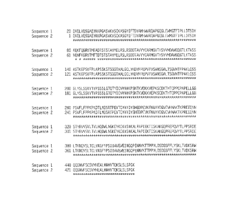
BRIEF DESCRIPTION OF THE FIGURE
Figure 1. Alignment of SEQ ID NO:11 of the instant application against SEQ ID
NO:14 of
W02008/076321.
DETAILED DESCRIPTION
As used herein, including the appended claims, the singular forms of words
such as "a,"
"an," and "the," include their corresponding plural references unless the
context clearly dictates
otherwise.
I. Definitions
"Activation," "stimulation," and "treatment," as it applies to cells or to
receptors, may
have the same meaning, e.g., activation, stimulation, or treatment of a cell
or receptor with a
ligand, unless indicated otherwise by the context or explicitly. "Ligand"
encompasses natural
and synthetic ligands, e.g., cytokines, cytokine variants, analogues, muteins,
and binding
compositions derived from antibodies. "Ligand" also encompasses small
molecules, e.g.,
peptide mimetics of cytokines and peptide mimetics of antibodies. "Activation"
can refer to
cell activation as regulated by internal mechanisms as well as by external or
environmental
factors. "Response," e.g., of a cell, tissue, organ, or organism, encompasses
a change in
biochemical or physiological behavior, e.g., concentration, density, adhesion,
or migration
within a biological compartment, rate of gene expression, or state of
differentiation, where the
change is correlated with activation, stimulation, or treatment, or with
internal mechanisms such
as genetic programming.
"Activity" of a molecule may describe or refer to the binding of the molecule
to a ligand
or to a receptor, to catalytic activity; to the ability to stimulate gene
expression or cell signaling,
differentiation, or maturation; to antigenic activity, to the modulation of
activities of other
molecules, and the like. "Activity" can also mean specific activity, e.g.,
[catalytic activity]/[mg
protein], or [immunological activity]/[mg protein], concentration in a
biological compartment,
or the like.
"Administration" and "treatment," as it applies to an animal, human,
experimental
subject, cell, tissue, organ, or biological fluid, refers to contact of an
exogenous pharmaceutical,
therapeutic, diagnostic agent, or composition to the animal, human, subject,
cell, tissue, organ,
CA 02779384 2017-01-25
7
or biological fluid.
"Administration" and "treatment" can refer, e.g., to therapeutic,
pharmacolcinetic, diagnostic, research, and experimental methods. Treatment of
a cell
encompasses contact of a reagent to the cell, as well as contact of a reagent
to a fluid, where the
fluid is in contact with the cell. "Administration" and "treatment" also means
in vitro and ex
vivo treatments, e.g., of a cell, by a reagent, diagnostic, binding
composition, or by another cell.
"Treatment," as it applies to a human, veterinary, or research subject, refers
to therapeutic
treatment, prophylactic or preventative measures, to research and diagnostic
applications.
"Binding compound" refers to a molecule that comprises one or more amino acid
sequences that specifically bind to human TSLP. In one preferred embodiment,
the binding
compound is an antibody, preferably an isolated antibody. In another preferred
embodiment,
the binding compound comprises an antigen-binding fragment of an antibody.
"Binding composition" refers to a TSLP-binding compound in combination with a
stabilizer, excipient, salt, buffer, solvent, or additive, capable of binding
to a target.
The scope of the present invention also includes complexes comprising any
antibody or
antigen-binding fragment thereof of the present invention complexed with TSLP
polypeptide or
an antigenic fragment thereof. Complexes may be prepared by contacting the
antibody or
fragment with the TSLP polypeptide or antigen fragment.
As used herein, the term "antibody" refers to any form of antibody or fragment
thereof
that exhibits the desired biological activity. Thus, it is used in the
broadest sense and
specifically covers monoclonal antibodies (including full length monoclonal
antibodies),
polyclonal antibodies, multispecific antibodies (e.g., bispecific antibodies),
and antibody
fragments so long as they exhibit the desired biological activity. "Isolated
antibody" refers to
the purification status of a binding compound and in such context means the
molecule is
substantially free of other biological molecules such as nucleic acids,
proteins, lipids,
carbohydrates, or other material such as cellular debris and growth media.
Generally, the term
"isolated" is not intended to refer to a complete absence of such material or
to an absence of
water, buffers, or salts, unless they are present in amounts that
substantially interfere with
experimental or therapeutic use of the binding compound as described herein.
A ''Fab fragment" is comprised of one light chain and the CH1 and variable
regions of
one heavy chain. The heavy chain of a Fab molecule cannot form a disulfide
bond with another
heavy chain molecule.
An "Fc" region contains two heavy chain fragments comprising the CH2 and CH3
domains of an antibody. The two heavy chain fragments are held together by two
or more
disulfide bonds and by hydrophobic interactions of the CH3 domains.
CA 02779384 2017-01-25
8
A "Fab' fragment" contains one light chain and a portion or fragment of one
heavy chain
that contains the VH domain and the CH1 domain and also the region between the
CH1 and
CH2 domains, such that an interchain disulfide bond can be formed between the
two heavy
chains of two Fab' fragments to form a F(ab') 2 molecule.
A "F(ab')2 fragment" contains two light chains and two heavy chains containing
a
portion of the constant region between the CH1 and CH2 domains, such that an
interchain
disulfide bond is formed between the two heavy chains. A F(ab') 2 fragment
thus is composed
of two Fab' fragments that are held together by a disulfide bond between the
two heavy chains.
The "Fy region" comprises the variable regions from both the heavy and light
chains, but
lacks the constant regions.
As used herein, the term "TSLP binding fragment" or "binding fragment thereof'
encompasses a fragment or a derivative of an antibody (or another binding
substance) that still
substantially retain its biological activity of inhibiting TSLP activity.
Therefore, the term
"antibody fragment" or TSLP binding fragment refers to a portion of a full
length antibody,
generally the antigen binding or variable region thereof. Examples of antibody
fragments
include Fab, Fab', F(ab')2, and Fv fragments; diabodies; linear antibodies;
single-chain antibody
molecules, e.g., sc-Fv; domain antibodies; and multispecific antibodies formed
from antibody
fragments. Typically, a binding fragment or derivative retains at least 10% of
its TSLP
inhibitory activity. Preferably, a binding fragment or derivative retains at
least 25%, 50%, 60%,
70%, 80%, 90%, 95%, 99% or 100% (or more) of its TSLP inhibitory activity,
although any
binding fragment with sufficient affinity to exert the desired biological
effect will be useful. It
is also intended that a TSLP binding fragment can include conservative amino
acid substitutions
that do not substantially alter its biologic activity.
The term "monoclonal antibody", as used herein, refers to an antibody obtained
from a
population of substantially homogeneous antibodies, i.e., the individual
antibodies comprising
the population are identical except for possible naturally occurring mutations
that may be
present in minor amounts. Monoclonal antibodies are highly specific, being
directed against a
single antigenic epitope. In contrast, conventional (polyclonal) antibody
preparations typically
include a multitude of antibodies directed against (or specific for) different
epitopes. The
modifier "monoclonal" indicates the character of the antibody as being
obtained from a
substantially homogeneous population of antibodies, and is not to be construed
as requiring
production of the antibody by any particular method. For example, the
monoclonal antibodies
to be used in accordance with the present invention may be made by the
hybridoma method first
described by Kohler et al., (1975) Nature 256: 495, or may be made by
recombinant DNA
CA 02779384 2017-01-25
9
methods (see, e.g., U.S. Pat. No. 4,816,567). The "monoclonal antibodies" may
also be isolated
from phage antibody libraries using the techniques described in Clackson et
al., (1991) Nature
352: 624-628 and Marks et al., (1991) J. Mol. Biol. 222: 581-597, for example.
The monoclonal antibodies herein specifically include "chimeric" antibodies
(immunoglobulins) in which a portion of the heavy and/or light chain is
identical with or
homologous to corresponding sequences in antibodies derived from a particular
species or
belonging to a particular antibody class or subclass, while the remainder of
the chain(s) is
identical with or homologous to corresponding sequences in antibodies derived
from another
species or belonging to another antibody class or subclass, as well as
fragments of such
antibodies, so long as they exhibit the desired biological activity (U.S. Pat.
No. 4,816,567; and
Morrison et al., (1984) Proc. Natl. Acad Sci. USA 81: 6851-6855).
A "domain antibody" is an immunologically functional immunoglobulin fragment
containing only the variable region of a heavy chain or the variable region of
a light chain. In
some instances, two or more VH regions are covalently joined with a peptide
linker to create a
bivalent domain antibody. The two VH regions of a bivalent domain antibody may
target the
same or different antigens.
A "bivalent antibody" comprises two antigen binding sites. In some instances,
the two
binding sites have the same antigen specificities. However, bivalent
antibodies may be
bi specific .
As used herein, the term "single-chain Fv" or "scFv" antibody refers to
antibody
fragments comprising the VH and VL domains of antibody, wherein these domains
are present in
a single polypeptide chain. Generally, the Fv polypeptide further comprises a
polypeptide linker
between the VH and VL domains which enables the sFv to form the desired
structure for antigen
binding. For a review of sFv, see Pluckthun (1994) THE PHARMACOLOGY OF
MONOCLONAL
ANTIBODIES, vol. 113, Rosenburg and Moore eds. Springer-Verlag, New York, pp.
269-315.
The monoclonal antibodies herein also include camelized single domain
antibodies.
See, e.g., Muyldermans et al. (2001) Trends Biochem. Sci. 26:230; Reichmann et
al. (1999) J.
Immunol. Methods 231:25; WO 94/04678; WO 94/25591; U.S. Pat. No. 6,005,079).
In one embodiment, the present invention
provides single domain antibodies comprising two VH domains with modifications
such that
single domain antibodies are formed.
As used herein, the term "diabodies" refers to small antibody fragments with
two
antigen-binding sites, which fragments comprise a heavy chain variable domain
(VH) connected
to a light chain variable domain (VL) in the same polypeptide chain (VH-VL or
VL-VH). By
CA 02779384 2017-01-25
using a linker that is too short to allow pairing between the two domains on
the same chain, the
domains are forced to pair with the complementary domains of another chain and
create two
antigen-binding sites. Diabodies are described more fully in, e.g., EP
404,097; WO 93/11161;
and Holliger et al. (1993) Proc. Natl. Acad. Sci. USA 90: 6444-6448. For a
review of
engineered antibody variants generally see Holliger and Hudson (2005) Nat.
Biotechnol.
23:1126-1136.
As used herein, the term "humanized antibody" refers to forms of antibodies
that contain
sequences from non-human (e.g., murine) antibodies as well as human
antibodies. Such
antibodies contain minimal sequence derived from non-human immunoglobulin. In
general, the
humanized antibody will comprise substantially all of at least one, and
typically two, variable
domains, in which all or substantially all of the hypervariable loops
correspond to those of a
non-human immunoglobulin and all or substantially all of the FR regions are
those of a human
immunoglobulin sequence. The humanized antibody optionally also will comprise
at least a
portion of an immunoglobulin constant region (Fc), typically that of a human
immunoglobulin.
The prefix "h", "hu" or "hum" is added to antibody clone designations when
necessary to
distinguish humanized antibodies (e.g., "hu23B12") from parental rodent
antibodies (e.g., rat
23B12, or "r23B12"). The humanized forms of rodent antibodies will generally
comprise the
same CDR sequences of the parental rodent antibodies, although certain amino
acid
substitutions may be included to increase affinity or increase stability of
the humanized
antibody.
The antibodies of the present invention also include antibodies with modified
(or
blocked) Fc regions to provide altered effector functions. See, e.g., U.S.
Pat. No. 5,624,821;
W02003/086310; W02005/120571; W02006/0057702; Presta (2006) Adv. Drug Delivery
Rev.
58:640-656. Such modification can be used to enhance or suppress various
reactions of the
immune system, with possible beneficial effects in diagnosis and therapy.
Alterations of the Fc
region include amino acid changes (substitutions, deletions and insertions),
glycosylation or
deglycosylation, and adding multiple Fc. Changes to the Fc can also alter the
half-life of
antibodies in therapeutic antibodies, and a longer half-life would result in
less frequent dosing,
with the concomitant increased convenience and decreased use of material. See
Presta (2005) J.
Allergy Clin. Immunol.116:731 at 734-35.
The term "fully human antibody" refers to an antibody that comprises human
immunoglobulin protein sequences only. A fully human antibody may contain
murine
carbohydrate chains if produced in a mouse, in a mouse cell, or in a hybridoma
derived from a
CA 02779384 2017-01-25
11
mouse cell. Similarly, "mouse antibody" refers to an antibody which comprises
mouse
immunoglobulin sequences only.
As used herein, the term "hypervariable region" refers to the amino acid
residues of an
antibody that are responsible for antigen-binding. The hypervariable region
comprises amino
acid residues from a "complementarity determining region" or "CDR" (e.g.
residues 24-34
(CDRL1), 50-56 (CDRL2) and 89-97 (CDRL3) in the light chain variable domain
and residues
31-35 (CDRH1), 50-65 (CDRH2) and 95-102 (CDRH3) in the heavy chain variable
domain;
Kabat et al., (1991) Sequences of Proteins of Immunological Interest, 5th Ed.
Public Health
Service, National Institutes of Health, Bethesda, Md.) and/or those residues
from a
"hypervariable loop" (i.e. residues 26-32 (L1), 50-52 (L2) and 91-96 (L3) in
the light chain
variable domain and 26-32 (H1), 53-55 (H2) and 96-101 (H3) in the heavy chain
variable
domain; Chothia and Lesk, (1987) J. Mol. Biol. 196: 901-917). As used herein,
the term
"framework" or "FR" residues refers to those variable domain residues other
than the
hypervariable region residues defined herein as CDR residues. The residue
numbering above
relates to the Kabat numbering system and does not necessarily correspond in
detail to the
sequence numbering in the accompanying Sequence Listing.
"Binding" refers to an association of the binding composition with a target
where the
association results in reduction in the normal Brownian motion of the binding
composition, in
cases where the binding composition can be dissolved or suspended in solution.
"Conservatively modified variants" or "conservative substitution" refers to
substitutions
of amino acids are known to those of skill in this art and may be made
generally without
altering the biological activity of the resulting molecule. Those of skill in
this art recognize
that, in general, single amino acid substitutions in non-essential regions of
a polypeptide do not
substantially alter biological activity (see, e.g., Watson, et at., Molecular
Biology of the Gene,
The Benjamin/Cummings Pub. Co., p. 224 (4th Edition 1987)). Such exemplary
substitutions
are preferably made in accordance with those set forth in Table 1 as follows:
Table 1
Exemplary Conservative Amino Acid Substitutions
Original residue Conservative substitution
Ala (A) Gly; Ser
Arg (R) Lys, His
Asn (N) Gln; His
Asp (D) Glu; Asn
Cys (C) Ser; Ala
CA 02779384 2017-01-25
12
Original residueConservative substitution
Gln (Q) As n
Glu (E) Asp; Gin
Gly (G) Ala
His (H) Asn; Gin
Ile (I) Leu; Val
Leu (L) Ile; Val
Lys (K) Arg; His
Met (M) Leu; He; Tyr
Phe (F) Tyr; Met; Leu
Pro (P) Ala
Ser (S) Thr
Thr (T) Ser
Trp (W) Tyr; Phe
Tyr (Y) Trp; Phe
Val (V) Ile; Leu
"Effective amount" encompasses an amount sufficient to ameliorate or prevent a
symptom or sign of the medical condition. Effective amount also means an
amount sufficient to
allow or facilitate diagnosis. An effective amount for a particular patient or
veterinary subject
may vary depending on factors such as the condition being treated, the overall
health of the
patient, the method route and dose of administration and the severity of side
affects (see, e.g.,
U.S. Pat. No. 5,888,530 issued to Netti, et al.). An effective amount can be
the maximal dose
or dosing protocol that avoids significant side effects or toxic effects. The
effect will result in
an improvement of a diagnostic measure or parameter by at least 5%, usually by
at least 10%,
more usually at least 20%, most usually at least 30%, preferably at least 40%,
more preferably at
least 50%, most preferably at least 60%, ideally at least 70%, more ideally at
least 80%, and
most ideally at least 90%, where 100% is defined as the diagnostic parameter
shown by a
normal subject (see, e.g., Maynard, et al. (1996) A Handbook of SOPs for Good
Clinical
Practice, Interpharm Press, Boca Raton, FL; Dent (2001) Good Laboratory and
Good Clinical
Practice, Urch Publ., London, UK).
As used herein, the term "isolated nucleic acid molecule" refers to a nucleic
acid
molecule that is identified and separated from at least one contaminant
nucleic acid molecule
with which it is ordinarily associated in the natural source of the antibody
nucleic acid. An
isolated nucleic acid molecule is other than in the form or setting in which
it is found in nature.
Isolated nucleic acid molecules therefore are distinguished from the nucleic
acid molecule as it
CA 02779384 2017-01-25
13
exists in natural cells. However, an isolated nucleic acid molecule includes a
nucleic acid
molecule contained in cells that ordinarily express the antibody where, for
example, the nucleic
acid molecule is in a chromosomal location different from that of natural
cells.
The expression "control sequences" refers to DNA sequences necessary for the
expression of an operably linked coding sequence in a particular host
organism. The control
sequences that are suitable for prokaryotes, for example, include a promoter,
optionally an
operator sequence, and a ribosome binding site. Eukaryotic cells are known to
utilize
promoters, polyadenylation signals, and enhancers.
A nucleic acid is "operably linked" when it is placed into a functional
relationship with
another nucleic acid sequence. For example, DNA for a presequence or secretory
leader is
operably linked to DNA for a polypeptide if it is expressed as a preprotein
that participates in
the secretion of the polypeptide; a promoter or enhancer is operably linked to
a coding sequence
if it affects the transcription of the sequence; or a ribosome binding site is
operably linked to a
coding sequence if it is positioned so as to facilitate translation.
Generally, "operably linked"
means that the DNA sequences being linked are contiguous, and, in the case of
a secretory
leader, contiguous and in reading phase. However, enhancers do not have to be
contiguous.
Linking is accomplished by ligation at convenient restriction sites. If such
sites do not exist, the
synthetic oligonucleotide adaptors or linkers are used in accordance with
conventional practice.
As used herein, the expressions "cell," "cell line," and "cell culture" are
used
interchangeably and all such designations include progeny. Thus, the words
"transformants"
and "transformed cells" include the primary subject cell and cultures derived
therefrom without
regard for the number of transfers. It is also understood that all progeny may
not be precisely
identical in DNA content, due to deliberate or inadvertent mutations. Mutant
progeny that have
the same function or biological activity as screened for in the originally
transformed cell are
included. Where distinct designations are intended, it will be clear from the
context.
As used herein, "polymerase chain reaction" or "PCR" refers to a procedure or
technique
in which minute amounts of a specific piece of nucleic acid, RNA and/or DNA,
are amplified as
described in, e.g., U.S. Pat. No. 4,683,195. Generally, sequence information
from the ends of
the region of interest or beyond needs to be available, such that
oligonucleotide primers can be
designed; these primers will be identical or similar in sequence to opposite
strands of the
template to be amplified. The 5' terminal nucleotides of the two primers can
coincide with the
ends of the amplified material. PCR can be used to amplify specific RNA
sequences, specific
DNA sequences from total genomic DNA, and cDNA transcribed from total cellular
RNA,
bacteriophage or plasmid sequences, etc. See generally Mullis et al. (1987)
Cold Spring Harbor
CA 02779384 2017-01-25
14
Symp. Quant. Biol. 51:263; Erlich, ed., (1989) PCR TECHNOLOGY (Stockton Press,
N.Y.) As
used herein, PCR is considered to be one, but not the only, example of a
nucleic acid
polymerase reaction method for amplifying a nucleic acid test sample
comprising the use of a
known nucleic acid as a primer and a nucleic acid polymerase to amplify or
generate a specific
piece of nucleic acid.
As used herein, the term "germline sequence" refers to a sequence of um-
earranged
immunoglobulin DNA sequences. Any suitable source of unrearranged
immunoglobulin DNA
may be used.
"Inhibitors" are compounds that decrease, block, prevent, delay activation,
inactivate,
desensitize, or down regulate, e.g., a gene, protein, ligand, receptor, or
cell. An inhibitor may
also be defined as a composition that reduces, blocks, or inactivates a
constitutive activity. An
"antagonist" is a compound that opposes the actions of an agonist. An
antagonist prevents,
reduces, inhibits, or neutralizes the activity of an agonist. An antagonist
can also prevent,
inhibit, or reduce constitutive activity of a target, e.g., a target receptor,
even where there is no
identified agonist.
To examine the extent of inhibition, for example, samples or assays comprising
a given,
e.g., protein, gene, cell, or organism, are treated with a potential
activating or inhibiting agent
and are compared to control samples without the agent. Control samples, i.e.,
not treated with
agent, are assigned a relative activity value of 100%. Inhibition is achieved
when the activity
value relative to the control is about 90% or less, typically 85% or less,
more typically 80% or
less, most typically 75% or less, generally 70% or less, more generally 65% or
less, most
generally 60% or less, typically 55% or less, usually 50% or less, more
usually 45% or less,
most usually 40% or less, preferably 35% or less, more preferably 30% or less,
still more
preferably 25% or less, and most preferably less than 25%.
Endpoints in inhibition can be monitored as follows. Inhibition, and response
to
treatment, e.g., of a cell, physiological fluid, tissue, organ, and animal or
human subject, can be
monitored by an endpoint. The endpoint may comprise a predetermined quantity
or percentage
of, e.g., an indicia of inflammation, oncogenicity, or cell degranulation or
secretion, such as the
release of a cytokine, toxic oxygen, or a protease. The endpoint may comprise,
e.g., a
predetermined quantity of ion flux or transport; cell migration; cell
adhesion; cell proliferation;
potential for metastasis; cell differentiation; and change in phenotype, e.g.,
change in expression
of gene relating to inflammation, apoptosis, transformation, cell cycle, or
metastasis (see, e.g.,
Knight (2000) Ann. Clin. Lab. Sci. 30:145-158; Hood and Cheresh (2002) Nature
Rev. Cancer
2:91-100; Timme, et al. (2003) Carr. Drug Targets 4:251-261; Robbins and
Itzkowitz (2002)
CA 02779384 2017-01-25
Med. Gin. North Am. 86:1467-1495; Grady and Markowitz (2002) Annu. Rev.
Genomics Hum.
Genet. 3:101-128; Bauer, et at. (2001) Glia 36:235-243; Stanimirovic and Satoh
(2000) Brain
Pathol. 10:113-126).
An endpoint of inhibition is generally 75% of the control or less, preferably
50% of the
control or less, more preferably 25% of the control or less, and most
preferably 10% of the
control or less. Generally, an endpoint of activation is at least 150% the
control, preferably at
least two times the control, more preferably at least four times the control,
and most preferably
at least 10 times the control.
"Specifically" or "selectively" binds, when referring to a ligand/receptor,
antibody/antigen, or other binding pair, indicates a binding reaction which is
determinative of
the presence of the protein, e.g., TSLP, in a heterogeneous population of
proteins and/or other
biologics. Thus, under designated conditions, a specified ligand/antigen binds
to a particular
receptor/antibody and does not bind in a significant amount to other proteins
present in the
sample.
The antibody, or binding composition derived from the antigen-binding site of
an
antibody, of the contemplated method binds to its antigen with an affinity
that is at least ten
times greater, more preferably at least 20-times greater, and most preferably
at least 50-times
greater than the affinity with unrelated antigens. In a preferred embodiment
the antibody will
have an affinity that is greater than about 109 liters/mol, as determined,
e.g., by Scatchard
analysis (Munsen, et al. (1980) Analyt. Biochem. 107:220-239).
As used herein, the term "inflammatory disorder" refers to any disease or
disorder
characterized by local inflammation at a site of injury or infection and
includes, without
limitation, allergic inflammation, autoimmune diseases, and other disorders
characterized by
undesired immune cell accumulation at a local tissue site.
As used herein, the term "immunomodulatory agent" refers to natural or
synthetic agents
that suppress or modulate an immune response. The immune response can be a
humoral or
cellular response.
Immunomodulatory agents encompass immunosuppressive or anti-
inflammatory agents.
"Immunosuppressive agents," "immunosuppressive drugs," or "immunosuppressants"
as
used herein are therapeutics that are used in immunosuppressive therapy to
inhibit or prevent
activity of the immune system. Clinically they are used to prevent the
rejection of transplanted
organs and tissues (e.g. bone marrow, heart, kidney, liver), and/or in the
treatment of
autoimmune diseases or diseases that are most likely of autoimmune origin
(e.g. rheumatoid
arthritis, myasthenia gravis, systemic lupus erythematosus, ulcerative
colitis, multiple sclerosis).
CA 02779384 2017-01-25
16
Immunosuppressive drugs can be classified into four groups: glucocorticoids
cytostatics;
antibodies (including Biological Response Modifiers or DMARDs); drugs acting
on
immunophilins; other drugs, including known chemotherpeutic agents used in the
treatment of
proliferative disorders. For
multiple sclerosis, in particular, the antibodies of the present
invention can be administered in conjunction with a new class of myelin
binding protein-like
therapeutics, known as copaxones.
"Anti-inflammatory agents" or "anti-inflammatory drugs", is used to represent
both
steroidal and non-steroidal therapeutics. Steroids, also known as
corticosteroids, are drugs that
closely resemble cortisol, a hormone produced naturally by adrenal glands.
Steroids are used as
the main treatment for certain inflammatory conditions, such as: Systemic
vasculitis
(inflammation of blood vessels); and Myositis (inflammation of muscle).
Steroids might also
be used selectively to treat inflammatory conditions such as: rheumatoid
arthritis (chronic
inflammatory arthritis occurring in joints on both sides of the body);
systemic lupus
erythematosus (a generalized disease caused by abnormal immune system
function); SjOgren's
syndrome (chronic disorder that causes dry eyes and a dry mouth).
Non-steroidal anti-inflammatory drugs, usually abbreviated to NSAIDs, are
drugs with
analgesic, antipyretic and anti-inflammatory effects - they reduce pain, fever
and inflammation.
The term "non-steroidal" is used to distinguish these drugs from steroids,
which (amongst a
broad range of other effects) have a similar eicosanoid-depressing, anti-
inflammatory action.
NSAIDs are generally indicated for the symptomatic relief of the following
conditions:
rheumatoid arthritis; osteoarthritis; inflammatory arthropathies (e.g.
ankylosing spondylitis,
psoriatic arthritis, Reiter's syndrome); acute gout ; dysmenorrhoea;
metastatic bone pain;
headache and migraine; postoperative pain ; mild-to-moderate pain due to
inflammation and
tissue injury; pyrexia; and renal colic. NSAIDs include salicylates,
arlyalknoic acids, 2-
arylpropionic acids (profens), N-arylanthranilic acids (fenamic acids),
oxicams, coxibs
(selective COX-2 inhibitors), sulphonanilides, diclofenac, diflunisal,
etodolac, fenoprofen,
flurbiprofen, ibuprofen, indomethacin, ketoprofen, ketorolac, mefenamic acid,
meloxicam,
nabumetone, naproxen, oxaprozin, piroxicam, salsalate, sulindac or tolmetin
II. General
The present invention provides engineered anti-TSLP antibodies and uses
thereof to
treat inflammatory, and particularly allergic inflammatory, disorders. In a
preferred
embodiment, the inflammatory disorder is asthma. In a preferred embodiment,
the allergic
inflammatory disorder is allergic rhinosinusitis, allergic asthma, allergic
conjunctivitis, or atopic
CA 02779384 2017-01-25
17
dermatitis. The present invention also provides engineered anti-TSLP
antibodies to treat
fibrosis, inflammatory bowel disease or Hodgkin's lymphoma.
As used herin, the term "TSLP" includes variants, isoforms, homologs,
orthologs and
paralogs of TSLP. The amino acid sequence of human TSLP is set forth in SEQ ID
NO:4 of
International Publication No. W000/17362.
III. Engineered TSLP Specific Antibodies of the Invention
The invention relates to engineered anti-TSLP antibodies comprising specified
CDR
regions.
Methods for recombinantly engineering antibodies have been described, e.g., by
Boss et
al. (U.S. Pat. No. 4,816,397), Cabilly et al. (U.S. Pat. No. 4,816,567), Law
et al. (European
Patent Application Publication No. 438 310) and Winter (European Patent
Application
Publication No. 239400).
Engineered antibodies of the invention include those in which modifications
have been
made to framework residues within VH and/or VL, e.g. to improve the properties
of the
antibody. Typically such framework modifications are made to decrease the
immunogenicity of
the antibody. For example, one approach is to "backmutate" one or more
framework residues to
the corresponding germline sequence. More specifically, an antibody that has
undergone
somatic mutation can contain framework residues that differ from the germline
sequence from
which the antibody is derived. Such residues can be identified by comparing
the antibody
framework sequences to the germline sequences from which the antibody is
derived.
Another type of framework modification involves mutating one or more residues
within
the framework region, or even within one or more CDR regions, to remove T cell
epitopes to
thereby reduce the potential immunogenicity of the antibody. This approach is
also referred to
as "deimmunization" and is described in further detail in U.S. Patent
Publication No.
20030153043.
In addition or alternative to modifications made within the framework or CDR
regions,
antibodies of the invention can be engineered to include modifications within
the Fc region,
typically to alter one or more functional properties of the antibody, such as
serum half-life,
complement fixation, Fc receptor binding, and/or antigen-dependent cellular
cytotoxicity.
Furthermore, an antibody of the invention can be chemically modified (e.g.,
one or more
chemical moieties can be attached to the antibody) or be modified to alter its
glycosylation,
again to alter one or more functional properties of the antibody. Each of
these embodiments is
CA 02779384 2017-01-25
18
described in further detail below. The numbering of residues in the Fc region
is that of the EU
index of Kabat.
In one embodiment, the antibody is an IgG4 isotype antibody comprising a
Serine to
Proline mutation at a position corresponding to position 228 (S228P; EU index)
in the hinge
region of the heavy chain constant region. This mutation has been reported to
abolish the
heterogeneity of inter-heavy chain disulfide bridges in the hinge region
(Angal et al. supra;
position 241 is based on the Kabat numbering system.
In one embodiment, the hinge region of CH1 is modified such that the number of
cysteine residues in the hinge region is altered, e.g., increased or
decreased. This approach is
described further in U.S. Patent No. 5,677,425. The number of cysteine
residues in the hinge
region of CH1 is altered to, for example, facilitate assembly of the light and
heavy chains or to
increase or decrease the stability of the antibody.
In another embodiment, the Fc hinge region of an antibody is mutated to
decrease the
biological half life of the antibody. More specifically, one or more amino
acid mutations are
introduced into the CH2-CH3 domain interface region of the Fc-hinge fragment
such that the
antibody has impaired Staphylococcyl protein A (SpA) binding relative to
native Fc-hinge
domain SpA binding. This approach is described in further detail in U.S.
Patent No. 6,165,745.
In another embodiment, the antibody is modified to increase its biological
half life.
Various approaches are possible. For example, one or more of the following
mutations can be
introduced: T252L, T254S, T256F, as described in U.S. Patent No. 6,277,375.
Alternatively, to
increase the biological half life, the antibody can be altered within the CH1
or CL region to
contain a salvage receptor binding epitope taken from two loops of a CH2
domain of an Fc
region of an IgG, as described in U.S. Patent Nos. 5,869,046 and 6,121,022.
In yet other embodiments, the Fc region is altered by replacing at least one
amino acid
residue with a different amino acid residue to alter the effector function(s)
of the antibody. For
example, one or more amino acids selected from amino acid residues 234, 235,
236, 237, 297,
318, 320 and 322 can be replaced with a different amino acid residue such that
the antibody has
an altered affinity for an effector ligand but retains the antigen-binding
ability of the parent
antibody. The effector ligand to which affinity is altered can be, for
example, an Fc receptor or
the Cl component of complement. This approach is described in further detail
in U.S. Patent
Nos. 5,624,821 and 5,648,260.
In another example, one or more amino acids selected from amino acid residues
329,
331 and 322 can be replaced with a different amino acid residue such that the
antibody has
CA 02779384 2017-01-25
19
altered Clq binding and/or reduced or abolished complement dependent
cytotoxicity (CDC).
This approach is described in further detail in U.S. Patent No. 6,194,551.
In another example, one or more amino acid residues within amino acid
positions 231
and 239 are altered to thereby alter the ability of the antibody to fix
complement. This approach
is described further in PCT Publication WO 94/29351.
In yet another example, the Fc region is modified to increase the ability of
the antibody
to mediate antibody dependent cellular cytotoxicity (ADCC) and/or to increase
the affinity of
the antibody for an Fey receptor by modifying one or more amino acids at the
following
positions: 238, 239, 248, 249, 252, 254, 255, 256, 258, 265, 267, 268, 269,
270, 272, 276, 278,
280, 283, 285, 286, 289, 290, 292, 293, 294, 295, 296, 298, 301, 303, 305,
307, 309, 312, 315,
320, 322, 324, 326, 327, 329, 330, 331, 333, 334, 335, 337, 338, 340, 360,
373, 376, 378, 382,
388, 389, 398, 414, 416, 419, 430, 434, 435, 437, 438 or 439. This approach is
described
further in PCT Publication WO 00/42072. Moreover, the binding sites on human
IgG1 for
FcyR11, FcyRIII and FcRn have been mapped and variants with improved binding
have
been described (see Shields et al. (2001) J. Biol. Chem. 276:6591-6604).
Specific mutations at
positions 256, 290, 298, 333, 334 and 339 were shown to improve binding to
FcyRIII.
Additionally, the following combination mutants were shown to improve FcyRIII
binding:
T256A/S298A, S298A/E333A, S298A/K224A and S298A/E333A/K334A.
In still another embodiment, the glycosylation of an antibody is modified or
altered, to
delete or add carbohydrate moieties to the antibodies. For example, an
aglycoslated antibody
can be made (i.e., the antibody lacks glycosylation). Glycosylation can be
altered to, for
example, increase the affinity of the antibody for antigen. Such carbohydrate
modifications can
be accomplished by, for example, altering one or more sites of glycosylation
within the antibody
sequence. For example, one or more amino acid substitutions can be made that
result in
elimination of one or more variable region framework glycosylation sites to
thereby eliminate
glycosylation at that site. Such aglycosylation may increase the affinity of
the antibody for
antigen. See, e.g., U.S. Patent Nos. 5,714,350 and 6,350,861.
Additionally or alternatively, an antibody can be made that has an altered
type of
glycosylation, such as a hypofucosylated antibody having reduced amounts of
fucosyl residues
or an antibody having increased bisecting GlcNac structures. Such altered
glycosylation
patterns have been demonstrated to increase the ADCC ability of antibodies.
Such carbohydrate
modifications can be accomplished by, for example, expressing the antibody in
a host cell with
altered glycosylation machinery. Cells with altered glycosylation machinery
have been
described in the art and can be used as host cells in which to express
recombinant antibodies of
CA 02779384 2017-01-25
the invention to thereby produce an antibody with altered glycosylation. For
example, the cell
lines Ms704, Ms705, and Ms709 lack the fucosyltransferase gene, FUT8 (a (1,6)-
fucosyltransferase), such that antibodies expressed in the Ms704, Ms705, and
Ms709 cell lines
lack fucose on their carbohydrates. The Ms704, Ms705, and Ms709 FUT8-/- cell
lines were
created by the targeted disruption of the FUT8 gene in CHO/DG44 cells using
two replacement
vectors (see U.S. Patent Publication No. 20040110704 and Yamane-Ohnuki et al.
(2004)
Biotechnol Bioeng 87:614-22). As another example, EP 1,176,195 describes a
cell line with a
functionally disrupted FUT8 gene, which encodes a fucosyl transferase, such
that antibodies
expressed in such a cell line exhibit hypofucosylation by reducing or
eliminating the a-1,6
bond-related enzyme. EP 1,176,195 also describes cell lines which have a low
enzyme activity
for adding fucose to the N-acetylglucosamine that binds to the Fc region of
the antibody or does
not have the enzyme activity, for example the rat myeloma cell line YB2/0
(ATCC CRL 1662).
PCT Publication WO 03/035835 describes a variant CHO cell line, Lec13 cells,
with reduced
ability to attach fucose to Asn(297)-linked carbohydrates, also resulting in
hypofucosylation of
antibodies expressed in that host cell (see also Shields et al. (2002) J.
Biol. Chem. 277:26733-
26740). Antibodies with a modified glycosylation profile can also be produced
in chicken eggs,
as described in PCT Publication WO 06/089231. Alternatively, antibodies with a
modified
glycosylation profile can be produced in plant cells, such as Lemma. PCT
Publication WO
99/54342 describes cell lines engineered to express glycoprotein-modifying
glycosyl
transferases (e.g., 13(1,4)-N-acetylglucosaminyltransferase Ill (GnTIII)) such
that antibodies
expressed in the engineered cell lines exhibit increased bisecting GlcNac
structures which
results in increased ADCC activity of the antibodies (see also Umana et al.
(1999) Nat. Biotech.
17:176-180). Alternatively, the fucose residues of the antibody can be cleaved
off using a
fucosidase enzyme; e.g., the fucosidase a-L-fucosidase removes fucosyl
residues from
antibodies (Tarentino et al. (1975) Biochem. 14:5516-23).
Another modification of the antibodies herein that is contemplated by this
disclosure is
pegylation. An antibody can be pegylated to, for example, increase the
biological (e.g., serum)
half life of the antibody. To pegylate an antibody, the antibody, or fragment
thereof, typically is
reacted with polyethylene glycol (PEG), such as a reactive ester or aldehyde
derivative of PEG,
under conditions in which one or more PEG groups become attached to the
antibody or antibody
fragment. Preferably, the pegylation is carried out via an acylation reaction
or an alkylation
reaction with a reactive PEG molecule (or an analogous reactive water-soluble
polymer). As
used herein, the term "polyethylene glycol" is intended to encompass any of
the forms of PEG
that have been used to derivatize other proteins, such as mono (CI-CIO) alkoxy-
or aryloxy-
CA 02779384 2017-01-25
21
polyethylene glycol or polyethylene glycol-maleimide. In certain embodiments,
the antibody to
be pegylated is an aglycosylated antibody. Methods for pegylating proteins are
known in the art
and can be applied to the antibodies of the invention. See, e.g., EP 0 154 316
and EP 0 401
384.
Amino acid sequence variants of humanized anti-TSLP antibody of the invention
can be
prepared by introducing appropriate nucleotide changes into the humanized anti-
TSLP antibody
DNA, or by peptide synthesis. Such variants include, for example, deletions
from, and/or
insertions into and/or substitutions of, residues within the amino acid
sequences shown for the
humanized anti-TSLP antibodies disclosed and claimed herein. Any combination
of deletion,
insertion, and substitution can be made to arrive at the final construct,
provided that the final
construct possesses the desired characteristics. As discussed above, the amino
acid changes
also may alter post-translational processes of the humanized anti-TSLP
antibody, such as
changing the number or position of glycosylation sites.
A useful method for identification of certain residues or regions of the
humanized anti-
TSLP antibody polypeptide that are preferred locations for mutagenesis is
called "alanine
scanning mutagenesis," as described by Cunningham and Wells (1989) Science
244: 1081-1085.
Here, a residue or group of target residues are identified (e.g., charged
residues such as Arg,
Asp, His, Lys, and Glu) and replaced by a neutral or negatively charged amino
acid (most
preferably alanine or polyalanine) to affect the interaction of the amino
acids with TSLP
antigen. The amino acid residues demonstrating functional sensitivity to the
substitutions then
are refined by introducing further or other variants at, or for, the sites of
substitution. Thus,
while the site for introducing an amino acid sequence variation is
predetermined, the nature of
the mutation per se need not be predetermined. For example, to analyze the
performance of a
mutation at a given site, Ala scanning or random mutagenesis is conducted at
the target codon
or region and the expressed humanized anti-TSLP antibody variants are screened
for the desired
activity.
Amino acid sequence insertions include amino- and/or carboxyl-terminal fusions
ranging in length from one residue to polypeptides containing a hundred or
more residues, as
well as intrasequence insertions of single or multiple amino acid residues.
Examples of
terminal insertions include humanized anti-TSLP antibody with an N-terminal
methionyl
residue or the antibody fused to an epitope tag. Other insertional variants of
the humanized
anti-TSLP antibody molecule include the fusion to the N- or C-terminus of
humanized anti-
TSLP antibody of an enzyme or a polypeptide which increases the serum half-
life of the
antibody.
CA 02779384 2017-01-25
22
Another type of variant is an amino acid substitution variant. These variants
have at least
one amino acid residue in the humanized anti-TSLP antibody molecule removed
and a different
residue inserted in its place. The sites of greatest interest for
substitutional mutagenesis include
the hypervariable loops, but FR alterations are also contemplated.
Hypervariable region
residues or FR residues involved in antigen binding are generally substituted
in a relatively
conservative manner.
Yet another type of amino acid variant is the substitution of residues to
provide for
greater chemical stability of the final humanized antibody.
In certain embodiments, it will be desirable to change certain amino acids
containing
exposed side-chains to another amino acid residue in order to provide for
greater chemical
stability of the final antibody, as follows. For example, an asparagine (Asn)
residue may be
changed to Gin or Ala to reduce the potential for formation of isoaspartate at
any Asn-Gly
sequences within a CDR. A similar problem may occur at a Asp-Gly sequence.
Reissner and
Aswad (2003) Cell. MoL Life Sci. 60:1281. Isoaspartate formation may
debilitate or completely
abrogate binding of an antibody to its target antigen. See, Presta (2005) J.
Allergy Clin.
Irnmunol. 116:731 at 734. In one embodiment, the asparagine is changed to
glutamine (Gin). It
may also be desirable to alter an amino acid adjacent to an asparagine (Asn)
or glutamine (Gin)
residue to reduce the likelihood of dearnidation, which occurs at greater
rates when small amino
acids occur adjacent to asparagine or glutamine. See, Bischoff & Kolbe (1994)
J. Chromatog.
662:261. In addition, any methionine residues (typically solvent exposed Met)
in CDRs may be
changed to Lys, Leu, Ala, or Phe in order to reduce the possibility that the
methionine sulfur
would oxidize, which could reduce antigen binding affinity and also contribute
to molecular
heterogeneity in the final antibody preparation. Id. In one embodiment, the
methionine is
changed to alanine (Ala). Additionally, in order to prevent or minimize
potential scissile Asn-
Pro peptide bonds, it may be desirable to alter any Asn-Pro combinations found
in a CDR to
Gin-Pro, Ala-Pro, or Asn-Ala. Antibodies with such substitutions are
subsequently screened to
ensure that the substitutions do not decrease TSLP binding affinity or other
desired biological
activity to unacceptable levels.
CA 02779384 2017-01-25
23
TABLE 2
Exemplary stabilizing CDR variants
CDR Residue Stabilizing Variant
Sequence
Asn-Gly Gln-Gly, Ala-Gly, or Asn-Ala
(N-G) (Q-G), (A-G), or (N-A)
Asp-Gly Glu-Gly, Ala-Gly or Asp-Ala
(D-G) (E-G), (A-G), or (D-A)
Met (typically solvent exposed) Lys, Leu, Ala, or Phe
(M) (K), (L), (A), or (F)
Asn Gin or Ala
(N) (Q) or (A)
A sn-Pro Gln-Pro, Ala-Pro, or Asn-Ala
(N-P) (Q-P), (A-P), or (N-A)
In addition, methionine residues in rodent CDRs may be changed to reduce the
possibility that the methionine sulfur would oxidize, which could reduce
antigen binding
affinity and also contribute to molecular heterogeneity in the final antibody
preparation. Id. In
one embodiment, the methionine is changed to alanine (A). Antibodies with such
substitutions
are subsequently screened to ensure that the substitutions do not decrease
TSLP binding affinity
to unacceptable levels.
Nucleic acid molecules encoding amino acid sequence variants of humanized TSLP
specific antibody are prepared by a variety of methods known in the art. These
methods
include, but are not limited to, isolation from a natural source (in the case
of naturally occurring
amino acid sequence variants) or preparation by oligonucleotide-mediated (or
site-directed)
mutagenesis, PCR mutagenesis, and cassette mutagenesis of an earlier prepared
variant or a
non-variant version of humanized anti-TSLP antibody.
Ordinarily, amino acid sequence variants of the humanized anti-TSLP antibody
will
have an amino acid sequence having at least 97% amino acid sequence identity
with the original
humanized antibody amino acid sequences of either the heavy or the light chain
more preferably
at least 98%, more preferably at least 99%. Identity or homology with respect
to this sequence
is defined herein as the percentage of amino acid residues in the candidate
sequence that are
identical with the humanized anti-TSLP residues, after aligning the sequences
and introducing
gaps, if necessary, to achieve the maximum percent sequence identity, and not
considering any
conservative substitutions as part of the sequence identity. None of N-
terminal, C-terminal, or
internal extensions, deletions, or insertions into the antibody sequence shall
be construed as
affecting sequence identity or homology.
CA 02779384 2017-01-25
24
The humanized antibody can be selected from any class of immunoglobulins,
including
IgM, IgG, IgD, IgA, and IgE. Preferably, the antibody is an IgG antibody. Any
isotype of IgG
can be used, including IgGI, IgG2, IgG3, and IgG4. Variants of the IgG
isotypes are also
contemplated. The humanized antibody may comprise sequences from more than one
class or
isotype. Optimization of the necessary constant domain sequences to generate
the desired
biologic activity is readily achieved by screening the antibodies in the
biological assays
described below.
Likewise, either class of light chain can be used in the compositions and
methods herein.
Specifically, kappa, lambda, or variants thereof are useful in the present
compositions and
methods.
Any suitable portion of the CDR sequences from the non-human antibody can be
used.
The CDR sequences can be mutagenized by substitution, insertion or deletion of
at least one
residue such that the CDR sequence is distinct from the human and non-human
antibody
sequence employed. It is contemplated that such mutations would be minimal.
Typically, at
least 95% of the humanized antibody residues will correspond to those of the
non-human CDR
residues, and most preferably greater than 97%.
Any suitable portion of the FR sequences from the human antibody can be used.
The FR
sequences can be mutagenized by substitution, insertion or deletion of at
least one residue such
that the FR sequence is distinct from the human and non-human antibody
sequence employed.
It is contemplated that such mutations would be minimal. Typically, at least
75% of the
humanized antibody residues will correspond to those of the human FR residues,
more often
90%, and most preferably greater than 95%.
CDR and FR residues are determined according to the standard sequence
definition of
Kabat. Kabat et al., Sequences of Proteins of Immunological Interest, National
Institutes of
Health, Bethesda Md. (1987).
In a preferred embodiment, a binding composition according to the invention
comprises,
one or more of the following sequences:
= The CDR-H1 sequence GY1FTDYAMH (SEQ ID NO: 1).
= The CDR-H2 sequence TFIPLLDTSDYAQKFQG (SEQ ID NO: 2).
= The CDR-H3 sequence MGVTHSYVMDA (SEQ ID NO: 3).
= The CDR-L1 sequence RASQPISISVH (SEQ ID NO: 4).
= The CDR-L2 sequence FASQSIS (SEQ ID NO: 5).
= The CDR-L3 sequence QQTFSLPYT (SEQ ID NO: 6).
= The variable heavy chain amino acid sequence shown in SEQ ID NO:7.
CA 02779384 2017-01-25
= The variable light chain amino acid sequence shown in SEQ ID NO: 8.
= The nucleic acid sequence encoding the variable heavy chain shown in SEQ
ID
NO:9.
= The nucleic acid sequence encoding the variable light chain shown in SEQ
ID
NO:10.
= The heavy chain amino acid sequence shown in SEQ ID NO:11. This sequence
can further comprise the following leader
sequence:
MAVLGLLFCLVTFPSCVLS (SEQ ID NO:15).
= The light chain amino acid sequence shown in SEQ ID NO: 12. This sequence
can further comprise the following leader
sequence:
MAPVQLLGLLVLFLPAMRC (SEQ ID NO:16).
= The nucleic acid sequence encoding the heavy chain is shown in SEQ ID
NO:13.
This sequence can further comprise a sequence encoding a leader sequence,
preferably the following leader sequence: MAVLGLLFCLVTFPSCVLS (SEQ
ID NO:15).
= The nucleic acid sequence encoding the light chain is shown in SEQ ID
NO:14.
This sequence can further comprise a sequence encoding a leader sequence,
preferably the following leader sequence: MAPVQLLGLLVLFLPAMRC (SEQ
ID NO:16).
For example, the present invention includes an isolated antibody or antigen-
binding
fragment thereof comprising a light chain immunoglobulin comprising CDR-L1,
CDR-L2 and
CDR-L3 (as set forth above) and a heavy chain immunoglobulin comprising CDR-
H1, CDR-H2
and CDR-H3 (as set forth above). The present invention also includes an
isolated antibody or
antigen-binding fragment thereof comprising a light chain immunoglobulin
variable region
comprising the amino acid sequence set forth in SEQ ID NO: 8 or 12 and a heavy
chain
immunoglobulin variable region comprising the amino acid sequence set forth in
SEQ ID NO: 7
and 11 (e.g., SEQ ID NO: 7 paired with SEQ ID NO: 8; or, SEQ ID NO: 11 paired
with SEQ ID
NO: 12). Such an antibody or fragment can, in an embodiment of the invention,
be linked to an
immunoglobulin constant domain such as IgG (e.g., IgGI, IgG2, IgG3 or Igat). A
pharmaceutical composition thereof, comprising said antibody or fragment and a
pharmaceutically acceptable carrier, is also part of the present invention.
In some embodiments, different constant domains may be appended to the
humanized
VL and VH regions provided herein. For example, if a particular intended use
of an antibody (or
fragment) of the present invention were to call for altered effector
functions, a heavy chain
CA 02779384 2017-01-25
26
constant domain other than IgG1 may be used. Although IgG1 antibodies provide
for long half-
life and for effector functions, such as complement activation and antibody-
dependent cellular
cytotoxicity, such activities may not be desirable for all uses of the
antibody. In such instances
an IgG4 constant domain, for example, may be used.
IV. Antibody Conjugates
The binding compounds of the invention, for example the antibody or antibody
fragments of the invention, may also be conjugated to a chemical moiety. The
chemical moiety
may be, inter alia, a polymer, a radionuclide or a cytotoxic factor.
Preferably the chemical
moiety is a polymer which increases the half-life of the antibody molecule in
the body of a
subject. Suitable polymers include, but are not limited to, polyethylene
glycol (PEG) (e.g., PEG
with a molecular weight of 2kDa, 5 kDa, 10 kDa, 12kDa, 20 kDa, 30kDa or
40kDa), dextran
and monomethoxypolyethylene glycol (mPEG). Lee, et al., (1999) (Bioconj. Chem.
10:973-981)
discloses PEG conjugated single-chain antibodies. Wen, et al., (2001)
(Bioconj. Chem. 12:545-
553) disclose conjugating antibodies with PEG which is attached to a
radiometal chelator
(diethylenetriaminpentaacetic acid (DTPA)).
The antibodies and antibody fragments of the invention may also be conjugated
with
111/n, , 32p 14C, 1251, 3H, 1311, 11c, 150, 13N, , ,
, 18F 35s 51cr,
labels such as 99Tc,9 Y, 57T0,
226Ra,
60 -0,
C 59Fe,
57Se, 152Eu, 67c-u, 2170, 211At, 212pb, 47su, 109pd, 234Th, and 4 K, I57Gd,
55Mn, 52Tr
and 56Fe.
The antibodies and antibody fragments of the invention may also be conjugated
with
fluorescent or chemilluminescent labels, including fluorophores such as rare
earth chelates,
fluorescein and its derivatives, rhodamine and its derivatives,
isothiocyanate, phycoerythrin,
phycocyanin, allophycocyanin, o-phthaladehyde, fluorescamine, I52Eu, dansyl,
umbelliferone,
luciferin, luminal label, isoluminal label, an aromatic acridinium ester
label, an imidazole label,
an acridimium salt label, an oxalate ester label, an aequorin label, 2,3-
dihydrophthalazinediones, biotin/avidin, spin labels and stable free radicals.
The antibody molecules may also be conjugated to a cytotoxic factor such as
diptheria
toxin, Pseudomonas aeruginosa exotoxin A chain , ricin A chain, abrin A chain,
modeccin A
chain, alpha-sarcin, Aleurites fordii proteins and compounds (e.g., fatty
acids), dianthin
proteins, Phytoiacca americana proteins PAPI, PAPII, and PAP-S, momordica
charantia
inhibitor, curcin, crotin, saponaria officinalis inhibitor, mitogellin,
restrictocin, phenomycin,
and enomycin.
CA 02779384 2017-01-25
27
Any method known in the art for conjugating the antibody molecules of the
invention to
the various moieties may be employed, including those methods described by
Hunter, et at.,
(1962) Nature 144:945; David, et al., (1974) Biochemistry 13:1014; Pain, et
al., (1981) J.
Immunol. Meth. 40:219; and Nygren, J., (1982) Histochem. and Cytochem. 30:407.
Methods
for conjugating antibodies are conventional and very well known in the art.
In yet other embodiments, different constant domains may be appended to
humanized
VL and VH regions derived from the CDRs provided herein. For example, if a
particular
intended use of an antibody (or fragment) of the present invention were to
call for altered
effector functions, a heavy chain constant domain other than IgG1 may be used,
or hybrid
IgGl/IgG4 may be utilized.
Although IgG1 antibodies provide for long half-life and for effector
functions, such as
complement activation and antibody-dependent cellular cytotoxicity, such
activities may not be
desirable for all uses of the antibody. In such instances an IgG4 constant
domain, for example,
may be used. In hu Mab8D5, the IgG4 constant domain differs from the native
human IgG4
constant domain (Swiss-Prot Accession No. P01861.1)
at position 108, where the native Ser108 is replaced with Pro, in
order to prevent a potential inter-chain disulfide bond between Cys106 and
Cys109 that could
interfere with proper intra-chain disulfide bond formation. See Angal et al.
(1993) Mol.
30:105.
V. Biological Activity of the Binding Compounds of the Invention
Binding compounds having the characteristics identified herein as being
desirable in a
humanized anti-TSLP antibody can be screened for inhibitory biologic activity
in vitro or for
suitable binding affinity.
Antibody affinities (e.g. for human TSLP) may be determined using standard
analysis.
Preferred humanized antibodies are those which bind human TSLP with a KD value
of no more
than about 1 x10 7 M; preferably no more than about 1 x10⁻⁸ M; more preferably
no more than
about 1x10⁻⁹ M; and most preferably no more than about 1x10⁻¹ M.
The antibodies and fragments thereof useful in the present compositions and
methods
are biologically active antibodies and fragments. As used herein, the term
"biologically active"
refers to an antibody or antibody fragment that is capable of binding the
desired the antigenic
epitope and directly or indirectly exerting a biologic effect. Typically,
these effects result from
the failure of TSLP to bind its receptor. In one embodiment, the antibody and
fragments thereof
useful in the present compositions and methods inhibit: hTSLP induced
proliferation of a Baf-3
CA 02779384 2017-01-25
28
cell line transfected with hTSLP-receptor and IL-7Ralpha; hTSLP induced
luciferase expression
from a Baf-3 cell line transfected with the TSLP-receptor and a luciferase
reporter system;
hTSLP induced TARC secretion from human primary monocytes isolated from PBMCs;
and
induction of Th2 differentiation.
As used herein, the term "specific" refers to the selective binding of the
antibody to the
target antigen epitope. Antibodies can be tested for specificity of binding by
comparing binding
to TSLP to binding to irrelevant antigen or antigen mixture under a given set
of conditions. If
the antibody binds to TSLP at least 10, and preferably 20 or 50 times more
than to irrelevant
antigen or antigen mixture then it is considered to be specific. An antibody
that "specifically
binds" to TSLP does not bind to proteins that do not comprise the TSLP-derived
sequences, i.e.
"specificity" as used herein relates to TSLP specificity, and not any other
sequences that may be
present in the protein in question. For example, as used herein, an antibody
that "specifically
binds" to TSLP will typically bind to FLAG-h TSLP, which is a fusion protein
comprising
TSLP and a FLAG peptide tag, but it does not bind to the FLAG peptide tag
alone or when it
is fused to a protein other than TSLP.
VI. Pharmaceutical Compositions
To prepare pharmaceutical or sterile compositions, the antibody or fragment
thereof is
admixed with a pharmaceutically acceptable carrier or excipient, see, e.g.,
Remington's
Pharmaceutical Sciences and U.S. Pharmacopeia: National Formulary, Mack
Publishing
Company, Easton, PA (1984). Formulations of therapeutic and diagnostic agents
may be
prepared by mixing with physiologically acceptable carriers, excipients, or
stabilizers in the
form of, e.g., lyophilized powders, slurries, aqueous solutions or suspensions
(see, e.g.,
Hardman, et al. (2001) Goodman and Gilman 's The Pharmacological Basis of
Therapeutics,
McGraw-Hill, New York, NY; Gennaro (2000) Remington: The Science and Practice
of
Pharmacy, Lippincott, Williams, and Wilkins, New York, NY; Avis, et al. (eds.)
(1993)
Pharmaceutical Dosage Forms: Parenteral Medications, Marcel Dekker, NY;
Lieberman, et al.
(eds.) (1990) Pharmaceutical Dosage Forms: Tablets, Marcel Dekker, NY;
Lieberman, et al.
(eds.) (1990) Pharmaceutical Dosage Forms: Disperse Systems, Marcel Dekker,
NY; Weiner
and Kotkoskie (2000) Excipient Toxicity and Safety, Marcel Dekker, Inc., New
York, NY).
Toxicity and therapeutic efficacy of the antibody compositions, administered
alone or in
combination with an immunosuppressive agent, can be determined by standard
pharmaceutical
procedures in cell cultures or experimental animals, e.g., for determining the
LD50 (the dose
lethal to 50% of the population) and the ED50 (the dose therapeutically
effective in 50% of the
CA 02779384 2017-01-25
29
population). The dose ratio between toxic and therapeutic effects is the
therapeutic index and it
can be expressed as the ratio between LD50 and ED50. Antibodies exhibiting
high therapeutic
indices are preferred. The data obtained from these cell culture assays and
animal studies can
be used in formulating a range of dosage for use in humans. The dosage of such
compounds
lies preferably within a range of circulating concentrations that include the
ED50 with little or no
toxicity. The dosage may vary within this range depending upon the dosage form
employed and
the route of administration utilized.
Suitable routes of administration include parenteral administration, such as
intramuscular, intravenous, or subcutaneous administration. Administration of
antibody used in
the pharmaceutical composition or to practice the method of the present
invention can be
carried out in a variety of conventional ways, such as oral ingestion,
inhalation, topical
application or cutaneous, subcutaneous, intraperitoneal, parenteral,
intraarterial or intravenous
injection. In one embodiment, the binding compound of the invention is
administered
intravenously. In another embodiment, the binding compound of the invention is
administered
subcutaneously.
Alternately, one may administer the antibody in a local rather than systemic
manner, for
example, via injection of the antibody directly into an arthritic joint or
pathogen-induced lesion
characterized by immunopathology, often in a depot or sustained release
formulation.
Furthermore, one may administer the antibody in a targeted drug delivery
system, for example,
in a liposome coated with a tissue-specific antibody, targeting, for example,
arthritic joint or
pathogen-induced lesion characterized by immunopathology. The liposomes will
be targeted to
and taken up selectively by the afflicted tissue.
Selecting an administration regimen for a therapeutic depends on several
factors,
including the serum or tissue turnover rate of the entity, the level of
symptoms, the
immunogenicity of the entity, and the accessibility of the target cells in the
biological matrix.
Preferably, an administration regimen maximizes the amount of therapeutic
delivered to the
patient consistent with an acceptable level of side effects. Accordingly, the
amount of biologic
delivered depends in part on the particular entity and the severity of the
condition being treated.
Guidance in selecting appropriate doses of antibodies, cytokines, and small
molecules are
available (see, e.g., Wawrzynczak (1996) Antibody Therapy, Bios Scientific
Pub. Ltd,
Oxfordshire, UK; Kresina (ed.) (1991) Monoclonal Antibodies, Cytokines and
Arthritis, Marcel
Dekker, New York, NY; Bach (ed.) (1993) Monoclonal Antibodies and Peptide
Therapy in
Autoimmune Diseases, Marcel Dekker, New York, NY; Baert, et al. (2003) New
Engl. J. Med.
348:601-608; Milgrom, et al. (1999) New EngL J. Med. 341:1966-1973; Slamon, et
al. (2001)
CA 02779384 2017-01-25
New EngL J. Med. 344:783-792; Beniaminovitz, et al. (2000) New EngL J. Med.
342:613-619;
Ghosh, et al. (2003) New Engl. I Med. 348:24-32; Lipsky, et al. (2000) New
Engl. J. Med.
343:1594-1602).
Determination of the appropriate dose is made by the clinician, e.g., using
parameters or
factors known or suspected in the art to affect treatment or predicted to
affect treatment.
Generally, the dose begins with an amount somewhat less than the optimum dose
and it is
increased by small increments thereafter until the desired or optimum effect
is achieved relative
to any negative side effects. Important diagnostic measures include those of
symptoms of, e.g.,
the inflammation or level of inflammatory cytokines produced. Preferably, a
biologic that will
be used is derived from the same species as the animal targeted for treatment,
thereby
minimizing an inflammatory, autoimmune, or proliferative response to the
reagent.
Antibodies, antibody fragments, and cytokines can be provided by continuous
infusion,
or by doses at intervals of, e.g., one day, one week, or 1-7 times per week.
Doses may be
provided intravenously, subcutaneously, topically, orally, nasally, rectally,
intramuscular,
intracerebrally, intraspinally, or by inhalation. A preferred dose protocol is
one involving the
maximal dose or dose frequency that avoids significant undesirable side
effects. A total weekly
dose is generally at least 0.05 pg/kg body weight, more generally at least 0.2
rig/kg, most
generally at least 0.5 p,g/kg, typically at least 1 gg/kg, more typically at
least 10 fig/kg, most
typically at least 100 gg/kg, preferably at least 0.2 mg/kg, more preferably
at least 1.0 mg/kg,
most preferably at least 2.0 mg/kg, optimally at least 10 mg/kg, more
optimally at least 25
mg/kg, and most optimally at least 50 mg/kg (see, e.g., Yang, et al. (2003)
New EngL J. Med.
349:427-434; Herold, et al. (2002) New Engl. J. Med. 346:1692-1698; Liu, et
al. (1999) J.
NeuroL Neurosurg. Psych. 67:451-456; Portielji, et al. (20003) Cancer Immunol.
Immunother.
52:133-144). The desired dose of a small molecule therapeutic, e.g., a peptide
mimetic, natural
product, or organic chemical, is about the same as for an antibody or
polypeptide, on a moles/kg
basis.
As used herein, "inhibit" or "treat" or "treatment" includes a postponement of
development of the symptoms associated with autoimmune disease or pathogen-
induced
immunopathology and/or a reduction in the severity of such symptoms that will
or are expected
to develop. The terms further include ameliorating existing uncontrolled or
unwanted
autoimmune-related or pathogen-induced immunopathology symptoms, preventing
additional
symptoms, and ameliorating or preventing the underlying causes of such
symptoms. Thus, the
terms denote that a beneficial result has been conferred on a vertebrate
subject with an
inflammatory disease.
CA 02779384 2017-01-25
31
As used herein, the term "therapeutically effective amount" or "effective
amount" refers
to an amount of an anti-TSLP antibody or fragment thereof, that when
administered alone or in
combination with an additional therapeutic agent to a cell, tissue, or subject
is effective to
prevent or ameliorate the autoimmune disease or pathogen-induced
inununopathology
associated disease or condition or the progression of the disease. A
therapeutically effective
dose further refers to that amount of the compound sufficient to result in
amelioration of
symptoms, e.g., treatment, healing, prevention or amelioration of the relevant
medical
condition, or an increase in rate of treatment, healing, prevention or
amelioration of such
conditions. When
applied to an individual active ingredient administered alone, a
therapeutically effective dose refers to that ingredient alone. When applied
to a combination, a
therapeutically effective dose refers to combined amounts of the active
ingredients that result in
the therapeutic effect, whether administered in combination, serially or
simultaneously. An
effective amount of therapeutic will decrease the symptoms typically by at
least 10%; usually by
at least 20%; preferably at least about 30%; more preferably at least 40%, and
most preferably
by at least 50%.
Methods for co-administration or treatment with an anti-TSLP antibody or
antigen-
binding fragment thereof of the present invention and a second therapeutic
agent, e.g., a
cytokine, steroid, chemotherapeutic agent, antibiotic, or radiation (or any
such agent discussed
herein) form part of the present invention, see, generally, e.g., Hardman, et
al. (eds.) (2001)
Goodman and Gilman 's The Pharmacological Basis of Therapeutics, 10th ed.,
McGraw-Hill,
New York, NY; Poole and Peterson (eds.) (2001) Pharmacotherapeutics for
Advanced
Practice: A Practical Approach, Lippincott, Williams & Wilkins, Phila., PA;
Chabner and
Longo (eds.) (2001) Cancer Chemotherapy and Biotherapy, Lippincott, Williams &
Wilkins,
Phila., PA. The
antibodies and antigen-binding fragments thereof and pharmaceutical
compositions thereof of the invention may also contain other immunosuppressive
or
immunomodulating agents. Any suitable immunosuppressive agent can be employed,
including
but not limited to anti-inflammatory agents, corticosteroids, cyclosporine,
tacrolimus (i.e., FK-
506), sirolimus, interferons, soluble cytokine receptors (e.g., sTNRF and slL-
1R), agents that
neutralize cytokine activity (e.g., inflixmab, adalimumab, golimumab,
etanercept),
mycophenolate mofetil, 15-deoxyspergualin, thalidomide, glatiramer,
azathioprine, leflunomide,
cyclophosphamide, methotrexate, and the like. Non-steroidal anti-inflammatory
drugs may also
be provided with an antibody or antigen-binding fragment thereof or
pharmaceutical
composition thereof of the present invention. The pharmaceutical composition
can also be
employed with other therapeutic modalities such as phototherapy and radiation.
The scope of
CA 02779384 2017-01-25
32
the present invention includes compositions comprising any antibody or antigen-
binding
fragment thereof of the present invention and any second therapeutic agent
(e.g., as discussed
herein, e.g., wherein the antibody or fragment is formulated separately from
the second
therapeutic agent or wherein they are formulated together).
Typical veterinary, experimental, or research subjects include monkeys, dogs,
cats, rats,
mice, rabbits, guinea pigs, horses, and humans.
VII. Antibody Production
For recombinant production of the antibodies of the present invention, the
nucleic acids
encoding the two chains are isolated and inserted into one or more replicable
vectors for further
cloning (amplification of the DNA) or for expression. DNA encoding the
monoclonal antibody
is readily isolated and sequenced using conventional procedures (e.g., by
using oligonucleotide
probes that are capable of binding specifically to genes encoding the heavy
and light chains of
the antibody). Many vectors are available. The vector components generally
include, but are
not limited to, one or more of the following: a signal sequence, an origin of
replication, one or
more marker genes, an enhancer element, a promoter, and a transcription
termination sequence.
In one embodiment, both the light and heavy chains of the humanized anti-TSLP
antibody of
the present invention are expressed from the same vector, e.g. a plasmid or an
adenoviral vector.
Antibodies and antigen-binding fragments thereof of the present invention may
be
produced by any method known in the art. In one embodiment, antibodies are
expressed in
mammalian or insect cells in culture, such as Chinese hamster ovary (CHO)
cells, human
embryonic kidney (HEK) 293 cells, mouse myeloma NSO cells, baby hamster kidney
(BHK)
cells, Spodoptera frugiperda ovarian (Sf9) cells. In one embodiment of the
invention, the
antibodies and antigen-binding fragments thereof are produced in fungal cells
such as Pichia
cells, Pichia pastoris cells, Pichia flnlandica cells, Pichia trehalophila
cells, Pichia koclamae
cells, Pichia membranaefaciens cells, Pichia minuta cells (Ogataea minuta,
Pichia lindneri),
Pichia opuntiae cells, Pichia thermotolerans cells, Pichia salictaria cells,
Pichia guercuum
cells, Pichia pljperi cells, Pichia stiptis cells, Pichia methanolica cells,
Saccharomyces
cerevisiae cells, Saccharomyces cells, Hans,8nula polymorpha cells,
Kluyverornyces cells,
Kluyveromyces lactis cells, Candida albi cans cells, Aspergillus nidulans
cells, Aspergillus niger
cells, Aspergillus oryzae cells, Trichoderma reesei cells, Chrysosporium
lucknowense cells,
Fusarivm cells, Fusarium gramineum cells, Fusarium venenatum cells or
Neuraspora crassa
cells.
CA 02779384 2017-01-25
33
In one embodiment, antibodies secreted from CHO cells are recovered and
purified by
standard chromatographic methods, such as protein A, cation exchange, anion
exchange,
hydrophobic interaction, and hydroxyapatite chromatography. Resulting
antibodies are
concentrated and stored in 20 mM sodium acetate, pH 5.5.
In another embodiment, the antibodies of the present invention are produced in
yeast
according to the methods described in W02005/040395. Briefly, vectors encoding
the
individual light or heavy chains of an antibody of interest are introduced
into different yeast
haploid cells, e.g. different mating types of the yeast Pichia pastoris, which
yeast haploid cells
are optionally complementary auxotrophs. The transformed haploid yeast cells
can then be
mated or fused to give a diploid yeast cell capable of producing both the
heavy and the light
chains. The diploid strain is then able to secret the fully assembled and
biologically active
antibody. The relative expression levels of the two chains can be optimized,
for example, by
using vectors with different copy number, using transcriptional promoters of
different strengths,
or inducing expression from inducible promoters driving transcription of the
genes encoding
one or both chains.
In one embodiment, the respective heavy and light chains of the anti-TSLP
antibody are
introduced into yeast haploid cells to create a library of haploid yeast
strains of one mating type
expressing a plurality of light chains, and a library of haploid yeast strains
of a different mating
type expressing a plurality of heavy chains. These libraries of haploid
strains can be mated (or
fused as spheroplasts) to produce a series of diploid yeast cells expressing a
combinatorial
library of antibodies comprised of the various possible permutations of light
and heavy chains.
The combinatorial library of antibodies can then be screened to determine
whether any of the
antibodies has properties that are superior (e.g. higher affinity for TSLP) to
those of the original
antibodies. See. e.g., W02005/040395.
In another embodiment, antibodies of the present invention are human domain
antibodies in which portions of an antibody variable domain are linked in a
polypeptide of
molecular weight approximately 13 kDa. See, e.g., U.S. Pat. Publication No.
2004/0110941.
Such single domain, low molecular weight agents provide numerous advantages in
terms of
ease of synthesis, stability, and route of administration.
VIII. Uses
The present invention provides methods for using engineered anti-TSLP for the
treatment and diagnosis of inflammatory disorders (for example, in mammals
such as humans).
In a preferred embodiment, the inflammatory disorder is asthma.
CA 02779384 2017-01-25
. .
34
In another preferred embodiment, the inflammatory disorder is an allergic
inflammatory
disorder. In a preferred embodiment, the allergic inflammatory disorder is
allergic
rhinosinusitis, allergic asthma, allergic conjunctivitis, or atopic
dermatitis.
The present invention provides methods for using engineered anti-TSLP for the
treatment and diagnosis of fibrosis, inflammatory bowel disease, Hodgkin's
lymphoma,
respiratory viral infections or other viral infections, rheumatoid arthritis,
or any other disorder
characterized by inflammation at the site of injury.
The broad scope of this invention is best understood with reference to the
following
examples, which are not intended to limit the inventions to the specific
embodiments.
Many modifications and variations of this invention can be made without
departing from
its spirit and scope, as will be apparent to those skilled in the art. The
specific embodiments
described herein are offered by way of example only, and the invention is to
be limited by the
terms of the appended claims, along with the full scope of equivalents to
which such claims are
entitled; and the invention is not to be limited by the specific embodiments
that have been
presented herein by way of example.
CA 02779384 2017-01-25
Example 1
General Methods
Standard methods in molecular biology are described (Maniatis et al. (1982)
Molecular
Cloning, A Laboratory Manual, Cold Spring Harbor Laboratory Press, Cold Spring
Harbor,
NY; Sambrook and Russell (2001) Molecular Cloning, 3rd ed., Cold Spring Harbor
Laboratory
Press, Cold Spring Harbor, NY; Wu (1993) Recombinant DNA, Vol. 217, Academic
Press, San
Diego, CA). Standard methods also appear in Ausbel et al. (2001) Current
Protocols in
Molecular Biology, Vols.1-4, John Wiley and Sons, Inc. New York, NY, which
describes
cloning in bacterial cells and DNA mutagenesis (Vol. 1), cloning in mammalian
cells and yeast
(Vol. 2), glycoconjugates and protein expression (Vol. 3), and bioinformatics
(Vol. 4).
Methods for protein purification including immunoprecipitation,
chromatography,
electrophoresis, centrifugation, and crystallization are described (Coligan et
al. (2000) Current
Protocols in Protein Science, Vol. 1, John Wiley and Sons, Inc., New York).
Chemical
analysis, chemical modification, post-translational modification, production
of fusion proteins,
glycosylation of proteins are described (see, e.g., Coligan et al. (2000)
Current Protocols in
Protein Science, Vol. 2, John Wiley and Sons, Inc., New York; Ausubel et al.
(2001) Current
Protocols in Molecular Biology, Vol. 3, John Wiley and Sons, Inc., NY, NY, pp.
16Ø5-
16.22.17; Sigma-Aldrich, Co. (2001) Products for Life Science Research, St.
Louis, MO; pp.
45-89; Amersham Pharmacia Biotech (2001) BioDirectory, Piscataway, N.J., pp.
384-391).
Production, purification, and fragmentation of polyclonal and monoclonal
antibodies are
described (Coligan et al. (2001) Current Protcols in Immunology, Vol. /, John
Wiley and Sons,
Inc., New York; Harlow and Lane (1999) Using Antibodies, Cold Spring Harbor
Laboratory
Press, Cold Spring Harbor, NY; Harlow and Lane, supra). Standard techniques
for
characterizing ligand/receptor interactions are available (see, e.g., Coligan
et al. (2001) Current
Protcols in Immunology, Vol. 4, John Wiley, Inc., New York).
Single chain antibodies and diabodies are described (see, e.g., Malecki et al.
(2002)
Proc. Natl. Acad. Sci. USA 99:213-218; Conrath et al. (2001) J. Biol. Chem.
276:7346-7350;
Desmyter et al. (2001) J. Biol. Chem. 276:26285-26290; Hudson and Kortt (1999)
J. ImmunoL
Methods 231:177-189; and U.S. Pat. No. 4,946,778). Bifunctional antibodies are
provided (see,
e.g., Mack, et al. (1995) Proc. Natl. Acad. Sci. USA 92:7021-7025; Carter
(2001) ImmunoL
Methods 248:7-15; Volkel, et al. (2001) Protein Engineering 14:815-823; Segal,
et al. (2001) J.
Immunol. Methods 248:1-6; Brennan, et al. (1985) Science 229:81-83; Raso, et
al. (1997) J.
Biol. Chem. 272:27623; Morrison (1985) Science 229:1202-1207; Traunecker, et
al. (1991)
EMBO J. 10:3655-3659; and U.S. Pat. Nos. 5,932,448, 5,532,210, and 6,129,914).
CA 02779384 2017-01-25
36
Bispecific antibodies are also provided (see, e.g., Azzoni et at. (1998) J.
Immunol.
161:3493; Kita et at. (1999) J. Immunol. 162:6901; Merchant et at. (2000) J.
Biol. Chem.
74:9115; Pandey et at. (2000) J. Biol. Chem. 275:38633; Zheng et al. (2001) J.
Biol Chem.
276:12999; Propst et al. (2000) J. Immunol. 165:2214; Long (1999) Ann. Rev.
Immunol.
17:875).
Antibodies can be conjugated, e.g., to small drug molecules, enzymes,
liposomes,
polyethylene glycol (PEG). Antibodies are useful for therapeutic, diagnostic,
kit or other
purposes, and include antibodies coupled, e.g., to dyes, radioisotopes,
enzymes, or metals, e.g.,
colloidal gold (see, e.g., Le Doussal et at. (1991) J. Immunol. 146:169-175;
Gibellini et at.
(1998) J. Immunol. 160:3891-3898; Hsing and Bishop (1999) J. Immunol. 162:2804-
2811;
Everts et al. (2002) J. Immunol. 168:883-889).
Methods for flow cytometry, including fluorescence activated cell sorting
(FACS), are
available (see, e.g., Owens et al. (1994) Flow Cytornetry Principles for
Clinical Laboratory
Practice, John Wiley and Sons, Hoboken, NJ; Givan (2001) Flow Cytometry, 2nd
ed.; Wiley-
Liss, Hoboken, NJ; Shapiro (2003) Practical Flow Cytometry, John Wiley and
Sons, Hoboken,
NJ). Fluorescent reagents suitable for modifying nucleic acids, including
nucleic acid primers
and probes, polypeptides, and antibodies, for use, e.g., as diagnostic
reagents, are available
(Molecular Probes (2003) Catalogue, Molecular Probes, Inc., Eugene, OR; Sigma-
Aldrich
(2003) Catalogue, St. Louis, MO).
Standard methods of histology of the immune system are described (see, e.g.,
Muller-
Harmelink (ed.) (1986) Human Thymus: Histopathology and Pathology, Springer
Verlag, New
York, NY; Hiatt, et al. (2000) Color Atlas of Histology, Lippincott, Williams,
and Wilkins,
Phila, PA; Louis, et at. (2002) Basic Histology:Text and Atlas, McGraw-Hill,
New York, NY).
Software packages and databases for determining, e.g., antigenic fragments,
leader
sequences, protein folding, functional domains, glycosylation sites, and
sequence alignments,
are available (see, e.g., GenBank, Vector NT1 Suite (Informax, Inc, Bethesda,
MD); GCG
Wisconsin Package (Accelrys, Inc., San Diego, CA); DeCypher (TimeLogic Corp.,
Crystal
Bay, Nevada); Menne et at. (2000) Bioinformatics 16: 741-742; Menne et at.
(2000)
Bioinforrnatics Applications Note 16:741-742; Wren et at. (2002) Comput.
Methods Programs
Biomed. 68:177-181; von Heijne (1983) Eur. Biochem. 133:17-21; von Heijne
(1986) Nucleic
Acids Res. 14:4683-4690).
CA 02779384 2017-01-25
37
Example 2
Optimization of Anti-TSLP Antibody Sequence to Avoid Deamidation Problems
A humanized antibody that binds to human and cyno TSLP was disclosed in
International Patent Publication W02008/076321. Upon further analysis of this
sequence, the
inventor identified that the CDR-H2 of this antibody contains two asparagine
(N) residues at
positions 61 and 63 of SEQ ID NO: 4 of W02008/076321 which could potentially
deamidate
and thereby disrupt the structure of the antibody potentially causing severe
unintended problems
affecting the safety and/or efficacy of the antibody. In order to avoid these
problems, the
inventors created an improved antibody that avoided these deamidation
problems, yet preserved
the affinity for human and cyno TSLP and avoided further problems relating to
immunogenicity. This improved antibody comprises the variable heavy chain
amino acid
sequence of SEQ ID NO:?. The CDR-H2 of this amino acid sequence corresponds to
SEQ ID
NO:2.
Figure 1 provides an alignment of SEQ ID NO:11 of the instant application
against SEQ
ID NO:14 of W02008/076321 (i.e., an alignment of the heavy chains of the
antibody claimed
herein and the antibody disclosed in W02008/076321. In Figure 1, "Sequence 1"
corresponds
to SEQ 1D NO:11 of the instant application and "Sequence 2" corresponds to ID
NO:14 of
W02008/076321. hi the antibody claimed herein, the asparagine (N) at position
at position 61
of SEQ ID NO: 14 of W02008/076321 was changed to alanine (A) and the
asparagine at
position 63 of SEQ ID NO: 14 was changed to lysine (K). These changes were
made to avoid
the potential deamidation of these residues. Additionally, the lysine (K) at
position 65 of SEQ
ID NO: 4 of W02008/076321 was changed to Glutamine (Q). This change was made
to
decrease the chances of creating immunogenicity. A further change was made at
position 72 of
SEQ ID NO: 14 of W02008/076321, where a threonine (T) was changed to an
alanine (A).
This change was made to improve the binding affinity of the antibody.
It was surprisingly found that the changes in CDR-H2 did not substantially
affect the
binding affinity of the resulting antibody.
A vector containing the genes encoding the heavy and light chain of the
antibody
disclosed herein was deposited with the ATCC, 10801 University Blvd.,
Manassas, VA 20110-
2209, on November 17, 2009 and received ATCC Deposit No. PTA-10482. This
deposit was
made under the conditions provided by the Budapest Treaty. The nucleic acid
sequences
encoding the light and heavy chains (including signal peptides) are in a
single plasmid, and both
genes are expressed from the human cytomegalovirus (CMV) promoter. The plasmid
also
CA 02779384 2017-01-25
38
contains an ampicillin resistant gene for selection in mammalian cells and a
DHFR gene for
gene amplification.
Example 3
Determining the Equilibrium Dissociation Constant (KD) for Humanized Anti-
human TSLP
Using KinExA Technology
The equilibrium dissociation constant (KD) was determined using the KinExA
3000
instrument (Sapidyne Instruments Inc., www.sapidyne.com ). The KinExA uses the
principle of
the Kinetic Exclusion Assay method based on measuring the concentration of
uncomplexed
antibody in a mixture of antibody, antigen and antibody-antigen complex. The
concentration of
free antibody is measured by exposing the mixture to a solid-phase immobilized
antigen for a
very brief period of time. In practice, this is accomplished by flowing the
solution phase
antigen-antibody mixture past antigen-coated particles trapped in a flow cell.
Data generated by
the instrument are analyzed using custom software. Equilibrium constants are
calculated using
a mathematical theory based on the following assumptions:
1. The binding follows the reversible binding equation for equilibrium:
kon [Ag] = korr[AbAg]
2. Antibody and antigen bind 1:1 and total antibody equals antigen-antibody
complex
plus free antibody.
3. Instrument signal is linearly related to free antibody concentration.
Materials
Antibodies:
= Antibody 1: Parental rat antibody 23B12
= Antibody 2: Humanized anti hu TSLP mAb 23B12 (comprising the heavy chain
of
SEQ ID NO:14 and the light chain of SEQ ID NO:16 of W02008/076321)
= Antibody 3: Humanized anti hu TSLP mAb 23B12 (comprising the heavy chain
of SEQ
ID NO:14 with a mutation at position 72 from T to A, and the light chain of
SEQ ID
NO:16 of W02008/076321
= Antibody 4: Humanized anti hu TSLP mAb 23B12 (comprising the heavy chain
of SEQ
ID NO:11 and the light chain of SEQ ID NO:12 of the instant application)
Antigens:
= Recombinant human TSLP
Biotinylated antigens:
= Biotinylated human TSLP
CA 02779384 2017-01-25
39
Other reagents:
= PMMA particles, 98 micron (Sapidyne, Cat No. 440198)
= Neutravidin (Pierce, Cat No. 31000)
= Cy5-conjugated Goat anti-rat IgG (H+L) (Jackson Immunoresearch
Laboratories Cat. No
112-175-167, Lot 60306)
= Cy5-conjugated Goat anti-HuIgG (H+L) (Jackson Immunoresearch Laboratories
Cat. No
109-175-088, lot 49069 and lot 58552)
Experimental conditions:
PMMA particles were coated with biotinylated human TSLP according to Sapidyne
"Protocol for coating PMMA particles with biotinylated ligands having short or
nonexistent
linker arms". All experimental procedures were done according to the KinExA
3000 manual.
All runs were done in duplicate.
The following conditions were used:
Sample volume: 2 mL
Sample flow rate: 0.25 mL/min
Label volume: 1 mL
Label flow rate: 0.25 mL/min
Antibody conc.: 0.1 nM
Highest antigen conc.: 10 nM
Lowest antigen conc.: 10 pM
Two-fold serial dilutions of the antigen were prepared and mixed with the
antibody at
constant concentration. The mixture was incubated for 2 hours at 25 C to
equilibrate.
Table 3
KD Values Determined by KinExa
mAb TSLP Expression KD (pM)
Antibody 1 human HEK293 1.1
Antibody 2 human HEK293 7.7
Antibody 3 human HEK293 1.6
Antibody 4 human HEK293 3.2
CA 02779384 2017-01-25
Example 4
Affinity Of Antibodies For Human And Cyno TSLP
The kinetic binding activities of the parental rat and the various humanized
derivative
anti human TSLP antibodies against both human (hu) and cynomolgus monkey
(cyno) TSLP
were measured by surface plasmon resonance using a BIAcore T100 system
(BIAcore AB,
Upsalla, Sweden). Approximately 100RUs of human TSLP or cyno TSLP were
immobilized
via amine coupling chemistry onto a Sensor Chip CM5 (Research grade, BR-1006-
68). HBS-
EP buffer (BR-1006-69) was used as the running buffer with a flow rate of
301.iL/min. rat and
humanized 23B12 antibodies at varying concentrations ranging from 0.82 to 600
nM were
injected over the immobilized hu or cyno TSLP surfaces at a flow rate of
301.iL/min. Following
each injection cycle the CM5 chip surface was regenerated using a series of
solutions (10 mM
Glycine pH 1.5 and 25 mM NaOH repectively) at a flow rate of 75uL/min.
Background subtraction binding sensorgrams were used for analyzing the rate
constant
of association (ka) and dissociation (kd), and the equilibrium dissociation
constant K. The
resulting data sets were fitted with a bivalent analyte model using the
BIAevaluation software
(version 1.0). The KD determined for the various antibodies are shown in Table
4. The results
of individual experiments are shown in separate lines.
Table 4
BIAcore Analysis
ICD (pM)
TSLP Antibody 1 Antibody 2 Antibody 3 Antibody 4
human 141 Not determined Not determined
172
human 130 150 128 Not determined
human Not determined Not determined Not determined
155, 142, 339,170, 153
human* Not determined Not determined
Not determined 339
human*, ** Not determined Not determined
Not determined 299
cyno 159 Not determined Not determined 138, 80, 115
cyno Not determined Not determined Not determined
127
cyno ** Not determined Not determined
Not determined 381
* This particular experiment used TSLP purchased from R&D and expressed in E.
coli. The other experiments
were conducted with TSLP expressed in HEK293 cells.
** These experiments were conducted at 37 C. All other experiments were
conducted at room temperature.
Antibody 1: Parental rat antibody 23B12
Antibody 2: Humanized anti hu TSLP mAb 23B12 (comprising the heavy chain
of SEQ ID NO:14
and the light chain of SEQ ID NO:16 of W02008/076321)
Antibody 3: Humanized anti hu TSLP mAb 23B12 (comprising the heavy chain
of SEQ ID NO:14
with a mutation at position 72 from T to A; and the light chain of SEQ ID
NO:16 of
W02008/076321)
Antibody 4: Humanized anti hu TSLP mAb 23B12 (comprising the heavy chain
of SEQ ID NO:11
and the light chain of SEQ ID NO:12 of the instant application)
CA 02779384 2017-01-25
41
Example 5
Proliferation Bioassay For The Assessment Of Neutralizing Anti-TSLP Antibody
The ability of the various anti-TSLP antibodies to biologically neutralize
human and
cyno TSLP was assessed by the application of short-term proliferation
bioassays that utilize
cells which express recombinant human and cyno TSLP receptors. The
transfectant Ba/F3-
TSLPR-IL7Ra cells proliferate in response to TSLP and the response can be
inhibited by a
neutralizing anti-TSLP antibody. Each antibody was titrated against a
concentration of TSLP
chosen within the linear region of the TSLP dose-response curve, near plateau
and above the
TSLP EC50. Proliferation, or lack thereof, is measured by colorimetric means
using Alamar
Blue, a growth indicator dye based on detection of metabolic activity. The
ability of an
antibody to neutralize TSLP is assessed by its EC50 value, or concentration of
antibody that
induces half-maximal inhibition of TSLP proliferation.
Ba/F3 transfectants are maintained in RPMI-1640 medium, 10% fetal calf serum,
50 M
2-mercaptoethanol, 2 mM L-Glutamine, 50 g/n1L penicillin-streptomycin, and 10
ng/mL
mouse IL-3.
Ba/F3 proliferation bioassays are performed in RPMI-1640 medium, 10% fetal
calf
serum, 50 M 2-mercaptoethanol, 2 mM L-Glutamine, and 50 i.ig/mL penicillin-
streptomycin.
The assay is performed in 96-well flat bottom plates (Falcon 3072 or similar).
All
preparations of reagents and cell suspensions utilize the appropriate bioassay
medium. The
assay volume is 150 4 per well. Titrations of an anti-TSLP antibody are pre-
incubated with
TSLP for 30-60 minutes at room temperature, during which time cells are
prepared. Cells are
added to plates following the antibody-cytokine pre-incubation. Bioassay
plates are incubated
in a humidified tissue culture chamber (37C, 5% CO2) for 40-48 hours. At the
end of the
culture time, Alamar Blue (Biosource Cat #DAL1100) is added and allowed to
develop for 8-12
hours. Absorbance is then read at 570 nm and 600 nm (VERSAmax Microplate
Reader,
Molecular Probes), and an 0D570-600 is obtained. Duplicates or triplicates are
recommended.
Cells are used in a healthy growth state, generally at densities of 3-8 x
105/mL. Cells are
counted, pelleted, washed twice in bioassay medium, and suspended to the
appropriate density
for plating.
TSLP was prepared to working concentration and added to first well at 75 4.
Serial
dilutions of 1:3 were made by titrating 25:50 I, in bioassay medium across
wells, leaving 50
4/well. Cells were suspended to the appropriate density for plating at 100 4
per well.
The antibody was prepared to working concentration and added to the first well
at 75
L. Serial dilutions of 1:3 were made by titrating 25:50 1.1.1_, in bioassay
medium across wells,
CA 02779384 2017-01-25
42
leaving 50 L, per well. TSLP at the appropriate concentration was added at 50
1., per well to
the wells containing the titrated antibody. Cells were suspended to the
appropriate density for
plating at 50 [IL per well, and added following the antibody-cytokine pre-
incubation.
Using GraphPad Prism 3.0 software, absorbance was plotted against cytokine or
antibody concentration and EC50 values were determined using non-linear
regression (curve fit)
of sigmoidal dose-response.
The assay results are shown in Table 5. The results of individual experiments
are shown
in separate lines.
Table 5
Inhibition Of Proliferation
EC 50 (pg/m1)
TSLP Antibody 1 Antibody 2 Antibody 3 Antibody 4
human 0.022 Not determined Not determined 0.041
human 0.025 0.092 0.054 Not determined
human 0.014 Not determined Not determined 0.024
human* 0.083 Not determined Not determined 0.215,
0.137
cyno 0.064 Not determined Not determined 0.117,
0.077
cyno 0.122 Not determined Not determined 0.158
cyno 0.054 Not determined Not determined 0.067
* This particular experiment used TSLP purchased from R&D and expressed in E.
coli. The other experiments
were conducted with TSLP expressed in HEK293 cells.
Antibody 1: Parental rat antibody 23B12
Antibody 2: Humanized anti hu TSLP mAb 23B12 (comprising the heavy chain of
SEQ ID NO:14 and
the light chain of SEQ ID NO:16 of W02008/076321)
Antibody 3: Humanized anti hu TSLP mAb 23B12 (comprising the heavy chain of
SEQ ID NO:14 with a
mutation at position 72 from T to A; and the light chain of SEQ ID NO:16 of
W02008/076321)
Antibody 4: Humanized anti hu TSLP mAb 23B12 (comprising the heavy chain of
SEQ ID NO:11 and
the light chain of SEQ ID NO:12 of the instant application)
A summary of the results presented in Table 5 including average values and SD
(standard
deviation) is provided in Table 6. (Only the values obtained using TSLP
expressed in HEK293
cells were used to calculate the values provided in Table 6.)
Table 6
Bioassay (pM)
Ba/F3 transfectant
hTSLP (SD) cTSLP (SD)
Antibody 1 120 (37) 528 (242)
Antibody 4 214 (80) 691 (274)
CA 02779384 2017-01-25
43
Example 6
Neutralizing Activity Of Anti-TSLP On TSLP Induced TARC Production By Human
Primary
Dendritic Cells
Peripheral blood mononuclear cells (PBMCs) were isolated from buffy coats
obtained
from healthy blood donors (Stanford Medical School Blood Center, Stanford, CA)
by Ficoll
centrifugation and CD11c+ Dendritic Cells were obtained by MACS (Miltenyi
Biotech,
Auburn, CA) using negative selection followed by cell sorting using a FACS.
Lineage negative
(Lin-) cells were obtained by MACS depletion of T cells, B cells, NK cells,
red blood cells and
monocytes form PBMC using mouse anti-human CD3 mAb (OKT3, DNAX) and mouse anti-
CD16 mAb and goat anti-mouse IgG coated magnetic beads (Miltenyi Biotech), and
using
magnetic beads directly coated with anti-CD19, CD56 and CD14 mAbs (Miltenyi
Biotech).
Subsequently, Lin- cells were stained with TC-anti-CD4 (Caltag, Burlingame,
CA), PE-anti-
CD11c and FITC-anti-CD3, -CD14, -CD19, -CD56, -CD16, and -CD20 (all BD
Biosciences,
San Diego, CA) and CD11c+ DC sorted on a Vantage FACsorterTM (BD Biosciences)
to a
purity > 99% of CD11c+ CD4+ Lin- cells.
CD11c+ CD4+ DCs were cultured immediately after sorting in RPMI (Mediatech,
Herndon, VA) containing 10% FCS and 1% pyruvate (Mediatech), HEPES
(Invitrogen, Grand
Island, NY) and penicillin-streptomycin (Mediatech). Cells were seeded at 0.5
x 106/m1 in flat-
bottomed 96-well plates in the presence of medium alone, TSLP (15 ng/ml,
DNAX), or in a
combination of TSLP and the neutralizing anti-TSLP mAb (clone 23B12) or an
anti-TSLPR
monoclonal antibody or an isotype control rat IgG2a (R&D Systems, Minneapolis,
MN). DC
culture supernatants were collected after 24 h of culture, stored frozen at -
20 C and analyzed
for TARC protein levels by ELISA (R&D Systems).
The results are summarized in Table '7. The results of individual experiments
are shown
in separate lines.
CA 02779384 2017-01-25
=
44
Table 7
EC 50 (1-tgim1)
TSLP Antibody 1 Antibody 4
human 0.12 0.16
human 0.0069 0.0077
human 0.031 0.060
human (R&D)* 0.050 0.102, 0.126
human (R&D)* 0.113 0.067, 0.173
human (R&D)* 0.228 0.424
human (R&D)* 0.404 0.164
* These experiments used TSLP purchased from R&D and expressed in E. coli. The
other experiments were
conducted with TSLP expressed in HEK293 cells.
Antibody 1: Parental rat antibody 23B12
Antibody 4: Humanized anti hu TSLP mAb 23B12 (comprising the heavy chain of
SEQ ID NO:11 and
the light chain of SEQ ID NO:12 of the instant application)
A summary of the results presented in Table 7 including average values and SD
(standard
deviation) is provided in Table 8.
Table 8
Bioassay (pM)
TARC production by human DCs
hTSLP hTSLP*
(SD) (SD)
Antibody 1 345 (389) 1312 (1025)
Antibody 4 492 (502) 1161(843)
* These experiments used TSLP purchased from R&D and expressed in E. coli. The
other experiments were
conducted with TSLP expressed in HEK293 cells.
Example 7
Neutralizing Activity Of Anti-TSLP Antibodies On TSLP-Induced MDC Production
By
Cynomolgus Monkey Splenocytes
Total splenocyte suspensions were prepared from Cynomolgus monkey spleen by
disruption of the tissue and passing it through a 50 mesh stainless steel
tissue sieve (Bellco)
followed by passage through a 70 micrometer Nylon Cell strainer (BD Falcon).
Cell
suspensions were washed in DPBS by centrifugation and cell pellets were
resuspended in pre-
warmed 37 C ACK lysis Buffer (BioWhittaker) to lyse Red Blood Cells and
incubated for 5
minutes at 37 C. Cells were diluted in DPBS, washed twice and resuspended in
culture
medium.
CA 02779384 2017-01-25
Splenocytes were cultured in RPMI (Mediatech, Herndon, VA) containing 10% FCS
and
1% pyruvate (Mediatech), HEPES (Invitrogen, Grand Island, NY) and penicillin-
streptomycin
(Mediatech). Cells were seeded at 1.0 x 10⁶/m1 in flat-bottomed 96-well plates
in the presence
of medium alone, TSLP (0.1 ng/ml), or in a combination of TSLP and the
neutralizing anti-
TSLP mAb (Antibody 1 or Antibody 4). Splenocyte culture supernatants were
collected after
120 h of culture, stored frozen at -20 C and analyzed for MDC protein levels
by a human MDC
ELISA (R&D Systems).
The results are summarized in Table 9. The results of individual experiments
are shown
in separate lines.
Table 9
IC 50 (ug/m1)
TSLP Antibody 1 Antibody 4
cyno 0.012 0.012
cyno 0.030 0.028, 0.008
cyno 0.018 0.054
cyno 0.033 0.023
Antibody 1: Parental rat antibody 23B12
Antibody 4: Humanized anti hu TSLP mAb 23B12 (comprising the heavy chain of
SEQ ID NO:11 and
the light chain of SEQ ID NO:12 of the instant application)
Many modifications and variations of this invention can be made without
departing from
its spirit and scope, as will be apparent to those skilled in the art. The
specific embodiments
described herein are offered by way of example only, and the invention is to
be limited by the
terms of the appended claims, along with the full scope of equivalents to
which such claims are
entitled; and the invention is not to be limited by the specific embodiments
that have been
presented herein by way of example.
Citation of the above publications or documents is not intended as an
admission that any
of the foregoing is pertinent prior art, nor does it constitute any admission
as to the contents or
date of these publications or documents.
CA 02779384 2017-01-25
=
46
SEQUENCE LISTING
The present invention includes any isolated polypeptide or isolated nucleic
acid
including any of the following amino acid or nucleotide sequences,
respectively:
SEQ Sequence
ID
NO:
1 GYIFTDYAMH
2 TFIPLLDTSDYAQKFQG
3 MGVTHSYVMDA
4 RASQPISISVH
FASQSIS
6 QQTFSLPYT
7 variable heavy chain amino acid sequence
QVQLVQSGAEVKKPGASVKVSCKASGYIFTDYAMHWVRQAPGQGLEWMGTFIPLLDTSDYAQK
FQGRVTMTADTSTSTAYMELRSLRSDDTAVYYCARMGVTHSYVMDAWGQGTLVTVSS
8 variable light chain amino acid sequence
EIVLTQSPGTLSLSPGERATLSCRASQPISISVHWYQQKPGQAPRLLIYFASQSISGIPDRFSGSG
SGTDFTLTISRLEPEDFAVYYCQQTFSLPYTFGQGTKVEIKRT
9 nucleic acid sequence encoding variable heavy chain
caggtgcagctggtgcagtctggcgccgaagtgaagaaacctggcgcctccgtgaaggtgtectgcaaggcctccggct
acatcttca
ccgactacgccatgcactgggtccgccaggctccaggacagggcctggaatggatgggcaccttcatccctctgctgga
cacctctga
ctacgcccagaaattccagggcagagtgaccatgaccgccgacacctccacctccaccgcctacatggaactgcggtcc
ctgagatc
cgacgacaccgccgtgtactactgcgcccggatgggcgtgacacactcctacgtgatggacgcttggggccagggcacc
ctggtcac
cgtgtcctcc
nucleic acid sequence encoding the variable light chain
gagatcgtgctgacccagtcccctggcaccctgtctctgtctcccggcgagagagccaccctgtcctgccgggcctccc
agcctatctcc
atctccgtgcactggtatcagcagaagccaggacaggcccctcggctgctgatctacttcgcttctcagtctatctctg
gcatccctgaccg
gttctccggctctggctccggcaccgacttcaccctgaccatctcccggctggaacctgaggacttcgcogtgtactac
tgccagcagac
cttctccctgccttacaccttcggccagggcaccaaggtggagatcaagcgtacg
11 heavy chain amino acid sequence
QVQLVQSGAEVKKPGASVKVSCKASGYIFTDYAMHWVRQAPGQGLEWMGTFIPLLDTSDYAQK
FQGRVTMTADTSTSTAYMELRSLRSDDTAVYYCARMGVTHSYVMDAWGQGTLVTVSSASTKG
PSVFPLAPSSKSTSGGTAALGCLVKDYFPEPVTVSW NSGALTSGVHTFPAVLQSSGLYSLSSVV
TVPSSSLGTQTYICNVNHKPSNTKVDKKVEPKSCDKTHTCPPCPAPELLGGPSVFLFPPKPKDTL
MISRTPEVTCVVVDVSHEDPEVKFNWYVDGVEVHNAKTKPREEQYNSTYRVVSVLTVLHQDWL
NGKEYKCKVSNKALPAPIEKTISKAKGQPREPQVYTLPPSRDELTKNQVSLTCLVKGFYPSDIAVE
WESNGQPENNYKTTPPVLDSDGSFFLYSKLTVDKSRWQQGNVFSCSVMHEALHNHYTQKSLSL
SPGK
12 light chain amino acid sequence
EIVLTQSPGTLSLSPGERATLSCRASQPISISVHWYQQKPGQAPRWYFASOSISGIPDRFSGSG
SGTDFTLTISRLEPEDFAVYYCQQTFSLPYTFGQGTKVEIKRTVAAPSVFIFPPSDEQLKSGTASV
VCLLNNFYPREAKVQWKVDNALQSGNSQESVTEQDSKDSTYSLSSTLTLSKADYEKHKVYACE
VTHQGLSSPVTKSFNRGEC
13 nucleic acid sequence encoding the heavy chain
caggtgcagctggtgcagtctggcgccgaagtgaagaaacctggcgcctccgtgaaggigtcctgcaaggcctccggct
acatcttca
ccgactacgccatgcactgggtccgccaggctccaggacagggcctggaatggatgggcaccttcatccctctgctgga
cacctctga
ctacgcccagaaattccagggcagagtgaccatgaccgccgacacctccacctccaccgcctacatggaactgcggtcc
ctgagatc
cgacgacaccgccgtgtactactgcgcccggatgggcgtgacacactcctacgtgatggacgcttggggccagggcacc
ctggtcac
cgtgtcctccgctagcaccaagggcccttccgtgttccctctggccccttcctccaagtctacctctggcggcaccgct
gctctgggctgtct
ggtcaaggactacttccctgagcctgtgacagtctcttggaactctggcgccctgacctccggcgtgcacaccttccct
gccgtgctgcagt
ctagtggcctgtactccctgtcctccgtggtcacagtgccttcatcatccctgggcacccagacctacatctgcaacgt
gaaccacaagcc
ttccaacaccaaggtggacaagaaggtggagcctaagtcctgcgacaagacccacacctgtcctccatgccctgcccct
gagctgctg
ggcggaccctccgtgttcctgttccctectaagcctaaggacaccctgatgatctcccggaccoctgaagtgacctgcg
tggtggtggac
gtgtcccacgaggacccagaagtgaagttcaattggtacgtggacggcgtggaggtgcacaacgccaagaccaagccte
gggagg
aacagtacaactccacctaccgggtggtgtccgtgctgaccgtgctgcaccaggactggctgaacggcaaagaatacaa
gtgcaagg
tgtccaacaaggccctgcctgcccctatcgaaaagaccatctccaaggccaagggccagccaagagaacctcaggtgta
caccctg
cctccctctcgggacgagctgaccaagaaccaggtgtecctgacatgcctggtcaagggatctacccttccgatatcgc
cgtggagtgg
gagtctaacggccagcctgagaacaactacaagaccacccctcctgtgctggactccgacggctccttcttcctgtact
ccaagctgacc
gtggacaagteccggtggcagcagggcaacgtgttctcctgctccgtgatgcacgaggccctgcacaaccactacaccc
agaagtcc
ctgtccctgtctcctggcaag
CA 02779384 2017-01-25
, .
47
14 nucleic acid sequence encoding the light chain
gagatcgtgctgacccagtcccctggcaccctgtctctgtctcccggcgagagagccaccctgtcctgccgggcctccc
agcctatctcc
atctccgtgcactggtatcagcagaagccaggacaggccectcggctgctgatctacttcgcttctcagtctatctctg
gcatccctgaccg
gttctccggctctggctccggcaccgacttcaccctgaccatctcccggctggaacctgaggacttcgccgtgtactac
tgccagcagac
cttctccctgccttacaccttcggccagggcaccaaggtggagatcaagcgtacggtggccgctccttccgtgttcatc
ttccctccctccg
acg agcagctg aagtccggcaccgcctctgtcgtctgcctgctg aacaacttctaccctcggg
aggccaaggtgcagtggaaggtgg a
caacgccctgcagtccggcaactcccaggaatccgtcaccgagcaggactccaaggactctacctactccctgtcctcc
accctgacc
ctgtccaaggccgactacgagaagcacaaggtgtacgcctgcgaagtgacccaccagggcctgIcatctccagtgacta
agtctttca
accmggcgagtqc
15 MAVLGLLFCLVTFPSCVLS
16 MAPVOLLGLLVLFLPAMRC