Note: Descriptions are shown in the official language in which they were submitted.
WO 2011/060080 PCT/US2010/056252
GENES DIFFERENTIALLY EXPRESSED BY CUMULUS CELLS AND
ASSAYS USING SAME TO IDENTIFY PREGNANCY COMPETENT
OOCYTES
Cross Reference to Related Applications
[0001] This application claims priority to US provisional application Serial
No. 61/388,296 filed September 30, 2010; US provisional application Serial No.
61/387,313 and 61/387,286 both filed September 28, 2010; US provisional
application Serial No. 61/360,556 filed on July 1, 2010 and US provisional
application Serial No. 61/259,783 filed on November 10, 2009. This application
also relates to US Serial No 11/584,580 filed on October 23, 2006 which is a
continuation in part of US Serial No. 11/437,797 filed on May 22, 2006, which
is
in turn a continuation-in-part of US Serial No. 11/091,883 filed on March 29,
2005. and which in turn claims the benefit of provisional application No.
60/556,875 filed March 29, 2004. All of these applications are incorporated
herein by reference in their entirety.
Field of the Invention
[00021 The present invention identifies a genus of 227 human genes, as well
as a preferred set of 14 genes, the expression of which on cumulus cells
correlates
to whether an oocyte that is associated with said cumulus cell, or which is
obtained from the same donor, are pregnancy competent, i.e., capable of
resulting
in a viable pregnancy upon in vitro fertilization. In addition the present
invention provides gene expression detection methods and statistical analysis
methods that resulted in the identification of these 227 genes and the
-1-
WO 2011/060080 PCT/US2010/056252
identification of the preferred set of 14 genes the expression of which on
cumulus
cells correlates to oocyte competency.
[00031 Based on this discovery, the present invention provides methods and
test kits for identifying human oocytes which are potentially suitable for use
in
IVF procedures by detecting the level of expression of one or more of these
227
genes, or one or more of these 14 genes, by a cumulus cell associated with
said
oocyte or derived from the same donor. In addition, based on this discovery
the
invention further provides test kits for the identification of human oocytes
that
when fertilized and when transferred to a suitable uterine environment are
more
likely, to yield a viable pregnancy. The set of 227 genes, the expression of
which
on cumulus cells correlates to pregnancy potential are contained in Table 4
infra
In addition the preferred set of 14 genes are found in Table 12 and consist of
ABCA6, DDIT4, DUSP1, GPR137B, IDUA, KCTD5, KRAS, NCAM1, NDNL2,
OLFML3, PTPRA, SDF4, SLC26A3, and TERF2TP.
[00041 Based on the foregoing, the present invention further provides genetic
methods of identifying female subjects, preferably human females, having
impaired fertility function, e.g., as a result of impaired ovarian function
because
of age (menopause), underlying disease condition or drug therapy by analyzing
the expression of one or more of these 227 specific genes contained in Table 4
or
the preferred set of 14 genes on cumulus cells obtained from oocytes isolated
from said female subject.
-2-
WO 2011/060080 PCT/US2010/056252
[0005] Also, the invention provides methods of evaluating the efficacy of a
putative fertility or hormonal treatment by assessing its effect on the
expression
of one or more of these 227 or 14 specific genes by cumulus cells of a female
subject receiving this fertility or hormonal treatment.
[0000] Background of the Invention
[0007] Currently, there is no reliable commercially available genetic or non-
genetic procedure for identifying whether a female subject produces oocytes
that
are "pregnancy competent", i.e., oocytes which when fertilized by natural or
artificial means are capable of giving rise to embryos that in turn are
capable of
yielding viable offspring when transferred to an appropriate uterine
environment. Rather, conventional fertility assessment methods assess
fertility
e.g., based on hormonal levels, visual inspection of numbers and quality of
oocytes, surgical or non-invasive (MRI) inspection of the female reproduction
system organs, and the like. Often, when a woman has a problem in producing a
viable pregnancy after a prolonged duration, e.g., more than a year, the
diagnosis
may be an "unexplained" fertility problem and the woman advised to simply keep
trying or to seek other options, e.g., adoption or surrogacy.
[00081 Perhaps in part of the lack of a means for identifying pregnancy
competent oocytes, the success rate for assisted reproductive technology
(ART),
pregnancy and birth rates following in vitro fertilization (IVF) attempts
remain
low. Subjective morphological parameters are still a primary criterion to
select
healthy embryos used for in IVF and ICSI programs. However, such criteria do
-3-
WO 2011/060080 PCT/US2010/056252
not truly predict the competence of an embryo. Many studies have shown that a
combination of several different morphologic criteria leads to more accurate
embryo selection. Morphological criteria for embryo selection are assessed on
the
day of transfer, and are principally based on early embryonic cleavage (25-27b
post insemination), the number and size of blastomeres on day two, day three,
or
day five, fragmentation percentage and the presence of multi-nucleation in the
4
or 8 cell stage (Fenwick et al., Hum Reprod, 17, 407-12. (2002).
[0009] A recent study has shown that the selection of oocytes for insemination
does not improve outcome of ART as compared to the transfer of all available
embryos, irrespective of their quality (La Sala et al., Fertil SteriL (2008)).
[0010] There is a need to identify viable embryos with the highest
implantation potential to increase 1VF success rates, reduce the number of
embryos for fresh replacement and lower multiple pregnancy rates. For all
these
reasons, several biomarkers for embryo selection are currently being
investigated (Haouzi et al., Gynecol Obstet Fertil, 36, 730-742. (2008); He et
al.,
Nature, 444, 12-3. (2006)).
[0011] As embryos that result in pregnancy differ in their metabolic profiles
compared to embryos that do not, some studies are trying to identify a
molecular
signature that can be detected by non-invasive evaluation of the embryo
culture
medium (Brison et al.,. Hum Reprod, 19, 2319-24. (2004); Gardner et al.,
Fertil
Steril, 76, 1175-80. (2001); Sakkas and Gardner, Curr Opin Obstet Gynecol, 17,
-4-
WO 2011/060080 PCT/US2010/056252
283-8 (2005); Seli et al., Fertil Steril, 88, 1350-7. (2007); Zhu et al.
Fertil Steril.
(2007).
[0012] Genomics are also providing vital knowledge of genetic and cellular
function during embryonic development. McKenzie et al., Hum Reprod, 19, 2869-
74. (2004); Feuerstein et al., Hum Reprod, 22, 3069-77 have reported, that the
expression of several genes in cumulus cells, such as cyclooxygenase 2 (COX2),
was indicative of oocyte and embryo quality. In addition Gremlin 1 (GREMI),
hyaluronic acid synthase 2 (HAS2), steroidogenic acute regulatory protein
(STAR), stearoyl-coenzyme A desaturase 1 and 5 (SCDI and 5), amphiregulin
(AREG) and pentraxin 3 (PTX3) have also been reported to be positively
correlated with embryo quality (Zhang et al., Fertil Steril, 83 Suppl 1, 1169-
79.
(2005)). More recently, the expression of glutathione peroxidase 3 (GPX3),
chemokine receptor 4 (CXCR4), cyclin D2 (CCND2) and catenin delta 1
(CTNNDI) in human cumulus cells have been shown to be inversely correlated
with embryo quality, based on early-cleavage rates during embryonic
development (van Montfoort et al., (2008) Mol Hum Reprod, 14, 157-68.(2008)).
[0013] Also Cillo et al., Reprod. 134:645-50 (2007) suggests a correlation
between the expression of certain cumulus genes, i.e., HAS2, GREM 1 and PTX3
and oocyte quality and embryo development. Still further Assidi et al. Biol.
Reprod. 79(2) 209-222 (2008) suggest a correlation as to the expression of
certain
cumulus genes, i.e., EGFR, CD44, HAS2, PTSG2 and BTC and oocyte quality
and development of embryos therefrom. Further, Bettegowda et al., Biol.
Reprod.
-5-
WO 2011/060080 PCT/US2010/056252
79(2):301-309 (2008) suggest a correlation as to the expression of certain
proteinase cathepsin genes and bovine oocyte quality and development of
offspring therefrom.
[0014] In addition, a patent was recently issued to Zhang et al. (August 11,
2009) claims the detection of pentraxin 3 and a BCL-2 member on cumulus cells
to assess oocyte quality. Also, US20040058975 published on March 25, 2004
teaches that antagonism of the EP2 receptor and/or cycloxygenase COX-2
promotes cumulus cell proliferation and oocyte development.
[0015] Also, while early cleavage has been shown to be a reliable biomarker
for predicting pregnancy (Lundin et al, Hum Reprod, 16, 2652-7. (2001); Van
Montfoort et al., Hum Reprod, 19, 2103-8 (2004; Yang et al, Fertil Steril, 88,
1573-8 (2007)), little has been reported correlating gene expression profiles
of
cumulus cells with respect to pregnancy outcome (but see Assou et al., Mol Hum
Reprod. 2008 Dec;14(12):711-9. Epub 2008 Nov 21).
[0016] Therefore, notwithstanding the foregoing, providing alternative and
more predictive methods for identifying oocytes suitable for use in IVF
procedures and in identifying the genetic bases of fertility problems in women
would be highly desirable. In particular an identification of other genes, and
biomarkers, the expression of which by cumulus cells correlates to pregnancy
competency of oocytes and test kits and assays using same would be highly
desirable as this could enhance the outcome of IVF procedures.
-6-
WO 2011/060080 PCT/US2010/056252
[0017] These methods and test kits would in addition provide for the
identification of women with oocyte related fertility problems, which is
desirable
as such fertility problems may correlate to other health issues that preclude
pregnancy, e.g., cancer, menopausal condition, hormonal dysfunction, ovarian
cyst, or other underlying disease or health related problems.
Brief Description and Objects of the Invention
[0018] The present invention relates to a method for selecting a competent
oocyte, comprising a step of measuring the expression level of one of 227
genes in
Table 4 or the 14 genes selected from the group consisting of ABCA6, DDIT4,
DUSP1, GPR137B, IDUA, KCTD5, KRAS, NCAM1, NDNL2, OLFML3, PTPRA,
SDF4, SLC26A3, and TERF2IP.
[OO191 The present invention also relates to a method for selecting a
competent embryo, comprising a step of measuring the expression level of
specific genes in a cumulus cell surrounding the embryo, wherein said genes
are
those in Table 4 or the 14 genes selected from the group consisting of ABCA6,
DDIT4, DUSP1, GPR137B, IDUA, KCTD5, KRAS, NCAM1, NDNL2, OLFML3,
PTPRA, SDF4, SLC26A3, and TERF2IP.
[0020] The present invention also relates to a method for selecting a
competent oocyte or a competent embryo, comprising a step of measuring in a
cumulus cell surrounding said oocyte or said embryo the expression level of
one
or more genes selected from the 227 genes in Table 4 or the 14 genes selected
-7-
WO 2011/060080 PCT/US2010/056252
from the group consisting of ABCA6, DDIT4, DUSP1, GPR137B, IDUA, KCTD5,
KRAS, NCAM1, NDNL2, OLFML3, PTPRA, SDF4, SLC26A3, and TERF2IP.
[0021] Aberrant expression levels of one or more of these genes are predictive
of a non competent oocyte or embryo due to early embryo arrest.
[0022] As discussed infra, it has been found that the level of expression of
these genes by a cumulus cell of a woman donor correlates to the likelihood
that
an oocyte associated with said cumulus cell or derived from the same subject
are
"pregnancy competent" when fertilized by natural or artificial means. These
genes and expression levels constitute what Applicants refer to as the
"pregnancy signature". In addition the pregnancy signature may further include
one or more of the genes disclosed in Applicant's prior applications
identified
supra.
[0023] It is a related object of the invention to provide a novel method of
determining whether an individual has a genetic associated fertility problem
which potentially renders the individual's oocytes unsuitable for use in IVF
methods based on the detected level of expression of one or more genes or
corresponding polypeptides which constitute the "pregnancy signature." The
genes and gene products which constitute the pregnancy signature are again
preferably selected from those contained in Table 4 and/or are selected from
the
group consisting of ABCA6, DDIT4, DUSP1, GPR137B, IDUA, KCTD5, KRAS,
NCAM1, NDNL2, OLFML3, PTPRA, SDF4, SLC26A3, and TERF2IP.
-8-
WO 2011/060080 PCT/US2010/056252
[0024] It is another object of the invention to provide a method of evaluating
the efficacy of a female fertility treatment which comprises:
(i) treating a female subject putatively having a problem that prevents
or inhibits her from having a "viable pregnancy" and
(ii) isolating at least one oocyte from said female subject and cells
associated therewith after said fertility treatment;
(iii) isolating at least one cumulus cell associated with said isolated
oocyte, and detecting the level of expression of at least one gene selected
from
those in Table 4 or at least one gene selected from ABCA6, DDIT4, DUSP1,
GPR137B, IDUA, KCTD5, KRAS, NCAM1, NDNL2, OLFML3, PTPRA, SDF4,
SLC26A3, and TERF21P that is expressed at a characteristic level of expression
in "pregnancy competent" oocytes; and
(iv) determining the putative efficacy of said fertility treatment based
on whether said gene is expressed at a level characteristic of "pregnancy
competent" oocytes as a result of treatment.
[0025] It is another specific object of the invention to provide novel methods
of
treating infertility by modulating the expression of one or more genes that
constitute the pregnancy signature. These methods include the administration
of compounds that agonize or antagonize the expression of one or more of the
genes contained in Table 4 or ABCA6, DDIT4, DUSP1, GPR137B, IDUA,
-9-
WO 2011/060080 PCT/US2010/056252
KCTD5, KRAS, NCAM1, NDNL2, OLFML3, PTPRA, SDF4, SLC26A3, and
TERF2IP and their splice or allelic variants.
[0026] It is another object of the invention to provide animal models for
evaluating the efficacy of putative fertility treatments comprising
identifying
genes which are expressed at characteristic levels in cumulus cells associated
with pregnancy competent oocytes of a non-human animal, e.g., a non-human
primate; and assessing the efficacy of a putative fertility treatment in said
non-
human animal based on its effect on said gene expression levels, i.e., whether
said treatment results in said gene expression levels better mimicking gene
expression levels observed in cumulus cells associated with pregnancy
competent
oocytes, ("pregnancy signature"). i.e. one or more of the 227 genes in Table 4
or
one or more of the 14 gene genus consisting of ABCA6, DDIT4, DUSP1,
GPR137B, IDUA, KCTD5, KRAS, NCAM1, NDNL2, OLFML3, PTPRA, SDF4,
SLC26A3, and TERF2IP.
Detailed Description of the Figures
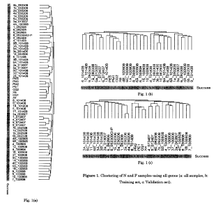
[0027] In Figure la-c, the inventors separately show the clustering of all F
and N samples (65 samples), training set samples (33 samples), and validation
set samples (32 samples) using all genes (a: all samples, b: Training set, c:
Validation set). Samples are clustered with hierarchical clustering utilizing
average linkage method using Pearson's correlation as the metric of similarity
following row normalization Sneath, P. (1973) Numerical taxonomy; the
principles and practice of numerical classification. W. H. Freeman, San
.10-
WO 2011/060080 PCT/US2010/056252
Francisco, CA USA. Figure 1: Clustering of N and F samples using all genes (a:
all samples, b: Training set, c: Validation set).
100281 Figure 2a-b: shows the clustering of N and F samples using 1180
descriptive genes (a: Training set, b: Validation set). The results therein
show
differential expression based on t-test (p<0.05 with Bonferroni correction for
multiple hypothesis testing) which were identified in the training set (F vs.
N).
Resulting 1180 genes, called "descriptive genes", were used to cluster
Training
and Validation sets separately.
100291 Figure 3 shows the clustering of N and F samples using 227 predictive
genes (a: Training set, b: Validation set). The data reveals that the only
sample
incorrectly predicted in the training set is misplaced in the clustering as
well,
however, the mixed behavior of F and N samples in validation set clustering
emphasizes the contribution made by the weighted voting approach.
10030] Figure 4 schematically depicts methods to A) assign significance to the
predictor gene set (PG's) in Table 4, B) Refine PG's, and C) further analyze
final
predictor gene set
(0031] Figure 5. For each gene, number of samples for which the gene has a
value of 40 is shown. Results are calculated for the "old" 35 samples. In
Figure 5,
we show the number of samples with a value of 40 for each gene, separately
plotted for our genes (196 genes labeled "Hasan genes") and all 379 genes on
TLDA (labeled "All genes").
-11-
WO 2011/060080 PCT/US2010/056252
[0032] Figure 6. Number of genes with a value of 40 is shown for each sample.
Results are calculated for the "old" 35 samples: For each gene, number of
samples for which the gene has a value of 40 is shown. Results are calculated
for
the "new" 14 samples.
[0033] Figure 7. For each gene, number of samples for which the gene has a
value of 40 is shown. Results are calculated for the "new" 14 samples.
10034] Figure S. Number of genes with a value of 40 is shown for each sample.
Results are calculated for the "new" 14 samples.
[0035] Figure 9 :: Distribution of genes based on following factors: Group the
gene belongs to (P or A); Agreement of the gene's up/down regulation in TLDA
and microarray (10, if the direction is the same and -10, otherwise); Number
of
samples for which the gene has a value of 40. The analysis is performed
separately for scaled and unsealed values with varying number of outliers
excluded
[0036] Detailed Description. of the Invention
10037] Prior to discussing the invention in more detail, the following
definitions are provided. Otherwise all words and phrases in this application
are
to be construed by their ordinary meaning, as they would be interpreted by an
ordinary skilled artisan within the context of the invention.
-12-
WO 2011/060080 PCT/US2010/056252
[0038] "Pregnancy-competent oocvte": refers to a female gamete or egg that
when fertilized by natural or artificial means is capable of yielding a viable
pregnancy when it is comprised in a suitable uterine environment.
[0039] "The term "competent embryo" similarly refers to an embryo with a
high implantation rate leading to pregnancy. The term "high implantation rate"
means the potential of the embryo when transferred in uterus, to be implanted
in
the uterine environment and to give rise to a viable fetus, which in turn
develops
into a viable offspring absent a procedure or event that terminates said
pregnancy.
[0040] "Viable-pregnancy": refers to the development of a fertilized oocyte
when contained in a suitable uterine environment and its development into a
viable fetus, which in turn develops into a viable offspring absent a
procedure or
event that terminates said pregnancy.
[0041] "Cumulus cell" refers to a cell comprised in a mass of cells that
surrounds an oocyte. This is an example of an "oocyte associated cell". These
cells are believed to be involved in providing an oocyte some of its
nutritional and
or other requirements that are necessary to yield an oocyte which upon
fertilization is "pregnancy competent" (Buccione, R., Schroeder, A.C., and
Eppig,
J.J. (1990). Interactions between somatic cells and germ cells throughout
mammalian oogenesis. Biol Reprod 43, 543-547.)
-13-
WO 2011/060080 PCT/US2010/056252
[00421 "Differential gene expression" refers to genes the expression of which
varies within a tissue of interest; herein preferably a cell associated with
an
oocyte, e.g., a cumulus cell.
[0043] "Real Time RT-PCR": refers to a method or device used therein that
allows for the simultaneous amplification and quantification of specific RNA
transcripts in a sample.
[0044] "Microarray analysis": refers to the quantification of the expression
levels of specific genes in a particular sample, e.g., tissue or cell sample.
[0045] "Pregnancy signature": herein refers to the normal level of expression
of one or more genes or polypeptides that are selected or encoded by the
specific
genes in Table 4 or the 14 genes selected from the group consisting of ABCA6,
DDIT4, DUSP1, GPR137B, IDUA, KCTD5, KRAS, NCAM1, NDNL2, OLFML3,
PTPRA, SDF4, SLC26A3, and TERF2IP and their orthologs, splice or allelic
variants wherein these genes or polypeptides are expressed in normal cumulus
cells at levels which correlate to the likelihood that an oocyte that is
associated
with a cumulus cell which expresses said one or more genes or polypeptides at
these characteristic levels are more likely to give rise to a viable
pregnancy.
[0046] "Characteristic level of expression of a cumulus gene" herein with
respect to a particular detected expressed nucleic acid sequence or
polypeptide
means that the particular gene or polypeptide is expressed at levels which are
substantially similar to the levels observed in cumulus cells that are
associated
-14-
WO 2011/060080 PCT/US2010/056252
with a normal cumulus cell or one associated with a normal or developmentally
competent oocyte.
[00471 By "substantially similar" is meant that the levels of expression of
individual genes are preferably within the range of +/- 1-5 fold of the level
of
expression by a normal cumulus cell, more preferably within the range of +1- 1-
3
-fold, still more preferably within the range of +/- 1-1.5 fold and most
preferably
within the range of +/- 1.0-1.3, 1.0-1.2 or 1.0- 1.2 fold of the detected
levels of
expression of the gene or polypeptide by a normal cumulus cell.
[00481 According to the invention, the oocyte may result from a natural cycle,
a modified natural cycle or a stimulated cycle for cIVF or ICSI. The term
"natural cycle" refers to the natural cycle by which the female or woman
produces an oocyte. The term "modified natural cycle" refers to the process by
which, the female or woman produces an oocyte or two under a mild ovarian
stimulation with GnRH antagonists associated with recombinant FSH or hMG.
The term "stimulated cycle" refers to the process by which a female or a woman
produces one or more oocytes under stimulation with GnRH agonists or
antagonists associated with recombinant FSH or hMG.
[0049] "Oocyte or cumulus cell determined to possess suitable pregnancy
signature or to be pregnancy competent" refers to an oocyte or a cumulus cell
associated with the oocyte or an oocyte derived from the same subject at
around
the same time (within 0-6 months) as the tested cumulus cell which has been
determined to express at least one of the genes or polypeptides encoded by
those
-15-
WO 2011/060080 PCT/US2010/056252
in Table 4 or the 14 genes selected from the group consisting of ABCA6, DDIT4,
DUSP1, GPR137B,. IDUA, KCTD5, KRAS, NCAM1, NDNL2, OLFML3, PTPRA,
SDF4, SLC26A3, and TERF2IP or an ortholog or splice or allelic variant thereof
in a manner characteristic of the level of expression by a normal cumulus
cell.
Preferably at least 2 or 3 genes are expressed in a characteristic manner,
more
preferably at least 3-10 genes, 10-50 genes and even up to 100 genes or more
of
those contained in Table 4 or their allelic or splice variants. It should be
understood that if the expression of numerous genes are evaluated in the
subject
genetic based assays, such as in the order of 10 or more, that a suitable
pregnancy signature means that all or substantially all, i.e. at least 70-80%
of
the detected genes are expressed in a manner characteristic of a normal
cumulus
cell. For example if the expression of 10 genes is detected at least 7, 8 or 9
of the
genes will preferably be expressed at the levels consistent with a normal
cumulus cell, i.e. one associated with an oocyte capable of giving rise to a
normal
embryo and viable pregnancy.
[00501 In general with respect to the pregnancy signature the characteristic
levels of expression is observed for at least 3-5, 5-10, 10 to 20, and
potentially at
least 50 to 100 genes, that are expressed at characteristic levels in cumulus
cells,
that surround "pregnancy competent" oocytes. This is intended to encompass the
level at which the gene is expressed and the distribution of gene expression
within cumulus cells analyzed.
-16-
WO 2011/060080 PCT/US2010/056252
[0051] "Pregnancy signature gene": refers to a gene which is expressed at
characteristic levels by a cumulus cell, which is associated with a normal or
"pregnancy competent" oocyte. These genes are contained in Table 4 and further
include the 14 genes selected from the group consisting of ABCA6, DDIT4,
DUSP1, GPRI3 7B, IDUA, KCTD5, KRAS, NCAMI, NDNL2, OLFML3, PTPRA,
SDF4, SLC26A3, and TERF2IP and their orthologs, splice and allelic variants.
In the table the genes are referenced by their name as well as Accession
number.
It should be understood that the invention further encompasses detection of
allelic and splice variants of these genes and orthologs.
[0052] "Probe suitable for detection of the expression of a pregnancy
signature
gene or polypeptide" refers to a nucleic acid sequence or sequences or ligand
such
as an antibody that specifically detects the expression of the transcribed
gene or
corresponding polypeptide. In a preferred embodiment expression is selected by
use of real time PCR detection methods.
[0053] "IVF": refers to in vitro fertilization.
[0054] The term "classical in vitro fertilization" or "cIVF" refers to a
process
by which oocytes are fertilized by sperm outside of the body, in vitro. IVF is
a
major treatment in infertility when in vivo conception has failed. The term
"intracytoplasmic sperm injection" or "ICSI" refers to an in vitro
fertilization
procedure in which a single sperm is injected directly into an oocyte. This
procedure is most commonly used to overcome male infertility factors, although
it may also be used where oocytes cannot easily be penetrated by sperm, and
-17-
WO 2011/060080 PCT/US2010/056252
occasionally as a method of in vitro fertilization, especially that associated
with
sperm donation.
[0055] "Zona pellucida" refers to the outermost region of an oocyte.
10056] "Method for detecting differential expressed genes" encompasses any
known method for quantitatively evaluating differential gene expression using
a
probe that specifically detects for the expressed gene transcript or encoded
polypeptide. Examples of such methods include indexing differential display
reverse transcription polymerise chain reaction (DDRT-PCR; Mahadeva et al,
1998, J. Mol. Biol. 284:1391-1318; WO 94/01582; subtractive mRNA
hybridization (See Advanced Mel. Biol.; R.M. Twyman (1999) Bios Scientific
Publishers, Oxford, p. 334, the use of nucleic acid arrays or microarrays (see
Nature Genetics, 1999, vol. 21, Suppl. 1061) and the serial analysis of gene
expression. (SAGE) See e.g., Valculesev et al, Science (1995) 270:484-487) and
real time PCR (RT-PCR). For example, differential levels of a transcribed gene
in an oocyte cell can be detected by use of Northern blotting, and/or RT-PCR.
[00571 A referred method is the CRL amplification protocol refers to the novel
total RNA amplification protocol disclosed in Applicant's earlier applications
that
combines template-switching PCR and T7 based amplification methods. This
protocol is well suited for samples wherein only a few cells or limited total
RNA
is available.
[00581 Preferably, the "pregnancy signature" genes are detected by
hybridization of RNA or DNA to DNA chips, e.g., filter arrays comprising cDNA
-18-
WO 2011/060080 PCT/US2010/056252
sequences or glass chips containing cDNA or in situ synthesized
oligonucleotide
sequences. Filtered arrays are typically better for high and medium abundance
genes. DNA chips can detect low abundance genes. In the exemplary
embodiment the sample may be probed with Affyxnetrix GeneChips comprising
genes from the human genome or a subset thereof.
[0059] Alternatively, polypeptide arrays comprising the polypeptides encoded
by pregnancy signature genes or antibodies that bind thereto may be produced
and used for detection and diagnosis.
10060] "EASE" is a gene ontology protocol that from a list of genes forms
subgroups based on functional categories assigned to each gene based on the
probability of seeing the number of subgroup genes within a category given the
frequency of genes from that category appearing on the microarray.
[00611 Based on the foregoing the present invention provides a novel method
of detecting whether a female, preferably human or non-human mammal,
produces "pregnancy competent" oocytes or whether a particular oocyte is
pregnancy competent. The method involves detecting the levels of expression of
one or more genes in Table 4 or the 14 genes selected from the group
consisting
of ABCA6, DDIT4, DUSP1, GPR137B, IDUA, KCTD5, KRAS, NCAM1, NDNL2,
OLFML3, PTPRA, SDF4, SLC26A3, and TERF2IP that are expressed at
characteristic levels by cumulus cells associated with (surrounding) oocytes
that
are "pregnancy competent", i.e., these oocytes when fertilized by natural or
artificial means (IVF), and transferred into a suitable uterine environment
are
-19-
WO 2011/060080 PCT/US2010/056252
capable of yielding a viable pregnancy, i.e., embryo that develops into a
viable
fetus and eventually an offspring unless the pregnancy is terminated by some
event or procedure, e.g., a surgical or hormonal intervention.
[0062] As described herein the inventors have determined as set of genes
expressed in cumulus cells that are biomarkers for embryo potential and
pregnancy outcome. They demonstrated that genes expression profile of cumulus
cells which surrounds oocyte correlated to different pregnancy outcomes,
allowing the identification of a specific expression signature of embryos
developing toward pregnancy. Their results indicate that analysis of cumulus
cells surrounding the oocyte is a non-invasive approach for embryo selection.
[0063] The set of predictive genes in Table 4 and the 14 gene set identified
in
Table 12 are known human genes. However, the expression of these genes (on
cumulus cells) had not heretofore been correlated to oocyte competency or
embryo development. Therefore, this invention relates to a method for
selecting a
competent oocyte, comprising a step of measuring the expression level of
specific
genes in a cumulus cell surrounding said oocyte, wherein said genes include at
least one of the 227 genes in Table 4 or the 14 genes selected from the group
consisting of ABCA6, DDIT4, DUSP1, GPR137B, IDUA, KCTD5, KRAS,
NCAM1, NDNL2, OLFML3, PTPRA, SDF4, SLC26A3, and TERF2IP.
[0064] The methods of the invention may further comprise a step consisting of
comparing the expression level of the genes in the sample with a control,
wherein
detecting differential in the expression level of the genes between the sample
and
-20-
WO 2011/060080 PCT/US2010/056252
the control is indicative whether the oocyte is competent. The control may
consist
in sample comprising cumulus cells associated with a competent oocyte or in a
sample comprising cumulus cells associated with an unfertilized oocyte.
[00651 The methods of the invention are applicable preferably to human
women but may be applicable to other mammals (e.g., primates, dogs, cats,
pigs,
cows...).
[0066] The methods of the invention are particularly suitable for assessing
the
efficacy of an in vitro fertilization treatment. Accordingly the invention
also
relates to a method for assessing the efficacy of a controlled ovarian
hyperstimulation (COS) protocol in a female subject comprising: i) providing
from said female subject at least one oocyte with its cumulus cells; ii)
determining by a method of the invention whether said oocyte is a competent
oocyte.
[0067] Then after such a method, the embryologist may select the competent
oocytes and in vitro fertilize them, for example using a classical in vitro
fertilization (cIVF) protocol or under an intracytoplasmic sperm injection
(ICSI)
protocol.
[0068] A further object of the invention relates to a method for monitoring
the
efficacy of a controlled ovarian hyperstimulation (COS) protocol comprising:
i)
isolating from said woman at least one oocyte with its cumulus cells under
natural, modified or stimulated cycles; ii) determining by a method of the
- 21 -
WO 2011/060080 PCT/US2010/056252
invention whether said oocyte is a competent oocyte; iii) and monitoring the
efficacy of COS treatment based on whether it results in a competent oocyte.
[0069] The COS treatment may be based on at least one active ingredient
selected from the group consisting of GnRH agonists or antagonists associated
with recombinant FSH or hMG-
[0070] The present invention also relates to a method for selecting a
competent embryo, comprising a step of measuring the expression level of at
least one of the 227 genes in Table 4 or the 14 genes selected from the group
consisting of ABCA6, DDIT4, DUSP1, GPR137B, IDUA, KCTD5, KRAS,
NCAM1, NDNL2, OLFML3, PTPRA, SDF4, SLC26A3, and TERF2IP.
[0071] The methods of the invention may further comprise a step consisting of
comparing the expression level of the genes in the sample with a control,
wherein
detecting differential in the expression level of the genes between the sample
and
the control is indicative whether the embryo is competent. The control may
consist in sample comprising cumulus cells associated with an embryo that
gives
rise to a viable fetus or in a sample comprising cumulus cells associated with
an
embryo that does not give rise to a viable fetus.
[0072] It is noted that the methods of the invention leads to an independence
from morphological considerations of the embryo. Two embryos may have the
same morphological aspects but by a method of the invention may present a
different implantation rate leading to pregnancy.
-22-
WO 2011/060080 PCT/US2010/056252
[0073] The methods of the invention are applicable preferably to human
women but may be applicable to other mammals (e.g. primates, dogs, cats, pigs,
cows...).
[0074] The present invention also relates to a method for determining
whether an embryo is a competent embryo, comprising a step consisting in
measuring the expression level of 45 genes in a cumulus cell surrounding the
embryo, wherein said genes include at least one of the 227 genes in Table 4 or
the 14 genes selected from the group consisting of ABCA6, DDIT4, DUSP1,
GPR137B, IDUA, KCTD5, KRAS, NCAM1, NDNL2, OLFML3, PTPRA, SDF4,
SLC26A3, and TERF2IP.
[0075] The present invention also relates to a method for determining
whether an embryo is a competent embryo, comprising: i) providing an oocyte
with its cumulus cells; ii) in vitro fertilizing said oocyte; and iii)
determining
whether the embryo that results from step ii) is competent by determining by a
method of the invention whether said oocyte of step i), is a competent oocyte.
[0076] The present invention also relates to a method for selecting a
competent oocyte or a competent embryo, comprising a step of measuring in a
cumulus cell surrounding said oocyte or said embryo the expression level of
one
or more genes selected from at least one of the 227 genes in Table 4 or the 14
genes selected from the group consisting of ABCA6, DDIT4, DUSP1, GPR137B,
IDUA, KCTD5, KRAS, NCAM1, NDNL2, OLFML3, PTPRA, SDF4, SLC26A3,
and TERF2IP. Aberrant expression of one or more of these genes selected may be
-23-
WO 2011/060080 PCT/US2010/056252
predictive of a non competent oocyte or embryo, the inability of the embryo
being
unable to implant or of a non competent oocyte or embryo due to early embryo
arrest.
100771 The methods of the invention are particularly suitable for enhancing
the pregnancy outcome of a female. Accordingly the invention also relates to a
method for enhancing the pregnancy outcome of a female comprising: i)
selecting
a competent embryo by performing a method of the invention ;iii) implanting
the
embryo selected at step i) in the uterus of said female, wherein said female
may
or may not be the oocyte donor.
[0078] The method as above described will thus help embryologist to avoid the
transfer in uterus of embryos with a poor potential for pregnancy out come.
The
method as above described is also particularly suitable for avoiding multiple
pregnancies by selecting the competent embryo able to lead to an implantation
and a pregnancy.
[0079] In all above cases, the methods described the relationship between
genes expression profile of cumulus cells and embryo and pregnancy outcomes.
[00801 Methods for determining the expression level of the genes of the
invention:
[00811 Determination of the expression level of the genes in the "pregnancy
signature" i.e., at least one of the 227 genes in Table 4 or at least one of
the 14
genes selected from the group consisting of ABCA6, DDIT4, DUSP1, GPR137B,
-24-
WO 2011/060080 PCT/US2010/056252
IDUA, KCTD5, KRAS, NCAM1, NDNL2, OLFML3, PTPRA, SDF4, SLC26A3,
and TERF2IP.can be performed by a variety of techniques. Generally, the
expression level as determined is a relative expression level.
[0082[ More preferably, the determination comprises contacting the sample
with selective reagents such as probes, primers or ligands, and thereby
detecting
the presence, or measuring the amount, of polypeptide or nucleic acids of
interest
originally in the sample. Contacting may be performed in any suitable device,
such as a plate, microtitre dish, test tube, well, glass, column, and so
forth. In
specific embodiments, the contacting is performed on a substrate coated with
the
reagent, such as a nucleic acid array or a specific ligand array. The
substrate
may be a solid or semi-solid substrate such as any suitable support comprising
glass, plastic, nylon, paper, metal, polymers and the like. The substrate may
be
of various forms and sizes, such as a slide, a membrane, a bead, a column, a
gel,
etc. The contacting may be made under any condition suitable for a detectable
complex, such as a nucleic acid hybrid or an antibody- antigen complex, to be
formed between the reagent and the nucleic acids or polypeptides of the
sample.
[0083[ In a preferred embodiment, the expression level may be determined by
determining the quantity of mRNA.
[0084] Methods for determining the quantity of mRNA are well known in the
art. For example the nucleic acid contained in the samples (e.g., cell or
tissue
prepared from the patient) is first extracted according to standard methods,
for
example using lytic enzymes or chemical solutions or extracted by nucleic-acid-
-25-
WO 2011/060080 PCT/US2010/056252
binding resins following the manufacturer's instructions. The extracted mRNA
is
then detected by hybridization (e. g., Northern blot analysis) and/or
amplification
(e.g., RT-PCR). Preferably quantitative or semi-quantitative RT-PCR is
preferred. Real-time quantitative or semi-quantitative RT-PCR is particularly
advantageous. Other methods of amplification include ligase chain reaction
(LCR), transcription- mediated amplification (TMA), strand displacement
amplification (SDA) and nucleic acid sequence based amplification (NASBA).
[0085] Nucleic acids having at least 10 nucleotides and exhibiting sequence
complementarity or homology to the mRNA of interest herein find utility as
hybridization probes or amplification primers. It is understood that such
nucleic
acids need not be identical, but are typically at least about 80% identical to
the
homologous region of comparable size, more preferably 85% identical and even
more preferably 90-95% identical. In certain embodiments, it is advantageous
to
use nucleic acids in combination with appropriate means, such as a detectable
label, for detecting hybridization. A wide variety of appropriate indicators
are
known in the art including, fluorescent, radioactive, enzymatic, or other
ligands
(e. g. avidin/biotin).
[0086] Probes typically comprise single-stranded nucleic acids of between 10
to 1000 nucleotides in length, for instance of between 10 and 800, more
preferably of between 15 and 700, typically of between 20 and 500. Primers
typically are shorter single- stranded nucleic acids, of between 10 to 25
nucleotides in length, designed to perfectly or almost perfectly match a
nucleic
-26-
WO 2011/060080 PCT/US2010/056252
acid of interest, to be amplified. The probes and primers are "specific" to
the
nucleic acids they hybridize to, i.e. they preferably hybridize under high
stringency hybridization conditions (corresponding to the highest melting
temperature Tm, e.g., 50 % formamide, 5x or 6x SCC. SCC is a 0.15 M NaCl,
0.015 M Na-citrate). The nucleic acid primers or probes used in the above
amplification and detection method may be assembled as a kit. Such a kit
includes consensus primers and molecular probes. A preferred kit also includes
the components necessary to determine if amplification has occurred. The kit
may also include, for example, PCR buffers and enzymes; positive control
sequences, reaction control primers; and instructions for amplifying and
detecting the specific sequences.
[0087] In a particular embodiment, the methods of the invention comprise the
steps of providing total RNAs extracted from cumulus cells and subjecting the
RNAs to amplification and hybridization to specific probes, more particularly
by
means of a quantitative or semi quantitative RT-PCR.
[0088] In another preferred embodiment, the expression level is determined
by DNA chip analysis. Such DNA chip or nucleic acid microarray consists of
different nucleic acid probes that are chemically attached to a substrate,
which
can be a microchip, a glass slide or a micro sphere- sized bead. A microchip
may
be constituted of polymers, plastics, resins, polysaccharides, silica or
silica-based
materials, carbon, metals, inorganic glasses, or nitrocellulose. Probes
comprise
nucleic acids such as cDNAs or oligonucleotides that may be about 10 to about
60
-27-
WO 2011/060080 PCT/US2010/056252
base pairs. To determine the expression level, a sample from a test subject,
optionally first subjected to a reverse transcription, is labeled and
contacted with
the microarray in hybridization conditions, leading to the formation of
complexes
between target nucleic acids that are complementary to probe sequences
attached to the microarray surface. The labeled hybridized complexes are then
detected and can be quantified or semi-quantified. Labeling may be achieved by
various methods, e.g. by using radioactive or fluorescent labeling. Many
variants
of the microarray hybridization technology are available to the man skilled in
the
art (see e.g. the review by Hoheisel, Nature Reviews, Genetics, 2006, 7:200-
210)
[0089] In this context, the invention further provides a DNA chip comprising a
solid support which carries nucleic acids that are specific to at least one of
the
227 genes in Table 4 or the 14 genes selected from the group consisting of
ABCA6, DDIT4, DUSP1, GPR137B, IDUA, KCTD5, KRAS, NCAMI, NDNL2,
OLFML3, PTPRA, SDF4, SLC26A3, and TERF2IP.
[0090] Other methods for determining the expression level of said genes
include the determination of the quantity of proteins encoded by said genes.
[0091] Such methods comprise contacting the sample with a binding partner
capable of selectively interacting with a marker protein present in the
sample.
The binding partner is generally an antibody that may be polyclonal or
monoclonal, preferably monoclonal.
[0092] The presence of the protein can be detected using standard
electrophoretic and immunodiagnostic techniques, including immunoassays such
-28-
WO 2011/060080 PCT/US2010/056252
as competition, direct reaction, or sandwich type assays. Such assays include,
but are not limited to, Western blots; agglutination tests; enzyme-labeled and
mediated immunoassays, such as ELISAs; biotin/avidin type assays;
radioimmunoassays; immunoelectrophoresis; immunoprecipitation, etc. The
reactions generally include revealing labels such as fluorescent,
chemiluminescent, radioactive, enzymatic labels or dye molecules, or other
methods for detecting the formation of a complex between the antigen and the
antibody or antibodies reacted therewith.
[00931 The aforementioned assays generally involve separation of unbound
protein in a liquid phase from a solid phase support to which antigen-antibody
complexes are bound. Solid supports which can be used in the practice of the
invention include substrates such as nitrocellulose (e. g., in membrane or
microtitre well form); polyvinylchloride (e. g., sheets or microtitre wells);
polystyrene latex (e.g., beads or microtitre plates); polyvinylidine fluoride;
diazotized paper; nylon membranes; activated beads, magnetically responsive
beads, and the like. More particularly, an ELISA method can be used, wherein
the wells of a microtiter plate are coated with an antibody against the
protein to
be tested. A biological sample containing or suspected of containing the
marker
protein is then added to the coated wells. After a period of incubation
sufficient
to allow the formation of antibody antigen complexes, the plate (s) can be
washed
to remove unbound moieties and a detectably labeled secondary binding molecule
added. The secondary binding molecule is allowed to react with any captured
-29-
WO 2011/060080 PCT/US2010/056252
sample marker protein, the plate washed and the presence of the secondary
binding molecule detected using methods well known in the art.
[0094] Alternatively an immunohistochemistry (IHC) method may be
preferred. IHC specifically provides a method of detecting targets in a sample
or
tissue specimen in situ. The overall cellular integrity of the sample is
maintained
in IHC, thus allowing detection of both the presence and location of the
targets of
interest. Typically a sample is fixed with formalin, embedded in paraffin and
cut
into sections for staining and subsequent inspection by light microscopy.
Current
methods of IHC use either direct labeling or secondary antibody-based or
hapten-
based labeling. Examples of known IHC systems include, for example,
EnVision(TM) (DakoCytomation), Powervision(R) (Immunovision, Springdale,
AZ), the NBA(TM) kit (Zymed Laboratories Inc., South San Francisco, CA),
HistoFine(R) (Nichirei Corp, Tokyo, Japan).
[0095] In particular embodiment, a tissue section (e.g. a sample comprising
cumulus cells) may be mounted on a slide or other support after incubation
with
antibodies directed against the proteins encoded by the genes of interest.
Then,
microscopic inspections in the sample mounted on a suitable solid support may
be performed. For the production of photomicrographs, sections comprising
samples may be mounted on a glass slide or other planar support, to highlight
by
selective staining the presence of the proteins of interest.
[00961 Therefore IHC samples may include, for instance: (a) preparations
comprising cumulus cells (b) fixed and embedded said cells and (c) detecting
the
-30-
WO 2011/060080 PCT/US2010/056252
proteins of interest in said cells samples. In some embodiments, an IHC
staining
procedure may comprise steps such as: cutting and trimming tissue, fixation,
dehydration, paraffin infiltration, cutting in thin sections, mounting onto
glass
slides, baking, deparaffination, rehydration, antigen retrieval, blocking
steps,
applying primary antibodies, washing, applying secondary antibodies
(optionally
coupled to a suitable detectable label), washing, counter staining, and
microscopic examination.
[0097] The invention also relates to a kit for performing the methods as above
described, wherein said kit comprises means for measuring the expression level
the levels of at least one of the 227 genes in Table 4 or the 14 genes
selected from
the group consisting of ABCA6, DDIT4, DUSP1, GPR137B, IDUA, KCTD5,
KRAS, NCAM1, NDNL2, OLFML3, PTPRA, SDF4, SLC26A3, and TERF2IP that
are indicative whether the oocyte or the embryo is competent.
[0098] The invention is further illustrated by the following description of
how
the inventors determined that the expression of the 227 genes in Table 4 and
the
14 gene set on a cumulus cell correlates to oocyte competency and embryo
development upon implantation and working examples. However, these
examples and description should not be interpreted in any way as limiting the
scope of the present invention.
[00991 An exemplary means by which this was effected is described in detail
as follows.
-31-
WO 2011/060080 PCT/US2010/056252
[00100] The first aspect of reducing the invention to practice involved
identifying genes which constitute the pregnancy signature in women and
potentially other mammals and was achieved by identifying and comparing the
expression of genes in cumulus cells collected from women donors which are
pregnancy competent or not. This was effected by collecting cumulus cells from
different human oocytes of donor women and implanting patients with one or
two putatively fertilized eggs. These patients were then, based on the results
of
the implantation, divided into three groups based on full, partial, and no
pregnancy. For each oocyte used in the process, the transcriptional profile of
at
least one cumulus cell surrounding the particular oocyte was determined using
Affymetrix HG 133 Plus 2 arrays containing over 54,000 transcripts. Patients
were included in the study only if they did not meet any of the exclusion
criteria
identified in Table 1.
Table 1. Patient Exclusion Criteria
On Female Side:
> 35 years of age
Low Ovarian Reserve
PcOS
>NFcycle 2
Presence of > 4cm fibroids
BMl > 35
History of chemotherapy of
radiation to abdomen or elvis
On. Male Side:
History of testicular biopsy
million sperm
-32-
WO 2011/060080 PCT/US2010/056252
[001011 More particularly, in order to find gene signatures predictive of an
oocyte's ability to produce a healthy baby, the inventors profiled the
transcriptome of cumulus cells surrounding the oocyte using Affymetrix HG 133
Plus 2 arrays containing over 54,000 transcripts. Total RNA from individual
cumulus samples was isolated using the PicoPure RNA isolation kit (Molecular
Devices, Sunnyvale, CA). Sample RNA was amplified using a protocol developed
in-house which ensures faithful and consistent amplification of small amounts
of
RNA to levels required for microarray analysis (Kocabas, et al., Proc 1Vati
Acad
Sci USA, 103, 14027-14032 (2006)).
[00102] Resulting amplified RNA (aRNA) was hybridized to the Affymetrix
arrays. Thirty-six samples were used for which none of the embryo transfers
led
to successful pregnancies (labeled N for No success) and 30 samples for which
all
of the transfers led to successful pregnancies (labeled F for Full success).
There
were no known confounding factors to effect pregnancy success and relevant
clinical parameters such as age or IVF cycle number did not vary significantly
between the F and N groups.
[001031 Quality Control (QC) parameters were calculated for all 65 samples
using Expression ConsoleTM (EC) software freely available by the manufacturer
(Affymetrix). All QC parameters including scaling factor (coefficient needed
to
equate the 2% trimmed mean of overall chip intensity), percentage of probe
sets
called present, 3'-5' ratios for spike and labeling controls and housekeeping
genes
were within acceptable ranges (as described in manufacturer's guidelines) for
all
-33-
WO 2011/060080 PCT/US2010/056252
the samples. There were no known confounding factors to affect pregnancy
success and relevant clinical parameters such as oocyte age or IVF cycle
number
did not vary significantly (t-test p > 0.05) between F and N groups (see Table
1).
Additional criteria for acceptance included absence of Polycystic Ovarian
Syndrome (PCOS), no history of chemotherapy or radiation to the abdomen or
pelvis, absence of >4 cm intramural or submucosal fibroids, and on the male
side,
no history of testicular biopsy and sperm count of > 5 million
[00104] In order to prove the soundness of the prediction model, F and N
samples were divided randomly into training and validation sets. The goal was
to
find a predictive set of genes developed on the training set and then test the
performance of the predictive genes on the validation set, which has not been
used in development of the predictive model. This strategy (as opposed to
using
all the samples to develop a signature) prevents over-fitting and provides an
assessment of predictive signature's robustness (Nevins, J.R. and Potti, A.
(2007)
Mining gene expression profiles: expression signatures as cancer phenotypes,
Nat Rev Genet, 8, 601-609.)
[oolo5l As shown in Table 2, 33 samples (15F; 18N) were used in the training
set and 32 samples (15F; 18N) in the validation set.
[00106] Samples used in training and validation sets are shown in Table 2.
-34-
WO 2011/060080 PCT/US2010/056252
Table 2. Samples used in the training and validation sets for prediction
purposes.
Training Validation
Sample Sample
Name Success Name Success
8_100908 F 1B 100908 F
4B100908 F 5B 100908 F
6_092308 F 1 092308 F
6A_101408 F 3B_101408 F
1A_100908 F 12100908 F
15_100908 F 4_100908 F
6101408 F 37_100908 F
1A_101408 F 8_092308CHP F
9092308 F 4072407 F
6A100908 F lb 032306 F
1072407 F 2a 013007 F
5a013007 F 2a030206 F
3a013007 F 4a 030206 F
3b_091406 F 2a 062807 F
6072407 F 9_072407 F
1C_101408 N 5C_101408 N
4A_100908 N 2A_101408 N
10_100908 N 4C 100908 N
9101408 N 9 100908 N
la 092308 N 8101408 N
4_101408 N 5B 101408 N
6100908 N 11_101408 N
5101408 N 3A_101408 N
9A_101408 N 6b 092308 N
lb_092308 N 7b_092308 N
5b_092308 N 7 100908 N
5C_100908 N 6_062906 N
10062906 N 5a_030206 N
la 030206 N C Q1 N
C92 N PEI N
PE5 N PM2 N
PM1 N X4 N
X6 N
[00107] In Figure 1, the inventors separately show the clustering of all F and
N
samples (65 samples), training set samples (33 samples), and validation set
samples (32 samples) using all genes. Samples are clustered with hierarchical
clustering utilizing average linkage method using Pearson's correlation as the
metric of similarity following row normalization Sneath, P. (1973) Numerical
-35-
WO 2011/060080 PCT/US2010/056252
taxonomy; the principles and practice of numerical classification. W. H.
Freeman, San Francisco, CA USA.
100108] During the clustering process complete transcriptional profiling on
the
chips, i.e. all 54K+ transcripts are used. The clustering of all three data
sets
indicates a lack of separation based on pregnancy success. This in turn
suggests
the need for supervised learning analysis to find phenotype specific gene
identification to correlate the expression results with success and to
eventually
identify a predictive gene set.
[00109] In order to find genes that correlate with success, genes that show
differential expression based on t-test (p<0.05 with Bonferroni correction for
multiple hypothesis testing) were identified in the training set (F vs. N).
Resulting 1180 genes, called "descriptive genes", were used to cluster
Training
and Validation sets separately (Figure 2). The results show successful
separation
of N and F samples, especially in the training set as samples in this set have
been used to identify the pregnancy signature genes in Table 4 and the 14
genes
selected from the group consisting of ABCA6, DDIT4, DUSP1, GPR137B, IDUA,
KCTD5, KRAS, NCAM1, NDNL2, OLFML3, PTPRA, SDF4, SLC26A3, and
TERF2IP.
[00110] Next, using these 1180 genes, leave-one-out-cross-validation (L1OXV)
was performed in the training set. In this method, first number of genes in
the
predictive gene set, say P, is fixed. Then one sample in the training set is
left-out
and top P genes using the remaining samples that differentiate between N and F
-36-
WO 2011/060080 PCT/US2010/056252
are calculated. Using these P genes, the sample that is left out is predicted
as N
or F. This process is cycled through all 33 samples in the training set
(leaving
one out at a time). The total number of correct predictions is listed as the
accuracy of the predictor on the training set.
1001111 During L1OXV process, different values for P, number of predictor
genes, are tried and for ones that show good L1OXV prediction accuracy, these
genes are applied on the validation set. The number of samples correctly
predicted in the validation set is reported as prediction accuracy in the
validation
set. The smallest P that yields high training and validation accuracies, i.e.
P for
which accuracy graph reaches a plateau, are reported as the predictor gene
set.
Prediction algorithms employed were Weighted Voting (Golub et al., Molecular
classification of cancer: class discovery and class prediction by gene
expression
monitoring, Science, 286, 531-537(1999), Bayesian Compound Covariate); Wright
et 1., Proc Natl Acad Sci U S A, 100, 9991-9996 (1999)); Diagonal Linear
Discriminant, Dudoit et al., citation, 97, 77-87); Nearest Centroid, k-Nearest
Neighbors (Golub et al., Molecular classification of cancer: class discovery
and
class prediction by gene expression monitoring, Science, 286, 531-537 (1999));
Shrunken Centroids, (Tibshirani et al., Proc Natl Acad Sci U S A, 99, 6567-
6572
(2002) ); Support Vector Machines (Radmacher, et al., J Comput Biol, 9, 505-
511
(2002)) and Compound Covariate (Hedenfalk et al., N Engl J Med, 344, 539-548
(2001)).
-37-
WO 2011/060080 PCT/US2010/056252
100112] In our analysis the weighted voting approach performed the best
prediction with a 227 gene predictor set yielding 97% L1OXV accuracy (32/33
correct predictions) and 88% (28/32 correct predictions) validation set
accuracy.
These 227 genes, called "predictive genes" yielded significant (p<0.05)
prediction
results on both training and validation sets using Fisher's tests. Prediction
results are shown in Table 3.
-38-
WO 2011/060080 PCT/US2010/056252
[001131 Table 3. Prediction results for training and validation data sets.
Training Validation
True Predi- Pred-
Sample Name cted Sample Name True icted
Class Class Class Class
lb_092308 N N 092308 N N
5b_092308 N N 7b_092308 N N
la_092308 N N 9_100908 N N
6100908 N N 40_100908 N N
4A_100908 N N 7_100908 N N
10100908 N N 11_101408 N N
5C_100908 N N 8_101408 N N
5101408 N N 3A_101408 N N
9101408 N -N 5B101408 N N
9A_101408 N N 2A_101408 N N
4_101408 N N 50_1.01408 N N
1C_101408 R -N 5a_030206 N N
la_030206 N N 6062906 N N-
062906 N F CQ1 N N
CQ2 N N PE1 N N
X6 N N PM2 N N
PE5 N N X4 N N
PM1 N N 8092308CHP F F
9092308 F F 1_092308 F F
6092308 F F 4_100908 F F
15100908 F F 12100908 F F
1A_100908 F F 37_100908 F F
8_100908 F F 1B_100908 F F
4B_100908 F F 5B_100908 F N
6A 100908 F F 313101408 F N
1A 101408 F F lb_032306 F F
6_101408 F F 2a_013007 F F
6A_101408 F F 2a 030206 F N
1_072407 F F 2a 062807 F F
3a013007 F F 4072407 F F
3b091406 F F 4a_030206 F N
5a013007 F F 9_072407 F F
6072407 F F
[001141 In Figure 3, we show clustering of training and validation sets using
the 227 predictive gene list. Note that the only sample incorrectly predicted
in
the training set is misplaced in the clustering as well, however, the mixed
-39-
WO 2011/060080 PCT/US2010/056252
behavior of F and N samples in validation set clustering emphasizes the
contribution made by the weighted voting approach.
[00115] Based on the foregoing the inventors have identified a set of human
genes (contained in Table 4) the expression of which may be assessed on
cumulus
cells and compared to the level of expression of said gene that correlates to
that
of a normal oocyte, i.e., one associated with an oocyte that is capable of
giving
rise to a viable pregnancy, in order to assess the pregnancy potential of an
oocyte
associated with said cumulus cell (or an oocyte from the same donor that was
used to isolate the tested cumulus cell). Analysis of additional samples
resulted
in identification of a preferred set of 14 genes (contained in Table 12) the
expression of which may be assessed on cumulus cells and compared to the level
of expression of said gene that correlates to that of a normal oocyte, i.e.,
one
associated with an oocyte that is capable of giving rise to a viable
pregnancy, in
order to assess the pregnancy potential of an oocyte associated with said
cumulus cell (or an oocyte from the same donor that was used to isolate the
tested cumulus cell).
[00116] While these lists of genes are clinically relevant, we anticipate that
these lists of 227 and 14 predictor genes may be further refined, subject to
functional analysis, and further validated in order to identify a more optimal
pregnancy signature set of genes.
-40-
WO 2011/060080 PCT/US2010/056252
[001171 With respect thereto, the following techniques which may be used to
refine this list of predictor genes contained in Table 4 and in the 14 gene
set
contained in Table 12 are described below.
[001181 The inventors will assign significance to these 227 or 14 predictor
genes (PG's) using two strategies: i) we will permute class labels, identify
optimum 227 gene predictors using the same method applied previously, and
calculate their performance on the perturbed data set; ii) we will test the
performance of randomly chosen 227 gene predictors using the original data
set.
Performance comparison of PG'S against results obtained using aforementioned
strategies are used to assess PG'S's significance. In order to refine PG'S, we
will
divide the complete data set into alternative training and validation sets and
for
each split calculate an optimum predictive gene set. These predictors are
compared to each other and PG'S to obtain a final predictive gene set composed
of genes consistently coming up as good predictors. This final set will then
be
evaluated for its significance and accuracy. Finally, we will further analyze
this
final predictor gene set for i) Gene Ontology (GO) functional classification
to find
significantly over-represented GO categories, ii) involvement in known
biological
pathways, gene networks, disease pathways, small molecule and drug target
interactions, iii) promoter region analysis to identify common/distinct
transcriptional regulatory elements and miRNA target analysis, and iv) their
localization, secretion potential, and ligation properties. In what follows we
explain in detail this overall workflow, which is summarized in Figure 4.
-41-
WO 2011/060080 PCT/US2010/056252
[00119] We have previously applied the approaches defined here on finding
predictive biomarkers in diabetes, myelodysplastic syndrome, chronic liver
disease and various forms of cancer using both high throughput transcriptional
profiling and proteomic data sets (See: Aivadoet al., Proc Natl Acad Sci U S
A,
104, 1307-1312 2007).; Jones et al., Clin Cancer Res, 11, 5730-5739 (2007);
Jones,
et al., J Urol, 179, 730-736 (2007) ; Out et al. Diabetes Care, 30, 638-643
(2007) ;
Out et al., J Biol Chem, 282, 11197-11204 (2007).; Prall et al., Int J
Hematol, 89,
173-187(2009); Spentzos et al., J Clin Oncol, 23, 7911-7918 (2005). and Zinkin
et
al., Clin Cancer Res, 14, 470-477 (2008). In all these studies, the biomarkers
were found to be statistically significantly predictive and were independently
validated both experimentally and analytically on new data sets that have not
been used to define the predictor set.
[00120] Prediction Algorithm
[00121] The "signal to noise ratio" (SNR) is used to assess predictor value of
a
gene g (Golub et al., Science, 286, 531-537 (1999).) Let jF(g) and pN(g) be
the
mean value of gene g in F (successful pregnancy) and N (failed pregnancy)
sample groups, respectively. Similarly, let oF(g) and oN(g) be the standard
deviation of gene g in F and N sample groups, respectively. We define SNR(g) =
[1pF(g) - pN(g)] / [oF(g) + oN(g)]. This metric defines a neighborhood in RM
around ideal gene expression vectors for both groups where M = I F I + I N I,
total
number of samples in the data set. SNR punishes genes with an expression
highly deviant in either group and provides a signed ranking method for a
gene's
-42-
WO 2011/060080 PCT/US2010/056252
membership. In this case large positive values indicate a good predictor for
the F
group and large negative values (in absolute value) indicate a good predictor
for
the N group. We also define the boundary between the correlation between
idealized expression patterns and a given gene g as B(g) = [pF(g) + pN(g)}I2.
[001221 In this method we assess the predictor gene set of P genes G = {gl.,
g2,
..., gP}, a group of F and N samples and a new sample S to be predicted. The
vote
of gi, I. < i <- P, is defined as Vi = SNR(gi) [S(gi) - B(gi)], where S(gi)
represents
the signal value of gene gi in S. Vi represents how well S(gi) relates to the
"behavior" of gi in F and N samples. If Vi is positive, we conclude that based
on
gi, S is predicted to be F and if Vi is negative gi predicts S as N. Cycling
through
all genes in the predictor set we obtain P votes and let VF be the sum of all
positive votes and VN be the sum of all negative votes. If VF is greater than
VN
in absolute value, we predict sample S as F; otherwise we predict S as N. In
our
previous studies we have obtained a robust predictor gene set using a training
set, which was tested on an independent validation set.
[00123] Significance of Predictor Gene Set
[00124] In order to obtain a robust predictor gene set, samples are randomly
divided into training and validation sets. We have previously left out cross
validation (LI.OXV) using weighted voting to obtain the 227 predictor gene
set.
In this analysis method we let T and V be the number of F and N samples used
in training and validation sets, respectively. In this context, we left one of
the
samples in the training group out and found P genes with the highest SNR value
-43-
WO 2011/060080 PCT/US2010/056252
in T-1 F and N samples in the training set. Using these P genes, the sample
that
was left out is predicted either as F or N using the procedure described
above.
Number of correct predictions amount to LIOXV prediction accuracy on the
training set and minimum P with highest prediction accuracy is carried on to
be
tested on the validation set. In our preliminary studies P was found to be 227
and our prediction accuracies were 97% and 88% on training and validation
sets,
respectively.
[00125] We also assess the statistical significance of these 227 predictive
genes.
In order to assess this, we employ a two step strategy. In the first phase, we
randomly shuffle the class labels of the samples used in the training set,
i.e., we
call a random subset of F samples as N and similarly call a random subset of N
samples as F. Using this perturbed data set, we calculate LIOXV accuracy of
227
genes with the weighted voting method on the training set. In this setting, we
choose 227 genes that have the highest SNR values using the permuted class
labels. Assume the class label permutation is performed B times and the L1OXV
accuracy of lath permutation is A. We assess L1OXV accuracy significance of
our
original 227 gene predictor set as p = 1/B Y_ I(Ab > 97%), where I is an
indicator
function and assumes a value of 1 if Ab > 97% and 0 otherwise. Similarly, we
let
A'b be the prediction accuracy of 227 genes obtained using both permutations
on
the validation set. We then assess the prediction accuracy significance of our
227
gene predictor set on the validation set as p = 1/B I(A'b > 88%).
-44-
WO 2011/060080 PCT/US2010/056252
[00126] In the second phase to assess significance, we retain the original
class
labels but pick random 227 gene sets and evaluate their L1OXV accuracy on the
training set and prediction accuracy in the validation set. We perform this
random selection B times and let Ab and A'b be the L1OXV accuracy on the
training set and prediction accuracy in the validation set for the bth
selection,
respectively. Similarly, the significance for L1OXV accuracy is calculated as
p =
1/B T, 1(Ab > 97%), and the significance for the validation set prediction
accuracy
are calculated as p I/B Y 1(A'b > 88%). In both phases B=1000.
(00127] Further Refinement of the Predictor Gene Set
[00128] In order to further refine the 227 gene predictor set we also employ
different training and validation set splits and look for the overlap in the
resulting gene sets. With an attempt to find well defined predictors, in the
refinement process, we do not split our whole data set in half into training
and
validation sets; rather, we use 75% of the samples for the training set and
25% of
the samples for the validation set. Furthermore, we adopt a ten-fold cross
validation strategy instead of a L1OXV. In this case, when finding a predictor
on
the training set, we do not leave one sample out at a time; instead, we leave
10%
of the total number of samples in the training set out at a time. At each
iteration,
a predictor set is calculated using the remaining 90% samples and the left out
10% samples is predicted with the predictor set. Total number of correct
predictions is then used to calculate prediction accuracy.
-45-
WO 2011/060080 PCT/US2010/056252
[00129] For each data split, we evaluate the prediction accuracy on the
validation set and the significance of the predictor gene set as described
above.
We then intersect the predictor gene sets found at each split with each other
and
with the original 227 gene predictor set to find genes that consistently come
up
as good predictors. To this end, we form a more refined predictor gene set and
calculate its prediction accuracy and significance on the overall data set
using
cross validation.
[00130] Clustering and Functional Analysis
[00131] Clustering of samples and genes are performed using Unweighted Pair
Group Method with Arithmetic-mean (UPGMA)( Sneath, P.H.A. (1973)
Numerical taxonomy; the principles and practice of numerical classification.
W.
H. Freeman, San Francisco, CA USA.), a hierarchical clustering technique used
to construct a similarity tree, and principal components analysis (PCA)(Otu,
et
al., J Biol Chem, 282, 11197-11204 (2007)). In hierarchical clustering,
expression
data matrix is row-normalized for each gene prior to the application of
average
linkage clustering and Pearson's correlation is used as the distance measure.
In
PCA, which projects multivariate data objects into a lower dimensional space
while retaining as much of the original variance as possible, each sample is
normalized to mean zero and standard deviation one.
[00132] Functional analysis is comprised of finding Gene Ontology (GO)
categories in the gene lists of interest that warrant further investigation
(Ashburner, et al., Nat Genet, 25, 25-29 (2000)). Expression Analysis
Systematic
-46-
WO 2011/060080 PCT/US2010/056252
Explorer (EASE) identifies biologically-relevant categories that are over-
represented in the set and therefore may be of further interest (Hosack et
al.,
Genome Biol, 4, R70) (2003).
[00133] To accomplish this, EASE maps each probe to an Entrez Gene
identifier (Maglott, et al., Nucleic Acids Res, 35, D26-31 (2001)) that is
associated
with a GO category. GO Consortium (Geneontology.org) assigns each gene
(where applicable) to one or more classes in the three GO categories:
biological
function, cellular process, and molecular function. EASE identifies GO
categories
in the input gene list that are over-represented using jackknife iterative
resampling of Fisher exact probabilities, with Bonferroni multiple testing
correction. The "EASE score" is the upper bound of the distribution of
Jackknife
Fisher exact probabilities. Categories containing low numbers of genes are
under-weighted so that the EASE score is more robust than the Fisher exact
test. EASE analysis will test the predictor gene list against all genes on the
chip, and an EASE score are calculated for likelihood of overrepresentation of
a
GO category in the input list. Overrepresentation describes a group of genes
that
belong to a certain GO category, e.g. cell cycle, that appear more often in
the
given input list than would be expected to occur if the distribution were
random.
The EASE score is a significance level with smaller EASE scores indicating
increasing confidence in overrepresentation. We select GO categories that have
EASE scores of 0.05 or lower as significantly over-represented.
[00134] Pathway Analysis
-47-
WO 2011/060080 PCT/US2010/056252
[00135] Pathway analysis, functional enrichment analysis or gene set analysis
focuses on predefined gene sets or classes that are significantly regulated in
a
microarray study. We use Ingenuity Software Knowledge Base (IKB), (Redwood
City, CA) to identify networks and pathways that best explain underlying
transcriptional regulations. IKB uses interactions between genes and/or gene
products based on manual curation of scientific literature providing a robust
interaction database. Once a gene list of interest is analyzed the results are
viewed in two ways: i) networks formed using input gene list and a limited
expansion ii) known biological pathway that significantly host a subset of the
input genes. Both results can further be analyzed in terms of drugs, small
metabolites, functions, and diseases known to interact partly with the final
gene
networks. In this way, we identify pathways best explained by our predictive
gene set and infer further perturbations to these networks. Furthermore, we
are
able to generate new interaction networks that do not necessarily fall in pre-
defined canonical pathways.
[00136] Promoter and miRNA target analysis
[00137] We will interrogate 5kb upstream of genes in our predictive signature
and analyze these identified promoter regions using promoter analysis and
interaction tool, PAINT (Vadigepalli et al., OMICS, 7, 235-252 (2003)) and
oPOSSUM (Ho Sui et al., Nucleic Acids Res, 33, 3154-3164 (2005)). These
methods model the promoter regions of input gene lists and identify
transcription regulatory elements (TRE) in these regions. The results are then
-48-
WO 2011/060080 PCT/US2010/056252
analyzed for over-representations of TREs and discover potential transcription
factors that are important in gene regulation for the underlying microarray
data.
[00138] Another potential mechanism of regulation of predictive genes is via
miRNAs that regulate gene expression primarily through post-transcriptional
repression or mRNA degradation in a sequence-specific manner. We can identify
miRNA targets sites for predictive genes using TargetScanS
(http://www.targetscan.org/). TargetScanS is an improved version of the
Targetscan that searches the UTRs for segments of perfect Watson-Crick
complementarities to base 2-8 of the miRNA, calculates a folding free energy G
for each miRNA-target site interaction using RNAeval, and then assigns a Z
score to each UTR (Lewis et al., Cell, 115, 787-798 (2003)). This way miRNA
targets in the input gene list are identified through computation of miRNA
binding sites.
[00139] Prediction of secreted proteins and membrane ligands
[00140] Localization and transport of proteins in the cell are governed by
intrinsic signals in their amino acid sequences. We can analyze our predictive
gene list in this context via the proteins they code for. In cases where
annotation
of a protein is sufficient to inform us about its localization, secretion
potential,
ligation and other properties, a manual filtering will suffice to isolate
targets for
further study. When substantial annotation is lacking, we are able to predict
aforementioned properties of the protein using a highly accurate computational
algorithm, TargetP, that uses neural networks and takes in consideration the
-49-
WO 2011/060080 PCT/US2010/056252
characteristic of the N terminus of the proteins (Emanuelsson et al. Nat
Protoc,
2, 953-971 (2007)). In case of secreted protein predictions, it helps to
filter out
ones that have transmembrane domains which would lessen the secretion
potential of the protein. The transmembrane domain prediction on these
proteins
is performed using the standalone version of TMHMM, which uses a hidden
Markov model for prediction (Sonnhammer et al., Proc Int Conf Intell Syst Mol
Biol, 6, 175-182 (1998)). We also can supplement this analysis by the approach
defined in PSORT9 Horton et al, Nucleic Acids Res, 35, W585-587 (2007)), which
uses k-nearest neighbor classification and weighted matrices resulting from
gapless multiple alignment in its prediction strategy.
[00141] Alternative Strategies
[00142] In addition we can optionally enhance our analysis in two ways:
[00143] Signal value calculation and normalization: Although model based
methods such as dChip (Li et al., Genome Biol, 2, RESEARCH0032 (2001).) and
RMA (Irizarry et al., Nucleic Acids Res, 31, e15. (2001)) may be used to
normalize and summarize gene chip data and have been shown to be superior to
Affymetrix MAS 5.0 normalization (Hubbell et al., Bioinformatics, 18, 1585-
1592.(2002)), we have avoided using these methods as they depend on a baseline
sample and do not adapt to addition of new samples (Barash et al.,
Bioinformatics, 20, 839-846 (2004)). In other words, as new samples are added
to
the data set, previous model based signal analysis becomes invalid and
complete
analysis workflow is therefore repeated. Because of our constantly growing
data
-50-
WO 2011/060080 PCT/US2010/056252
set and need to validate our previous findings in newly added samples, the
inventors use MAS 5.0, a signal value calculation and normalization method
that
is robust to addition of new samples. A new method called Probe Logarithmic
Intensity Error (PLIER) workflow in the Expression Console from Affymetrix
(www.affymetrix.com) includes quintile normalization and produces improved
signal values by utilizing the probes affinities, empirical probe performance
and
by handling the error appropriately across low and high concentrations (Katz
et
al., BMC Bioinformatics, 7, 464 (2006). Although PLIER is not as robust as MAS
5.0 to addition of new samples, it provides a better performance compared to
model based methods and therefore are our alternative strategy for signal
value
calculation and normalization.
[00144] Prediction algorithm:
[001451 In addition to weighted voting, which has been the best performing
prediction algorithm in our previous studies, we plan to try the following
prediction algorithms in case our results do not conform to the success
criteria
set forth in this application: Bayesian Compound Covariate (Dudoit et al.,
Journal of the American Statistical Association, 97, 77-87 (2002); Wright et
al.,
Proc Natl Acad Sci U S A, 100, 9991-9996 (2003)), Diagonal Linear
Discriminant,
Nearest Centroid, k-Nearest Neighbors(Golub et al., Science, 286, 531-537
(1999), Shrunken Centroids (Tibshirani et al., Proc Natl Acad Sci U S A, 99,
6567-6572 (2002), Support Vector Machines (Radmacher et al., J Comput Biol, 9,
-51.
WO 2011/060080 PCT/US2010/056252
505-511 (2002)), and Compound Covariate (Hedenfalk et al., N Engl J Med, 344,
539-548 (2001)).
[00146] Validation of predictive gene set using qRT-PCR-based TagmanTM Low
Density Arrays (LDAs)
1001471 Taqman LDAs (Applied Biosystems) are microfluidic plates which
allow for the simultaneous qRT-PCR analysis of 384 genes, from very small
amounts of RNA, and with high fidelity. We use a single custom LDA to validate
two sets of genes: 1.) the PG'S identified in the preliminary microarray
study,
and 2.) a set supplied by our collaborator, Dagan Wells, of Oxford University.
In
an independent study, Wells and colleagues identified a set of genes expressed
in
cumulus cells which are associated with aneuploidy in oocytes. Cytogenetic
studies have revealed that, in women with an average age of 32, one quarter of
oocytes are aneuploid (Fragouli Cytogenet Genome Res;114:30-38 (2006). By
combining both of these gene sets, a more optimal stronger pregnancy signature
may be identified.
[00148] Based thereon we randomly select 25 F (Full pregnancy success) and
24 N (No pregnancy success) cumulus samples from the set of 65 that were
previously subjected to microarray analysis for processing on our custom LDA.
These cDNA (remaining from microarray analysis) are prepared and processed
on an ABI 7900HT machine, one sample per LDA, according to Applied
Biosystems' Taqman LDA instructions. Absolute quantification of each
transcript is performed. Resultant amplification data are analyzed using 7000
-52-
WO 2011/060080 PCT/US2010/056252
System SDS Software (Applied Biosystems) and each gene intensity value are
normalized to a control gene.
[00149] Comparison with microarray results
[00150] We apply Spearman's rank correlation and Mann-Whitney U test to
compare expression levels for each gene across samples obtained via microarray
and LDA. We will adopt these non-parametric and rank-based approaches as
distributional assumptions in parametric models may not be valid in the two
platforms and the signal values may not be comparable between the two
platforms due to the difference in underlying technologies and normalization
methods. We also calculate the degree of fold change between the F and N
samples used on LDA for each of the genes tested. If the direction of this
change
is in accord with what we see in microarray results, gene's expression profile
across the two platforms remain not-significantly changed (p>0.05) and highly
correlated (r>0.9), we will label these genes as validated. We will assess
validated genes' prediction power on samples used in LDA experiment using both
LDA and microarray signal values separately. We also assess validated genes'
prediction accuracy using the complete data set using microarray signal
values.
In these prediction processes we divide the samples into training and
validation
sets (75%-25% split) and calculate prediction accuracy by building a model on
the
training set and testing it on the validation set. In the prediction strategy
we
apply the weighted voting method as previously described.
[00151] Alternative Strategies
-53-
WO 2011/060080 PCT/US2010/056252
[00152] If the predictor genes are not validated as described above on the LDA
platform or if a good prediction result is not obtained using validated genes,
the
prediction strategy can be modified to obtain higher accuracy and genes well
correlated with pregnancy outcome by sacrificing the robustness of the
predictive
gene signature. For this purpose, we do not split the original data set into
training and validation sets to calculate a predictive gene signature.
Instead, we
use the complete microarray data set to build a predictor, calculate its
accuracy,
and assess its statistical significance as previously described. During this
prediction strategy we will use leave -ten-fold-out cross validation applied
on the
complete data set using weighted voting.
[00153] Application of pregnancy predictors in other samples
[00154] 15 F (Full pregnancy success) and 15 N (No pregnancy success) new
cumulus samples are collected. RNAs are isolated as described above. Reverse
transcription are completed using ABI's High Capacity cDNA Reverse
Transcription kit and cDNA amplification are completed using custom
preamplification pools (ABI). Pre amplification and qRT-PCR cycling will occur
sequentially in an ABI 7900HT machine, one sample per LDA, according to
Applied Biosystems' Taqman LDA instructions. Absolute quantification of each
transcript is performed. Resultant amplification data are analyzed using 7000
System SDS Software (Applied Biosystems) and each gene intensity value are
normalized to a control gene.
-54-
WO 2011/060080 PCT/US2010/056252
100155] Using the final predictor genes that have been validated on the LDA
platform, we build a prediction model on the complete data set consisting of
samples in the prospective study. We then apply this model on the new samples
using the signal values obtained in the retrospective study. In the prediction
strategy we apply weighted voting method as previously described.
[00156] Alternative Strategies
[00157] Optionally, we may repeat our experiments performed in the
retrospective study in order to get a more precise signal value, or increase
the
number of samples used in order to potentially enhance the statistical power
of
our validation efforts.
[00158] Therefore, based on the foregoing, in preferred embodiments the
inventive methods are used to identify women subjects who produce or do not
produce pregnancy competent oocytes based on the levels of expression of a set
of
differentially expressed genes contained in Table 4 or the set of 14 genes
contained in Table 12 or the corresponding encoded polypeptides. However, the
inventive methods are applicable to non-human animals as well, e.g., other
mammals, including dogs, bats, bovines, horses, avians, amphibians, reptiles,
et
al. For example, the subject invention may be used to derive animal models for
the study of putative female fertility treatments., i.e. by screening for
compounds
that modulate the expression of one or more of the pregnancy signature genes
in
Table 4 or the 14 gene set contained in Table 12 or a complement thereof.
Additionally, the present invention may be used to identify female subjects
who
-55-
WO 2011/060080 PCT/US2010/056252
have an abnormality that precludes or inhibits their ability to produce
pregnancy
competent oocytes, e.g., ovarian dysfunction, ovarian cyst, pre-menopausal or
menopausal condition, cancer, autoimmune disorder, hormonal dysfunction, cell
proliferation disorder, or another health condition that inhibits or precludes
the
development of pregnancy competent oocytes.
1001591 For example, subjects who do not express specific pregnancy signature
genes at characteristic expression levels are screened to assess whether they
have an underlying health condition that precludes them from producing
pregnancy competent oocytes. Particularly, such subjects are screened to
assess
whether they are exhibiting signs of menopause, whether they have a cancer,
autoimmune disease or ovarian abnormality, e.g., ovarian cyst, or whether they
have another health condition, e.g., hormonal disorder, allergic disorder,
etc.,
that may preclude the development of "pregnancy competent" oocytes.
[00160] Additionally, the subject methods may be used to assess the efficacy
of
putative female fertility treatments in humans or non-human female subjects-
Essentially, such methods will comprise treating a female subject, preferably
a
woman, with a putative fertility enhancing treatment, isolating at least one
oocyte and associated surrounding cumulus cells from said woman after
treatment, optionally further isolating at least one oocyte and associated
surrounding cells prior to treatment, isolating at least one cumulus cell from
each of said isolated oocytes; detecting the levels of expression of at least
one
gene that is expressed or not expressed at characteristic levels by cumulus
cells
-56-
WO 2011/060080 PCT/US2010/056252
that are associated with (surround) pregnancy competent oocytes; and assessing
the efficacy of said putative fertility treatment based on whether it results
in
cumulus cells that express at least one pregnancy signature gene at levels
more
characteristic of cumulus cells that surround pregnancy competent oocytes
(than
without treatment). As noted, while female human subjects are preferred, the
subject methods may be used to assess the efficacy of putative fertility
treatments in non-human female animals, e.g., female non-human primates or
other suitable animal models for the evaluation of putative human fertility
treatments.
[00161] Still further, the present invention may be used to enhance the
efficacy
of in vitro or in vivo fertility treatments. Particularly, oocytes that are
found to
be "pregnancy incompetent", or are immature, may be cultured in a medium
containing one or more gene products that are encoded by genes identified as
being "pregnancy signature" genes, e.g., hormones, growth factors,
differentiation factors, and the like, prior to, during, or after in vivo, or
in vitro
fertilization. Essentially, the presence of these gene products should
supplement
for a deficiency in nutritional gene products that are ordinarily expressed by
cumulus cells that surround "pregnancy competent" oocytes, and which normally
nurture oocytes and thereby facilitate the capability of these oocytes to
yield
viable pregnancies upon fertilization.
[00162] Alternatively, one or more gene products encoded by said pregnancy
signature genes or compounds that modulate the expression of such genes may
-57-
WO 2011/060080 PCT/US2010/056252
be administered to a subject who is discovered not to produce pregnancy
competent oocytes according to the methods of the invention. Such
administration may be parenteral, e.g., by intravenous, intramuscular,
subcutaneous injection or by oral or transdermal administration.
Alternatively,
these gene products may be administered locally to a target site, e.g., a
female
ovarian or uterine environment. For example, a female subject may have her
uterus or ovary implanted with a drug delivery device that provides for the
sustained delivery of one or more gene products encoded by "pregnancy
signature" genes. or modulators of such genes.
[001631 Thus, in general, the present invention involves the identification
and
characterization, in terms of gene identity and relative abundance, of genes
that
are expressed by, cumulus cells derived from an egg, preferably human egg, at
the time of ovulation, preferably cumulus cells, the expression levels of
which
correlate to the capability of said egg to give rise to a viable pregnancy
upon
natural or artificial fertilization and transferral to a suitable uterine
environment.
[00164] In one exemplary embodiment, of the invention assays the expression
of any combination, i.e., at least 1, 2, 3, 4, 5, 6, 10, 50, ...100.....200
....227 of the
genes in Table 4 or any combination of the 14 genes in Table 12 by cumulus
cells
relative to levels expressed by normal cumulus cells using known nucleic acid
or
protein detection testing methods as exemplified above as a means of assessing
oocyte competency or whether an individual produces competent oocytes.
-58-
WO 2011/060080 PCT/US2010/056252
100165] However, while the invention in an exemplary embodiment will select
any combination of the genes in Table 4 and Table 12 genes in a preferred
embodiment the invention will assay the expression of at least 2 of the genes
in
Table 12, more preferably at least 3 and up to and including all 14 of these
genes
as a means of assessing oocyte competency. In addition the inventive methods
alternatively may be practiced by monitoring the expression levels of one or
more
of the differentially expressed cumulus cell expressed genes selected from
those
in Table 4 and 12, the expression of which correlates to oocyte competency, in
association with other genes, the expression of which is similarly found to be
predictive.
100166] Table 4: Human Genes Differentially Expressed By Human
Cumulus Cells Associated With Pregnancy Competency
Fold
Represent<kti\ e Lntrez Chalu'le
C~etic Narrrc fiche Symbol Ptohiic 11) (kyle (F,N)
........
claudin 1 CLDNI AF101051 9076 5.413001
CDNA FLJ20134 fis,
clone COL06604 --- AK000141 --- 1.622237
CDNA FLJ31010 fis,
clone HLUNG2000174 --- AK055572 --- 1.616133
calciunl/calmodulin-
dependent protein kinase II
inhibitor I CAMK2NI AW162846 55450 1.555865
hypothetical protein
DKFZ 547J222 DKFZp547J222 AL512720 84237 1.486741
s ntaxin I1 STX1I AF071504 8676 1.478423
chromosome 1 open
reading frame 180 Clorfl 80 AK092806 439927 1,447737
CDNA FLJ36648 fis,
clone UTERU1000138 --- R08650 --- 1.446137
s nuclein, beta SNCB NM 003085 6620 1.439097
-59-
WO 2011/060080 PCT/US2010/056252
chromosome 15 open
reading frame 41 C l 5orf41 AK026504 84529 1.411212
multiple EGF-like-domains
I1 i MEGFII AL834326 84465 1.404959
DNA-damage-inducible
transcript4 DDIT4 NM 019058 54541 1.390497
transforming growth
Factor, beta 2 TGFB2 M19154 7042 1.390203
CD22 molecule CD22 X59350 933 1.366033
Transcribed locus --- AL080072 --- 1.363304
protein tyrosine
phosphatase, receptor type,
M PTPRM BC029442 5797 1.348507
NM 025062 --- 1.336922
hypothetical protein
L0C100128822 L0C100128822 AW952781 1E+08 1.330737
DTW domain containin I DTWD1 AW977964 56986 1.307044
dual specificity
phosphatase I DUSPI AA530892 1843 1,2986
tripartite motif containing
TRIM10 X90539 10107 1.295834
G protein-coupled receptor
137B GPR137B NM 003272 7107 1.287113
reticulon 4 receptor-like 2 RTN4RL2 A1240883 349667 1.27649
CDNA clone
IMAGE:3946787 --- BC009873 --- 1.239883
Glutamate receptor,
metabotropic 5 GRM5 D60132 2915 1.234105
tripartite motif-containing
4 TRIM4 BE501464 89122 1.233475
Transcribed locus --- AW271932 --- 1.233033
Transcribed locus --- A1808830 --- 1.232386
bromodomain and WD
repeat domain containing 1 BRWD1 A1638279 54014 1.227435
Transcribed locus --- A1821085 --- 1.22535
Transcribed locus --- A1659426 --- 1.225214
RAD9 homolog B (S.
cerevisiae) RAD9B NM 152442 144715 1.219942
sialidase 2 (cytosolic
sialidase) NEU2 NM 005383 4759 1.218876
necdin-like 2 NDNL2 AA627644 56160 1.21764
- 60 -
WO 2011/060080 PCT/US2010/056252
CDNA FLJ11975 fis,
clone HEMBB 1001249 --- AK022037 --- 1.214013
fibroblast growth factor 12 FGF12 ALI 19322 2257 1.204025
COBL-like 1 COBLLI NM 014900 22837 1.194374
S m lekin SYMPK Y10931 8189 1.193895
Wilms tumor 1 associated
protein WTAP NM 004906 9589 1.190418
Transcribed locus AA251561 1.189774
tenascin XB TNXB NM 019105 7148 1.188202
an io oietin-like 2 ANGPTL2 NM 012098 23452 1.185842
CDNA FLJ36107 fis,
clone TEST12021819 AW629387 --- 1.183773
CDNA FLJ36457 fis,
clone T14YM1J2014500 --- H23431 --- 1.183173
potassium channel
tetramerisation domain
containing 5 KCTD5 AA872593 54442 1.179654
- --- AV650953 --- 1.175211
Transcribed locus --- A1732331 --- 1.17038
hypothetical protein
LOC339978 LOC339978 BC043566 339978 1.168337
CTAGE family, member 5 CTAGE5 NM 005930 4253 1,164094
Rap guanine nucleotide
exchange factor GEF) 1 RAPGEFI NM 005312 2889 1.160436
Cholinergic receptor,
nicotinic, alpha 3 CHRNA3 B0006114 1136 1.158333
MOCO sulphurase C-
terminal domain
containing 2 MOSC2 NM 017898 54996 1.156247
keratin associated protein KRTAP3-3 /// 1001322
3-3 /// hypothetical protein 76 ///
LOC100132276 L0C100132276 AJ406933 85293 1.155302
transmembrane protein 9 TMEM9 AF151020 252839 1.151528
KIAA0467 KIAA0467 AB007936 23334 1.140189
activating transcription
factor 6 ATF6 NM 007348 22926 1.139882
zinc finer protein 404 ZNF404 AA084273 342908 1.138368
'uncta hilin 3 JPH3 A1680727 57338 1.138319
-61-
WO 2011/060080 PCT/US2010/056252
coiled-coil and C2 domain
containin 2A ' CC2D2A BE893129 57545 1.136331
olfactory receptor, family
1, subfamily A, member 2 OR1A2 NM 012352 26189 1.131217
family with sequence
similarity 90, member Al FAM90AI NM 018088 55138 1.130777
chromosome 4 open
reading frame 42 C4orf42 AL390154 92070 1.129559
Nucleoporin (GYLZ-
RCC18) mRNA, GYLZ-
RCC 18-NUP2 allele --- AY064415 --- 1.127627
hypothetical protein
LOC729178 LOC729178 BC035182 729178 1.12651
Transcribed locus --- A1288796 --- 1.124611
zinc finer protein 79 ZNF79 X65232 7633 1.124606
Interleukin I family,
member 8 (eta) IL I F8 NM 014438 27177 1,124428
1001311
KAT protein hCG 20857 /// RPI1- 87 ///
hypothetical protein 1001348
LOC100134860 544M22,4 AI814545 60 1.121303
hypothetical protein
LOC285370 LOC285370 A1357576 285370 1.120968
chromosome 1 open
reading frame 74 C 1 orf74 AW295407 148304 1.120949
CD2 (cytoplasmic tail)
binding protein 2 CD2BP2 NM 006110 10421 1.120897
heterogeneous nuclear
ribonueleo rotein R HNRNPR B0001449 10236 1.112295
Na+/H+ exchanger domain
containin 2 NHEDC2 BF433180 133308 1,112154
SH2 domain containing 4B SH2D4B AK091518 387694 1.109436
solute carrier family 4,
sodium bicarbonate
cotransporter, member 5 SLC4A5 AF453528 57835 1.109098
hairy and enhancer of split
7 (Drosophila) HES7 AB049064 84667 1.105084
Serine/threonine kinase 35 STK35 AW292935 140901 1.102551
aristaless-like homeobox 4 ALX4 NM 021926 60529 1.102434
nuclear receptor subfamily
2, group F, member 6 NR2F6 BF000629 2063 1.102388
telomeric repeat binding
factor 2. interacting protein TERF2IP NM 018975 54386 1.102095
transmembrane protein
87A TMEM87A BC005335 25963 1.097199
dihydropyrimidine
dehydro enase DPYD BC008379 1806 1.09698
-62-
WO 2011/060080 PCT/US2010/056252
HEAT repeat containing 3 HEATR3 BC033077 55027 1.096173
CDNA FLJ39333 fis,
clone OCBBF2017306 --- AK025002 --- 1,089562
zinc finer protein 132 E ZNF132 NM 003433 7691 1.088678
potassium voltage-gated
channel, shaker-related
subfamil , member 6 KCNA6 NM 002235 3742 1.08425
Meis homeobox 2 MEIS2 NM 020149 4212 1.080438
calcium regulated heat
stable protein 1, 24kDa CARIISPI NM 014316 23589 1.077448
spare/osteonectin, cwcv
and kazal-like domains
roteo 1 can (testican) 2 SPOCK2 A1952009 9806 1.076532
hypothetical LOC642757 FLJ32756 BC041833 642757 1.075501
solute carrier family 2 1
(facilitated glucose
transporter), member 9 SLC2A9 NM 020041 56606 1.069368
Chromosome 3 open
reading frame 62 C3orf62 BC032616 375341 1.067068
fibroblast growth factor 12 FGF12 NM 004113 2257 1.066428
zinc finger and BTB
domain containing 47 ZBTB47 AL133062 92999 1.065563
CDNA clone
IMAGE:5294477 --- BC031274 --- 1.065299
chromosome 8 open
reading frame 31 C8orf31 NM 173687 286122 1.064842
Chordin CHRD AF209929 8646 1.06252
hypothetical protein
LOC284865 LOC284865 AK092552 284865 1.060695
Immunoglobulin lambda
joining 3 IGL@ D87016 3535 1.057531
CDNA FLJ13557 fis,
clone PLACE 1007737 --- AU157438 --- 1.056414
fibronectin type III and
SPRY domain containing
1-like FSD I L A1970348 83856 1.0517
signal-regulatory protein
delta SIRPD AL049634 128646 1.046758
Transcribed locus --- A1732305 --- 1.043952
Hemo Tobin, e silon 1 HBE1 AA1 15963 3046 1.043912
- - - A1969784 --- 1.043734
RUN domain containing
3A RUNDC3A NM 006695 10900 1.036916
-63-
WO 2011/060080 PCT/US2010/056252
Transcribed locus, strongly
similar to NP 060631.2
NAD synthetase I [Homo
sa iens] --- BC015237 --- 1.036842
zinc finger, SWIM-type
containing 4 ZSWIM4 AK024452 65249 1.032107
killer cell
immunoglobulin-like
receptor, two domains,
long cytoplasmic tail, 4 KIR2DL4 NM 002255 3805 1.031994
ATPase,
aminophospholipid
transporter (APLT), class I,
t e 8A, member I ATP8AI
BC020943 10396 1.029996
Chromosome 9 open
reading. frame 44 C9orf44 BF591554 158314 1.028594
arachidonate 15-
li ox enase, type B ALOX15B AF468053 247 1.02354
rhomboid, veinlet-like 2
Drosa hila RHBDL2 NM 017821 54933 1.016638
hypothetical LOC255031 FLJ35390 BC024303 255031 1.015271
KRT6A 1/l KRT6B 1/I 286887
keratin 6A /// keratin 6B 1/I /// 3853
keratin 6C KRT6C AL569511 /// 3854 1.014761
hypothetical protein
MGC3196 MGC3196 A1760124 79064 1.014488
family with sequence
similarity 36, member A FAM36A AV694386 116228 1.0105
MRNA; cDNA
DKFZp434A2111 (from
clone DKFZ 434A2111) --- AL137596 --- 1.005173
dual specificity
hosphatase 12 DUSP12 NM 007240 11266 -1.00293
hypothetical gene
supported by AK056507 FLJ3 1945 A1911996 440137 -1.01104
CDNA FLJ36291 fis,
clone THYMU2004003 --- AK093610 --- -1..01273
CDNA FLJ11818 fis,
clone HEMBA1006424 --- AK021880 --- -1.01378
solute carrier family 26,
member 3 SLC26A3 NM 000111 1811 -1.01479
iduronidase, alpha-L- IDUA A1762782 3425 -1,02249
CDNA clone
IMAGE:3920493 --- BC016176 --- -1.02256
RIB43A domain with
coiled-coils 1 RIBC1 NM 144968 158787 -1.02633
DET I and DDB 1
associated 1 DDA1 A13046843 79016 -1.02796
-64-
WO 2011/060080 PCT/US2010/056252
--- --- AW242763 --- -1.02872
ninein (GSK3B interacting
_protein) NIN AF223937 51199 -1.03046
CDNA FLT 11662 fis,
clone HEMBA1004629 --- AU145365 --- -1.03369
--- --- AL833072 --- -1.03543
Transcribed locus --- A1760944 --- -1.03574
AT rich interactive domain
lB (SWI1-like ARIDIB Y08266 57492 -1.03865
hypothetical protein
L0C100129792 L0C100129792 BF432331 1E+08 -1.0404
DiGeorge syndrome
critical region gene 6
DiGeorge syndrome 8214 ///
critical region gene 6-like DGCR6 Ill DGCR6L NM 005675 85359 -1.04304
Ubiquitin protein ligase
E3A (human papilloma
virus E6-associated
protein, Angelman
syndrome) UBE3A AF037219 7337 -1.04342
solute carrier family 12,
(potassium-chloride
transporter) member 5 SLC12A5 AF208159 57468 -1.04431
Membrane-associated ring
finer (C3HC4) 11 MARCHI I AA383208 441061 -1.05134
AW984341 --- -1.05212
hypothetical protein
LOC338579 LOC338579 BC031237 338579 -1.05648
Homo sapiens, clone
IMAGE:5575984, mRNA --- BC035649 --- -1.05814
pentatricopeptide repeat
domain I PTCDI ABO14532 26024 -1.06376
Full length insert cDNA
clone ZD58FO1 --- AF088044 --- -1.06778
hypothetical protein
LOC284352 LOC284352 AC005757 284352 -1.06893
--- --- AU150926 --- -1.07019
7TV 1 gene JTV 1 AF116615 7965 -1.07128
Hypothetical protein
L0C100132891 L0C100132891 A1948599 1E+08 -1.07645
cytochrome P450, family
4, subfamily F, polypeptide
2 CYP4F2 D26480 8529 -1.07856
HCG1732469 hCG 1732469 NM 017624 729164 -1.07929
-65-
WO 2011/060080 PCT/US2010/056252
CDNA FLJ12204 fis,
clone MAMMA1000921 --- AK022266 --- -1.08062
pro-melanin-concentrating
hormone PMCH NM 002674 5367 -1.08286
Transcribed locus 3 - BE220224 --- -1.08358
dihydrofolate reductase DHFR A1144299 1719 -1.0836
CDNA FLJ35054 fis,
clone OCBBF2018380 --- BF678148 --- -1.08411
Homo sapiens, clone
IMAGE:5171167, mRNA --- BC043545 --- -1.0845
CDNA clone
IMAGE:5311357 --- BC042007 --- -1.08983
--- --- BE327552 --- -1.09536
lipid storage droplet
stein 5 LSDP5 BC033570 440503 -1.09813
Transcribed locus --- A1288679 --- -1.09816
Mucin BF476613 --- -1.09898
--- AJ242956 --- -1.10143
pyrin and HIN domain
famil , member I PYHINI A1827431 149628 1.1053
similar to hCG2038397 L0C100130264 AK097497 IE+08 -1.10606
KIAA1305 KIAA1305 NM 025081 57523 -1.10626
Transcribed locus --- A1668649 --- -1.10773
MRNA; eDNA
DKFZp761BO218 (from
clone DKFZp761BO218) --- AL831948 --- -1.10859
P143 --- AF334792 --- -1.10946
--- --- AK000293 --- -1.11212
Transcribed locus --- BF432946 --- -1.11417
--T-
tubb homolog (mouse) TUB AK022297 7275 -1.11822
Transcribed locus --- AI023133 --- -1.12015
ATP synthase, H+
transporting, mitochondria].
FO complex, subunit E ATP5I NM 007100 521 -1,12194
CDNA clone
IMAGE:4861280 --- BC015784 --- -1.1287
Transcribed locus, strongly
similar to A1912965 --- -1.13484
-66-
WO 2011/060080 PCT/US2010/056252
XP001102524.1
PREDICTED: similar to
Olfactory receptor 211
[Macaca mulatta]
Scaffold attachment factor
B SAFE A1761858 6294 -1.13835
transmembranne protein TMEM 183A ///
183A /// transmembrane 653659
protein 183B TMEM183B AF070537 /// 92703 -1.14637
CDNA FLJ33813 fis,
clone CTONG2002744 --- AW376955 --- -1.14739
Transcribed locus, weakly
similar to XP 524364.2
PREDICTED: zinc finger
protein 649 [Pan
troglodytes] --- AA861192 --- -1.14786
solute carrier family 6
(neurotransmitter
transporter, GABA),
member I SLC6A1 A1003579 6529 -1.14947
tubulin t rosine ligase TTL BG 115434 150465 -1.1509
Transcribed locus -- AW015319 --- -1.15215
--- --- AK026890 --- -1.16401
hypothetical protein
LOC338667 LOC338667 BC043578 338667 -1.16948
CDNA FLJ25946 fis,
clone JTH14258 -- AK098812 --- -1.1715
Transcribed locus , --- BE889628 --- -1.17156
GLB2 gene, upstream
regulatory region --- AF091526 --- -1.17286
FK506 binding protein 8,
38kDa FKBP8 L37033 23770 -1,17332
ST8 alpha-N-acetyl-
neuraminide alpha-2,8-
sialyltransferase 3 ST8SIA3 NM 015879 51046 -1.17723
chromosome 1 open
reading frame 116 C1orfl16 NM 024115 79098 -1.20692
fatty acid binding protein
7, brain FABP7 AL512688 2173 -1.22273
Transcribed locus --- A1990286 --- -1.22525
suppressor of cytokine
signaling I SOCS1 AA877218 8651 -1.22717
CDC14 cell division cycle 1001314
14 homolog B (S. CDC 14B /// CDC 14C 47///
cerevisiae) III CDC14 cell 168448
division cycle 14 homolog III LOC 100131447 NM 152627 ///8555 -1.23322
-67-
WO 2011/060080 PCT/US2010/056252
C (S. cerevisiae) /1/
hypothetical
LOC 100 131447
Transcribed locus --- BE222041 --- -1,23323
RUN and SH3 domain
containing 1 RUSCI NM 014328 23623 -1,24197
chromosome 16 open
reading frame 72 C16orf72 BG495327 29035 -1.24528
C-t e lectin-like I CLECLI BC042176 160365 -1.26061
arginase, type II ARG2 NM 001172 384 -1.26369
eyes absent homolog 3
(Drosophila) EYA3 BC041667 2140 -1.26724
aquaporin 3 (Gill blood
group) AQP3 N74607 360 -1.27083
eDNA FLJ39819 fis, clone
SPLEN2010534 --- BM676963 --- -1.27232
G protein-coupled receptor
110 GPRIIO AA746038 266977 -1.27246
inositol 1,4,5-trisphosphate
3-kinase B ITPKB NM 002221 3707 -1.27508
similar to developmental
pluripotency associated 5;
embryonal stem cell
specific gene 1 LOC341912 AF111167 341912 -1.27998
calcium channel, voltage-
dependent, alpha 2/delta
subunit 2 CACNA2D2 NM 006030 9254 -1.28016
--- --- AF361491 --- -1.28916
CXXC finer 4 CXXC4 841728 80319 -1.29067
hypothetical gene
supported b AK123662 LOC388692 AA713827 388692 -1.30283
AI191591 --- -1.31547
eDNA FLJ33029 fis, clone
THYMU2000162 --- AW954539 --- -1.31899
Hypothetical gene
supported by BC040060 LOC387895 A1138766 387895 -1.32544
hypothetical protein
FLJ39743 FLJ39743 AK097062 283777 -1.33788
CCR4-NOT transcription
...complex, subunit 6-like CNOT6L NM 144571 246175 -1.34995
coiled-coil domain
containin 64B CCDC64B AW139399 146439 -1.35553
nucleoporin 133kDa NUP133 AU146738 55746 -1.36092
Transcribed locus --- AL832727 --- -1.37903
-68-
WO 2011/060080 PCT/US2010/056252
pre-B-cell leukemia
homeobox 4 PBX4 AJ300182 80714 -1.39029
zinc finer protein 93 ZNF93 NM 031218 81931 -1.39104
une-93 homolog A (C.
ele ans UNTC93A AL021331 54346 -1.39988
coronin, actin binding
protein, 113 CORO1B A1341234 57175 -1.40703
hypothetical protein
FLJ37396 FLJ37396 NM 173671 285754 -1.45559
chromosome 14 open
reading-frame 166B Cl4orfl66B AF111169 145497 -1.45812
hypothetical protein Ã
BC009862 LOC90113 BC001200 90113 -1.4584
hypothetical protein
LOC284926 LOC284926 BG828817 284926 -1.49174
Transcribed locus A1565624 --- -1.56866
leucine rich repeat
containing 3B LRRC3B AW027879 116135 -1.61032
hypothetical locus
LOC401237 FLJ22536 H14782 401237 -1.70566
TABLE 5:
HUMAN GENES USED TO VALIDATE DIFFERENTIAL EXPRESSION
DATA
Genes Used to Validate Differential
Expression Microarray Data Results
Gene Symbol TLDA Assay ID
Ct.DN1 Hs01076359 ml
PDE7B Hs01054008 ml
CAMK2N1 Hs00218591 ml
DKFZ 547.1222 Hs00298862 sl
Clorfl80 Hs03026345 ul
SNCB Hs00608185 ml
C15orf41 Hs01029993 ml
MEGF11 Hs00260981 ml
DDIT4 Hs01111686 1
TGFB2 Hs00234244 ml
MAG Hs01114387 ml
-69-
WO 2011/060080 PCT/US2010/056252
PTPRM Hs00267809 ml
DTWDI Hs00737889 ml
DUSPI Hs(}0510257 ;1
TRIMIO Hs00232497 ml
GPR137B Hs00162803 ml
RTN4RL2 Hs00604888 ml
PPPIRl5B Hs00262481 ml
GRM5 Hs00168275 ml
TRIM4 Hs00263522 ml
BMWD:1. Hs00219I.11 rigi
RAD9B Hs00332650 ml
NEU2 Hs00193573 ml
NDNL2 Hs00328952 sl
FGF12 Hs00374427 ml
COBLLI Hs00208564 ml
SYMPK Hs00191361 ml
WTAP :1SUO3 /4488 tmm 1
TNXB Hs00372889 l
ANGPTL2 Hs00765775 ml
KCTD5 Hs00368026 ml
PTPSA1 Hs00323527 rail
RAPGEF1 Hs00178409 ml
CHRNA3 Hs01095115 ml
MOSC2 Hs00215486 ml
KRTAP3-3 Hs00953462 sl
TMEM9 Hs00212825 ml
KIAA0467 Hs00390302 ml
ATF6 Hs00232586 ml
JPH3 Hs00221053 ml
ORIA2 Hs00360084 Si
FAM90A1 Hs00216400 ml
C4orf42 Hs00364580 si
LOC729178 Hs01384704 ml
ZNF79 Hs00287927 ml
IL1F8 Hs00758166 ml
F1IR Hs00375889 ml
-70-
WO 2011/060080 PCT/US2010/056252
Clorf74 Hs00331881 ml
CD2BP2 Hs00272036 ml
HNRNPR Hs00195167 mi
NHEDC2 Hs00604979 m l
SH2D4B Hs02575381 sl
SLC4A5 Hs01121579 ml
HES7 Hs00261517 ml
STK35 Hs00369871 ml
ALX4 Hs00222494 ml
NR2F6 Hs00172870 ml
TERF2IP Hs00430292 mi
TMEM87A Hs01064936 ml
DPYD Hs02510591 sl
ARRDCI Hs00326522 ml
ZNF132 Hs01036387 ml
KCNA6 Hs00266903 si
CARHSPI HsOOi83933 ml
SPOCK2 Hs00360339 ml
TBCID22A Hs00378709 ml
SLC2A9 Hs00417125 ml
C3orf62 Hs00737144 ml
ZBTB47 Hs00378996 ml
C8orf3l Hs00543617 ml
CH R D Hs01000656_gl
LOC284865 Hs01376340 ml
FSD1L Hs00736434 ml
SIRPD Hs00988049 ml
MTUS1 Hs00826834 ml
HBE1 Hs00362216 ml
EHD4 Hs00248124 ml
RUNDC3A Hs00198594 ml
ZSWIM4 Hs00397653 ml
KIR2014 Hs00427106 ml
ALOX15B Hs00153988 mi
RHBDL2 Hs00384848 mi
KRT6A Hs01699178 gI
71-
WO 2011/060080 PCT/US2010/056252
FAM36A HsO0831105 sl
DUSP12 HsO0170898 ml
SLC26A3 Hs00995363 ml
IDUA Hs00164940 ml
RIBC1 Hs00330280 ml
DDAI Hs00610984 ml
NIN Hs00794913 ml
FOYPI Hs00415004 ml
ARIDIB Hs00368175 ml
DGCR6 Hs00606390 mH
UBE A ftOO166580 ml
SLC12A5 Hs01110928 ml
PTCD1 Hs00248918 m1
MVK Hs00176077 m1
CYP4F2 Hs00426608 m i
PMCH Hs00173595 m1
CTTNBP2 Hs00364312 m l
DHFR Hs00758822 sl
LSDPS Hs00965990 ml
PYHIN1 Hs00378651 ml
LOCIOD130264 Hs01382384 ml
KIAA1305 Hs00830469 sl
BARD1 HsO0184427 ml
PDSS2 Hs00220614 m l
TUB Hs00163231 ml
ATP51 Hs00273015 ml
SAFB 11500161495 ml
TMEM183A Hs02577166_gl
SESN3 Hs00376220 ml
SLC6AI Hs01104469 ml
TTL Hs00542266 ml
ERAP2 Hs01073631 ml
FKBP8 Hs00273319 ml
ST8SIA3 Hs00288761 sl
FABP7 Hs00361426 ml
SOCS1 Hs00705164 s1
-72-
WO 2011/060080 PCT/US2010/056252
TDP1 Hs00217832 ml
RUSC1 Hs00204904 ml
C16orf72 Hs00415599 ml
CLECLI Hs00416849 ml
ARG2 Hs00982837 ml
EYA3 Hs00157443 ml
AQP3 HsOOl$5420 ml
GPR11O Hs00228100 ml
ITPKB Hs0O176666 ml
CACNA2D2 HsOlO21049 ml
CXXC4 Hs00228693 ml
DGKE Hs40177537 ml
FU39743 Hs00753595 sl
CNOT6L Hs00375913 ml
NUP133 Hs00217272 ml
FYB Hs01061561 ml
PBX4 Hs00257935 ml
ZNF93 Hs01656246sl
UNC93A Hs00219157 ml
CORO1B Hs00252726 ml
C14orfl66B Hs00332462 ml
LRRC3B Hs00364791 m1
PTPRA Hs0O160751 ml
OLFML3 Hs00220180 ml
CXCL2 Hs00601975 ml
NCATVI1 Hs0094182J ml
LSAMP Hs00158884 ml
HNF4A Hs01023298 ml
KANK2 Hs00795260 ml
ADAMTS2 Hs00247980 ml
CTNND1 Hs00931670 r-rl
FCHSD2 Hs00207952 ml
SDF4 Hs00275083 ml
EGFR Hs01076086 ml
T'HGC1 HsOO192903 Ã i
CRP Hs00265044 ml
73_
WO 2011/060080 PCT/US2010/056252
IFT140 Hs00206938 ml
GPC6 Hs00170677 ml
TPM4 Hs01861627 1,1
ABCA6 Hs00365329 ml
TAS1R2 Hs01027711 ml
CRIM1 Hs00212750 ml
RBM6 Hs00172915 ml
THBS1 Hs00962914 ml
CADM3 Hs01003862 ml
SOX4 Hs00268388 sl
CDR2L Hs00412746 ml
B3GN 7 .iS01912656 sI
DAAM1 Hs00323674 mi
RWDD2B Hs00213555 ml
SFRP4 Hs00180066 ml
CBX6 Hs00204726 m1
PTBP1 Hs00243060 ml
C6orfl45 Hs00406043 ml
DNA]C15 HW03877b3 m1
RENBP Hs00234138 ml
C15orf43 Hs00415148 ml
KRAS Hs00270666 ml
BMPRIB Hs00176144 ml
STXBP2 Hs00199557 ml
DTNB Hs00222463 ml
MYOD1 Hs00159528 m1
ZAN Hs00361830 ml
NTS Hs00175048 ml
MAPK81P2 Hs00183753 ml
LOC55908 Hs00218820 ml
PTPRS HsOO161009 ml
DGCR7 Hs01561390 sl
RUFY2 Hs00396174 ml
TK2 Hs00936918 ml
XYLT2 Hs01048792 m1
SLC35F2 Hs00213850 ml
-74-
WO 2011/060080 PCT/US2010/056252
PUM2 Hs01093540 ml
LPP Hs00194400 ml
6 WDI Hs00251184 m1
DMRTB1 Hs00380834 m1
APCDDIL Hs00542128 ml
[00167] As shown in Table 4 we identified 227 genes predictive of 1VF success
and 4128 differentially expressed genes. On the TLDA chips, we represented 199
of these genes, representing 196 unique ones (B3GNT7, ATP8A1, and DNAJC15
are represented by two probe sets). Out of these 196, 141 belong to the
predictive
set (represented by P in the "group" column of Excel files) and 55 belong to
differentially expressed set (represented by A after "All" as all samples were
used
to find differentially expressed genes).
[00168] The output of TLDA experiments are threshold cycle (Ct) values, which
are "the fractional cycle number at which the fluorescence passes the
threshold".
These values are in logarithmic scale (base 2) and are inversely related to
expression. For example, let Ct values for genes X and Y be 18 and 19,
respectively. This means we "detect" X "well" at 18th cycle, which is, in
comparison to Y (detected well at 19th cycle) renders two times greater
expression level. Hence, to calculate classical fold change between X and Y
(X/Y),
we need to subtract X from Y and take that to power 2.
[00169] There are no spike-in controls to provide normalization across arrays.
To do approximate normalization, Ct values of all genes are considered across
-75-
WO 2011/060080 PCT/US2010/056252
samples and one with the most stable expression is chosen. Delta Ct (dCt)
value
for each gene in a plate (sample) is calculated by subtracting the Ct value of
the
"stable" gene (in that plate) from each gene's Ct value. Statminer analysis
found
the "stable" gene to be GAPDH.
100170] Samples used on TLDA from our "old samples used in prediction
analysis" are given in Table 6 below. In the Table the "*" corresponds to an
incorrectly predicted sample in previous analysis.
[001711 Table 6. Samples from previous analysis used in TLDA experiments
Sample Name Group Sample Name Group
10_062906* N 1072407 F
la 030206 N 1092308 F
la 092308 N 15.100908 F
1C 101408 N IA 100908 F
3A 101408 N 1A 101408 F
4C 100908 N lb 032306 F
5a 030206 N 2a013007 F
5B 101408 N A041406 F
5C 100908 N 6 072407 F
7100908 N 6092308 F
9100908 N 6A 100908 F
PE5 N 6A 101408 F
PM 1 N 8100908 F
PM2 N 9072407 F
X6 N 9092308 F
CQ2 N 5b-100908* F
2a-030206* F
4a-030206* F
3b-101408* F
Therefore, we have 19 F and 16 N samples for a total of 35. We also ran 14 new
samples: 11
N 3 F, as shown in Table 7.
-76-
WO 2011/060080 PCT/US2010/056252
Table 7. New samples used in TLDA experiments.
Sample Name GrouP4 Sample Name Group
42-082609-2 N 54-072909-9 F
MAC#4 092308 N SMG#10 111609 F
MAC#5 092308 N 17-100709-22 F
BJP#9 111709 N
JRC#1111708 N
CMA#3 090208 N
JRC#2 111708 N
AMP#2 021609 N
BAS#1030309 N
CMA#2 090208 N
MAC#6 092308 N
[00172] Data Quality
[00173] We checked distribution for the "detection" call of genes. If a gene's
expression level is undetermined, it is assigned a value of 40. In Figure 5,
we
show the number of samples with a value of 40 for each gene, separately
plotted
for our genes (196 genes labeled "Hasan genes") and all 379 genes on TLDA
(labeled "All genes"). In this Figure for each gene, number of samples wherein
the gene has a value of 40 is shown. In the Figure the results are calculated
for
all samples.
[00174] In toto for our genes, we have 2237 values equal to 40 out of 6860
(196*35) values indicating that 32.6% of measurements yield undetected genes.
In the inventive quantitative analyses it is permissible to have genes with a
value of 40 as we expect some genes to be almost non-existent in some samples.
By contrast, in our methods it is not permissible to have a gene undetected in
all
35 samples. For example, for our genes, we have 62 genes for which there has
been no detection for 30 or more samples. When all genes are considered, the
-77-
WO 2011/060080 PCT/US2010/056252
percentage of measurements with a value of 40 is about 27.4%: 3631 out of
13230
(378*35).
[00175] In Figure 6, we show number of genes called 40 for each sample. This
in a way shows the detection level we get for each sample. In this figure we
see
some samples with suboptimal overall detection like 9_072407, which has 117 of
Hasan Genes and 212 of all genes with a value of 40. The average st. dev.
value
of number of genes called 40 across samples is 63.9 14.5 for Hasan Genes and
103.7 28.6 for all genes.
[00176] In addition, we effected a similar analysis for 14 "new" samples.
Therein we see 541 out of 2744 (14*196) values equal to 40 in Hasan Genes
(corresponding to 19.7% of measurements) and 896 out of 5292 (14*378) values
equal to 40 in All Genes (corresponding to 16.9% of measurements). These are
more than 10% better than what we see in "old" samples. We show in Figures 7
and 8 a number of samples and number of genes with a value of 40 when data is
analyzed with respect to genes and samples, respectively.
[00177] Figure 7 shows for each gene, the number of samples for which the
gene has a value of 40. Results are calculated for the tested 14 samples.
[00178] For our gene list, we have 25 genes that have 12 or more samples for
which they have a value of 40. In Figure 8 the number of genes with a value of
40
is shown for each sample and results are calculated for the 14 "new" samples.
-78-
WO 2011/060080 PCT/US2010/056252
[00179] Therein, we see one sample with bad overall detection: 1710070922,
which has 85 of a human gene set that are differentially expressed on cumulus
cells which we refer to as "Hasan Genes" and 140 of genes with a value of 40
in
the old data set. The average st. dev. value of number of genes called 40
across
samples is 38.6 13.7 for Hasan Genes and 64.0 22.8 for all genes, in the new
data set, which is improved relative to the prior data set.
[00180] These results suggest that TLDA detection for the new samples was an
improvement relative to the old samples and some sort of filtering on the
genes
would be required. This filtering could be based on number of samples for
which
the gene has an expression value of 40, i.e. undetected. As discussed above,
for
example, for the prior samples, we have numerous genes where the gene is not
detected in more than 85% of the samples, regardless of sample's group.
[00181] Data Analysis
[00182] There are various issues to consider such as handling of data points
that have a value of 40, calculating fold change, and whether or not to use
logged
values. Below, we address such issues providing potential solutions.
[00183] Scaling: We have two sets of output: Ct values (logged expression
levels) and dCt values, where for a given sample, each gene's dC value is
calculated by subtracting GAPDH's Ct value from the gene's Ct value. Since Ct
values are logarithmic, this corresponds to dividing each gene's expression
value
by GAPDH's expression value. In other words, it is the fold change between a
gene and GAPDH. Moving on with these values mean calculating fold change
-79-
WO 2011/060080 PCT/US2010/056252
between groups based on genes' fold change with respect to GAPDH. Since
GAPDH is not one of the endogenous controls used on the array, there are no
spike-in controls used in TLDA, and small variations in logarithmic scale may
imply large differences in real values, we approach this with some caution.
Nevertheless, we provide analysis both using scaled and unsealed values. For
the
remainder of this report unsealed values refer to Ct values as obtained in
amplification files and scaled values refer to dCt values obtain by
subtracting
GAPDH.
[001841 Fold Change:
[00185) Assuming we have two samples A and B, and gene X's expression
values in these samples are aX and bX, respectively. What we see in TLDA
output (Ct values) are log (aX) and log (bX). If you want to calculate fold
change
between these two samples, you would subtract Ct values and take that to power
of 2. That is, FC = 2 log (aX) - log (bX). The reason for this is the
following rules:
log p - log q = log (p/q) and 21og2p = p. However, since Ct values are
reversed, i.e.
a smaller value means larger expression, this FC gives you the fold change
B/A.
To exemplify, if we see a Ct value of 10.8 in A and 12.3 in B, this means this
gene
is upregulated in A and fold change for B/A is 2 10.8 - 12.3 = 2 -1.5 = 0.35.
In
other words, this gene is upregulated in A by 1/0.35 = 2.8 times. Another way
to
arrive this point is first to unlog Ct values and then calculate FC as we know
it,
except that the direction is reversed, i.e. in Ct world less means more.
Hence, we
-80-
WO 2011/060080 PCT/US2010/056252
have the expression level for A = 2 10.8 = 1782, the expression level for B =
2
12.3 = 5042, and FC B/A = 1782/5042 = 0.35.
[001861 FC values less than 1 are hard to interpret so what we do is we
reverse
them and put a minus sign. For the above example, instead of saying FC for B/A
is 0.35, we say FC for B/A is -1/0.35 = -2.8. In all my calculations, we
always
subtracted F values from N values (if we were using log scale) or divided N
values by F values (if we used unlogged values) and calculated FC for FIN. we
used negative values to depict FCs less than 1 as explained above.
[001871 As if it has not been complicated enough to calculate a simple FC, we
have more to think about. The example above contained only two samples, or,
you can view it as having one sample in each group. How about if we have more
than one sample in each group, as in our case (16 N, 19F)? If you average Ct
values, you indeed get a geometric mean of expression levels. If you then
subtract averages of Ct values in two groups and then take that to the power
of
two, this in turn means calculating FC by dividing geometric means of
expressions in two groups. The reason for this is the following rules: alogX =
logXa and logp + logq = log (pq).
[00188] To give an example, assume you have expression levels a, b, and c in
group N and d, e, f, and g in group F. What we see in TLDA output is loga,
logb,
..., etc. In order to calculate FC (FIN), if we subtract the average value in
F from
the average value in N and then take that to power 2, we get the following:
[00189] Average in N= 1/3 [loga + logb + loge] = 1/31og[abc] = log (abc)1/3
-81-
WO 2011/060080 PCT/US2010/056252
[00190] Average in F = 1/4 [logd + loge + logf + logg] = 1/41og[defg] = log
(defg) 1/4
[00191] FC (F/N) = 2 ^ [log (abc)1/3 - log (defg)1/4] = 2 A {log [(abc)1/3 /
(defg)1/4]}= (abc) 1/3 / (defg) 1/4
[00192] Recall that geometric mean of n numbers is nth root of their products.
Therefore, we always choose to work with unlogged values. That is, we first
took
Ct values to the power of 2 and then did my analyses.
[00193] 40: 40 is an arbitrary Ct value considered high enough to represent a
gene that has not been detected. However, if you set it to 42 instead of 40,
all
your results will change. Therefore, we resolved this by first looking at all
values
that are not 40 and ranked them. For Hasan Genes, this corresponds to ranking
4623 values. We then looked at the bottom 2% of these genes, that is the
lowest
92 genes; calculated their average and standard deviation, which turned out to
be 37.9 and 0.8. We then replaced each 40 by a number randomly chosen
between the interval [37.9-0.8, 37.9+0.8].
[00194] Outliers: When you manually look at the expression levels, you often
see samples that behave as outliers for a given gene. In order to overcome
this
we removed the highest and lowest expression levels in a group (N or F) when
calculating FC. We also repeated this procedure by removing highest two and
lowest two samples in each group.
-82-
WO 2011/060080 PCT/US2010/056252
[00195] In conclusion, using the foregoing statistical methods we found that
the level of expression of genes in Table 4 (which set of genes includes
ABCA6,
DDIT4, DUSP1, GPR173B, IDUA, KCTD5, NDNL2, SLC26A3 and TERF2IP as
well as an additional 6 genes KRAS, NCAM 1, OLFML3, PTPRA, and SDF4) by
cumulus cells correlates to the capability of an oocyte associated therewith
or
from the same women donor to result in a viable pregnancy. Therefore, methods
which detect the expression of one or more of these genes by a cumulus cell
may
be used in order to determine whether an oocyte associated therewith or from
the
same women donor is suitable for use in an IVF procedure.
[00196] To confirm these results we did a 14N vs. 17F comparison and a 12N
and 15F comparison as described below.
[00197] Filtering Genes: There are some genes that are undetected in most of
the samples. In the extreme, we have 20 genes designated "Masan Genes" that
are given a Ct value of 40 in all 35 samples. The use of these genes is not of
predictive value. As a general approach, we eliminated genes that are
undetected
in 25 or more samples.
[001981 Results
[00199] Using the foregoing methods we have generated three data sets based
on removal of outliers, where outlier is the sample(s) with highest or lowest
expression in a group given a gene: i) we removed no samples (denoted by
"remove none" in Excel file names) ii) we removed samples with highest and
lowest expression value in each group (N and F), i.e. FC is calculated using
14N
- 83 -
WO 2011/060080 PCT/US2010/056252
and 17F samples (denoted by "remove 1" in Excel file names) iii) we removed
samples with top two highest and top two lowest expression values in each
group
(N and F), i.e. FC is calculated using 12N and 15F samples (denoted by "remove
2" in Excel file names).
[00200] For each data set, we handled 40s as explained above, i.e. we replaced
them with random values between average of the lowest two percent of all
detected values plus/minus standard deviation of those values. We then
unlogged expression levels and generated two files for each data set: Scaled
(for a
given sample, each gene's expression level is divided by GAPDH's expression
level in that sample) and Unsealed (no GAPDH or any other scaling is applied).
[002011 For each Excel file (totaling G, 2 for each of 3 data sets), we also
included columns "group": P (gene is from the predictive signature) A (gene is
from differential expression analysis using combined training and validation
samples, i.e. all samples in microarray analysis; "count 40": showing number
of
samples a gene assumes the value 40; "FC TLDA": FC calculated as ratio of
averages; "FC Affy": FC coming from microarray analysis; "Agree": showing if
the
direction of increase is the same in TLDA and Affy; we used 10 to indicate
agreement and -10 otherwise.
[00202] In Figure 9 we show the distribution of genes that are in agreement
between TLDA and microarray. A tabulated form of these results is in Table 8,
where we break down the level of agreement based on genes' up/down regulation
-84-
WO 2011/060080 PCT/US2010/056252
from microarray results. Here, we use all genes, i.e. no filtering is applied
based
on the number of samples where the gene has been shown to be 40.
[002031 In Table 8 below we show the number of genes that show agreement in
direction of upregulation between TLDA and microarray analysis in direction of
upregulation between TLDA and microarray analysis. S: scaled, U: unscaled. P:
predictive gene list. A: Genes obtained when all samples were analyzed
together
in microarray data set. T: Total genes. No genes were filtered out.
[002041 Table 8:
Removed GAPDH Agree All Agree Up in F Agree Down in F
P: 73/141 (52%) P. 39/78 (50%) P: 34/63 (54%)
None S A: 32/55 (58%) A. 17/30 (57%) A: 15/25 (60%)
T: 105/196 (54%) T: 56/108 (52%) T: 49/88 (56%)
P: 77/141 (55%) P: 43/78 (55%) P: 34/63 (54%)
U A: 23/55 (42%) A: 1.3/30 (43%) A: 10/25 (40%)
T: 100/196 (51%) T: 56/108 (52%) T: 44/88 (50%)
P: 84/141 (60%) P: 43178 (55%) P. 41/63 (65%)
One S A: 36/55 (65%) A: 19/30 (63%) A: 17/25 (68%)
T: 120/196 (61%) T: 62/108 (57%) T: 58/88 (66%)
P: 77/141 (55%) P: 60/78 (77%) P: 17/63 (27%)
U A: 33/55 (60%) A. 24/30 (80%) A: 9/25 (36%)
T: 110/196 (56%) T: 84/108 (78%) T: 26/88 (30%)
P: 83/141 (59%) P: 40/78 (51%) P: 43/63 (68%)
Two S A: 37/55 (67%) A: 18/30 (60%) A: 19/25 (76%)
L.1.20/196 (61%) T: 58/108 (54%) 1: 62/88 (70%)
P: 78/141 (55%) P: 61/78 (78%) P: 17/63 (27%)
U A: 32/55 (58%) A: 24/30 (80%) A: 8/25 (32%)
T. 110/196 (56%) T: 85/108 (79%) T: 25/88 (28%)
1002051 These results suggest removing top one or two outliers in each group
and best overall agreement is achieved at 61% using scaled data. In addition,
we
repeated this analysis by filtering out genes that are "undetected" in 25 or
more
-85-
WO 2011/060080 PCT/US2010/056252
samples. Results showing the distribution of agreement between Affy and TLDA
using genes that are detected in 11 or more samples are tabulated in Table 9.
Table 9.
Removed GAPDH Agree All Agree Up in F Agree Down in F
P: 53/101 (52%) P: 38/57 (67%) P: 15/44 (54%)
None S A: 26/43 (60%) A: 17/24 (71%) A: 9/19 (60%)
T: 78/144 (54%) T: 56/81 (69%) T: 24/63 (38%)
P: 50/101 (50%) P. 30/57 (53%) P: 20/44 (45%)
U A: 17/43 (40%) A: 10/24 (42%) A: 7/19 (37%)
T: 67/144 (47%) T: 40/81 (49%) T: 27/63 (43%)
P: 65/101 (64%) P: 43/57 (75%) P: 22/44 (50%)
One S A: 30/43 (70%) A: 19/24 (79%) A: 11/19 (58%)
T: 95/144 (66%) T: 62/81 (77%) T: 33/63 (52%)
P: 54/101 (53%) P: 47/57 (82%) P: 7/44 (16%)
U A: 24/43 (56%) A: 19/24 (79%) A: 5/19 (26%)
T: 78/144 (54%) T: 66/81 (81%) T: 12/63 (19%)
P: 64/101 (63%) P: 40/57 (70%) P: 24/44 (55%)
Two S A: 31/43 (71%) A: 18/24 (75%) A: 13/19 (68%)
T: 95/144 (66%) T: 58/81 (72%) T: 37/63 (59%)
P: 55/101 (54%) P: 52/57 (91%) P: 3/44 (7%)
U A: 29/43 (67%) A: 24/24 (100%) A. 5/19 (26%)
T: 84/144 (60%) T. 76/81 (94%) T: 8/63 (13%)
[002061 These results are further optimized as there is an overall 66%
agreement when the top 1 or 2 outliers are removed on either side in both
groups
and scaled values are used. Also, of interest is that in unsealed values,
almost all
genes show upregulation in F group.
[002071 Prediction
[002081 In the prediction analysis, we used genes that are in agreement
between Affy and TLDA when genes that are "undetected" in 25 or more samples
are filtered out (Table 9). We applied leave one out cross validation using
weighted voting. Best results were obtained when unsealed data was used (27/35
-86-
WO 2011/060080 PCT/US2010/056252
= 77% prediction accuracy) with 6 genes. In case of scaled data, best
prediction
accuracy was (22/35 = 63%). These genes along with their fold change value
(F/N)
in TLDA using 35 samples and in Affy is shown in Table 10, along with the fold
change value (F/N) of these genes in "new" 14 samples.
Table 10. Six predictive genes and corres ondin fold than a values.
Gene FC TLDA (F/N) FC Affy TLDA (F/N)
35 Samples y 14 Samples
DUSP1 4.61 6.10 11.49
TGFB2 3.60 1.50 1.40
SDF4 3.52 1.84 5.83
SYMPK 2.60 1.57 6.30
NCAMI 2.51 2.12 5.41
IDUA 2.49 1.56 2.34
[00209] Prediction results for the 35 samples are given in Table 11.
Incorrectly
predicted samples are shaded. In total, 3 N and 5 F samples were predicted
incorrectly.
-87-
WO 2011/060080 PCT/US2010/056252
Table 11. L1OXV prediction results for the "old" 35 samples.
Sample True Predicted Sample True Predicted
Class Class Class Class
CQ2 N N 8_100908 F F
PE5 N N 1072407 F F
PM1 N N 1092308 F F
PM2 N N 15100908 F F
062906 N N 1A_100908 F F
la 030206 N N 1A_101408 F F
1a 092308 N N lb 032306 I' N
IC 101408 N F 2a013007 F N
3A_101408 N F 2a 030206 r F --~
4C_100908 N N 3b 091406 F
5a 030206 N N 3B101408 F F
5B 101408 f' 4a_030206 F F
SC_100908 N N 5B 100908 F F
X6 N N 6 072407 F N
7 100908 N N 6092308 F F
9100908 N N 6A_100908 F F
_ 6A_101408 F F
9072407 FN
9 092308 F F
-88-
WO 2011/060080 PCT/US2010/056252
Table 12 List of 14 Preferred Pregnancy Signature Genes
Pred- Pred-
True icted True icted Predictive
Sample Name Class Class Sample Name Class Class Genes FC Affy FC TLDA
C02 N N 8_100908 F F ABCA6 1.73201 2.266364
P6 N N 1_072407 F F DDIT4 1.431242 3.590461
PM1 N N 1092308 F F DUSP1 6.097665 3.859997
PM2 N N 15 100908 F F GPR137B 1.351784 2.580321
10062906 N N 1A_100908 F F IDUA 1.155538 1.877405
1a_030206 N N 1A101408 F F KCTD5 1.18101 1.543389
1a_092308 N N lb_032306 F N KRAS 1.313773 1.686065
1C101408 N F 2a_013007 F N NCAM1 2.121727 2.729792
3A_101408 N N 2a030206 F F NDNL2 1.339368 3.989482
4C100908 N N 3b_091406 F N OLFML3 4.039102 1.926399
5a030206 N N 313101408 F F PTPRA 3.192034 2.282193
513101408 N F 4a_030206 F N SDF4 1.84401 3.262687
5C_100908 N N 513100908 F N SLC26A3 1.097162 2.022467
X6 N N 6072407 F N TERF2IP 1.123738 2.363746
7_100908 N N 6092308 F F
9_100908 N N 6A_100908 F F
10_100908 N N 6A_101408 F F
11 101408 N N 9_072407 F N
1b 0923118 N N 9092308 F N
X4 N N 4_072407 F F
08_092308C
5101408 N F HP F F
5C_101408 N N 12_100908 F F
6_100908 N N 113 100908 F F
8_101408 N F 4_100908 F N
5a_013007 F N
1002101 In conclusion, using the foregoing statistical methods we found that
the
level of expression of one or more genes in Table 4 and more preferably one or
more of the 14 genes selected from the group consisting of ABCA6, NCAM1,
OLFML3, PTPRA, SDF4, GPR137B, DDIT4, DUSP1, GPR137B, IDUA, KCTD5,
NDNL2, SLC26A3, and TERF2IP by cumulus cells correlates to the capability of
an oocyte associated therewith or from the same women donor to result in a
viable pregnancy. Therefore, methods which detect the expression of one or
more
-89-
WO 2011/060080 PCT/US2010/056252
of these genes by a cumulus cell may be used in order to determine whether an
oocyte associated therewith or from the same women donor is suitable for use
in
an IVF procedure., as well as for identifying individuals with conditions that
result in oocytes unsuitable for use in IVF procedures, and for monitoring the
success of fertility treatments.
[00211] REFERENCES
[00212] Throughout this application, various references describe the state of
the art to which this invention pertains. The disclosures of these references
are
hereby incorporated by reference into the present disclosure.
-90-