Note: Descriptions are shown in the official language in which they were submitted.
CA 02780512 2012-05-10
WO 2011/035067 PCT/US2010/049181
ISOLATED AUSTRALIAN CORAL REEF FLUORESCENT PROTEINS AND
CELL-BASED KINASE OR PHOSPHATASE PLATFORMS FOR CANCER DRUG
DEVELOPMENT
CROSS-REFERENCE TO RELATED APPLICATIONS
This application claims the benefit of priority of i) U.S. Provisional
Application No.
61/243,054, filed on September 16, 2009; and ii) U.S. Provisional Application
No. 61/350.630,
filed on June 2, 2010, the entire content of each of which is incorporated
herein by reference.
SEQUENCE LISTING
The instant application contains a Sequence Listing which has been submitted
in
ASCII format via EFS-Web and is hereby incorporated by reference in its
entirety. Said ASCII
copy, created on September 18, 2010, is named 26228PCT.txt and is 100,737
bytes in size.
FIELD OF THE DISCLOSURE
Generally disclosed are isolated fluorescent proteins from organisms of the
order
Scleractinia, and variants of such proteins. Further disclosed are methods of
using and making
the disclosed proteins and variants thereof, as well as kits for performing
the methods.
Additionally disclosed are cell-based assays for detecting kinase and
phosphatase activities and
for identifying kinase and phosphatase modulators by utilizing a fluorescent
protein disclosed
herein.
BACKGROUND OF THE DISCLOSURE
Fluorescent proteins are proteins that absorb electromagnetic radiation of a
particular
wavelength and emit electromagnetic radiation of a different longer
wavelength. The marine
organisms that express fluorescent proteins are predominantly within the
phylum Cnidaria, and
are estimated to have evolved over 700 million years ago, before organisms of
the phylum
Cnidaria and the bilateria separated (Shagin ei al. (2004), Mol. Biol. Evol.
21, pp. 841-850).
Fluorescent proteins exhibit a wide diversity of excitation/emission spectra
that extend from cyan
to far red, but are generally grouped according to four basic colors: three
fluorescent colors
(cyan, green, and red) and a non-fluorescent color (purple-blue) (Kelmanson
and Matz (2003),
1
CA 02780512 2012-05-10
WO 2011/035067 PCT/US2010/049181
Mol. Biol. Evol. 20, pp. 1125-1133). Single organisms have been shown to
express multiple
fluorescent protein genes, to emit a variety of fluorescent colors (Kelmanson
and Matz (2003),
Mol. Biol. Evol. 20, pp. 1125-1133; Kao et al. (2007), Mar. Biotechnol. (NY)
9, pp. 733-746),
and to express fluorescent proteins in distinct anatomical patterns (Gruber et
al. (2008), Biol.
Bull. 215, pp. 143-154).
The identification and isolation of fluorescent proteins in various organisms,
including marine organisms, has provided a valuable tool to molecular biology.
The green
fluorescent protein (GFP) of the jellyfish Aequorea Victoria (A. victoria),
for example, has
become a commonly used reporter molecule for examining various cellular
processes, including
the regulation of gene expression, the localization and interactions of
cellular proteins, the pH of
intracellular compartments, and the activities of enzymes (see, e.g., U.S.
Patent Nos. 5,491,084,
5,777,079, and 7,329,735).
The usefulness of A. victoria GFP has led to the identification of numerous
other
fluorescent proteins, such as fluorescent proteins with emission wavelengths
or brightness
different from that of GFP. In addition, spectral variants of A. victoria GFP
have been disclosed
that are excited or emit at wavelengths, for different periods of time, and
under different
conditions in comparison to the respective properties of native GFP.
Although a number of fluorescent proteins have been disclosed, there still
exists a
need for fluorescent proteins that exhibit unique biochemical properties. For
example, a
fluorescent protein that fluoresces with a greater intensity than those
previously disclosed would
be beneficial, inter (ilia, in the fields of molecular biology, biochemistry,
and drug discovery.
Additionally, fluorescent proteins that can detect the interaction of specific
molecules and that
can track the intra- and intercellular movements of specific molecules would
be beneficial, inter
alia. in the fields of molecular biology, biochemistry, and drug discovery.
A hallmark of cancer is the imbalance between protein kinase and phosphatase
activity. In many cases, overactive protein kinases drive the uncontrolled
proliferation of
tumors. Aktl kinase is a well studied kinase that promotes angiogenesis and
the development of
new blood vessels that feed the uncontrolled growth of solid tumors. Aktl
kinase has several
established inhibitors that have shown great potential in retarding the growth
of tumors.
Over the past decade, several inhibitors have been identified that target
specific
protein kinases, and these inhibitors have been developed into highly
effective anti-cancer
7
CA 02780512 2012-05-10
WO 2011/035067 PCT/US2010/049181
agents. One such example is imatinib mesylate (Gleevec ), a tyrosine kinase
inhibitor marketed
by Novartis that successfully treats chronic myeloid leukemia and generates
over $3.7
billion/year in revenue. There has been great difficulty in finding selective
kinase inhibitors, and
presently fewer than 15 have been approved by the FDA.
SUMMARY OF THE DISCLOSURE
Disclosed herein are fluorescent proteins isolated from organisms of the order
Scleractina, and variants of such proteins.
In one embodiment, an isolated fluorescent polypeptide is provided, which
comprises
an amino acid sequence selected from the group consisting of SEQ ID NOs: 10,
12, 14, 16, 18,
20, 22, 24, 26, 28, 30, 32, 34, 36, 38. 40, 42, and 44.
In another embodiment, an isolated fluorescent polypeptide variant is
provided,
wherein the fluorescent polypeptide variant comprises an amino acid sequence
having at least
80% sequence identity to an amino acid sequence selected from the group
consisting of SEQ ID
NOs: 10, 12, 14, 16, 18, 20, 22, 24, 26, 28, 30, 32, 34, 36, 38, 40, 42, and
44.
In yet another embodiment, an isolated nucleic acid encoding for a fluorescent
protein
is provided. The nucleic acid comprises a nucleotide sequence selected from
the group
consisting of SEQ ID NOs: 11, 13, 15, 17, 19, 21, 23, 25, 27, 29, 31, 33, 35,
37. 39, 41, 43, and
45. In a further embodiment, the nucleic acid comprises a codon-usage variant
of a nucleotide
sequence selected from the group consisting of SEQ ID NOs: 11, 13, 15, 17, 19,
21, 23, 25, 27,
29, 31, 33, 35, 37, 39, 41, 43, and 45. In this embodiment, the variant
nucleotide sequence of the
nucleic acid differs from the nucleotide sequence of the reference nucleic
acid, but each
nucleotide sequence nevertheless encodes a polypeptide comprising the same
amino acid
sequence.
In still another embodiment, an isolated nucleic acid encoding for a
fluorescent
protein is provided, wherein the nucleic acid comprises a nucleotide sequence
having at least
80% sequence identity to a nucleotide sequence selected from the group
consisting of SEQ ID
. 1 1 , I n embodiment, the nucleic acid comprises a codon-usage variant of a
nucleotide sequence having
at least 80% sequence identity to a nucleotide sequence selected from the
group consisting of
SEQ ID NOs: .11, 13, 15, 17, 19, 21, 23, 25, 27, 29, 31, 33, 35, 37, 39, 41,
43, and 45.. In this
3
CA 02780512 2012-05-10
WO 2011/035067 PCT/US2010/049181
embodiment, the variant nucleotide sequence of the nucleic acid differs from
the nucleotide
sequence of the reference nucleic acid, but each nucleotide sequence
nevertheless encodes a
polypeptide comprising the same amino acid sequence.
In another embodiment, the present invention provides fusion proteins
comprising a
protein of interest operatively joined to at least one fluorescent protein of
the invention, or
variant thereof (e.g., a protein comprising the amino acid sequence of any of
SEQ ID NOs: 10,
12, 14, 16, 18, 20, 22, 24, 26, 28, 30. 32, 34, 36, 38, 40, 42 and 44, or a
sequence that has at least
80% sequence identity to any of these sequences). In some embodiments, the
fusion protein
contains an epitope tag, such as a polyhistine tag.
In still another embodiment, nucleic acid molecules encoding a fusion protein
are
provided. In some such embodiments, a nucleotide sequence encoding a protein
of interest is
operatively linked to a nucleotide sequence selected from the group consisting
of SEQ ID NOs:
It, 13, 15, 17, 19, 21, 23, 25, 27, 29, 31, 33, 35, 37, 39, 41, 43, and 45. In
other such
embodiments, a nucleotide sequence encoding a protein of interest is
operatively linked to a
nucleotide sequence having at least 80% sequence identity to a nucleotide
sequence selected
from the group consisting of SEQ ID NOs: 11, 13. 15, 17, 19, 21, 23, 25, 27,
29, 31, 33, 35, 37,
39, 41, 43, and 45. In still other such embodiments, a nucleotide sequence
encoding a protein of
interest is operatively linked to a codon-usage variant of a nucleotide
sequence having at least
80% sequence identity to a nucleotide sequence selected from the group
consisting of SEQ ID
NOs: 1 1 , 13, 1 5 , 1 7 , 19, 21, 23, 25, 27, 29, 31, 33, 35, 37, 39, 41, 43,
and 45. In these still other
such embodiments, the variant nucleotide sequence of the nucleic acid differs
from the
nucleotide sequence of the reference nucleic acid, but each of the variant
nucleotide sequence
and the nucleotide sequence of the reference nucleic acid nevertheless encode
a polypeptide
comprising the same amino acid sequence.
In one embodiment, the present invention provides vectors that encode the
fluorescent
protein variants disclosed herein, as well as host cells containing such
vectors. The invention
also provides expression vectors suitable for the expression of the disclosed
fluorescent
polypeptides, fluorescent polypeptide variants, or fusion proteins, as well as
host cells containing
such expression vectors.
As further disclosed herein, the fluorescent protein variants of the invention
can be
used in various applications.
4
CA 02780512 2012-05-10
WO 2011/035067 PCT/US2010/049181
In one embodiment, the invention provides a method for detecting
transcriptional
activity, where the method utilizes a host cell comprising a vector encoding a
fluorescent protein
comprising the amino acid sequence of any of SEQ ID NOs: 10, 12, 14, 16, 18,
20, 22, 24, 26,
28, 30, 32, 34, 36, 38, 40, 42 and 44, operably linked to at least one
expression control sequence,
and a means to detecting fluorescence. In this method, assaying the
fluorescence of the
fluorescent protein produced by the host cell is indicative of transcriptional
activity.
In one embodiment, the present invention is directed to a kit for the
detection of
protein-protein interactions. The kit comprises an isolated fluorescent
polypeptide comprising an
amino acid sequence selected from the group consisting of SEQ ID NOs: 10, 12,
14, 16, 18, 20,
22, 24, 26, 28, 30, 32, 34, 36, 38, 40, 42 and 44, or a variant thereof.
In other embodiments, the invention also provides a polypeptide probe suitable
for
use in fluorescence resonance energy transfer (FRET), comprising at least one
fluorescent
protein variant of the invention.
In a further embodiment, the present invention is directed to a novel FRET
pair. The
novel FRET pair comprises an isolated fluorescent polypeptide which comprises
an amino acid
sequence selected from the group consisting of SEQ ID NOs: 10, 12, 14, 16, 18,
20, 22, 24, 26,
28, 30, 32, 34, 36, 38, 40, 42 and 44. In still another embodiment, the FRET
pair comprises a
polypeptide having an amino acid sequence with at least 80% sequence identity
to any of SEQ
ID NOs: 10, 12, 14, 16, 18, 20, 22. 24, 26, 28, 30, 32, 34, 36, 38, 40, 42 or
44.
In another embodiment, the present invention provides fluorescent proteins
having an
increased intensity of emission with respect to fluorescent proteins known in
the art.
In still another embodiment, the invention provides a method for the analysis
of in
vivo localization or trafficking of a polypeptide of interest, where the
method uses a fluorescent
fusion protein of the invention in a host cell or tissue, and where the fusion
protein can be
visualized in the host cell or tissue.
In one embodiment, the invention concerns a method for the analysis of in vivo
localization or trafficking of a polypeptide of interest, comprising the steps
of: (a) providing a
polynucleotide encoding a fusion protein, comprising at least one fluorescent
protein encoded by
the polynucleotides discussed above, operatively joined to at least one other
polypeptide of
interest and a host cell or tissue, and (b) visualizing said fusion protein
that is expressed in said
host cell or tissue.
CA 02780512 2012-05-10
WO 2011/035067 PCT/US2010/049181
Also disclosed are methods for detecting protein kinase or phosphatase
activity. In
one embodiment, the disclosed fluorescent polypeptides and variants thereof
are used to detect
protein kinase or phosphatase activity, wherein the fluorescence emission of
the disclosed
fluorescent polypeptides and variants thereof is indicative of the activity of
a protein kinase or
phosphatase.
Further disclosed are cell-based methods for screening for kinase or
phosphatase
modulators by utilizing a fluorescent protein disclosed herein.
In one embodiment, the cell-based method of the invention utilizes the
fluorescent
protein, PhosFluor, which comprises the amino acid sequence as set forth in
SEQ ID NO: 10, or
a variant or derivative thereof. Such fluorescent protein has been identified
as capable of
optically detecting protein phosphorylation in living cells in real time.
In a specific embodiment, the cell-based assay is designed to screen for
protein kinase
and phosphatase inhibitors, including, for example, inhibitors of the Aktl
kinase.
BRIEF DESCRIPTION OF THE DRAWINGS
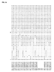
FIG. 1, which is presented in the drawings as FIGS. I A-G, is an alignment of
a
subset of fluorescent proteins spanning representative genus (SEQ ID NOS 49-
65. 10, 14, 28, 42,
and 66-70, respectively, in order of appearance). The highlighted portion of
the sequences
corresponds to the chromophore region of the fluorescent proteins.
FIG. 2 is a map of a typical fluorescent protein, shown with terminal regions
separated from the internal chromophore region. The 3-residue chromophore is
shown in the
internal region with a shaded box. Below the map are lines representing 40-
residue sliding
window segments (with region designated) that were examined for congruence
with the terminal
regions. Dark lines indicate sliding windows that were shown to be incongruent
with the terminal
regions with statistical significance. The histogram below the map plots the
difference in number
of steps it takes to construct a phylogenetic tree using the N/C terminal
regions versus the middle
region, for each residue in the protein. Residue position is indicated on the
bottom of the
histogram.
FIGS. 3A-D are diagrams of the crystal structure of a red fluorescent protein
from
Discosoma sp. ("DsRed"). The conserved central domain (dark) and flanking
variable domains
(white/grey) are imposed on a ribbon diagram of monomer (A) and tetramer (B
and C) of the red
6
CA 02780512 2012-05-10
WO 2011/035067 PCT/US2010/049181
fluorescent protein crystal structure. (B) Standard view of the tetramer. (C)
depicts a slight
rotation to highlight the proximity of the fI strands of the conserved region.
(D) Electron density
map created in Chimera (Pettersen et al. (2004). J. Comp. Chemistry 25, pp
1605-1612)
depicting residues (dark) corresponding to the middle conserved region mapped
to the crystal
structure of DsRed.
FIG. 4 is a ball-and-stick diagram of the crystal structure of DsRed. Residues
undergoing rapid molecular change are shown in a darker shade. These residues
were determined
by analyses of fluorescent proteins derived from specimens of Montastrea
cavernosa from
different geographic regions.
FIG. 5 shows the amino acid sequence of red fluorescent protein (SEQ ID NO:
71),
and the respective residues which are highly variable, as compared to other
fluorescent proteins
(asterisks). Residues 61-105 correspond to the conserved central fluorescent
protein region.
This region is flanked by the N- and C-terminal variable regions (residues I
to 60, and 106 to
225) respectively. Residues 13, 30, 32, 34, 36, 41, 43, 45, 201, and 211 are
fluorescent protein
residues homologous to Perlecan-binding residues in Nidogens. The boxed
residues are
homologous to both Perlecan-binding residues in Nidogen proteins, and highly
variable residues
in fluorescent proteins.
FIG. 6 shows the emission spectra of both eGFP and the fluorescent polypeptide
comprising the sequence set forth in SEQ ID NO: 24.
FIG. 7 shows the emission and excitation spectra at 25 C and 37 C for the
isolated
fluorescent polypeptide comprising the sequence set forth in SEQ ID NO: 12.
FIG. 8 sets forth a schematic diagram of a disclosed method. Cells expressing
both a
protein kinase of interest and PhosFluor (the PhosFluor Detection System) are
plated onto a
multi-well grid for compound screening. Compounds are then added to the wells
to test for
kinase inhibition, in the presence or absence of a stimulus. Fluorescence is
used as the readout
of kinase activity. Both the IC50 and kinetic data can be obtained using this
system.
FIGS. 9A-9B disclose the modulation of PhosFluor by alkaline pH and
phosphorylation. A. Equal quantities of the indicated fluorescent proteins
were subjected to
different pH by addition of Tris buffer. Normalized fluorescence intensity
(excitation at 485 nm
and emission at 538 nm, compared to the value at pH 7.0) was measured using a
SpectraMax M5
Microplate Reader. B. Equal quantities of the recombinant PhosFluor or eGFP
were subjected to
7
CA 02780512 2012-05-10
WO 2011/035067 PCT/US2010/049181
phosphorylation by the indicated protein kinases or to dephosphorylation with
alkaline
phosphatase (Alk Phos). Fluorescence intensity (representing the change in
emission of a part of
the emission spectrum compared to the value at 0 hours) was measured over time
during the
reaction. The PhosFluor reaction containing no enzyme showed an increase in
fluorescence over
time, but virtually no change was observed when the reaction was incubated
with alkaline,
thereby suggesting the presence of an endogenous bacterial protein kinase that
was co-purified
with PhosFluor.
FIG. 10 discloses that PhosFluor is an avid substrate for PKC and Src. Equal
quantities of PhosFluor or eGFP incubated with either PKC or Src for the
indicated times (in
hours) were resolved on 10% SDS-PAGE gels and immunoblotted to antibodies that
reveal total
protein (a polyhistidine monoclonal antibody) or phospho-tyrosine. M
designates the position of
the monomeric form of fluorescent protein while P designates the position of
phosphorylated
forms of the protein that have undergone a motility shift due to increased
negative charge.
FIG. 11 discloses that phosphorylation of PhosFluor alters its spectral
properties.
The excitation and emission spectra of PhosFluor and eGFP are depicted under
control
conditions (no kinase) and after phosphorylation by the indicated kinases. The
excitation and
emission spectra for eGFP are all superimposed under a single curve, while
phosphorylation of
PhosFluor alters both spectra compared to control conditions.
FIG. 12 discloses the expression of PhosFluor in mammalian cells. HEK-293
cells
were transfected with PhosFluor driven by the CMV promoter, and visualized
with a fluorescent
microscope 1 week later.
DETAILED DESCRIPTION OF THE DISCLOSURE
De initions
Unless specifically indicated otherwise, all technical and scientific terms
used herein
have the same meaning as commonly understood by those of ordinary skill in the
art to which
this disclosure pertains, or with which it is most nearly connected. For
purposes of the present
invention, the following terms are defined.
The term "nucleic acid molecule" or "polynucleotide" refers to a
deoxyribonucleotide
or ribonucleotide polymer in either single-stranded or double-stranded form,
and, unless
specifically indicated otherwise, encompasses polynucleotides containing known
analogs of
8
CA 02780512 2012-05-10
WO 2011/035067 PCT/US2010/049181
naturally-occurring nucleotides which can function in a similar manner as
naturally-occurring
nucleotides. It will be understood that when a nucleic acid molecule is
represented by a DNA
sequence, this also includes RNA molecules comprising a nucleotide sequence
which
corresponds to a DNA sequence in which "U" (uridine) replaces "T" (thymidine).
The tern "recombinant nucleic acid molecule" refers to a non-naturally
occurring
nucleic acid molecule containing two or more linked polynucleotide sequences.
A recombinant
nucleic acid molecule can be produced by recombination methods, particularly
genetic
engineering techniques, or can be produced by a chemical synthesis method. A
recombinant
nucleic acid molecule can encode a fusion protein, for example, a disclosed
fluorescent protein
variant linked to a polypeptide or peptide of interest. The term "recombinant
host cell" refers to
a cell that contains a recombinant nucleic acid molecule. As such, a
recombinant host cell can
express a polypeptide from a "gene" that is not found within the native (non-
recombinant) form
of the cell.
Reference to a polynucleotide "encoding" a polypeptide means that, upon
transcription of the polynucleotide and translation of the mRNA transcript, a
polypeptide is
produced. The encoding polynucleotide is considered to include both the coding
strand, whose
nucleotide sequence is identical to the nucleotide sequence of an mRNA, as
well as its
complementary strand, whose nucleotide sequence is complementary to the
nucleotide sequence
of an mRNA. It will be recognized that such a polynucleotide encoding a
polypeptide is
considered to include degenerate nucleotide sequences, which encode the same
amino acid
residues. Nucleotide sequences encoding a polypeptide can include
polynucleotides containing
introns as well as the encoding exons.
The term "expression control sequence" refers to a nucleotide sequence that
regulates
the transcription or translation of a polynucleotide or the localization of a
polypeptide to which
to which it is operatively linked. Expression control sequences are
"operatively linked" when the
expression control sequence controls or regulates the transcription and, as
appropriate,
translation of the nucleotide sequence which encodes a polypeptide (i.e., a
transcription or
translation regulatory element, respectively), or localization of the encoded
polypeptide to a
specific compartment of a cell or tissue. Thus, an expression control sequence
can be a
promoter, enhancer, transcription terminator, a start codon (ATG), a splicing
signal for intron
excision and maintenance of the correct reading frame, a STOP codon, a
nucleotide sequence
9
CA 02780512 2012-05-10
WO 2011/035067 PCT/US2010/049181
encoding a ribosome binding site, or a sequence that targets a polypeptide to
a particular
location, for example, a cell compartmentalization signal which can target a
polypeptide to the
cytosol, nucleus, plasma membrane, endoplasmic reticulum, mitochondrial
membrane or matrix,
chloroplast membrane or lumen, medial trans-Golgi cistemae, or a lysosome or
endosome. Cell
compartmentalization domains are well known in the art and include, for
example, a peptide
containing amino acid residues 1 to 81 of human type 11 membrane-anchored
protein
gal actosyltransferase, or amino acid residues 1 to 12 of the presequence of
subunit IV of
cytochrome c oxidase (see, also, Hancock et al. (1991), EMBO J. 10, pp. 4033-
4039; Buss et al.
(1988), Mol. Cell. Biol. 8, pp. 3960-3963; U.S. Patent No. 5,776,689, each of
which is
incorporated herein by reference).
The term "operatively linked" or "operably linked" or "operatively joined,"
when used
herein to describe fusion proteins, refers to polypeptide sequences that are
placed in a physical
and/or functional relationship with each other. The functional activity of the
components of a
given fusion protein are preferably unchanged compared to the functional
activities of the
individual components when the components are not operatively joined. For
example, a
disclosed fluorescent polypeptide or variant thereof, can be fused to a
polypeptide of interest. In
this case, it is preferable that the fusion molecule retains its fluorescence,
and the polypeptide of
interest retains its original biological activity. In some embodiments of the
present invention, the
activities of either the fluorescent protein or the protein of interest can be
reduced relative to their
activities in isolation. Such fusions can also find use with the present
invention. As used herein,
the chimeric fusion molecules of the invention can be in a monomeric state, or
in a multimeric
state (e.g., dimeric, trimeric or tetrameric).
The term "oligomer" refers to a complex formed by the specific interaction of
two or
more polypeptides. A "specific interaction" or "specific association" is one
that is relatively
stable under specified conditions, for example, physiologic conditions.
Reference to a
"propensity" of proteins to oligomerize indicates that the proteins can form
dimers, trimers,
tetramers, or the like under specified conditions. Generally, fluorescent
proteins such as GFPs
have a propensity to oligomerize under physiologic conditions although, as
disclosed herein,
fluorescent proteins also can oligomerize, for example, under pH conditions
other than
physiologic conditions. The conditions under which fluorescent proteins
oligomerize or have a
CA 02780512 2012-05-10
WO 2011/035067 PCT/US2010/049181
propensity to oligomerize can be determined using well known methods as
disclosed herein or
otherwise known in the art.
The terms "polypeptide" and "protein" are synonymous, and refer to a polymer
of two
or more amino acid residues. The terms apply to amino acid polymers in which
one or more
amino acid residue is an artificial chemical analog of a corresponding
naturally-occurring amino
acid, as well as to naturally-occurring amino acid polymers.
The term "recombinant protein" refers to a protein that is produced by
expression of a
recombinant polynucleotide encoding the protein.
The term "isolated" or "purified" refers to a material that is substantially
or essentially
free from components that normally accompany the material in its native state.
Purity or
homogeneity generally is determined using analytical techniques such as
polyacrylamide gel
electrophoresis and high performance liquid chromatography. A polynucleotide
or a polypeptide
is considered to be isolated when it is the predominant polynucleotide or a
polypeptide present in
a preparation, respectively. An isolated protein or nucleic acid molecule
represents greater than
80% of the macromolecular species present in a preparation, greater than 90%
of all
macromolecular species present, greater than 95% of all macromolecular species
present, greater
than 96% of all macromolecular species present, greater than 97% of all
macromolecular species
present, greater than 98% of all macromolecular species present, greater than
99% of the
macromolecular species, and, in particular, is a polypeptide or polynucleotide
that purified
essentially to homogeneity such that the polypeptide or polynucleotide is the
only
macromolecular species detected when examined using conventional methods for
determining
purity of such a molecule.
The term "naturally-occurring" is used to refer to a protein, nucleic acid
molecule,
cell, or other material that occurs in nature (i.e., wild type molecule), for
example, a polypeptide
or polynucleotide sequence that is present in an organism, including in a
virus. A naturally-
occurring material can be in its form as it exists in nature, and can be
modified by the hand of
man such that, for example, is in an isolated form.
The term "antibody" refers to a polypeptide encoded by at least one portion of
an
immunoglobulin gene. The recognized immunoglobulin genes include the kappa,
lambda, alpha,
gamma, delta, epsilon and mu constant region genes, as well as the myriad of
immunoglobulin
variable region genes. Antibodies exist as intact immunoglobulins and as well
characterized
11
CA 02780512 2012-05-10
WO 2011/035067 PCT/US2010/049181
antigen-binding fragments of an antibody which can be produced by digestion
with a peptidase
or by recombinant DNA methods. Such antigen-binding fragments of an antibody
include, for
example, Fv, Fab' and F(ab)'2 fragments. "Antibody," as used herein, includes
antibody
fragments either produced by the modification of whole antibodies or those
synthesized de novo
using recombinant DNA methodologies.
The term "identical" is used herein in reference to two or more polynucleotide
sequences or, alternatively, two or more polypeptide sequences. The term
"identical" refers to
nucleotides in one nucleotide sequence that are the same as nucleotides in
another nucleotide
sequence when the nucleotide sequences are aligned for maximum correspondence.
Similarly,
the term "identical" refers to amino acid residues in one amino acid sequence
that are the same as
amino acid residues in another amino acid sequence when the amino acid
sequences are aligned
for maximum correspondence. When percentage of sequence identity is used in
reference to a
polypeptide, it is recognized that one or more residue positions that are not
otherwise identical
can differ by a conservative amino acid substitution, in which a first amino
acid residue is
substituted for another amino acid residue having similar chemical properties
such as a similar
charge. hydrophobic character, or hydrophilic character and, therefore, does
not substantially
change the functional properties of the polypeptide. Where polypeptide
sequences differ in
conservative substitutions, the percent sequence identity can be adjusted
upwards to correct for
the conservative nature of the substitution. Such an adjustment can be made
using well known
methods, for example, scoring a conservative substitution as a partial rather
than a full mismatch,
thereby increasing the percentage sequence identity. Thus, for example, where
an identical
amino acid is given a score of 1 and a non-conservative substitution is given
a score of zero, a
conservative substitution is given a score between zero and 1. The scoring of
conservative
substitutions can be calculated using any well known algorithm (see, e.g.,
Meyers and Miller
(1988), Comp. Appl. Biol. Sci. 4, pp. 11-17; Smith and Waterman (1981), Adv.
Appl. Math. 2, p.
482; Needleman and Wunsch (1970), J. Mol. Biol. 48, p. 443; Pearson and Lipman
(1988), Proc.
Nall. Acad. Sci., USA 85, p. 2444; Higgins and Sharp (1988), Gene 73, pp. 237-
244). Manual
alignment also can be performed by simple visual inspection and manual
alignment of
sequences. Such manual alignments are well known in the art.
Two or more nucleotide sequences are considered to be "substantially
identical" if the
nucleotide sequences share at least 80% sequence identity with each other, or
with a reference
12
CA 02780512 2012-05-10
WO 2011/035067 PCT/US2010/049181
sequence over a given comparison window. Similarly, two or more amino acid
sequences are
considered to be "substantially identical" or "substantially similar" if the
amino acid sequences
share at least 80% sequence identity with each other, or with a reference
sequence over a given
comparison window. Thus, substantially similar sequences include those having,
for example, at
least 85% sequence identity, at least 88% sequence identity, at least 90%
sequence identity, at
least 92% sequence identity, at least 95% sequence identity, at least 97%
sequence identity, or at
least 99% sequence identity.
A "variant" polypeptide or "variant" polynucleotide is substantially identical
in
sequence to the respective native (wild type) polypeptide or polynucleotide.
Fluorescent molecules are useful in fluorescence resonance energy transfer,
FRET,
which involves a donor molecule and an acceptor molecule. To optimize the
efficiency and
detectability of FRET between a donor and acceptor molecule, several factors
need to be
balanced. The emission spectrum of the donor should overlap as much as
possible with the
excitation spectrum of the acceptor to maximize the overlap integral. Also,
the quantum yield of
the donor moiety and the extinction coefficient of the acceptor should be as
high as possible to
maximize Ro, which represents the distance at which energy transfer efficiency
is 50c%c.
However, the excitation spectra of the donor and acceptor should overlap as
little as possible so
that a wavelength region can be found at which the donor can be excited
efficiently without
directly exciting the acceptor because fluorescence arising from direct
excitation of the acceptor
can be difficult to distinguish from fluorescence arising from FRET.
Similarly, the emission
spectra of the donor and acceptor should overlap as little as possible so that
the two emissions
can be clearly distinguished. High fluorescence quantum yield of the acceptor
moiety is
desirable if the emission from the acceptor is to be measured either as the
sole readout or as part
of an emission ratio. One factor to be considered in choosing the donor and
acceptor pair is the
efficiency of fluorescence resonance energy transfer between them. Preferably,
the efficiency of
FRET between the donor and acceptor is at least 10%, more preferably at least
50% and even
more preferably at least 80%.
The term "fluorescent property" or "fluorescent characteristics" refers to the
molar
extinction coefficient at an appropriate excitation wavelength, the
fluorescence quantum
efficiency, the shape of the excitation spectrum or emission spectrum, the
excitation wavelength
maximum and emission wavelength maximum, the ratio of excitation amplitudes at
two different
13
CA 02780512 2012-05-10
WO 2011/035067 PCT/US2010/049181
wavelengths, the ratio of emission amplitudes at two different wavelengths,
the excited state
lifetime, fluorescence intensity, fluorescence lifetime, multiphoton cross-
section, fluorescence
resonance energy transfer efficiency, bioluminescence resonance energy
transfer efficiency, or
the fluorescence anisotropy. A measurable difference in any one of these
properties between the
wild type GFP of A. victoria and a spectral variant thereof, or the wild type
GFP of A. victoria
and a mutant of a spectral variant thereof, is useful. A measurable difference
can be determined
by determining the amount of any quantitative fluorescent property, e.g., the
amount of
fluorescence at a particular wavelength, or the integral of fluorescence over
the emission
spectrum.
As used herein, the term "fluorescent protein" refers to any protein that can
fluoresce
when excited with an appropriate electromagnetic radiation, except that
chemically tagged
proteins, wherein the fluorescence results from the chemical tag, and
polypeptides that fluoresce
only due to the presence of certain amino acids such as tryptophan or
tyrosine, whose emission
peaks at ultraviolet wavelengths (i.e., less that about 400 nm) are not
considered fluorescent
proteins for purposes of the present disclosure. In general, a disclosed
fluorescent protein is a
protein which derives its fluorescence from autocatalytically forming a
chromophore. A
fluorescent protein can contain amino acid sequences that are naturally
occurring or that have
been engineered (i.e., variants or mutants). When used in reference to a
fluorescent protein, the
term "mutant" or "variant" refers to a protein that is different from a
reference fluorescent
protein. For example, a spectral variant of Aequorea GFP can be derived from
the naturally
occurring GFP by engineering mutations such as amino acid substitutions into
the reference GFP
protein (see, e.g., U.S. Patent No. 5,777,079). A "spectral variant" or
"spectral mutant" of a
fluorescent protein indicates a mutant fluorescent protein which has a
different fluorescence
characteristic with respect to the corresponding wild type or reference
fluorescent protein.
The term "coral" as used herein encompasses species within the class Anthozoa
(e.g.,
species of the order Scleractinia) and includes corals, stony corals and
corallimorphs.
Fluorescent Proteins
The GFP of A. victoria and blue, cyan, and yellow variants thereof have found
widespread use as both genetically-encoded indicators for tracking gene
expression and as
donor/acceptor pairs for fluorescence resonance energy transfer (FRET).
However, extending
the spectrum of available colors to red wavelengths, and the further
engineering of these proteins
14
CA 02780512 2012-05-10
WO 2011/035067 PCT/US2010/049181
to create biosensors and to detect novel protein-protein interactions, e.g.,
for high throughput
drug screening would provide distinct new labels for multicolor tracking of
fusion proteins and
the detection of various interactions.
Accordingly, disclosed herein are isolated fluorescent proteins from organisms
of the
order Scleractinia, which are indigenous to the Australian Great Barrier Reef.
Further disclosed
are fluorescent proteins with fluorescent properties disclosed above.
Illustrative examples of the disclosed isolated fluorescent proteins include
proteins
comprising an amino acid sequence selected from the groups consisting of SEQ
ID NOs: 10, 12,
14, 16, 18, 20, 22, 24, 26, 28, 30, 32, 34. 36, 38, 40, 42, and 44. The
sequences associated with
these SEQ ID NOs are provided in the Sequence Listing.
Also disclosed herein are polynucleotides encoding the isolated fluorescent
proteins.
In one embodiment. a nucleic acid molecule is provided that comprises a
nucleotide sequence
selected from the group consisting of SEQ ID NOs: 11, 13, 15, 17, 19, 21, 23,
25, 27, 29, 31, 33,
35, 37, 39, 41, 43, and 45. The sequences associated with these SEQ ID NOs are
provided in the
Sequence Listing.
The disclosed isolated fluorescent proteins contain three distinct regions: a
first
region of 45 amino acid residues which includes a chromophore; a second region
of 50- amino
acid residues N-terminal of the first region; and a third region of 140 amino
acid residues C-
terminal of the first).
The chromophore region of 45 amino acid residues, specifically residues 70 to
115,
displays a sharply divergent evolutionary pattern from the rest of the
protein. The chromophore
region evolved slowly under stabilizing selection. The structure of the
chromophore region
contains an a helix and a single ~3 strand. The ~3 strand faces inward in the
tetrameric fluorescent
protein complex (see FIG. 3).
The N-terminal and C-terminal regions are under intense Darwinian selection
and
evolve rapidly with mutations appearing at sites of putative protein-protein
interactions (see
FIG. 4). In addition, fluorescence color is significantly associated with the
N-terminal and C-
terminal hypervariable regions and not with the middle conserved region.
CA 02780512 2012-05-10
WO 2011/035067 PCT/US2010/049181
Fluorescent Protein Variants
The present invention provides variant fluorescent proteins, which differ from
the
fluorescent proteins having a polypeptide sequence as set forth in any of SEQ
ID NOS: 10, 12,
14, 16, 18, 20, 22, 24, 26, 28, 30, 32, 34, 36, 38, 40, 42 or 44.
In one embodiment, the present invention is directed to a novel fluorescent
protein
having an amino acid sequence comprising a sequence with at least 80% identity
to that of any of
sequences identified as SEQ ID NOS: 10, 12, 14, 16, 18, 20, 22, 24, 26, 28,
30, 32, 34, 36, 38,
40, 42 and 44. In a further embodiment, the variant has at least 85%, 90%,
92%, 959c, 97c%c,
98% or 99c%o identity to the aforementioned sequences.
In one embodiment, the fluorescent protein variant of the present invention
has a
mutation in at least one of the hypervariable residues. In a further
embodiment, the one or more
mutations results in a fluorescent protein with less of a propensity to
oligomerize, or a protein
that specifically binds a protein or small molecule of interest.
As stated above, variant fluorescent proteins of the invention can have a
reduced
propensity to oligomerize, due to the presence of one or more mutations at the
fluorescent
protein's surface. In one embodiment, one of the starred residues in FIG. 5 is
mutated to arrive at
a variant fluorescent protein with a reduced propensity to oligomerize.
Amino acids with charged (ionized D, E, K, and R), dipolar (H, N, Q, S, T, and
uncharged D, E and K), and polarizable side groups (e.g., C, F, H, M, W and Y)
are useful for
altering the ability of fluorescent proteins to oligomerize or interact with
other proteins,
especially when they substitute an amino acid with an uncharged, nonpolar or
non-polarizable
side chain.
In one embodiment, the present invention provides a variant fluorescent
protein that
fluoresces at a different wavelength, as compared to the protein the variant
was derived from. In
this embodiment, one or more residues in the fluorescent protein's terminal
hypervariable region
(region does not include the chromophore) is mutated.
In another embodiment, a variant fluorescent protein of the present invention
only
fluoresces when binding to a protein of interest.
Fusion Proteins Comprising the Disclosed Fluorescent Proteins
Fluorescent proteins fused to target proteins can be prepared, for example
using
recombinant DNA methods, and used as markers to identify the location and
amount of the target
16
CA 02780512 2012-05-10
WO 2011/035067 PCT/US2010/049181
protein produced. Accordingly, the present invention provides fusion proteins
comprising a
fluorescent protein (including those described above and variants thereof) and
a polypeptide or
peptide of interest. The polypeptide of interest can be of any length, for
example, about 15
amino acid residues, about 50 residues, about 150 residues, or up to about
1000 amino acid
residues or more, provided that the fluorescent protein component of the
fusion protein can
fluoresce or can be induced to fluoresce when exposed to electromagnetic
radiation of the
appropriate wavelength. The polypeptide of interest can be, for example, a
peptide tag such as a
polyhistidine sequence, a c-myc epitope, a FLAG epitope, and the like; can be
an enzyme, which
can be used to effect a function in a cell expressing a fusion protein
comprising the enzyme or to
identify a cell containing the fusion protein; can be a protein to be examined
for an ability to
interact with one or more other proteins in a cell, or any other protein as
disclosed herein or
otherwise desired.
A fusion protein of the present invention can include a fluorescent protein
disclosed
herein operatively linked to one or more polypeptides of interest. The two or
more polypeptides
of the fusion protein can be linked through peptide bonds, or the fluorescent
protein can be
linked to the one or more polypeptides of interest through a linker molecule.
In one embodiment, a linker can be present to join the fluorescent protein of
the
present invention and a polypeptide of interest. If the linker between the two
moieties is a non-
peptide linker, the two subunits will be encoded by separate polynucleotide
molecules, produced
separately, and subsequently linked by methods known in the art.
In another embodiment, the fusion protein is expressed from a recombinant
nucleic
acid molecule containing a polynucleotide encoding a fluorescent protein
disclosed herein
operatively linked to one or more polynucleotides encoding one or more
polypeptides of interest.
A polypeptide of interest can be any polypeptide, including, for example, a
peptide
tag such as a polyhistidine peptide, or a cellular polypeptide such as an
enzyme, a G-protein, a
growth factor receptor, or a transcription factor; and can be one of two or
more proteins that can
associate to form a complex. In one embodiment, the fusion protein is a tandem
fluorescent
protein variant construct, which includes a donor fluorescent protein
disclosed herein, an
acceptor fluorescent protein disclosed herein, and a peptide linker moiety
coupling said donor
and said acceptor, wherein cyclized amino acids of the donor emit light
characteristic of said
donor, and wherein the donor and the acceptor exhibit fluorescence resonance
energy transfer
Z__
CA 02780512 2012-05-10
WO 2011/035067 PCT/US2010/049181
when the donor is excited, and the linker moiety does not substantially emit
light to excite the
donor. As such, a fusion protein of the invention can include two or more
operatively linked
fluorescent proteins, which can be linked directly or indirectly, and can
further comprise one or
more polypeptides of interest.
Preparation of Fluorescent Proteins
The present invention also provides polynucleotides encoding fluorescent
proteins, or
variants thereof, where the protein can be a fluorescent protein isolated from
Scleractinia (Lizard
Island, Australia), a variant thereof, or a fusion protein comprising such a
fluorescent protein (or
variant) operatively linked to one or more polypeptides of interest.
The invention further provides vectors containing such polynucleotides, and
host cell
containing a polynucleotide or vector. Also provided is a recombinant nucleic
acid molecule,
which includes at least one polynucleotide encoding a fluorescent protein
operatively linked to
one or more other polynucleotides. The one or more other polynucleotides can
be, for example,
a transcription regulatory element such as a promoter or polyadenylation
signal sequence, or a
translation regulatory element such as a ribosome binding site. Such a
recombinant nucleic acid
molecule can be contained in a vector, which can be an expression vector, and
the nucleic acid
molecule or the vector can be contained in a host cell.
The vector generally contains elements required for replication in a
prokaryotic or
eukaryotic host system or both, as desired. Such vectors, which include
plasmid vectors and
viral vectors such as bacteriophage, baculovirus, retrovirus, lentivirus,
adenovirus, vaccinia
virus. semliki forest virus and adeno-associated virus vectors, are well known
and can be
purchased from a commercial source (Promega, Madison Wis.; Stratagene, La
Jolla Calif.;
GIBCO/BRL. Gaithersburg Md.) or can be constructed by one skilled in the art
(see, e.g., Meth.
Enzymol., Vol. 185, Goeddel, ed. (Academic Press, Inc., 1990); Jolly (1994),
Canc. Gene her.
1, pp. 51-64; Flotte (1993), Bioenerg. Biomemb. 25, pp. 37-42; Kirshenbaum et
al. (1992). J.
Clin. Invest. 92, pp. 381-387; each of which is incorporated herein by
reference in its entirety).
A vector for containing a polynucleotide encoding a fluorescent protein can be
a
cloning vector or an expression vector, and can be a plasmid vector, viral
vector, and the like.
Generally, the vector contains a selectable marker independent of that encoded
by a
polynucleotide of the invention, and further can contain transcription or
translation regulatory
elements, including a promoter sequence, which can provide tissue specific
expression of a
18
CA 02780512 2012-05-10
WO 2011/035067 PCT/US2010/049181
polynucleotide operatively linked thereto, which can, but need not, be the
polynucleotide
encoding the fluorescent protein, for example, a variant fluorescent protein
with a decreased
propensity to oligomerize, thus providing a means to select a particular cell
type from among a
mixed population of cells containing the introduced vector and recombinant
nucleic acid
molecule contained therein.
Where the vector is a viral vector, it can be selected based on its ability to
infect one
or few specific cell types with relatively high efficiency. For example, the
viral vector also can
be derived from a virus that infects particular cells of an organism of
interest, for example,
vertebrate host cells such as mammalian host cells. Viral vectors have been
developed for use in
particular host systems, particularly mammalian systems and include, for
example, retroviral
vectors, other lentivirus vectors such as those based on the human
immunodeficiency virus
(HIV), adenovirus vectors, adeno-associated virus vectors, herpesvirus
vectors, vaccinia virus
vectors, and the like (see Miller and Rosman (1992), BioTechniques 7, pp. 980-
990; Anderson et
al. (1998), Nature 392, pp. 25-30 Suppl.; Verma and Somia (1997). Nature 389,
pp. 239-242;
Wilson (1996), New Engl..1. Med. 334, pp. 1185-1187).
Recombinant production of a fluorescent protein, which can be a component of a
fusion protein, involves expressing a polypeptide encoded by a polynucleotide.
A
polynucleotide encoding the fluorescent protein is a useful starting material.
In one embodiment,
the polynucleotide comprises one or more of the sequences identified as SEQ ID
NOS: 11, 13,
15, 17, 19, 21, 23, 25, 27 ,29, 31, 33, 35, 37, 39, 31 or 43 or 45 is
employed. In another
embodiment, a polynucleotide comprises a polynucleotide sequence having at
least 80% identity
to one of the sequences identified as SEQ ID NOS: 11, 13, 15, 17, 19, 21, 23,
25, 27, 29, 31, 33,
35, 37, 39, 31 or 43 or 45. In a further embodiment, the variant has at least
85%, at least 87%, at
least 90%, at least 929c. at least 95% or at least 97% identity to one of the
sequences identified as
SEQ ID NOS: 11, 13, 15, 17, 19, 21, 23, 25, 27, 29, 31, 33, 35, 37, 39, 31 or
43 or 45.
Polynucleotides encoding a fluorescent protein are disclosed herein or
otherwise
known in the art, and can be obtained using routine methods, then can be
modified such that the
encoded fluorescent protein has a biophysical property altered. For example,
the resulting
fluorescent protein variant may be engineered to bind specifically to a
protein target.
Alternatively or additionally, the variant may emit fluorescence only when the
binding occurs.
19
CA 02780512 2012-05-10
WO 2011/035067 PCT/US2010/049181
For example, a polynucleotide encoding a fluorescent protein of the present
invention, can be isolated by PCR of cDNA from Sclereaciinia using primers
based on the
polynucleotide sequences provided as SEQ ID NOS: 11, 13, 15, 17, 19, 21, 23,
25, 27, 29, 31,
33, 35, 37, 39, 41, 43 or 45, or alternatively, sets of degenerate primers
(e.g., primers comprising
a sequence identified as any of SEQ ID NOS: 1-9). PCR methods are well known
and routine in
the art (see, e.g., U.S. Pat. No. 4,683,195; Mullis et al. (1987), Cold Spring
Harbor Syrup. Quarrl.
Biol. 51, p. 263; Erlich, ed., "PCR Technology" (Stockton Press, NY, 1989)). A
variant form of
the fluorescent protein then can be made by site-specific mutagenesis of the
polynucleotide
encoding the fluorescent protein.
The construction of expression vectors and the expression of a polynucleotide
in
transfected cells involves the use of molecular cloning techniques also well
known in the art (see
Sambrook et al., In "Molecular Cloning: A Laboratory Manual" (Cold Spring
Harbor Laboratory
Press 1989); "Current Protocols in Molecular Biology" (eds., Ausubel et al.;
Greene Publishing
Associates, Inc., and John Wiley & Sons, Inc. 1990 and supplements)).
Expression vectors
contain expression control sequences operatively linked to a polynucleotide
sequence of interest,
for example, that encodes a fluorescent protein variant, as indicated above.
The expression
vector (for example, pCR4Blunt-TOPO (Invitrogen, Carlsbad, CA)) can be adapted
for function
in prokaryotes or eukaryotes by inclusion of appropriate promoters,
replication sequences,
markers, and the like. An expression vector can be transfected into a
recombinant host cell for
expression of a fluorescent protein variant, and host cells can be selected,
for example, for high
levels of expression in order to obtain a large amount of isolated protein. A
host cell can be
maintained in cell culture, or can be a cell in vivo in an organism. A
fluorescent protein or
variant thereof can be produced by expression from a polynucleotide encoding
the protein in a
host cell such as E. coll.
An expressed fluorescent protein of the present invention, or variant thereof,
can be
operatively linked to a first polypeptide of interest, further can be linked
to a second polypeptide
of interest, for example, a peptide tag, which can be used to facilitate
isolation of the fluorescent
protein variant, including any other polypeptides linked thereto. For example,
a polyhistidine tag
containing, for example, six histidine residues, can be incorporated at the N-
terminus or C-
terminus of the fluorescent protein (or variant thereof), which then can be
isolated in a single
step using nickel-chelate chromatography. Additional peptide tags, including a
c-myc peptide, a
CA 02780512 2012-05-10
WO 2011/035067 PCT/US2010/049181
FLAG epitope, or any other ligand, including any peptide epitope (or antibody,
or antigen
binding fragment thereof, that specifically binds the epitope are well known
in the art and
similarly can be used (see, e.g., Hopp et at. (1988). Biotechnology 6, pp.
1204; U.S. Patent No.
5,011,912).
Kits
The present invention is also directed to kits, in order to facilitate and/or
standardize
use of compositions provided by the present invention, as well as to
facilitate the methods of the
present invention. Materials and reagents to carry out these various methods
can be provided in
kits to facilitate execution of the methods. As used herein, the term "kit" is
used in reference to a
combination of articles that facilitate a process, assay, analysis or
manipulation.
Kits can contain chemical reagents (e.g., polypeptides or polynucleotides) as
well as
other components. In addition, kits of the present invention can also include,
for example but not
limited to, apparatus and reagents for sample collection and/or purification,
apparatus and
reagents for product collection and/or purification, reagents for bacterial
cell transformation,
reagents for eukaryotic cell transfection, previously transformed or
transfected host cells, sample
tubes, holders, trays, racks, dishes, plates, instructions to the kit user,
solutions, buffers or other
chemical reagents (e.g., oligonucleotide primers), suitable samples to be used
for standardization,
normalization, and/or control samples. Kits of the present invention can also
be packaged for
convenient storage and safe shipping, for example, in a box having a lid.
In some embodiments, for example, kits of the present invention can provide a
fluorescent protein of the invention, a polynucleotide vector (e.g., a
plasmid) encoding a
fluorescent protein of the invention (including variant(s) thereof), bacterial
cell strains suitable
for propagating the vector, and reagents for purification of expressed fusion
proteins.
Alternatively, a kit of the present invention can provide the reagents
necessary to conduct
mutagenesis of fluorescent proteins isolated from Scleractinia, in order to
generate a fluorescent
protein variant of the present invention having a novel biophysical or
biochemical property.
A kit can contain one or more compounds of the invention, for example, one or
a
plurality of fluorescent proteins or variants, which can be a portion of a
fusion protein, or one or
a plurality of polynucleotides that encode the polypeptides. The fluorescent
protein variant can
be a mutated fluorescent protein having a fluorescent emission spectrum at a
wavelength
different than the native protein. In one embodiment, the kit comprises a
plurality of fluorescent
21
CA 02780512 2012-05-10
WO 2011/035067 PCT/US20101049181
protein variants, or at least one isolated fluorescent protein of the present
invention, and reagents
sufficient to carry out site directed mutagenesis.
A kit of the invention also can contain one or a plurality of recombinant
nucleic acid
molecules, which encode, in part or full, a fluorescent protein of the present
invention or variant
thereof, and can further include, for example, an operatively linked second
polynucleotide
containing or encoding a restriction endonuclease recognition site or a
recombinase recognition
site, or any polypeptide of interest. In addition, the kit can contain
instructions for using the
components of the kit, particularly the compositions of the invention that are
contained in the kit.
Such kits can be particularly useful where they provide a plurality of
different
fluorescent proteins or variants because the artisan can conveniently select
one or more proteins
having the fluorescent properties desired for a particular application.
Similarly, a kit containing
a plurality of polynucleotides encoding different fluorescent protein variants
provides numerous
advantages. For example, the polynucleotides can be engineered to contain
convenient
restriction endonuclease or recombinase recognition sites, thus facilitating
operative linkage of
the polynucleotide to a regulatory element or to a polynucleotide encoding a
polypeptide of
interest or, if desired, for operatively linking two or more the
polynucleotides encoding the
fluorescent protein variants to each other.
Uses of Disclosed Fluorescent Proteins
An isolated fluorescent protein of the present invention (i.e., isolated from
Scleractinia), or variant of the isolated protein, is useful in any method
that employs a
fluorescent protein. Thus, the fluorescent proteins and variants, are useful
as fluorescent markers
in the many ways fluorescent markers already are used, including, for example,
coupling
fluorescent protein variants to antibodies, polynucleotides or other receptors
for use in detection
assays such as immunoassays or hybridization assays, to track the movement of
proteins in cells,
or for the identification of protein-protein interactions, or protein-small
molecule interactions.
For intracellular tracking studies, a first polynucleotide encoding the
fluorescent
protein variant is fused to a second polynucleotide encoding a protein of
interest and the
construct, if desired, can be inserted into an expression vector. Upon
expression inside the cell,
the protein of interest can be localized based on fluorescence, without
concern that localization
of the protein is an artifact caused by oligomerization of the fluorescent
protein component of the
fusion protein. In one embodiment of this method, two proteins of interest
independently are
CA 02780512 2012-05-10
WO 2011/035067 PCT/US2010/049181
fused with two fluorescent protein variants that have different fluorescent
characteristics. This
allows for the tracking of two proteins simultaneously.
The isolated fluorescent proteins and variants of this invention are useful in
systems
to detect induction of transcription. For example, a nucleotide sequence
encoding an isolated
Scleractinia fluorescent protein or variant can be fused to a promoter or
other expression control
sequence of interest, which can be contained in an expression vector. The
construct can be
transfected into a cell, and induction of the promoter (or other regulatory
element) can be
measured by detecting the presence or amount of fluorescence, thereby allowing
a means to
observe the responsiveness of a signaling pathway from receptor to promoter.
Fluorescent proteins and variants of the invention also are useful in
applications
involving fluorescence resonance energy transfer (FRET), which can detect
events as a function
of the movement of fluorescent donors and acceptors towards or away from each
other. One or
both of the donor/acceptor pair can be a fluorescent protein of the present
invention (or variant(s)
thereof). Such a donor/acceptor pair provides a wide separation between the
excitation and
emission peaks of the donor, and provides good overlap between the donor
emission spectrum
and the acceptor excitation spectrum.
FRET can be used to detect cleavage of a substrate having the donor and
acceptor
coupled to the substrate on opposite sides of the cleavage site. Upon cleavage
of the substrate,
the donor/acceptor pair physically separate, eliminating the energy transfer,
and therefore the
fluorescence emission of the acceptor molecule. Such an assay can be
performed, for example,
by contacting the substrate with a sample, and determining a qualitative or
quantitative change in
FRET (see, e.g., U.S. Pat. No. 5,741,657). A fluorescent protein or variant
donor/acceptor pair
also can be part of a fusion protein coupled by a peptide having a proteolytic
cleavage site (see,
e.g., U.S. Pat. No. 5,981,200). FRET also can be used to detect changes in
potential across a
membrane. For example, a donor and acceptor can be placed on opposite sides of
a membrane
such that one translates across the membrane in response to a voltage change,
thereby producing
a measurable FRET (see, e.g., U.S. Pat. No. 5,661,035).
In other embodiments, fluorescent proteins and variants of the invention are
useful for
making fluorescent biosensors for protein kinase and phosphatase activities or
indicators for
small ions and molecules such as Ca 21, Zn2 , cyclic 3', 5'-adenosine
monophosphate, and cyclic
3', 5'-guanosine monophosphate. In these embodiments, the fluorescence
emission of a protein
23
CA 02780512 2012-05-10
WO 2011/035067 PCT/US2010/049181
of the present invention is correlated with the protein kinase or phosphatase
activity,
respectively.
Fluorescence in a sample generally is measured using a fluorimeter, wherein
excitation radiation from an excitation source having a first wavelength,
passes through
excitation optics, which cause the excitation radiation to excite the sample.
In response, a
fluorescent protein variant in the sample emits radiation having a wavelength
that is different
from the excitation wavelength. Collection optics then collect the emission
from the sample.
The device can include a temperature controller to maintain the sample at a
specific temperature
while it is being scanned, and can have a multi-axis translation stage, which
moves a microtiter
plate holding a plurality of samples in order to position different wells to
be exposed. The multi-
axis translation stage, temperature controller, auto-focusing feature, and
electronics associated
with imaging and data collection can be managed by an appropriately programmed
digital
computer, which also can transform the data collected during the assay into
another format for
presentation. This process can be miniaturized and automated to enable
screening many
thousands of compounds in a high throughput format. These and other methods of
performing
assays on fluorescent materials are well known in the art (see, e.g.,
Lakowicz, "Principles of
Fluorescence Spectroscopy" (Plenum Press 1983); Herman, "Resonance energy
transfer
microscopy" In "Fluorescence Microscopy of Living Cells in Culture" Part B,
Meth. Cell Biol.
30:219-243 (ed. Taylor and Wang; Academic Press 1989); Turro, "Modem Molecular
Photochemistry" (Benjamin/Cummings Pub]. Co., Inc. 1978), pp. 296-361, each of
which is
incorporated herein by reference).
A fluorescent protein can be linked to a molecule directly or indirectly,
using any
linkage that is stable under the conditions to which the protein-molecule
complex is to be
exposed. Thus, the fluorescent protein and molecule can be linked via a
chemical reaction
between reactive groups present on the fluorescent protein and molecule, or
the linkage can be
mediated by linker moiety, which contains reactive groups specific for the
fluorescent protein
and the molecule. It will be recognized that the appropriate conditions for
linking the fluorescent
protein of the present invention and the molecule are selected depending, for
example, on the
chemical nature of the molecule and the type of linkage desired. Where the
molecule is a
polypeptide, a convenient means for linking a fluorescent protein variant and
the molecule is by
expressing them as a fusion protein from a recombinant nucleic acid molecule,
which comprises
24
CA 02780512 2012-05-10
WO 2011/035067 PCT/US2010/049181
a polynucleotide encoding, for example, an isolated coral reef fluorescent
protein operatively
linked to a polynucleotide encoding the polypeptide molecule.
A method of identifying an agent or condition that regulates the activity of
an
expression control sequence also is provided. Such a method can be performed,
for example, by
exposing a recombinant nucleic acid molecule, which includes a polynucleotide
encoding a
fluorescent protein variant operatively linked to an expression control
sequence, to an agent or
condition suspected of being able to regulate expression of a polynucleotide
from the expression
control sequence, and detecting fluorescence of the fluorescent protein
variant due to such
exposure. Such a method is useful, for example, for identifying chemical or
biological agents,
including cellular proteins, which can regulate expression from the expression
control sequence,
including cellular factors involved in the tissue specific expression from the
regulatory element.
As such, the expression control sequence can be a transcription regulatory
element such as a
promoter, enhancer, silencer, intron splicing recognition site,
polyadenylation site, or the like; or
a translation regulatory element such as a ribosome binding site.
The fluorescent proteins and variants of the invention also are useful in a
method of
identifying a specific interaction of a first molecule and a second molecule.
Such a method can
be performed, for example, by contacting the first molecule, which is linked
to a donor first
fluorescent protein, and the second molecule, which is linked to an acceptor
second fluorescent
protein, under conditions that allow a specific interaction of the first
molecule and second
molecule; exciting the donor; and detecting fluorescence or luminescence
resonance energy
transfer from the donor to the acceptor, thereby identifying a specific
interaction of the first
molecule and the second molecule. The conditions for such an interaction can
be any conditions
under which is expected or suspected that the molecules can specifically
interact. In particular,
where the molecules to be examined are cellular molecules, the conditions
generally are
physiological conditions. As such, the method can be performed in vitro using
conditions of
buffer, pH, ionic strength, and the like, that mimic physiological conditions,
or the method can
be performed in a cell or using a cell extract.
The first and second molecules can be cellular proteins that are being
investigated to
determine whether the proteins specifically interact, or to confirm such an
interaction. Such first
and second cellular proteins can be the same, where they are being examined,
for example, for an
ability to oligomerize, or they can be different where the proteins are being
examined as specific
CA 02780512 2012-05-10
WO 2011/035067 PCT/US2010/049181
binding partners involved, for example, in an intracellular pathway. The first
and second
molecules also can be a polynucleotide and a polypeptide, for example, a
polynucleotide known
or to be examined for transcription regulatory element activity and a
polypeptide known or being
tested for transcription factor activity. For example, the first molecule can
comprise a plurality
of nucleotide sequences, which can be random or can be variants of a known
sequence, that are
to be tested for transcription regulatory element activity, and the second
molecule can be a
transcription factor, such a method being useful for identifying novel
transcription regulatory
elements having desirable activities.
The present invention also provides a method for determining whether a sample
contains an enzyme, e.g., a protein kinase or phosphatase. Such a method can
be performed, for
example, by contacting a sample with a tandem fluorescent protein disclosed
herein (including a
variant fluorescent protein); exciting the donor, and determining a
fluorescence property in the
sample, wherein the presence of an enzyme in the sample results in a change in
the degree of
fluorescence resonance energy transfer. Similarly, the present invention
relates to a method for
determining the activity of an enzyme in a cell. Such a method can be
performed, for example,
providing a cell that expresses a tandem fluorescent protein construct,
wherein the peptide linker
moiety comprises a cleavage recognition amino acid sequence specific for the
enzyme coupling
the donor and the acceptor; exciting said donor, and determining the degree of
fluorescence
resonance energy transfer in the cell, wherein the presence of enzyme activity
in the cell results
in a change in the degree of fluorescence resonance energy transfer.
Also provided is a method for determining the pH of a sample. Such a method
can be
performed, for example, by contacting the sample with a first fluorescent
protein of the
invention, wherein the emission intensity of the first fluorescent protein
changes as pH varies
between pH 5 and pH 10, and in some embodiments, varies between pH6 and pH9,
and in some
specific embodiments, varies between pH6.3 and pH8.5; exciting the indicator;
and determining
the intensity of light emitted by the first fluorescent protein at a first
wavelength, wherein the
emission intensity of the first fluorescent protein indicates the pH of the
sample. The first
fluorescent protein useful in this method can comprise a polypeptide sequence
as set forth in any
one of SEQ ID NOS: 10, 12, 14, 16, 18, 20, 22, 24, 26, 28, 30, 32, 34, 36, 38,
40, 42 or 44.
Alternatively, the protein can comprise an amino acid sequence with at least
80% identity, or at
least 85% identity, or at least 90% identity, or at least 95% identity with
one or more of the
26
CA 02780512 2012-05-10
WO 2011/035067 PCT/US2010/049181
sequences set forth in SEQ ID NOS: 10, 12, 14, 16, 18, 20, 22, 24, 26, 28, 30,
32, 34, 36, 38, 40,
42 or 44. In a specific embodiment, the method utilizes the fluorescent
protein having the amino
acid sequence set forth in SEQ ID NO: 10.
The sample used in a method for determining the pH of a sample can be any
sample,
including, for example, a biological tissue sample, or a cell or a fraction
thereof.
Cell-Based Method for Detecting Kinase and Phosphatase Modulators
In a further aspect, the invention provides a cell-based method for detecting
kinase
and phosphatase activities, and for detecting kinase and phosphatase
modulators. The method is
based on the surprising discovery that one or more fluorescent characteristics
of a fluorescent
protein disclosed herein are modulated by phosphorylation. The method utilizes
a genetically
encoded fluorescent protein which, when expressed within living cells,
exhibits a change in a
fluorescent characteristic concordant with the temporal and spatial activity
of a kinase or
phosphatase of interest.
This method of the invention provides a powerful cellular screening platform
for new
drugs to target kinases and phosphatases. Cell-based phosphorylation detection
platforms to
screen for kinase and phosphatase modulators offer significant advantages to
current strategies
that involve purified reagents. For instance, considerable effort has been
made to determine
protein kinase structures in order to synthesize chemical inhibitors. Cell-
based screening for
kinase and phosphatase inhibitors has the potential to lead to the discovery
of compounds with
mechanisms that are either not apparent from the kinase structure, or not
dependent on the
protein kinase alone. Another advantage to a cell-based detection approach is
that the problem
of cell membrane permeability and access to the kinase and phosphatase is
automatically
addressed by the design of the approach. Further, a cell-based detection
approach permits
identification of any undesirable effects a compound may have, e.g.,
undesirable effects on other
molecules within the cell (such as related kinases and phosphotases) that may
cause, e.g.,
apoptosis, which would not be detected by assays using purified kinases or
phosphatases, and
reagents. Cell-based phosphorylation detection platforms provided by the
invention also offer
significant advantages over assays which detect activities of specific kinases
and phosphatases
within cells using phosphorylation state-specific antibodies, which are
typically performed in
Western Blot or ELISA format (see, e.g., Naim et al., Nature 299, 734-736,
1982). Such
antibody-based phosphorylation assays only provide a "snapshot" of the dynamic
process of
27
CA 02780512 2012-05-10
WO 2011/035067 PCT/US2010/049181
phosphorylation within cells, which involves both kinase and phosphatase
activities, and are also
expensive and difficult to scale up for purposes of drug screening.
In accordance with the present invention, a cell-based detection method
utilizes host
cells that express a protein kinase or phosphatase of interest and a
fluorescent protein, wherein
one or more fluorescent characteristics of the fluorescent protein change upon
phosphorylation
by the kinase or phosphatase. Such host cells are contacted with a candidate
compound under
conditions that permit phosphorylation of the fluorescent protein by the
kinase or phosphatase,
and the compound is identified as an modulator of the kinase or phosphatase if
the compound
causes any alteration in the change of a fluorescent characteristic of the
fluorescent protein upon
phosphorylation in the presence of the compound as compared to in the absence
of the
compound.
The term "modulator" as used herein refers to a molecule that modulates,
directly or
indirectly, the activity of a protein kinase or phosphatase. A modulator
includes both molecules
that enhance and molecules that inhibit, directly or indirectly, the activity
of a protein kinase or
phosphatase. In some embodiments, the cell-based detection method of the
invention is directed
to identifying inhibitors of a protein kinase or phosphatase of interest.
The cell-based detection method of the invention can be applied to screen for
modulators of a wide array of protein kinases, including serine kinases,
threonine kinases, and
tyrosine kinases. Examples of protein kinases suitable for use in accordance
with the present
invention include, but not limited to, Aktl, Akt2, Akt3, VEGF, Src, MET, KIT,
ERBB2. FAK,
PKA. PKC, PKG, PKD, MAPK (such as MAPK4, MAPK6, MAPK7, MAPK8, MAPK9,
MAPKIO, MAPKI 1, MAPK12, MAPK13, MAPK14, and MAPK15), cdc2, CDK. ERK2, CKI,
CK2, GSK3. CaMKI, CaMK2, CaMK4, ABL, EGFR, IRK, PIMI-3, CLKI, DMPK-E, Piml,
RSKI, SLK1, ZIPK, NIMA, DCKI-b2, CHKI, MSKI/2, PAK, PDKI, LKBI, mTOR, MEK3,
BARK, ATM, DNAPK, SIM, ERK1. GSK3, TGF-betaR1, TrkB, Fyn among others (see,
e.g.,
Ubersax et al., Nature Reviews Mol. Cell Biol. 8: 530-541 (2007), the content
of which is
incorporated herein by reference)
In a specific embodiment, the cell-based detection method is designed to
identify
modulators of Aktl. Akt 1 is well characterized (for review, see Gonzalez and
McGraw, Cell
Cycle 8, 2502-2508, 2009), allowing testing of candidate compounds and
comparison with
available inhibitor compounds of Akt1.
28
CA 02780512 2012-05-10
WO 2011/035067 PCT/US2010/049181
The cell-based detection method of the invention utilizes a fluorescent
protein, which
exhibits a change in one or more fluorescent characteristics upon
phosphorylation. As described
hereinabove, the term "fluorescent property" or "fluorescent characteristic"
refers to the molar
extinction coefficient at an appropriate excitation wavelength, the
fluorescence quantum
efficiency, the shape of the excitation spectrum or emission spectrum, the
excitation wavelength
maximum and emission wavelength maximum, the ratio of excitation amplitudes at
two different
wavelengths, the ratio of emission amplitudes at two different wavelengths,
the excited state
lifetime, the fluorescence anisotropy, intensity of fluorescence at a
particular wavelength, or the
integral of fluorescence over the emission spectrum.
In some embodiments, the fluorescent characteristic that exhibits a change
upon
phosphorylation is the fluorescence intensity at a particular wavelength,
and/or the integral of
fluorescence over part or all of the emission spectrum. In these cases, the
change can be
measured and is indicative of the activity of a kinase or phosphatase being
examined.
As used herein, the chance of a fluorescent characteristic (such as change in
fluoresce
intensity) is considered to be significant if the change is at least 20%, or
at least 30%, or at least
40%, or at least 50%, or at least 60%, or at least 70%, or at least 80%, or at
least 90%, or at least
100% or greater, when comparing the measurement of the fluorescent
characteristic in question
in the presence of a kinase or phosphatase relative to in the absence of the
kinase or phosphatase.
In specific embodiments, the fluorescence intensity at a particular wavelength
and/or
the integral of fluorescence over the emission spectrum is increased when the
fluorescent protein
is phosphorylated. Thus, detection of the increase in fluorescence intensity
is indicative of the
kinase or phosphatase activity.
For purposes of the cell-based assay disclosed herein, a fluorescent
characteristic is
being examined and detected, and the detected characteristic is correlated
with a kinase or
phosphatase activity. The term "correlated with", as used in this context, is
meant to include a
correlation where the detected characteristic (e.g., fluorescence intensity)
is compared to a
control value, and the difference relative to the control value is used as an
indicator of the kinase
or phosphatase activity, as well as a co-relation where the detection device
or data processing
unit has been calibrated based on a built-in control value, such that the
detected characteristic or
value is used directly as an indicator of a kinase or phosphatase activity.
29
CA 02780512 2012-05-10
WO 2011/035067 PCTIUS2010/049181
In other embodiments, the fluorescent characteristic that exhibits a change is
the
shape of the excitation spectrum or emission spectrum. Changes in the emission
spectrum of a
fluorescent protein permit ratiometric measurements to be carried out, which
allows for the
development of a highly robust method for quantifying spectral change in a
fluorescent protein.
In some embodiments, a portion of the emission or excitation spectrum is
enhanced while
another portion of the emission or excitation spectrum is reduced. In such
embodiments, both
portions are simultaneously measured. In such embodiments, the signals
produced are
normalized for probe levels. In some embodiments, a single narrow excitation
filter centered
around 490 nm causes the emission of the probe to decrease with
phosphorylation. In other
embodiments, the excitation wavelength is switched between 490 nm and 420 nm
to produce a
ratio in which the 420 nm signal decreases and the 490 nm signal increases
following
phosphorylation. In some embodiments, a single narrow excitation filter
centered around 490
nm causes the emission of the probe to decrease with phosphorylation. In other
embodiments,
switching between exciting at 490 nm and 420 nm produces a ratio in which the
420 nm signal
decreases and the 490 nm increase following phosphorylation.
The cell-based kinase or phosphatase assays of the invention can be
implemented
using a fluorescent protein or a variant disclosed herein and above. In a
specific embodiment,
the assay utilizes the fluorescent protein having the amino acid sequence set
forth in SEQ ID
NO: 10. also referred to herein as "PhosFluor". PhosFluor has been identified
in accordance
with the invention as a protein that exhibits increased fluorescence intensity
and an altered
emission spectrum upon phosphorylation. It is believed that specific sites
within the PhosFluor
protein alter the excitation and emission spectrum of the protein in response
to phosphorylation
or dephosphorylation.
In other embodiments, the assay utilizes a variant of the PhosFluor protein
which
exhibits increased fluorescence intensity and an altered emission spectrum
upon phosphorylation
or dephosphorylation.
In one embodiment, a PhosFluor variant is generated by inserting into the
naturally-
occurring PhosFluor protein, one or more consensus sites for phosphorylation
by a specific
kinase or phosphatase, or a group of kinases or phosphatases, without
necessarily replacing any
of the phosphorylation sites within the naturally-occurring PhosFluor protein.
Phosphorylation
sites can be inserted at an internal location of the PhosFluor protein, or at
the termini of the
CA 02780512 2012-05-10
WO 2011/035067 PCT/US2010/049181
PhosFluor protein. In eukaryotic cells, phosphorylation can occur on several
amino acids within
a protein, with phosphorylation on serine being the most common, followed by
threonine, and
with tyrosine phosphorylation being relatively rare. The term "phosphorlation
consensus site"
refers to a relatively short amino acid sequence motif, generally 4-10
residues, which includes a
phosphoryation residue (serine, threonine or tyrosine) flanked by amino acid
residues having
certain characteristics (e.g., hydrophobic, polar, acidic, basic, neutral, or
kink), and which
sequence motif defines substrate specificity for a specific kinase or
phosphatase, or group of
kinases or phosphatases. For example, the concensus phosphorylation sites for
Akt, PKA and
Src are RXRXX(S/T) (SEQ ID NO: 46), RRX(S/T)O (SEQ ID NO: 47), and EEIY(E/G)XF
(SEQ ID NO: 48), respectively, where X represents any amino acid and 0
represents a
hydrophobic residue (Ubersax et al., Nature Reviews Mol. Cell Biol. 8: 530-541
(2007)).
Additionally, a number of databases are available which provide experimental
verified
phosphorylation sites, accessible on line, such as Phosphobase as reported by
Blom et al.
(Nucleic Acids Research 26(1): 382-386, 1998).
In another embodiment, a PhosFluor variant is generated by replacing one or
more
native phosphorylation sites within the naturally-occurring PhosFluor with one
or more
consensus phosphorylation sites for a specific kinase or phosphatase, or group
of kinases or
phosphatases. Such replacement engineering is believed to enhance the
specificity of the
resulting variant to phosphorylation by the specific kinase or phosphatase, or
group of kinases or
phosphatatases which recognize and acts on the phosphorylation site(s). In a
specific
embodiment, all of the native phosphorylation sites in PhosFluor have been
replaced with the
consensus phosphorylation sites for a specific kinase or phosphatase, e.g.,
the Aktl kinase.
Any of the above-described PhosFluor variants can be generated using genetic
engineering techniques such as, e.g., site-directed mutagenesis. Additional
molecular
engineering can be performed to improve solubility, folding, expression,
fluorescence intensity,
and monomer state in a host cell. For instance, previous studies have
identified amino acid
modifications that optimize protein folding of coral GFPs (Campbell et al.,
Proc Nall Acad Sci U
S A 99, 7877-7882, 2002). A similar approach can be taken to improve the
folding of PhosFluor
in host cells such as mammalian cells. Furthermore, mutations can be
introduced into PhosFluor
which can reduce the affinity of paired proteins with one another thereby
permitting more
effective spatial resolution of phosphorylation detection in cells. Another
fluorescent protein
31
CA 02780512 2012-05-10
WO 2011/035067 PCT/US2010/049181
isolated from warm water coral, Cyphastrea microphthalma, vivid Verde
fluorescent protein (or
"vVFP"), has been successfully monomerized based on introducing mutations into
the region(s)
involved in oligomerization (Ilagan et al., 2010, incorporated herein by
reference). The
PhosFluor protein, which exhibits 68.9% identity to vVFP, can be mutated in a
similar manner.
To practice the cell-based method of the invention, a variety of cells can be
used as
host cells, including but not limited to bacterial and eukaryotic cell lines,
such as fungal, plant,
avian, insect and mammalian cell lines suitable for expression of a protein
kinase or phosphatase
of interest and a fluorescent protein disclosed herein. In specific
embodiments, the kinase or
phosphatase detection method of the invention utilizes a mammalian cell line.
A host cell may express a protein kinase or phosphatase of interest at a
desirable level
endogenously, or may be engineered or further modified to achieve an effective
expression of the
protein kinase or phosphatase of interest. For example, a host cell can be
transformed with an
expression vector coding for the kinase or phosphatase, and expression of the
kinase or
phosphatase is driven by a strong promoter, which can be either inducible or
constituitive.
Suitable promoters include the cytomegalovirus (CMV) and retroviral long
terminal-repeat
(LTR) promoters. Cell lines that normally express low basal levels of protein
kinases, such as
NIH 3T3 cells, may be desirable to permit introduction of expression vectors
that direct
expression of a specific kinase. The expression vectors can, in some
embodiments, be integrated
into the genome of the host cell to achieve stable, high level expression of
the kinase or
phosphatase.
The host cell is also engineered to express a fluorescent protein disclosed
herein
above. This can be accomplished by conventional transformation of the host
cell with an
expression vector which encodes a desirable fluorescent protein, e.g.,
PhosFluor or a variant
thereof.
Expression of a protein kinase or phosphatase of interest and a fluorescent
protein
may be enhanced by optimizing the codons encoding these proteins, i.e.,
selecting codons that
are preferably used for expression in the host cells such as mammalian cells,
as well as utilizing
an optimized Shine-Dalgamo sequence in the expression vector. Similar methods
were used to
create enhanced GFP (eGFP) (Yang et al., Nucleic Acids Res 24, 4592-4593,
1996).
To perform a cell-based assay for detection of kinase or phosphatase activity,
an
appropriate host cell which expresses a protein kinase or phosphatase of
interest and a
32
CA 02780512 2012-05-10
WO 2011/035067 PCT/US2010/049181
fluorescent protein is monitored. A change in one or more fluorescent
characteristic, e.g., an
increase in fluorescence intensity within the cell, can be detected and is
indicative of the spatial
and temporal activity of the kinase or phosphatase. Animal models can also be
developed based
on such cell-based approach to detect the spatial and temporal activity of a
kinase or phosphatase
in vivo within an animal.
To perform a cell-based assay for screening for kinase inhibitors, an
appropriate host
cell which expresses a protein kinase of interest and a fluorescent protein is
contacted with a
candidate compound under conditions that permit phosphorylation of the
fluorescent protein by
the kinase. Host cells without being contacted by the compound are used as
control. In cases
where the fluorescent protein exhibits increased fluorescence intensity upon
phosphorylation, a
reduction in the increase of fluorescence intensity in the presence of the
compound is indicative
of an inhibitory effect of the compound on the kinase activity. As disclosed
above, once
properly calibrated, the fluorescence intensity detected in the presence of a
compound can be
correlated directly with the effect of the compound on the kinase or
phosphatase activity.
To perform a cell-based assay for screening for phosphatase inhibitors, an
appropriate
host cell which expresses a protein phosphatase of interest and a
phosphorylated fluorescent
protein is contacted with a candidate compound under conditions that permit
phosphorylation of
the fluorescent protein. In some embodiments, the fluorescent protein is
phosphorylated by a
specific kinase, for example, by PKC or Src, prior to the performance of the
assay. In certain
embodiments, the host cell expresses a exogenous kinase, for example, PKC or
Src. The
phosphorylated fluorescent protein is contacted with a candidate compound
under conditions that
permit dephosphorylation of the phosphorylated fluorescent protein by the
phosphatase. Host
cells without being contacted by the compound are used as control. In cases
where the
fluorescent protein exhibits increased fluorescence intensity upon
phosphorylation, a decline in
the decrease of fluorescence intensity in the presence of the compound is
indicative of an
inhibitory effect of the compound on the phosphatase activity. As disclosed
above, once
properly calibrated, the fluorescence intensity detected in the presence of a
compound can be
correlated directly with the effect of the compound on the phosphatase
activity.
In a specific embodiment, the cell-based assay of the invention is performed
in an
array or multiwall fornat, which allows high throughput screening of
combinatorial libraries for
kinase or phosphatase inhibitors. In this embodiment, cells expressing both a
protein kinase or
33
CA 02780512 2012-05-10
WO 2011/035067 PCT/US2010/049181
phosphatase of interest and a fluorescent protein (e.g., the PhosFluor protein
or variant) are
plated onto a multi-well grid. Compounds are then added to the wells to test
for kinase or
phosphatase inhibition, followed with an addition of a stimulus that triggers
the phosphorylation
or dephosphorylation reaction, or without addition of such stimulus (as
control). Suitable stimuli
include cyclic AMP (cAMP) and brain-derived neurotrophic factor (BDNF).
Fluorescence is
used as the readout of kinase or phosphatase activity. Both the ICS() and
kinetic data (based on
fluorescence over time) can be obtained from such an assay.
In one embodiment, the assay is performed in vivo, i.e. within an intact
animal. In
this embodiment, cells of the animal express both a protein kinase or
phosphatase of interest and
a fluorescent protein. The cells of the animal are then contacted with one or
more compounds to
test for kinase or phosphatase inhibition. The cells of the animal are
contacted with a stimulus
that triggers the phosphorylation or dephosphorylation reaction, or without
addition of such
stimulus (as control). Suitable stimuli include cyclic AMP (cAMP) and brain-
derived
neurotrophic factor (BDNF). Fluorescence is used as the readout of kinase or
phosphatase
activity. Both the ICSO and kinetic data (based on fluorescence over time) can
be obtained from
such an assay.
The present invention is further illustrated by reference to the following
Examples.
However, it should be noted that these Examples, like the embodiments
described above, are
illustrative and are not to be construed as restricting the enabled scope of
the invention in any
way.
EXAMPLE 1- cDNA Synthesis, Cloning, Isolation and Sequencing of Disclosed
Fluorescent Proteins
Biological samples were isolated from organisms of the order Scleractinia,
found in
the Australian Great Barrier Reef.
Methods for RNA extraction, cDNA synthesis, and specific cloning of
fluorescent
proteins from the Australian Great Barrier Reef and Montastrea cavernosa have
been described
previously (Kao et al. (2007). Mar Biotechnol. (NY) 9, pp. 733-746).
A set of degenerate primers was used to amplify a conserved region of each
fluorescent molecule. The degenerate primers comprising the nucleotide
sequences set forth in
34
CA 02780512 2012-05-10
WO 2011/035067 PCT/US2010/049181
SEQ ID NOs: 1-5 (1 tM total concentration with each primer present in
equimolar
concentrations) were used for the 5' end.
The degenerate primers comprising the nucleotide sequences set forth in SEQ ID
NOs: 6-9 (at 1 p M total concentration with each primer present in equimolar
concentrations)
were used for the 3' end.
For each of the primers comprising the nucleotide sequences set forth in SEQ
ID NO:
1-9, "B" = G, C or T; "K"= G or T; "M" = A or C; "N" = A, C, G or T; "R" = A
or G; "S" = G or
C; "W" = A or T; "Y" = C or T.
The degenerate primers were used to amplify cDNA derived from the coral
specimens, and the resulting DNAs were cloned into pCR4Blunt-TOPO (Invitrogen,
Carlsbad,
CA) and sequenced. Sequences that were homologous to previously known
fluorescent proteins
were used to design internal primers for amplifying the entire cDNA.
The internal primers were used in inverse PCR assays to obtain the full length
clones
(Kuniyoshi, Fukui, and Sakai (2006), Biosci. Biotechnol. Biochem. 70, pp. 1983-
1986).
Fluorescent proteins were constitutively expressed in pCR4Blunt-TOPO
(Invitrogen,
Carlsbad, CA). Expression was visualized by plating bacteria onto CircleGrow
agar plates (MP
Biomedicals, Irvine, CA) supplemented with kanamycin (20 g/mL) and charcoal
(2% w/v) to
suppress endogenous fluorescence from bacterial media. Colonies were
visualized using
Illumatool (Lightools Research, Encinitas, CA).
Sequences and alignment
The Australian Great Barrier Reef fluorescent proteins in this study were
obtained
from sequencing and from GenBank. The nucleotide sequences encoding the
disclosed
fluorescent polypeptides are set forth in SEQ ID NOs: 11, 13, 15, 17, 19, 21,
23, 25. 27, 29, 31,
33, 35, 37, 39, 41, 43, and 45. The amino acid sequences encoding the
disclosed fluorescent
polypeptides are set forth in SEQ ID NOs: 10, 12, 14, 16, 18, 20, 22, 24, 26,
28, 30, 32, 34, 36,
38, 40, 42, and 44.
For the nucleotide sequences, the upper case letters denote the coding
sequence. The
novel amino acid and nucleotide sequences of the present invention were
collated and aligned
with the sequences of known fluorescent proteins using MAFFT default settings
(Katoh ei al.
CA 02780512 2012-05-10
WO 2011/035067 PCT/US2010/049181
(2005), Nucleic Acids Res. 33, pp. 511-518). While some gaps were observed in
the alignment
in the N terminal region, most of the protein is trivial with respect to
alignment (see FIG. 1).
EXAMPLE 2 - Phylogenetic Tree Generation for the Disclosed Fluorescent
Proteins
All phylogenetic trees were generated using PAUP* (Swofford (2000), PAUP*.
Phylogenetic Analysis Using Parsimony (* and Other Methods). Version 4.
Sinauer Associates,
Sunderland, Mass). Standard parsimony settings were used in all analyses, and
robustness was
assessed with bootstrap and jackknife analyses as well as Bayesian approaches
using MrBayes
(using the parsmodel option). In general, trees generated were well resolved
and supported
despite the small number of characters present (45 for the conserved
chromophore regions and
225 for the flanking non-chromophore regions) in each of the partitioned
matrices.
The phylogenetic matrix was partitioned using the charset option in PAUP*. The
interior potential chromophore region was partitioned into 40 residue sliding
windows as
indicated in the charpar partitions. The congruence of each of these internal
sliding windows as
well as the congruence of the N terminal end with the C terminal end was
determined using the
"hompart" option in PAUP* utilizing 100 random partitioning steps.
Molecular evolutionary analysis of the novel polypeptides of the present
invention,
and 74 additional fluorescent protein sequences (encompassing fluorescent
protein sequences
derived from organisms of the order Scleractinia, of the order Actiniaria, of
the order
Corallimorpharia, of the order Ceriantharia, of the order Hvdroida, of the
subclass Copepoda,
and of the order Amphioxus) of known geographic origin revealed a conserved
region located
approximately in the middle of the molecule that includes the light-emitting
tripeptide
chromophore (e.g., for eGFP, Ser65-Tyr66-Gly67) (see FIGS. 1, 2 and 5).
Molecular phylogenetic analyses were then undertaken by partitioned analysis
of the
conserved region and the remainder of the protein. The initial analyses using
the incongruence
length difference (ILD) test revealed distinct evolutionary processes at work
on a central
conserved region and two flanking regions (null hypothesis of congruence is
rejected at p>0.25).
The analysis was repeated by sliding a 40 amino acid window (representing the
potential boundary size of the region) in the carboxyl direction by 5 amino
acid increments (see
FIG. 2) to precisely locate the boundary of the interior conserved region.
This revealed a distinct
central region, demarcated by residues 70 to 115 (see FIGS. I and 2; residues
correspond to
36
CA 02780512 2012-05-10
WO 2011/035067 PCT/US2010/049181
sequence alignment shown in FIG. 1). The central region displays a sharply
divergent
evolutionary pattern from the rest of the protein (ILD test; p>0.25). The
central region evolves
slowly under stabilizing selection. Consistent with this finding, the rate of
molecular change in
this middle conserved region is much slower than the terminal regions
(relative ratio of rates of
terminal regions to the middle conserved region range from 1.68 to 1.77
depending on input
criteria). This central region consists of the chromophore containing n helix
and a single (3
strand. The (3 strand faces inward in the tetrameric fluorescent complex (see
FIG. 3).
The terminal regions are under intense Darwinian selection and evolve rapidly
with
mutations appearing at sites of putative protein-protein interactions (see
FIG. 4), with no
difference observed between the amino and carboxyl regions GILD test; p<0.01).
In addition,
phylogenetic trees generated from the middle region and from the combined
terminal regions
revealed that fluorescence color is significantly associated with the terminal
hypervariable
regions and not with the middle conserved region (KH test - p< 0.0 13 - 0.039;
Templeton test -
p< 0.022 - 0.039; marginal significance winning site test p <0.071 - 0.125).
The results indicate that the fluorescent proteins analyzed herein possess two
regions
under distinct molecular evolutionary pressures. When aligned to the crystal
structure, residues
undergoing rapid evolution map to a single patch on the exterior of the
tetramer and point
outward (see FIG. 4). By contrast, the middle conserved region contains the
chromophore
followed by a single beta-strand, part of which forms a pocket or channel (see
FIGS. 3 and 4) in
the center of the tetrameric structure.
However this central conserved region does not appear to contain those
residues
necessary for tetramerization of fluorescent proteins. Based on the sequence
of the entire
protein, fluorescent separate on the basis of color (Ugalde, Chang, and Matz
(2004), Science 305,
p. 1433; Field et al. (2006). J. Molecular Evolution 62, pp. 332-U315; Kao et
al. (2007). Mar
Biotechnol (NY) 9, pp. 733-746). However, only the terminal hypervariable
regions of
fluorescent proteins, which do not include the chromophore, track with color
evolution.
Conversely, the region containing the chromophore. evolves independently from
the rest of the
protein and does not track fluorescence color (see FIG. 2).
For a class of compact protein appreciated mainly for its chromatic
properties,
fluorescent proteins contain distinct regions - one containing the chromophore
(45 internal
residues) and the other enclosing it (50 residues on the N terminus and 140
residues on the C
37
CA 02780512 2012-05-10
WO 2011/035067 PCT/US2010/049181
terminus) - with sharply contrasting evolutionary behavior largely unrelated
to chromatic
properties. The highly divergent and externally facing terminal regions are
likely involved in
protein-protein interactions with a highly variable protein of external
origin. In addition there
are additional hypermutable sites (19 sites; FIG. 4 and 5) in this region,
consistent with the
findings of Field et al., who reported 11 hypermutable sites (Field et al.
(2006), J. Molecular
Evolution 62, pp. 332-U315).
EXAMPLE 3 - Determination of Surface Residues of the Disclosed Fluorescent
Polypeptides
The amino acid sequences of the disclosed fluorescent polypeptides, as well as
sequences of other fluorescent proteins, were aligned to the structure of
nidogens, a family of
extracellular matrix proteins that unexpectedly displayed a nearly identical
crystal structure to
that of fluorescent proteins (Hopf, et. al. (2001). Nat. Struct. Biol. 8, pp.
634-640). The N-
terminal hypervariable amino acids of fluorescent proteins form a surface
patch that closely
aligns with the conserved binding region of the nidogens (see FIG. 5).
Alignment of amino acid sequences of the disclosed fluorescent polypeptides
with the
globular extracellular region of nidogens reveals that the N-terminal
hypervariable amino acids
of the respective fluorescent polypeptide forms a surface patch that closely
aligns with the
conserved binding region of the nidogens. This conserved nidogen region is the
surface that
interacts with perlecans, the major protein binding partner of nidogens
(Kvansakul et al. (2001).
EMBO 20, pp. 5342-5346). Accordingly, a main function of the hypervariable
terminal regions
of the fluorescent proteins disclosed herein may be to bind to other protein
targets.
EXAMPLE 4 - Comparison of a Disclosed Fluorescent Polypeptide With eGFP
The fluorescence emission of a disclosed fluorescent polypeptide having the
amino
acid sequence set forth in SEQ ID NO: 24 was compared to eGFP. eGFP is one of
the brightest
fluorescent proteins created to date. Both the disclosed fluorescent
polypeptide having the amino
acid sequence set forth in SEQ ID NO: 24 and eGFP were cloned into a modified
pET-HT vector
with a TEV-cleavable His-tag, expressed in BL21-DE3 F. coli, and purified via
Ni-NTA resin.
The concentration of each sample was normalized by the intensity of the
respective absorption
spectrum at 480 nm.
38
CA 02780512 2012-05-10
WO 2011/035067 PCT/US2010/049181
Fluorescence correlation spectroscopy (FCS) measurements (not shown) also
indicated the protein having the sequence set forth in SEQ ID NO: 24 is
monomeric and about
1.4 times as bright as eGFP. Additionally, as FIG. 6 clearly indicates,
the'peak emission
intensity of the disclosed fluorescent polypeptide having the amino acid
sequence set forth in
SEQ ID NO: 24 is about 50% greater than that of eGFP.
EXAMPLE 5 - Maturation Kinetics of the Disclosed Fluorescent Polypeptides
The kinetics of maturation of the disclosed fluorescent polypeptide having the
amino
acid sequence set forth in SEQ ID NO: 12 were monitored in E. coli grown at
two different
temperatures (25 C and 35 C). FIG. 7 shows the excitation and emission
spectra, measured
under identical conditions, and normalized to absorbance at 600 nm (i.e., an
equal number of
cells) for the proteins grown at both 25 C and 37 C. Distinct, high peaks were
seen for the
sample grown at 37 C, in contrast to the 25 C sample. This indicates that the
disclosed
fluorescent polypeptide having the amino acid sequence set forth in SEQ ID NO:
12 grown at
37 C is soluble and correctly folded.
EXAMPLE 6 - PhosFluor Exhibited Fluorescence Modulation in Response to
Alkaline pH
It was hypothesized that a single fluorescent protein could exhibit altered
fluorescence when phosphorylated, and that such proteins could exist in
nature. Phosphorylation
bestows a phosphate group to a protein, rendering additional negative charge
and a more basic
isoelectric point. Since this is roughly equivalent to creating a basic
environment for the protein,
it was reasoned that proteins exhibiting a change of fluorescence with basic
pH could be
engineered into phosphorylation sensors.
One of the earliest examples of a pH-sensitive fluorescent protein is
pHluorin, an
engineered fluorescent protein that exhibits very low or no fluorescence below
pH 6, but bright
fluorescence at pH 7 (Miesenbock et al., 1998). When pHluorins (also known as
SynaptopHluorins) are engineered to reside within acidic organelles (e.g.
vesicles of lysosomes)
no fluorescence is detected until the organelle is brought to a neutral pH.
Thus, pHluorins
fluoresce when vesicles or lysosomes are discharged, providing a fluorescent
readout of a
physiological process. It was believed, however, that there are no proteins
that exhibit increased
fluorescence with basic pH.
39
CA 02780512 2012-05-10
WO 2011/035067 PCT/US2010/049181
A panel of fluorescent proteins was subjected to different pH conditions. As
shown
in FIG. 9A, PhosFluor (the fluorescent protein having the amino acid sequence
set forth in SEQ
ID NO: 10) exhibited >100% increase in bri ghtness from pH 6.3 to pH 8.5. By
contrast, none of
the other proteins, including Green Fluorescent Protein (GFP) or its
derivatives, enhanced GFP
(eGFP), CFP or YFP, or another coral protein (vVFP) exhibited any change in
fluorescence
intensity at basic pH (FIG. 9A).
EXAMPLE 7 - PhosFluor Underwent Dramatic Spectral Changes When Phosphorylated
Analyses of the PhosFluor protein suggest that several sites within the
molecule are
strong substrates for many different protein kinases (Blom et al., .I Mol Biol
294, 1351-1362.
1999), particularly protein kinase C (PKC). Indeed, Western blot analyses
revealed that serine
and threonine sites on PhosFluor are phosphorylated by PKA, PKC, mitogen-
activated protein
kinase (MAPK), cell division control protein 2 (cdc2), and Akt 1.
To determine if the fluorescence of PhosFluor could be modulated by
phosphorylation, recombinant PhosFluor was incubated with different protein
kinases and
fluorescence was monitored (FIG. 9B). PhosFluor demonstrated a dramatic
increase in
fluorescence intensity when treated with various protein kinases, consistent
with the increased
brightness of this molecule when exposed to alkaline pH (FIG. 9A). eGFP, one
of the most
widely used fluorescent proteins, showed no change in fluorescence intensity
when incubated
with kinases (FIG. 9B). However, fluorescence intensity also increased to a
lesser degree in the
absence of added protein kinase. It was thought that endogenous bacterial
protein kinases were
expressed in E. coli (Enami and Ishihama, J Biol Chem 259, 526-533, 1984);
therefore PhosFluor
was incubated in the presence of alkaline phosphatase to remove phosphate
groups. This
treatment virtually abolished the increase in fluorescence intensity of
PhosFluor over time (FIG.
9B). The fluorescence intensity of PhosFluor also increased above background
levels when the
protein was incubated with cdc2 and MAPK (data not shown). Collectively, these
experiments
demonstrate that PhosFluor increases its fluorescence intensity upon
phosphorylation.
Phosphorylation is known to increase the negative charge on proteins, which
commonly results in an upward shift in the apparent molecular weight of the
protein when
resolved by SDS-PAGE (sodium dodecyl sulfate-polyacrylamide gel
electrophoresis). Indeed,
when phosphorylated by PKC, multiple higher molecular weight species of
PhosFluor were
CA 02780512 2012-05-10
WO 2011/035067 PCT/US2010/049181
resolved by SDS-PAGE (Fig. 9C), indicating that several sites on this molecule
were
phosphorylated by this protein kinase. To a lesser extent, higher molecular
weight species were
also observed upon phosphorylation by PKA (Fig. 9C).
The excitation and emission spectra of PhosFluor were also obtained under
control
conditions and when phosphorylated (FIG. l0A). Remarkably, the excitation
spectrum of
PhosFluor was dramatically altered upon phosphorylation by different protein
kinases, including
Aktl (FIG. l0A). A marked shift in the emission spectrum of PhosFluor was
observed when
this molecule was incubated with PKA, PKC or Aktl (FIG. 10B). Similar changes
in the
emission spectrum were also observed when this protein was phosphoryated by
CamKII and
MAPK (data not shown). By contrast, no changes were observed in either the
excitation or
emission spectra of eGFP under the same conditions (FIGS. 10A-10B). These
observations are
highly significant because changes in the emission spectrum permits
ratiometric measurements
to be carried out, a highly robust method for quantitating spectral change in
a fluorophore.
Moreover, the effects reported here were carried out on the unmodified native
PhosFluor protein.
Enhanced effects would be expected if this protein is optimized for
phosphorylation and/or
expression as disclosed herein.
EXAMPLE 8 - PhosFluor Was Expressed Stably in a Mammalian Cell Line
PhosFluor was readily expressed in E. coli, but requires a few hours at room
temperature or 4 C to attain maximal fluorescence. To determine if this
protein can be expressed
in mammalian cells, PhosFluor was expressed in HEK-293 cells, a transformed
cell line that
expresses a multitude of different protein kinases. Fluorescence was observed
in these cells five
days post-transfection (FIG. 11), and was still stably expressed in cells
selected for stable
transfection 3 weeks later. This stable transfection indicated that the
disclosed fluorescent
polypeptide could be used in a cell-based phosphorylation detection system.
EXAMPLE 9 - Converting Phosfluor Into An Avid Substrate For Aktl Kinase
Preliminary data indicate the presence of several phosphorylation consensus
sites in
PhosFluor. These sites are confirmed in the recombinant PhosFluor protein by
phosphorylation
in vitro using purified kinases, followed by digestion with trypsin and
analysis by mass
spectroscopy (Porton et at., Biol Psychiatry 55, 118-125, 2004). Peptides of
interest are
41
CA 02780512 2012-05-10
WO 2011/035067 PCT/US2010/049181
identified by an 80 Dalton mass shift (the molecular weight of a phosphate
group) upon
phosphorylation, and the identity of the phosphorylation site are inferred
from the peptide's
molecular weight and predicted sequence.
Conversion of functional phosphorylation sites into optimal substrates for
Aktl in
PhosFluor is achieved by site-directed mutagenesis, a routine procedure (Kao
et al., Nat Neurosci
5, 431-437, 2002). The effect of adding multiple Aktl phosphorylation sites to
the PhosFluor
molecule is determined. These additional sites are believed to enhance the
shift in the isoelectric
point of the protein, resulting in a greater alteration of its spectral
properties.
Recombinant PhosFluor protein or a variant thereof is produced in E. coli and
purified using established techniques (Malhotra, Methods Enzvmol 463, 239-258,
2009).
Purified proteins are subjected to phosphorylation in vitro using recombinant
Aktl kinase.
Reactions are monitored in a spectrofluorimeter after the addition of Aktl
kinase to record
changes in intensity, excitation spectra or emission spectra.
Patents, patent applications, publications, product descriptions, and
protocols which
are cited throughout this application are incorporated herein by reference in
their entireties. The
embodiments illustrated and discussed in this specification are intended only
to teach those
skilled in the art the best way known to the inventors to make and use the
invention. Nothing in
this specification should be considered as limiting the scope of the present
invention.
Modifications and variation of the above-described embodiments of the
invention are possible
without departing from the invention, as appreciated by those skilled in the
art in light of the
above teachings. It is therefore understood that, within the scope of the
claims and their
equivalents, the invention may be practiced otherwise than as specifically
described.
4 ''