Note: Descriptions are shown in the official language in which they were submitted.
11
CA 2780536 2017-04-13
STABILIZATION OF RNA IN AND EXTRACTING FROM
INTACT CELLS WITHIN A BLOOD SAMPLE
FIELD OF THE INVENTION
[0001] This invention relates to the identification and isolation of
nucleic acids in
blood samples and more particularly to the stabilization of cellular RNA
within a
blood sample.
BACKGROUND OF THE INVENTION
[0002] Messenger RNA (mRNA) in a cell is a snapshot of the real time
activity of
its genome, depicting what genes are expressed and to what extent. Profiling
of
cellular mRNA expression patterns is typically done by use of microarrays,
quantitative reverse transcriptase real time PCR and molecular beacons.
Profiling of
cellular mRNA is becoming important in disease diagnosis, prognosis and in
clinical
trials for biomarker discovery. Such cellular mRNA profiling has relied on
tumor and
other biopsy material from affected and unaffected tissues. However, such
tissue
biopsies may not be readily available and sampling often requires highly
invasive
procedures of the human body. Therefore, human peripheral blood and blood
cells
have been explored as a possible source of material for gene expression
profiling,
which are readily available via a relatively noninvasive procedure.
[0003] Some issues inherent to gene profiling in blood cells have the
significant
potential to influence data interpretation. One such issue is related to the
handling of
blood samples ex vivo prior to the extraction of mRNA. Expression levels for
many
genes in blood cells can be adversely effected by ex vivo incubation because
of the
metabolic stress brought on by the lack of oxygen and glucose sources. The
aftereffect of phlebotomy causes the simultaneous degradation of mRNA
molecules
and the unintended up-regulation of certain genes. Another issue is related to
the
widely used method for obtaining total RNA from blood cells, which includes
density-
gradient centrifugation to isolate white blood cells. This method needs
equipment
beyond what is available in the typical clinical setting and may require
shipment to
another site for the necessary processing. This causes delays in sample
processing
and may create significant changes in gene expression profiles. The above
1
CA 2780536 2017-04-13
observations emphasize the importance of developing blood collection devices
capable of stabilizing mRNA expression immediately upon blood draw. By
inhibiting
cellular metabolism and nuclease (RNase) action, RNA degradation and changes
in
the mRNA expression profile can be effectively overcome post-phlebotomy.
[0004] Several newer technologies have been introduced that have aimed to
stabilize whole blood RNA post-phlebotomy. These devices are capable of
inhibiting
RNase activity in blood cells and cell metabolism by lysing all blood cells at
the point
of collection and thereby stabilizing the RNA expression profile. However,
there are
some inherent disadvantages in these blood collection devices. Since all blood
cells
are lysed at the point of collection, there is a significant introduction of a-
and (3-
globin mRNA that is released from reticulocytes, which interferes with
microarray
and real time PCR detection methodologies. Excessive globin mRNA from whole
blood decreases mRNA transcript detection sensitivity and increases signal
variation
on microarrays. Another significant disadvantage of these devices is the
inability to
utilize molecular beacon technology, where it is imperative to have intact
cells so that
one can visualize the gene-specific fluorescence staining by histology or by
flow
cytometry. To circumvent these problems, additional methods are necessary to
reduce globin mRNA from whole blood RNA samples obtained using those blood
collection devices. As a result, additional costs are incurred and there is
increased
time required for sample preparation.
[0005] A number of patent documents address processes for the stabilization
and
identification of nucleic acids and other cellular materials and their
diagnostic
applications. See, generally, U.S. Patent Nos. 5,459,253; 6,043,032;
6,168,922;
6,218,531; 6,602,718; 6,645,731; 6,821,789; 7,282,371; 7,332,288; 7,445,901
and
U.S. Patent Publication Nos. 2006/0105372; 2006/0194192; 2008/0119645; and
2008/0318801. Notwithstanding the above, there remains a critical need for the
development of a blood collection device that stabilizes mRNA expression
profiles
immediately after a blood draw by completely inhibiting cell metabolism and
stabilizing nucleated blood cells while allowing blood cells to remain intact.
Such a
device would permit isolation of white blood cells by widely used
methodologies
without compromising the original gene expression profile. The device would
further
provide stabilized intact cells for use in gene expression profiling using
molecular
beacon technology.
[0006] The use of formaldehyde-donor preservatives for cell and tissue
stabilization has been described in U.S. Patent Nos. 5,196,182; 5,260,048;
5,459,073; 5,811,099; 5,849,517; and 6,337,189. While the use of formaldehyde-
donor preservatives for the fixation of cells and tissues is known,
formaldehyde-
donors have been shown to be less effective in
2
CA 02780536 2012-05-09
WO 2011/057184 PCT/US281(1/(155815
completely inhibiting cell metabolism at least during the first 24 hours of
post
phlebotomy. Further, the use of formaldehyde-donor preservatives alone have
not
shown to stabilize mRNA expression patterns in cells within a blood sample
post
phlebotomy.
[0007] The present invention addresses the need for an efficient and
consistent
method of preserving a blood sample containing diagnostically useful RNA so
that
the RNA can be effectively isolated and tested, which unexpectedly and
surprisingly
results in one or any combination of the following: short term inhibition of
metabolism
(i.e. RNA synthesis): fixation of the cellular RNA within the blood cells to
freeze the
mRNA expression pattern of the blood cells; stabilization of the RNA that is
in the
blood cells from nucleases and proteases; prevention of interference from
globin
RNA and cell-free RNA: and fixation of blood cells to prevent the loss of
cellular RNA
leaked from white blood cells during transportation or storage of blood
specimens.
SUMMARY OF THE INVENTION
[00081 The present invention provides a unique approach to the
preservation,
isolation, and analysis of nucleic acids. One aspect of the invention involves
use of a
unique protective agent composition, which includes at least one preservative
agent
which may include a formaldehyde donor. The nucleic acid may be DNA, RNA, or
any combination thereof. The samples from which the nucleic acids may be
isolated
include any blood sample. The nucleic acids may be cellular nucleic acids
(e.g.,
nucleic acids that are located within cells in vivo as opposed to cell-free
nucleic acids
found outside of cells in vivo). The method disclosed herein allows for
efficient
preservation and isolation of cellular nucleic acids while avoiding
contamination with
undesirable globin mRNA and cell-free nucleic acids that originate at extra-
cellular
locations in vivo (as compared to cellular RNA that becomes cell-free RNA due
to
cell metabolism and cell lysis post-blood draw).
[0009] In a first aspect, the present invention contemplates a screening
method
for the identification of a disease state. The screening method includes the
step of
contacting a drawn blood sample that includes a plurality of blood cells with
a
protective agent in an amount and for a time sufficient so that RNA synthesis
is
inhibited for at least two hours. The contact time with the protective agent
and
amount of protective agent used may also be sufficient so that the blood cells
of the
drawn blood sample are fixed to substantially prevent contamination of the
cellular
RNA with cell-free RNA or globin mRNA. Further, any cellular RNA that is
within the
blood cells at the time of the blood draw may be substantially preserved to
freeze the
mRNA expression pattern of the blood cells substantially as of the time of the
blood
draw (e.g., no longer than 10 minutes post-blood draw or even no longer than 5
3
CA 02780536 2012-05-09
WO 2011/057184 PCT/US201(1/(155815
minutes post-blood draw). The screening method may also include the step of
isolating white blood cells from the whole blood by lysing the red blood cells
and
isolating the white blood cells. The isolated white blood cells may then be
treated to
extract cellular RNA from the isolated white blood cells.
[0010] The
protective agent discussed above may include a preservative agent
selected from the group consisting of: diazolidinyl urea, imidazolidinyl urea,
dimethoylor-5,5climethylhydantoin, dimethylol urea, 2-bromo-2anitropropane-1,3-
diol,
oxazolidines, sodium hydroxymethyl glycinate, 5-hydroxymethoxymethyl-1-1aza-3,
7-d ioxabicyclo[3.3.0joctane, 5-hydroxymethyl-1-1aza-3,
7dioxabicyclo[3.3.0]octane,
5-hyclroxypolyimethyleneoxylmethyl-1-1aza-3, 7d
ioxabicyclo[3.3.01octane,
quaternary adamantine and any combination thereof. The protective agent may
also
include one or more metabolic inhibitors selected from the group consisting
of:
dihydroxyacetone phosphate, glyceraldehyde 3-phosphate, 1,3-
bisphosphoglycerate, 3-p hosphoglycerate, 2-
phosphoglycerate,
phosphoenolpyruvate. pyruvate and glycerate dihydroxyacetate. sodium fluoride,
K20204 and any combination thereof. The protective agent may further include
an
nuclease inhibitor selected from the group consisting of: dithiothreitol
(DTT),
iodoacetamide, iodoacetic acid, heparin, chitosan, cobalt chloride, diethyl
pyrocarbonate, ethanol, aurintricarboxylic acid (ATA), glyceraldehydes, sodium
fluoride, ethylenediamine tetraacetic acid (EDTA), forriamide, vanadyl-
ribonucleoside complexes, macaloid, hydroxylamine-oxygen-cupric ion,
bentonite,
ammonium sulfate, beta-mercaptoethanol, cysteine, dithioerythritol, tris (2-
carboxyethyl) phosphene hydrochloride, a divalent cation such as Mg*2, IVIn*2,
Zn+2,
Fe+2, Ca, Cu+2 and any combination thereof.
[00111 The
protective agent may also include an amino acid selected from the
group consisting of: isoleucine, leucine, lysine, valine, tryptophan,
threonine,
phenylalanine, methionine, alanine, histidine, asparagine, aspartate,
cysteine,
glutamate. glutamine, glycine, proline, serine, tyrosine, arginine, and any
combination thereof. The protective agent may include a substance to increase
the
permeability of cell membranes such as glycerol, dimethyl sulfoxide,
chloroquine,
BC-30 Tx and any combination thereof. The protective agent may also include a
metal ion chelator selected from the group consisting of: ethylene glycol
tetraacetic
acid (EGTA), 1,2-bis-(o-Aminophenoxy)-ethane-N,NaN'.N'-tetraace.tic
acid
tetraacetoxy-Methyl ester (BAPTA-AM), dietyldithiocarbamate (DEDTC),
ethylenediamine tetraacetic acid (EDTA), and any combination thereof. The
protective agent may also include an oxidative stress neutralizer selected
from the
group consisting of: N-acetyl-L-cysteine, D-Mannitol and any combination
thereof.
The protective agent may also include glycine. The protective agent may also
include a protease inhibitor selected from the group consisting of: antipain,
aprotinin,
4
CA 02780536 2012-05-09
WO 2011/057184 PCT/US281(1/(155815
chymostatin, elastatinal, phenylmethylsutfonyl fluoride (PMSF), APMSF, TLCK.
TPCK, leupeptin, soybean trypsin inhibitor, indoleacetic acid (IAA), E-64,
pepstatin,
VdLPFFVdt..., EDTA, 1,10-phenanthroline, phosphoramodon, amastatin, bestatin,
diprotin A, diprotin 13, alpha-2-macroglobulin, lima bean trypsin inhibitor,
pancreatic
protease inhibitor, egg white ovostatin egg white cystatin, and any
combination
thereof. The protective agent may also include a phosphatase inhibitor
selected from
the group consisting of: calyculin A, nodularin, NIPP-1, microcystin LR,
tautomycin,
okadaic acid, cantharidin, microcystin LR, okadalc acid. fostriecin,
tautomycin,
cantharidin, endothall, nodularin, cyclosporin A, FK 5061immunophilin
complexes,
cypermethrin, deltamethrin, fenvalerate, bpV(phen), dephostatin, mpV(pic)
DMHV,
sodium orthovanadate and any combination thereof.
[0012] As discussed above, the screening method may include the steps of
isolating white blood cells, extracting RNA from the isolated white blood
cells and
analyzing the extracted RNA. The isolating step, the analyzing step, or both
may
occur at least 2 hours after the blood sample is drawn. Either or both of the
isolating
and analyzing steps may occur without freezing the blood sample (e.g. to a
temperature colder than about -30"C (more preferably colder than about -
70"C)).
Either or both of the isolating and analyzing steps may occur at least 3 days
after the
blood sample is drawn.
[0013] The initial contacting step may take place in a blood collection
tube into
which the blood sample is drawn. The contacting step may take place as the
blood
sample is drawn. The contacting step may be sufficient so that after a period
of at
least 3 days from the time the blood sample is drawn, the amount of RNA
present in
the blood sample is at least about 90% of the amount of RNA present in the
blood
sample at the time the blood sample is drawn. The contacting step may be
sufficient
so that after a period of at least 3 days from the time the blood sample is
drawn, the
amount of RNA present in the sample is about 100% of the amount of RNA present
in the sample at the time the blood sample is drawn. The contacting step may
be
sufficient so that after a period of at least about 3 days from the time the
blood
sample is drawn, the concentration of RNA relative to the total nucleic acid
in the
blood sample that is present is at least 10 times the amount of RNA that would
be
present in the absence of the contacting step. The contacting step may be
sufficient
so that after a period of at least about 3 days from the time the blood sample
is
drawn, the concentration of RNA relative to the total nucleic acid in the
blood sample
that is present is at least about 20 to 50 times the amount of RNA that would
be
present in the absence of the contacting step.
[0014] The preservative agent may be added to a blood collection tube prior
to
blood draw and one or more additional components may be added to the blood
collection tube post-blood draw. All components of the protective agent may be
CA 02780536 2012-05-09
WO 2011/057184 PCT/US2010/055815
added to a blood collection tube post-blood draw. The preservative agent and
one or
more nuclease inhibitors may be placed within a blood collection tube in
substantially
solid form prior to blood draw. All components of the protective agent may be
placed
within a blood collection tube in substantially solid form prior to blood
draw. The
protective agent may be made up of multiple components that can be added to a
blood collection tube separately or simultaneously prior to blood draw or post-
blood
draw so that the cellular RNA within the blood cells of a drawn blood sample
remains
intact.
[0015] The screening method step of isolating the white blood cells from a
drawn
blood sample may include the steps of lysing the red blood cells, lysing the
white
blood cells, or both. The screening method may further include a step of
analyzing
(e.g., by quantity, quality, or both) the extracted RNA for the presence,
absence or
severity of a disease state.
[00161 The screening method of the present invention provides a process for
preserving a blood sample containing diagnostically useful RNA so that the RNA
can
be effectively isolated and tested. The preservation technique results in
short term
inhibition of metabolism (i.e., RNA synthesis), fixation of the cellular RNA
within the
blood cells to freeze the mRNA expression pattern of the blood cells,
protection of
the RNA that is in the blood cells from nucleases and proteases, prevention of
unwanted interference from globin RNA and cell-free RNA, and fixation of blood
cells
to prevent the loss of cellular RNA leaked from blood cells during
transportation or
storage of blood specimens.
BRIEF DESCRIPTION OF THE DRAWINGS
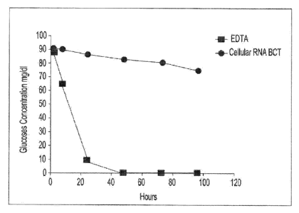
[0017] Fig. I is a graphic representation showing inhibition of metabolism
in blood
cells using glucose concentration as an indicator of metabolism.
[0018] Fig. 2 is a graphic representation showing the effectiveness of
nuclease
inhibitors present in the blood collection device in inhibiting RNase activity
present in
a plasma sample,
[0019] Fig. 3 is a graphic representation showing inhibition of unintended
up-
regulation of c-fos mRNA in a blood sample collected in accordance with the
present
invention as compared to collection in a standard EDTA blood collection
device, Real
Time Reverse Transcriptase PCR technology was used to detect the copy number
of
c-fos mRNA.
[0020] Fig. 4 is a graphic representation showing inhibition of unintended
up-
regulation of mRNA for glyceralciehydes-3-phosphate dehydrogenase (GADPH) in a
blood sample collected in accordance with the present invention as compared to
collection in a standard EDTA blood collection device. Real Time Reverse
6
CA 02780536 2012-05-09
WO 2011/057184 PCT/US281(1/(155815
Transcriptase PCR technology was used to detect the copy number of GAPDH
mRNA.
100211 Fig. 5 is a graphic representation showing inhibition of unintended
down
regulation of mRNA for RASSF1A in a blood sample collected in accordance with
the
present invention as compared to collection in a standard EDTA blood
collection
device. Real Time Reverse Transcriptase PCR technology was used to detect the
copy number of RASSF1A mRNA.
[0022] Fig, 6 is a graphic representation showing inhibition of unintended
up-
regulation of mRNA for glyceraldehydes-3-phosphate dehydnogenase (GADPH) in a
blood sample collected in accordance with the present invention as compared to
collection in a standard EDTA blood collection device. Molecular beacon for
GADPH
mRNA was used to detect GADPH mRNA in intact cells using flow cytometry.
DETAILED DESCRIPTION
[0023] In general, the invention herein contemplates a unique approach for
the
stabilization, isolation, and analysis of cellular RNA. The stabilization step
acts to
inhibit unwanted gene up-regulations and down-regulations after blood draw and
protect the quality of recoverable nucleic acids relative to samples that are
not
stabilized thereby improving the analytic detection sensitivity of the
isolated RNA and
their resulting diagnostic capabilities.
[002,1] In one aspect, the unique approach makes use of a particular
composition
that includes a preservative optionally in combination with one or more
metabolic
inhibitors, one or more nuclease inhibitors, one or more metal ion chelators
or
combinations thereof. In another aspect, the unique approach herein
contemplates
methods that include a step of contacting a blood sample with the compositions
herein. A sample so contacted may thereafter be analyzed. Thus, the method of
the
present invention may involve the steps stabilizing a blood sample, isolating
one or
more blood cells from the stabilized blood sample, extracting cellular nucleic
acids
from the isolated blood cells, and analyzing those cellular nucleic acids for
the
identification of a disease state. The stabilization step may include
contacting the
blood sample with a protective agent in an amount and for a time sufficient so
that
RNA synthesis is inhibited at least partially, if not entirely, for at least
two hours. The
contact time with the protective agent and amount of protective agent used may
also
be sufficient so that the blood cells within the blood sample are fixed to
substantially
prevent contamination of the cellular RNA with cell-free RNA or globin RNA.
Further,
7
CA 02780536 2012-05-09
WO 2011/057184 PCT/US2010/055815
any cellular RNA that is within the blood cells at the time of the blood draw
may be
substantially preserved to freeze the mRNA expression pattern of the blood
cells
substantially as of the time of the blood draw (e.g., no longer than 10
minutes post-
blood draw or even no longer than 5 minutes post-blood draw). The isolating
process
may include iysing the red blood cells and lysing the white blood cells. The
isolated
white blood cells may then be treated to extract cellular RNA from the
isolated white
blood cells and the extracted RNA may be analyzed for the presence, absence,
or
severity of a disease state. The methods disclosed herein allow for the
efficient
preservation, isolation and analysis of cellular nucleic acids while avoiding
contamination with undesirable globin RNA and cell-free nucleic acids that
originate
at extra-cellular locations in vivo (as compared to cellular RNA that becomes
cell-
free RNA due to cell metabolism and cell lysis post-blood draw),
[00251 The
process for improved nucleic acid preservation within a blood sample
may employ a step of contacting a blood sample with a protective agent
containing
one or more preservative agents to maintain the integrity of the components
within
the sample. Ingredients that may be used as preservative agents include, but
are not
limited to, diazolidinyl urea, imidazolidinyl urea, dimethoylo1-
5.5dimethylhydantoin,
dimethylot urea, 2-bromo-2.-nitropropane-1,3-diol, oxazolidines, sodium
hydroxymethyl g lycl nate, 5-
hydroxymethoxymethy1-1-1aza-3,7-
dioxabicyclo[3.3.01octane: 5-hydroxymethy1-1-1aza-
3,7dioxabicyclo[3.3.01octane, 5-
hydroxypolytmethyleneoxy]methyl-1-1aza-3, 7dioxabicyclo [3.3.01octane,
quaternary
adamantine or any combination thereof. Preferred ingredients are selected from
the
group consisting of diazolidinyl urea (DU), imidazolidinyl urea (IOU), and any
combination thereof. The amount of preservative agent used is generally about
10 to
about 400 grams per liter. For example, in certain preferred embodiments the
protective agent comprises about 4 to about 10 grams of !DU per 100 ml of
buffered
salt solution and/or about 1 to about 30 grams of DU per 100 ml of buffered
salt
solution. The preservative agent may be present in the protective agent in an
amount
of greater than about 0.019 per 5m1 blood sample post-blood draw. The
presentative
agent may be present in the protective agent in an amount of less than about
0.20g
per 5m1 blood sample post-blood draw. The concentration of the preservative
agent
within the protective agent may be greater than about 5% vviv prior to blood
draw.
8
CA 02780536 2012-05-09
WO 2011/057184 PCT/US281(1/(155815
The concentration of the preservative agent within the protective agent may be
less
than about 40% %/iv prior to blood draw.
[0026] As used throughout the present teachings, the protective agent
composition including the preservative agent discussed above is preferably
substantially non-toxic. For example, while many cell preservation techniques
make
use of formaldehyde products for purposes of fixation, the methods herein (and
compositions used herein) are free of separately adding andior handling of any
materially significant concentration (e.g., less than about 1% by weight, more
preferably less than about 2000 parts per million, more preferably less than
about
1000 parts per million, and still more preferably less than about 500 parts
per million)
of formaldehyde and/or paraformaldehyde prior to any contact with a blood
product
sample.
[0027] in order to further protect the nucleic acids from degradation, the
protective agent may also include one, or any combination of a cell membrane
permeablizer, a DNase and/or RNase inhibitor, a metal ion chelator, an
oxidative
stress neutralizer, a metabolic inhibitor, an amino acid, a cationic polymer
or a
polyamine. It is also possible that one or more components of the protective
agent
may be prevented from contacting one or more other components of the
protective
agent. This may be achieved by adding the one or more components to a blood
sample at different times or by adding the one or more components into a
container
in phases or locations that do not allow the one or more components to contact
one
another prior to blood draw. This may aid in preventing unwanted reactions
between
the one or more components prior to contact with a blood sample.
[0028] The protective agent may contain a nuclease inhibitor that acts to
prevent
DNase and/or RNase activity within a blood sample. The nuclease inhibitor is
preferably present in an amount sufficient to prevent a decrease in the amount
and/or quality of the nucleic acids recoverable from the blood sample as
compared
with a sample that does not include a nuclease inhibitor. Nuclease inhibitors
that
may be used include, but are not limited to dithiothreitol (DTI),
iodoacetamide,
iodoacetic acid, heparin, chitosan, cobalt chloride, diethyl pyrocarbonate,
ethanol,
aurintricarboxylic acid (ATA), glyceraldehydes, sodium fluoride,
ethylenediamine
tetraacetic acid (EDTA), formamide, vanatlyl-ribonucleosicte complexes,
macaloid,
hydroxylamine-oxygen-cupric ion, bentonite, ammonium sulfate, bete-
g
CA 02780536 2012-05-09
WO 2011/057184 PCT/US2010/055815
mercaptoethanol, cysteine, dithioarythritol, tris (2-carboxyethyl) phosphene
hydrochloride, a divalent cation such as Mg'2, Mn42, Z64.2, Fe.12, Ca+2. Cu+2
or any
combination thereof. One or more nuclease inhibitors may be present within the
protective agent in an amount of more than about 0.1% by weight. A nuclease
inhibitor may be present within the protective agent in an amount of less than
about
60% by weight. The nuclease inhibitor may be present in the protective agent
in an
amount of greater than about 0.00008g per 5m1 blood sample post-blood draw.
The
nuclease inhibitor may be present in the protective agent in an amount of less
than
about 0.2g per 5mlblood sample post-blood draw. The concentration of the
nuclease
inhibitor within the protective agent may be greater than about 0.016% w/v
prior to
blood draw. The concentration of the nuclease inhibitor within the protective
agent
may be less than about 5,0% w/v prior to blood draw. The concentration of the
nuclease inhibitor within the protective agent may be greater than about 0.5%
wiv
prior to blood draw. The concentration of the nuclease inhibitor within the
protective
agent may be less than about 2.0% w/v prior to blood draw. The concentration
of the
nuclease inhibitor within the protective agent may be from about 0.2% wiv to
about
2.0% wiv prior to blood draw.
[00291 The
protective agent may also include one or more metabolic inhibitors in
a suitable amount to reduce cell metabolism within a blood sample. Metabolic
inhibitors that may be used include, but are not limited to glyceraldehyde,
dihydroxyacetone phosphate, glyceraldehyde 3-phosphate. 1,3-
bisphosphoglycerate, 3-phosphoglycerate, 2-
phosphoglycerate,
phosphoenolpyruvate, pyruvate and glyce rate dihydroxyacetate, sodium
fluoride,
K2C204, or any combination thereof, One or more metabolic inhibitors may be
present
within the protective agent at a concentration of more than about 0.21% w/v.
One or
more metabolic inhibitors may be present within the protective agent at a
concentration of less than about 40% wiv. One or more metabolic inhibitors may
be
present within the protective agent at a concentration of more than about 1.0%
w/v,
A metabolic inhibitor may be present within the protective agent at a
concentration of
less than about 10% w/v. The concentration of the one or more metabolic
inhibitors
within the protective agent may be from about 2% wly to about 6% w/v.
[0030] The
protective agent may also include one or more chelators capable of
bonding with metal ions. The purpose of the one or more chelators is to
further
I I
= CA 2780536 2017-04-13
minimize nuclease activity and the resulting nucleic acid degradation. RNA
cleavage
via RNase activity requires the presence of divalent metal ions. The metal ion
chelators will act by bonding with the metal ions thereby inactivating the
ions and
reducing the RNase effect of the metal ions. Possible metal ion chelators for
addition
to the protective agent include but are not limited to one or any combination
of
ethylene glycol tetraacetic acid (EGTA), 1,2-bis-(o-Aminophenoxy)-ethane-N,N,-
N',N'-tetraacetic acid tetraacetoxy-Methyl ester (BAPTA-AM),
dietyldithiocarbamate
(DEDTC), ethylenediaminetetraacetic acid (EDTA), dicarboxymethyl- glutamic
acid,
nitrilotriacetic acid (NTA), or ethylenediaminedisuccinic acid (EDDS). A metal
ion
chelator may be present within the protective agent at a concentration of more
than
about 0.1% w/v. A metal ion chelator may be present within the protective
agent at a
concentration of less than about 40% w/v. A metal ion chelator may be present
within the protective agent at a concentration of more than about 1% w/v. A
metal ion
chelator may be present within the protective agent at a concentration of less
than
about 20% by w/v. The concentration of the one or more metal ion chelators may
be
from about 4% to about 12% w/v.
[0031] As mentioned above, it may be possible for the protective agent
composition to employ a substance to cause cell membrane permeablization in an
effort to increase blood cell uptake of the protective agent thereby improving
fixation.
A selected permeablization substance should generally function to improve the
cell
membrane's ability to selectively allow access to the protective agent while
maintaining desired cell structure and avoiding damage to cell surface
proteins.
Examples of such cell permeablization substance may include but are not
limited to
one or any combination of glycerol, chloroquine, ceteth-15 (C56F1114021),
Triton X-
IOOTM ((C14H220(C2F140)), or saponin. A permeablization substance may be
present
within the protective agent in an amount of more than about 0.5% by weight. A
permeablization substance may be present within the protective agent in an
amount
of less than about 40% by weight. A permeablization substance may be present
within the protective agent in an amount of greater than about 0.001% by
weight. A
permeablization substance may be present within the protective agent in an
amount
of less than about 10% by weight. A permeablization substance may be present
within the protective agent in an amount of greater than about 0.3% by weight.
11
CA 02780536 2012-05-09
WO 2011/057184 PCT/US201(1/(155815
[0032] The protective agent may also include a substance that acts to
prevent
oxidative stress. Nucleic acids and RNA in particular have been found to be
highly
susceptible to oxidative stress. Thus, the addition of antioxidants and/or
reactive
oxygen species (ROS) scavengers to the protective agent may help to protect
the
cellular RNA from the deleterious effects of oxidative damage. As used herein,
the
term "reactive oxygen species (ROS) scavenger group" refers to a group capable
of
acting as a scavenger of, or reacting with, superoxide (02-) or other reactive
oxygen
species (ROS) including hydroxyl radicals, peroxynitrite, hypochlorous acid
and
hydrogen peroxide. Additional examples of such antioxidants and reactive
oxygen
species scavengers include but are not limited to one or any combination of
polyphenols such as flavonoidsõ phenolic acids, D-Mannitol, N-acetyl-L-
cysteine,
natural phenolic antioxidants (alpha-hydroxytyrosol, tyrosol, caffeic acid,
alpha-
tocopherol) as well as commercial phenolic antioxidants (BHT and BHA) or
carotenoids. An ROS scavenger may be present within the protective agent in an
amount of more than about 0.1% by weight. An ROS scavenger may be present
within the protective agent in an amount of less than about 40% by weight_ An
ROS
scavenger may be present within the protective agent in an amount of more than
about 2% by weight. An ROS scavenger may be present within the protective
agent
in an amount of less than about 15% by weight.
[00331 The protective agent may also include one or more polycations
(preferably
polyamines) in a suitable amount such that they are capable of binding with
any
nucleic acids thereby preventing degradation of the nucleic acids. While
polycations
generally bind to nucleic acids, many polycations also alter cell membrane
structure
which may be associated with the loss of cell markers located on the cell
membrane.
Poiyamines are naturally synthesized cations that do not compromise the
structure
of the cell membrane and thus are highly preferred for their ability to bind
specifically
to cellular RNA, based upon the polyanionic nature of the RNA. In binding to
the
RNA, the polyamines are able to protect the nucleic acids from RNase activity.
The
polyamines that may be added include but are not limited to protamine,
spermine,
spermidine, putrescine, cadaverine, or any combination thereof. The polyamines
may be present in the protective agent in an amount of greater than about
0.003g
per 5m1 blood sample post-blood draw. The polyamines may be present in the
protective agent in an amount of less than about 0.1g per 5m1 blood sample
post-
12
CA 02780536 2012-05-09
WO 2011/057184 PCT/US281(1/(155815
blood draw. The concentration of the polyamines within the protective agent
may be
greater than about 77,5 mM prior to blood draw. The concentration of the
polyamines
within the protective agent may be less than about 1562.5 mM prior to blood
draw.
The concentration of the polyamines within the protective agent may be greater
than
about 5% wiv prior to blood draw. The concentration of the polyamines within
the
protective agent may be less than about 50% wiv prior to blood draw.
[0034]
Additional classes of cationic compositions (included in the polyamines
discussed above) may also be included in the protective agent. Certain
cationic
polymers are used in DNA transfection processes such as those disclosed in
U.S.
Patent No, 6,013,240, incorporated by reference herein. The affinity of these
cationic
polymers in binding with nucleic acids may aid in protecting the nucleic acids
from
nuclease activity. Cationic polymers that may be used include but are not
limited to
polylysine, polyarnidoarnine dendrirner, polyethylenimine,
(poly(dimethylamino)ethyl
rnethylactylate (pDMAEMA), polypropylenimine, or any combination thereof. The
PEI
may be low molecular weight PEI, such as about 400 g/mol to about 1000 gimol.
The
cationic polymers (polyamines) may be present in the protective agent in an
amount
of greater than about 0.01g per 5m1 blood sample post-blood draw. The
polyamines
may be present in the protective agent in an amount of less than about 0.1g
per 5m1
blood sample post-blood draw. The concentration of the polyamines within the
protective agent may be greater than about 5% INA, prior to blood draw. The
concentration of the polyamines within the protective agent may be less than
about
50% wiv prior to blood draw.
[0035] The
protective agent may also include one or more amino acids that react
in a manner similar to the one or polyamines discussed above. By binding to
cellular
nucleic acids, the amino acids may protect the nucleic acids from deleterious
nuclease activity. The amino acids may include but are not limited to
isoleucine,
leudne, lysine, valine, tryptophan, threonine, phenylalanine, methionine,
alanine,
histidine, asparagine, aspartate, cysteine, glutamate, glutamine, glycine,
proline,
serine, tyrosine, arginine, or any combination thereof. One or more amino
acids may
be present within the protective agent in an amount of more than about 0.001%
by
weight. One or more amino acids may be present within the protective agent in
an
amount of less than about 30% by weight. One or more amino acids may be
present
within the protective agent in an amount of more than about 0.1% by weight.
One or
13
CA 02780536 2012-05-09
WO 2011/057184 PCT/US201(1/(155815
more amino acids may be present within the protective agent in an amount of
less
than about 10% by weight.
[0036] The protective agent may also include one or more protease
inhibiting
compounds for inhibiting enzyme activity that may have deleterious effects on
the
integrity of any nucleic acids present in a blood sample. These protease
inhibiting
compounds may include but are not limited to antipain, aprotinin, chymostatin,
elastatinai, phenylmethylsulfonyl fluoride (PMSF), APMSF, TLCK, TPCK,
leupeptin,
soybean trypsin inhibitor, indoleacetic acid (IAA), E-64, pepstatin,
VdLPFFVd1_,
EDTA, 1,10-phenanthroline, phosphoramodon, amastatin, bestatin, diprotin A,
diprotin B, alpha-2-macroglobulin, lima bean trypsin inhibitor, pancreatic
protease
inhibitor, egg white ovostatin, egg white cystatin or any combination thereof.
Combinations of protease inhibitors, commonly referred to as a "protease
inhibition
cocktail" by commercial suppliers of such inhibitors, may also be used as the
stabilizing agent. Such "cocktails" may be generally advantageous in that they
provide stabilization for a range of proteins of interest. A protease
inhibitor may be
present within the protective agent in an amount of more than about 0.1% by
weight.
A protease inhibitor may be present within the protective agent in an amount
of less
than about 40% by weight. A protease inhibitor may be present within the
protective
agent in an amount of greater than about 0.001% by weight. A protease
inhibitor
may be present within the protective agent in an amount of less than about 10%
by
weight. A protease inhibitor may be present within the protective agent in an
amount
of greater than about 0.1% by weight.
[0037] The protective agent may further include one or more phosphatase
inhibitors for inhibiting enzyme activity that may have deleterious effects on
the
integrity of any nucleic acids present in a blood sample. These phosphatase
inhibiting compounds may include but are not limited to calyculin A,
nodularin, NIPP-
1 microcystin LR, tautomycin, okadaic acid, cantharidin, calyculin A,
microcystin LR,
okadaic acid, fostriecin, tautomycin, cantharidin, endothall, nodularin,
cyclosporin A,
FK 5061immunophilin complexes, cypermethrin, deitamethrin, fenvalerate,
bpV(phen), dephostatin, mpV(pic) OMFIV, sodium orthovanadate or any
combination
thereof. A phosphatase inhibitor may be present within the protective agent in
an
amount of more than about 0.1% by weight. A phosphatase inhibitor may be
present
within the protective agent in an amount of less than about 40% by weight. A
14
CA 02780536 2012-05-09
WO 2011/057184 PCT/US281(1/(155815
phosphatase inhibitor may be present within the protective agent in an amount
of
more than about 1% by weight. A phosphatase inhibitor may be present within
the
protective agent in an amount of less than about 20% by weight.
[00381 The protective agent or any of the overall compositions may also be
substantially free of guanidinium salts, sodium dodecyl sulfate (SDS), or any
combination thereof.
[0039] The initial contacting of the blood sample will be for a time
sufficient to
inhibit one or both of cell lysis and nuclease activity, or any combination
thereof.
Contacting may occur for at least about 10 seconds, more preferably at least
about 1
minute, still more preferably at least about 2 minutes. Contacting may also
occur for
longer periods of time. For example, contacting may be commenced substantially
contemporaneously from the time of blood draw (e.g., within less than about 10
minutes of the blood draw) and it may last until nucleic acids are isolated,
screened,
and/or tested. The contacting step may also be employed to provide a sample
with a
longer shelf life. Thus, it is possible that a lapse of time of at least about
2 hours,
more preferably at least about 6 hours, at least about 24 hours, at least
about 7 days
or even at least about 14 days can elapse between the time of blood draw
(which
may be substantially contemporaneous with the contacting step), and the time
of any
testing or screening of the sample, and or isolation of the nucleic acids. The
protective agent may comprise an active agent in solution. Suitable solvents
include
water, saline, dirnethylsutfoxide, alcohol or any mixture thereof. The
protective agent
may comprise diazolidinyl urea (DU) and/or imidazolidinyl urea (IOU) in a
buffered
salt solution. The compositions herein (e.g. the protective agent) may further
comprise one or more of spermine, spermidine, polyethylenimine, and histidine.
The
protective agent may contain only a fixative and is free of any additional
additives.
[0040] The present invention may include one or more preservative agents,
one
or more metabolic inhibitors, one or more nuclease inhibitors and one or more
metal
ion chelators. The amount of any preservative agent within the protective
agent is
generally at least about 10% by weight. The amount of any preservative agent
within
the protective agent may be generally less than about 70% by weight. The
preservative agent may comprise at least about 20% IDU by weight, and
generally
less than 40% IDU by weight. The preservative agent may comprise at least
about
20% DU by weight, and generally less than 40% DU by weight. The protective
agent
CA 02780536 2012-05-09
WO 2011/057184 PCT/US201(1/(155815
may further contain a metal ion chelator such as at least about 5% EDTA by
weight.
For example, the protective agent may contain about 8% EDTA by weight. The
protective agent may contain less than about 50% EDTA by weight. The
protective
agent may include from about 0.001% to about 30% by weight of one or more
metabolic inhibitors. For example, the protective agent may contain at least
about
3% glyceraldehyde by weight and at least about 0.1% sodium fluoride by weight.
The
protective agent may include from about 0.001% to about 20% by weight of one
or
more nuclease inhibitors. For example, the protective agent may contain at
least
about 0.5% aurintricarboxylic acid (ATA) by weight. The protective agent may
contain less than about 5% aurintricarboxylic acid (ATA) by weight.
100411 The amount of preservative agent relative to the amount of EDTA is
preferably about 1 to about 10 parts (more preferably about 2 to about 5
parts) by
weight of preservative agent to about 1 part by weight EDTA. The amount of
preservative agent relative to the amount of metabolic inhibitors may be about
1 to
about 10 parts (more preferably about 2 to about 8 parts) by weight of
preservative
agent to about 1 part by weight of metabolic inhibitors. The amount of
preservative
agent relative to the amount of nuclease inhibitors may be about 1 to about 30
parts
(more preferably about 15 to about 22 parts) by weight of preservative agent
to
about 1 part by weight of nuclease inhibitors. The amount of protective agent
within a
tube or other receptacle for receiving a biological specimen prior to blood
draw is
preferably about 300 to 1000 giliter and more preferably about 400 to about
700 g!
liter.
[00421 The combination of one or more presentative agents, one or more
metabolic inhibitors, one or more nuclease inhibitors and one or more
chelators
within the protective agent results in improved ability to maintain the amount
and
quality of RNA within a blood sample. These results are believed unexpected
and
superior to results obtained by the use of only the one or more preservative
agents,
only the one or more metabolic inhibitors, only the one or more nuclease
inhibitors,
only the one or more chelators or any combination including at least two but
less
than all of the one or more preservative agents, the one or more metabolic
inhibitors,
the one or more nuclease inhibitors, or the one or more chelators. Therefore
it is
contemplated that a synergistic effect occurs when one or more preservative
agents,
16
I I
CA 2780536 2017-04-13
one or more metabolic inhibitors, one or more nuclease inhibitors, and one or
more
chelators are combined.
[0043] Additionally, multiple components of the protective agent may
undergo a
lyophilization process so that each component is added either prior to or post-
blood
draw in a substantially solid form to prevent unwanted reactions between one
or
more components of the protective agent. Similar agents in substantially solid
form
and associated blood screening devices are disclosed in U.S. Publication No.
2010/0167271. Liquid removal techniques can be performed on the protective
agent
in order to obtain a substantially solid state protective agent. Liquid
removal
conditions may preferably be such that they result in removal of at least
about 50%
by weight, more preferably at least about 75% by weight, and still more
preferably at
least about 85% by weight of the original amount of the dispensed liquid
protective
agent. Liquid removal conditions may preferably be such that they result in
removal
of sufficient liquid so that the resulting composition is in the form of a
film, gel or
other substantially solid or highly viscous layer; for example it may result
in a
substantially immobile coating (preferably a coating that can be re-dissolved
or
otherwise dispersed upon contact with a blood product sample). Thus, liquid
removal
conditions may preferably be such that they result in a material that upon
contact
with the sample under consideration (e.g., a blood sample) the protective
agent will
disperse in the sample, and substantially preserve components (e.g., cellular
nucleic
acids) in the sample. Liquid removal conditions may preferably be such that
they
result in a remaining composition that is substantially free of crystallinity;
has a
viscosity that is sufficiently high that the remaining composition is
substantially
immobile at ambient temperature (e.g., it does not exhibit any visibly
detectable (as
seen by the naked eye) flow when held in a storage device at room temperature
on
an incline of at least about 45 for at least one hour); or both. In this
regard as taught
in the forgoing application a colorant may also be employed. In one
embodiment,
one or more polyamines may be combined prior to lyophilization and then
lyophilized. One or more preservative agents may also be combined with one or
more enzyme inhibitors prior to lyophilization and then the combined
preservative
agents and enzyme inhibitors may be lyophilized. Lyophilization of one or more
polyamines and one or more preservative agents prior to any contact between
the
one or more polyamines and the one or more preservative agents may prevent any
17
,;
I
CA 2780536 2017-04-13
undesired effects (e.g., a loss of cationic function) that may occur during
contact in
any substantially liquid form.
[0044] A blood screening device (e.g., a specimen container) may further
include
a structure for physically separating any components of the protective agent
that
should be prevented from contacting one another prior to blood draw. This
means
may require removal post-blood draw or may simply breakdown upon placement of
a
blood sample into the specimen container. The blood screening device may
include
a receptacle that receives a sample of blood and that is substantially
transparent
over at least a portion of its area. The device may include a first end, a
second end,
a base portion located a distance between the first end and second end that
divides
the receptacle into a receiving portion and an elongated channel portion,
wherein the
first end and second end are both open. The device may further include the
protective agent composition placed within the receptacle and being visible
through
the substantially transparent window, the protective agent composition being
in solid
form and located in the top portion of the receptacle and being of sufficient
concentration so that upon contact with the sample of blood the protective
agent
composition will disperse in the sample, and substantially preserve white
blood cell
components in the sample.
[0045] The protective agent may be located within a specialized device,
wherein
the protective agent is already present in the device prior to addition of the
blood
sample, such as those disclosed in U.S. Patent Publication No. 2004/0137417.
The
device may also be an evacuated collection container, such as a tube. The tube
may
preferably be made of a transparent material that will also resist adherence
of the
cells within a given sample. The interior wall of the tube may be coated or
otherwise
treated to modify its surface characteristics, such as to render it more
hydrophobic
and/or more hydrophilic, over all or a portion of its surface. The tube may
have an
interior wall flame sprayed, subjected to corona discharge, plasma treated,
coated or
otherwise treated. The tube may be treated by contacting an interior wall with
a
substance so that the nucleic acids of interest will resist adhering to the
tube walls.
The surface of the tube may be modified to provide a dual functionality that
simultaneously provides an appropriate balance of desired hydrophilicity and
hydrophobicity, to allow collection of blood, dispersion of the preservatives
herein,
and resistance of adhesion of nucleic acids to the inner wall of a blood
collection
tube. Thus it is possible that any coating may be a functionalized polymeric
coating
18
I I
CA 2780536 2017-04-13
that includes a first polymer and one or more second monomeric and/or
polymeric
functionalities that are different from (e.g., chemically different from) the
first polymer.
The coating may include one or more co-polymers (e.g., block copolymer, graft
copolymer, or otherwise). For example, it may include a copolymer that
includes a
first hydrophobic polymeric portion, and a second hydrophilic polymeric
portion. The
coating may be a water based coating. The coating may optionally include an
adhesion promoter. The coating may be applied in any suitable manner, it may
be
sprayed, dipped, swabbed, or otherwise applied onto some or all of the
interior of the
blood collection tube. The coating may also be applied in the presence of
heat.
Preferably any coating applied to the inner wall of a blood collection tube
will form a
sufficiently tenacious bond with the glass (e.g., borosilicate glass) or other
material
(e.g., polymeric material) of the tube so that it will not erode or otherwise
get
removed from the inner wall. Examples of suitable polymeric coatings may
include
silicon containing polymers (e.g., silanes, siloxanes, or otherwise);
polyolefins such
as polyethylene or polypropylene; polyethylene terephthalate; fluorinated
polymers
(e.g., polytetrafluoroethylene); polyvinyl chloride, polystyrene or any
combination
thereof. Examples of teachings that may be employed to coat an interior of a
blood
collection tube may be found in U.S. Patent Nos. 6,551,267; 6,077,235;
5,257,633;
and 5,213,765.
[0046] The protective agent may also be placed prior to or post-blood draw
into a
receptacle employing a blood sample identification system such as that
disclosed in
co-owned U.S. Publication No. US2011/0053208 entitled "Blood Sample
Identification System". The identification system includes a handling device
for a
biological specimen within a receptacle comprising an initially planar
substrate
including at least one crease that divides the substrate into a handle portion
having
at least one peripheral edge portion and a receiving portion that includes at
least a
portion of an aperture having a perimeter that is configured so that it
receives a
container having a cover that includes a biological specimen for test, and
resists pull-
through of the container relative to the substrate. The substrate may further
include
identification information about the sample contained within the receptacle
and/or its
source.
19
I 1.
CA 2780536 2017-04-13
[0047] As discussed above, the tube may include a metal ion chelator
(which may
also be an anticoagulant agent), one or more metabolic and/or nuclease
inhibitors
and a preservative agent such as a fixative agent including but not limited to
those
disclosed above. The tube may also include one or any combination of one or
more
polyamines, a cell membrane permeablizer, and an antioxidant or reactive
oxygen
species scavenger. Preferably, the compounds included in the tube are in an
amount
sufficient to preserve cell morphology and prevent cell degradation while also
preventing deleterious DNase and RNase activity within the nucleic acids. In
preferred embodiments, blood may be fixed simultaneously as it is drawn into
the
specialized tube. The tube may also be coated over an exterior wall with a
protective
coating (e.g., a containment barrier that helps control glass shard
fragmentation)
such as that disclosed in U.S Patent No. 7,419,832.
[0048] As discussed herein, a step of contacting a blood sample with
the
protective agent allows the sample to be stored for a period of time prior to
isolating
and testing the nucleic acids. A blood sample may be drawn at one location,
contacted with the protective agent, and later transported to a different
remote or off-
site location for the nucleic acid isolation and testing process. The nucleic
acids may
be isolated from the blood sample and tested at the remote location and the
resulting
diagnostic information is then reported to the site of the original blood
draw. The
nucleic acid isolation process may be performed at one remote location and the
resulting data can be analyzed to identify the presence, absence or relative
severity
of a disease state at a third location. Alternatively the results of the
nucleic acid
isolation process may be sent back to the site of the initial blood draw and
analyzed
there. The resulting diagnostic information may then be sent to a third
location or
back to the remote location or the site of the initial blood draw. The blood
draw
location and the testing site and/or analysis site are separated by at least
about 0.5
km, 1km, 100km, or longer.
[0049] At any time after the initial contact of the blood sample with
the protective
agent, the sample can be treated to isolate one or more blood cells from the
sample
and extract the cellular nucleic acids located within the isolated blood
cells. The
nucleic acids may be extracted and isolated using any method including those
methods disclosed in commonly owned US Publication No. 2009/0081678. The
I
CA 2780536 2017-04-13
preservative agent acts to prevent cell lysis so that the blood cells remain
intact and
substantially all cellular nucleic acids remain intra-cellular to avoid
unwanted
contamination with cell-free RNA and globin RNA.
[0050] After the cellular RNA has been extracted, it can be tested to
identify the
presence, absence or severity of a disease state. The methods herein thus
further
contemplate a step of nucleic acid testing. Testing of the nucleic acids can
be
performed using any nucleic acid testing method including, but not limited to
polymerase chain reaction (PCR), reverse transcription polymerase chain
reaction
(RT-PCR), quantitative real time polymerase chain reaction (Q-PCR), gel
electrophoresis, capillary electrophoresis, mass spectrometry, fluorescence
detection, ultraviolet spectrometry, DNA hybridization, allele specific
polymerase
chain reaction, polymerase cycling assembly (PCA), asymmetric polymerase chain
reaction, linear after the exponential polymerase chain reaction (LATE-PCR),
helicase-dependent amplification (HDA), hot-start polymerase chain reaction,
intersequence-specific polymerase chain reaction (ISSR), inverse polymerase
chain
reaction, ligation mediated polymerase chain reaction, methylation specific
polymerase chain reaction (MSP), multiplex polymerase chain reaction, nested
polymerase chain reaction, solid phase polymerase chain reaction, or any
combination thereof.
[0051] The present invention also contemplates a method for isolating and
testing
cellular RNA. The method can be performed on a single sample or on a multitude
of
samples (e.g., in a multi-well plate). The method includes contacting a blood
sample
with a protective agent. The protective agent includes a preservative agent as
previously discussed so that the blood cells remain intact throughout the
blood draw
and RNA isolation process. The protective agent further includes a component
to
protect the RNA from RNase activity or to inhibit RNase activity altogether.
Post-
blood draw, the blood sample may be contacted with a red blood cell lysis
buffer
which may include NH4CI, KHCO3, and EDTA in water. The blood sample may then
be placed in ice water for about 15 to about 20 minutes. The sample may then
be
centrifuged at about 1000 rpm at about 1 C to about 10 C for about 10 minutes.
The
supernatant may then be removed and a red blood cell lysis buffer may be added
for
removal of red blood cells. The resulting white blood cell pellet may then be
resuspended and centrifuged at about 1000 rpm at about 1 C to about 10 C for
21
CA 02780536 2012-05-09
WO 2011/057184 PCT/US201(1/(155815
about 10 minutes. The supernatant may then be removed by aspiration in an
effort to
avoid any disruption to the white blood cell pellet. The RNA may then be
isolated
using any nucleic acid isolation method. As an example, the AllPrep DNA/RNA
Mini
Kit manufactured by QIAGEN, Inc. of Valencia, California may be used. The
white
blood cell pellet may be initially contacted with a cell lysis buffer which
may contain
guanidine hydrochloride and 13-mercaptoethanol. The cell pellet may then be
vortexed to promote cell lysis. After cell lysis, cell lysate is introduced to
a
homogenizing device by microcentrifuge at about 13000 rpm at room temperature
for
about 1 minute to about 4 minutes to ensure disruption and homogenization of
the
cell lysate. The homogenized cell lysate may then be applied to a DNA binding
column and microcentrifuged at about 8000 g at room temperature for about 1
minute to remove the majority ef DNA. The flow-through may then be collected
and
mixed with ethanol to adjust the salt concentration and pH for proper binding
on the
RNA column. The mixture may then be applied to an RNA column and contacted
with one or more buffers to remove any impurities including protein and salts.
The
RNA may then be eluted by RNase-free water and stored at about -80 C for long
term storage or at about O'C for short term storage. For use in a UV
spectrophotometer, the RNA samples may be kept at 0 C but analyzed at room
temperature. For use in a bioanalyzer, the RNA samples may first be denatured
at
about 70 C for about 2 minutes then immediately cooled at about 0 C on ice to
keep
the RNA denatured and free of any tertiary structure. The RNA samples may
remain
on ice until loaded onto the bioanalyzer chips for analysis at room
temperature.
[0052] Example 1
[0053] Blood samples from the same donor are drawn into two separate blood
collection tubes (tube 1 (EDTA tube) and tube 2 (RNA BCT tube)). Tube 1
contains
only EDTA. Tube 2 contains DU. EDTA, ATA, glyceraldehydc.1 and sodium
fluoride.
Both tubes are stored at room temperature and 1ml aliquots of blood are
removed
from each tube at hours 1.5, 8, 24, 48, 72 and 96. The blood glucose levels of
each
sample are measured using a YSI blood glucose meter available from YSI Life
Sciences (Yellow Springs, OH). The blood glucose concentration of samples from
tube 2 were the only samples that maintained relatively consistent glucose
levels
over the test period, indicating that the combination of EDTA, DU, ATA,
22
CA 02780536 2012-05-09
WO 2011/057184 PCT/US20 1o/055815
glyceraidehyde and sodium fluoride provided reduced levels of cell metabolism,
The
results of this example are shown in a graphic format at Figure 1.
[0054] Example 2
[00551 Blood samples from the same donor are drawn into two separate blood
collection tubes (tube 1 and tube 2). Tube I contains EDTA. Tube 2 contains
DU,
EDTA, ATA, glyceraldehyde and sodium fluoride. Both tubes are stored for 2 h
at
room temperature before plasma was separated. RNase activity of plasma from
tube
1 and tube 2 was measured using a commercially available RNase activity
detection
kit, RNaseAlert Lab Test Kit (Applied Biosystems. Foster City, CA). Two
additional
control experiments were also carried out with purified RNase A enzyme alone
and
RNase A treated with chemical mixture present in tube 2. RNase activity is
presented as relative fluorescence, Results of this example are illustrated in
a
graphic format at Figure 2,
[0056] Example 3
[0057] Two blood samples from the same donor are drawn into two separate
blood collection tubes, tube A (RNA BCT) and tube B (EDTA). Tube A contains
DU,
EDTA, ATA, glyceraldehyde and sodium fluoride. Tube B contains only EDTA. Both
tubes are stored at room temperature and 5m1 aliquots of blood are removed
from
each tube on day 0, day 1, day 2, and day 3 and plasma is separated. All
samples
are centrifuged at 800 g for 10 minutes at room temperature to separate the
plasma
The plasma is then transferred into new tubes and centrifuged at 1500 g for 10
minutes at room temperature. Free circulating RNA is purified using the Q1Aamp
circulating nucleic acid kit available from Qiagen Inc. (Valencia, CA). RNA is
extracted from each plasma sample. The samples are then amplified by Real Time
PCR (using TagMan RT PCR reagents available from Applied Biosystems, Foster
City, California) to identify the c-fos mRNA copy number per ml of plasma.
Results
showed a consistent copy number of c-fos mRNA per ml of plasma in samples
originating from tube A at each measurement, indicating little or no change in
c-fos
mRNA level in tube A over a 3 day period. The c-fos mRNA copy number per ml of
plasma showed elevated levels at every measurement in those samples
originating
in tube B, indicating an increase in the amount of c-fos mRNA present as a
result of
increased cell metabolism. The results of this example are shown in a graphic
format
at Figure 3.
23
CA 02780536 2012-05-09
WO 2011/057184 PCT/US2010/055815
[0058] Example 4
[0059] Two blood samples from the same donor are drawn into two separate
blood collection tubes, tube A (RNA SOT) and tube B (EDTA). Tube A contains
DU,
EDTA, ATA, glyceraldehyde and sodium fluoride. Tube B contains only EDTA. Both
tubes are stored at room temperature and 5m1 aliquots of blood are removed
from
each tube on day 0, day 1, day 2, and day 3 and plasma is separated. All
samples
are centrifuged at 800 g for 10 minutes at room temperature to separate the
plasma.
The plasma is then transferred into new tubes and centrifuged at 1500 g for 10
minutes at room temperature. Free circulating RNA is purified using the QiAamp
circulating nucleic acid kit available from Qiagen Inc. (Valencia, CA). RNA is
extracted from each plasma sample. The samples are then amplified by Real Time
PCR (using TagMan RT PCR reagents available from Applied Biosystems, Foster
City, California) to identify the GAPDH mRNA copy number per ml of plasma.
Results showed a consistent copy number of GAPDH mRNA per ml of plasma in
samples originating in tube A at each measurement, indicating little or no
change in
GAPDH mRNA level in tube A over a 3 day period. The GAPDH mRNA copy number
per ml of plasma showed elevated levels at every measurement in those samples
originating in tubes B, indicating an increase in the amount of GAPDH mRNA
present as a result of increased cell metabolism. The results of this example
are
shown in a graphic format at Figure 4.
[0060] Example 5
[0061] Two blood samples from the same donor are drawn into two separate
blood collection tubes, tube A (RNA SOT) and tube B (EDTA). Tube A contains
DU,
EDTA, ATA, glyceraldehyde and sodium fluoride. Tube B contains only EDTA. Both
tubes are stored at room temperature and 5m1 aliquots of blood are removed
from
each tube on day 0, day 1, day 2, and day 3 and plasma is separated. All
samples
are centrifuged at 800 g for 10 minutes at room temperature to separate the
plasma.
The plasma is then transferred into new tubes and centrifuged at 1500 g for 10
minutes at room temperature. Free circulating RNA is purified using the QIAamp
circulating nucleic acid kit available from Qiagen Inc, (Valencia, CA). RNA is
extracted from each plasma sample. The samples are then amplified by Real Time
PCR (using TagMan RI PCR reagents available from Applied Biosystems, Foster
City, California) to identify the RASSF1A mRNA copy number per ml of plasma.
24
CA 02780536 2012-05-09
WO 2011/057184 PCPUS201(1/(155815
Results showed a consistent copy number of RASSF1A mRNA per ml of plasma at
each measurement, indicating little or no change in c-fos mRNA level in tube A
over
a 3 day period. The RASSF1A mRNA copy number per ml of plasma showed
declined at every measurement in those samples originating in tubes B,
indicating an
decrease in the amount of RASSF1A mRNA present as a result of RASSF1A mRNA
down-regulation. The results of this example are shown in a graphic format at
Figure
5.
[0062] Example 6
[0063] Two blood samples from the same donor are drawn into two separate
blood collection tubes, tube A (RNA GOT) and tube B (EDTA), Tube A contains
DU,
EDTA, ATA, glyceraldehyde and sodium fluoride. Tube B contains only EDTA. Both
tubes are stored at room temperature and 5m1 aliquots of blood are removed
from
each tube on day 0, day 1, day 2, and day 3 and white blood cells were
isolated by
either density gradient centrifugation or by removing red cells using a red
cell lysis
solution. White blood cells isolated from tube A and tube B were treated with
ice-cold
100% methanol for 10 ruin separately. Then cells from both tubes were washed
with
PBS for two times and suspended in PBS and incubated with a molecular beacon
for
GAPDH mRNA for 1 h at room temperature. After this incubation period cells
from
both tube A and tube B were analyzed by flowcytometry to quantify the GADPH
mRNA Level in intact white blood cells, Results showed a consistent level of
GAPDH
mRNA indicating little or no change in GAPDH mRNA level in tube A over a 3 day
period, The GAPDH mRNA level showed elevated at every measurement in those
samples originating in tubes B, indicating an increase in the amount of GAPDH
mRNA present as a result of increased cell metabolism. The results of this
example
are shown in a graphic format at Figure 6.
[0064] Examples 1 through 6 above demonstrate an unexpected synergistic
effect occurring in blood samples contacted by a preservative, one or more
metabolic inhibitors and/or nuclease inhibitors, and one or more chelators.
Blood
samples contacted by only a preservative, only one or more metabolic
inhibitors
and/or nuclease inhibitors, only one or more chelators, or any combination of
only
some but not all of those do not demonstrate the ability to maintain the
integrity of
the blood cells or the integrity of the nucleic acids. The combined effect of
the DU
and one or more metabolic and/or nuclease inhibitors far exceeds any
expectations
!I = CA 2780536 2017-04-13
based on the effect, or lack thereof, of the DU or one or more metabolic
and/or
nuclease inhibitors used alone.
[0065] Any
numerical values recited herein include all values from the lower value
to the upper value in increments of one unit provided that there is a
separation of at
least 2 units between any lower value and any higher value. As an example, if
it is
stated that the amount of a component or a value of a process variable such
as, for
example, temperature, pressure, time and the like is, for example, from 1 to
90,
preferably from 20 to 80, more preferably from 30 to 70, it is intended that
values
such as 15 to 85, 22 to 68, 43 to 51, 30 to 32 etc. are expressly enumerated
in this
specification. For values which are less than one, one unit is considered to
be
0.0001, 0.001, 0.01 or 0.1 as appropriate. These are only examples of what is
specifically intended and all possible combinations of numerical values
between the
lowest value and the highest value enumerated are to be considered to be
expressly
stated in this application in a similar manner. As can be seen, the teaching
of
amounts expressed as "parts by weight" herein also contemplates the same
ranges
expressed in terms of percent by weight. Thus, an expression in the Detailed
Description of the Invention of a range in terms of at "sx' parts by weight of
the
resulting polymeric blend composition" also contemplates a teaching of ranges
of
same recited amount of "x" in percent by weight of the resulting polymeric
blend
composition."
[0066]
Unless otherwise stated, all ranges include both endpoints and all
numbers between the endpoints. The use of "about" or "approximately" in
connection
with a range applies to both ends of the range. Thus, "about 20 to 30" is
intended to
cover "about 20 to about 30", inclusive of at least the specified endpoints.
[0067] The
term "consisting essentially of' to describe a combination shall
include the elements, ingredients, components or steps identified, and such
other
elements ingredients, components or steps that do not materially affect the
basic and
novel characteristics of the combination. The use of the terms "comprising" or
"including" to describe combinations of elements, ingredients, components or
steps
herein also contemplates embodiments that consist essentially of the elements,
ingredients, components or steps. By use of the term "may" herein, it is
intended that
any described attributes that "may" be included are optional.
[0068]
Plural elements, ingredients, components or steps can be provided by a
single integrated element, ingredient, component or step. Alternatively, a
single
26
r
CA 2780536 2017-04-13
integrated element, ingredient, component or step might be divided into
separate
plural elements, ingredients, components or steps. The disclosure of "a" or
"one" to
describe an element, ingredient, component or step is not intended to
foreclose
additional elements, ingredients, components or steps. All references herein
to
elements or metals belonging to a certain Group refer to the Periodic Table of
the
Elements published and copyrighted by CRC Press, Inc., 1989. Any reference to
the
Group or Groups shall be to the Group or Groups as reflected in this Periodic
Table
of the Elements using the IUPAC system for numbering groups.
[0069] It will be appreciated that concentrates or dilutions of the
amounts recited
herein may be employed. In general, the relative proportions of the
ingredients
recited will remain the same. Thus, by way of example, if the teachings call
for 30
parts by weight of a Component A, and 10 parts by weight of a Component B, the
skilled artisan will recognize that such teachings also constitute a teaching
of the use
of Component A and Component B in a relative ratio of 3:1. Teachings of
concentrations in the examples may be varied within about 25% (or higher) of
the
stated values and similar results are expected. Moreover, such compositions of
the
examples may be employed successfully in the present methods.
[0070] It will be appreciated that the above is by way of
illustration only. Other
ingredients may be employed in any of the compositions disclosed herein, as
desired, to achieve the desired resulting characteristics. Examples of other
ingredients that may be employed include antibiotics, anesthetics,
antihistamines,
preservatives, surfactants, antioxidants, unconjugated bile acids, mold
inhibitors,
nucleic acids, pH adjusters, osmolarity adjusters, or any combination thereof.
[00711 It is understood that the above description is intended to
be illustrative
and not restrictive. Many embodiments as well as many applications besides the
examples provided will be apparent to those of skill in the art upon reading
the above
description. The scope of the invention should, therefore, be determined not
with
reference to the above description, but should instead be determined with
reference
to the appended claims, along with the full scope of equivalents to which such
claims
are entitled. The omission in the following claims of any aspect of subject
matter that
is disclosed herein is not a disclaimer of such subject matter, nor should it
be
regarded that the inventors did not consider such subject matter to be part of
the
disclosed inventive subject matter.
27
CA 02780536 2012-05-09
WO 2011/057184 PCT/US2010/055815
applications and publications, are incorporated by reference for all purposes.
The
omission in the following claims of any aspect of subject matter that is
disclosed
herein is not a disciairner of such subject matter, nor should it be regarded
that the
inventors did not consider such subjeet matter to be part of the disclosed
inventive
subject matter.
28