Note: Descriptions are shown in the official language in which they were submitted.
CA 02780827 2012-05-11
WO 2011/082293 PCT/US2010/062485
METHODS FOR PRODUCING UNIQUELY SPECIFIC NUCLEIC ACID
PROBES
CROSS REFERENCE TO RELATED APPLICATION
This claims the benefit of U.S. Provisional Application No. 61/291,750, filed
December 31, 2009, and U.S. Provisional Application No. 61/314,654, filed
March
17, 2010, both of which are incorporated herein by reference in their
entirety.
FIELD
This disclosure relates to the field of molecular detection of nucleic acid
target sequences (e.g., genomic DNA or RNA). More specifically, this
disclosure
relates to methods of producing nucleic acid probes that include uniquely
specific
nucleic acid sequences which are represented only once in the haploid genome
of an
organism, and probes generated by the disclosed methods.
BACKGROUND
Molecular cytogenetic techniques, such as fluorescence in situ hybridization
(FISH), chromogenic in situ hybridization (CISH) and silver in situ
hybridization
(SISH), combine visual evaluation of chromosomes (karyotypic analysis) with
molecular techniques. Molecular cytogenetics methods are based on
hybridization
of a nucleic acid probe to its complementary nucleic acid within a cell. A
probe for
a specific chromosomal region will recognize and hybridize to its
complementary
sequence on a metaphase chromosome or within an interphase nucleus (for
example
in a tissue sample). Probes have been developed for a variety of diagnostic
and
research purposes. For example, certain probes produce a chromosome banding
pattern that mimics traditional cytogenetic staining procedures and permits
identification of individual chromosomes for karyotypic analysis. Other probes
are
derived from a single chromosome and when labeled can be used as "chromosome
paints" to identify specific chromosomes within a cell. Yet other probes
identify
particular chromosome structures, such as the centromeres or telomeres of
chromosomes. Additional probes hybridize to single copy DNA sequences in a
-1-
CA 02780827 2012-05-11
WO 2011/082293 PCT/US2010/062485
specific chromosomal region or gene. These are the probes used to identify the
critical chromosomal region or gene associated with a syndrome or condition of
interest. On metaphase chromosomes, such probes hybridize to each chromatid,
usually giving two small, discrete signals per chromosome.
Hybridization of such chromosomal or gene-specific probes has made
possible detection of chromosomal abnormalities associated with numerous
diseases
and syndromes, including constitutive genetic anomalies, such as microdeletion
syndromes, chromosome translocations, gene amplification and aneuploidy
syndromes, neoplastic diseases, as well as pathogen infections. Most commonly
these techniques are applied to standard cytogenetic preparations on
microscope
slides. In addition, these procedures can be used on slides of formalin-fixed
tissue,
blood or bone marrow smears, and directly fixed cells or other nuclear
isolates.
Chromosomal or gene-specific probes can also be used in comparative genomic
hybridization (CGH) to determine gene copy number in a genome.
The genome of many organisms contains repetitive nucleic acid sequences,
which are series of nucleotides that are repeated multiple times, often in
tandem
arrays. The presence of such repetitive sequences in a probe results in
increased
background staining and requires the use of blocking DNA during hybridization.
"Repeat-free" probes which lack such repetitive sequences are often generated
(for
example using a computer algorithm) to reduce this problem. However, even
"repeat-free" probes require the use of substantial amounts of blocking DNA in
order to reduce background staining to acceptable levels.
SUMMARY
Disclosed herein are uniquely specific nucleic acid probes and methods for
their use and production. The disclosed probes have reduced or eliminated
background signal while reducing or eliminating the use of blocking DNA during
hybridization. In some examples, probes are produced by a method that includes
joining at least a first binding region and a second binding region in a pre-
determined order and orientation, wherein the first binding region and second
binding region are complementary to uniquely specific nucleic acid sequences,
-2-
CA 02780827 2012-05-11
WO 2011/082293 PCT/US2010/062485
wherein the uniquely specific nucleic acid sequences are represented only once
in a
genome of an organism and wherein the first binding region and the second
binding
region include about 20% or less (for example 20%, 19%, 18%, 17%, 16%, 15%,
14%,13%,12%,11%,10%, 9%, 8%, 7%, 6%, 5%,4%, 3%, 2%, 1%, or less) of a
genomic target nucleic acid molecule. In some examples, the first binding
region
and the second binding region include about 10% or less of a genomic target
nucleic
acid molecule. In particular examples, the binding regions ("uniquely specific
binding regions") are complementary to non-contiguous portions of the genomic
target nucleic acid. In some examples, the uniquely specific binding regions
are at
least about 20 base pairs (bp) in length (for example, about 35-500 bp, such
as about
100 bp). In some examples, the genomic target nucleic acid is from a
eukaryotic
genome (such as a mammalian genome, for example a human genome).
In particular embodiments, the uniquely specific binding regions are
generated by one or more of the following: separating the genomic target
nucleic
acid into a plurality of segments (for example, separating the genomic nucleic
acid
sequence into segments, such as in silico); comparing each segment with a
genome
including the genomic target nucleic acid (for example, using a computer
algorithm,
such as BLAT); selecting at least two segments which are uniquely specific to
the
genomic target nucleic acid (such as at least two segments that are each
represented
only once each in the genomic target nucleic acid molecule); removing
repetitive
DNA sequences from the genomic target nucleic acid (for example, using a
computer algorithm, such as RepeatMasker); and selecting at least two segments
having a GC nucleotide content between about 30% and 70%.
In other embodiments, the uniquely specific binding regions are generated by
one or more of the following: separating the genomic target nucleic acid into
a
plurality of segments (for example, separating the genomic nucleic acid
sequence
into segments, such as in silico); synthesizing the plurality of nucleic acid
segments;
attaching the synthesized plurality of nucleic acid segments to an array;
hybridizing
the array with total genomic DNA and blocking DNA; selecting at least two
segments which are uniquely specific to the genomic target nucleic acid (such
as at
least two segments that are each represented only once each in the genomic
target
-3-
CA 02780827 2012-05-11
WO 2011/082293 PCT/US2010/062485
nucleic acid molecule); removing repetitive DNA sequences from the genomic
target nucleic acid (for example, using a computer algorithm, such as
RepeatMasker); and selecting at least two segments having a GC nucleotide
content
between about 30% and 70%.
In some examples, the uniquely specific binding regions are generated by
synthesizing a plurality of nucleic acid segments including the target genomic
region, attaching the synthesized plurality of nucleic acid segments to an
array,
hybridizing the array with total genomic DNA and blocking DNA, and selecting
at
least two segments which are uniquely specific to the genomic target nucleic
acid
(such as at least two segments that are each represented only one each in the
genomic target nucleic acid molecule).
In some examples, the pre-determined order and orientation is generated by
the following: ordering the selected uniquely specific binding regions to
produce a
candidate nucleic acid probe (for example, ordering in the chromosomal order
and
orientation); separating the candidate nucleic acid probe into a plurality of
segments
(for example, separating the genomic nucleic acid sequence into segments, such
as
in silico); comparing each segment with a genome including the genomic target
nucleic acid (for example, using a computer algorithm, such as BLAT);
selecting at
least one order and orientation of the selected segments that is uniquely
specific to
the genomic target nucleic acid (for example, does not include any sequence
represented more than once in the genome of the organism); and joining the
selected
uniquely specific binding regions in the selected order and orientation. In
other
examples, the pre-determined order and orientation is generated by ordering
the
selected uniquely specific binding regions to produce a nucleic acid probe
(for
example in the chromosomal order and/or orientation) and joining the selected
uniquely specific binding regions in the selected order and orientation.
Methods of using the disclosed probes include, for example, detecting (and
in some examples quantifying) a genomic target nucleic acid sequence. For
example, the method can include contacting the disclosed probes with a sample
containing nucleic acid molecules under conditions sufficient to permit
hybridization between the nucleic acid molecules in the sample and the
plurality of
-4-
CA 02780827 2012-05-11
WO 2011/082293 PCT/US2010/062485
nucleic acid molecules of the probe. Resulting hybridization is detected,
wherein
the presence of hybridization indicates the presence (and in some examples,
the
quantity) of the genomic target nucleic acid sequence.
Kits including the probes and/or reagents for producing or using the probes
are also disclosed.
The foregoing and other features will become more apparent from the
following detailed description, which proceeds with reference to the
accompanying
figures.
BRIEF DESCRIPTION OF THE DRAWINGS
FIG. 1 shows an example of a portion of a Met proto-oncogene genomic
nucleic acid sequence (SEQ ID NO: 1) that is enumerated and separated into 100
bp
fragments. The repetitive sequence is replaced with "n", followed by
replacement of
the number of "n"s by their numerical value. For example, there were 38 "n"s
that
were replaced by "*38*" in the line labeled "600."
FIG. 2A shows BLAT results for a non-uniquely specific 100 bp segment of
human chromosome 7.
FIG. 2B shows BLAT results for a uniquely specific 100 bp segment of
human chromosome 7.
FIG. 3 is a digital image of a dot blot of selected segments 185 to 271 of an
exemplary Met proto-oncogene (MET) probe in the form of 100 bp
oligonucleotides
immobilized on a membrane and hybridized with a human DNA probe. The three
spots in the bottom right of the membrane correspond to human DNA controls (1
ng,
10 ng, and 100 ng).
FIG. 4A is a digital image of MDA-361 cells comparing ISH using a repeat-
free MET probe made using prior methods (human placental blocking DNA was
included during hybridization) to ISH using a uniquely specific MET probe of
the
present disclosure. No human blocking DNA was included during the uniquely
specific probe hybridization; however salmon sperm DNA was included in the
hybridization to counteract background binding of nucleic acids to non-nucleic
acid
reaction components, for example. Detection was via SISH colorimetric
detection.
-5-
CA 02780827 2012-05-11
WO 2011/082293 PCT/US2010/062485
FIG. 4B is a digital image of MDA-361 cells comparing ISH using a repeat-
free IGF1R probe made using prior methods (human placental blocking DNA was
included during hybridization) to ISH using a uniquely specific IGF1R probe of
the
present disclosure. Human placental blocking DNA (minimal amounts compared to
the repeat-free probe hybridization) and salmon sperm DNA were included during
the uniquely specific probe hybridization. Detection was via SISH colorimetric
detection.
FIG. 5A is a pair of digital images showing ISH performed with uniquely
specific IGF1R probes to IGF1R target nucleic acids in a lung cancer tissue
sample
with (left) and without (right) human placental blocking DNA.
FIG. 5B is a pair of digital images showing ISH performed with uniquely
specific TS probes to TS target nucleic acids in a lung cancer tissue sample
with
(left) and without (right) human placental blocking DNA.
FIG. 5C is a pair of digital images showing ISH performed with uniquely
specific MET probes to Met proto-oncogene target nucleic acids in a lung
cancer
tissue sample with (left) and without (right) human placental blocking DNA.
FIG. 5D is a pair of digital images showing ISH performed with uniquely
specific KRAS probes to KRAS target nucleic acids in a lung cancer tissue
sample
with (left) and without (right) human placental blocking DNA.
FIG. 6A is a plot of signal from hybridization of sequences targeting the
CCND1 gene analyzed using a NimbleGen array. Pass/Fail criteria were
established
by including a series of positive and negative controls and using the data to
establish
thresholds for cutoffs.
FIG. 6B is a plot of signal from hybridization of sequences targeting the
CDK4 gene analyzed using a NimbleGen array. Pass/Fail criteria were
established
by including a series of positive and negative controls and using the data to
establish
thresholds for cutoffs.
FIG. 6C is a plot of signal from hybridization of sequences targeting the Myb
gene analyzed using a NimbleGen array. Pass/Fail criteria were established by
including a series of positive and negative controls and using the data to
establish
thresholds for cutoffs.
-6-
CA 02780827 2012-05-11
WO 2011/082293 PCT/US2010/062485
FIG. 7A is a digital image showing ISH performed with a uniquely specific
CCND1 probe in a lung cancer tissue sample without human placental blocking
DNA.
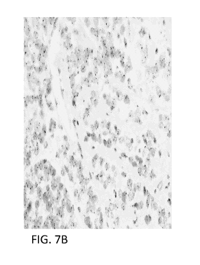
FIG. 7B is a digital image showing ISH performed with uniquely specific
CDK4 probe in a lung cancer tissue sample without human placental blocking
DNA.
FIG. 7C is a digital image showing ISH performed with uniquely specific
Myb probe in a lung cancer tissue sample without human placental blocking DNA.
FIG. 8 is a digital image showing ISH performed with a uniquely specific
EGFR probe in a lung cancer tissue sample without human placental blocking DNA
and detected with tyramide signal amplification.
SEQUENCE LISTING
Any nucleic acid and amino acid sequences listed herein or in the
accompanying sequence listing are shown using standard letter abbreviations
for
nucleotide bases, and three letter code for amino acids, as defined in 37
C.F.R.
1.822. In at least some cases, only one strand of each nucleic acid sequence
is
shown, but the complementary strand is understood as included by any reference
to
the displayed strand.
The Sequence Listing is submitted as an ASCII text file in the form of the
file named Sequence_Listing.txt, which was created on December 28, 2010, and
is
2,017 bytes, which is incorporated by reference herein.
SEQ ID NO: 1 is an exemplary enumerated and separated Met proto-
oncogene genomic sequence wherein repetitive sequences are replaced with "n."
DETAILED DESCRIPTION
1. Introduction
Production of probes corresponding to selected target nucleic acid sequences
(e.g., genomic target nucleic acid sequences) for molecular analysis can be
complicated by the presence of undesired sequences in the probe that can
potentially
increase the amount of background signal. Examples of undesired sequences
include, but are not limited to, interspersed repetitive nucleic acid elements
present
-7-
CA 02780827 2012-05-11
WO 2011/082293 PCT/US2010/062485
throughout eukaryotic (e.g., human) genomes and nucleic acid sequences that
are
present more than once in a genome (e.g. a "non-unique" sequence).
Historically, the selection of probes typically attempts to balance the
strength
of a target specific signal against the level of non-specific background. For
example, in previous methods, when selecting a probe corresponding to a
target,
signal is generally maximized by increasing the sequence content of the probe.
However, as the sequence content of a probe (e.g., for genomic target nucleic
acid
sequences) increases, so does the amount of undesired (e.g., repetitive and/or
non-
unique) nucleic acid sequence included in the probe. Attempts to increase the
specificity of probes by decreasing the sequence content of the probe does not
eliminate the inclusion of DNA sequences that maintain non-unique nucleic acid
sequences that exist multiple times in the genome of interest (for example,
the
human genome). Such probes can contain sequences that are present numerous
times (for example, up to 150-200 times) in the genome.
When the probe is labeled (either directly with a detectable moiety, such as a
fluorophore, or indirectly with a moiety such as a hapten, which can be
indirectly
detected based on binding and detection of additional components), the
undesired
(e.g., repetitive and/or non-unique) nucleic acid sequence elements are
labeled along
with the target-specific elements within the target sequence. During
hybridization,
binding of the labeled undesired (e.g., repetitive and/or non-unique) nucleic
acid
sequences results in a dispersed background signal, which can confound
interpretation, for example when numerical or quantitative data (such as copy
number of a sequence or copy number difference between genomes) is desired.
Reduction of background due to hybridization of labeled repetitive or other
undesired nucleic acid sequences in the probe has typically been accomplished
by
adding blocking DNA (e.g., unlabeled repetitive DNA, such as Cot-1 DNA or
total genomic DNA) to the hybridization reaction.
The present disclosure provides an approach to reducing or eliminating
background signal due to the presence of repetitive or other undesired (e.g.
non-
unique) nucleic acid sequences in a probe. In particular, the present
disclosure
provides probes and methods of producing probes that have reduced or
eliminated
-8-
CA 02780827 2012-05-11
WO 2011/082293 PCT/US2010/062485
background signal while reducing or eliminating the use of blocking DNA (such
as
human blocking DNA, for example, human placental DNA) and methods for
producing such probes. Some exemplary probes disclosed herein are
substantially
or entirely free of repetitive or other non-unique nucleic acid sequences,
such as
probes that include substantially only uniquely specific nucleic acid
sequences (for
example, sequences that are represented in a genome only once).
II. Abbreviations
aCGH: array comparative genomic hybridization
BLAT: BLAST-like alignment tool
bp: base pair(s)
CCND1: cyclin D1
CDK4: cyclin-dependent kinase 4
CGH: comparative genomic hybridization
CISH: chromogenic in situ hybridization
EGFR: epidermal growth factor receptor
FISH: fluorescent in situ hybridization
IGF1R: insulin-like growth factor 1 receptor
ISH: in situ hybridization
MET: Met proto-oncogene (also known as hepatocyte growth factor
receptor)
SISH: silver in situ hybridization
III. Terms
Unless otherwise noted, technical terms are used according to conventional
usage. Definitions of common terms in molecular biology may be found in
Benjamin Lewin, Genes VII, published by Oxford University Press, 2000 (ISBN
019879276X); Kendrew et al. (eds.), The Encyclopedia of Molecular Biology,
published by Blackwell Publishers, 1994 (ISBN 0632021829); Robert A. Meyers
(ed.), Molecular Biology and Biotechnology: a Comprehensive Desk Reference,
published by Wiley, John & Sons, Inc., 1995 (ISBN 0471186341); and George P.
Redei, Encyclopedic Dictionary of Genetics, Genomics, and Proteomics, 2nd
Edition, 2003 (ISBN: 0-471-26821-6).
The following explanations of terms and methods are provided to better
describe the present disclosure and to guide those of ordinary skill in the
art to
practice the present disclosure. The singular forms "a," "an," and "the" refer
to one
-9-
CA 02780827 2012-05-11
WO 2011/082293 PCT/US2010/062485
or more than one, unless the context clearly dictates otherwise. For example,
the
term "comprising a cell" includes single or plural cells and is considered
equivalent
to the phrase "comprising at least one cell." The term "or" refers to a single
element
of stated alternative elements or a combination of two or more elements,
unless the
context clearly indicates otherwise. As used herein, "comprises" means
"includes."
Thus, "comprising A or B," means "including A, B, or A and B," without
excluding
additional elements.
All publications, patent applications, patents, and other references mentioned
herein are incorporated by reference in their entirety for all purposes. All
sequences
associated with the GenBank Accession Nos. mentioned herein are incorporated
by
reference in their entirety as were present on December 31, 2009, to the
extent
permissible by applicable rules and/or law. In case of conflict, the present
specification, including explanations of terms, will control.
Although methods and materials similar or equivalent to those described
herein can be used to practice or test the disclosed technology, suitable
methods and
materials are described below. The materials, methods, and examples are
illustrative
only and not intended to be limiting.
To facilitate review of the various embodiments of this disclosure, the
following explanations of specific terms are provided:
Array: An arrangement of molecules, such as biological macromolecules
(such as peptides or nucleic acid molecules) or biological samples (such as
tissue
sections), in addressable locations on or in a substrate. A "microarray" is an
array
that is miniaturized so as to require or be aided by microscopic examination
for
evaluation or analysis. Arrays are sometimes called chips or biochips.
The array of molecules ("features") makes it possible to carry out a very
large number of analyses on a sample at one time. In certain example arrays,
one or
more molecules (such as a nucleic acid molecule) will occur on the array a
plurality
of times (such as twice), for instance to provide internal controls. The
number of
addressable locations on the array can vary, for example from at least one, to
at least
2, to at least 5, to at least 10, at least 20, at least 30, at least 50, at
least 75, at least
100, at least 150, at least 200, at least 300, at least 500, least 550, at
least 600, at
-10-
CA 02780827 2012-05-11
WO 2011/082293 PCT/US2010/062485
least 800, at least 1000, at least 10,000, or more. In particular examples, an
array
includes nucleic acid molecules, such as nucleic acid molecules that are at
least 20
nucleotides in length, such as about 20-500 nucleotides in length. In
particular
examples, an array includes nucleic acid molecules generated by separating a
genomic target nucleic acid into a plurality of segments, for example using
the
methods provided herein.
Within an array, each arrayed sample is addressable, in that its location can
be reliably and consistently determined within at least two dimensions of the
array.
The feature application location on an array can assume different shapes. For
example, the array can be regular (such as arranged in uniform rows and
columns)
or irregular. Thus, in ordered arrays the location of each sample is assigned
to the
sample at the time when it is applied to the array, and a key may be provided
in
order to correlate each location with the appropriate target or feature
position.
Often, ordered arrays are arranged in a symmetrical grid pattern, but samples
could
be arranged in other patterns (such as in radially distributed lines, spiral
lines, or
ordered clusters). Addressable arrays usually are computer readable, in that a
computer can be programmed to correlate a particular address on the array with
information about the sample at that position (such as hybridization or
binding data,
including for instance signal intensity). In some examples of computer
readable
formats, the individual features in the array are arranged regularly, for
instance in a
Cartesian grid pattern, which can be correlated to address information by a
computer.
In some examples, the array includes positive controls, negative controls, or
both, for example nucleic acid molecules specific for known repetitive
elements or
nucleic acid molecules specific for an unrelated genome or organism. In one
example, the array includes 1 to 100 controls, such as 1 to 60 or 1 to 20
controls.
Binding or stable binding: The association between two substances or
molecules, such as the hybridization of one nucleic acid molecule (e.g., a
binding
region) to another (or itself) (e.g., a target nucleic acid molecule). A
nucleic acid
molecule (such as a binding region) binds or stably binds to a target nucleic
acid
-11-
CA 02780827 2012-05-11
WO 2011/082293 PCT/US2010/062485
molecule if a sufficient amount of the nucleic acid molecule forms base pairs
or is
hybridized to its target nucleic acid molecule to permit detection of that
binding.
Binding can be detected by any procedure known to one skilled in the art,
such as by physical or functional properties of the target:binding region
complex.
Physical methods of detecting the binding of complementary strands of nucleic
acid
molecules include, but are not limited to, such methods as DNase I or chemical
footprinting, gel shift and affinity cleavage assays, Northern blotting, dot
blotting
and light absorption detection procedures. In another example, the method
involves
detecting a signal, such as a detectable label, present on one or both nucleic
acid
molecules (e.g., a label associated with the binding region).
Binding region: A segment or portion of a target nucleic acid molecule (for
example, at least 20 bp, such as about 20-500 bp, or about 100 bp) that is
uniquely
specific to the target molecule. The nucleic acid sequence of a binding region
and
its corresponding target nucleic acid molecule have sufficient nucleic acid
sequence
complementarity such that when the two are incubated under appropriate
hybridization conditions, the two molecules will hybridize to form a
detectable
complex. A target nucleic acid molecule can contain multiple different binding
regions, such as at least 10, at least 50, at least 100, at least 1000, at
least 1500 or
more unique binding regions. In particular examples, a binding region is
approximately 20 to 500 bp in length. When obtaining binding regions from a
target
nucleic acid sequence, the target sequence can be obtained in its native form
in a
cell, such as a mammalian cell, or in a cloned form (e.g., in a vector).
Complementary: A nucleic acid molecule is said to be complementary with
another nucleic acid molecule if the two molecules share a sufficient number
of
complementary nucleotides to form a stable duplex or triplex when the strands
bind
(hybridize) to each other, for example by forming Watson-Crick, Hoogsteen, or
reverse Hoogsteen base pairs. Stable binding occurs when a nucleic acid
molecule
(e.g., a uniquely specific nucleic acid molecule) remains detectably bound to
a target
nucleic acid (e.g., genomic target nucleic acid) under the required
conditions.
Complementarity is the degree to which bases in one nucleic acid molecule
(e.g., a probe nucleic acid molecule) base pair with the bases in a second
nucleic
- 12-
CA 02780827 2012-05-11
WO 2011/082293 PCT/US2010/062485
acid molecule (e.g., genomic target nucleic acid molecule). Complementarity is
conveniently described by percentage, that is, the proportion of nucleotides
that
form base pairs between two molecules or within a specific region or domain of
two
molecules. For example, if 10 nucleotides of a 15 contiguous nucleotide region
of a
probe nucleic acid molecule form base pairs with a target nucleic acid
molecule, that
region of the probe nucleic acid molecule is said to have 66.67%
complementarity to
the target nucleic acid molecule.
In the present disclosure, "sufficient complementarity" means that a
sufficient number of base pairs exist between one nucleic acid molecule or
region
thereof (such as a uniquely specific binding region) and a target nucleic acid
sequence (e.g., genomic target nucleic acid sequence) to achieve detectable
binding.
A thorough treatment of the qualitative and quantitative considerations
involved in
establishing binding conditions is provided by Beltz et al. Methods Enzymol.
100:266-285, 1983, and by Sambrook et al. (ed.), Molecular Cloning: A
Laboratory
Manual, 2nd ed., vol. 1-3, Cold Spring Harbor Laboratory Press, Cold Spring
Harbor, NY, 1989.
Computer implemented algorithm: An algorithm or program (set of
executable code in a computer readable medium) that is performed or executed
by a
computing device at the command of a user. In the context of the present
disclosure,
computer implemented algorithms can be used to facilitate (e.g., automate)
selection
of polynucleotide sequences with particular characteristics, such as
identification of
uniquely specific nucleic acid sequences of a target nucleic acid sequence.
Typically, a user initiates execution of the algorithm by inputting a command,
and
setting one or more selection criteria, into a computer, which is capable of
accessing
a sequence database. The sequence database can be encompassed within the
storage
medium of the computer or can be stored remotely and accessed via a connection
between the computer and a storage medium at a nearby or remote location via
an
intranet or the internet. Following initiation of the algorithm, the algorithm
or
program is executed by the computer, e.g., to compare one or more segments of
a
target nucleic acid with the genome comprising the target nucleic acid
molecule.
-13-
CA 02780827 2012-05-11
WO 2011/082293 PCT/US2010/062485
Most commonly, the results of the comparison are then displayed (e.g., on a
screen)
or outputted (e.g., in printed format or onto a computer readable medium).
Detectable label: A compound or composition that is conjugated directly or
indirectly to another molecule (such as a uniquely specific nucleic acid
molecule) to
facilitate detection of that molecule. Specific, non-limiting examples of
labels
include fluorescent and fluorogenic moieties, chromogenic moieties, haptens,
affinity tags, and radioactive isotopes. The label can be directly detectable
(e.g.,
optically detectable) or indirectly detectable (for example, via interaction
with one
or more additional molecules that are in turn detectable). Exemplary labels in
the
context of the probes disclosed herein are described below. Methods for
labeling
nucleic acids, and guidance in the choice of labels useful for various
purposes, are
discussed, e.g., in Sambrook and Russell, in Molecular Cloning: A Laboratory
Manual, 3rd Ed., Cold Spring Harbor Laboratory Press (2001) and Ausubel et
al., in
Current Protocols in Molecular Biology, Greene Publishing Associates and Wiley-
Intersciences (1987, and including updates).
DNA blocking reagent: A preparation of genomic DNA (such as human
genomic DNA, for example human placental DNA) that is included in a
hybridization reaction to decrease binding (for example, hybridization) of a
nucleic
acid probe to non-target nucleic acids (e.g., repetitive nucleic acid
sequences) in a
sample. In some examples, a blocking reagent is unlabeled repetitive DNA, for
example, Cot-1 DNA. Blocking DNA is distinguished from carrier DNA (such as
salmon sperm DNA or herring sperm DNA), which is included in a hybridization
reaction to reduce non-specific binding of a probe to non-nucleic acid
components
(for example, a tube, slide, membrane, protein, or other non-nucleic acid
component
that a probe contacts during experimental handling).
Genome: The total genetic constituents of an organism. In the case of
eukaryotic organisms, the genome is contained in a haploid set of chromosomes
of a
cell. The genome of an organism may also include non-chromosomal DNA, such as
mitochondrial DNA or chloroplast DNA. In particular examples, a genome is a
mammalian genome (for example, a human genome).
- 14-
CA 02780827 2012-05-11
WO 2011/082293 PCT/US2010/062485
Hybridization: To form base pairs between complementary regions of two
strands of DNA, RNA, or between DNA and RNA, thereby forming a duplex
molecule. Hybridization conditions resulting in particular degrees of
stringency will
vary depending upon the nature of the hybridization method and the composition
and length of the hybridizing nucleic acid sequences. Generally, the
temperature of
hybridization and the ionic strength (such as the Na' concentration) of the
hybridization buffer will determine the stringency of hybridization. The
presence of
a chemical which decreases hybridization (such as formamide) in the
hybridization
buffer will also determine the stringency (Sadhu et al., J. Biosci. 6:817-821,
1984).
Calculations regarding hybridization conditions for attaining particular
degrees of
stringency are discussed in Sambrook et al., (1989) Molecular Cloning, second
edition, Cold Spring Harbor Laboratory, Plainview, NY (chapters 9 and 11).
Hybridization conditions for ISH are also discussed in Landegent et al., Hum.
Genet.
77:366-370, 1987; Lichter et al., Hum. Genet. 80:224-234, 1988; and Pinkel et
al.,
Proc. Natl. Acad. Sci. USA 85:9138-9142, 1988.
Isolated: An "isolated" biological component (such as a nucleic acid
molecule, protein, or cell) has been substantially separated or purified away
from
other biological components in the cell of the organism, or the organism
itself, in
which the component naturally occurs, such as other chromosomal and extra-
chromosomal DNA and RNA, proteins and cells. Nucleic acid molecules and
proteins that have been "isolated" include nucleic acid molecules and proteins
purified by standard purification methods. The term also embraces nucleic acid
molecules and proteins prepared by recombinant expression in a host cell as
well as
chemically synthesized nucleic acid molecules and proteins.
Joined or joining: Physically connected or linked. In particular examples,
the binding regions (such as uniquely specific binding regions) described
herein are
joined or linked together to produce a uniquely specific probe. Typically the
binding regions are joined enzymatically by a ligase in a ligation reaction.
However, binding regions can also be joined chemically, for example, by
incorporating appropriate modified nucleotides (as described in Dolinnaya et
al.,
Nucleic Acids Res. 16:3721-38, 1988; Mattes and Seitz, Chem.. Commun. 2050-
-15-
CA 02780827 2012-05-11
WO 2011/082293 PCT/US2010/062485
2051, 2001; Mattes and Seitz, Agnew. Chem. Int. 40:3178-81, 2001; Ficht et
al., J.
Am. Chem. Soc. 126:9970-81, 2004) or by chemical synthesis of the
polynucleotide
including the binding regions. Alternatively, two binding regions can be
joined in
an amplification reaction, or using a recombinase.
Nucleic acid: A deoxyribonucleotide or ribonucleotide polymer in either
single or double stranded form, and unless otherwise limited, encompassing
analogs
of natural nucleotides that hybridize to nucleic acids in a manner similar to
naturally
occurring nucleotides. The term "nucleotide" includes, but is not limited to,
a
monomer that includes a base (such as a pyrimidine, purine or synthetic
analogs
thereof) linked to a sugar (such as ribose, deoxyribose or synthetic analogs
thereof),
or a base linked to an amino acid, as in a peptide nucleic acid (PNA). A
nucleotide
is one monomer in a polynucleotide. A nucleotide sequence refers to the
sequence
of bases in a polynucleotide.
A nucleic acid "segment" is a subportion or subsequence of a target nucleic
acid molecule. A nucleic acid segment can be derived hypothetically or
actually
from a target nucleic acid molecule in a variety of ways. For example, a
segment of
a target nucleic acid molecule (such as a genomic target nucleic acid
molecule) can
be obtained by digestion with one or more restriction enzymes to produce a
nucleic
acid segment that is a restriction fragment. Nucleic acid segments can also be
produced from a target nucleic acid molecule by amplification, by
hybridization (for
example, subtractive hybridization), by artificial synthesis, or by any other
procedure that produces one or more nucleic acids that correspond in sequence
to a
target nucleic acid molecule. Nucleic acid segments may also be produced in
silico,
for example using a computer-implemented algorithm. A particular example of a
nucleic acid segment is a binding region.
Probe: A nucleic acid molecule that is capable of hybridizing with a target
nucleic acid molecule (e.g., genomic target nucleic acid molecule) and, when
hybridized to the target, is capable of being detected either directly or
indirectly.
Thus probes permit the detection, and in some examples quantification, of a
target
nucleic acid molecule. In particular examples, a probe includes at least two
binding
regions, such as two or more binding regions complementary to uniquely
specific
-16-
CA 02780827 2012-05-11
WO 2011/082293 PCT/US2010/062485
nucleic acid sequences of a target nucleic acid molecule and are thus capable
of
specifically hybridizing to at least a portion of the target nucleic acid
molecule.
Generally, once at least one binding region or portion of a binding region has
(and
remains) hybridized to the target nucleic acid molecule other portions of the
probe
may (but need not) be physically constrained from hybridizing to those other
portions' cognate binding sites in the target (e.g., such other portions are
too far
distant from their cognate binding sites); however, other nucleic acid
molecules
present in the probe can bind to one another, thus amplifying signal from the
probe.
A probe can be referred to as a "labeled nucleic acid probe," indicating that
the
probe is coupled directly or indirectly to a detectable moiety or "label,"
which
renders the probe detectable.
Repeat-free sequence: A nucleic acid that does not include an appreciable
amount of repetitive nucleic acid (e.g., DNA) sequences or "repeats." However,
in
some examples, "repeat-free" sequences may still include one or more nucleic
acid
segments including repetitive nucleic acid sequences or having homology or
sequence identity to multiple portions of the genome. Repetitive nucleic acid
sequences are nucleic acid sequences within a nucleic acid (such as a genome,
for
example a mammalian genome) which encompass a series of nucleotides which are
repeated many times, often in tandem arrays. The repetitive nucleic acid
sequences
can occur in a nucleic acid sequence (e.g., a mammalian genome) in multiple
copies
ranging from two to hundreds of thousands of copies, and can be clustered or
interspersed on one or more chromosomes throughout a genome. In some examples,
the presence of significant repetitive nucleic acid sequences in a probe can
increase
background signal. Repetitive nucleic acid sequences include, but are not
limited to
for example in humans, telomere repeats, subtelomeric repeats, microsatellite
repeats, minisatellite repeats, Alu repeats, L1 repeats, Alpha satellite DNA,
and
satellite 1, H, and III repeats.
Sample: A biological specimen containing DNA (for example, genomic
DNA), RNA (including mRNA), protein, or combinations thereof, obtained from a
subject. Examples include, but are not limited to, chromosomal preparations,
peripheral blood, urine, saliva, tissue biopsy, surgical specimen, bone
marrow,
-17-
CA 02780827 2012-05-11
WO 2011/082293 PCT/US2010/062485
amniocentesis samples, and autopsy material. In one example, a sample includes
genomic DNA. In some examples, the sample is a cytogenetic preparation, for
example which can be placed on microscope slides. In particular examples,
samples
are used directly, or can be manipulated prior to use, for example, by fixing
(e.g.,
using formalin).
Sequence identity: The identity (or similarity) between two or more nucleic
acid sequences is expressed in terms of the identity or similarity between the
sequences. Sequence identity can be measured in terms of percentage identity;
the
higher the percentage, the more identical the sequences are. Sequence
similarity can
be measured in terms of percentage similarity (which takes into account
conservative
amino acid substitutions); the higher the percentage, the more similar the
sequences
are.
Methods of alignment of sequences for comparison are well known in the art.
Various programs and alignment algorithms are described in: Smith & Waterman,
Adv. Appl. Math. 2:482, 1981; Needleman & Wunsch, J. Mol. Biol. 48:443, 1970;
Pearson & Lipman, Proc. Natl. Acad. Sci. USA 85:2444, 1988; Higgins & Sharp,
Gene, 73:237-44, 1988; Higgins & Sharp, CABIOS 5:151-3, 1989; Corpet et al.,
Nuc.
Acids Res. 16:10881-90, 1988; Huang et al. ComputerAppls. in the Biosciences
8,
155-65, 1992; and Pearson et al., Meth. Mol. Bio. 24:307-31, 1994. Altschul et
al., J.
Mol. Biol. 215:403-10, 1990, presents a detailed consideration of sequence
alignment
methods and homology calculations.
The NCBI Basic Local Alignment Search Tool (BLAST) (Altschul et al., J.
Mol. Biol. 215:403-10, 1990) is available from several sources, including the
National
Center for Biotechnology (NCBI, National Library of Medicine, Building 38A,
Room
8N805, Bethesda, MD 20894) and on the Internet, for use in connection with the
sequence analysis programs blastp, blastn, blastx, tblastn and tblastx.
Additional
information can be found at the NCBI web site.
BLASTN may be used to compare nucleic acid sequences, while BLASTP
may be used to compare amino acid sequences. If the two compared sequences
share homology, then the designated output file will present those regions of
-18-
CA 02780827 2012-05-11
WO 2011/082293 PCT/US2010/062485
homology as aligned sequences. If the two compared sequences do not share
homology, then the designated output file will not present aligned sequences.
The BLAST-like alignment tool (BLAT) may also be used to compare
nucleic acid sequences (Kent, Genome Res. 12:656-664, 2002). BLAT is available
from several sources, including Kent Informatics (Santa Cruz, CA) and on the
Internet (genome.ucsc.edu).
Once aligned, the number of matches is determined by counting the number
of positions where an identical nucleotide or amino acid residue is presented
in both
sequences. The percent sequence identity is determined by dividing the number
of
matches either by the length of the sequence set forth in the identified
sequence, or
by an articulated length (such as 100 consecutive nucleotides or amino acid
residues
from a sequence set forth in an identified sequence), followed by multiplying
the
resulting value by 100. For example, a nucleic acid sequence that has 1166
matches
when aligned with a test sequence having 1554 nucleotides is 75.0 percent
identical
to the test sequence (1166-1554*100=75.0). The percent sequence identity value
is
rounded to the nearest tenth. For example, 75.11, 75.12, 75.13, and 75.14 are
rounded down to 75.1, while 75.15, 75.16, 75.17, 75.18, and 75.19 are rounded
up to
75.2. The length value will always be an integer. In another example, a target
sequence containing a 20-nucleotide region that aligns with 15 consecutive
nucleotides from an identified sequence as follows contains a region that
shares 75
percent sequence identity to that identified sequence (that is, 15-20*
100=75).
Subject: Any multi-cellular vertebrate organism, such as human and non-
human mammals (e.g., veterinary subjects).
Target nucleic acid sequence or molecule: A defined region or particular
portion of a nucleic acid molecule, for example a portion of a genome (such as
a
gene or a region of mammalian genomic DNA containing a gene of interest). In
an
example where the target nucleic acid sequence is a target genomic sequence,
such a
target can be defined by its position on a chromosome (e.g., in a normal
cell), for
example, according to cytogenetic nomenclature by reference to a particular
location
on a chromosome; by reference to its location on a genetic map; by reference
to a
hypothetical or assembled contig; by its specific sequence or function; by its
gene or
-19-
CA 02780827 2012-05-11
WO 2011/082293 PCT/US2010/062485
protein name; or by any other means that uniquely identifies it from among
other
genetic sequences of a genome. In some examples, the target nucleic acid
sequence
is mammalian genomic sequence (for example human genomic sequence).
In some examples, alterations of a target nucleic acid sequence (e.g.,
genomic nucleic acid sequence) are "associated with" a disease or condition.
That
is, detection of the target nucleic acid sequence can be used to infer the
status of a
sample with respect to the disease or condition. For example, the target
nucleic acid
sequence can exist in two (or more) distinguishable forms, such that a first
form
correlates with absence of a disease or condition and a second (or different)
form
correlates with the presence of the disease or condition. The two different
forms can
be qualitatively distinguishable, such as by polynucleotide polymorphisms,
and/or
the two different forms can be quantitatively distinguishable, such as by the
number
of copies of the target nucleic acid sequence that are present in a cell.
Uniquely specific sequence: A nucleic acid sequence of any length that is
present only one time in a genome of an organism. In a particular example, a
uniquely specific nucleic acid sequence is a nucleic acid sequence from a
target
nucleic acid that has 100% sequence identity with the target nucleic acid and
has no
significant identity to any other nucleic acid sequences present in the
specific
genome that includes the target nucleic acid. In some examples, uniquely
specific
nucleic acid sequences can be identified using a computer-implemented
algorithm,
for example, BLAT. In other examples, uniquely specific nucleic acid sequences
can be identified empirically, for example, using hybridization to nucleic
acid
sequences on an array.
Vector: Any nucleic acid that acts as a carrier for other ("foreign") nucleic
acid sequences that are not native to the vector. When introduced into an
appropriate host cell a vector may replicate itself (and, thereby, the foreign
nucleic
acid sequence) or express at least a portion of the foreign nucleic acid
sequence. In
one context, a vector is a linear or circular nucleic acid into which a
nucleic acid
sequence of interest is introduced (for example, cloned) for the purpose of
replication (e.g., production) and/or manipulation using standard recombinant
nucleic acid techniques (e.g., restriction digestion). A vector can include
nucleic
-20-
CA 02780827 2012-05-11
WO 2011/082293 PCT/US2010/062485
acid sequences that permit it to replicate in a host cell, such as an origin
of
replication. A vector can also include one or more selectable marker genes and
other genetic elements known in the art. Common vectors include, for example,
plasmids, cosmids, phage, phagemids, artificial chromosomes (e.g., BAC, PAC,
HAC, YAC) and hybrids that incorporate features of more than one of these
types of
vectors. Typically, a vector includes one or more unique restriction sites
(and in
some cases a multi-cloning site) to facilitate insertion of a target nucleic
acid
sequence.
In one example discussed herein, two or more binding regions
complementary to uniquely specific nucleic acid sequences are introduced and
replicated in a vector, such as a plasmid or an artificial chromosome (e.g.,
yeast
artificial chromosome, P1 based artificial chromosome, bacterial artificial
chromosome (BAC)).
IV. Methods for Producing Uniquely Specific Probes
Methods of producing nucleic acid probes including binding regions that are
complementary to uniquely specific nucleic acid sequences of a target nucleic
acid
molecule are disclosed herein. In particular examples, the methods include
joining
at least a first binding region and a second binding region in a pre-
determined order
and orientation, wherein the binding regions are complementary to uniquely
specific
nucleic acid sequences (for example, sequences that are represented only once
in a
genome of an organism) and the binding regions include about 20% or less of a
genomic target nucleic acid molecule.
In one example, at least two uniquely specific binding regions (such as at
least 5, 10, 50, 100, 200, 300, 400, 500, 600, 700, 800, 900, 1000, 1200,
1500, 1800,
2000, 2500, 3000, or more binding regions) are included in a nucleic acid
probe. In
particular examples, about 200 to 3000 (such as about 300 to 600, about 350 to
550,
about 500 to 600, or about 500 to 3000, about 500 to 2000, or about 2000 to
3000)
uniquely specific binding regions are included in a nucleic acid probe.
The method disclosed herein provides for generation of a nucleic acid probe
that includes at least two binding regions complementary to uniquely specific
-21-
CA 02780827 2012-05-11
WO 2011/082293 PCT/US2010/062485
nucleic acid sequences. Much of the genome of an organism (for example, a
eukaryotic organism, such as a mammal, e.g., a human) consists of non-uniquely
specific nucleic acid sequence (for example, repetitive sequence or sequences
represented more than once in the genome). For example, the proportion of
mammalian genome that consists of repetitive sequence is estimated to be
approximately 40-50% (e.g., Lander et al., Nature 409:860-921, 2001). Thus,
the
portion of a genomic target nucleic acid molecule that is uniquely specific
will be
only a fraction of the target nucleic acid molecule. There are also regional
differences within genomes, for example the human genome. For example,
regional
differences comprise differences between centromeric DNA, telomeric DNA, etc.
In
some examples, the binding regions selected for the probe are non-contiguous
and/or
are distributed throughout the genomic target nucleic acid molecule. In
particular
examples, the binding regions complementary to uniquely specific nucleic acid
sequence represent less than about 20% (such as less than about 20%, 19%, 18%,
17%,16%,15%,14%,13%,12%,11%,10%, 9%, 8%, 7%, 6%, 5%, 4%, 3%, 2%,
1%, or even less) of the genomic target nucleic acid molecule. For example,
the
binding regions complementary to uniquely specific nucleic acid sequence may
represent about 1-20% (such as about 15-20%, about 10-15%, about 2-8%, about 3-
6%, or about 2-3%) of the genomic target nucleic acid molecule.
A. Identifying Uniquely Specific Sequences
The disclosed methods include identifying two or more nucleic acid
segments that are uniquely specific to a target nucleic acid. A uniquely
specific
nucleic acid sequence is a nucleic acid sequence of at least 20 bp (such as at
least 20
bp, 30 bp, 40 bp, 50 bp, 60 bp, 70 bp, 80 bp, 90 bp, 100 bp, or more) that is
present
only one time in the genome of the organism in which the target nucleic acid
is
present or from which the target nucleic acid is derived. For example, a
uniquely
specific nucleic acid sequence can be a nucleic acid sequence from a region of
the
target nucleic acid that has 100% sequence identity with that region of the
target
nucleic acid and has no significant identity to any other nucleic acid
sequence in the
genome which includes the target nucleic acid molecule.
-22-
CA 02780827 2012-05-11
WO 2011/082293 PCT/US2010/062485
In particular examples, a genomic target nucleic acid molecule of interest is
selected (such as one or more of those discussed in Section V, below). The
nucleic
acid sequence of the genomic target nucleic acid is obtained, for example, by
in
silico methods (such as from a database) or by direct sequencing. In some
examples, the genomic target nucleic acid (for example, a eukaryotic gene
target)
includes at least about 10,000 bp, such as at least about 20,000, 30,000,
40,000,
50,000, 100,000, 250,000, 500,000, 600,000, 700,000, 800,000, 900,000,
1,000,000,
1,500,000, 2,000,000, 3,000,000, 4,000,000 bp, or more (such as an entire
chromosome or even an entire genome).
Following selection of a genomic target nucleic acid sequence, repetitive
sequences are optionally detected and removed from the sequence. In some
examples, most or substantially all repetitive nucleic acid sequences (for
example,
substantially all known repeat sequences for the particular genome) are
identified
and removed from the sequence. For example, repetitive sequences (such as
telomere repeats, subtelomeric repeats, microsatellite repeats, minisatellite
repeats,
Alu repeats, L1 repeats, Alpha satellite DNA, and satellite 1, H, and III
repeats) can
be identified using a computer implemented algorithm. Such algorithms are
known
in the art and include software applications such as RepeatMasker (available
on the
World Wide Web at repeatmasker.org) and CENSOR (Kohany et al., BMC
Bioinformatics 7:474, 2006; available on the World Wide Web at
girinst.org/censor/index.php). In a particular example, RepeatMasker is used
to
identify repetitive sequences. Once repetitive sequences are identified, they
are
removed from the genomic target nucleic acid sequence, or "masked" (for
example,
the repetitive sequence may be replaced with a non-nucleotide character, such
as
"N" or with a number indicating the number of consecutive base pairs that are
masked). Some computer algorithms for identifying repetitive nucleic acid
sequences also "mask" the repetitive sequences (for example, RepeatMasker and
CENSOR). This generates a substantially repeat-free genomic target nucleic
acid
sequence.
To facilitate the automation of sequence selection for DNA probes, in one
example, the selected genomic target nucleic acid sequence (such as a
substantially
-23-
CA 02780827 2012-05-11
WO 2011/082293 PCT/US2010/062485
repeat-free genomic target nucleic acid sequence) is enumerated (numbered) and
separated in silico into segments, such as segments of about 20-500 bp (for
example,
about 50-250 bp, about 75-250 bp, about 100-200 bp, about 250-500 bp, or about
35-50 bp). In a particular example, the segments are each about 100 bp. The
genomic target nucleic acid sequence may be enumerated and separated in non-
overlapping, consecutive segments or into overlapping, consecutive segments
(for
example, overlapping by at least one base pair, such as 1, 2, 3, 4, 5, 10, 15,
20, 50, or
more bp). In one example, the genomic target nucleic acid sequence is
separated
into consecutive non-overlapping 100 base pair segments (for example, bases 1-
100,
101-200, 201-300 of the genomic target nucleic acid sequence, and so on). In
another example, the genomic target nucleic acid sequence is separated into
consecutive 100 base pair segments that overlap by at least one base pair
(such as
overlap of 99, 98, 97, 96, 95, 90, 85, 80 base pairs, and so on), for example,
bases 1-
100, 2-101, 3-102, 4-103 and so on; or bases 1-100, 5-105, 10-110, and so on;
or
bases 1-100, 10-110, 20-120 of the genomic target nucleic acid sequence, and
so on.
In a particular example, the genomic target nucleic acid sequence is separated
into
consecutive 100 base pair segments that overlap by at least ten base pairs,
such as
bases 1-100, 10-110, 20-120, 30-130 of the genomic target nucleic acid
sequence,
and so on.
One of skill in the art can select the amount of sequence overlap used in the
disclosed methods, for example, based on the size of the target sequence or
the
amount of non-repetitive and/or unique sequence present in the target. In some
examples, if the target sequence is relatively small or includes a high number
of
repetitive sequences, it may be desirable to utilize a larger overlap (for
example, 100
bp segments that overlap by at least 99, 98, 97, 96, 95, 94, 93, 92, 91, or 90
base
pairs). In other examples, if the target sequence is relatively large or
contains a low
number of repetitive sequences, a smaller overlap (for example, 100 bp
segments
that overlap by 10, 9, 8, 7, 6, 5, 4, 3, 2, or 1 base pairs) or no overlap may
be
utilized. In some examples, if a selected number of uniquely specific
sequences
from a genomic target region is not obtained with a particular overlap, the
overlap
-24-
CA 02780827 2012-05-11
WO 2011/082293 PCT/US2010/062485
amount is increased until the desired number of uniquely specific sequences
from
the genomic target region is obtained.
In other examples, the enumeration and separation of sequences are carried
out using a computer implemented algorithm (for example, a macro-embedded word
processing file). In one example, the MATLAB programming language (version
7.9Ø529 (R2009b); The MathWorks, Inc., Natick, MA) is used to develop an
algorithm to identify multiple 100 bp segments that are tiled (overlap) by at
least one
base pair (such as at least 1, 2, 3, 4, 5, 10, 15, 20, 50, or more base
pairs). In another
example, the enumeration and separation of sequences is carried out using a
sliding
window reading frame where every possible sequence of a selected length (such
as
20-500 bp) is analyzed for any given target nucleic acid sequence.
In some examples, the nucleic acid segments are about 100 bp. For example,
segments of about 20-500 bp can be used for the disclosed methods. Commonly
used methods for probe labeling (such as nick translation) result in labeled
fragments of approximately 100-500 bp. Thus, having uniquely specific segments
of greater than about 500 bp may not improve probe signal strength. In
addition,
because the labeled probe fragments are generally longer than the uniquely
specific
nucleic acid sequences, each labeled fragment may contain multiple non-
contiguous
portions of the target nucleic acid sequence. This allows the probe fragments
to
form scaffolds, thereby increasing the signal strength of the probe. Having
uniquely
specific segments of about 20-500 bp also allows the probe to be spread out
over the
larger target nucleic acid sequence. In some examples, the selected uniquely
specific segments are separated by at least about 100 bp to about 70,000 bp
(such as
at least about 200-50,000 bp, about 500-25,000 bp, about 1000-10,000 bp, or
about
500-5000 bp) in the genomic target nucleic acid. In particular examples, the
selected uniquely specific segments are noncontiguous, for example, separated
by
about 1500-2500 bp in the genomic target nucleic acid.
The segments of the selected genomic target nucleic acid sequence are
optionally screened for G/C nucleotide content (for example, percentage of
bases in
a nucleic acid sequence that are either guanine or cytosine). In some
examples, the
selected segments included in the probe hybridize to the genomic target
nucleic acid
-25-
CA 02780827 2012-05-11
WO 2011/082293 PCT/US2010/062485
under similar hybridization conditions. In addition to potentially maintaining
more
homogeneous probe fragment-target hybridization, probe G/C content below 65%
can facilitate chemical synthesis of the DNA. Therefore, segments having a G/C
nucleotide content of more than about 65% or less than about 30% (such as more
than about 70% or 80% or less than about 30%, such as less than about 20% or
15%)
may be removed. Methods for determining G/C nucleotide content of a sequence
are known in the art. In some examples, G/C content may be calculated using
the
formula [(G + C)/(A+ T+ G + C)]x100. In other examples, methods for
determining
G/C content include a computer implemented algorithm, such as OligoCalc
(Kibbe,
Nucl. Acids Res. 35:W43-46, 2007; available on the World Wide Web at
basic.northwestern.edu/biotools/oligocalc.html) or a macro-embedded
spreadsheet
file. In another example, the MATLAB programming language can be used to
analyze the percent G/C content of a sequence.
The segments of the selected genomic target nucleic acid sequence are
optionally screened for endonuclease restriction sites (such as type II
restriction
sites, for example, ASCUPacI, BbsI, BsmBI, Bsal, BtgZI, Aarl, and Sapl).
Presence
of such sequences can make gene synthesis and/or subsequent subcloning
difficult,
and eliminating such sequences creates a wider variety of DNA cloning options.
Therefore, in some examples, segments including one or more type II
restriction
sites selected from ASCUPacI, BbsI, BsmBI, Bsal, BtgZI, Aarl, and Sapl are
removed. Methods for determining the presence of restriction sites are known
in the
art. In some examples, methods for identifying restriction enzyme sites
include a
computer implemented algorithm, such as NEBcutter (New England BioLabs,
Ipswich, MA; available on the internet at tools.neb.com/NEBcutter2/index.php)
or
Sequencher (Gene Codes Corp., Ann Arbor, MI). In other examples, methods for
identifying restriction sites utilize the MATLAB programming language and
software.
A skilled artisan will appreciate that hybridization between a probe and that
of a target sequence depends on a number of factors, regardless of whether the
probe
is a probe produced using previously known methods (such as a "repeat-free"
probe)
or a uniquely specific probe of the present disclosure. For example, homology
-26-
CA 02780827 2012-05-11
WO 2011/082293 PCT/US2010/062485
between a nucleic acid probe and its target sequence is important in
hybridization
kinetics, as are hybridization conditions, which can vary according to
individual
applications. For example, the stringency of hybridization conditions, washes,
etc.,
such as those typically employed during microarray analysis may require
different
G/C content to preserve probe/target hybridizations than, for example,
hybridization
conditions typically utilized for in situ hybridization on tissue samples. As
such, the
G/C content of a probe useful in maintaining probe/target hybridizations may
vary
from application to application. For example, if the probe is intended for use
in
microarray applications, segments having a G/C nucleotide content of more than
about 60% or less than about 30% (such as more than about 65%, 70%, or 80% or
less than about 30%, such as less than about 20% or 15%) may be removed. In
other examples, segments having a G/C nucleotide content of more than about
50%
(such as more than about 55%, 60%, or 65%) are removed for probes intended for
use in microarray applications.
1. In silico Identification of Uniquely Specific Segments
In some embodiments, following selection of genomic target nucleic acid
sequence, optional repeat masking, separation into segments of the selected
length,
and optional screening for G/C nucleotide content and/or presence of selected
restriction sites, individual segments (such as 100 base pair segments) are
screened
in silico to identify segments which have a sequence that is uniquely specific
(such
as represented only once in the genome of the organism). Segments that are
uniquely specific are selected as binding regions, which are then joined (for
example, ligated or linked) to produce the desired uniquely specific nucleic
acid
probe.
In some examples, each segment is compared to the genomic nucleic acid
sequence of the organism from which the genomic target nucleic acid sequence
is
selected. Homology (for example, sequence identity) with the target nucleic
acid
sequence, as well as any non-target nucleic acid sequence in the genome is
identified
(for example, displayed as a sequence alignment). In a particular example,
homology with the genome of the organism is identified and displayed using the
-27-
CA 02780827 2012-05-11
WO 2011/082293 PCT/US2010/062485
computer algorithm BLAT (Blast-Like Analysis Tool; Kent, Genome Res. 12:656-
644, 2002).
BLAT is an alignment tool which compares an input sequence to an index
derived from an entire genome assembly. DNA BLAT keeps an index consisting of
all non-overlapping 11-mers of an entire genome in random access memory,
except
for those areas that include high levels of repetitive sequence. BLAT scans
through
the input sequence to find areas of probable homology, which are then loaded
into
memory for a detailed alignment. DNA BLAT is designed to find sequences of 95%
and greater similarity of length 25 bases or more. It may miss more divergent
or
shorter sequence alignments; however, BLAT will find perfect sequence matches
of
as few as 20-25 bases. In some examples, any segments including a perfect
sequence match of more than about 20 bp (such as 20, 21, 22, 23, 24, 25 bp, or
more) are eliminated.
In contrast, BLAST is an alignment tool which compares an input sequence
to a database of GenBank sequences (Altschul et al., J. Mol. Biol. 215:403-
410,
1990; Altschul et al., Nucl. Acids Res. 25:3389-3402, 1997). BLAST builds an
index from the input sequence and scans linearly through the database. BLAST
is
less sensitive than BLAT for detecting uniquely specific nucleic acid
sequences in a
genomic target nucleic acid sequence. Due to the algorithm used in BLAST,
sensitivity is sacrificed for speed, thus BLAST determines "best fit" and will
not
generate uniquely specific nucleic acid sequences. For example, BLAST will
produce false positives (for example, identify a sequence segment as occurring
only
one time in the genome, where BLAT will identify multiple areas of homology in
the genome to the same sequence segment). Therefore, BLAST is generally not
suitable for use in the methods described herein.
The acceptance criterion for including a segment in a uniquely specific probe
is a segment that is complementary to a uniquely specific nucleic acid
sequence,
such as a segment that is homologous to one and only one region of the genome
(for
example, the genomic target nucleic acid molecule). An accepted segment
(designated a "binding region" or a "uniquely specific binding region") may be
included in a nucleic acid probe produced by the methods disclosed herein. Any
-28-
CA 02780827 2012-05-11
WO 2011/082293 PCT/US2010/062485
segment that has homology (for example, is identical to another sequence over
at
least about 20-25 consecutive bp) to more than one region of the genome fails
the
acceptance criterion, and is not included in the nucleic acid probe. If a
probe target
area does not yield enough uniquely specific nucleic acid sequences, it can be
supplemented with nucleic acid segments that include some nucleotides (for
example, about 25 or less) that are identical to more than one region (such as
10 or
less, for example, 2, 3, 4, 5, 6, 7, 8, 9, or 10 regions) of the genome may be
included
in the probe.
Uniquely specific binding regions selected using the in silico methods
described above may optionally be tested empirically for the presence of
repetitive
or other non-unique sequences (such as previously unidentified repetitive
sequences). In some examples, the selected binding regions are prepared (for
example by oligonucleotide synthesis) and tested for hybridization with
genomic
DNA from the organism containing the genomic target nucleic acid.
Hybridization
methods are well known in the art, such as membrane-based hybridization
techniques (for example, Southern blot, slot-blot, or dot-blot). In a
particular
example, hybridization is tested by dot-blotting. For example, the sequence
segments can be synthesized as oligonucleotides, spotted onto a membrane, and
hybridized with labeled genomic DNA probe. If there is no hybridization (for
example, no detectable hybridization) to the genomic DNA probe, the segment is
confirmed to be a uniquely specific binding region and may be selected for
inclusion
in a nucleic acid probe produced by the methods disclosed herein. If there is
any
hybridization (for example, any detectable hybridization) to the genomic DNA
probe, the segment may be excluded from the nucleic acid probe.
In other examples, a microarray including the selected binding regions is
prepared. In some examples, the array optionally includes positive and
negative
controls. Positive controls can include repetitive element sequences, similar
to the
examples given above, for example Alul alpha satellite (such as D17Z1), LINE
element (such as Sau3), and/or telomeric sequences (such as pHuR93Telo).
Negative controls can include genomic sequences from an unrelated organism
(such
as rice), or randomized sequences (such as those commonly used on commercially
-29-
CA 02780827 2012-05-11
WO 2011/082293 PCT/US2010/062485
available arrays). In a particular example, the microarray is probed with
labeled
total genomic DNA (such as human total genomic DNA) and labeled repetitive
DNA (such as Cot-1TM DNA). In some examples, the array is probed
simultaneously with the total genomic DNA and the repetitive DNA. In other
examples, two separate, identical, arrays are probed, one with the total
genomic
DNA and one with the repetitive DNA. Data is collected and analyzed by
standard
methods and software (for example, NimbleScan software, Roche Nimblegen).
In some examples, selection criteria are established to screen the test
sequences by deriving a linear regression of all the positive control
sequences and
decreasing the linear regression by one standard deviation. In addition, the
minimum human genomic score from the positive controls (such as the Alul
positive
controls), and a predetermined value (such as 12) for the repetitive DNA probe
(such
as Cot-1TM) are established as additional positive control cutoffs. The cutoff
for
negative controls is established by using the mean of the total genomic DNA
score
of the negative control sequences. Such cutoffs differentiate the
hybridization
intensities of a subset of test sequences, such that the sequences that
perform more
similar to the positive and negative controls are segregated. Sequences that
fall
within the selection criteria are included in the probe, whereas sequences
that fall
outside of the selection criteria are eliminated. In some examples, sequences
that
fall within the selection criteria are considered to be uniquely specific
sequences
(such as sequences that occur only once in the genome of the organism). One
skilled in the art of array data analysis will understand that many different
statistical
methods can be used to derive meaningful cutoffs that can be used to
exclude/include test sequences.
2. Empiric Identification of Uniquely Specific Segments
In other embodiments, empiric testing of enumerated sequence is utilized to
identify uniquely specific binding regions. Empiric analysis may be used in
place of
in silico methods (for example, BLAT analysis), described in section 1
(above).
In some examples, following selection of genomic target nucleic acid
sequence, optional repeat masking, separation into segments of the selected
length,
-30-
CA 02780827 2012-05-11
WO 2011/082293 PCT/US2010/062485
and optional screening for G/C nucleotide content and/or presence of selected
restriction sites, individual segments (such as 15-500 base pair segments, for
example, 100 base pair segments) are synthesized and attached to an array. Any
number of individual segments for testing (such as at least 10, 50, 100, 200,
300,
400, 500, 600, 700, 800, 900, 1000, 2000, 4000, 5000, 8000, 10,000, 50,000,
100,000, 200,000, or more) can be attached to the array. In some examples, the
array optionally includes positive and negative controls. Positive controls
can
include repetitive element sequences, for example Alul alpha satellite (such
as
D17Z1), LINE element (such as Sau3), and/or telomeric sequences (such as
pHuR93Telo). In particular examples, a positive control is a sequence with a
known
copy number in the genome of the organism including the target genomic
sequence.
In some examples, a negative control is a randomized sequence, such as a
sequence
that has little to no homology to the genome of the organism. Negative
controls can
also include genomic sequences from an unrelated organism, such as from a
plant
(for example, rice), bacterial, viral, or yeast genome.
The arrays of the present disclosure can be prepared by a variety of
approaches. In one example, nucleic acid molecules are synthesized separately
and
then attached to a solid support (see U.S. Patent No. 6,013,789). In another
example, nucleic acid molecules are synthesized directly onto the support to
provide
the desired array (see U.S. Patent No. 5,554,501). Suitable methods for
covalently
coupling nucleic acids to a solid support and for directly synthesizing the
nucleic
acids onto the support are known to those working in the field; a summary of
suitable methods can be found in Matson et al., Anal. Biochem. 217:306-10,
1994.
In one example, the nucleic acid molecules are synthesized onto the support
using
conventional chemical techniques for preparing oligonucleotides on solid
supports
(such as PCT applications WO 85/01051 and WO 89/10977, or U.S. Patent No.
5,554,501). The solid support of the array can be formed from an organic
polymer.
Suitable materials for the solid support include, but are not limited to:
polypropylene, polyethylene, polybutylene, polyisobutylene, polybutadiene,
polyisoprene, polyvinylpyrrolidine, polytetrafluroethylene, polyvinylidene
difluoride, polyfluoroethylene-propylene, polyethylenevinyl alcohol,
-31-
CA 02780827 2012-05-11
WO 2011/082293 PCT/US2010/062485
polymethylpentene, polycholorotrifluoroethylene, polysulfornes, hydroxylated
biaxially oriented polypropylene, aminated biaxially oriented polypropylene,
thiolated biaxially oriented polypropylene, ethyleneacrylic acid, thylene
methacrylic
acid, and blends of copolymers thereof (see U.S. Patent No. 5,985,567).
In some examples, the microarray is probed with labeled total genomic DNA
from the organism of interest and labeled repetitive DNA from the genome of
the
organism. In a particular example, human total genomic DNA and Cot-1TM DNA
are used. In some examples, the array is probed sequentially with the total
genomic
DNA and the repetitive DNA. In other examples, two separate, identical, arrays
are
probed, one with the total genomic DNA and one with the repetitive DNA. Data
is
collected and analyzed by standard methods and software (for example,
NimbleScan
software, Roche Nimblegen).
In some examples, uniquely specific sequences are selected by deriving a
linear regression of hybridization scores of total genomic DNA and blocking
DNA
and selecting sequences falling within one or more predetermined cutoffs. In
some
examples, selection criteria are established to screen the test sequences by
deriving a
linear regression of all the positive control sequences and decreasing the
linear
regression by one standard deviation. In addition, the minimum human genomic
score from a positive control (such as an Alul positive control), and a
predetermined
value (such as 11, 12, 13, or 14, for example, 12) for the blocking DNA (such
as the
Cot-1TM DNA) are established as additional positive control cutoffs. The
cutoff for
negative controls can be established by using the mean of the total human
genomic
DNA score of the negative control sequences. Such cutoffs differentiate the
hybridization intensities of a subset of test sequences, such that the
sequences that
perform more similarly to the positive and negative controls will be
segregated.
Sequences that fall within the selection criteria are included in the probe,
whereas
sequences that fall outside of the selection criteria are eliminated. In some
examples, sequences that fall within the selection criteria are considered to
be
uniquely specific sequences (such as sequences that occur only once in the
genome
of the organism). One skilled in the art of array data analysis will
understand that
many different statistical methods can be used to derive meaningful cutoffs
that can
-32-
CA 02780827 2012-05-11
WO 2011/082293 PCT/US2010/062485
be used to exclude/include test sequences. In further examples, if the array
does not
include positive and negative controls, the sequence selection criteria is the
distance
from the population origin of the mean of all sequences included in the array.
In this
case, a defined number of sequences are chosen with respect to their radial
distance
from this origin, which can be established hierarchically.
In some embodiments, the uniquely specific sequences selected using the
criteria described above are placed in an order and orientation that is as
they occur in
the genomic target. In other examples, the methods of determining an order and
orientation of the selected sequences in the probe can include those methods
described in Part IV, Section B (below).
B. Determining Order and Orientation of Uniquely Specific Sequences
The method further includes determining an order and orientation of the
selected binding regions complementary to uniquely specific nucleic acid
sequences,
prior to joining the binding regions to generate the nucleic acid probe
(identifying a
pre-determined order and orientation). The uniquely specific binding regions
are
selected as described in Section IV, Part A (above). However, it is possible
that
non-uniquely specific nucleic acid sequence (such as a nucleic acid sequence
that is
represented more than once in the haploid genome, for example, a repetitive
sequence or homology to a non-target nucleic acid) may be generated when the
selected uniquely specific binding regions are joined. For example, a non-
uniquely
specific sequence may be generated from a sequence that includes an
overlapping
region between two or more binding regions (such as at the site where two
uniquely
specific sequences are joined). Therefore, the nucleic acid probe sequence can
be
analyzed to assure that the generated probe does not include non-uniquely
specific
nucleic acid sequences. If the probe contains non-uniquely specific nucleic
acid
sequence, the order and/or orientation of the binding regions in the probe is
changed
and re-analyzed.
Determining the order and orientation of the binding regions in the probe
includes placing the selected uniquely specific binding regions in an initial
order and
orientation. In some examples, the binding regions utilized to produce that
initial
-33-
CA 02780827 2012-05-11
WO 2011/082293 PCT/US2010/062485
order include a number of uniquely specific binding regions that provide a
convenient total sequence length. The total sequence length can include any
length
that can be included in a vector (such as a plasmid, cosmid, bacterial
artificial
chromosome or yeast artificial chromosome), including, but not limited to at
least
1000 bp, at least 10,000 bp, at least 20,000 bp, at least 50,000 bp, for
example about
1000 bp to about 60,000 bp (for example, about 1000 bp, 2000 bp, 3000 bp, 4000
bp, 4500 bp, 5000 bp, 5500 bp, 6000 bp, 7000 bp, 8000 bp, 10,000 bp, 20, 000
bp,
30,000 bp, 40,000 bp, 50,000 bp, or 60,000 bp) total length of uniquely
specific
binding regions. In some examples, the total size of the selected uniquely
specific
binding regions from a genomic target nucleic acid sequence may exceed a
sequence
length that may be conveniently included in a plasmid vector. In such
examples, the
selected uniquely specific binding regions may be divided into groups, such
that
each group includes a total sequence length suitable for insertion in a vector
(such as
a plasmid, cosmid, bacterial artificial chromosome or yeast artificial
chromosome).
In some examples, the initial ordering of the selected uniquely specific
binding regions may be in the order that the uniquely specific binding regions
occur
in the genomic target nucleic acid. For example, the selected binding region
that is
located most 5' in the genomic target nucleic acid is placed first in the
initial
ordering, followed by the selected binding region that occurs next in the
genomic
target nucleic acid moving in a 5' to 3' direction, and so on, until the
selected
binding region that is located most 3' in the genomic target nucleic acid is
placed
last in the initial ordering. In addition, each of the binding regions is
placed in the
same orientation in the initial ordering as it occurs in the genomic target
nucleic
acid. Alternatively, each of the binding regions may be placed in reverse
orientation
in the initial ordering as it occurs in the genomic target nucleic acid, or a
mixture of
forward and reverse orientations may be used.
In another example, the initial ordering of the selected uniquely specific
binding regions may be every 1+ n binding regions as they occur in the genomic
target nucleic acid, where n is 1, 2, 3, 4, 5, 6, 7, 8, 9, or 10. For example,
the initial
ordering could be every second selected binding region, every third selected
binding
region, every fourth selected binding region, every fifth selected binding
region, and
-34-
CA 02780827 2012-05-11
WO 2011/082293 PCT/US2010/062485
so on. The initial ordering of the selected uniquely specific binding regions
may
also include the reverse order to the order that they occur in the genomic
target
nucleic acid. The orientation of the selected uniquely specific binding
regions may
be in the orientation that they occur in the genomic target nucleic acid, the
reverse
orientation, or may be random. In other examples, the initial ordering of the
selected uniquely specific binding regions may be in reverse order from how
they
occur in the genome, or may be in a randomly selected order.
Following the initial ordering of the binding regions, the resulting sequence
is analyzed for the de novo generation of any non-uniquely specific nucleic
acid
sequence. This is performed as described for the selection of uniquely
specific
segments (Section IV, Part A, above). In some examples, the initial order and
orientation of the binding regions does not include any non-uniquely specific
nucleic
acid sequences. In such an example, the initial ordering is the same order and
orientation selected for linking the binding regions to generate the probe
(the "pre-
determined" order and orientation).
In other examples, the initial order and orientation of the binding regions
generates at least one non-uniquely specific segment. If the initial ordering
generates at least one non-uniquely specific segment, the order and
orientation of the
selected binding regions is adjusted to identify an order and orientation that
consists
of uniquely specific nucleic acid sequences. In one example, the binding
region that
resulted in the formation of a non-uniquely specific nucleic acid sequence in
the
initial ordering is moved to an end of the ordered binding regions (for
example, the
5' end or the 3' end of the ordered binding regions).
In other examples, the binding region that resulted in the formation of a non-
uniquely specific nucleic acid sequence may remain in the same order, but be
placed
in the opposite orientation, or it may be both moved to an end of the ordered
binding
region and placed in the opposite orientation. In another example, the binding
region that resulted in the formation of a non-uniquely specific nucleic acid
sequence may be excluded from the probe. In a further example, all of the
selected
binding regions may be re-ordered, for example by choosing a different order
and/or
orientation, such as those described above for the initial ordering. The
sequence
-35-
CA 02780827 2012-05-11
WO 2011/082293 PCT/US2010/062485
consisting of the adjusted or re-ordered segments is then analyzed for the de
novo
generation of any non-uniquely specific nucleic acid sequence. This is
performed as
described for the selection of uniquely specific segments (Section IV, Part A,
above).
In some examples, the adjusted order and orientation of the binding regions
does not include any non-uniquely specific nucleic acid sequences. In such an
example, the adjusted order and orientation is the order and orientation
selected for
joining the binding regions to generate the probe (the "pre-determined" order
and
orientation). In other examples, the adjusted ordering generates at least one
non-
uniquely specific segment. If the adjusted ordering generates at least one non-
uniquely specific segment, the order and orientation of the selected binding
regions
is re-adjusted to identify an order and orientation that consists of uniquely
specific
nucleic acid sequences, as described above. This process is repeated as many
times
as necessary to identify an order and orientation of the selected binding
regions that
does not include any non-uniquely specific nucleic acid sequences.
Once an order and orientation of the uniquely specific binding regions is
determined, the binding regions are joined (e.g., ligated or linked) in the
pre-
determined order and orientation. In some examples, the individual binding
region
sequences are produced (for example by oligonucleotide synthesis or by
amplification of the sequences from the genomic target nucleic acid) and
joined
together in the selected order and orientation. In other examples, the nucleic
acid
probe is synthesized as a series of oligonucleotides (such as individual
oligonucleotides of about 20-500 bp), which are joined together. For example,
the
binding regions may be joined or ligated to one another enzymatically (e.g.,
using a
ligase). For example, binding regions can be joined in a blunt-end ligation or
at a
restriction site. In another example, the binding regions may be synthesized
with
complementary nucleic acid overhangs (such as at least a 3 bp overhang),
annealed,
and joined to one another, for example with a ligase. Chemical ligation and
amplification can also be used to join binding regions. In some examples, the
binding regions are separated by linkers. In another example, the entire
nucleic acid
probe including the selected binding regions in the selected order and
orientation is
-36-
CA 02780827 2012-05-11
WO 2011/082293 PCT/US2010/062485
synthesized and the binding regions are directly joined during synthesis. In
particular examples, the plurality of joined (e.g., ligated or linked) binding
regions
are inserted into a plasmid vector to allow production of the nucleic acid
probe by
standard molecular biology techniques.
V. Target Nucleic Acid Sequences
Target nucleic acid sequences or molecules include genomic DNA target
sequences. Nucleic acid molecules including at least a first binding region
and a
second binding region complementary to uniquely specific nucleic acid
sequences
can be generated which correspond to essentially any genomic target sequence.
In
some examples, a target sequence is selected that is associated with a disease
or
condition, such that detection of hybridization can be used to infer
information (such
as diagnostic or prognostic information for the subject from whom the sample
is
obtained) relating to the disease or condition. In a specific example, the
genomic
target nucleic acid sequence is selected from a target genome such as a
eukaryotic
genome, for example, a mammalian genome, such as a human genome.
The disclosed uniquely specific nucleic acid molecules can be generated
which correspond to essentially any genomic target sequence that includes at
least a
portion of uniquely specific DNA. For example, the genomic target sequence can
be
a portion of a eukaryotic genome, such as a mammalian (e.g., human) genome.
The
uniquely specific nucleic acid molecules and probes including such molecules
can
correspond to one or more individual genes (including coding and/or non-coding
portions of genes), regions of one or more chromosomes (e.g., a region that
includes
one or more genes of interest or includes no known genes) or even one or more
entire chromosomes.
The target nucleic acid sequence (e.g., genomic target nucleic acid sequence)
can span any number of base pairs. In one example, such as a genomic target
nucleic acid sequence selected from a mammalian or other genome with
substantial
interspersed repetitive nucleic acid sequence (for example, a human genome),
the
target nucleic acid sequence spans at least 100,000 bp. In specific examples,
a target
nucleic acid sequence (e.g., genomic target nucleic acid sequence) is at least
about
-37-
CA 02780827 2012-05-11
WO 2011/082293 PCT/US2010/062485
100,000 bp, such as at least about 150,000, 250,000, 500,000, 600,000,
700,000,
800,000, 900,000, 1,000,000, 1,500,000, 2,000,000, 3,000,000, 4,000,000 bp, or
more (such as an entire chromosome).
In specific non-limiting examples, a genomic target nucleic acid sequence
associated with a neoplasm (for example, a cancer) is selected. Numerous
chromosome abnormalities (including translocations and other rearrangements,
reduplication (amplification) or deletion) have been identified in neoplastic
cells,
especially in cancer cells, such as B cell and T cell leukemias, lymphomas,
breast
cancer, colon cancer, neurological cancers and the like. Therefore, in some
examples, at least a portion of the target nucleic acid sequence (e.g.,
genomic target
nucleic acid sequence) is reduplicated or deleted in at least a subset of
cells in a
sample.
Translocations involving oncogenes are known for several human
malignancies. For example, chromosomal rearrangements involving the SYT gene
located in the breakpoint region of chromosome 18g11.2 are common among
synovial sarcoma soft tissue tumors. The t(18g11.2) translocation can be
identified,
for example, using probes with different labels: the first probe includes
uniquely
specific nucleic acid molecules generated from a target nucleic acid sequence
that
extends distally from the SYT gene, and the second probe includes uniquely
specific
nucleic acid molecules generated from a target nucleic acid sequence that
extends 3'
or proximal to the SYT gene. When probes corresponding to these target nucleic
acid sequences (e.g., genomic target nucleic acid sequences) are used in an in
situ
hybridization procedure, normal cells, which lack a t(18g11.2) in the SYT gene
region, exhibit two fusion (generated by the two labels in close proximity)
signals,
reflecting the two intact copies of SYT. Abnormal cells with a t(18g11.2)
exhibit a
single fusion signal.
Numerous examples of reduplication of genes (also known as gene
amplification) involved in neoplastic transformation have been observed, and
can be
detected cytogenetically by in situ hybridization using the disclosed probes.
In one
example, the genomic target nucleic acid sequence is selected to include a
gene
(e.g., an oncogene) that is reduplicated in one or more malignancies (e.g., a
human
-38-
CA 02780827 2012-05-11
WO 2011/082293 PCT/US2010/062485
malignancy). For example, HER2, also known as c-erbB2 or HER2/neu, is a gene
that plays a role in the regulation of cell growth (a representative human
HER2
genomic sequence is provided at GENBANKTM Accession No. NC_000017,
nucleotides 35097919-35138441). The gene codes for a 185 kD transmembrane cell
surface receptor that is a member of the tyrosine kinase family. HER2 is
amplified
in human breast, ovarian, gastric, and other cancers. Therefore, a HER2 gene
(or a
region of chromosome 17 that includes a HER2 gene) can be used as a genomic
target nucleic acid sequence to generate probes that include uniquely specific
binding regions for HER2.
In other examples, a genomic target nucleic acid sequence is selected that is
a tumor suppressor gene that is deleted (lost) in malignant cells. For
example, the
p16 region (including D9S 1749, D9S 1747, p16(INK4A), p14(ARF), D9S 1748,
p15(INK4B), and D9S1752) located on chromosome 9p21 is deleted in certain
bladder cancers. Chromosomal deletions involving the distal region of the
short arm
of chromosome 1 (that encompasses, for example, SHGC57243, TP73, EGFL3,
ABL2, ANGPTLI, and SHGC-1322), and the pericentromeric region (e.g., 19p13-
19g13) of chromosome 19 (that encompasses, for example, MAN2B1, ZNF443,
ZNF44, CRX, GLTSCR2, and GLTSCRI) ) are characteristic molecular features of
certain types of solid tumors of the central nervous system.
The aforementioned examples are provided solely for purpose of illustration
and are not intended to be limiting. Numerous other cytogenetic abnormalities
that
correlate with neoplastic transformation and/or growth are known to those of
skill in
the art. Genomic target nucleic acid sequences, which have been correlated
with
neoplastic transformation and which are useful in the disclosed methods and
for
which disclosed probes can be prepared, also include the EGFR gene (7pl2;
e.g.,
GENBANKTM Accession No. NC_000007, nucleotides 55054219-55242525), the
MET gene (7g31; e.g., GENBANKTM Accession No. NC_000007, nucleotides
116099695-116225676), the C-MYC gene (8q24.21; e.g., GENBANKTM Accession
No. NC_000008, nucleotides 128817498-128822856), IGF1R (15q26.3; e.g.,
GENBANKTM Accession No. NC_000015, nucleotides 97010284-97325282),
D5S271 (5p15.2), KRAS (12p12.1; e.g. GENBANKTM Accession No. NC_000012,
-39-
CA 02780827 2012-05-11
WO 2011/082293 PCT/US2010/062485
complement, nucleotides 25249447-25295121), TYMS (18p11.32; e.g.,
GENBANKTM Accession No. NC_000018, nucleotides 647651-663492), CDK4
(12g14; e.g., GENBANKTM Accession No. NC_000012, nucleotides 58142003-
58146164, complement), CCND1 (11g13, GENBANKTM Accession No.
NC_000011, nucleotides 69455873-69469242), MYB (6q22-q23, GENBANKTM
Accession No. NC_000006, nucleotides 135502453-135540311), lipoprotein lipase
(LPL) gene (8p22; e.g., GENBANKTM Accession No. NC_000008, nucleotides
19840862-19869050), RB1 (13g14; e.g., GENBANKTM Accession No. NC_000013,
nucleotides 47775884-47954027), p53 (17p13.1; e.g., GENBANKTM Accession
No. NC_000017, complement, nucleotides 7512445-7531642), N-MYC (2p24; e.g.,
GENBANKTM Accession No. NC_000002, complement, nucleotides
15998134-16004580), CHOP (12g13; e.g., GENBANKTM Accession
No. NC_000012, complement, nucleotides 56196638-56200567), FUS (16pll.2;
e.g., GENBANKTM Accession No. NC_000016, nucleotides 31098954-31110601),
FKHR (13p14; e.g., GENBANKTM Accession No. NC_000013, complement,
nucleotides 40027817-40138734), as well as, for example: ALK (2p23; e.g.,
GENBANKTM Accession No. NC_000002, complement,
nucleotides 29269144-29997936), Ig heavy chain, CCND1 (11g13; e.g.,
GENBANKTM Accession No. NC_000011, nucleotides 69165054-69178423), BCL2
(18g21.3; e.g., GENBANKTM Accession No. NC_000018, complement, nucleotides
58941559-59137593), BCL6 (3q27; e.g., GENBANKTM Accession No. NC_000003,
complement, nucleotides 188921859-188946169), AP1 (lp32-p3l; e.g.,
GENBANKTM Accession No. NC_000001, complement, nucleotides
59019051-59022373), TOP2A (17g21-q22; e.g., GENBANKTM Accession
No. NC_000017, complement, nucleotides 35798321-35827695), TMPRSS
(21g22.3; e.g., GENBANKTM Accession No. NC_000021, complement, nucleotides
41758351-41801948), ERG (21g22.3; e.g., GENBANKTM Accession
No. NC_000021, complement, nucleotides 38675671-38955488); ETV1 (7p2l.3;
e.g., GENBANKTM Accession No. NC_000007, complement, nucleotides
13897379-13995289), EWS (22g12.2; e.g., GENBANKTM Accession
No. NC_000022, nucleotides 27994017-28026515); FLIT (11g24.1-g24.3; e.g.,
-40-
CA 02780827 2012-05-11
WO 2011/082293 PCT/US2010/062485
GENBANKTM Accession No. NC_000011, nucleotides 128069199-128187521),
PAX3 (2q35-q37; e.g., GENBANKTM Accession No. NC_000002, complement,
nucleotides 222772851-222871944), PAX7 (lp36.2-p36.12; e.g., GENBANKTM
Accession No. NC_000001, nucleotides 18830087-18935219), PTEN (10g23.3; e.g.,
GENBANKTM Accession No. NC_000010, nucleotides 89613175-89718512),
AKT2 (19g13.1-g13.2; e.g., GENBANKTM Accession No. NC_000019,
complement, nucleotides 45428064-45483105), MYCL1 (lp34.2; e.g.,
GENBANKTM Accession No. NC_000001, complement, nucleotides
40133685-40140274), REL (2p13-p12; e.g., GENBANKTM Accession
No. NC_000002, nucleotides 60962256-61003682) and CSF1R (5q33-q35; e.g.,
GENBANKTM Accession No. NC_000005, complement, nucleotides
149413051-149473128). A disclosed probe or method may include a region of the
respective human chromosome containing at least a portion of any one (or more,
as
applicable) of the foregoing genes.
In certain embodiments, the probe specific for the genomic target nucleic
acid molecule is assayed (in the same or a different but analogous sample) in
combination with a second probe that provides an indication of chromosome
number, such as a chromosome specific (e.g., centromere) probe. For example, a
probe specific for a region of chromosome 17 containing at least uniquely
specific
nucleic acid sequences of the HER2 gene (a HER2 probe) can be used in
combination with a CEP 17 probe that hybridizes to the alpha satellite DNA
located
at the centromere of chromosome 17 (17pl l.1-g1l.l). Inclusion of the CEP 17
probe allows for the relative copy number of the HER2 gene to be determined.
For
example, normal samples will have a HER2/CEP17 ratio of less than 2, whereas
samples in which the HER2 gene is reduplicated will have a HER2/CEP17 ratio of
greater than 2Ø Similarly, CEP centromere probes corresponding to the
location of
any other selected genomic target sequence can also be used in combination
with a
probe for a unique target on the same (or a different) chromosome.
-41-
CA 02780827 2012-05-11
WO 2011/082293 PCT/US2010/062485
VI. Detectable Labels and Methods of Labeling
The nucleic acid probes generated by the disclosed methods can include one
or more labels, for example to permit detection of a target nucleic acid
molecule
using the disclosed probes. In various applications, such as in situ
hybridization
procedures, a nucleic acid probe includes a label (e.g., a detectable label).
A
"detectable label" is a molecule or material that can be used to produce a
detectable
signal that indicates the presence or concentration of the probe (particularly
the
bound or hybridized probe) in a sample. Thus, a labeled nucleic acid molecule
provides an indicator of the presence or concentration of a target nucleic
acid
sequence (e.g., genomic target nucleic acid sequence) (to which the labeled
uniquely
specific nucleic acid molecule is bound or hybridized) in a sample. The
disclosure
is not limited to the use of particular labels, although examples are
provided.
A label associated with one or more nucleic acid molecules (such as a probe
generated by the disclosed methods) can be detected either directly or
indirectly. A
label can be detected by any known or yet to be discovered mechanism including
absorption, emission and/or scattering of a photon (including radio frequency,
microwave frequency, infrared frequency, visible frequency and ultra-violet
frequency photons). Detectable labels include colored, fluorescent,
phosphorescent
and luminescent molecules and materials, catalysts (such as enzymes) that
convert
one substance into another substance to provide a detectable difference (such
as by
converting a colorless substance into a colored substance or vice versa, or by
producing a precipitate or increasing sample turbidity), haptens that can be
detected
by antibody binding interactions, and paramagnetic and magnetic molecules or
materials.
Particular examples of detectable labels include fluorescent molecules (or
fluorochromes). Numerous fluorochromes are known to those of skill in the art,
and
can be selected, for example from Life Technologies (formerly Invitrogen),
e.g., see,
The Handbook A Guide to Fluorescent Probes and Labeling Technologies).
Examples of particular fluorophores that can be attached (for example,
chemically
conjugated) to a nucleic acid molecule (such as a uniquely specific binding
region)
are provided in U.S. Patent No. 5,866,366 to Nazarenko et al., such as 4-
acetamido-
-42-
CA 02780827 2012-05-11
WO 2011/082293 PCT/US2010/062485
4'-isothiocyanatostilbene-2,2'disulfonic acid, acridine and derivatives such
as
acridine and acridine isothiocyanate, 5-(2'-aminoethyl)aminonaphthalene-l-
sulfonic
acid (EDANS), 4-amino-N-[3-vinylsulfonyl)phenyl]naphthalimide-3,5 disulfonate
(Lucifer Yellow VS), N-(4-anilino-l-naphthyl)maleimide, anthranilamide,
Brilliant
Yellow, coumarin and derivatives such as coumarin, 7-amino-4-methylcoumarin
(AMC, Coumarin 120), 7-amino-4-trifluoromethylcouluarin (Coumarin 151);
cyanosine; 4',6-diaminidino-2-phenylindole (DAPI); 5', 5"-dibromopyrogallol-
sulfonephthalein (Bromopyrogallol Red); 7-diethylamino-3-(4'-
isothiocyanatophenyl)-4-methylcoumarin; diethylenetriamine pentaacetate; 4,4'-
diisothiocyanatodihydro-stilbene-2,2'-disulfonic acid; 4,4'-
diisothiocyanatostilbene-
2,2'-disulfonic acid; 5-[dimethylamino]naphthalene- 1-sulfonyl chloride (DNS,
dansyl chloride); 4-(4'-dimethylaminophenylazo)benzoic acid (DABCYL); 4-
dimethylaminophenylazophenyl-4'-isothiocyanate (DABITC); eosin and derivatives
such as eosin and eosin isothiocyanate; erythrosin and derivatives such as
erythrosin
B and erythrosin isothiocyanate; ethidium; fluorescein and derivatives such as
5-
carboxyfluorescein (FAM), 5-(4,6-dichlorotriazin-2-yl)aminofluorescein (DTAF),
2'7'-dimethoxy-4'5'-dichloro-6-carboxyfluorescein (JOE), fluorescein,
fluorescein
isothiocyanate (FITC), and QFITC (XRITC); 2, 7'-difluorofluorescein (OREGON
GREEN ); fluorescamine; IR144; IR1446; Malachite Green isothiocyanate; 4-
methylumbelliferone; ortho cresolphthalein; nitrotyrosine; pararosaniline;
Phenol
Red; B-phycoerythrin; o-phthaldialdehyde; pyrene and derivatives such as
pyrene,
pyrene butyrate and succinimidyl 1-pyrene butyrate; Reactive Red 4 (Cibacron
Brilliant Red 3B-A); rhodamine and derivatives such as 6-carboxy-X-rhodamine
(ROX), 6-carboxyrhodamine (R6G), lissamine rhodamine B sulfonyl chloride,
rhodamine (Rhod), rhodamine B, rhodamine 123, rhodamine X isothiocyanate,
rhodamine green, sulforhodamine B, sulforhodamine 101 and sulfonyl chloride
derivative of sulforhodamine 101 (Texas Red); N,N,N',N'-tetramethyl-6-
carboxyrhodamine (TAMRA); tetramethyl rhodamine; tetramethyl rhodamine
isothiocyanate (TRITC); riboflavin; rosolic acid and terbium chelate
derivatives.
Other suitable fluorophores include thiol-reactive europium chelates which
emit at approximately 617 nm (Heyduk and Heyduk, Analyt. Biochem. 248:216-27,
-43-
CA 02780827 2012-05-11
WO 2011/082293 PCT/US2010/062485
1997; J. Biol. Chem. 274:3315-22, 1999), as well as GFP, LissamineTM,
diethylaminocoumarin, fluorescein chlorotriazinyl, naphthofluorescein, 4,7-
dichlororhodamine and xanthene (as described in U.S. Patent No. 5,800,996 to
Lee
et al.) and derivatives thereof. Other fluorophores known to those skilled in
the art
can also be used, for example those available from Life Technologies
(Invitrogen;
Molecular Probes (Eugene, OR)) and including the ALEXA FLUOR series of
dyes (for example, as described in U.S. Patent Nos. 5,696,157, 6,130,101 and
6,
716,979), the BODIPY series of dyes (dipyrrometheneboron difluoride dyes, for
example as described in U.S. Patent Nos. 4,774,339, 5,187,288, 5,248,782,
5,274,113, 5,338,854, 5,451,663 and 5,433,896), Cascade Blue (an amine
reactive
derivative of the sulfonated pyrene described in U.S. Patent No. 5,132,432)
and
Marina Blue (U.S. Patent No. 5,830,912).
In addition to the fluorochromes described above, a fluorescent label can be
a fluorescent nanoparticle, such as a semiconductor nanocrystal, e.g., a
QUANTUM
DOTTM (obtained, for example, from Life Technologies (QuantumDot Corp,
Invitrogen Nanocrystal Technologies, Eugene, OR); see also, U.S. Patent Nos.
6,815,064; 6,682596; and 6,649,138). Semiconductor nanocrystals are
microscopic
particles having size-dependent optical and/or electrical properties. When
semiconductor nanocrystals are illuminated with a primary energy source, a
secondary emission of energy occurs of a frequency that corresponds to the
bandgap
of the semiconductor material used in the semiconductor nanocrystal. This
emission
can be detected as colored light of a specific wavelength or fluorescence.
Semiconductor nanocrystals with different spectral characteristics are
described in
e.g., U.S. patent No. 6,602,671. Semiconductor nanocrystals that can be
coupled to
a variety of biological molecules (including dNTPs and/or nucleic acids) or
substrates by techniques described in, for example, Bruchez et al., Science
281:2013-2016, 1998; Chan et al., Science 281:2016-2018, 1998; and U.S. Patent
No. 6,274,323.
Formation of semiconductor nanocrystals of various compositions are
disclosed in, e.g., U.S. Patent Nos. 6,927,069; 6,914,256; 6,855,202;
6,709,929;
6,689,338; 6,500,622; 6,306,736; 6,225,198; 6,207,392; 6,114,038; 6,048,616;
-44-
CA 02780827 2012-05-11
WO 2011/082293 PCT/US2010/062485
5,990,479; 5,690,807; 5,571,018; 5,505,928; 5,262,357 and in U.S. Patent
Publication No. 2003/0165951 as well as PCT Publication No. 99/26299
(published
May 27, 1999). Separate populations of semiconductor nanocrystals can be
produced that are identifiable based on their different spectral
characteristics. For
example, semiconductor nanocrystals can be produced that emit light of
different
colors based on their composition, size or size and composition. For example,
quantum dots that emit light at different wavelengths based on size (565 nm,
655
nm, 705 nm, or 800 nm emission wavelengths), which are suitable as fluorescent
labels in the probes disclosed herein are available from Life Technologies
(Carlsbad,
CA).
Additional labels include, for example, radioisotopes (such as 3H), metal
chelates such as DOTA and DPTA chelates of radioactive or paramagnetic metal
ions like Gd3+, and liposomes.
Detectable labels that can be used with nucleic acid molecules (such as a
probe generated by the disclosed methods) also include enzymes, for example
horseradish peroxidase, alkaline phosphatase, acid phosphatase, glucose
oxidase, f3-
galactosidase, 0-glucuronidase, or (3-lactamase. Where the detectable label
includes
an enzyme, a chromogen, fluorogenic compound, or luminogenic compound can be
used in combination with the enzyme to generate a detectable signal (numerous
of
such compounds are commercially available, for example, from Life
Technologies,
Carlsbad, CA). Particular examples of chromogenic compounds include
diaminobenzidine (DAB), 4-nitrophenylphosphate (pNPP), fast red, fast blue,
bromochloroindolyl phosphate (BCIP), nitro blue tetrazolium (NBT), BCIP/NBT,
AP Orange, AP blue, tetramethylbenzidine (TMB), 2,2'-azino-di-[3-
ethylbenzothiazoline sulphonate] (ABTS), o-dianisidine, 4-chloronaphthol (4-
CN),
nitrophenyl-(3-D-galactopyranoside (ONPG), o-phenylenediamine (OPD), 5-bromo-
4-chloro-3-indolyl-(3-galactopyranoside (X-Gal), methylumbelliferyl-(3-D-
galactopyranoside (MU-Gal), p-nitrophenyl- a-D- galactopyrano side (PNP), 5-
bromo-4-chloro-3-indolyl- 0 -D-glucuronide (X-Gluc), 3-amino-9-ethyl carbazol
(AEC), fuchsin, iodonitrotetrazolium (INT), tetrazolium blue and tetrazolium
violet.
-45-
CA 02780827 2012-05-11
WO 2011/082293 PCT/US2010/062485
Alternatively, an enzyme can be used in a metallographic detection scheme.
For example, silver in situ hybridization (SISH) procedures involve
metallographic
detection schemes for identification and localization of a hybridized genomic
target
nucleic acid sequence. Metallographic detection methods include using an
enzyme,
such as alkaline phosphatase, in combination with a water-soluble metal ion
and a
redox-inactive substrate of the enzyme. The substrate is converted to a redox-
active
agent by the enzyme, and the redox-active agent reduces the metal ion, causing
it to
form a detectable precipitate. (See, for example, U.S. Patent Application
Publication No. 2005/0100976, PCT Publication No. 2005/003777 and U.S. Patent
Application Publication No. 2004/0265922). Metallographic detection methods
also
include using an oxido-reductase enzyme (such as horseradish peroxidase) along
with a water soluble metal ion, an oxidizing agent and a reducing agent, again
to
form a detectable precipitate. (See, for example, U.S. Patent No. 6,670,113).
In non-limiting examples, nucleic acid probes (such as a probe generated by
the disclosed methods) are labeled with dNTPs covalently attached to hapten
molecules (such as a nitro-aromatic compound (e.g., dinitrophenyl (DNP)),
biotin,
fluorescein, digoxigenin, etc.). Methods for conjugating haptens and other
labels to
dNTPs (e.g., to facilitate incorporation into labeled probes) are well known
in the
art. For examples of procedures, see, e.g., U.S. Patent Nos. 5,258,507,
4,772,691,
5,328,824, and 4,711,955. Indeed, numerous labeled dNTPs are available
commercially, for example from Life Technologies (Molecular Probes, Eugene,
OR). A label can be directly or indirectly attached to a dNTP at any location
on the
dNTP, such as a phosphate (e.g., a, 0 or y phosphate) or a sugar. Detection of
labeled nucleic acid molecules can be accomplished by contacting the hapten-
labeled nucleic acid molecules bound to the genomic target sequence with a
primary
anti-hapten antibody. In one example, the primary anti-hapten antibody (such
as a
mouse anti-hapten antibody) is directly labeled with an enzyme. In another
example, a secondary anti-antibody (such as a goat anti-mouse IgG antibody)
conjugated to an enzyme is used for signal amplification. In CISH a
chromogenic
substrate is added, for SISH, silver ions and other reagents as outlined in
the
referenced patents/applications are added.
-46-
CA 02780827 2012-05-11
WO 2011/082293 PCT/US2010/062485
In some examples, a probe is labeled by incorporating one or more labeled
dNTPs using an enzymatic (polymerization) reaction. For example, the nucleic
acid
probe (such as at least two uniquely specific binding regions, such as
incorporated
into a plasmid vector) can be labeled by nick translation (using, for example,
biotin,
2,4-dinitrophenol, digoxigenin, etc.) or by random primer extension with
terminal
transferase (e.g., 3' end tailing). In some examples, the nucleic probe is
labeled by a
modified nick translation reaction where the ratio of DNA polymerase Ito
deoxyribonuclease I (DNase I) is modified to produce greater than 100% of the
starting material. In particular examples, the nick translation reaction
includes DNA
polymerase Ito DNase I at a ratio of at least about 800:1, such as at least
2000:1, at
least 4000:1, at least 8000:1, at least 10,000:1, at least 12,000:1, at least
16,000:1,
such as about 800:1 to 24,000:1 and the reaction is carried out overnight (for
example, for about 16-22 hours) at a substantially isothermal temperature, for
example, at about 16 C to 25 C (such as room temperature). See, e.g., U.S.
Provisional Patent Application No. 61/291,741, entitled "Methods and
Compositions
for Nucleic Acid Labeling and Amplification," filed on December 31, 2009;
incorporated herein by reference.
If the nucleic acid probe includes multiple plasmids (such as 2, 3, 4, 5, 6,
7,
8, 9, 10, or more plasmids), the plasmids may be mixed in an equal molar ratio
prior
to performing the labeling reaction (such as nick translation or modified nick
translation), to insure that all binding regions are equally abundant
following
labeling.
In other examples, chemical labeling procedures can also be employed.
Numerous reagents (including hapten, fluorophore, and other labeled
nucleotides)
and other kits are commercially available for enzymatic labeling of nucleic
acids,
including nucleic acid probes produced by the methods disclosed herein. As
will be
apparent to those of skill in the art, any of the labels and detection
procedures
disclosed above are applicable in the context of labeling a probe, e.g., for
use in in
situ hybridization reactions. For example, the Amersham MULTIPRIME DNA
labeling system, various specific reagents and kits available from Molecular
Probes/Life Technologies, or any other similar reagents or kits can be used to
label
-47-
CA 02780827 2012-05-11
WO 2011/082293 PCT/US2010/062485
the nucleic acids disclosed herein. In particular examples, the disclosed
probes can
be directly or indirectly labeled with a hapten, a ligand, a fluorescent
moiety (e.g., a
fluorophore or a semiconductor nanocrystal), a chromogenic moiety, or a
radioisotope. For example, for indirect labeling, the label can be attached to
nucleic
acid molecules via a linker (e.g., PEG or biotin).
Additional methods that can be used to label probe nucleic acid molecules
are provided in U.S. Application Pub. No. 2005/0158770.
VII. Methods of Using Probes
Probes made using the disclosed methods can be used for nucleic acid
detection, such as ISH procedures (for example, fluorescence in situ
hybridization
(FISH), chromogenic in situ hybridization (CISH) and silver in situ
hybridization
(SISH)) or comparative genomic hybridization (CGH). Exemplary uses are
discussed below.
A. In Situ Hybridization
In situ hybridization (ISH) involves contacting a sample containing target
nucleic acid sequence (e.g., genomic target nucleic acid sequence) in the
context of a
metaphase or interphase chromosome preparation (such as a cell or tissue
sample
mounted on a slide) with a labeled probe specifically hybridizable or specific
for the
target nucleic acid sequence (e.g., genomic target nucleic acid sequence). The
slides
are optionally pretreated, e.g., to remove paraffin or other materials that
can interfere
with uniform hybridization. The chromosome sample and the probe are both
treated, for example by heating to denature the double stranded nucleic acids.
The
probe (formulated in a suitable hybridization buffer) and the sample are
combined,
under conditions and for sufficient time to permit hybridization to occur
(typically to
reach equilibrium). The chromosome preparation is washed to remove excess
probe, and detection of specific labeling of the chromosome target is
performed
using standard techniques.
For example, a biotinylated probe can be detected using fluorescein-labeled
avidin or avidin-alkaline phosphatase. For fluorochrome detection, the
-48-
CA 02780827 2012-05-11
WO 2011/082293 PCT/US2010/062485
fluorochrome can be detected directly, or the samples can be incubated, for
example,
with fluorescein isothiocyanate (FITC) -conjugated avidin. Amplification of
the
FITC signal can be effected, if necessary, by incubation with biotin-
conjugated goat
anti-avidin antibodies, washing and a second incubation with FITC-conjugated
avidin. For detection by enzyme activity, samples can be incubated, for
example,
with streptavidin, washed, incubated with biotin-conjugated alkaline
phosphatase,
washed again and pre-equilibrated (e.g., in alkaline phosphatase (AP) buffer).
The
enzyme reaction can be performed in, for example, AP buffer containing
NBT/BCIP
and stopped by incubation in 2 X SSC. For a general description of in situ
hybridization procedures, see, e.g., U.S. Patent No. 4,888,278.
Numerous procedures for FISH, CISH, and SISH are known in the art. For
example, procedures for performing FISH are described in U.S. Patent Nos.
5,447,841; 5,472,842; and 5,427,932; and for example, in Pinkel et al., Proc.
Natl.
Acad. Sci. 83:2934-2938, 1986; Pinkel et al., Proc. Natl. Acad. Sci. 85:9138-
9142,
1988; and Lichter et al., Proc. Natl. Acad. Sci. 85:9664-9668, 1988. CISH is
described in, e.g., Tanner et al., Am. J. Pathol. 157:1467-1472, 2000 and U.S.
Patent
No. 6,942,970. Additional detection methods are provided in U.S. Patent No.
6,280,929.
Numerous reagents and detection schemes can be employed in conjunction
with FISH, CISH, and SISH procedures to improve sensitivity, resolution, or
other
desirable properties. As discussed above, probes labeled with fluorophores
(including fluorescent dyes and QUANTUM DOTS ) can be directly optically
detected when performing FISH. Alternatively, the probe can be labeled with a
non-
fluorescent molecule, such as a hapten (such as the following non-limiting
examples: biotin, digoxigenin, DNP, and various oxazoles, pyrrazoles,
thiazoles,
nitroaryls, benzofurazans, triterpenes, ureas, thioureas, rotenones, coumarin,
courmarin-based compounds, Podophyllotoxin, Podophyllotoxin-based compounds,
and combinations thereof), ligand or other indirectly detectable moiety.
Probes
labeled with such non-fluorescent molecules (and the target nucleic acid
sequences
to which they bind) can then be detected by contacting the sample (e.g., the
cell or
tissue sample to which the probe is bound) with a labeled detection reagent,
such as
-49-
CA 02780827 2012-05-11
WO 2011/082293 PCT/US2010/062485
an antibody (or receptor, or other specific binding partner) specific for the
chosen
hapten or ligand. The detection reagent can be labeled with a fluorophore
(e.g.,
QUANTUM DOT ) or with another indirectly detectable moiety, or can be
contacted with one or more additional specific binding agents (e.g., secondary
or
specific antibodies), which can in turn be labeled with a fluorophore.
Optionally,
the detectable label is attached directly to the antibody, receptor (or other
specific
binding agent). Alternatively, the detectable label is attached to the binding
agent
via a linker, such as a hydrazide thiol linker, a polyethylene glycol linker,
or any
other flexible attachment moiety with comparable reactivities. For example, a
specific binding agent, such as an antibody, a receptor (or other anti-
ligand), avidin,
or the like can be covalently modified with a fluorophore (or other label) via
a
heterobifunctional polyalkyleneglycol linker such as a heterobifunctional
polyethyleneglycol (PEG) linker. A heterobifunctional linker combines two
different reactive groups selected, e.g., from a carbonyl-reactive group, an
amine-
reactive group, a thiol-reactive group and a photo-reactive group, the first
of which
attaches to the label and the second of which attaches to the specific binding
agent.
In other examples, the probe, or specific binding agent (such as an antibody,
e.g., a primary antibody, receptor or other binding agent) is labeled with an
enzyme
that is capable of converting a fluorogenic or chromogenic composition into a
detectable fluorescent, colored or otherwise detectable signal (e.g., as in
deposition
of detectable metal particles in SISH). As indicated above, the enzyme can be
attached directly or indirectly via a linker to the relevant probe or
detection reagent.
Examples of suitable reagents (e.g., binding reagents) and chemistries (e.g.,
linker
and attachment chemistries) are described in U.S. Patent Application
Publication
Nos. 2006/0246524; 2006/0246523, and 2007/0117153.
In further examples, a signal amplification method is utilized, for example,
to
increase sensitivity of the probe. In particular examples, signal
amplification is
utilized with probes of about 5000 bp or less (such as about 5000, 4500, 4000,
3500,
3000, 2500, 2000, 1500, 1000, 900. 800, 700, 600, 500, 400, 300, 200, or 100
bp).
One of skill in the art can select probes for which signal amplification is
appropriate.
For example, CAtalyzed Reporter Deposition (CARD), also known as Tyramide
-50-
CA 02780827 2012-05-11
WO 2011/082293 PCT/US2010/062485
Signal Amplification (TSATM) may be utilized. In one variation of this method
a
biotinylated nucleic acid probe detects the presence of a target by binding
thereto.
Next a streptavidin-peroxidase conjugate is added. The streptavidin binds to
the
biotin. A substrate of biotinylated tyramide (tyramine is 4-(2-
aminoethyl)phenol) is
used, which presumably becomes a free radical when interacting with the
peroxidase
enzyme. The phenolic radical then reacts quickly with the surrounding
material,
thus depositing or fixing biotin in the vicinity. This process is repeated by
providing
more substrate (biotinylated tyramide) and building up more localized biotin.
Finally, the "amplified" biotin deposit is detected with streptavidin attached
to a
fluorescent molecule. Alternatively, the amplified biotin deposit can be
detected
with avidin-peroxidase complex, that is then fed 3,3'-diaminobenzidine to
produce a
brown color. It has been found that tyramide attached to fluorescent molecules
also
serve as substrates for the enzyme, thus simplifying the procedure by
eliminating
steps.
In other examples, the signal amplification method utilizes branched DNA
signal amplification. In some examples, target-specific oligonucleotides
(label
extenders and capture extenders) are hybridized with high stringency to the
target
nucleic acid. Capture extenders are designed to hybridize to the target and to
capture probes, which are attached to a microwell plate. Label extenders are
designed to hybridize to contiguous regions on the target and to provide
sequences
for hybridization of a preamplifier oligonucleotide. Signal amplification then
begins
with preamplifier probes hybridizing to label extenders. The preamplifier
forms a
stable hybrid only if it hybridizes to two adjacent label extenders. Other
regions on
the preamplifier are designed to hybridize to multiple bDNA amplifier
molecules
that create a branched structure. Finally, alkaline phosphatase (AP)-labeled
oligonucleotides, which are complementary to bDNA amplifier sequences, bind to
the bDNA molecule by hybridization. The bDNA signal is the chemiluminescent
product of the AP reaction See, e.g., Tsongalis, Microbiol. Inf. Dis. 126:448-
453,
2006; U.S. Pat. No. 7,033,758.
In further examples, the signal amplification method utilizes polymerized
antibodies. In some examples, the labeled probe is defected by using a primary
-51-
CA 02780827 2012-05-11
WO 2011/082293 PCT/US2010/062485
antibody w the label (such as an anti-DIG or anti-DNP antibody). The primary
antibody is detected by a polymerized secondary antibody (such as a
polymerized
HRP-conjugated secondary antibody or an AP-conjugated secondary antibody). .
The enzymatic reaction of AP or I-IRP lead:. to the formation of strong
signals that
can be visualized.
It will be appreciated by those of skill in the art that by appropriately
selecting labeled probe-specific binding agent pairs, multiplex detection
schemes
can be produced to facilitate detection of multiple target nucleic acid
sequences
(e.g., genomic target nucleic acid sequences) in a single assay (e.g., on a
single cell
or tissue sample or on more than one cell or tissue sample). For example, a
first
probe that corresponds to a first target sequence can be labeled with a first
hapten,
such as biotin, while a second probe that corresponds to a second target
sequence
can be labeled with a second hapten, such as DNP. Following exposure of the
sample to the probes, the bound probes can be detected by contacting the
sample
with a first specific binding agent (in this case avidin labeled with a first
fluorophore, for example, a first spectrally distinct QUANTUM DOT , e.g., that
emits at 585 nm) and a second specific binding agent (in this case an anti-DNP
antibody, or antibody fragment, labeled with a second fluorophore (for
example, a
second spectrally distinct QUANTUM DOT , e.g., that emits at 705 nm).
Additional probes/binding agent pairs can be added to the multiplex detection
scheme using other spectrally distinct fluorophores. Numerous variations of
direct,
and indirect (one step, two step or more) can be envisioned, all of which are
suitable
in the context of the disclosed probes and assays.
Additional details regarding certain detection methods, e.g., as utilized in
CISH and SISH procedures, can be found in Bourne, The Handbook of
Immunoperoxidase Staining Methods, published by Dako Corporation, Santa
Barbara, CA.
B. Microarray Applications
Comparative genomic hybridization (CGH) is a molecular-cytogenetic
method for the analysis of copy number changes (gain/loss) in the DNA content
of
-52-
CA 02780827 2012-05-11
WO 2011/082293 PCT/US2010/062485
cells. The contribution of genome structural variation to human disease is
found in
rare genomic disorders (for example, Trisomy 21, Prader-Willi Syndrome) and a
broad range of human diseases, such as genetic diseases, autism,
schizophrenia,
cancers, and autoimmune diseases. In one example, the method is based on the
hybridization of differently fluorescently labeled sample DNA (for example,
labeled
with fluorescein-FITC) and normal DNA (for example, labeled with rhodamine or
Texas red) to normal human metaphase preparations. Using methods known in the
art, such as epifluorescence microscopy and quantitative image analysis,
regional
differences in the fluorescence ratio of sample versus control DNA can be
detected
and used for identifying abnormal regions in the sample cell genome. CGH
detects
unbalanced chromosomes changes (such as increase or decrease in DNA copy
number). See, e.g., Kallioniemi et al., Science 258:818-821, 1992; U.S. Pat.
Nos.
5,665,549 and 5,721,098.
Genomic DNA copy number may also be determined by array CGH (aCGH).
See, e.g., Pinkel and Albertson, Nat. Genet. 37:S11-S17, 2005; Pinkel et al.,
Nat.
Genet. 20:207-211, 1998; Pollack et al., Nat. Genet. 23:41-46, 1999. Similar
to
standard CGH, sample and reference DNA are differentially labeled and mixed.
However, for aCGH, the DNA mixture is hybridized to a slide containing
hundreds
or thousands of defined DNA probes (such as probes that specifically hybridize
to a
genomic target nucleic acid of interest). The fluorescence intensity ratio at
each
probe in the array is used to evaluate regions of DNA gain or loss in the
sample,
which can be mapped in finer detail than CGH, based on the particular probes
which
exhibit altered fluorescence intensity.
In general, CGH (and aCGH) does not provide information as to the exact
number of copies of a particular genomic DNA or chromosomal region. Instead,
CGH provides information on the relative copy number of one sample (such as a
tumor sample) compared to another (such as a reference sample, for example a
non-
tumor cell or tissue sample). Thus, CGH is most useful to determine whether
genomic DNA copy number of a target nucleic acid is increased or decreased as
compared to a reference sample (such as a non-tumor cell or tissue sample)
thereby
-53-
CA 02780827 2012-05-11
WO 2011/082293 PCT/US2010/062485
determining the copy number variation of a target nucleic acid sample relative
to a
reference sample.
In a particular example, probes generated using the methods disclosed herein
(for example, a probe including uniquely specific binding regions from one or
more
individual genes (including coding and/or non-coding portions of genes), one
or
more regions of a chromosome (e.g., regions include one or more genes of
interest
or no known genes) or even one or more entire chromosomes) may be utilized for
aCGH. For example, an unlabeled probe prepared utilizing the methods described
herein may be immobilized on a solid surface (such as nitrocellulose, nylon,
glass,
cellulose acetate, plastics (for example, polyethylene, polypropylene, or
polystyrene), paper, ceramics, metals, and the like). Methods of immobilizing
nucleic acids on a solid surface are well known in the art (see, e.g.,
Bischoff et al.,
Anal. Biochem. 164:336-344, 1987; Kremsky et al., Nuc. Acids Res. 15:2891-
2910,
1987). As discussed above, differently fluorescently labeled sample DNA (for
example, labeled with fluorescein-FITC) and reference DNA (for example,
labeled
with rhodamine or Texas red) is hybridized to the probe array and regional
differences in the fluorescence ratio of sample versus reference DNA can be
detected and used for identifying abnormal regions in the sample cell genome.
In another example, uniquely specific oligonucleotide probe nucleic acids
designed as described herein are synthesized in situ on a solid surface (such
as
nitrocellulose, nylon, glass, cellulose acetate, plastics (for example,
polyethylene,
polypropylene, or polystyrene), paper, ceramics, metals, and the like). For
example,
uniquely specific segments defined using the methods described herein are
utilized
for printing, in situ, the oligonucleotide probes on a solid support utilizing
computer
based microarray printing methodologies, such as those described in U.S. Pat.
Nos.
6,315,958; 6,444,175; and 7,083,975 and U.S. Pat. Application Nos.
2002/0041420,
2004/0126757, 2007/0037274, and 2007/0140906. In some examples, using a
maskless array synthesis (MAS) instrument, oligonucleotides synthesized in
situ on
the microarray are under software control resulting in individually customized
arrays
based on the particular needs of an investigator. The number of uniquely
specific
oligonucleotides synthesized on a microarray varies, for example presently
-54-
CA 02780827 2012-05-11
WO 2011/082293 PCT/US2010/062485
anywhere from 50,000 to 2.1 million probes, in various configurations, can be
synthesized on a single microarray slide (for example, Roche NimbleGen CGH
microarrays contain from 385,000 to 4 million or more probes/array).
Uniquely specific oligonucleotides probe sequences are synthesized either in
situ by MAS instruments, or alternatively by utilizing photolithographic
methods as
described in U.S. Pat. Nos. 5,143,854; 5,424,186; 5,405,783; and 5,445,934.
Utilizing the disclosed uniquely specific probes for microarray applications
is not
limited by their method of manufacture, and a skilled artisan will understand
additional methods of creating microarrays with uniquely specific
oligonucleotide
probes thereon that are equally applicable. For example, historical methods of
spotting nucleic acid sequences onto solid supports are also contemplated,
such that
historically utilized nucleic acid probes are replaced by uniquely specific
oligonucleotide probes as described herein. Regardless of method used to place
probes on a microarray, the uniquely specific oligonucleotide probes can be
used to
target one or more nucleic acid samples, either individually or on the same
array.
Applications of uniquely specific probes as designed herein that are in situ
synthesized or otherwise immobilized on a microarray slide can be utilized for
aCGH as well as other microarray based genomic target enrichment applications
such as those described in U.S. Pat. Publication Nos. 2008/0194413,
2008/0194414,
2009/0203540, and 2009/0221438. Utilizing uniquely specific probes for
generating
in situ synthesized microarrays provides many improvements over current
microarray probe designs. For example, use of uniquely specific probes allows
for
more specific binding of target sequences as compared to current probes,
therefore
not as many probes are needed per target and/or in conjunction more can be
added to
capture additional targets. Further, the need for blocking DNA (for example,
Cot-
1TM DNA) typically utilized in microarray experiments is reduced or eliminated
when utilizing uniquely specific oligonucleotide probes.
For CGH applications, typically both target and reference genomic DNA are
hybridized on one array for comparison on one microarray substrate. The CGH
Analysis User's Guide (version 5.1, Roche NimbleGen, Madison, WI; available on
the World Wide Web at nimblegen.com) describes methods for performing CGH
-55-
CA 02780827 2012-05-11
WO 2011/082293 PCT/US2010/062485
analysis utilizing microarrays. In general, two genomic DNA samples, a target
sample and a reference sample, are fragmented and labeled with different
detection
moieties (for example, Cy-3 and Cy-5 fluorescent moieties). The two labeled
samples are mixed and hybridized to a microarray support, in this case a
microarray
comprising uniquely specific oligonucleotide probes, and the microarray is
subsequently assayed for both detection moieties. The microarrays are scanned
and
detection data captured, for example by scanning a microarray with a
microarray
scanner (for example, a MS200 Microarray Scanner; Roche NimbleGen). The data
is analyzed using analysis software (for example, NimbleScan; Roche
NimbleGen).
The target genomic sequence data is compared to the reference and DNA copy
number gains and losses in target samples are thereby characterized. The
target
genomic sequences can be, for example, from targeted region(s) of one or more
chromosome(s), one whole chromosome, or the total genomic complement of an
organism (for example, a eukaryotic genome, such as a mammalian genome, for
example a human genome).
For genomic enrichment (also known as sequence capture), typically a
genomic sample is hybridized to a microarray support comprising targeted
sequence
specific probes for specific target enrichment prior to downstream
applications, such
as sequencing. The Sequence Capture User's Guide (version 3.1, Roche
NimbleGen, incorporated by reference herein) describes methods for performing
genomic enrichment. In general, a genomic DNA sample is prepared for
hybridization to a microarray support, in this case a microarray comprising
the
disclosed uniquely specific oligonucleotide probes designed to capture
targeted
sequences from a genomic sample for enrichment. The captured genomic sequences
are then eluted from the microarray support and sequenced, or used for other
applications.
C. Blocking DNA
Genome-specific blocking DNA (such as human DNA, for example, total
human placental DNA or Cot- 1TM DNA) is usually included in a hybridization
solution (such as for in situ hybridization or CGH) to suppress probe
hybridization
-56-
CA 02780827 2012-05-11
WO 2011/082293 PCT/US2010/062485
to repetitive DNA sequences or to counteract probe hybridization to highly
homologous (frequently identical) off target sequences when a probe
complementary to a human genomic target nucleic acid is utilized. In
hybridization
with standard probes, in the absence of genome-specific blocking DNA, an
unacceptably high level of background staining (for example, non-specific
binding,
such as hybridization to non-target nucleic acid sequence) is usually present,
even
when a "repeat-free" probe is used. Nucleic acid probes produced by the
methods
disclosed herein exhibit reduced background staining, even in the absence of
blocking DNA. In particular examples, the hybridization solution including the
disclosed uniquely specific probe does not include genome-specific blocking
DNA
(for example, total human placental DNA or Cot- 1TM DNA, if the probe is
complementary to a human genomic target nucleic acid). This advantage is
derived
from the uniquely specific nature of the target sequences included in the
nucleic acid
probe; each labeled probe sequence binds only to the cognate uniquely specific
genomic sequence. This results in dramatic increases in signal to noise ratios
for
ISH and CGH techniques.
Including blocking DNA in hybridization experiments not only adds an
additional unwanted variable which can contribute to background staining, but
it is
also a costly component of hybridization experiments. In some examples, by
utilizing uniquely specific probes generated using the methods of the present
disclosure, experimental variability, background staining, and additional
experimental cost can be bypassed.
In some examples the hybridization solution may contain carrier DNA from
a different organism (for example, salmon sperm DNA or herring sperm DNA, if
the
genomic target nucleic acid is a human genomic target nucleic acid) to reduce
non-
specific binding of the probe to non-DNA materials (for example to reaction
vessels
or slides) with high net positive charge which can non-specifically bind to
the
negatively charged probe DNA.
-57-
CA 02780827 2012-05-11
WO 2011/082293 PCT/US2010/062485
VIII. Kits
Kits including at least one nucleic acid probe including at least two binding
regions complementary to uniquely specific nucleic acid sequences generated as
described herein are also a feature of this disclosure. For example, kits for
in situ
hybridization procedures such as FISH, CISH, and/or SISH include at least one
probe (such as at least two, at least three, at least five, or at least 10
probes) as
described herein. In another example, kits for array CGH include at least one
probe
as described herein. Accordingly, kits can include one or more nucleic acid
probes
including at least two binding regions complementary to uniquely specific
nucleic
acid sequences generated using the methods disclosed herein.
The kits can also include one or more reagents for performing an in situ
hybridization or CGH assay, or for producing a probe. For example, a kit can
include at least one uniquely specific nucleic acid probe (or population of
such
probes), along with one or more buffers, labeled dNTPs, a labeling enzyme
(such as
a polymerase), primers, nuclease free water, and instructions for producing a
labeled
probe.
In one example, the kit includes one or more uniquely specific nucleic acid
probes (unlabeled or labeled) along with buffers and other reagents for
performing
in situ hybridization. For example, if one or more unlabeled uniquely specific
nucleic acid probes are included in the kit, labeling reagents can also be
included,
along with specific detection agents and other reagents for performing an in
situ
hybridization assay, such as paraffin pretreatment buffer, protease(s) and
protease
buffer, prehybridization buffer, hybridization buffer, wash buffer,
counterstain(s),
mounting medium, or combinations thereof. In some examples, such kit
components are present in separate containers.
The kit can optionally further include control slides for assessing
hybridization and signal of the probe.
In certain examples, the kits include avidin, antibodies, and/or receptors (or
other anti-ligands). Optionally, one or more of the detection agents
(including a
primary detection agent, and optionally, secondary, tertiary or additional
detection
reagents) are labeled, for example, with a hapten or fluorophore (such as a
-58-
CA 02780827 2012-05-11
WO 2011/082293 PCT/US2010/062485
fluorescent dye or QUANTUM DOT ). In some instances, the detection reagents
are labeled with different detectable moieties (for example, different
fluorescent
dyes, spectrally distinguishable QUANTUM DOT s, different haptens, etc.). For
example, a kit can include two or more different uniquely specific nucleic
acid
probes that correspond to and are capable of hybridizing to different genomic
target
nucleic acid sequences (for example, any of the target sequences disclosed
herein).
The first probe can be labeled with a first detectable label (e.g., hapten,
fluorophore,
etc.), the second probe can be labeled with a second detectable label, and any
additional probes (e.g., third, fourth, fifth, etc.) can be labeled with
additional
detectable labels. The first, second, and any subsequent probes can be labeled
with
different detectable labels, although other detection schemes are possible. If
the
probe(s) are labeled with indirectly detectable labels, such as haptens, the
kits can
include detection agents (such as labeled avidin, antibodies or other specific
binding
agents) for some or all of the probes. In one embodiment, the kit includes
probes
and detection reagents suitable for multiplex ISH.
In one example, the kit also includes an antibody conjugate, such as an
antibody conjugated to a label (e.g., an enzyme, fluorophore, or fluorescent
nanoparticle). In some examples, the antibody is conjugated to the label
through a
linker, such as PEG, 6X-His, streptavidin, and GST.
In another example, the kit includes one or more uniquely specific nucleic
acid probes affixed to a solid support (such as an array) along with buffers
and other
reagents for performing CGH. Reagents for labeling sample and control DNA can
also be included, along with other reagents for performing an aCGH assay,
prehybridization buffer, hybridization buffer, wash buffer, or combinations
thereof.
The kit can optionally further include control slides for assessing
hybridization and
signal of the labeled DNAs.
The disclosure is further illustrated by the following non-limiting Examples.
-59-
CA 02780827 2012-05-11
WO 2011/082293 PCT/US2010/062485
EXAMPLES
Example 1
Generation of Uniquely Specific Gene Probes
This example describes the design and production of a gene probe consisting
of uniquely specific nucleic acid sequences.
To generate a uniquely specific gene probe, an approximately 700,000 bp
region of human chromosome 7g31.2 including the MET gene located between base
pairs 115809695-116513594 (using the March 2006 [hgl8] build of the human
genome; UCSC Genome browser; genome.ucsc.edu) was selected. The sequence
was screened to identify repetitive nucleic acid sequences using RepeatMasker,
enumerated, and separated into 100 bp segments with the repetitive sequences
replaced by the number of bp within the repetitive element (FIG. 1). The
repeat-free
100 bp segments within the region were then analyzed with BLAT (BLAST-Like
Alignment Tool). Segments that did not have any sequence identity to any other
region of chromosome 7 or any other human chromosome were identified as
uniquely specific nucleic acid sequences.
For example, a 100 bp segment (nucleotides 116103296-116103395 of
chromosome 7) had regions of sequence identity to sequences on chromosomes 3,
16, and 10 (FIG. 2A). Therefore, this sequence is not a uniquely specific
nucleic
acid sequence and was not included in the uniquely specific gene probe. In
contrast,
another 100 bp segment (nucleotides 115809695-115809794 of chromosome 7) did
not have any regions of sequence identity to any other region of the human
genome
(FIG. 2B). Therefore, this sequence is a uniquely specific nucleic acid
sequence,
which was included in the uniquely specific gene probe.
Table 1. Summary of uniquely specific MET probe sequences
Plasmid Name Size of Plasmid Identity Chr 7 bp Chr 7 bp Chromosomal
Insert (Probe with Chr 7 Start End Span (bp span)
Length)
MET Plasmid 1 5500 100.00% 115809695 116504794 695,099
MET Plasmid 2 5499 100.00% 115812695 116505594 692,899
MET Plasmid 3 5500 100.00% 115817594 116512994 695,400
MET Plasmid 4 5300 100.00% 115820694 116513194 692,500
MET Plasmid 5 5400 100.00% 115822495 116513594 691,099
-60-
CA 02780827 2012-05-11
WO 2011/082293 PCT/US2010/062485
Plasmid Name Size of Plasmid Identity Chr 7 bp Chr 7 bp Chromosomal
Insert (Probe with Chr 7 Start End Span (bp span)
Length)
TOTAL 27199 100.00% 703,899
Following one pass of the 700,000 base pair region, 273 uniquely specific
100 bp sequences were identified. Each of the uniquely specific 100 bp
sequences
was synthesized as an oligonucleotide. Each oligonucleotide was spotted on a
membrane (15 pg oligonucleotide per spot). The membrane was prehybridized for
2
hours at 42 C with a buffer containing 50% formamide and 1 mg/ml salmon sperm
DNA (Life Technologies, Carlsbad, CA). A nick-translated human placental DNA
probe (labeled with DNP-dCTP through nick-translation; Sambrook et al.,
Molecular Cloning: A Laboratory Manual, 2nd ed., Cold Spring Harbor Laboratory
Press, 1989, substituting hapten-labeled dCTP for 32P-dNTP) was added at a
final
concentration of 1 pg/ml, and incubated for 18 to 24 hours at 42 C. Following
probe hybridization, the membranes were washed three times in a buffer
containing
2x SSC with 1% Brij 35 at 42 C. The probe hybridization was detected using the
CDP Star detection kit from Sigma-Aldrich (St. Louis, MO), using an alkaline
phosphatase conjugated mouse monoclonal anti-DNP antibody (Sigma-Aldrich, Cat.
No. 066K4842). The probe did not hybridize with any of the oligonucleotides
(FIG.
3), indicating that all the identified sequences were uniquely specific to the
human
genome.
The sequences were initially organized in five approximately 5500 bp
segments. The sequences were organized in the order that they occurred in the
target and then placed in the plasmids such that the first plasmid contained
sequences 1, 6, 11, 16, and so on; the second plasmid contained sequences 2,
7, 12,
l7and so on; the third plasmid contained sequences 3, 8, 13, 18, and so on;
the
fourth plasmid contained sequences 4, 9, 14, 19, and so on; and the fifth
plasmid
contained sequences 5, 10, 15, 20, and so on. Each of the initially ordered
5500 bp
segments was analyzed using BLAT to determine if any non-uniquely specific
nucleic acid sequences were produced. One of the initial 5500 bp segments
resulted
in a non-uniquely specific nucleic acid sequence. The 100 bp segment that
produced
the non-uniquely specific nucleic acid sequence was moved to the 3' end of the
-61-
CA 02780827 2012-05-11
WO 2011/082293 PCT/US2010/062485
order; this placement resulted in a 5500 bp segment that consisted only of
uniquely
specific nucleic acid sequence.
Each 5500 bp sequence was synthesized in vitro (GeneArt, Regensburg,
Germany) and inserted into a modified pUC plasmid backbone. Five plasmids
containing a total of 27,199 bp of sequence were generated. The plasmids were
pooled together in an equimolar ratio and labeled by nick translation for use
for in
situ hybridization (see Example 2). The nick translation reaction included 8 U
DNA
polymerase I (Roche Applied Science) and 0.0025 U DNasel (Roche Applied
Science) per microgram of DNA, 3 MM M902, and 2:1 DNP-dCTP:dCTP (66
M:34 M) and was incubated at 22 C for 17 hours.
An approximately 1,000,000 bp region of human chromosome 15q26 was
selected to generate an IGF1R probe. Sequence analysis, dot-blotting, and
ordering
were performed as described for the MET probe. The plasmids generated are as
shown in Table 2.
Table 2. Summary of uniquely specific IGF1R probe sequences
Size of Plasmid Identity Chr. 15 Chr. 15 Chromosomal
Plasmid Name Insert (Probe with Chr. base pair base pair Span (base pair
Length) 15 Start End span)
IGF1R Plasmidl 5300 100.00% 96661884 96826583 164,700
IGF1R Plasmid2 5303 100.00% 96828084 97015583 187,500
IGF1R Plasmid3 5300 100.00% 97016784 97107783 91,000
IGF1R Plasmid4 5300 100.00% 97112884 97216783 103,900
IGF1R Plasmid5 5200 100.00% 97216984 97309083 92,100
IGF1R Plasmid6 5000 100.00% 97309584 97481983 172,400
IGF1R Plasmid7 5200 100.00% 97482284 97674883 192,600
TOTAL 36,603 100.00% 1,012,999
An approximately 1,000,000 bp region of human chromosome 12p12.1 was
selected to generate a KRAS probe. Sequence analysis, dot-blotting, and
ordering
were performed as described for the MET probe. The plasmids generated are as
shown in Table 3.
-62-
CA 02780827 2012-05-11
WO 2011/082293 PCT/US2010/062485
Table 3. Summary of uniquely specific KRAS probe sequences
Size of Plasmid Identity Chr. 12 Chr. 12 Chromosomal
Plasmid Name Insert (Probe with Chr. base pair base pair Span (base pair
Length) 12 Start End span)
KRAS Plasmidl 5300 100.00% 25610831 25783130 172,300
KRAS Plasmid2 5600 100.00% 25426731 25601430 174,700
KRAS Plasmid3 5500 100.00% 25265931 25425430 159,500
KRAS Plasmid4 5500 100.00% 25045731 25261430 215,700
KRAS Plasmid5 5500 100.00% 24886231 25042430 156,200
KRAS Plasmid6 5500 100.00% 24788631 24885730 971,00
TOTAL 33,100 100.00% 994,499
An approximately 1,000,000 bp region of human chromosome 18p11.32 was
selected to generate a TS probe. Sequence analysis, dot-blotting, and ordering
were
performed as described for the MET probe. The plasmids generated are as shown
in
Table 4.
Table 4. Summary of uniquely specific TS probe sequences
Size of Plasmid Identity Chr. 18 Chr. 18 Chromosomal
Plasmid Name Insert (Probe with Chr. base pair base pair Span (base pair
Length) 18 Start End span)
TS Plasmid 1 4858 100.00% 649404 763303 113,900
TS Plasmid 2 4859 100.00% 763304 895303 132,000
TS Plasmid 3 4859 100.00% 896704 1040903 144,200
TS Plasmid 4 4855 100.00% 1063804 1294103 230,300
TS Plasmid 5 4855 100.00% 1294804 1480703 185,900
TS Plasmid 6 4460 100.00% 1490104 1642803 152,700
TOTAL 28,746 100.00% 993,399
Example 2
Comparison of Uniquely Specific Probes with Repeat-Free Probes
This example compares the performance of uniquely specific probes and
repeat-free probes for in situ hybridization.
The uniquely specific MET probe was prepared as described in Example 1.
The repeat-free MET probe was prepared by PCR amplifying 156 non-repetitive
DNA sequences within a 500,000 bp region of chromosome 7g31.2. The repeat free
-63-
CA 02780827 2012-05-11
WO 2011/082293 PCT/US2010/062485
MET probe has an overall coverage of approximately 425,000bp on chromosome 7
at 7g31.2, which includes the MET gene sequence. Following the PCR, the
purified
amplicons were screened using a dot blot, as described in Example 1. The PCR
fragments that did not hybridize to the human DNA probe were pooled together
at
an equal molar concentration, and randomly ligated together using DNA ligase.
The
resulting ligated concatenated DNA product was amplified using Whole Genome
Amplification (Qiagen, Valencia, CA).
Both the uniquely specific probe and a repeat-free probe were used on the
Ventana BENCHMARK XT with silver in situ hybridization (SISH) detection. The
probes were labeled with DNP-dCTP using nick-translation as described in
Example
1. The repeat-free probe was used at a concentration of 10 pg/ml with 2 mg/ml
human placental blocking DNA (FIG. 4A, left panel). The uniquely specific
probe
was used at a concentration of 20 pg/ml with 1 mg/ml sheared salmon sperm DNA
(Life Technologies) (FIG. 4A, right panel). Staining with the uniquely
specific
probe was comparable to staining with the repeat-free probe, however human DNA
blocking reagent was not required.
The uniquely specific IGF1R probe was prepared as described in Example 1.
The repeat-free IGF1R probe was prepared by PCR amplifying 200 non-repetitive
DNA sequences within a 500,000 bp region of chromosome 15q26.3. Following the
PCR, the purified amplicons were screened using a dot blot, as described in
Example 1. The PCR fragments that did not hybridize to the human DNA probe
were pooled together at an equal molar concentration, and randomly ligated
together
using DNA ligase. The resulting ligated, concatenated DNA product was
amplified
using Whole Genome Amplification (Qiagen).
Both the uniquely specific IGF1R probe and the repeat-free IGF1R probe
were used on the Ventana BENCHMARK XT with silver in situ hybridization
(SISH) detection. The probes were labeled with DNP-dCTP using nick-translation
as described in Example 1. The repeat-free IGF1R probe was used at a
concentration of 10 pg/ml with 2 mg/ml whole male placental human DNA (FIG.
4B, left panel). The uniquely specific IGF1R probe was used at a concentration
of
-64-
CA 02780827 2012-05-11
WO 2011/082293 PCT/US2010/062485
30 pg/ml with 0.25 mg/ml human placental blocking DNA and 1.75 mg/ml sheared
salmon sperm DNA (FIG. 4B, right panel).
Example 3
Comparison of Probe Hybridization With and Without Blocking DNA
This example describes experiments demonstrating that blocking DNA is not
required when using the uniquely specific probes of the present disclosure in
in situ
hybridizations.
Lung cancer test tissue array slides were obtained from US Biomax, Inc.
(Rockville, MD; Cat. No. TMA-T044). Uniquely specific probes to MET, IGF1R,
KRAS, and TS were generated as described in Example 1.
Lung cancer slides were processed and stained on the BENCHMARK XT
system (Ventana Medical Systems) and detected by SISH detection. In situ
hybridizations were performed with 10 g/ml of nick-labeled uniquely specific
probe DNA with or without 0.1 mg/ml human placental blocking DNA (hpDNA) in
the presence of carrier DNA (herring DNA at 1 mg/ml; Roche Diagnostics). As
seen in FIGS. 5A-D, when using the uniquely specific probes, there was no need
for
blocking DNA during hybridization. In general, probe signal was equivalent, or
even better, when human blocking DNA was omitted.
Example 4
Generation of Uniquely Specific Probes Utilizing Empiric Selection
An approximately 1,000,000 bp region of human chromosome 11g31.2 was
selected to generate a CCND1 probe. MATLAB software was used to separate the
acquired target sequence into 100 bp sequences, tiling by 10 bp. Following the
enumeration of all 100 bp candidate sequences, the percentage of guanosine and
cytosine was determined in MATLAB and all sequences above 65% and below
35% were eliminated. The remaining candidate 100 bp sequences were printed on
a
NimbleGen 2.1M CGH slide and probed simultaneously with a total human genomic
probe, and a Cot-1TM DNA probe according to NimbleGen processes. Positive
controls (positive DNA sequences were ALU1, D17Z1 alpha satellite, the Sau3
-65-
CA 02780827 2012-05-11
WO 2011/082293 PCT/US2010/062485
LINE element, and the pHuR93Telo telomeric repetitive element) and negative
controls (DNA sequences from the rice genome) were included on the array to
establish cutoffs for selection criteria. Fifty-eight rice genome sequences
were
selected from chromosome 5 (base pairs 20,000,000 to 21,000,000) of Oryza
sativa.
Data acquisition and normalization were provided by NimbleGen. MATLAB was
used to analyze the NimbleGen data and establish sequence selection criteria
by
deriving a linear regression of all the positive control sequences, followed
by
decreasing the linear regression by one standard deviation. The cut off for
the
negative controls (rice DNA sequences) was established by using the mean of
the
total human genomic DNA score of the negative control sequences. Two
additional
cut offs were created by using the minimum human genomic score from the ALU1
sequences, and a hard cut of for the Cot- TM score was set at 12 (FIG. 6A).
MATLAB was then utilized to eliminate overlapping candidate sequences.
Five hundred 100 bp uniquely specific candidate sequences were organized into
5000 bp concatenated sequences in the order they appear on the genomic target.
The 5000 bp sequences were then synthesized in vitro (GeneWiz, South
Plainfield,
NJ) and inserted into a modified pUC plasmid backbone. Ten plasmids each
containing 5000 bp of sequences were synthesized.
An approximately 1,000,000 bp region of human chromosome 12g14.1 was
selected to generate a CDK4 probe. Sequence analysis, array analysis, and
ordering
were performed as described for the CCND1 probe (FIG. 6B).
An approximately 1,000,000 bp region of human chromosome 6q23.3 was
selected to generate a Myb probe. Sequence analysis, array analysis, and
ordering
were performed as described for the CCND 1 probe (FIG. 6C).
Plasmid pooling, labeling and staining with each of the probes was
performed as described for the MET probe (Example 1). Each probe was
hybridized
to a BioMax lung cancer array without use of human placental blocking DNA, and
detected using SISH (FIG. 7A-C).
-66-
CA 02780827 2012-05-11
WO 2011/082293 PCT/US2010/062485
Example 5
In situ Hybridization with a Single Plasmid Probe
An approximately 60,000 bp region of human chromosome 7p11.2 was
selected to generate an EGFR probe. Sequence analysis, array analysis, and
ordering were performed as described for the CCND 1 probe (Example 4), with
the
exception that only a single 5000 bp plasmid was used as the probe. The EGFR
probe (5 g/ml) was hybridized to a BioMax lung cancer array without use of
human placental blocking DNA, and detected using HRP activated tyramide
conjugated to hydroxyquinoxaline (HQ), followed by SISH detection with an anti-
HQ monoclonal antibody conjugated to HRP (FIG. 8).
Example 6
Microarray Methods
This example describes methods for comparing performance of uniquely
specific probes generated using the methods described herein with repeat-free
probes generated by previously utilized methods hybridized to a comparative
genomic hybridization (CGH) array.
A uniquely specific probe is generated as described in Example 1 or
Example 4 (for example, an epidermal growth factor receptor (EGFR) probe). A
repeat-free probe that hybridizes to the same target nucleic acid (such as
EGFR) is
generated by methods previously known in the art (for example, the methods
described in Example 2). Individual binding regions (uniquely specific
segments)
from the uniquely specific probe are printed on one CGH array. Individual
repeat-
free segments from the repeat-free probe are printed on a second CGH array.
CGH is performed using routine methods (e.g., NimbleGen Array User's
Guide, CGH Analysis version 4.0, Roche NimbleGen, Madison, WI). Genomic
DNA samples are prepared and labeled (for example, with Cy3 or Cy5). The
labeled genomic DNA is hybridized to each of the CGH arrays. Appropriate
stringency washes are performed following hybridization. The array is then
scanned
(for example, using a GenePix 4000B scanner) and the data is analyzed (for
example, with NimbleScan software).
-67-
CA 02780827 2012-05-11
WO 2011/082293 PCT/US2010/062485
Hybridization with the uniquely specific probe array is comparable to
hybridization with the repeat-free probe array.
Example 7
Diagnostic Methods
This example describes particular methods that can be used for determining a
diagnosis or prognosis of a subject (such as a subject with cancer) utilizing
probes
generated by the methods described herein. However, one skilled in the art
will
appreciate that methods that deviate from these specific methods can also be
used to
successfully provide a diagnosis or prognosis of a subject.
A sample, such as a tumor sample, is obtained from the subject. Tissue
samples are prepared for ISH, including deparaffinization and protease
digestion.
In one example, the diagnosis of a tumor (for example, a lung tumor, such as
a non-small cell lung carcinoma (NSCLC)) is determined by determining MET gene
copy number by in situ hybridization in a tumor sample obtained from a
subject.
For example, the sample, such as a tissue or cell sample present on a
substrate (such
as a microscope slide) is incubated with a MET probe complementary to uniquely
specific nucleic acid sequence, such as a MET probe generated as described in
Example 1. The hybridization is carried out in the absence of human DNA
blocking
reagent (for example, in the absence of Cot-1TM DNA). Hybridization of the MET
probe to the sample is detected, for example, using microscopy. The MET gene
copy number is determined by counting the number of MET signals per nucleus in
the sample and calculating an average MET gene copy number/cell. An increase
in
MET gene copy number/cell in the tumor sample (such as a MET gene copy number
of more than 2, 3, 4, 5, 10, 20, or more) or an increase in MET gene copy
number
relative to a control (such as a non-neoplastic sample or a reference value)
indicates
a diagnosis of cancer (such as NSCLC). In contrast, no substantial change in
MET
gene copy number (such as an MET gene copy number of about 2 or less) or no
substantial change in MET gene copy number relative to a control (such as a
non-
neoplastic sample or a reference value) does not indicate a diagnosis of
cancer (such
as the absence of NSCLC).
-68-
CA 02780827 2012-05-11
WO 2011/082293 PCT/US2010/062485
In another example, the prognosis of a tumor (for example, a lung tumor,
such as a NSCLC) is determined by determining IGF1R gene copy number by in
situ hybridization in a tumor sample obtained from a subject. For example, the
sample, such as a tissue or cell sample present on a substrate (such as a
microscope
slide) is incubated with a IGF1R probe complementary to uniquely specific
nucleic
acid sequence, such as an IGF1R probe generated as described in Example 1. The
hybridization is carried out in the absence of human DNA blocking reagent (for
example, in the absence of Cot-1TM DNA). Hybridization of the IGF1R probe to
the
sample is detected, for example, using microscopy. The IGF1R gene copy number
is determined by counting the number of IGF1R signals per nucleus in the
sample
and calculating an average IGF1R copy number/cell. An increase in IGF1R gene
copy number/cell in the tumor sample (such as an IGF1R gene copy number of
more
than 2, 3, 4, 5, 10, 20, or more) or an increase in IGF1R gene copy number
relative
to a control (such as a non-neoplastic sample or a reference value) indicates
a good
prognosis, such as an increase in the likelihood of survival, for the subject.
In
contrast, no substantial change or a decrease in IGF1R gene copy number (such
as
an IGF1R gene copy number of about 2 or less) or no substantial change or a
decrease in IGF1R gene copy number relative to a control (such as a non-
neoplastic
sample or a reference value) indicates a poor prognosis, such as a decrease in
the
likelihood of survival, for the subject.
In view of the many possible embodiments to which the principles of the
disclosure may be applied, it should be recognized that the illustrated
embodiments
are only examples and should not be taken as limiting the scope of the
invention.
Rather, the scope of the invention is defined by the following claims. We
therefore
claim as our invention all that comes within the scope and spirit of these
claims.
-69-