Note: Descriptions are shown in the official language in which they were submitted.
1 05 11
WO 2011/059397 PCT/SE2010/051256
1
SELF-FLOWING MEASURING SYSTEM
Technical field
The present invention generally relates to continuous measurement of
substances present in body fluid.
In particular, the present invention can be used when measuring substances
that are indicators of pathological conditions and the sampling probe may be
placed in a blood vessel.
Background
It is known that certain substances which may be present in the
body can function as indicators for various pathological conditions in the
body. Such substances are hereafter called indicator substances. Examples of
indicator substances are glucose, lactate, pyruvate, glycerol, glutamate, and
glutamine, cytokines and heart specific enzymes. Pathological conditions that
may be indicated or detected, or as well forecasted, include ischemia,
hypoglycemia, hyperglycemia, sepsis, cell membrane damage or lipolysis,
vasospasms, metabolic disorders and inflammatory disorders. By measuring
indicator substances, pathological conditions may be detected before they lead
to clinical signs. It may even be possible to detect processes or conditions
that
2o eventually may lead to a pathological condition. In many cases it would be
advantageous to have the possibility to measure the concentration of indicator
substances directly in a blood stream, or in tissue fluid. However, until now
there have not existed any systems suitable for clinical use for continuous
measurement of indicator substances. Systems known from the background
art all have different drawbacks. Examples of common drawbacks in
background art systems are that the measurement delay is extensive and that
1 05 11
WO 2011/059397 PCT/SE2010/051256
2
those systems have measured phenomena that are the result of a pathological
condition, e.g. ischemia. This is clearly disadvantageous. With measurement
delay is meant the time that passes from the moment that a sample is taken
until the moment that a measurement value relating to this sample is obtained.
Also, in background art systems measurement values can often only be
obtained with relatively extended time periods between each measurement
value, e.g. if sample fluid is collected in micro-vials. In another system,
according to prior art, blood samples are drawn from a patient before being
analysed with a blood gas analyser. In a further system, also according to
lo prior art, a microdialysis probe provided with a semi-permeable membrane is
inserted into a vein of a patient. A perfusion fluid (perfusate) is pumped
into
an inlet lumen before entering a microdialysis chamber on the inside of the
membrane. The perfusate absorbs substances in the blood through the
membrane and passes into an outlet lumen of the probe and then flows
through a sensor where the substances are measured.
US-A-5 078 135 describes a measuring system where a drug is
administrated to a rat and where a microdialysis probe is placed in the vein
of
the rat. Mass spectrometry is used to batchwise analyse the dialysate for
obtaining pharmacokinetic data.
US-A1-2004/0191848 describes a system for measuring the
concentration of glucose in tissue fluid. The system uses a microdialysis
probe which is fed with a perfusate already containing glucose. The
concentration of glucose in the perfusate is controlled using self-adaptive
control.
In view of the prior art, there is a need for a more reliable and accurate
measuring system that can be used in monitoring the condition of a critical
care patient.
1 05 11
WO 2011/059397 PCT/SE2010/051256
3
Description of the invention
It is an object of the present invention to provide a measuring
system that is improved with respect to the background art. Further, it is an
object of the present invention to provide a mechanism for measuring the
current amount of substances in body fluid, with relevant accuracy and
without introducing much delay. It is also an object of the invention to
provide a measuring probe which is simplified in construction and operable
without pumps and other accessories normally associated with, for instance,
microdialysis equipment. These objects may be met by an arrangement and a
lo method according to the attached independent claims.
Briefly described, the present invention provides a solution for enabling a
less
complex and more exact system for measuring of substances in a body fluid.
The term "analysate" is used throughout this description to define an outflow
from the probe transported to the sensor and then subsequently analysed.
The term "ultrafiltration" refers to a membrane filtration in which pressure
forces a liquid against a semipermeable membrane. Suspended solids and
solutes of high molecular weight are retained, while water and low molecular
weight solutes pass through the membrane.
The term "probe" refers to a catheter or probe suitable to be inserted into a
living body. The term "membrane" refers to a microporous semipermeable
structure.
The term "flow lumen" refers to a channel inside the probe that actively
carries a liquid to and/or from the membrane of a probe.
The terms "spontaneous flow or spontaneous fluid flow" used in the
following section means that the flow is generated from the pressurized body
fluid entirely without any energized device, such as pumps. In other terms no
1 05 11
WO 2011/059397 PCT/SE2010/051256
4
external or extracorporeal force is used to generate a flow through the
membrane and into the flow lumen.
In general terms the present invention relates to a probe which is
adapted to be inserted into a pressurized body fluid and to receive a fluid
flow
that is subsequently analyzed. The probe comprises an essentially cylindrical
elongated body with a proximal part, a distal part and at least one chamber
part covered by a membrane, The chamber part is in fluid connection with a
flow lumen The probe body is provided with one single flow lumen for
transporting a spontaneous flow of fluid, continuously obtained from the body
lo fluid through the membrane, from the chamber to the proximal part of the
probe for subsequent measurement of the concentration of one or more
substances present in the pressurized body fluid. In the present context, the
term "one single flow lumen" means that the probe is devoid of any other
flow lumen or comprising one or more inoperable flow lumens, for example
conventional flow lumens extending from the chamber part to the proximal
part which are plugged. The probe further preferably comprises a through-
hole extending from the chamber to the single flow lumen in order to admit
passage of fluid flow. In one embodiment, the chamber is essentially annular
in cross-section and extends laterally along the probe body with a generally
cylindrical shape and communicates with single flow lumen with a single
through-hole so that fluid communication is established.
It is an important part of the invention that the membrane is
selected with respect to the pressure in the body fluid, so as to accomplish
ultrafiltration and thereby generate a spontaneous fluid flow through the
membrane and in the lumen of the probe in the range of 1 to 50 l/min.
The pressurized body fluid will have a mean pressure of about 2-250 mmHg.
In the arteries a systolic pressure of about 80-200 mmHg and a diastolic
pressure of about 50-120 mmHg, and in the veins the pressure is in the range
1 05 11
WO 2011/059397 PCT/SE2010/051256
of 2-8 mmHg. In order to accomplish ultrafiltration and the fluid flow in the
meaning of the present invention, a membrane is selected with suitable liquid
permeability, membrane area, thickness, as well as a suitable pore size and
surface roughness adapted to face the body fluid.
5 Generally, the liquid permeability (Lp) of the membranes
applicable with the present invention vary from between about 1 to 150* 10-4
cm/(bar*s). For an arterial probe, a suitable liquid permeability is about 5
to
50 *10-4 cm/(bar*s) in order to obtain a suitable fluid flow rate in the probe
from about 2 to 10 l/min. It lies within the concept of the present invention
to select appropriate flow rates in the probe by selecting suitable membrane
parameters. On one hand a too low flow rate will generate unacceptable delay
times from the pick-up of the flow through the membrane to the moment the
carried analytes reach a sensor or a sampling function. On the other hand a
too high flow rate may risk causing clinical complications by draining the
body site from fluid and generate unnecessary waste. A preferred surface area
of membranes with the probe is within the range of 5 to 500 mm2, more
preferably of about 30 to 200 mm2 and the membrane has a thickness of 30 to
80 m.
The probes can generally be adapted to be inserted into a blood
vessel and have a length of about 5 to 60 cm, while the single flow lumen has
an internal diameter of about 0.05 to 0.3 mm, preferably of about 0.15 mm.
An especially suitable such probe for insertion into an artery, has a membrane
with a liquid permeability of 5-50* 10-4 cm/(bar*s), a membrane area of 30 to
200 mm2 and a thickness of 30 to 80 pm
Probes especially suitable for arterial applications include an
approximately 50-250 mm long catheter having an external diameter of about
0,7-1.4 mm, an internal flow lumen of about 0,1-0,3 mm, a membrane of a
hollow-fibre type with an outer diameter of about 0.9-1.6 mm and a wall
1 05 11
WO 2011/059397 PCT/SE2010/051256
6
thickness of about 30-80 m, a surface area of about 30-100 mm2 and a liquid
permeability of about 20-50* 10-4 cm/(bar*s). It is understood that the above
values are approximate and may be adapted depending on in which artery the
probe is to be placed.
A working embodiment of a probe for an arterial application
includes an approximately 70 mm long catheter having an external diameter
of about 1. 1 mm, an internal flow lumen of about 0,15 mm, a membrane
hollow-fibre with an outer diameter of about 1.3 mm and a wall thickness of
about 50 m, a surface area of about 60 mm2 and a liquid permeability of
lo about 40* 10-4 cm/(bar*s).
The probes can be further adapted for continuous measurement by
including sensing functions, or adapted to collect at least one sample for
other types of analyze. The probes can also include additional lumens for
other conventional purposes than fluid transport, for example admitting
direct access (without any membrane) to the pressurized body fluid.
In another aspect, the invention relates to a method of
manufacturing a probe for insertion into a pressurized body fluid that
ascertains a continuous fluid flow through a membrane contacting the
body fluid flow lumen extending from a distal to a proximal part of the
probe for sampling or sensing of one or more compounds in the body
fluid. The method typically comprises the steps of providing an
elongated probe body having an internal flow lumen connected to a
chamber coverable with a membrane; estimating the pressure range of
the body fluid in a selected body site; selecting a membrane that at the
estimated pressure range of body fluid provides a spontaneous fluid flow
of about 1-50 l/min; and finally attaching the membrane to sealingly
cover the probe chamber. The membrane is selected in accordance with
what have been discussed above regarding consideration to the pressure
1 05 11
WO 2011/059397 PCT/SE2010/051256
7
range of the body fluid with respect to the mentioned important
membrane characteristics in order to obtain a desired flow rate in the
probe. The elongated probe body can be provided with a single flow
internal lumen connected to the chamber and a single through-hole
connecting the chamber and the lumen, or alternatively there are one or
more additional internal flow lumens made inoperable for fluid transport,
for example by a plug. The method can involve selecting a membrane
from a kit of membranes, wherein each membrane has a liquid
permeability, area and adapted to a pressure range of the body fluid.
In yet another aspect, the present invention relates to a method for
measuring the concentration of one or more substances in a pressurized
body fluid with a pressure of about 2 to 250 mmHg. The method
comprises the steps of inserting a probe as described above in a body site
containing the pressurized body fluid; establishing a spontaneous fluid
flow through a membrane of the probe and transporting at least a part of
said fluid flow to a sensor adjacent to the probe outlet or to sampling
function associated with the probe and detecting continuously a
substance present in the body fluid or analyzing the collected samples.
According to a still further aspect, a self-flowing system for
measuring the concentration of one or more substances or analytes in a
pressurized body fluid is provided. The system comprises the above described
probe, and a sensor adapted to receive and analyze the fluid. The sensor is
connected to the outlet lumen of the probe described above. The sensor
continuously provides data to monitoring means.The above described method
and arrangements may be used for continuous measurement of the current
amount of substance(s) in a pressurised body fluid, with relevant accuracy and
1 05 11
WO 2011/059397 PCT/SE2010/051256
8
reasonable response times. Further features and benefits of the present
invention will become apparent from the detailed description.
1 05 11
WO 2011/059397 PCT/SE2010/051256
9
Brief description of the drawings
The present invention will now be described in more detail by means of
exemplary embodiments and with reference to the accompanying drawings, in
which:
Figure 1 is a basic overview illustrating a scenario where a substance in a
pressurised body fluid y is analysed, in accordance with one
embodiment.
Figure 2 is a block diagram illustrating a system for analysing a substance
in a pressurized body fluid, in accordance with another
embodiment.
Figure 3 is a schematic part of a cross-sectional view longitudinal through
a probe, in accordance with a further embodiment.
Figure 4a is a schematic cross-sectional view transverse a probe, according
to an example.
Figure 4b is a part of a schematic cross-sectional view transverse a probe,
in accordance with another embodiment.
Figure 5 demonstrates results with a system according to the present
invention with the probe placed in arterial blood of a test animal.
1 05 11
WO 2011/059397 PCT/SE2010/051256
Detailed description
Before the system described herein is described in detail, it is to
be understood that this system is not limited to the particular component
parts
of the devices described or steps of the methods described as such devices and
5 methods may vary. It is also to be understood that the terminology used
herein is for purposes of describing particular embodiments only, and is not
intended to be limiting. It must be noted that, as used in the specification
and
the appended claims, the singular forms "a," "an" and "the" also include
plural
referents unless the context clearly dictates otherwise. Thus, for example,
lo reference to "a substance" includes more than one such substance, and the
like.
With reference to FIGURE 1, a self-flowing measuring system
100 for continuous measurement of substances in a pressurised body fluid,
according to an embodiment, will now be described.
A measuring probe 102 is inserted into a pressurised body fluid of a patient
104. Typically, the pressurised body fluid is the blood flowing in a suitable
artery of the patient, e.g. the radial artery. However, the invention is not
limited to measurements of substances in arteries; a skilled person may easily
modify the method to be able to perform measurements of substances in any
other pressurised body fluid, e.g. any pressurised artery or vein, in the
manner
described. Typically, the pressure of the body fluid will be in the range of 2
to
250 mmHg.
The probe 102 is connected to a monitoring means 106, via a
sensor 108. The probe 102, the monitoring means 106, and the sensor 108
will be described in more detail in embodiments below. According to this
embodiment, the length of the probe will be 5-60 cm. It should be noted that,
even if the above described self-flowing system is adapted to be applied for
1 05 11
WO 2011/059397 PCT/SE2010/051256
11
continuous measurements, a skilled person will easily realise how to modify
the system, e.g. to enable collection of samples for analysis in vitro.
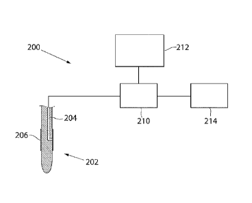
With reference to FIGURE 2, showing a block diagram, a self-flowing system
200 for measuring the concentration of one or more substances or analytes,
according to another embodiment will now be described. The self-flowing
system 200 comprises a probe 202, a sensor 210, a monitoring means 212,
and a waste container 214. The probe 202 is inserted into a suitable
pressurised body fluid of a patient (not shown). The probe 202 further
comprises an outlet lumen 204, one or more through-holes (not shown)
lo connecting the outer surface of the probe 202 with the outlet lumen 204,
and
an interface 206 covering the through-hole(s). In this embodiment, the
membrane has a very smooth surface on the part of the membrane being in
contact with the body fluid. The sensor 210 is situated adjacent to the
proximal end of the outlet lumen 204, and detects the concentration of at
least
one substance from a pressurised body fluid, when the substance passes from
the patient through the membrane via the outlet lumen 204 and into the
sensor 210. However, the invention is not limited thereto; the sensor 210 may
alternatively be situated in the outlet lumen 204. According to this
embodiment, the sensor 210 is connected to the proximal end of the outlet
lumen 204 of the probe 202, and conveys data regarding the detected
concentration to the monitoring means 212. The sensor 210 and the
monitoring means 212 may be connected wirelessly or by a direct cable. Such
monitoring means can be realised as a computer monitor, a display device,
etc. Furthermore, the sensor 210 is a flow-through sensor and the fluid flow
passing the sensor 210 is collected in the waste container 214. The collection
of fluid flow enables further analyses of the fluid flow, e.g.
spectrophotometric analysis in vitro. It should also be noted that the
invention is not limited to the above described embodiments of self-flowing
1 05 11
WO 2011/059397 PCT/SE2010/051256
12
systems 100, 200, a skilled person may easily realise how to modify the self-
flowing system 100, 200, e.g. by omitting the waste container 214, by
selecting an alternative sensor type and/or another type of monitoring device,
etc.
With reference to FIGURE 3, showing a part of a cross-sectional
longitudinal view through a probe 300, the design of the probe 300 according
to a specific embodiment will now be described. The probe 300 comprises a
body 302 and a membrane 304. The probe body 302 is partly provided with
an outlet lumen 306, and at least one through-hole 308 connecting the outside
of the probe body 302 with the outlet lumen 306. The probe body 302 is
covered with the membrane304. The semipermeable membrane 304 is
selected with special characteristics regarding the liquid permeability Lp,
the
surface area, as well as -the pores sizes and the surface roughness facing the
pressurized body fluid. According to this embodiment, the membrane is a
PAES hollow-fibre membrane from Gambro, with an outer diameter of 1.55
mm and a wall thickness of about 50 m. The liquid permeability Lp, also
called hydraulic permeability, hydraulic conductivity or the filtration
coefficient (Kf), is 6.6*10-4 cm/(bar*s), the surface area of the membrane is
about 195 mm2. Regarding surface roughness, pore sizes and other overall
membrane characteristics, suitable membranes, for this application and other
applications discussed with the present invention, are found in WO
2008/046779 (Gambro Lundia AB). The through-hole 308 is situated at the
distal part of the probe body 302. The outlet lumen 306 transports a flow of a
liquid comprising substance(s) from a body fluid, which flows through the
membrane via the through-hole 308 into the distal end of the outlet lumen 306
and then to the proximal end of the outlet lumen 306. A skilled person
realises easily how to manufacture the through-hole 308. In this embodiment,
a cut is made in the outside of the probe body 302, connecting the outside of
1 05 11
WO 2011/059397 PCT/SE2010/051256
13
the probe body 302 with the outlet lumen 306. The manufacturing of the
outlet lumen 306 may, for instance, be performed by forming a longitudinal
lumen through the probe body 302 during extrusion, and then providing a
stopper (not shown) in the outlet lumen 306 distally from the through-hole
308. The stopper prevents the outlet flow from flowing distally in the outlet
lumen 306. Alternatively, the interface 304 will cover just the through-
hole(s)
308 of the probe body 302, instead of surrounding the complete probe body
302. Furthermore, a chamber 3 10 may be created between the interface 304
and the probe body 302.
With reference to FIGURE 4a, showing a transversal cross-
sectional view, seen from the distal side, a conventional microdialysis probe
400 is described. The microdialysis probe 400 comprises a probe body 402,
an inlet lumen 404, an outlet lumen 406, a membrane 408, a microdialysis
chamber 410, and through-holes 412, 414. The microdialysis probe 400 is
adapted to be inserted into a body fluid of a patient, e.g. in an artery or
vein.
The inlet lumen 404 is provided in the probe body 402 and transports a
perfusate to the microdialysis chamber 410 via the through-hole 412, which
connects the inlet lumen 404 with the microdialysis chamber 410. Typically,
the perfusate is pumped into the proximal end of the inlet lumen 404. In the
microdialysis chamber 410, the perfusate absorbs substances from the body
fluid surrounding the microdialysis probe 400, through the membrane 408.
The perfusate, which have been absorbing substances, will be denoted as
analysate. The through-hole 414 is provided in the probe body 402 and
transports the analysate from the microdialysis chamber 410 to the outlet
lumen 406, to be transported to the proximal end of the probe 400. Adjacent
to the proximal end of the probe 400, a sensor (not shown) may be provided,
adapted to analyse the analysate.
1 05 11
WO 2011/059397 PCT/SE2010/051256
14
With reference to FIGURE 4b, showing a transversal cross-
sectional view, seen from the distal side, a self-flowing probe 450 according
to an embodiment will now be described. The self-flowing probe 450
comprises a probe body 452, an outlet lumen 454, a membrane 456, at least
one through-hole 460. The self-flowing probe 450 is adapted to be inserted
into a pressurised body fluid of a patient, e.g. in a suitable artery or vein.
The probe body 452 is covered with the membrane 456, at least where the
through-hole is located. The self-flowing probe 452 is adapted to absorb
substances and liquid from the surrounding body fluid through the membrane
456, and transport via the through-hole 460 to the outlet lumen 454. The
outlet lumen 454 is adapted to further transport the substances and liquid to
its
proximal, e.g. to be analysed. The analysis may be performed by a flow-
through sensor (not shown) at the proximal end of the self-flowing probe 450
and/or by collecting the analysate and analyse it in vitro. How the analysis
is
performed can easily be realised by a skilled person, and is therefore not
necessary to be further discussed here.
Alternatively, the self-flowing probe 450 comprises a chamber 458, defining a
space between the membrane and the probe body 452. The probe body 452
may further comprise additional components or means for providing
functionality to the probe 450. For instance, an additional lumen 470 to
facilitate insertion, measure blood pressure, and draw blood samples may be
provided in the probe body 452.
An advantage with the self-flowing probe 450 is that no perfusate needs to be
supplied to the probe 450. Consequently, no inlet lumen needs to be provided
in the self-flowing probe 450, and the design of the self-flowing probe 450
therefore is simplified. Moreover, the probe can be designed with a smaller
diameter, or can contain additional components without increasing the
diameter of the probe. Additionally, because a system applying the above
1 05 11
WO 2011/059397 PCT/SE2010/051256
described self-flowing probe 450 is self-flowing, the system does not need to
apply a pump and syringe for supplying perfusate, which makes the system
less complex.
With reference to FIGURE 5, a comparison between a real-time
5 analysis and a blood gas analysis will now be described. A comparison test
was made with a self-flowing system, described in an embodiment above. A
self-flowing probe as described with FIGURE 3 was inserted into a femoral
artery of a pig. The diagram comprises two graphs; a first graph indicated by
a line illustrates the result of an analysis of glucose performed by applying
a
lo flow-through sensor at the proximal end of the self-flowing probe, and
analysing the analysate flowing through the sensor. A second graph indicated
by black dots illustrates the result of a blood gas analysis of the blood in
the
femoral artery of the pig. The system will always provide a fluid flow, having
the correct concentration of analytes, to the sensor. The liquid and the
15 analytes present in the surrounding body fluid will spontaneously be forced
through the membrane. The rate at which the liquid and the analytes will pass
depends mostly on the surrounding pressure as well as the liquid permeability
and the surface area of the membrane. At a higher pressure as in an artery a
lower liquid permeability is suitable. At a lower surrounding pressure as in a
vein a higher liquid permeability would be more suitable.
By means of the present invention, a system for continuous
measurement of substances in a pressurised body fluid without needing to
provide a perfusion fluid to the probe is achieved. The system may be
designed without pump, syringe, or perfusion fluid, and will therefore be less
complex.
Furthermore, the probe may be designed without an inlet channel for
perfusion fluid, resulting in that the probe may be designed with smaller
dimensions, or contain additional lumens and/or components.
1 05 11
WO 2011/059397 PCT/SE2010/051256
16
Moreover, since no perfusion fluid needs to be provided, the spontaneous
fluid flow from the probe to be analysed will not be diluted and will always
exactly reflect the concentration in the body fluid.
Although particular embodiments have been disclosed herein in
detail, this has been done by way of example for purposes of illustration
only,
and is not intended to be limiting with respect to the scope of the appended
claims that follow. In particular, it is contemplated by the inventors that
various substitutions, alterations, and modifications may be made to the
invention without departing from the spirit and scope of the invention as
lo defined by the claims.
The invention is generally defined by the following independent claims.