Note: Descriptions are shown in the official language in which they were submitted.
CA 02782078 2015-10-27
OPHTHALMIC VALVED TROCAR CANNULA
FIELD OF THE INVENTION
The present invention generally pertains to ophthalmic surgery. More
particularly,
but not by way of limitation, the present invention pertains to ophthalmic
trocar cannulas
and vents.
DESCRIPTION OF THE RELATED ART
Microsurgical instruments may be used by surgeons for removal of tissue from
delicate and restricted spaces in the human body, e.g., in surgery on the eye
(such as
procedures for removal of the vitreous body, blood, scar tissue, or the
crystalline
lens). Such instruments may include a control console and a surgical handpiece
with
which the surgeon dissects and removes the tissue. With respect to posterior
segment
surgery, the handpiece may be a vitreous cutter probe, a laser probe, or an
ultrasonic
fragmenter for cutting or fragmenting the tissue and may be connected to the
control
console by a long air-pressure (pneumatic) line and/or power cable, optical
cable, or
flexible tubes for supplying an infusion fluid to the surgical site and for
withdrawing or
aspirating fluid and cut/fragmented tissue from the site. The cutting,
infusion, and
aspiration functions of the handpiece may be controlled by the remote control
console
that not only provides power for the surgical handpiece(s) (e.g., a
reciprocating or
1
CA 02782078 2012-05-28
WO 2011/087577
PCT/US2010/057582
rotating cutting blade or an ultrasonically vibrated needle), but may also
control the
flow of infusion fluid and provide a source of vacuum (relative to atmosphere)
for the
aspiration of fluid and cut/fragmented tissue. The functions of the console
may be
controlled manually by the surgeon, (e.g., through use of a foot-operated
switch or
proportional control).
During posterior segment surgery, the surgeon may use several handpieces or
instruments during the procedure. This procedure may require that these
instruments
be inserted into, and removed out of the incision. This repeated removal and
insertion
may cause trauma to the eye at the incision site. To address this concern,
hubbed
cannulae were developed at least by the mid-1980s. These devices may include a
narrow tube with an attached hub. The tube may be inserted into an incision in
the eye
up to the hub, which may act as a stop, preventing the tube from entering the
eye
completely. rfhe hub may be stitched to the eye to prevent inadvertent
removal.
Surgical instruments can be inserted into the eye through the tube, and the
tube may
protect the incision sidewall from repeated contact by the instruments. In
addition, the
surgeon may use the instrument, by manipulating the instrument when the
instrument
is inserted into the eye through the tube, to help position the eye during
surgery.
Disadvantages of prior art cannulae may include the height of the projection
on the
surface of the eye, as well as the lack of any means to control loss of
intraocular
pressure during instrument exchange or removal. The eye, being a pressurized
globe,
may expel aqueous or vitreous out of the open cannula when a surgical device
is not
present. With prior art cannulae, loss of intraocular pressure was prevented
by the
insertion of a plug or cap into the tube to seal the cannula and prevent the
expression
of fluid and tissue. This may be a time-consuming process that may require
additional
instrumentation as well as the assistance of other operating room personnel
and may
increase the risk of post-operative infection.
2
CA 2782078 2017-03-01
SUMMARY OF THE INVENTION
In various embodiments, a trocar cannula may be configured for insertion into
an eye to facilitate insertion and removal of instruments during surgery. The
cannula
may be affixed to an overcap (affixed to inhibit rotation of the overcap
relative to the
cannula). The overcap may include a seal for inhibiting the flow of fluids out
of the
cannula (when an instrument is not inserted) while the cannula is inserted in
the eye.
In some embodiments, the seal may be molded into the overcap or may include a
wafer that is fixed between the cannula and the overcap such that the seal
does not
rotate relative to the cannula and the overcap. In some embodiments, the
cannula and
overcap may snap together through a tab/slot interface in a permanent fashion
such
that the cannula and overcap many not be separated without damaging at least
part of
the cannula or overcap. In some embodiments, a vent cannula may be slidably
receivable in the slit of the seal for allowing fluids to vent from the eye
through the
cannula. In some embodiments, the cannula may include at least one indentation
to
frictionally engage a portion of the vent when the vent is inserted into the
cannula.
In one particular embodiment there is provided an apparatus, comprising:
cannula configured for insertion into an eye; an overcap affixed to the
cannula;
wherein the cannula or the overcap includes at least one tab and wherein the
other of
the cannula or overcap includes at least one slot, wherein a tab of the at
least one tab
extends along less than three hundred and sixty degrees of a peripheral of the
cannula
or the overcap and wherein a slot of the at least one slot terminates in an
overlapping
portion of the peripheral of the cannula or the overcap that does not include
the tab;
wherein the overcap is affixed to the cannula by the at least one tab being
received in
the at least one slot; wherein the overcap is configured not to rotate
relative to the
cannula; and a seal between the cannula and the overcap, wherein the seal is
configured to allow passage of a surgical tool into the cannula through a slit
in the seal
while inhibiting fluid flow through the seal when the surgical tool is not
present in the
seal.
3
CA 2782078 2017-03-01
In a further embodiment there is provided an apparatus, comprising: a cannula
configured for insertion into an eye; an overcap affixed to the cannula,
wherein the
overcap is configured not to rotate relative to the cannula; and a seal
between the
cannula and the overcap, wherein the seal is configured to allow passage of
surgical
tools into the cannula through a slit in the seal while inhibiting fluid flow
through the
seal when a surgical tool is not present in the seal; characterized in that
the seal is
made of an elastomer overmolded into a depression in the overcap; and wherein
the
seal is molded into at least one hole in the overcap.
There is also provided a method, comprising: forming an overcap; forming a
cannula configured for insertion into the eye; securing an elastomer seal to
the overcap
by overmolding the seal into a depression in the overcap, wherein the seal is
configured to allow passage of surgical tools into the cannula through a slit
in the seal
while inhibiting fluid flow through the seal when a surgical tool is not
present in the
seal; and affixing the overcap to the cannula, wherein the overcap is
configured not to
rotate relative to the cannula.
3a
CA 02782078 2012-05-28
WO 2011/087577
PCT/US2010/057582
BRIEF DESCRIPTION OF THE DRAWINGS
For a more complete understanding of the present invention, reference is made
to the following description taken in conjunction with the accompanying
drawings in
which:
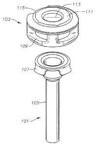
FIG, 1 illustrates a cannula and an overcap, according to an embodiment;
FIG. 2 illustrates the cannula affixed to the overcap, according to an
embodiment;
FIG. 3a illustrates a top view showing the slit in the seal on the overcap,
according to an embodiment;
FIG. 3b illustrates a side view of the cannula and overcap with several
example dimensions, according to an embodiment;
FIGs. 4a-d illustrate cross-sections of embodiments of the overcap and seal;
FIG. 5a illustrates the cannula on a trocar inserter, according to an
embodiment;
FIG. 5b illustrates the cannula on a trocar inserter with a shipping cap,
according to an embodiment;
FIGs. 6a-b illustrates a vent, according to an embodiment;
FIG. 7 illustrates a vent in the valved trocar cannula, according to an
embodiment;
4
CA 02782078 2012-05-28
WO 2011/087577
PCT/US2010/057582
FIG. 8 illustrates a cross section of the vent in the valved trocar cannula,
according to an embodiment;
FIGs. 9a-b illustrate a second embodiment of a vent;
FIGs. 10a-c illustrate a third embodiment of a vent;
FIG. 11 illustrates a flowchart of a method for forming the valved trocar
cannula, according to an embodiment;
FIG. 12 illustrates a flowchart of a method for forming the valved trocar
cannula, according to another embodiment;
FIG. 13 illustrates a flowchart of a method for using the vent with the valved
trocar cannula, according to an embodiment; and
FIG. 14 illustrates a valved trocar cannula inserted into an eye, according to
an
embodiment.
It is to be understood that both the foregoing general description and the
following detailed description are exemplary and explanatory only and are
intended to
provide a further explanation of the present invention as claimed.
5
CA 02782078 2012-05-28
WO 2011/087577
PCT/US2010/057582
DETAILED DESCRIPTION OF THE EMBODIMENTS
FIG. 1 illustrates an embodiment of a trocar cannula 101 and an overcap 103.
The trocar cannula 101 may be configured for insertion into an eye to
facilitate
insertion and removal of instruments during surgery. The cannula 101 may
include a
shaft 105 capable of extending into the eye (e.g., through a sclera,
conjunctiva, etc).
In some embodiments, the cannula 101 may be attached to an overcap 103. For
example, the cannula 101 may include one or more tabs 107 configured to engage
corresponding slots 109 on the cannula 101 (e.g., the cannula 101 illustrated
in FIG. 1
includes four tabs 107 to engage four corresponding slots 109 on the overcap
103).
Other attachments are also contemplated. For example, the cannula 101 may
include
the slots and the overcap may include the tabs. In some embodiments, the
cannula
101 may be attached to the overcap 103 through adhesive, thermal bonding, etc.
In
some embodiments, a seal 111 may be coupled to the overcap 103 (e.g., the seal
111
may be disposed at least partially between the shaft 105 and the overcap 109)
to form
an overmolded valve. As shown in FIG. 1, a surface of the seal 111 may be
exposed
on the overcap 109. In some embodiments, the exposed surface of the seal 111
may
include one or more slits 113 to allow passage of surgical tools into the
cannula 101.
In the absence of a surgical instrument, the seal 111 may inhibit fluid flow
through the
seal 111.
FIG. 2 illustrates an embodiment of the cannula 101 affixed to the overcap 103
(e.g., after engagement of the tabs 107 in respective slots 109). In some
embodiments, the tab/slot interface may prevent rotation of the overcap 103
relative
to the cannula 101 (e.g., during insertion of the cannula 101 into the eye).
In some
embodiments, the tabs 107 may be configured to permanently hold the overcap
103 to
the cannula 101 (such that the overcap 103 may not be removed from the cannula
101
without destroying part of the cannula 101 and/or overcap 103). For example,
the
tabs 107 (and cannula 101) may be made of stainless steel and the overcap 103
may
be made of plastic (e.g., polycarbonate). Other materials are also
contemplated. The
6
CA 02782078 2012-05-28
WO 2011/087577
PCT/US2010/057582
permanent hold between the overcap 103 and the cannula 101 may prevent
inadvertent removal of the overcap 103 from the cannula 101 during surgery
(e.g.,
vitreoretinal surgery).
FIG. 3a illustrates a top view of an embodiment of the slit 113 in the seal
111
on the overcap 103. FIG. 3b illustrates a side view of an embodiment of the
cannula
101 and overcap 103 with several example dimensions (provided in inches).
Other
dimensions are also contemplated. For example, while the outer diameter of the
cannula 101 is shown as 0.029 inches (corresponding to a 23 gauge cannula), in
another embodiment, the outer diameter of the cannula may be 0.0243 inches
(for a 25
gauge cannula). Other outer diameters are also contemplated.
FIGs. 4a-c illustrate cross-sections of an embodiment of the overcap 103 and
seal 111. The seal 111 may be made of an elastomer (e.g., silicone). In some
embodiments, the seal 111 may be attached to the overcap 103 to inhibit
rotation of
the seal 111 relative to the overcap 103. For example, the seal 111 may be
overmolded into a depression 403 and one or more holes 401 in the overcap 103.
In
some embodiments, the seal 111 may include a silicon wafer 405 that is formed
separately from the overcap 103 and inserted between the overcap 103 and the
cannula 101 during assembly of the overcap 103 onto the cannula 101. In such a
case,
the seal 111 may be attached to the overcap 103 and cannula 101 through a
friction
fit. Other attachments are also contemplated (e.g., adhesive).
FIG. 5a illustrates an embodiment of the cannula 101 on a trocar inserter 501.
In some embodiments, the trocar inserter 501 may include a trocar blade 503
attached
to a handle 505. In some embodiments, the handle 505 may be made of plastic
and
the blade 503 may be made of stainless steel. Other materials are also
contemplated.
The trocar blade 503 may extend past the end of the shaft 105 and may include
one or
more sharp edges to pierce an eye 1401 (e.g., pierce a hole through the sclera
1403
and into the vitreous body 1405) for insertion of the cannula 101. In some
7
CA 02782078 2012-05-28
WO 2011/087577
PCT/US2010/057582
embodiments, a guide 507 may fit into guide slot 115 to inhibit rotation of
the overcap
103/cannula 101 relative to the handle 505 during insertion of the eannula 101
into
eye 1401. In some embodiments, the guide 507 may releasably engage the guide
slot
115 such that when the trocar inserter 501 is withdrawn from the overcap
103/cannula
101, the guide 507 does not pull the overcap 103/cannula 101 out of the eye
1401.
For example, the guide 507 may frictionally engage the guide slot 115 with a
friction
force that is less than a friction force exerted by the eye on the external
sides of the
cannula 101 when the cannula 101 is in the eye.
While the guide 507 is depicted as a tab to be received into guide slot 115,
other interlocking features are also contemplated. For example, the guide 507
and
guide slot 115 may include different interlocking features (such as a ring and
a rod) or
may include other interlocking components (e.g., interlocking magnets (one on
each
of the handle and overcap 103), engaging o-rings (one on each of the handle
and
overcap 103), etc). In some embodiments, the guide 507/guide slot 115
interaction
may prevent rotation between the cannula 101 and the overcap 103 so that any
angular movement of the trocar handle 505 about the handle's axis may be
transmitted to the overcap 103 and then to the eannula 101. This interaction
may
provide vitreoretinal surgeons angular control of the cannula 101 relative to
the trocar
handle 505 during insertion of the cannula 101 into the sclera 1403. FIG.
5b
illustrates an embodiment of the cannula 101 on a trocar inserter 501 with a
shipping
cap 511 (which may be snapped on over the cannula 101 and/or over the trocar
inserter 501 to protect the cannula 101 and/or trocar inserter 501.
FIGs. 6a-b illustrates an embodiment of a vent 60]. While the seal 111 of the
valved trocar cannula may close off the cannula from fluid flow into or out of
the
cannula when, for example, a surgical instrument is occluding the cannula, a
vent
cannula 603 may be configured to slide into the slit 113 of the seal 111 to
allow fluids
to vent from the eye through the cannula 101 (e.g., see FIG. 7). In some
embodiments, the vent 601 may hold the seal 111 in an open position to allow
fluid
8
CA 02782078 2012-05-28
WO 2011/087577
PCT/US2010/057582
(e.g., a gas or liquid) to vent through the cannula 101. For example, a gas
(or another
fluid) may flow through the cannula 101 and out of vent 601 during a procedure
to
replace the gas with another fluid. The vent 601 may further include a rim 609
to
provide a stop for preventing the vent from slipping all the way into the seal
111. The
vent cannula 603 may have an outer diameter that is smaller than an inner
diameter of
trocar cannula 101 to allow the vent cannula 603 to slide past the seal 111
and into the
trocar cannula 101. The vent cannula 603 may further include a rim 609 with at
least
one dimension that is large enough to prevent the vent 601 from slipping
completely
into the trocar cannula 101 (e.g., the diameter of the rim 609 may be larger
than an
inner diameter of the trocar cannula 101).
In some embodiments, the vent 601 may be a separate device from the cannula
to allow the vent 601 to be inserted and removed without adding or removing
parts of
the cannula 101 (e.g., without having to remove the overcap 103 of the cannula
101).
The size of the vent 601 may also allow a user (e.g., a surgeon) to handle the
vent 601
with fingers (or, for example, forceps) during the insertion and removal of
the vent
601.
In some embodiments, the vent 601 may include a flexible tube 605 (e.g., a
silicone tube) frictionally engaging the vent cannula 603. The tube 605 may
provide a
visual indicator (e.g., be at least partially transparent) of the venting
process (e.g., if a
substance is overflowing from the eye (such as silicone during a viscous fluid
control
injection procedure), the silicone may flow into the tube 605 and be visible
to a user.
In some embodiments, the tube 605 may be used as a grasping surface for vent
removal from the cannula 101 (e.g., to assist grasping by fingers or forceps).
Vent
cannula 603 may include a tube portion 607 configured to receive the flexible
tube
605 along an outer perimeter of the tube 607 (which may be made of, for
example,
stainless steel). In some embodiments, the tube 607 and vent cannula 603 may
be
formed of one piece. FIG. 6b illustrates several example dimensions (provided
in
inches), according to an embodiment. Other dimensions are also contemplated.
In
9
CA 02782078 2012-05-28
WO 2011/087577
PCT/US2010/057582
some embodiments, the dimensions of the vent 601 may allow for the passage of
instruments through the vent 601 when the vent 601 is in seal 111.
FIG. 8 illustrates a cross section of an embodiment of the vent 601 in the
cannula 101. In some embodiments, the cannula 101 may include at least one
indentation 801 to frictionally engage a portion of the vent cannula 603 when
the vent
601 is inserted into the cannula 101. The indentation 801 may be dimensioned
to
provide enough resistance to the vent 601 to keep the vent 601 in place during
a
procedure. In some embodiments, the resistance between the indentation 801 and
vent 601 may be less than needed to pull the cannula 101 out of the eye when
withdrawing the vent 601 from the cannula 101 (such that the cannula 101 is
not
pulled out of the eye when the vent 601 is pulled out of the cannula 101 while
the
cannula 101 is in the eye).
Other embodiments of the vent are also contemplated. For example, an
embodiment of a vent 901 is shown in FIGs. 9a-b. As seen in FIGs. 9a-b, the
vent
901 may not include a tube 605, but may instead be a single piece. The vent
901 may
be deep drawn and may include a retention feature for mating with a retention
feature
on a cannula 101. Other formation techniques are also contemplated (e.g., the
vent
901 may be molded). In some embodiments, the vent may not include a retention
feature. Yet another embodiment is shown as vent 1001 in FIGs. 10a-c. Vent
1001
may include a large bell shaped inlet that may make it easier to insert and
remove
tools through the vent 1001 when the vent 1001 is inserted into a cannula 101.
The
vent 1001 may also include one or more retention features 1003 to increase a
grip
between the vent 1001 and a cannula 101 when the vent 1001 is inserted into a
cannula 101.
FIG. 11 illustrates a flowchart of a method for forming the valved trocar
cannula, according to an embodiment. The elements provided in the flowchart
are
illustrative only. Various provided elements may be omitted, additional
elements may
CA 02782078 2012-05-28
WO 2011/087577
PCT/US2010/057582
be added, and/or various elements may be performed in a different order than
provided below.
At 1101, the overcap 103 may be formed. For example, the overcap 103 may
be molded to include thru-holes 401 for receiving a silicone seal 111. Molding
processes for the overcap 103 may include injection molding, compression
molding,
blow molding, rotational molding, etc. Other techniques for forming the
overcap 103
arc also contemplated (e.g., casting).
At 1103, the seal 111 may be overmolded onto the overcap 103. For example,
the seal Ill may include an elastomer (such as silicone) molded into a
depression 403
of the overcap 103 and may flow into holes 401 to secure the seal 111 to the
overcap
103. In some embodiments, the overcap 103 may be placed into a mold that
defines
spaces through the overcap 103 for the seal 111. The elastomer may then be
injected
into the mold and flow through the defined spaces through the overcap 103 to
form
the seal 111 in the overcap 103. Other
manufacturing processes are also
contemplated. For example, the seal 111 and overcap 103 may be molded as one
piece (e.g., using the same material for both the overcap 103 and seal 111 in
a single
mold). In some embodiments, the seal 111 may be formed separately from the
overcap 103 (e.g., see FIG. 10). In some embodiments, the seal may be formed
with a
slit 113 or the slit 113 may be formed in the seal I 1 I after the seal is
formed (e.g., the
slit 113 may be cut into the seal 111 using a sharp edge).
At 1105, a cannula 101 may be formed. For example, the cannula 101 may be
deep drawn. Deep drawing the cannula 101 may include starting with a disc of
material that is pressed between one or more sets of male/female dies to deep
draw
the cannula 101. A final step in cannula formation may include removing excess
material and/or polishing the cannula 101. In some embodiments, material
between
the tabs 107 may be sheared off between a male and female die or may be
removed in
other ways (e.g., cut away). In some embodiments, the cannula 101 may be
molded
11
CA 02782078 2012-05-28
WO 2011/087577
PCT/US2010/057582
(e.g., injection molding, compression molding, blow molding, rotational
molding,
extrusion molding, etc). Other techniques for cannula formation are also
contemplated. In some embodiments, the cannula 101 may be made of stainless
steel
or plastic. Other materials may also be used. In some embodiments, the cannula
101
may be formed with snapping tabs 107. For example, the dies or mold for the
cannula
101 may include spaces for the formation of the tabs or the tabs may be formed
on the
cannula 101 through machining. Other tab forming techniques are also
contemplated.
At 1107, the overcap 103 may be affixed to the cannula 101. For example, the
tabs 107 may be snapped into corresponding slots 109. In some embodiments, the
overcap 103 may slightly deform to receive the tabs 107 or the tabs 107 may be
configured to slightly deform as the overcap 103 is pressed onto the cannula
101 and
then return to their initial condition as the corresponding slots 109 of the
overcap 103
pass over the tabs 107. The tabs 107 may be rigid enough (e.g., made of
stainless
steel) such that the overcap 103 may not be removed from the cannula 101
without
destroying part of the cannula 101 and/or overcap 103.
FIG. 12 illustrates a flowchart of a method for forming the valved trocar
cannula according to another embodiment. The elements provided in the
flowchart
are illustrative only. Various provided elements may be omitted, additional
elements
may be added, and/or various elements may be performed in a different order
than
provided below.
At 1201, an overcap 1103 may be formed (e.g., through molding). In some
embodiments, the overcap 1103 may be formed with receiving slots 109.
At 1203, a seal 111 (such as a silicone wafer 405) may be formed. In some
embodiments, the silicone wafer 405 may be molded with a slit 113 or the slit
113
may be formed in the silicone wafer 405 after molding (e.g., the slit 113 may
be cut
into the silicone wafer using a sharp edge).
12
CA 02782078 2012-05-28
WO 2011/087577
PCT/US2010/057582
At 1205, the cannula 101 may be formed. For example, the cannula 101 may
be molded out of stainless steel and may include tabs 107.
At 1207, the silicone wafer 405 may be inserted between the cannula 101 and
overcap 103 and the overcap 103 may be affixed to the cannula 101 (e.g., the
overcap
103 may be snapped onto the cannula 101 such that the slots 109 may receive
tabs
107).
FIG. 13 illustrates a flowchart of a method for using the vent with the valved
trocar cannula, according to an embodiment. The elements provided in the
flowchart
are illustrative only. Various provided elements may be omitted, additional
elements
may be added, and/or various elements may be performed in a different order
than
provided below.
At 1301, the trocar blade 503 may be inserted through the slit 113 in the seal
111 and through cannula 101. At 1303, the eye 1401 may be pierced with the
trocar
blade 503 and the cannula 101 may be pushed into the eye. At 1305, the trocar
blade
503 and cannula 101 may be rotated as needed during the insertion. At 1307,
the
trocar blade 503 may be withdrawn leaving the cannula 101 in the eye 1401. At
1309,
a vent 601 may be inserted as needed to open the seal 111 to allow fluid/gas
to vent
through the cannula 101 and out of the eye 1401. The vent 601 may be removed
and/or reinserted as needed into the seal 111 and cannula 101 without
withdrawing
the cannula 101 from the eye during the procedure. Inserting the withdrawing
the
vent 601 may be performed using, for example, the user's fingers or a pair of
forceps.
Various modifications may be made to the presented embodiments by a person
of ordinary skill in the art. Other embodiments of the present invention will
be
apparent to those skilled in the art from consideration of the present
specification and
practice of the present invention disclosed herein. It is intended that the
present
13
CA 02782078 2015-10-27
specification and examples be considered as exemplary only with a true scope
being
indicated by the following claims and equivalents thereof.
14