Note: Descriptions are shown in the official language in which they were submitted.
CA 02782304 2012-05-29
WO 2011/067756 PCT/IL2010/001007
1
DEVICE SYSTEM AND METHOD FOR TISSUE ACCESS SITE CLOSURE
FIELD AND BACKGROUND OF THE INVENTION
The present invention relates to a device, system and method which can be used
to partially or fully close tissue access sites and in particular; access
sites in tubular
vessels such as blood vessels (vascular access sites).
More than five million percutaneous interventions are performed annually in
the
United States, involving femoral artery catheterization for diagnostic or
therapeutic
purposes.
Most procedures are performed through small sheath access sites (5-8F) and
thus
closure of such access sites can be effected using manual or mechanical
compression for
15-30 minutes, typically combined with an extended bed-rest of three to six
hours.
However, manual compression can cause patient discomfort, and is time- and
resource-intensive, and as such, a need for quicker, more patient compatible
closure has
led to the introduction of closure devices in the early 1990s. Since then,
vascular closure
systems have been simplified to provide wider patient access to a range of
vascular
procedures. Now available from many sources, these devices shorten procedure
times,
allow patients to ambulate earlier, minimize bleeding and possibly reduce
costs
associated with hospital care.
At present there are dozens of devices on the market or at various stages of
development, such devices employ sutures, patches, glue, coagulants and/or
staples or a
source of energy to effectively seal access sites post procedure. ;
Although these devices were specifically designed for closure of small access
sites (<10F), there have been attempts since the late 90s to utilize suture
closure devices
(specifically the SuturaTM and PercloseTM devices) in large bore access sites
>18F,
illustrating at least a limited need for `automated' closure of large access
sites. Large
bore access site closure is typically effected via manual suturing of an
exposed artery
and thus requires presence of a specialist while being time consuming as well
as more
invasive.
The studies performed to date illustrate that closure of access sites less
than 18F
in size via such devices is effective and highly successful, whereas closure
of larger bore
access sites (e.g. 22F) is less effective.
CA 02782304 2012-05-29
WO 2011/067756 PCT/IL2010/001007
2
Although at present the number of procedures effected through large bore
access
sites is small, current trends anticipate that the number of such procedures
will rise in the
future and although a concomitant reduction in sheath sizes might also take
place, such
reduction will still place average sheath size at over 18F.
While reducing the present invention to practice, the present inventors have
devised an access site closure system which enables reliable closure of large
bore access
sites while providing re-access if necessary.
SUMMARY OF THE INVENTION
According to one aspect of the present invention there is provided a system
for
closure of a vascular access site comprising: (a) a radially expandable device
sized and
configured for positioning within a blood vessel; and (b) a delivery catheter
for (i)
delivering the radially expandable device through the vascular access site;
and (ii)
expanding the radially expandable device in a region within the blood vessel
spanning
the vascular access site, thereby at least partially closing the vascular
access site.
According to further features in preferred embodiments of the invention
described below, the radially expandable device is a stent graft.
According to still further features in the described preferred embodiments the
radially expandable device is self expanding.
According to still further features in the described preferred embodiments the
radially expandable device includes two opposing expandable rings
interconnected via a
sleeve.
According to still further features in the described preferred embodiments
each
of the two opposing expandable rings is less than 10 mm in width.
According to still further features in the described preferred embodiments the
sleeve is fabricated from a tubular sheet of ePTFE.
According to still further features in the described preferred embodiments the
delivery catheter includes a mechanism for expanding the radially expandable
device.
According to still further features in the described preferred embodiments the
radially expandable device includes a balloon and the mechanism is an
inflation
mechanism.
CA 02782304 2012-05-29
WO 2011/067756 PCT/IL2010/001007
3
According to still further features in the described preferred embodiments the
radially expandable device is compressed within a sheath and the mechanism is
a sheath
removal mechanism.
According to still further features in the described preferred embodiments the
radially expandable device includes an aperture in a side wall along a length
thereof, the
aperture being for providing the delivery catheter access to a lumen of the
expandable
tubular body.
According to still further features in the described preferred embodiments the
delivery catheter and the radially expandable device form a t-shape when the
radially
expandable device is positioned in the blood vessel.
According to still further features in the described preferred embodiments the
delivery catheter engages the radially expandable device through the aperture,
such that
a guide wire can be threaded through the delivery catheter though a lumen of
the radially
expandable device and into a lumen of the blood vessel.
According to still further features in the described preferred embodiments the
aperture is capable of at least partially closing when the delivery catheter
is disengaged
therefrom.
According to another aspect of the present invention there is provided a
device
for closure of a vascular access site comprising a radially expandable tubular
body
having an aperture in a side wall along a length thereof, the aperture being
for providing
a delivery catheter access to a lumen of the expandable tubular body.
According to still further features in the described preferred embodiments the
radially expandable tubular body includes two opposing expandable rings
interconnected
via a sleeve.
According to still further features in the described preferred embodiments
each
of the two opposing expandable rings is less than 10 mm in width.
According to still further features in the described preferred embodiments the
sleeve is fabricated from a tubular sheet of ePTFE.
According to another aspect of the present invention there is provided a
method
of at least partially closing a vascular access site comprising: (a)
positioning a radially
expandable device having an aperture in a side wall along a length thereof
within a
blood vessel through the vascular access site; and (b) aligning the aperture
with the
CA 02782304 2012-05-29
WO 2011/067756 PCT/IL2010/001007
4
access site and expanding the radially expandable device within the blood
vessel at a
region spanning the vascular access site; and (c) at least partially closing
the aperture
thereby at least partially closing the vascular access site.
According to still further features in the described preferred embodiments
steps
(a) and (b) are effected using a delivery catheter.
According to still further features in the described preferred embodiments the
delivery catheter engages the radially expandable device through the aperture,
such that
a guide wire can be threaded through the delivery catheter though a lumen of
the radially
expandable device and into a lumen of the blood vessel.
According to still further features in the described preferred embodiments the
delivery catheter includes a mechanism for expanding the radially expandable
device.
According to still further features in the described preferred embodiments the
radially expandable device is disposed over a balloon and the mechanism is an
inflation
mechanism.
According to still further features in the described preferred embodiments the
radially expandable device is compressed within a sheath and the mechanism is
a sheath
removal mechanism.
According to still further features in the described preferred embodiments the
delivery catheter and the radially expandable device form a t-shape when the
radially
expandable device is positioned in the blood vessel.
According to still further features in the described preferred embodiments the
aperture is capable of at least partially closing when the delivery catheter
is disengaged
therefrom.
According to yet another aspect of the present invention there is provided a
system for delivering a stent-graft to a body lumen comprising: (a) a radially
expandable
device sized and configured for positioning within the body lumen, the
radially
expandable device including two opposing expandable rings interconnected via a
sleeve
having an aperture in a side wall along a length thereof; and (b) a delivery
catheter
including an internal sheath and an external sheath, wherein the radially
expandable
device is packable within the external sheath with a first ring of the two
opposing
expandable rings being disposed around the internal sheath and the second ring
of the
two opposing expandable rings being disposed adjacent to the internal sheath.
CA 02782304 2012-05-29
WO 2011/067756 PCT/IL2010/001007
According to still further features in the described preferred embodiments the
radially expandable device is packable within the external sheath with the
internal sheath
inserted through the aperture.
5 The present invention successfully addresses the shortcomings of the
presently
known configurations by providing a closure device and system that enables
rapid and
easy closure of, for example, a vascular access site while also enabling
subsequent
vascular reentry.
Unless otherwise defined, all technical and scientific terms used herein have
the
same meaning as commonly understood by one of ordinary skill in the art to
which this
invention belongs. Although methods and materials similar or equivalent to
those
described herein can be used in the practice or testing of the present
invention, suitable
methods and materials are described below. In case of conflict, the patent
specification,
including definitions, will control. In addition, the materials, methods, and
examples are
illustrative only and not intended to be limiting.
BRIEF DESCRIPTION OF THE DRAWINGS
The invention is herein described, by way of example only, with reference to
the
accompanying drawings. With specific reference now to the drawings in detail,
it is
stressed that the particulars shown are by way of example and for purposes of
illustrative
discussion of the preferred embodiments of the present invention only, and are
presented
in the cause of providing what is believed to be the most useful and readily
understood
description of the principles and conceptual aspects of the invention. In this
regard, no
attempt is made to show structural details of the invention in more detail
than is
necessary for a fundamental understanding of the invention, the description
taken with
the drawings making apparent to those skilled in the art how the several forms
of the
invention may be embodied in practice.
In the drawings:
FIGs. 1-8 illustrate one embodiment of the present system through stages of
deployment and stent-graft positioning.
CA 02782304 2012-05-29
WO 2011/067756 PCT/IL2010/001007
6
FIG. 9 illustrates one embodiment of a stent-graft constructed in accordance
with
the teachings of the present invention.
FIG. 10 depicts the setup used for testing feasibility of the present
approach.
FIGs. 11A-E illustrate the steps in deploying the tubular element
(representing
the stent-graft) out of the delivery catheter assembly and into a silicone
tube representing
an artery.
FIGs. 12A-D illustrate a platform used to test blood flow through an artery
having an access site closed using the device of the present invention. Figure
12B
illustrates the silicone tube 'artery' portion of the platform shown in Figure
12A showing
the access site formed therein via a cross-shaped incision. Figure 12C
illustrates the
device of the present invention (stent-graft, schematically illustrated in
Figure 13D)
positioned within the silicon tube (Figure 12B) of the testing platform.
Figure 12D
illustrates inward collapse of one of the stent-like rings of the present
device due to
application of external pressure.
FIGs. 13A-C illustrate another embodiment of the present system through stages
of deployment and stent-graft positioning.
FIG. 13D illustrates the embodiment of the present device formed from two-
opposing rings connected via a tubular sheet.
DESCRIPTION OF THE PREFERRED EMBODIMENTS
The present invention is of a system which can be used to partially or fully
close
a vascular access site. In one embodiment, the present invention employs a
stent-graft
which can be positioned in a lumen of a blood vessel across the access site
thereby
sealing/reducing the access site hole and preventing blood leakage therefrom.
The principles and operation of the present invention may be better understood
with reference to the drawings and accompanying descriptions.
Before explaining at least one embodiment of the invention in detail, it is to
be
understood that the invention is not limited in its application to the details
set forth in the
following description or exemplified by the Examples. The invention is capable
of other
embodiments or of being practiced or carried out in various ways. Also, it is
to be
understood that the phraseology and terminology employed herein is for the
purpose of
description and should not be regarded as limiting.
CA 02782304 2012-05-29
WO 2011/067756 PCT/IL2010/001007
7
Percutaneous access to coronary and other major blood vessels is slowly
replacing open surgical access and is driving a need for accessory
technologies such as
access site closure systems.
Although small access sites (<8-10F) can be effectively closed using existing
technologies, solutions for effective closure of large access sites (>10-12F)
are still
lacking.
Since existing approaches co-apt tissue edges surrounding the access site
hole,
use of such approaches in closure of large access sites can lead to a
substantial reduction
in blood vessel diameter as well as vessel kinking. As a result, the present
inventors have
postulated that effective closure of large access sites requires a new
approach rather than
modification of existing approaches.
Thus, according to one aspect of the present invention there is provided a
system
for closure of a vascular access site.
As used herein, the phrase "vascular access site" refers to the tissue site
through
which vasculature of a subject is accessed. The access site can be formed in
any blood
vessel suitable for access. Examples include the femoral artery, the radial
artery and the
subclavian artery.
The system of the present invention includes a radially expandable device
(also
referred to herein as the "device") which is sized and configured for
positioning within a
blood vessel and a delivery catheter for delivering the radially expandable
device
through the vascular access site and positioning it such that it spans the
vascular access
site thereby partially or fully blocking the access site hole.
The radially expandable device can be any device that can be expanded within
the blood vessel lumen to apply a force -onto the inner wall of the blood
vessel
surrounding the access site hole.
In that respect, when expanded within the lumen, the radially expandable
device
can expand to assume a substantially tubular configuration with, for example,
closed (0-
shaped cross section) or open (C-shaped cross section) profiles.
Several configurations of the radially expandable device can be used with the
present invention, including, but not limited to, closed or open tubes
fabricated from
rolled sheets, coated/covered wire frames (e.g. stent-grafts) or tubular
sheets
interconnected via two opposing stent-like rings.
CA 02782304 2012-05-29
WO 2011/067756 PCT/IL2010/001007
8
The radially expandable device of the present invention can be fabricated by
laser cutting a polymeric or alloy (e.g. Nitinol, Cobalt Chromium or stainless
steel) tube,
or by braiding or knitting polymeric or alloy wires over a mandrel to form a
tubular
structure or portions thereof.
The tubular structure can be fabricated as two opposing expandable rings
connected via rods or struts or interconnected via the tubular graft material
described
below. Each of the rings can have a width of 5-20 mm, preferably, 5-10 mm,
most
preferably 6-8 mm (along the axis defining the length of the device).
The radially expandable device can be pre-shaped as a tubular structure having
a
diameter of 7-14 mm which is capable of being compressed and folded into a 6-9
French
(F) sheath. Alternatively, the radially expandable device can be pre-shaped as
a tubular
structure having a diameter of 2-5 mm which can be crimped into a 6-9 F sheath
and
expanded (plastically) to a diameter of 7-14 mm. The tubular structure can be
15-80 mm
long, preferably, 20-70 mm long, more preferably 30-50 mm long.
The tubular structure can be covered with a graft on its external or internal
(luminal) surface. The graft can cover the entire circumference and length of
the tubular
structure or a portion thereof (e.g. less than 360 degrees around the tubular
structure
and/or a portion of its length). The graft can be fabricated from Dacron,
PTFE,
polyurethane and like materials and glued, stitched or cast onto a unitary sub
frame (e.g.
stent or strut-interconnected rings) or a sub frame including two discrete
rings.
Since closure requires sealing of the access site hole, the delivery catheter
and
the radially expandable device are configured such that the radially
expandable device
spans the vascular access site to thereby seal the access site hole following
positioning
thereof via the delivery catheter.
To achieve such functionality, both the delivery catheter and the radially
expandable device are designed and configured for T-shaped deployment within
the
lumen of the blood vessel. Such T-shaped deployment is achieved by attaching
the
delivery catheter (and guide wire threaded therethrough) through an aperture
provided in
a mid section of the radially expandable device. This ensures that when the
device is
delivered through the access site hole and deployed via the delivery catheter,
the
portions of the device disposed on either side of the aperture, flank the
access site hole,
while the aperture aligns with the access site hole.
CA 02782304 2012-05-29
WO 2011/067756 PCT/IL2010/001007
9
The above described configurations of the radially expandable device and
delivery catheter which are collectively referred to herein as system 10 are
described in
more detail below with respect to the accompanying drawings.
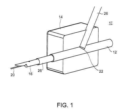
Figures 1-8 illustrates system 10 through stages of deployment within an
artery
12 surrounded by tissue 14. System 10 includes a delivery catheter and a
radially
expandable device which in the example illustrated below is a stent-graft.
Referring now to Figure 1 which illustrates delivery catheter 16 mounted over
guide wire 20, and within a standard introducer sheath 26 typically used for
percutaneous procedures; guide wire 20, introducer sheath 26 and delivery
catheter 16
are delivered through tissue 14 and access site hole 22 to the lumen of artery
12.
Typically, an access site hole 22 is formed using a needle and guide wire 20
is then
threaded therethrough. The needle is then removed and introducer sheath 26 is
mounted
over guide wire 20 and delivered through access site hole 22, widening it in
the process
to the desired diameter. Delivery catheter 16 can then be introduced into
artery 12 over
guide wire 20 and within introducer sheath 26.
Following positioning of delivery catheter 16 within artery 12, introducer
sheath
26 is pulled back through access site hole 22 and out of tissue 14 (Figure 2).
Delivery catheter 16 is then pulled back (in a proximal direction) until an
end
portion 15 thereof is positioned against the inner artery wall at access site
hole 22
(Figure 3). Distal portion 15 is pivotally attached and thus can angle and
align with the
longitudinal axis of artery 12 to facilitate deployment of expandable device
18
described below. Delivery catheter also includes an inspection hole 17 for
indicating
correct positioning of distal portion 15 of delivery catheter 16 within artery
12. Such an
indication can be provided by stoppage of blood dripping from inspection hole
17 when
delivery catheter is correctly positioned. To enable such functionality
inspection hole 17
is fluidly connected to a conduit which terminates at a distal region of
delivery catheter
16. When that distal region of delivery catheter is positioned within artery
12, the
conduit communicates blood flowing through artery 12 to inspection hole 17,
however,
when delivery catheter is pulled back to a position in which that distal
region is out of
the blood flow, no blood is communicated to inspection hole 17 and as such
dripping
stops. Thus, by slowly pulling out delivery catheter 16 and watching for
cessation of
blood.flow through inspection hole 17, one can correctly position delivery
catheter 16.
CA 02782304 2012-05-29
WO 2011/067756 PCT/IL2010/001007
Delivery catheter 16 includes radially expandable device 18 (also referred to
herein as device 18) within a lumen 24 thereof (device 18 shown in Figures 4-8
and
separately shown in Figure 9). In the present configuration of system 10,
guide wire 20
is threaded within a positioning tube 25 which runs through lumen 24 (within
internal
5 tube 23, described below) of delivery catheter and through an aperture 28 of
device 18;
guide wire exits system 10 through a distal end 30 of device 18. When pushed
out of
lumen 24 of delivery catheter 16, device 18 remains connected to delivery
catheter 16
(via the tube describe above) at a mid portion thereof and forms a T-shaped
end with
delivery catheter 16 as is further detailed below.
10 Delivery catheter 16 includes internal tube 23 for deploying radially
expandable
device 18. Internal tube 23 (pusher tube) is assembled over positioning tube
25 (both
within lumen 24) such that when internal tube 23 is advanced distally, it
pushes device
18 with attached positioning tube 25 out of the outer housing of delivery
catheter 16
(Figure 4). The distal portion of positioning tube 25 is preshaped with a 45-
90 degree
bend. When within tube 23, positioning tube 25 is held straight, however, when
it is
pushed out along with device 18 it assumes its preshaped (bent) position,
thereby
facilitating correct positioning of device 18.
Once radially expandable device 18 is exposed, it is maneuvered into a T-
position by pulling positioning tube 25 proximally (Figures 5-6). Positioning
tube 25
also serves to route guide wire 20 through delivery catheter 16 and into
radially
expandable device 18 through aperture 28.
When in the T-position, aperture 28 of device 18 is aligned with access site
hole
22 and enables expansion of device 18 in the correct position maintaining
alignment
between aperture 28 and access site hole 22. Device 18 is then expanded using
one or
more expansion mechanisms as described below (Figure 7). Once delivery
catheter 16
and introducer sheath 26 are removed, device 18 maintains its expanded
position across
access site hole 22 (Figure 8) with guide wire 20 running through tissue 14,
aperture 28
and out of distal end 30 of device 18 and into artery 12. Aperture 28 is
preferably
designed to close around guide wire 20 so as to minimize or prevent blood
leakage.
Self-sealing features of aperture 28 are further described hereinbelow.
CA 02782304 2012-05-29
WO 2011/067756 PCT/IL2010/001007
11
Device 18 can be actively expanded or it can be self expanding. For Example,
a stent graft configuration of device 18 can be wrapped by a thin sheath
(0.025-0.2mm
thick) of nylon, PTFE or Dacron and maintained at a diameter of 1-3 mm in a
non-
expanded (compressed) state. The sheath is glued mid length to positioning
tube 25 and
is locked over device 18 via a wire (Nitinol, silk or other). The
lock/stitching wire
extends from the sheath/device 18 into the catheter and out to an actuating
handle
attached proximally to delivery catheter 16. Puling the wire releases the
sheath and
enables expansion of device 18. Several locking options are contemplated. The
locking
wire can be stitched into the sheath along its length or it can glued thereto.
In any case,
deployment can be gradual along the length of device 18 (e.g. gradual
expansion from
one end to the other) or it can be stepwise, where one end (e.g. distal end of
device 18) is
deployed via pulling of locking wire to a first position following which
further pulling of
the locking wire releases the other end of device 18.
Since the locking wire connects to device 18 at a mid region (area of aperture
28)
it can also be configured to separated pull 2 ends of two locking wires
thereby opening
the wrapping sheath from both ends simultaneously.
Expansion can also be effected using a balloon. In such an approach, a balloon
is
used to tear open the wrapping sheath described above. Once the balloon is
inflated
device 18 expands, applies a radial force on the wrapping sheath and rips it
open at
predefined point or points along a predefined line along the wrapping sheath
(a precut
notch or a series of small holes). When the wrapping sheath is fully ripped
open (along
its longitudinal axis) device 18 expands to its final dimensions.
Following deployment, the balloon is deflated and is pulled out along with
along
with the wrapping sheath (both are connected to positioning tube 25) through
aperture
28 and delivery catheter 16, aperture 28 can then self-seal as described
below.
A balloon expanded configuration of device 18 is also envisaged. In such a
configuration, device 18 is fabricated in a compressed state and is actively
expanded (via
plastic deformation) using a balloon.
A stainless steel or Cobalt Chromium stent graft is positioned over a balloon
mounted and attached to a fluid filling tube 25 within delivery catheter 16.
Device 18 in
a compressed state (1-3 mm in diameter) is crimped over the balloon with the
fluid
filling tube routed through aperture 28. Inflating the balloon to 7-14mm in
diameter will
CA 02782304 2012-05-29
WO 2011/067756 PCT/IL2010/001007
12
plastically deform device 18 to the desired expanded size. Once device 18 is
deployed,
the balloon is deflated and delivery catheter 25 with enclosed balloon are
pulled out
through aperture 28 and access site hole 22.
Housing of delivery catheter 16 is constructed as a tube having a lumen which
includes device 18 and tubes 23 and 25 in a coaxial arrangement. The housing
and tubes
can be molded from any suitable material, examples include polymers, alloys,
ceramics
and the like.
Once delivery catheter 16 and guide wire 20 are removed from the body,
aperture
28 can either self seal or be sealed using an adhesive, a patch or a
combination thereof.
Several self-sealing mechanisms can be used to partially or fully close
aperture
28.
One sealing configuration can employ a wire frame oval as aperture 28 (oval
arcs
indicated by 40 and 42 in Figure 9) which is heat treated to a "normally
closed" position
in which opposing arcs 40 and 42 of the oval cross each other thereby
minimizing area
44. When device 18 is assembled within delivery catheter 16 such that
positioning tube
is fed through aperture 28, arcs 40 and 42 are forced apart thereby opening
aperture
28. Once delivery catheter is pulled out of the body, arcs 40 and 42 of
aperture 28 close
over guide wire 20, pulling out guide wire 20 allows final closure of aperture
28.
Aperture 28 designed for partial sealing can close to a predetermined point
and
20 then be completely sealed using an adhesive, pad, patch or a combination
thereof, or it
can be sealed via coagulation induced by a coagulant or manual pressure. In
any case,
closure is preferably effected using a mechanism that would allow for artery
re-entry
through aperture 28.
Figures 13A-C illustrate another embodiment of system 10 as operated through
25 the various stages of deployment.
System 10 packed with device 18 and ready for use is shown in Figure 13A.
This embodiment of system 10 includes an external sheath 50 which is delivered
as is or
through a standard delivery sheath (not shown) into a blood vessel (shown in
Figures
13B-C)) and an internal sheath 52 which is mountable over a guidewire 54.
System 10
further includes a device locking sheath 56 which locks device 18 in a
compressed state
around internal sheath 52 and within external sheath 50. Device 18 (separately
shown in
Figure 13D) includes a proximal stent-like ring 58 and a distal stent-like
ring 60
CA 02782304 2012-05-29
WO 2011/067756 PCT/IL2010/001007
13
interconnected by a graft 62. Proximal ring 58 is compressed over internal
sheath 52,
while distal ring 60 is compressed over a distal portion 64 of boom' arm 66.
Boom arm
66 and mounted distal ring 60 are held against internal sheath 52 and held in
position by
external sheath 50.
As is further described below with reference to Figure 13D, graft 62 includes
an
aperture 28 (noted by dotted line), through which internal sheath 52 is
routed. Thus,
device 18 is packed within external sheath 50 with proximal ring 58 disposed
(compressed) around internal sheath 52 and distal ring 60 (compressed)
disposed
adjacent to internal sheath 52.
Device locking sheath 56 is connected (e.g. glued, sutured) to a lock removal
mechanism 68 which functions in removing device locking sheath 56. Lock
removal
mechanism can be realized by a pair of pull wires, a sheath and the like.
System 10 as shown in Figure 13A is inserted through an access site 70 and
into
an artery 72 over a guidewire 54. External sheath 50 is then pulled back (out)
until blood
outflow is detected. One approach for detecting blood outflow (and thus
providing an
indication of external sheath 50 position) is via use of side holes in an
intermediate
sheath or tube disposed between external sheath 50 and internal sheath 52.
Such side
holes would be covered by external sheath 50 and thus no blood will flow out
through
such holes. However, pulling back external sheath 50 and exposing such side
holes will
lead to blood outflow and an indication of system 10 position within the
artery.
Alternatively, an indication of the correct positioning of system 10 can be as
described
with respect to Figure 3 above.
External sheath 50 is then held in position and the components housed within
external sheath 50 are advanced further into artery 72. As result, boom arm 66
which
was held against internal sheath 52 by external sheath 50 is released, such
that distal ring
60 now assumes a co-linear position with proximal ring 58 at this stage,
system
components are pulled back to allow distal ring 60 to be located distally to
the entry site
while proximal ring 58 is located proximally to the entry site (Figure 13B).
Lock
removal mechanism 68 is then pulled back and out releasing device locking
sheath 56
(tearing it) from device 18 and thereby expanding proximal ring 58 and distal
ring 60
(stepwise or concomitantly). Device locking sheath 56 can have a tear pattern
(formed
by perforations) along which it tears when pulled.
CA 02782304 2012-05-29
WO 2011/067756 PCT/IL2010/001007
14
Internal sheath 52 and external sheath 50 are then completely removed from
artery 72 and aperture 28 is using a self closing wire frame oval (not shown)
which is
glued or fastened to the graft material at the site of aperture 28.
Alternatively and
preferably, aperture 28 is partially or fully closed or via prepositioned
sutures (further
described hereinbelow with respect to Figure 13D). Guidewire 54 is then
removed from
the artery and aperture 28 completely sealed via these sutures or by an
adhesive or the
like.
It will be appreciated that although release of device locking sheath 56 and
expansion of proximal ring 58 and distal ring 60 is effected via release
mechanism 58
which pulls, tears and removes device locking sheath 56, other release
mechanisms such
as balloons mounted over internal sheath 52 (under proximal ring 58) and boom
arm 66
(under distal ring 60) can also be used to tear and release device locking
sheath 56.
As is mentioned above, this embodiment of system 10 includes a device 18
which is formed from a sleeve interconnecting two opposing stent-like rings.
As shown in Figure 13D, device 18 includes proximal ring 58, distal ring 60
and
graft 62. Graft 62 includes aperture 28 which is positioned along a length of
graft 62
preferably at a midway point between proximal ring 58 and distal ring 60.
Proximal ring 58 and distal ring 60 can be made from stainless steel, Nitinol
and
the like by laser cutting a stent pattern from a tube having a length of 6-12
mm (along
longitudinal axis of device 18). Rings 58 and 60 can be 2-3 mm in diameter
when
compressed and 7-12 mm in diameter when expanded. The total length of device
18
(distance between outer edges of rings 58 and 60) can be 20-40 mm. Graft 62
can be a
rolled sheet or a mandrel formed graft made from Dacron, ePTFE and the like.
Graft 62
can be glued, stapled or sutured onto rings 58 and 60. Aperture 28 can be 2-4
mm in
diameter with a capability of elastically expanding to accommodate
devices/sheaths
having diameters of 8 mm or more. Aperture 28 can be reduced to 1 mm or less
(0 mm)
in diameter via suturing as described below.
Figure 13D also illustrates an alternative aperture 28 closing approach. In
this
configuration of device 18, closure is preferably effected using one or more
sutures 74
that are prepositioned around aperture 28. The suture or sutures can be
threaded through
the graft material in a purse string configuration or any other configuration
which
CA 02782304 2012-05-29
WO 2011/067756 PCT/IL2010/001007
enables access through aperture 28 and simple closure following the procedure
[e.g., by
pulling one or more ends of the suture(s) outwardly].
The device 18 configuration shown in Figure 13D provides several advantages,
especially when used in femoral access site closure:
5 (i) stents positioned in a femoral arteries can be exposed to bending forces
(e.g.
caused by leg movement) that can potentially lead to breakage and stent
failure. Since
only a small portion of device 18 (the rings) is stent-like, it is less
susceptible to such
forces than a full stent body.
(ii) two independent anchoring regions reduce movement (creeping) of the
device
10 under the forces of pulsatile blood flow.
(iii) since in femoral closure the present device is positioned near the
pelvic joint leg
movement may lead to cyclic stress. A short device will be less exposed to
such stress
then a longer device. In fact, a device having the length of the present
device will be
exposed to little or no stress. In addition, since the present device includes
two narrow
15 rings interconnected by graft material, it will not be susceptible to the
"bending" fatigue
characteristic of full stent implants.
As is further described in Example 2 of the Examples section which follows, a
preferred configuration of such a device 18 includes self expanding super-
elastic alloy
rings each capable of applying a radial force of at least 0.8-2N when expanded
against
the inner arterial wall (intima). This ensures that device 18 does not migrate
under the
pulsatile flow of blood in the artery while it also ensures that non-
symmetrical
compression forces applied to each or any of the expanded rings do not lead to
non-
reversible buckling (inward collapse of a sector) without elastic rebound.
Device 18 can include a radio opaque marker or markers surrounding aperture
28, such markers would allow identification of aperture 28 once embedded in
the artery
using imaging techniques. Such identification could be used for re-entry if
necessary.
It will be appreciated that although the present system is described herein
with
respect to vascular access site closure. It can also be used for closure of
other tissue
opening of other tubular vessels or structures, such as for example, a
urethra, ureters,
portions of the GI tract, or for delivery of a stent-graft device into a
tubular vessel, such
as a blood vessel, for purposes not related to access site closure.
CA 02782304 2012-05-29
WO 2011/067756 PCT/IL2010/001007
16
As used herein the term "about" refers to 10 %.
Additional objects, advantages, and novel features of the present invention
will
become apparent to one ordinarily skilled in the art upon examination of the
following
examples, which are not intended to be limiting. Additionally, each of the
various
embodiments and aspects of the present invention as delineated hereinabove and
as
claimed in the claims section below finds experimental support in the
following
examples.
EXAMPLES
Reference is now made to the following examples, which together with the above
descriptions, illustrate the invention in a non limiting fashion.
EXAMPLE 1
T-graft deployment feasibility
A feasibility test was designed in order to illustrate the usability of the
deployment approach described herein (T-deployment). A tubular-shaped element
simulating a wrapped stent graft was connected to delivery catheter at a mid-
portion of
the element. A silicon tube simulating an artery with an access site hole was
wrapped in
a foam block simulating surrounding tissue and was used as a tissue phantom.
System
The delivery system included a 15 F external sheath with a 12 F pusher tube.
The radially expandable device (wrapped stent) was a tubular element 3 mm in
diameter
and 30 mm in length. A 6 F pigtail diagnostic catheter was inserted through a
side hole
in the tubular element and glued thereto to create the functionality for the
required T-
shaped delivery. The system was assembled by threading the 12 F pusher over
the 6 F
catheter. Both were inserted into the 15 F catheter (functioning as the
catheter housing)
while positioning the tubular element in line with the 12 F pusher.
Procedure
A 10 mm diameter silicone tube simulating a femoral artery was positioned
within a hole drilled through a foam block simulating surrounding tissue
(Figure 10). A
30-45 degree 8 mm diameter entry hole (access site hole) was drilled through
the foam
CA 02782304 2012-05-29
WO 2011/067756 PCT/IL2010/001007
17
block and into the silicone tube. A guide wire was threaded into the silicone
tube and the
assembled system including the catheter outer housing (15 F catheter)
containing the
12F pusher tube, the 6F catheter and the tubular element (connected to the 6 F
pigtail
catheter) was positioned within a 26 F introducer sheath and mounted over the
guide
wire (FigurellA, mounted system shown without foam block).
Deployment of the tubular element procedure was carried out while the silicone
tube was positioned within the foam block. However, for illustrative purposes
the foam
block was removed and the procedure repeated in order to clearly show the
stages of
deployment of the tubular element (Figures 11B-E).
Following positioning of the system within the silicone tube, the introducer
sheath was removed and the 15 F catheter was pulled back to a position near
the access
site hole (Figure 11B). While the 15 F was held in position, the 12 F pusher
was
advanced distally until the tubular element was pushed completely out of the
15 F
catheter (Figure 11C). The pusher and 6 F catheter along with attached tubular
element
were then pulled back (proximally) to thereby trap the tubular element in a t-
position
(Figure 11D). The 15 F catheter along with the pusher and attached pigtail
catheter and
tubular element (in the t-position) were then pulled back (proximally) to
align the
tubular element with the access site hole and the 15F was removed (Figure
11E).
EXAMPLE 2
Stent-Graft
A stent-graft fabricated from two opposing stet-like rings interconnected via
a
tubular sheet cover (Figure 13D) was fabricated and tested for sealing and
structural
integrity using a platform modeling flow in an artery (Figure 12A). The
platform
included a silicon tube simulating an artery (O.D. 10, I.D. 9mm) and a non-
pulsatile
fluid pressure source for simulating blood pressure within the simulated
artery (a
number 5 Syringe, digital pressure meter).
Two stent-graft configurations were fabricated by stitching an ePTFE tube
(Zeus, 0.415" id x 4 mil thick) over two discrete pre-shaped nitinol stents
(rings)
fabricated by laser cutting 3mm od nitinol tube in two rows of 14 cells
pattern. The first
configuration stent-graft was heat treated to 10.5mm diameter, 8mm in length
and
CA 02782304 2012-05-29
WO 2011/067756 PCT/IL2010/001007
18
0.08mm thick, the second configuration post treatment dimentions where 11.5mm
diameter, 7mm in length and 0.11mm thick.
The silicon tube was cut to simulate an access site (Figure 12B), and the
stent-
graft was positioned within the silicone tube using a delivery device (not
shown). The
platform was then used to test:
(i) stent-graft delivery, positioning and expansion within the,tube;
(ii) sealing of access site; and
(ii) stent-graft response to pressure.
Procedure
Using a simple axial delivery system the device was located within a silicone
tube and released under a pre-cut side hole in the silicone tube. The delivery
system was
removed and a syringe was used to inject water through the silicon tube,
pressure was
raised to 300mm Hg and leakage through the side hole was monitored.
Results
Configuration 1
Sealing was obtained under fluid pressures of 300mmHg. In this configuration,
the Stent length to ID ratio is approximately 1:1 when expanded, while in the
collapsed
state, this ratio is 1:3. As a result when expanded within the artery, the
stent distal side
will achieve full I.D. only following total expansion and anchoring of the
proximal side.
This can lead to release instability and may also affect graft behavior.
Configuration 2
Sealing was obtained under fluid pressures of 300mmHg. This configuration
was designed in order to traverse the limitations of configuration 1. Thus,
the radial
force and radial kink stability was enhanced in order to improve device
apposition and
device release stability. The radial force of this configuration was increased
by a factor
of 1.37, while kink resistance was improved by a factor of 2.5. This led to an
improved
kink resistance and improved stability in delivery and deployment.
The device was further tested for stability against external compression
forces
designed to mimic the forced encountered in an artery, namely forces due to
pulsatile
flow of blood and movement of the patient (e.g. bending and muscle forces
caused by
CA 02782304 2012-05-29
WO 2011/067756 PCT/IL2010/001007
19
limb movement). External forces applied to one of the rings lead to an inward
and
irreversible collapse of the ring (Figure 12D).
Analyzing these results led to the conclusions, that in order to improve
radial
stability (against collapse) of the device, wall thickness of individual stent
struts should
be increased a factor of 2. This will result in a 2X increase in radial force
and an 8X
increase in kink resistance.
It is appreciated that certain features of the invention, which are, for
clarity,
described in the context of separate embodiments, may also be provided in
combination
in a single embodiment. Conversely, various features of the invention, which
are, for
brevity, described in the context of a single embodiment, may also be provided
separately or in any suitable subcombination.
Although the invention has been described in conjunction with specific
embodiments thereof, it is evident that many alternatives, modifications and
variations
will be apparent to those skilled in the art. Accordingly, it is intended to
embrace all
such alternatives, modifications and variations that fall within the spirit
and broad scope
of the appended claims. All publications, patents and patent applications
mentioned in
this specification are herein incorporated in their entirety by reference into
the
specification, to the same extent as if each individual publication, patent or
patent
application was specifically and individually indicated to be incorporated
herein by
reference. In addition, citation or identification of any reference in this
application shall
not be construed as an admission that such reference is available as prior art
to the
present invention.